Bone Formation and Joints - Virtual MicroscopyLab 7 –Bone Formation and Joints Slide 130:...
35
Lab 7 – Bone Formation and Joints A560 – Fall 2015 I. Introduction II. Learning Objectives III. Slides and Micrographs A. Bone (cont.) 1. General structure 2. Cells a. Osteoblasts b. Osteoclasts B. Bone Formation 1. Intramembranous ossification 2. Endochondral ossification C. Joints 1. Synovial 2. Intervertebral IV. Summary Bone Formation and Joints
Transcript of Bone Formation and Joints - Virtual MicroscopyLab 7 –Bone Formation and Joints Slide 130:...

Lab7– BoneFormationandJointsA560– Fall2015
I. IntroductionII. LearningObjectivesIII. SlidesandMicrographs
A. Bone(cont.)1. Generalstructure2. Cellsa. Osteoblastsb. Osteoclasts
B. BoneFormation1. Intramembranousossification2. Endochondralossification
C. Joints1. Synovial2. Intervertebral
IV. Summary
BoneFormationandJoints

BoneFormationandJoints
1. Bone is a specialized type of connective tissue witha calcified (mineralized) extracellular matrix (ECM);it serves to support the body, protect internalorgans, and acts as the body’s calcium reservoir.
2. Major cells of bone include: osteoblasts (formosteoid which allows matrix mineralization tooccur), osteocytes (from osteoblasts; enclosed inlacunae and maintain the matrix), and osteoclasts(locally erode bone matrix during bone formationand remodeling).
3. Bone growth occurs via two basic mechanisms:intramembranous ossification (bone forms withinmesenchymal membrane) and endochondralossification (bone replaces hyaline cartilage)
4. Joints are places where bones meet (articulate),allowing at least the potential of bending ormovement; examples include, synovial joints(diarthrosis) and intervertebral joints
Lab7– BoneFormationandJointsA560– Fall2015
I. IntroductionII. LearningObjectivesIII. SlidesandMicrographs
A. Bone(cont.)1. Generalstructure2. Cellsa. Osteoblastsb. Osteoclasts
B. BoneFormation1. Intramembranousossification2. Endochondralossification
C. Joints1. Synovial2. Intervertebral
IV. Summary

Learning Objectives
1. Understand the differences and similarities between intramembranous andendochondral bone formation and the key function of the periosteum inbone growth.
2. Understand the organization of the epiphyseal growth plate and its role inendochondral bone formation and growth of long bones.
3. Understand the structure of a typical synovial joint, including the natureand functions of the synovium.
Lab7– BoneFormationandJointsA560– Fall2015
I. IntroductionII. LearningObjectivesIII. SlidesandMicrographs
A. Bone(cont.)1. Generalstructure2. Cellsa. Osteoblastsb. Osteoclasts
B. BoneFormation1. Intramembranousossification2. Endochondralossification
C. Joints1. Synovial2. Intervertebral
IV. Summary

104:Bone,H&EBone
Slide129:Tooth,H&E
Lab7– BoneFormationandJointsA560– Fall2015
I. IntroductionII. LearningObjectivesIII. SlidesandMicrographs
A. Bone(cont.)1. Generalstructure2. Cellsa. Osteoblastsb. Osteoclasts
B. BoneFormation1. Intramembranousossification2. Endochondralossification
C. Joints1. Synovial2. Intervertebral
IV. Summary
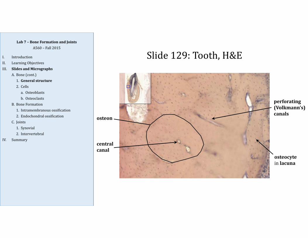
Slide129:Tooth,H&E
osteon
perforating(Volkmann’s)canals
centralcanal
osteocytein lacuna
Lab7– BoneFormationandJointsA560– Fall2015
I. IntroductionII. LearningObjectivesIII. SlidesandMicrographs
A. Bone(cont.)1. Generalstructure2. Cellsa. Osteoblastsb. Osteoclasts
B. BoneFormation1. Intramembranousossification2. Endochondralossification
C. Joints1. Synovial2. Intervertebral
IV. Summary

Slide104:Bone,H&E
osteon
centralcanal
resorptioncanals
Lab7– BoneFormationandJointsA560– Fall2015
I. IntroductionII. LearningObjectivesIII. SlidesandMicrographs
A. Bone(cont.)1. Generalstructure2. Cellsa. Osteoblastsb. Osteoclasts
B. BoneFormation1. Intramembranousossification2. Endochondralossification
C. Joints1. Synovial2. Intervertebral
IV. Summary
Osteon formation: resorption canals (with dimension of new osteon) are carved out of bone byosteoclasts; blood vessels and connective tissue invade and occupy the tunnel; osteoblasts beginto deposit new bone along the walls, forming lamellae; synthesis continues from periphery tocenter until only central canal with neurovascular bundle remains in center

Slide34:HealingBoneFracture,H&ELab7– BoneFormationandJointsA560– Fall2015
I. IntroductionII. LearningObjectivesIII. SlidesandMicrographs
A. Bone(cont.)1. Generalstructure2. Cellsa. Osteoblastsb. Osteoclasts
B. BoneFormation1. Intramembranousossification2. Endochondralossification
C. Joints1. Synovial2. Intervertebral
IV. Summary
1
2
Fracture
Callus
HyalineCartilage
ECOss
Corticalbone
Sections (1) and (2) are two rib segments from a fetal/newborn rabbit; (1) gives an example of a bonefracture and repair processes (the callus is a temporary formation of highly proliferative fibroblasts andchondroblasts extending from the periosteum down into the fracture to form new bone); (2) gives anexample of endochondral ossification (EC Oss) bone growth
Slide34:HealingBoneFracture,H&ELab7– BoneFormationandJointsA560– Fall2015
I. IntroductionII. LearningObjectivesIII. SlidesandMicrographs
A. Bone(cont.)1. Generalstructure2. Cellsa. Osteoblastsb. Osteoclasts
B. BoneFormation1. Intramembranousossification2. Endochondralossification
C. Joints1. Synovial2. Intervertebral
IV. Summary
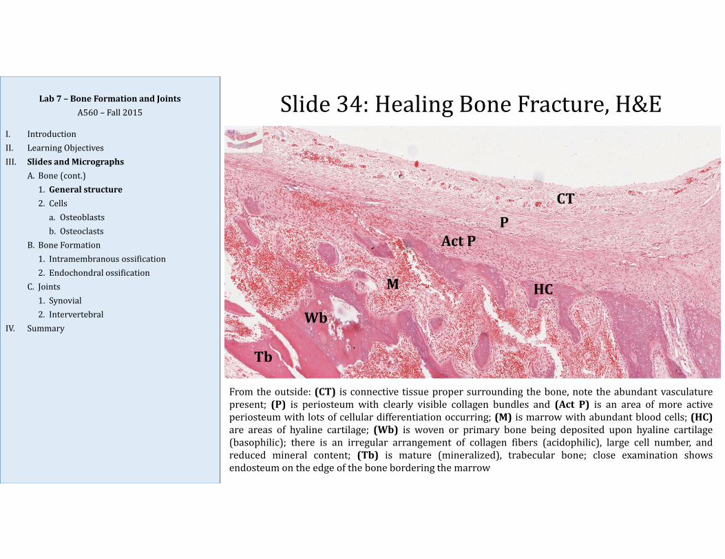
From the outside: (CT) is connective tissue proper surrounding the bone, note the abundant vasculaturepresent; (P) is periosteum with clearly visible collagen bundles and (Act P) is an area of more activeperiosteum with lots of cellular differentiation occurring; (M) is marrow with abundant blood cells; (HC)are areas of hyaline cartilage; (Wb) is woven or primary bone being deposited upon hyaline cartilage(basophilic); there is an irregular arrangement of collagen fibers (acidophilic), large cell number, andreduced mineral content; (Tb) is mature (mineralized), trabecular bone; close examination showsendosteum on the edge of the bone bordering the marrow
Tb
Wb
M
ActPP
CT
HC

Slide34:HealingBoneFracture,H&ELab7– BoneFormationandJointsA560– Fall2015
I. IntroductionII. LearningObjectivesIII. SlidesandMicrographs
A. Bone(cont.)1. Generalstructure2. Cellsa. Osteoblastsb. Osteoclasts
B. BoneFormation1. Intramembranousossification2. Endochondralossification
C. Joints1. Synovial2. Intervertebral
IV. Summary
From the outside (top): (CT) is connective tissue proper surrounding the bone; (P) is periosteum withclearly visible collagen bundles; (Cb) is cortical bone with lamellar arrangement, note the adjacent centralcanal with surrounding rings of cartilage; (M) is marrow with abundant blood cells; (Tb) is trabecularbone, distinguished from cortical bone by the lack of osteons; close examination shows endosteum on theedge of the bone bordering the marrow; (Wb) is woven or primary bone with an irregular arrangement ofcollagen fibers, large cell number, and reduced mineral content; osteoid (unmineralized bone) is the pale,acellular layer just below osteoblasts which line the edges of the nearby marrow cavities
CT P Cb
M
Tb
PCT
Wb

Slide34:HealingBoneFracture,H&E
endosteumliningtrabecula
marrow
Lab7– BoneFormationandJointsA560– Fall2015
I. IntroductionII. LearningObjectivesIII. SlidesandMicrographs
A. Bone(cont.)1. Generalstructure2. Cellsa. Osteoblastsb. Osteoclasts
B. BoneFormation1. Intramembranousossification2. Endochondralossification
C. Joints1. Synovial2. Intervertebral
IV. Summary
Endosteum lines all internal surfaces of bone (both cortical and trabecular); itis generally only a single cell‐layer thick, and consists of inactive and activeosteoblasts

Slide34:HealingBoneFracture,H&E
osteoidlighterstainedareabetweenendosteum andmaturebone
endosteum
maturebone
Lab7– BoneFormationandJointsA560– Fall2015
I. IntroductionII. LearningObjectivesIII. SlidesandMicrographs
A. Bone(cont.)1. Generalstructure2. Cellsa. Osteoblastsb. Osteoclasts
B. BoneFormation1. Intramembranousossification2. Endochondralossification
C. Joints1. Synovial2. Intervertebral
IV. Summary
Osteoid is collagen‐rich, non‐mineralized precursor to true bone matrix; itis secreted by osteoblasts during bone growth, repair, and remodeling;osteoblast subsequently calcify the osteoid into bony hard bony matrix; inthe process, they become trapped in the matrix (in lacunae) and becomeosteocytes
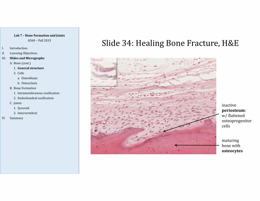
inactiveperiosteum:w/flattenedosteoprogenitorcells
maturingbonewithosteocytes
Slide34:HealingBoneFracture,H&ELab7– BoneFormationandJoints
A560– Fall2015
I. IntroductionII. LearningObjectivesIII. SlidesandMicrographs
A. Bone(cont.)1. Generalstructure2. Cellsa. Osteoblastsb. Osteoclasts
B. BoneFormation1. Intramembranousossification2. Endochondralossification
C. Joints1. Synovial2. Intervertebral
IV. Summary

Slide34:HealingBoneFracture,H&E
osteocyte
activeperiosteum:w/roundedorcuboidalosteoblasts
Lab7– BoneFormationandJointsA560– Fall2015
I. IntroductionII. LearningObjectivesIII. SlidesandMicrographs
A. Bone(cont.)1. Generalstructure2. Cellsa. Osteoblastsb. Osteoclasts
B. BoneFormation1. Intramembranousossification2. Endochondralossification
C. Joints1. Synovial2. Intervertebral
IV. Summary
Osteoblasts develop from osteoprogenitor cells (from mesenchymal cells) and are found lining theexternal and internal bone surfaces; they make osteoid (collagen‐rich matrix) which they thencalcify into true hard, bony matrix; they become trapped in the matrix and become osteocytes

Slide130:MembranousBone,FetalSkull
periosteum
osteoblasts
Lab7– BoneFormationandJointsA560– Fall2015
I. IntroductionII. LearningObjectivesIII. SlidesandMicrographs
A. Bone(cont.)1. Generalstructure2. Cellsa. Osteoblastsb. Osteoclasts
B. BoneFormation1. Intramembranousossification2. Endochondralossification
C. Joints1. Synovial2. Intervertebral
IV. Summary

Slide34:HealingBoneFracture,H&E
Osteoclast
Howship’s lacuna(space)
Osteoclasts are large, multinucleated cells; they resorb bone by secreting organic acids, whichdissolve hydroxyapatite, and lysosomal enzymes, which break down the osteoid matrix; at the bonesurface, osteoclasts lie in Howship's lacunae, surface depressions caused by the resorption of bone
Howmanynucleidoesthisosteoclasthave?
upto200ispossible,5‐20isusual
Lab7– BoneFormationandJointsA560– Fall2015
I. IntroductionII. LearningObjectivesIII. SlidesandMicrographs
A. Bone(cont.)1. Generalstructure2. Cellsa. Osteoblastsb. Osteoclasts
B. BoneFormation1. Intramembranousossification2. Endochondralossification
C. Joints1. Synovial2. Intervertebral
IV. Summary

Slide34:HealingBoneFracture,H&E
Osteoclastwithruffledborder(visibleinEM)inHowship’s lacuna
Lab7– BoneFormationandJointsA560– Fall2015
I. IntroductionII. LearningObjectivesIII. SlidesandMicrographs
A. Bone(cont.)1. Generalstructure2. Cellsa. Osteoblastsb. Osteoclasts
B. BoneFormation1. Intramembranousossification2. Endochondralossification
C. Joints1. Synovial2. Intervertebral
IV. Summary

Slide130:MembranousBone,FetalSkull
osteoclasts
Lab7– BoneFormationandJointsA560– Fall2015
I. IntroductionII. LearningObjectivesIII. SlidesandMicrographs
A. Bone(cont.)1. Generalstructure2. Cellsa. Osteoblastsb. Osteoclasts
B. BoneFormation1. Intramembranousossification2. Endochondralossification
C. Joints1. Synovial2. Intervertebral
IV. Summary
IntramembranousOssification
Lab7– BoneFormationandJointsA560– Fall2015
I. IntroductionII. LearningObjectivesIII. SlidesandMicrographs
A. Bone(cont.)1. Generalstructure2. Cellsa. Osteoblastsb. Osteoclasts
B. BoneFormation1. Intramembranousossification2. Endochondralossification
C. Joints1. Synovial2. Intervertebral
IV. Summary
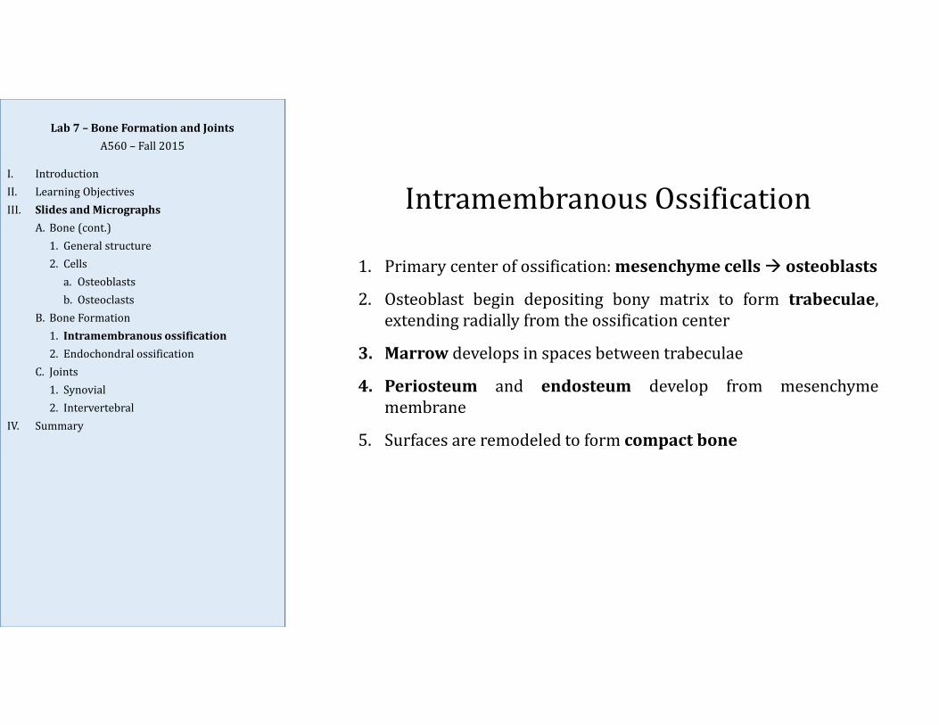
1. Primary center of ossification:mesenchyme cells osteoblasts
2. Osteoblast begin depositing bony matrix to form trabeculae,extending radially from the ossification center
3. Marrow develops in spaces between trabeculae
4. Periosteum and endosteum develop from mesenchymemembrane
5. Surfaces are remodeled to form compact bone

Slide130:MembranousBone,FetalSkullLab7– BoneFormationandJoints
A560– Fall2015
I. IntroductionII. LearningObjectivesIII. SlidesandMicrographs
A. Bone(cont.)1. Generalstructure2. Cellsa. Osteoblastsb. Osteoclasts
B. BoneFormation1. Intramembranousossification2. Endochondralossification
C. Joints1. Synovial2. Intervertebral
IV. Summary
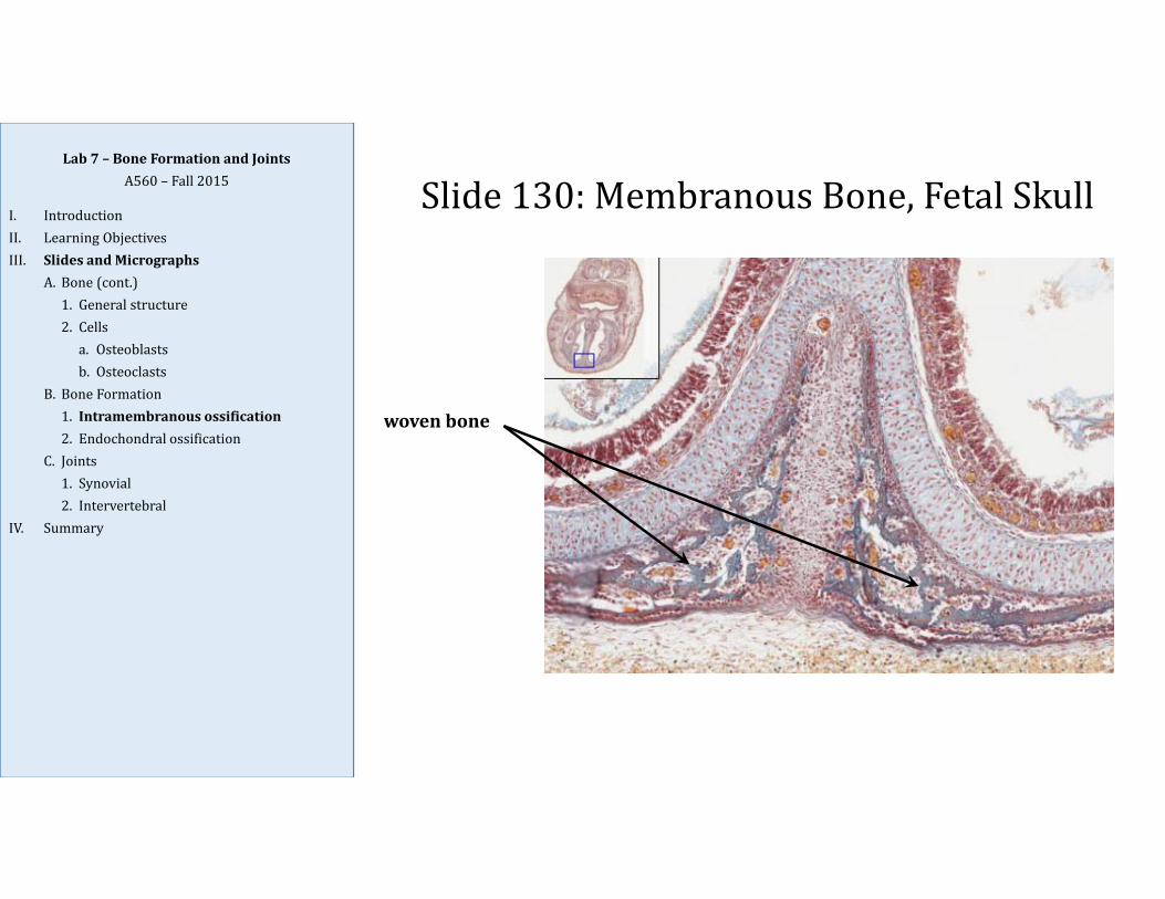
SideView FrontalView developingteethwithsurroundingintramembranousbonedevelopmentofthejaw
tongueintheoralcavity
nasalcavitywithcartilaginousnasalseptuminthemiddle;surroundedbyintramembranousbonedevelopmentoftheskull
Slide130– FetalSkull
Slide130:MembranousBone,FetalSkull
wovenbone
Lab7– BoneFormationandJointsA560– Fall2015
I. IntroductionII. LearningObjectivesIII. SlidesandMicrographs
A. Bone(cont.)1. Generalstructure2. Cellsa. Osteoblastsb. Osteoclasts
B. BoneFormation1. Intramembranousossification2. Endochondralossification
C. Joints1. Synovial2. Intervertebral
IV. Summary

Slide130:MembranousBone,FetalSkull
hyalinecartilage
periosteum
wovenbone
Lab7– BoneFormationandJointsA560– Fall2015
I. IntroductionII. LearningObjectivesIII. SlidesandMicrographs
A. Bone(cont.)1. Generalstructure2. Cellsa. Osteoblastsb. Osteoclasts
B. BoneFormation1. Intramembranousossification2. Endochondralossification
C. Joints1. Synovial2. Intervertebral
IV. Summary
EndochondralOssificationLab7– BoneFormationandJoints
A560– Fall2015
I. IntroductionII. LearningObjectivesIII. SlidesandMicrographs
A. Bone(cont.)1. Generalstructure2. Cellsa. Osteoblastsb. Osteoclasts
B. BoneFormation1. Intramembranousossification2. Endochondralossification
C. Joints1. Synovial2. Intervertebral
IV. Summary
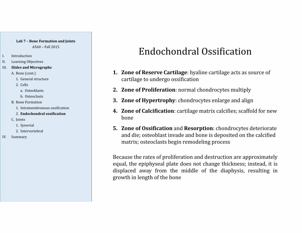
1. ZoneofReserveCartilage:hyalinecartilageactsassourceofcartilagetoundergoossification
2. ZoneofProliferation:normalchondrocytesmultiply
3. ZoneofHypertrophy:chondrocytesenlargeandalign
4. ZoneofCalcification:cartilagematrixcalcifies;scaffoldfornewbone
5. ZoneofOssification andResorption:chondrocytesdeteriorateanddie;osteoblastinvadeandboneisdepositedonthecalcifiedmatrix;osteoclastsbeginremodelingprocess
Because the rates of proliferation and destruction are approximatelyequal, the epiphyseal plate does not change thickness; instead, it isdisplaced away from the middle of the diaphysis, resulting ingrowth in length of the bone

Slide34:HealingBoneFracture,H&E
GrowthplateEpiphysis(End)
Diaphysis(Middle)
endochondralossification
Lab7– BoneFormationandJointsA560– Fall2015
I. IntroductionII. LearningObjectivesIII. SlidesandMicrographs
A. Bone(cont.)1. Generalstructure2. Cellsa. Osteoblastsb. Osteoclasts
B. BoneFormation1. Intramembranousossification2. Endochondralossification
C. Joints1. Synovial2. Intervertebral
IV. Summary

Slide34:HealingBoneFracture,H&E
zoneofreservecartilage
zoneofchondroblastproliferation
zoneofchondrocytehypertrophy
zoneofcalcification,
ossification,andresorption
Lab7– BoneFormationandJointsA560– Fall2015
I. IntroductionII. LearningObjectivesIII. SlidesandMicrographs
A. Bone(cont.)1. Generalstructure2. Cellsa. Osteoblastsb. Osteoclasts
B. BoneFormation1. Intramembranousossification2. Endochondralossification
C. Joints1. Synovial2. Intervertebral
IV. Summary
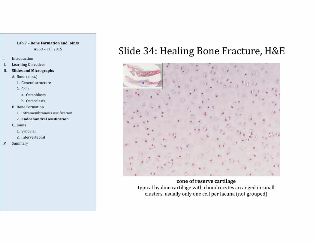
Slide34:HealingBoneFracture,H&E
zoneofreservecartilagetypicalhyalinecartilagewithchondrocytesarrangedinsmall
clusters,usuallyonlyonecellperlacuna(notgrouped)
Lab7– BoneFormationandJointsA560– Fall2015
I. IntroductionII. LearningObjectivesIII. SlidesandMicrographs
A. Bone(cont.)1. Generalstructure2. Cellsa. Osteoblastsb. Osteoclasts
B. BoneFormation1. Intramembranousossification2. Endochondralossification
C. Joints1. Synovial2. Intervertebral
IV. Summary

Slide34:HealingBoneFracture,H&E
zoneofchondroblast proliferationchondrocytesaredividingandincreasinginnumber;chondrocytesareslightlylargerinsize,areclosertoneighboringcells,andarebeginningtoformrowsorstacks
Lab7– BoneFormationandJointsA560– Fall2015
I. IntroductionII. LearningObjectivesIII. SlidesandMicrographs
A. Bone(cont.)1. Generalstructure2. Cellsa. Osteoblastsb. Osteoclasts
B. BoneFormation1. Intramembranousossification2. Endochondralossification
C. Joints1. Synovial2. Intervertebral
IV. Summary
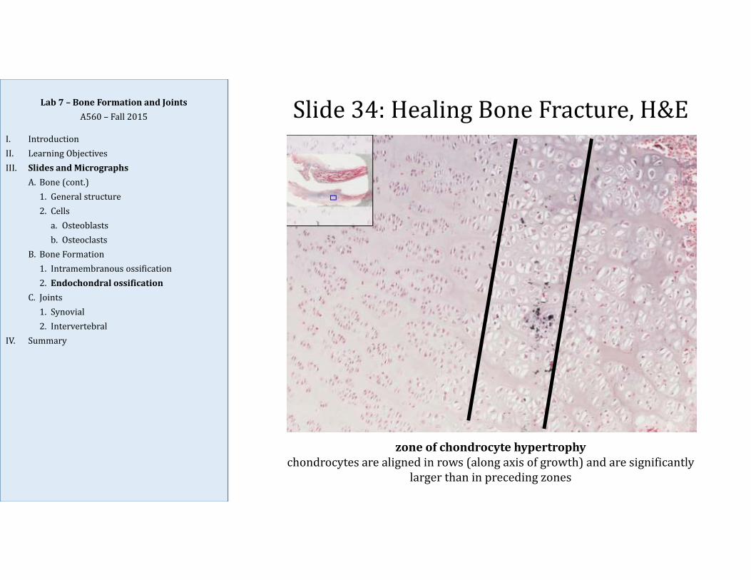
Slide34:HealingBoneFracture,H&E
zoneofchondrocytehypertrophychondrocytesarealignedinrows(alongaxisofgrowth)andaresignificantly
largerthaninprecedingzones
Lab7– BoneFormationandJointsA560– Fall2015
I. IntroductionII. LearningObjectivesIII. SlidesandMicrographs
A. Bone(cont.)1. Generalstructure2. Cellsa. Osteoblastsb. Osteoclasts
B. BoneFormation1. Intramembranousossification2. Endochondralossification
C. Joints1. Synovial2. Intervertebral
IV. Summary

Slide34:HealingBoneFracture,H&E
zoneofcalcification,ossification,andresorptionchondrocytesundergoapoptosis(sonucleiaremorecondensed);smallbloodvesselandosteoprogenitor cellsbegintoinvade,givingrisetoosteoblasts
whichbegintolaydownboneandbecomeosteocytes
Lab7– BoneFormationandJointsA560– Fall2015
I. IntroductionII. LearningObjectivesIII. SlidesandMicrographs
A. Bone(cont.)1. Generalstructure2. Cellsa. Osteoblastsb. Osteoclasts
B. BoneFormation1. Intramembranousossification2. Endochondralossification
C. Joints1. Synovial2. Intervertebral
IV. Summary
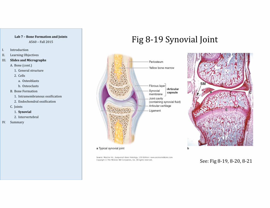
Fig8‐19SynovialJointLab7– BoneFormationandJointsA560– Fall2015
I. IntroductionII. LearningObjectivesIII. SlidesandMicrographs
A. Bone(cont.)1. Generalstructure2. Cellsa. Osteoblastsb. Osteoclasts
B. BoneFormation1. Intramembranousossification2. Endochondralossification
C. Joints1. Synovial2. Intervertebral
IV. Summary
See:Fig8‐19,8‐20,8‐21

Fig8‐22IntervertebralDiscLab7– BoneFormationandJointsA560– Fall2015
I. IntroductionII. LearningObjectivesIII. SlidesandMicrographs
A. Bone(cont.)1. Generalstructure2. Cellsa. Osteoblastsb. Osteoclasts
B. BoneFormation1. Intramembranousossification2. Endochondralossification
C. Joints1. Synovial2. Intervertebral
IV. Summary
Vertebrawithbonemarrowcavity(BM)
Vertebrawithbonemarrowcavity
Concentriclayersoffibrocartilageformtheannulusfibrosus (AF)(Lt.“fibrousring”)
Nucleuspulposus (NP)istheinnercoreofthevertebraldisc;itiscomposedofagel‐likematrixconsistingofwaterandaloosenetworkofcollagenfibers
Lab7– BoneFormationandJointsA560– Fall2015
I. IntroductionII. LearningObjectivesIII. SlidesandMicrographs
A. Bone(cont.)1. Generalstructure2. Cellsa. Osteoblastsb. Osteoclasts
B. BoneFormation1. Intramembranousossification2. Endochondralossification
C. Joints1. Synovial2. Intervertebral
IV. Summary
Common Confusion:Cartilage vs. Bone
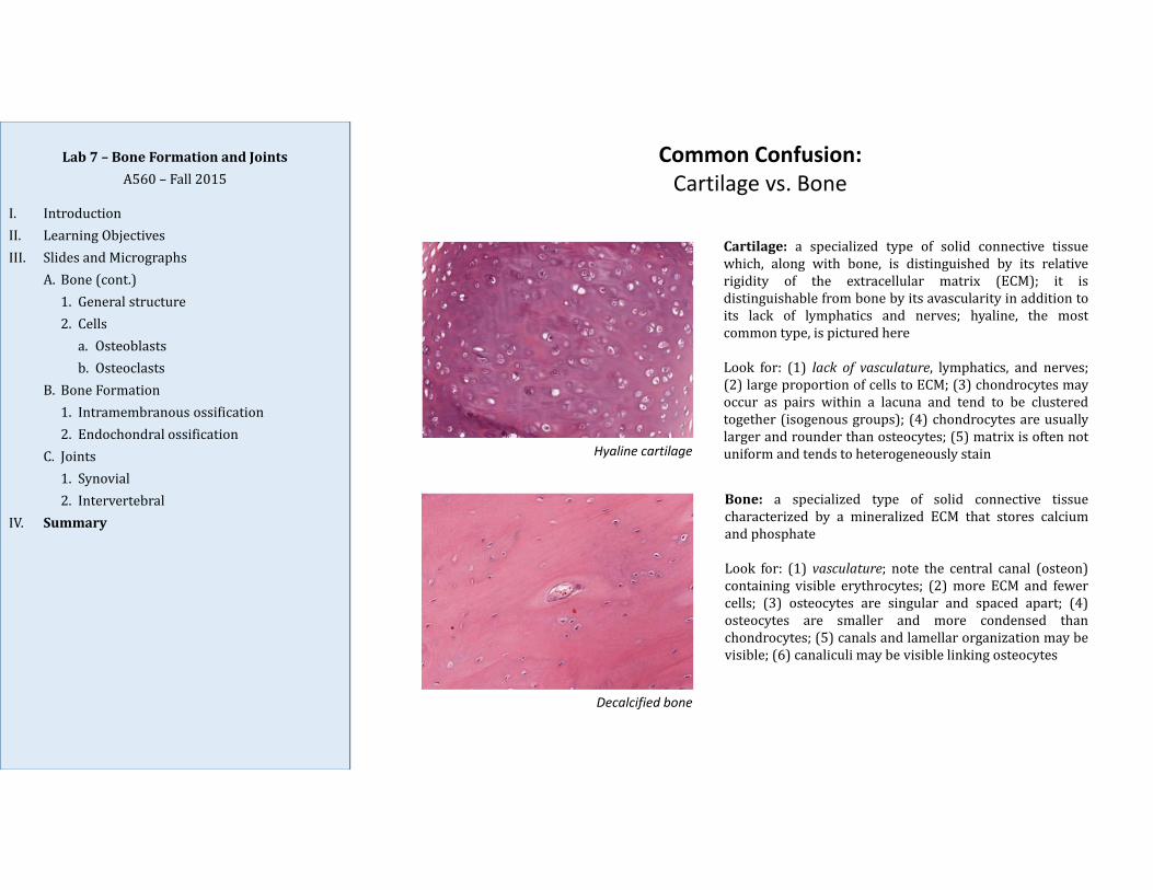
Hyaline cartilage
Cartilage: a specialized type of solid connective tissuewhich, along with bone, is distinguished by its relativerigidity of the extracellular matrix (ECM); it isdistinguishable from bone by its avascularity in addition toits lack of lymphatics and nerves; hyaline, the mostcommon type, is pictured here
Look for: (1) lack of vasculature, lymphatics, and nerves;(2) large proportion of cells to ECM; (3) chondrocytes mayoccur as pairs within a lacuna and tend to be clusteredtogether (isogenous groups); (4) chondrocytes are usuallylarger and rounder than osteocytes; (5) matrix is often notuniform and tends to heterogeneously stain
Bone: a specialized type of solid connective tissuecharacterized by a mineralized ECM that stores calciumand phosphate
Look for: (1) vasculature; note the central canal (osteon)containing visible erythrocytes; (2) more ECM and fewercells; (3) osteocytes are singular and spaced apart; (4)osteocytes are smaller and more condensed thanchondrocytes; (5) canals and lamellar organization may bevisible; (6) canaliculi may be visible linking osteocytes
Decalcified bone

Lab7– BoneFormationandJointsA560– Fall2015
I. IntroductionII. LearningObjectivesIII. SlidesandMicrographs
A. Bone(cont.)1. Generalstructure2. Cellsa. Osteoblastsb. Osteoclasts
B. BoneFormation1. Intramembranousossification2. Endochondralossification
C. Joints1. Synovial2. Intervertebral
IV. Summary
Summary
1. During bone formation, woven bone (haphazard arrangement of collagenfibers) gets remodeled into lamellar bone (parallel bundles of collagen inlayers known as lamellae).
2. Periosteum is a layer of dense connective tissue on the outer surface ofbone; endosteum is a thin layer (generally only one cell layer) which linesall the internal surfaces of bone.
3. Major cells of bone include: osteoblasts (from osteoprogenitor cells; formosteoid which allows matrix mineralization to occur), osteocytes (fromosteoblasts; enclosed in lacunae and maintain the matrix), and osteoclasts(from hematopoietic lineage; locally erode bone matrix during boneformation and remodeling).
4. Bone growth occurs via two basic mechanisms:• intramembranous ossification occurs when bone forms within
mesenchymal membrane; forms bones of skull and jaw; primarilyoccurs only during development or fracture repair
• endochondral ossification occurs when bone replaces hyalinecartilage; forms and grows all other bones except as noted for IM;occurs during development and throughout life

Lab7– BoneFormationandJointsA560– Fall2015
I. IntroductionII. LearningObjectivesIII. SlidesandMicrographs
A. Bone(cont.)1. Generalstructure2. Cellsa. Osteoblastsb. Osteoclasts
B. BoneFormation1. Intramembranousossification2. Endochondralossification
C. Joints1. Synovial2. Intervertebral
IV. Summary
Summary(cont.)
5. During epiphyseal growth (elongation of bone), the growth plate, with itszonal organization of endochondral ossification, allows bone to lengthenwithout the epiphyseal growth plate enlarging; zones include:
• Zone of reserve cartilage• Zone of proliferation• Zone of hypertrophy• Zone of calcification• Zone of ossification and resorption
6. Joints are places where bones meet (articulate), allowing at least thepotential of bending or movement; examples include, synovial joints(diarthrosis) and intervertebral joints (with tough outer layer offibrocartilage known as annulus fibrosus, and gel‐like core known asnucleus pulposus).
Osteoblasts Osteocytes Osteoclasts
Precursor cell
Location
Percentage of all cells in bone
Function
Appearance
Sketch
Lab 7: Summary Features of Major Cells of Bone Tissue

Intramembranous Ossification Endochondral Ossification
Bones produced
Cartilage present
Type of bone produced
When occurs
Steps Involved
Lab 7: Comparison of Mechanisms of Bone Formation