Bonamia ostreae in the New Zealand oyster Ostrea chilensis ...
9
DISEASES OF AQUATIC ORGANISMS Dis Aquat Org Vol. 118: 55–63, 2016 doi: 10.3354/dao02960 Published February 11 INTRODUCTION The haplosporidian genus Bonamia contains a number of morphologically similar species of mol- lusc parasites. Bonamia microcells are small (typi- cally 1-3 μm), uninucleated, and infect oyster hemocytes causing a disease known as bonamiosis (Hine & Jones 1994, OIE 2012). Molecular and ultra- structural studies have identified 4 Bonamia species from a number of hosts and locations throughout the world (Hine et al. 2001, Carnegie et al. 2006, Lohrmann et al. 2009, Hill et al. 2014). These include B. ostreae (see Pichot et al. 1979), B. exitiosa (see Hine et al. 2001), B. perspora (see Carnegie et al. 2006) and B. roughleyi (see Farley et al. 1988, Cochennec-Laureau et al. 2003), although B. rough- leyi has been assigned nomen dubium and is unlikely to be a distinct species of Bonamia (see Carnegie et al. 2014, Hill et al. 2014, Speirs et al. 2014). Due to the severity of disease that these para- sites cause, B. ostreae and B. exitiosa are listed as pathogens notifiable to the World Organisation of © Inter-Research 2016 · www.int-res.com *Corresponding author: [email protected] Bonamia ostreae in the New Zealand oyster Ostrea chilensis : a new host and geographic record for this haplosporidian parasite Henry S. Lane 1, *, Stephen Charles Webb 2 , John Duncan 2 1 Department of Zoology, University of Otago, PO Box 56, Dunedin 9054, New Zealand 2 Cawthron Institute, 98 Halifax Street East, Nelson 7010, New Zealand ABSTRACT: Previous reports of the haplosporidian parasite Bonamia ostreae have been restricted to the Northern Hemisphere, including Europe, and both eastern and western North America. This species is reported for the first time in New Zealand infecting the flat oyster Ostrea chilensis. Histological examination of 149 adult oysters identified 119 (79.9%) infected with Bonamia micro- cells. Bonamia generic PCR of several oysters followed by DNA sequencing of a 300 bp portion of the 18S rDNA gene produced a 100% match with that of B. ostreae. All DNA-sequenced products also produced a B. ostreae PCR-restriction fragment length polymorphism (PCR-RFLP) profile. Bonamia species-specific PCRs further detected single infections of B. exitiosa (2.7%), B. ostreae (40.3%), and concurrent infections (53.7%) with these 2 Bonamia species identifying overall a Bonamia prevalence of 96.6%. Detailed histological inspection revealed 2 microcell types. An infection identified by PCR as B. ostreae histologically presented small microcells (mean ± SE diameter = 1.28 ± 0.16 μm, range = 0.9-2 μm, n = 60) commonly with eccentric nuclei. A B. exitiosa infection exhibited larger microcells (mean ± SE diameter = 2.12 ± 0.27 μm, range = 1.5-4 μm, n = 60) with more concentric nuclei. Concurrent infections of both Bonamia species, as identified by PCR, exhibited both types of microcells. DNA barcoding of the B. ostreae-infected oyster host con- firmed the identification as O. chilensis. A suite of other parasites that accompany O. chilensis are reported here for the first time in mixed infection with B. ostreae including apicomplexan X (76.5%), Microsporidium rapuae (0.7%) and Bucephalus longicornutus (30.2%). KEY WORDS: Bonamiosis · Flat oyster · First report · Range extension · Restriction fragment length polymorphism · RFLP Resale or republication not permitted without written consent of the publisher
Transcript of Bonamia ostreae in the New Zealand oyster Ostrea chilensis ...

DISEASES OF AQUATIC ORGANISMSDis Aquat Org
Vol. 118: 55–63, 2016doi: 10.3354/dao02960
Published February 11
INTRODUCTION
The haplosporidian genus Bonamia contains anumber of morphologically similar species of mol-lusc parasites. Bonamia microcells are small (typi-cally 1−3 µm), uninucleated, and infect oysterhemocytes causing a disease known as bonamiosis(Hine & Jones 1994, OIE 2012). Molecular and ultra-structural studies have identified 4 Bonamia speciesfrom a number of hosts and locations throughout theworld (Hine et al. 2001, Carnegie et al. 2006,
Lohrmann et al. 2009, Hill et al. 2014). Theseinclude B. ostreae (see Pichot et al. 1979), B. exitiosa(see Hine et al. 2001), B. perspora (see Carnegie etal. 2006) and B. roughleyi (see Farley et al. 1988,Cochennec- Laureau et al. 2003), although B. rough-leyi has been assigned nomen dubium and isunlikely to be a distinct species of Bonamia (seeCarnegie et al. 2014, Hill et al. 2014, Speirs et al.2014). Due to the severity of disease that these para-sites cause, B. ostreae and B. exitiosa are listed aspathogens notifiable to the World Organisation of
© Inter-Research 2016 · www.int-res.com*Corresponding author: [email protected]
Bonamia ostreae in the New Zealand oyster Ostrea chilensis: a new host and geographic record
for this haplosporidian parasite
Henry S. Lane1,*, Stephen Charles Webb2, John Duncan2
1Department of Zoology, University of Otago, PO Box 56, Dunedin 9054, New Zealand2Cawthron Institute, 98 Halifax Street East, Nelson 7010, New Zealand
ABSTRACT: Previous reports of the haplosporidian parasite Bonamia ostreae have been restrictedto the Northern Hemisphere, including Europe, and both eastern and western North America.This species is reported for the first time in New Zealand infecting the flat oyster Ostrea chilensis.Histological examination of 149 adult oysters identified 119 (79.9%) infected with Bonamia micro-cells. Bonamia generic PCR of several oysters followed by DNA sequencing of a 300 bp portion ofthe 18S rDNA gene produced a 100% match with that of B. ostreae. All DNA-sequenced productsalso produced a B. ostreae PCR-restriction fragment length polymorphism (PCR-RFLP) profile.Bonamia species-specific PCRs further detected single infections of B. exitiosa (2.7%), B. ostreae(40.3%), and concurrent infections (53.7%) with these 2 Bonamia species identifying overall aBonamia prevalence of 96.6%. Detailed histological inspection revealed 2 microcell types. Aninfection identified by PCR as B. ostreae histologically presented small microcells (mean ± SEdiameter = 1.28 ± 0.16 µm, range = 0.9−2 µm, n = 60) commonly with eccentric nuclei. A B. exitiosainfection exhibited larger microcells (mean ± SE diameter = 2.12 ± 0.27 µm, range = 1.5−4 µm, n =60) with more concentric nuclei. Concurrent infections of both Bonamia species, as identified byPCR, exhibited both types of microcells. DNA barcoding of the B. ostreae-infected oyster host con-firmed the identification as O. chilensis. A suite of other parasites that accompany O. chilensis arereported here for the first time in mixed infection with B. ostreae including apicomplexan X(76.5%), Microsporidium rapuae (0.7%) and Bucephalus longicornutus (30.2%).
KEY WORDS: Bonamiosis · Flat oyster · First report · Range extension · Restriction fragmentlength polymorphism · RFLP
Resale or republication not permitted without written consent of the publisher
Dis Aquat Org 118: 55–63, 2016
Animal Health (OIE) and to the European Union(OIE 2012, Engelsma et al. 2014).
To date, the only Bonamia species re ported in NewZealand was B. exitiosa. Its type-host (Hine et al.2001) is the New Zealand dredge, or flat oysterOstrea (= Tiostrea) chilensis (= lutaria), a significantcustomary, recreational and commercial species pre-dominantly exploited in the Foveaux Strait (Ministryfor Primary Industries 2014). It is important to notethat Hill et al. (2014) incorrectly reported B. ostreaein the European flat oyster Ostrea edulis from NewZealand, when in fact it was reported in O. edulisfrom the Netherlands (R. Carnegie pers. comm.), andthat B. ostreae has not been reported from NewZealand until this study.
Between 1986−1992 and 2000−2005, B. exitiosawas responsible for 2 epizootic events in O. chilensiswithin the Foveaux Strait fishery that caused highmortalities, reducing oyster density to less than 10%of their pre-disease level (Doonan et al. 1994, Cran-field et al. 2005, Michael et al. 2013). B. exitiosa con-tinues to have a recurrent impact on this fishery andis the main influence on flat oyster numbers (Michaelet al. 2013). Surveillance of the Foveaux Strait O.chilensis fishery for Bonamia spp. using histologyand PCR has failed to detect any species of Bonamiaother than the endemic B. exitiosa (Michael et al.2013, H. Lane pers. obs.). A further possible host forB. exitiosa in New Zealand is Ostreola stentina fromthe Hauraki Gulf. DNA sequences matching B. exi-tiosa were reported from Hill et al. (2014), althoughthis was not corroborated by other methods such ashistology or in situ hybridisation (ISH).
Historically, Bonamia parasites were viewed ashaving a localised distribution, with B. ostreae re -stricted to the European flat oyster O. edulis in thetemperate Northern Hemisphere, and B. exitiosa toO. chilensis from southern New Zealand. However,the discovery of a Bonamia-like parasite in Crass-ostrea ariakensis in North Carolina, USA (Burresonet al. 2004), and the description of another parasite asB. perspora from O. stentina from the same area(Carnegie et al. 2006), in combination with the dis-covery of B. exitiosa and B. exitiosa-like parasites inO. stentina from the Mediterranean (Hill et al. 2010),O. chilensis from Chile (Campalans et al. 2000,Lohrmann et al. 2009), O. puelchana from Argentina(Kroeck & Montes 2005), O. angasi from Australia,and O. edulis from Europe (Abollo et al. 2008, Long-shaw et al. 2013) have initiated a change in percep-tion of the distribution of Bonamia (Engelsma et al.2014). Some Bonamia species are now reported withoverlapping geographic ranges (Abollo et al. 2008,
Narcisi et al. 2010, Carrasco et al. 2012, Longshaw etal. 2013) and in one case to the point of a concurrentinfection of B. exitiosa and B. ostreae within the sameO. edulis host (Abollo et al. 2008).
B. ostreae was first described from O. edulis inFrance (Pichot et al. 1979) and is considered to havebeen introduced into Europe by a transhipment ofinfected oysters from its putative endemic area ofeastern USA (Elston et al. 1986). Since then, it hasspread throughout Europe and has been responsiblefor extensive oyster mortalities and subsequent de -clines in production of O. edulis along the EuropeanAtlantic coast (Grizel et al. 1988) and eastern USA(Carnegie & Cochennec-Laureau 2004). B. ostreaehas also been reported from western North Americaand Morocco (Friedman et al. 1989, Marty et al. 2006,Belhsen et al. 2008).
A putative B. ostreae infection was first reportedfrom O. chilensis in the UK by Bucke & Hepper (1987)using histology. The reported infection of B. ostreaein O. chilensis occurred after the oyster host hadbeen transferred to a B. ostreae endemic area inCornwall, England (Bucke & Hepper 1987); however,there are still limited scientific data on the suscepti-bility of O. chilensis to infection by B. ostreae(Engelsma et al. 2014). Moreover, it is also uncertainif B. ostreae can infect other oyster species such as O.angasi (see Bougrier et al. 1986), O. puelchana (seePascual et al. 1991) and C. gigas (see Lynch et al.2010).
With the advent of molecular tools, reports of otherBonamia from new geographic regions (Burreson etal. 2004, Carnegie et al. 2006, Abollo et al. 2008, Hillet al. 2010, Carrasco et al. 2012) have proliferated.Most of these new Bonamia occurrences phylogenet-ically group with B. exitiosa (see Hill et al. 2010,Carnegie et al. 2014). In January 2015, B. ostreae wasidentified in New Zealand from the New Zealand flatoyster O. chilensis. This finding is the first report of B.ostreae from the Southern Hemisphere and repre-sents a further change in global distribution of thisparasite as well as a possible incursion of an exoticpathogen to New Zealand.
MATERIALS AND METHODS
Sample collection and histology
Ostrea chilensis (n = 149) from the MarlboroughSounds (Fig. 1) were collected on 18 November 2014during a health survey of a population that hadshown significant (60−70%) mortality (A. Elliot pers.
56

Lane et al.: Bonamia ostreae in Ostrea chilensis
comm.). Shell height was measured to the nearestmm, and, after shucking, oysters were prepared forhistology following Howard et al. (2004): a 3−5 mmthick section was taken from each oyster, includinggills, digestive gland, gonad and mantle. The sec-tions were placed into pre-labelled histo-cassettesand fixed in formalin/seawater (4% formaldehyde)for 48 h, after which they were stored in 70% ethanoluntil histological processing. Concurrently, a sampleof the oyster tissue including gills and mantle wasstored in a 1.5 ml microcentrifuge tube and frozen at−70°C for molecular analyses. All histo-cassetteswere processed using standard medical histologicaltechniques with haematoxylin and eosin (H&E) stain-ing. Cover-slipped histology slides were examinedunder an Olympus BX51 light microscope at 400×,and 1000× with oil immersion. Tissues examined in -cluded palps, gills, mantle, digestive gland, gastro -intestinal tract, heart, kidney, nerve and muscle. Pre -sence or absence of Bonamia microcells was recorded,and these data allowed prevalence of Bonamia in -fection (by histology) to be estimated. Any otherpathogens/ conditions seen during this examinationwere also recorded.
Molecular methods (see next section) distin-guished epidemiological subpopulations within theoriginal 149 oysters, including those infected with
B. exitiosa only, those infected with B. ostreaeonly, and those with concurrent infections. Thisfacilitated detailed comparative examination of re -presentative histology slide preparations under oilimmersion. Microcells were measured, and inten-sity of infection was estimated semi-quantitativelyusing the scoring method of Diggles et al. (2003).The data set (not shown) for the 149 oysters wasranked by shell height from smallest to largest(range 60−105 mm), and all distributions of infec-tion for pathogens noted (histo pathology and PCR)were inspected for any size-dependent changes infrequency.
Molecular analyses
Bonamia sequencing
Approximately 25 mg of gill and mantle tissue wasexcised from the frozen sampled tissue and used forDNA extraction with the QIAmp Mini Kit followingthe manufacturer’s protocol. DNA was quantifiedusing a Qubit® 2.0 fluorometer and stored at 4°C.
The first 12 Bonamia-positive samples identifiedby histology were subjected to molecular testing forBonamia species identification. Purified DNA wasfirst assayed using an 18S rRNA Internal−Control(#4308329 Life Technologies) real-time PCR to ensurethe presence of amplifiable DNA. For the Bonamia-genus conventional PCR, the forward primer Bo(5’-CAT TTA ATT GGT CGG GCC GC-3’) waspaired with the reverse primer Boas (5’-CTG ATCGTC TTC GAT CCC CC-3’) to produce an ampliconof ~300 bp (Cochennec et al. 2000). Thermal cyclingwas performed on a VeritiR Dx Thermal Cycler(Applied Biosystems): 95°C for 2 min, followed by35 cycles of 95°C for 10 s, 60°C for 10 s, and 72°C for1 s. The resultant amplicons were electrophoresedon a 1.5% agarose gel stained with gel-red andvisualised under UV. Bands from 4 of these 12 sam-ples — hereafter referred to as Samples 1, 2, 3, and4 — were gel-cut and purified using a Zymo GelPurification kit. These 4 samples were chosen be -cause they produced the strongest bands after elec-trophoresis. DNA sequencing was performed atEcoGene (Landcare Research) using primers de -scribed above. The returned DNA sequences wereimported in Geneious version 7.1.5 (www. geneious.com; Kearse et al. 2012), where they were assem-bled, Geneious aligned, and submitted to BLAST(Altschul et al. 1990) for species identification usingdefault parameters.
57
Fig. 1. Hauraki Gulf and Marlborough Sounds, where Bona -mia ostreae was first identified in New Zealand, and theFoveaux Strait, where the largest flat oyster fishery of NewZealand is located. Inset shows global location of New
Zealand
Dis Aquat Org 118: 55–63, 2016
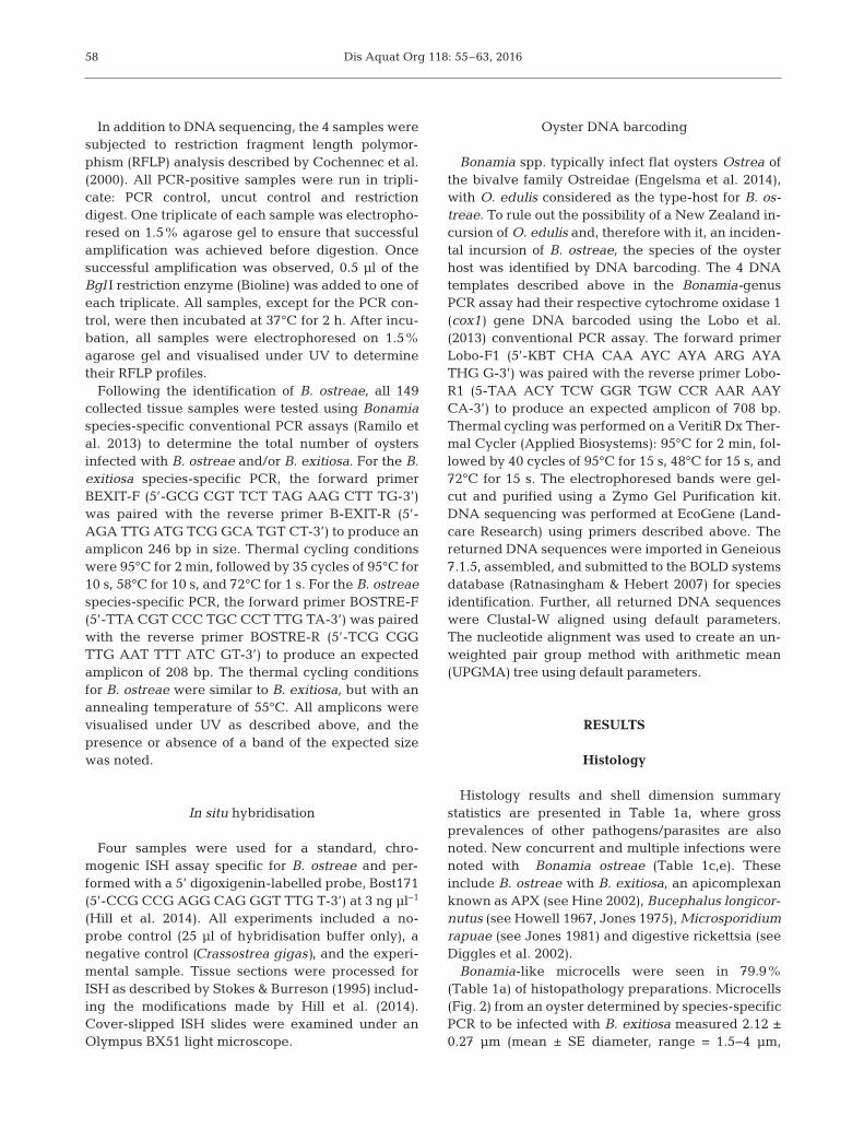
In addition to DNA sequencing, the 4 samples weresubjected to restriction fragment length polymor-phism (RFLP) analysis described by Cochennec et al.(2000). All PCR-positive samples were run in tripli-cate: PCR control, uncut control and restrictiondigest. One triplicate of each sample was electropho-resed on 1.5% agarose gel to ensure that successfulamplification was achieved before digestion. Oncesuccessful amplification was observed, 0.5 µl of theBgl I restriction enzyme (Bioline) was added to one ofeach triplicate. All samples, except for the PCR con-trol, were then incubated at 37°C for 2 h. After incu-bation, all samples were electrophoresed on 1.5%agarose gel and visualised under UV to determinetheir RFLP profiles.
Following the identification of B. ostreae, all 149collected tissue samples were tested using Bonamiaspecies-specific conventional PCR assays (Ramilo etal. 2013) to determine the total number of oystersinfected with B. ostreae and/or B. exitiosa. For the B.exitiosa species-specific PCR, the forward primerBEXIT-F (5’-GCG CGT TCT TAG AAG CTT TG-3’)was paired with the reverse primer B-EXIT-R (5’-AGA TTG ATG TCG GCA TGT CT-3’) to produce anamplicon 246 bp in size. Thermal cycling conditionswere 95°C for 2 min, followed by 35 cycles of 95°C for10 s, 58°C for 10 s, and 72°C for 1 s. For the B. ostreaespecies-specific PCR, the forward primer BOSTRE-F(5’-TTA CGT CCC TGC CCT TTG TA-3’) was pairedwith the reverse primer BOSTRE-R (5’-TCG CGGTTG AAT TTT ATC GT-3’) to produce an expectedamplicon of 208 bp. The thermal cycling conditionsfor B. ostreae were similar to B. exitiosa, but with anannealing temperature of 55°C. All amplicons werevisualised under UV as described above, and thepresence or absence of a band of the expected sizewas noted.
In situ hybridisation
Four samples were used for a standard, chro-mogenic ISH assay specific for B. ostreae and per-formed with a 5’ digoxigenin-labelled probe, Bost171(5’-CCG CCG AGG CAG GGT TTG T-3’) at 3 ng µl−1
(Hill et al. 2014). All experiments included a no-probe control (25 µl of hybridisation buffer only), anegative control (Crassostrea gigas), and the experi-mental sample. Tissue sections were processed forISH as described by Stokes & Burreson (1995) includ-ing the modifications made by Hill et al. (2014).Cover-slipped ISH slides were examined under anOlympus BX51 light microscope.
Oyster DNA barcoding
Bonamia spp. typically infect flat oysters Ostrea ofthe bivalve family Ostreidae (Engelsma et al. 2014),with O. edulis considered as the type-host for B. os-treae. To rule out the possibility of a New Zealand in-cursion of O. edulis and, therefore with it, an inciden-tal incursion of B. ostreae, the species of the oysterhost was identified by DNA barcoding. The 4 DNAtemplates described above in the Bonamia-genusPCR assay had their respective cytochrome oxidase 1(cox1) gene DNA barcoded using the Lobo et al.(2013) conventional PCR assay. The forward primerLobo-F1 (5’-KBT CHA CAA AYC AYA ARG AYATHG G-3’) was paired with the reverse primer Lobo-R1 (5-TAA ACY TCW GGR TGW CCR AAR AAYCA-3’) to produce an expected amplicon of 708 bp.Thermal cycling was performed on a VeritiR Dx Ther-mal Cycler (Applied Biosystems): 95°C for 2 min, fol-lowed by 40 cycles of 95°C for 15 s, 48°C for 15 s, and72°C for 15 s. The electro phoresed bands were gel-cut and purified using a Zymo Gel Purification kit.DNA sequencing was performed at EcoGene (Land-care Research) using primers described above. Thereturned DNA se quences were imported in Geneious7.1.5, assembled, and submitted to the BOLD systemsdatabase (Ratnasingham & Hebert 2007) for speciesidentification. Further, all returned DNA sequenceswere Clustal-W aligned using default parameters.The nucleotide alignment was used to create an un-weighted pair group method with arithmetic mean(UPGMA) tree using default parameters.
RESULTS
Histology
Histology results and shell dimension summarystatistics are presented in Table 1a, where grossprevalences of other pathogens/parasites are alsonoted. New concurrent and multiple infections werenoted with Bonamia ostreae (Table 1c,e). Theseinclude B. ostreae with B. exitiosa, an apicomplexanknown as APX (see Hine 2002), Bucephalus longicor-nutus (see Howell 1967, Jones 1975), Microsporidiumrapuae (see Jones 1981) and digestive rickettsia (seeDiggles et al. 2002).
Bonamia-like microcells were seen in 79.9%(Table 1a) of histopathology preparations. Microcells(Fig. 2) from an oyster determined by species-specificPCR to be infected with B. exitiosa measured 2.12 ±0.27 µm (mean ± SE diameter, range = 1.5−4 µm,
58

Lane et al.: Bonamia ostreae in Ostrea chilensis
n = 60). The nuclei were commonlyconcentric or nearly so.
Microcells (Fig. 3) from a B.ostreae species- specific PCR-posi-tive individual were smaller (mean± SE diameter = 1.28 ± 0.16 µm,range = 0.9− 2 µm, n = 60), and fre-quently contained eccentric nuclei.In fected hemocytes contained amean number of 2.3 microcells(range = 1−7, n = 20). Distributionof infection was patchy with mostinfected hemocytes oc curring inthe connective tissue betweendigestive gland tubules, proximalto the intestine, in clumps at mantlemargins, and in connective tissuebetween reproductive follicles. Nomicrocells or infected hemocyteswere seen in the kidneys or theinterior of reproductive follicles.Extracellular microcells were infre-quent, most being seen in connec-tive tissue just outside the repro-ductive follicles. Overall grade(Diggles et al. 2003) of infectionintensity was 3. Inspection of theshell size-ranked data set for the149 oysters disclosed no obvioussize-dependent changes in infec-tion frequency (as indicated byhistopathology and PCR) for any ofthe parasites/pathogens noted.
Molecular analyses
PCR results presented in Table 1distinguish 5 Bonamia infection-related subpopulations within the149 oysters in this study. An overallBonamia prevalence of 96.6%was observed (Table 1b), with 80(53.7%) having concurrent infec-tion of B. exitiosa and B. ostreae(Table 1c); 4 (2.7%) infectedonly by B. exitiosa (Table 1d); 60(40.3%) infected only by B. ostreae(Table 1e), and 5 (3.6%) infected byneither Bonamia species (Table 1f).The PCR Bonamia-positive results(Table 1b) included all of thosedetected by histology.
59
Histology results Prevalence n Shell height (mm) (%) Mean SD Max. Min.
(a) Total oysters surveyed – 149 77.76 7.69 105 60Bonamia sp. 79.9 119 Bucephalus 32.2 48 APX 78.5 117 M. rapuae 0.7 1 Ciliates 33.6 50 Copepod 5.4 8 Rickettsia 8.7 13
(b) Total Bonamia by PCR 96.6 144 77.5 7.4 105 60Bonamia sp. 79.9 119 Bucephalus 30.2 45 APX 76.5 114 M. rapuae 0.7 1 Ciliates 31.5 47 Copepod 5.4 8 Rickettsia 8.7 13
(c) Bonamia exitiosa and 53.7 80 77.8 6.8 93 66B. ostreae infection by PCR
Bonamia sp. 49 73 Bucephalus 12.1 18 APX 40.9 61 M. rapuae 0.7 1 Ciliates 22.8 34 Copepod 2 3 Rickettsia 4.7 7
(d) Bonamia exitiosa only by PCR 2.7 4 74.5 9.3 85 64Bonamia sp. 2.7 4 Bucephalus 1.3 2 APX 2 3 M. rapuae 0 0 Ciliates 0 0 Copepod 1.3 2 Rickettsia 0.7 1
(e) Bonamia ostreae only by PCR 40.3 60 77.4 8.1 105 60Bonamia sp. 28.2 42 Bucephalus 16.8 25 APX 33.6 50 M. rapuae 0 0 Ciliates 8.7 13 Copepod 2 3 Rickettsia 3.4 5
(f) No Bonamia detected by PCR 3.6 5 83.8 13.8 105 71Bonamia sp. 0 0 Bucephalus 2 3 APX 2 3 M. rapuae 0 0 Ciliates 2 3 Copepod 0 0 Rickettsia 0 0
Table 1. Ostrea chilensis summary shell height measurements and pathogensnoted by histology. (a) Total oysters in study, (b) all oysters PCR positive for anyBonamia spp., (c) oysters PCR positive for B. exitiosa and B. ostreae, (d) oystersPCR positive for B. exitiosa only, (e) oysters positive for B. ostreae only, (f) oystersPCR negative for any Bonamia. Histology results note Bonamia only to genus. Bu-cephalus: Bucephalus longicornutus; APX: apicomplexan X; M. rapue: Mi-crosporidium rapuae; Ciliates: ciliates in digestive tubule lumen; Copepod: cope-pod in digestive gland connective tissue; Rickettsia: Rickettsia in digestive tubule
epithelium

Dis Aquat Org 118: 55–63, 2016
Bonamia sequencing
All samples assayed by the Bonamia-genus con-ventional PCR produced a band of the expected size.DNA sequencing of this product from all 4 samplesproduced high quality sequence reads. After se -quence assembly and trimming, the DNA sequencesranged from 187 to 299 bp. Sequence alignment of 4consensus sequences show 100% base pair match toone another. The consensus sequence from all 4 sam-ples was extracted and submitted to BLAST. The299 bp sequence matched 100% with publishedsmall subunit ribosomal DNA (SSU rDNA) sequencesof B. ostreae (JQ936481, JN040831, AF192759), incomparison with only a 90% match to B. exitiosa(JF831802). All samples, 1c to 4c, as shown in Fig. 4,produced 2 bands of 120 and 180 bp when digestedwith Bgl I. This is the expected profile for B. ostreae.There was no digestion of the B. exitiosa-positivecontrol.
In situ hybridisation
B. ostreae microcells hybridised to the B. ostreaeprobe (Figs. 5 & 6) and all controls performed asexpected. That the probe is species-specific can beinferred from Fig. 6, which shows smaller labelledB. ostreae near unlabelled B. exitiosa — all approxi-mating to the expected dimensions for their respec-tive microcell types. This corroborates the concur-rent B. exitiosa and B. ostreae PCR result (data notshown) and the concurrent B. exitiosa and B.ostreae histology result from the same individual(Fig. 7).
DNA barcoding of Ostrea chilensis
CLUSTAL-W alignment of cox1 DNA sequencesshowed a 99% similarity between Samples 1−4and the reference cox1 sequence of O. chilensis(AF112285). When all 4 samples were compared withreference sequences of O. edulis (AF540599), O. an-gasi (AF112287), O. puelchana (DQ226521), and O.stentina (AF112288), there was an 81−87% sequencematch. The alignment view of the UPGMA phylo -genetic tree also shows Samples 1−4 grouping withO. chilensis (data not shown). BLAST results andBoldsystems ID of all 4 samples match O. chilensis.
60
Fig. 2. Bonamia exitiosa microcells (white arrows) in Ostrea chilensis hemocytes. Scale bar = 20 µm
Fig. 3. Ostrea chilensis hemocytes infected with Bonamia os-treae microcells (white arrows) in connective tissue between
the digestive tubules. Scale bar = 20 µm
Fig. 4. PCR-restriction fragment length polymorphism (PCR-RFLP) analysis. Molecular weight marker (100 bp). Lanes 1, 2, 3,4 are Samples 1, 2, 3, 4. Lane a: PCR control; Lane b: uncut con-trol; Lane c: sample digested by the Bgl I restriction enzyme;Lanes 5 and 6: no-template-controls; Lane 7: Bonamia exitiosa-
positive control; Lane 8: B. ostreae-positive control

Lane et al.: Bonamia ostreae in Ostrea chilensis
DISCUSSION
The confirmed identification of Bonamia ostreaewas guided by the OIE diagnostic manual (OIE 2012)and has been demonstrated by histopathology (Figs. 2,3 & 7), ISH (Fig. 6), DNA sequence data, and anexpected PCR-RFLP profile (Fig. 4). This first recordof B. ostreae from the Southern Hemisphere also doc-uments a concurrent infection with B. exitiosa (Figs. 6& 7, Table 1) and previously unreported co-infectionsof B. ostreae with APX, Bucephalus longicornutus,Microsporidium rapuae, and digestive rickettsia(Table 1). Similar concurrent infection of B. exitiosaand B. ostreae has been reported in Ostrea edulis(Abollo et al. 2008), and Hine (2002) has documentedco-infection of APX with B. exitiosa in O. chilensisfrom Foveaux Strait, New Zealand.
The source of this B. ostreae infection is currentlyunclear, but the introduction of flat oysters, whetherby natural or anthropogenic means (Howard 1994,Peeler et al. 2011) seems likely. Bonamia is directlytransmissible between oysters (Elston et al. 1987), butan exclusively direct life cycle has not yet been con-firmed. Other possible transmission pathways appearmore restricted: infective stages are short-lived andappear to be carried passively by water currents(Cranfield et al. 2005) or even by oyster larvae (Arzulet al. 2009), where they may remain viable in thewater column for, at the most, a week (Hollis 1962,Arzul et al. 2009), which seems insufficient to explainB. ostreae’s arrival in New Zealand. Transmission viaother, more resistant, water-borne stages seemsunlikely since within the genus Bonamia (with theexception of B. perspora) there is currently no molec-ular or histological evidence for spore formation(Carnegie et al. 2006).
Flat oysters of the genus Ostrea are similar in ap-pearance (Morton et al. 2003) and should such a hosthave arrived in New Zealand, either through anthro-pogenic or natural (O’Foighil et al. 1999) means, itcould easily have gone undetected among native spe-cies (Morton et al. 2003). In this re spect, O. edulis andO. chilensis are difficult to distinguish by morphology.Reported opportunities for introductions are sparse,but 2 O. edulis individuals surviving transhipmentfrom the UK to New Zealand were placed among O.chilensis in the Otago Harbour in 1896 (Cranfield etal. 1998). Elsewhere, the advent of B. exitiosa inCrassostrea ariakensis near a port in eastern USA isattributed to Bonamia-infected flat oyster tranship-ment (Burreson et al. 2004, Bishop et al. 2006), and B.exitiosa epizootics in Argentina first occurred in flatoyster beds next to an international ship re-fuelling
61
Fig. 5. In situ hybridisation of Ostrea chilensis tissue. Theblue colour is representative of hybridisation signals to theBost171 probe specific to Bonamia ostreae. Scale bar = 10 µm
Fig. 7. Concurrent infection in Ostrea chilensis with micro-cells of Bonamia exitiosa (red arrows) and B. ostreae (white
arrows). Scale bar = 20 µm
Fig. 6. In situ hybridisation of Ostrea chilensis tissue at highermagnification using the Bost171 probe. Bonamia ostreae mi-crocells (black arrows) stain blue while the larger B. exitiosacells (red arrows) remain unlabelled. Scale bar = 20 µm

Dis Aquat Org 118: 55–63, 2016
terminal (see Kroeck & Montes 2005). The absence ofexotic flat oysters in New Zealand is thus by no meansassured. Although oysters in the present study wereDNA barcoded as O. chilensis, a larger survey wouldbe required to rule out an exotic species such as O.edulis acting to spread the parasite within NewZealand. The ongoing collection and analysis of mo-lecular data may clarify this issue.
The world-wide range extension of Bonamia para-sites is to be expected as a result of (1) an actual in-crease in geographic spread of the parasite, (2) in-creased global surveillance of oyster populations,and/or (3) a wider application of molecular diagnostictools that aid detection of Bonamia (see Engelsma etal. 2014). The influence of (2) and (3) above will beparticularly telling. Since pathogens have optimal en-vironmental ranges and hosts, any such change islikely to modulate their infectivity (Snieszko 1974). Inresponse, the Ministry for Primary Industries has initi-ated a surveillance plan for B. ostreae to determinehow far it has spread throughout New Zealand withinflat oyster populations. Further, molecular data willenable the provenance of this parasite to be identified,which may give insights into incursion pathways.
Acknowledgements. We thank Andy Elliot and Bruce Hearnfor their invaluable input on flat oyster farming, theTaranaki Medlab for histology processing, and EcoGene forthe DNA sequencing. The Animal Health Laboratory, Wal-laceville, Ministry for Primary Industries, provided labora-tory resources during this work, which was carried out aspart of a PhD project at the University of Otago, within Prof.Robert Poulin’s laboratory group. Lastly we thank the re -viewers, whose comments greatly improved the manuscript.
LITERATURE CITED
Abollo E, Ramilo A, Casas SM, Comesaña P, Cao A, Car-ballal MJ, Villalba A (2008) First detection of the proto-zoan parasite Bonamia exitiosa (Haplosporidia) infectingflat oyster Ostrea edulis grown in European waters.Aquaculture 274: 201−207
Altschul SF, Gish W, Miller W, Myers EW, Lipman DJ (1990)Basic local alignment search tool. J Mol Biol 215: 403−410
Arzul I, Gagnaire B, Bond C, Chollet B and others (2009)Effects of temperature and salinity on the survival ofBonamia ostreae, a parasite infecting flat oysters O.edulis. Dis Aquat Org 85: 67−75
Belhsen O, Kodad S, Talbaoui E, Orbi A (2008) The Moroc-can plan of zoosanitary surveillance of shellfish. In: Vil-lalba A (ed) Workshop for the analysis of the impact ofperkinsosis to the European shellfish industry. Centro deInvestigacións Mariñas, Consellería de Pesca e AsuntosMarítimos da Xunta de Galicia, Vilanova de Arousa,Spain, and Centro Tecnológico del Mar − FundaciónCETMAR, Vigo, p 156
Bishop ML, Carnegie RB, Stokes NA, Peterson CH, Burre-son EM (2006) Complications of a non-native oyster
introduction: facilitation of a local parasite. Mar EcolProg Ser 325: 145−152
Bougrier S, Tigé G, Bachère E, Grizel H (1986) Ostreaangasi acclimatization to French coasts. Aquaculture 58: 151−154
Bucke D, Hepper B (1987) Bonamia ostreae infecting Ostrealutaria in the UK. Bull Eur Assoc Fish Pathol 7: 79−80
Burreson EM, Stokes NA, Carnegie RB, Bishop MJ (2004)Bonamia sp. (Haplosporidia) found in non-native oystersCrassostrea ariakensis in Bogue Sound, North Carolina.J Aquat Anim Health 16: 1−9
Campalans M, Rojas P, Gonzalez M (2000) Haemocytic par-asitosis in the farmed oyster Tiostrea chilensis. Bull EurAssoc Fish Pathol 20: 31−33
Carnegie RB, Cochennec-Laureau N (2004) Microcell para-sites of oysters: recent insights and future trends. AquatLiving Resour 17: 519−528
Carnegie RB, Burreson EM, Hine PM, Stokes NA, Aude-mard C, Bishop MJ, Peterson CH (2006) Bonamia per-spora n. sp. (Haplosporidia), a parasite of the oysterOstreola equestris, is the first Bonamia species known toproduce spores. J Eukaryot Microbiol 53: 232−245
Carnegie RB, Hill KM, Stokes NA, Burreson NM (2014) Thehaplosporidian Bonamia exitiosa is present in Australia,but the identity of the parasite described as Bonamia (for-merly Mikrocytos) roughleyi is uncertain. J InvertebrPathol 115: 33−40
Carrasco N, Villalba A, Andree KB, Engelsma MY and oth-ers (2012) Bonamia exitiosa (Haplosporidia) observedinfecting the European flat oyster Ostrea edulis culturedon the Spanish Mediterranean coast. J Invertebr Pathol110: 307−313
Cochennec N, LeRoux F, Berthe F, Gerard A (2000) Detec-tion of Bonamia ostreae based on small subunit riboso-mal probe. J Invertebr Pathol 76: 26−32
Cochennec-Laureau N, Reece KS, Berthe FCJ, Hine PM(2003) Mikrocytos roughleyi taxonomic affiliation leadsto the genus Bonamia (Haplosporidia). Dis Aquat Org 54: 209−217
Cranfield HJ, Gordon DP, Willan RC, Marshall BA and oth-ers (1998) Adventive marine species in New Zealand.NIWA Technical Report 34. NIWA, Wellington
Cranfield HJ, Dunn A, Doonan IJ, Michael KP (2005)Bonamia exitiosa epizootic in Ostrea chilensis fromFoveaux Strait, southern New Zealand between 1986and 1992. ICES J Mar Sci 62: 3−13
Diggles BK, Hine PM, Handley S, Boustead NC (2002) A hand-book of diseases of importance to aquaculture in NewZealand. NIWA Science and Technology Series No. 49.NIWA, Wellington
Diggles BK, Cochennec-Laureau N, Hine PM (2003) Com-parison of diagnostic techniques for Bonamia exitiosusfrom flat oysters Ostrea chilensis in New Zealand. Aqua-culture 220: 145−156
Doonan IJ, Cranfield HJ, Michael KP (1994) Catastrophicreduction of the oyster, Tiostrea chilensis (Bivalvia: Ostreidae), in Foveaux Strait, New Zealand, due to aninfestation by the protistan Bonamia sp. NZ J MarFreshw Res 28: 335−344
Elston RA, Farley CA, Kent ML (1986) Occurrence and sig-nificance of bonamiasis in European flat oysters Ostreaedulis in North America. Dis Aquat Org 2: 49−54
Elston RA, Kent ML, Wilkinson MT (1987) Resistance ofOstrea edulis to Bonamia ostreae infection. Aquaculture64: 237−242
62
Lane et al.: Bonamia ostreae in Ostrea chilensis
Engelsma MY, Culloty SC, Lynch SA, Arzul I, Carnegie RB(2014) Bonamia parasites: a rapidly changing perspec-tive on a genus of important mollusc pathogens. DisAquat Org 110: 5−23
Farley CA, Wolf PH, Elston RA (1988) A long-term study of‘microcell’ disease in oysters with a description of a newgenus, Mikrocytos (g. n.), and two new species, Mikrocy-tos mackini (sp. n.) and Mikrocytos roughleyi (sp. n.).Fish Bull 86: 581−593
Friedman CS, McDowell T, Groff JM, Hollibaugh JT,Manzer D, Hedrick RP (1989) Presence of Bonamiaostreae among populations of the European flat oyster,Ostrea edulis Linné‚ in California, USA. J Shellfish Res 8: 133−137
Grizel H, Mialhe E, Chagot D, Boulo V, Bachère E (1988)Bonamiasis: a model study of diseases in marine mol-luscs. In: Fisher WS (ed) Disease processes in marinebivalve molluscs. Am Fish Soc Spec Publ 18. AmericanFisheries Society, Bethesda, MD, p 1−4
Hill KM, Carnegie RB, Aloui-Bejaoui N, El Gharsalli R,White DM, Stokes NA, Burreson EM (2010) Observationof a Bonamia sp. infecting the oyster Ostrea stentina inTunisia, and a consideration of its phylogenetic affinities.J Invertebr Pathol 103: 179−185
Hill KM, Stokes NA, Webb SC, Hine PM and others (2014)Phylogenetics of Bonamia parasites based on small sub-unit and internal transcribed spacer region ribosomalDNA sequence data. Dis Aquat Org 110: 33−54
Hine PM (2002) Severe apicomplexan infection in oystersOstrea chilensis: a possible predisposing factor in bona -miosis. Dis Aquat Org 51: 49−60
Hine PM, Jones JB (1994) Bonamia and other aquatic para-sites of importance to New Zealand. NZ J Zool 21: 49−56
Hine PM, Cochennec-Laureau N, Berthe FCJ (2001) Bonamiaexitiosus n. sp. (Haplosporidia) infecting flat oysters Os-trea chilensis in New Zealand. Dis Aquat Org 47: 63−72
Hollis PJ (1962) Studies on the New Zealand mud-oysterOstrea lutaria Hutton, 1873. MSc thesis, Victoria Univer-sity of Wellington, Wellington
Howard AE (1994) The possibility of long distance transmis-sion of Bonamia by fouling on boat hulls. Bull Eur AssocFish Pathol 14: 211−212
Howard DW, Lewis EJ, Keller BJ, Smith CS (2004) Histolog-ical techniques for marine bivalve molluscs and crus-taceans. NOAA Tech Memo NOS NCCOS 5. NOAA,Oxford, MD
Howell M (1967) The trematode, Bucephalus longicornutus(Manter 1954) in the New Zealand mud-pyster Ostrealutaria. Trans R Soc NZ 22: 221−237
Jones JB (1975) Studies on animals closely associated withsome New Zealand shellfish. PhD thesis, Victoria Univer-sity of Wellington, Wellington
Jones JB (1981) A new Microsporidium from the oysterOstrea lutaria in New Zealand. J Invertebr Pathol 38: 67−70
Kearse M, Moir R, Wilson A, Stones-Havas S and others(2012) Geneious Basic: an integrated and extendabledesktop software platform for the organization andanalysis of sequence data. Bioinformatics 28: 1647−1649
Kroeck MA, Montes J (2005) Occurence of the haemocyteparasite Bonamia sp. in flat oysters Ostrea puelchanafarmed in San Antonio Bay (Argentina). Dis Aquat Org63: 231−235
Lobo J, Costa PM, Teixeira MAL, Ferreira MSG, Costa MH,Costa FO (2013) Enhanced primers for amplification ofDNA barcodes from a broad range of marine metazoans.BMC Ecol 13: 34
Lohrmann KB, Hine PM, Campalans M (2009) Ultrastructure
of Bonamia sp. in Ostrea chilensis in Chile. Dis AquatOrg 85: 199−208
Longshaw M, Stone DM, Wood G, Green MJ, White P (2013)Detection of Bonamia exitiosa (Haplosporidia) in Euro-pean flat oysters Ostrea edulis cultivated in mainlandBritain. Dis Aquat Org 106: 173−179
Lynch SA, Abollo E, Ramilo A, Cao A, Culloty SC, Villalba A(2010) Observations raise the question if the Pacific oys-ter, Crassostrea gigas, can act as either a carrier or areservoir for Bonamia ostreae or Bonamia exitiosa. Para-sitology 137: 1515−1526
Marty GD, Bower SM, Clarke KR, Meyer G and others(2006) Histopathology and a real-time PCR assay fordetection of Bonamia ostreae in Ostrea edulis cultured inwestern Canada. Aquaculture 261: 33−42
Michael KP, Fu D, Forman J, Hulston D (2013) The FoveauxStrait oyster (Ostrea chilensis, OYU5) stock assessmentsurvey and status of Bonamia infection and mortality,February 2012. New Zealand Fisheries Assessment Re -port 2013/09. Ministry for Primary Industries, Wellington
Ministry for Primary Industries (2014) Fisheries AssessmentPlenary, November 2014: stock assessments and stockstatus. Compiled by the Fisheries Science Group, Min-istry for Primary Industries, Wellington
Morton B, Lam K, Slack-Smith S (2003) First report of theEuropean flat oyster O. edulis, identified genetically fromOyster Harbour, Albany, south-western Western Aus-tralia. Molluscan Res 23: 199−208
Narcisi V, Arzul I, Cargini D, Mosca F and others (2010)Detection of Bonamia ostreae and B. exitiosa (Hap-losporidia) in Ostrea edulis from the Adriatic Sea (Italy).Dis Aquat Org 89: 79–85
O’Foighil D, Marshall BA, Hilbish TJ, Pino MA (1999) Trans-Pacific range extension by rafting is inferred for the flatoyster Ostrea chilensis. Biol Bull 196: 122−126
OIE (2012) Infection with Bonamia ostreae. In: Manual ofdiagnostic tests for aquatic animals, Chap 2.4.3. WorldOrganisation for Animal Health (OIE), Paris, p 463–474
Pascual M, Martin AG, Zampatti E, Coatanea D, Defossez J,Robert R (1991) Testing of the Argentina oyster, Ostreapuelchana, in several French oyster farming sites. ICESCM 1991 K: 30. International Council for the Explorationof the Sea, Copenhagen
Peeler EJ, Oidtmann BC, Midtlyng PJ, Miossec L, Gozlan RE(2011) Non-native aquatic animals introductions havedriven disease emergence in Europe. Biol Invasions 13: 1291−1303
Pichot Y, Comps M, Tigé G, Grizel H, Rabouin MA (1979)Recherches sur Bonamia ostreae gen. n., sp. n., parasitenouveau de l’huître plate Ostrea edulis. Rev Trav InstPêches Marit 43: 131−140
Ramilo A, Ignacio Navas J, Villalba A, Abollo E (2013) Spe-cies-specific diagnostic assays for Bonamia ostreae andB. exitiosa in the European flat oyster Ostrea edulis: con-ventional, real-time and multiplex PCR. Dis Aquat Org104: 149−161
Ratnasingham S, Hebert PDN (2007) BOLD: the barcode oflife data system (www.barcodinglife.org). Mol Ecol Notes7: 355−364
Snieszko SF (1974) The effects of environmental stress on out-breaks of infectious diseases of fishes. J Fish Biol 6: 197−208
Spiers ZB, Gabor M, Fell SA, Carnegie RB and others (2014)Longitudinal study of winter mortality disease in Sydneyrock oysters Saccostrea glomerata. Dis Aquat Org 110:151–164
Stokes NA, Burreson EM (1995) A sensitive and specificDNA probe for the oyster pathogen Haplosporidium nel-soni. J Eukaryot Microbiol 42: 350−357
63
Editorial responsibility: Mike Hine, Fouras, France
Submitted: May 21, 2015; Accepted: December 10, 2015Proofs received from author(s): January 29, 2016
➤
➤
➤
➤
➤
➤
➤
➤
➤
➤
➤
➤
➤
➤
➤
➤
➤
➤
➤
➤
➤