Body Trauma: An Unrecognized Impact · Crush injury to abdomen/thorax with no clear head trauma 2....
50
Tabitha Chettupally, Kriti Prasad, Gurjinder Sidhu, Maxwell Thorpe, Daniel Rafter, M.D., Uzma Samadani, M.D. Body Trauma: An Unrecognized Impact
Transcript of Body Trauma: An Unrecognized Impact · Crush injury to abdomen/thorax with no clear head trauma 2....

Tabitha Chettupally, Kriti Prasad, Gurjinder Sidhu, Maxwell Thorpe,
Daniel Rafter, M.D., Uzma Samadani, M.D.
Body Trauma: An Unrecognized Impact

Dr. Uzma SamadaniIntellectual Property related to:
•concussion and brain injury assessment
•assessment of dementia after brain injury
•treatment of intracranial hemorrhage
Grant funding, salary/employment, consulting fee, honorarium or
equity
•Abbott Diagnostic Laboratories
•Continuing Legal Education in MN, NY
•Hennepin County Medical Center
•Hennepin Health Foundation
•Integra Corporation
DISCLOSURES
•Islamic Medical Association of North America
•Medtronic Corporation
•Minnesota Brain Injury Alliance
•National Football League
•National Neurotrauma Society
•North American Brain Injury Society
•Oculogica Inc.
•Steven and Alexandra Cohen Foundation for Veteran Post
Traumatic Stress and Traumatic Brain Injury
•Texas, Minnesota, and Wisconsin High School Coaches
Associations
•United States Veterans Administration and Office of Research
and Development
•USA Football

- TABITHA CHETTUPALLY -
PRESENTERS
Washington University in St. Louis ‘17
Baba Farid University ‘16University of Minnesota ‘19B.S. in Neuroscience
- KRITI PRASAD - - GURJINDER SIDHU -

UZMA SAMADANI, MD, PhD
● Education:○ Undergraduate: University of Wisconsin○ Graduate and Medical School: University of Illinois○ Residency: Hospital University of Pennsylvania○ Fellowship: Georg-August-Universität Göttingen, Germany
● Accomplishments and Titles:○ Rockswold Kaplan Endowed Chair for TBI Research○ American Board of Neurological Surgeons Board-Certified○ Fellow of the American Association of Neurological Surgeons (FAANS)○ Fellow of the American College of Surgeons (FACS)○ Congress of Neurological Surgeons Executive Committee for Trauma and
Critical Care○ Executive Board of Women in Neurosurgery
B.I.R.L.
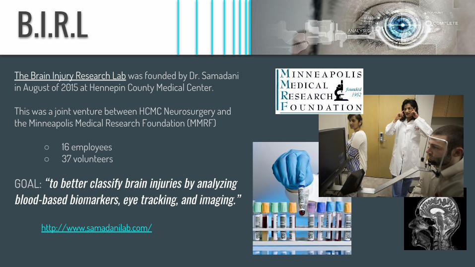
B.I.R.LThe Brain Injury Research Lab was founded by Dr. Samadani in August of 2015 at Hennepin County Medical Center.
This was a joint venture between HCMC Neurosurgery and the Minneapolis Medical Research Foundation (MMRF)
○ 16 employees○ 37 volunteers
GOAL: “to better classify brain injuries by analyzing blood-based biomarkers, eye tracking, and imaging.”
http://www.samadanilab.com/
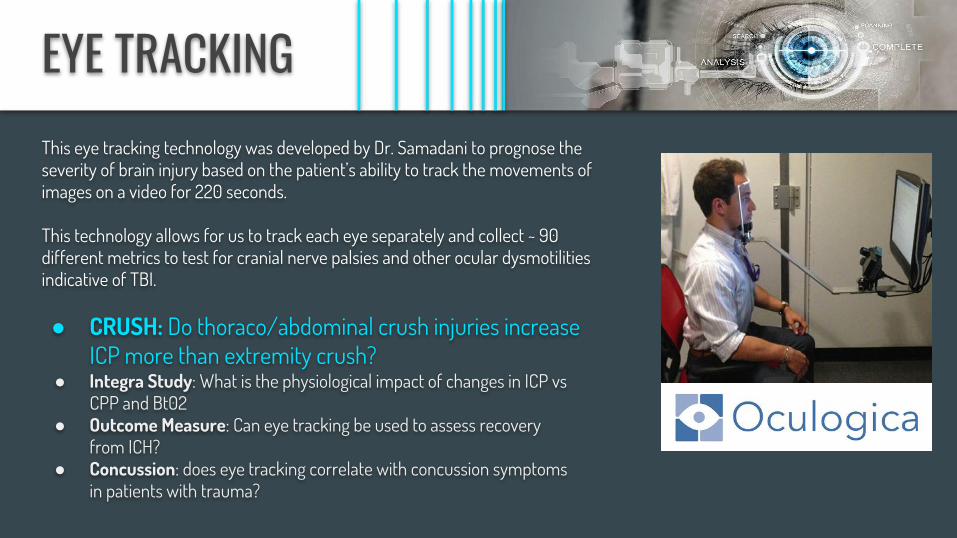
EYE TRACKINGThis eye tracking technology was developed by Dr. Samadani to prognose the severity of brain injury based on the patient’s ability to track the movements of images on a video for 220 seconds.
This technology allows for us to track each eye separately and collect ~ 90 different metrics to test for cranial nerve palsies and other ocular dysmotilities indicative of TBI.
● CRUSH: Do thoraco/abdominal crush injuries increase ICP more than extremity crush?
● Integra Study: What is the physiological impact of changes in ICP vs CPP and Bt02
● Outcome Measure: Can eye tracking be used to assess recovery from ICH?
● Concussion: does eye tracking correlate with concussion symptoms in patients with trauma?

The birth of Eye Tracking How does it work?
EYE TRACKING

The Basics
EYE TRACKING

● The video makes 5 clockwise rotations around the screen
● Each pupil’s movements are tracked and plotted
● All five of the plots are then combined and averaged
● Conjugacy is assessed by delta X and delta Y graphs
Video Summary
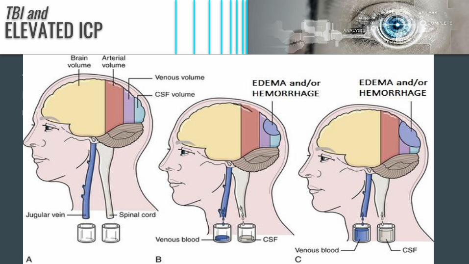
Traumatic Brain Injuries (TBIs) are associated with some degree of fluctuation in Intracranial Pressure (ICP) which are proportional to the primary severity and resultant secondary pathology.
○ Ranging from the most mild concussion to the most severe, catastrophic intracranial event.
○ However, we did not have a reliable or appropriate tool with which we can measure these changes in patients who do not warrant invasive ICP monitoring devices until now.
○ As such, these ICP changes in mild to moderate TBI have gone undiagnosed and unmeasured
○ Elevated ICP can result from TBI due to vascular swelling or obstruction of cerebral edema and cerebrospinal fluid (CSF) flow (2)
ELEVATED ICPTBI and

● Elevated ICP may eventually lead to intracranial hypertension (ICH) which is clinically defined as an ICP > 20 mmHg (3)
○ This is often the case in patients who suffered a severe TBI
ICP MONITORING
● Two current main methods of routine ICP monitoring:
○ Intraventricular catheter → difficulties in catheter placement and risk of ventriculitis (4)
○ Intraparenchymal microsensor → potential misrepresentation of global ICP value due to intraparenchymal pressure gradients (4)
● Indications for ICP Monitoring Device:○ Severe TBI with sustained ICP > 20 mmHg (5)○ GCS < 8 (5)○ Post-craniotomy (5)
TRADITIONAL

ANATOMY

● The intracranial contents, including the Cranial Nerves which exit at the base of the brain, are susceptible to changes in intracranial pressure (ICP) that result from Traumatic Brain Injury (1)
○ In particular CN III, IV, and VI are injured by the elevation of ICP
○ These nerves are often injured due to their long course and can be affected even in mild TBI’s where there is an increase in ICP
○ CN III: Oculomotor
○ CN IV: Trochlear nerve
○ CN VI: Abducens
● Eye tracking has been shown to pick up on these cranial nerve palsies
OCULAR DYSMOTILITY

CRANIAL NERVE
MUSCLE INNERVATION FUNCTION
Oculomotor Nerve (CN III)
Superior RectusInferior RectusMedial RectusInferior ObliqueLevator Palpebrae Superioris
AdductionDepressionElevationExternal Rotation
Trochlear Nerve(CN IV)
Superior Oblique AbductionInternal RotationDepression while adducting
Abducens Nerve (CN VI)
Lateral Rectus Abduction
OCULAR DYSMOTILITY[continued]

It has been previously shown that elevated ICP correlates with increasingly
abnormal eye tracking results (1)
EYE TRACKING AND ICP

EYE TRACKING AND ICP
● Elevated ICP also corresponds with decreasing box areas, and thus abnormal eye tracking, for the left and right eyes1.
○ CN III Palsy → assessed by measuring vertical distance moved by pupil1
○ CN VI Palsy → assessed by measuring horizontal distance moved by pupil1

EYE TRACKING
CRUSHINJURY
Fitting the Pieces

Crush Injury: Compression of extremities or different parts of the body that lead to muscle swelling and/or neurological disturbances in various areas of the body
CRUSH INJURIES
Extremity
● Soft tissue injuries● Femur fracture● Humerus fracture● Tibia fractures● Radial fractures● Ulnar fractures● Compartment Syndrome● Crushed Limb Syndrome
Abdominal
● Abdominal perforation● Abdominal hemorrhage● Splenic laceration● Lumbar fractures● Sacral fractures● Pelvic fractures● Bowel contusion● Liver laceration● Abdominal Compartment Syndrome (ACS)● Acute Renal Injury
Thoracic
● Penetrating chest injury● Rib/Sternal fracture● Vertebral fracture● Spinal cord Injury● Palmar contusion/hemorrhage● Cardiac contusion● Aortic Injury● Pneumothorax/hemothorax● Chest asphyxia● Esophageal/ Bronchial perforation● Bronchial perforation● Diaphragmatic Injury● Post-traumatic pneumonia● Pulmonary Embolism
Common Traumatic Pathology: Systemic ***
● Hypotension● Rhabdomyolysis

WHAT DOES THE LITERATURE SAY?

→ Case Report published in August, 2012 focused on a 44-year-old male who suffered from chest asphyxia after a crush injury to his chest and torso (1)
→ Patient was crushed by heavy vehicle parts and “developed traumatic asphyxia with severe thoracic injury and mild brain edema…” (1).
LITERATURE SAY?
Well-understood impact, focus of the case report
Less-understood impact, overlooked by case report
WHAT DOES THE
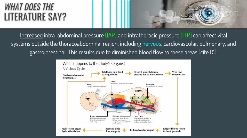
Increased intra-abdominal pressure (IAP) and intrathoracic pressure (ITP) can affect vital systems outside the thoracoabdominal region, including nervous, cardiovascular, pulmonary, and
gastrointestinal. This results due to diminished blood flow to these areas (cite R1).
LITERATURE SAY?WHAT DOES THE

Multiple Proposed Mechanisms:
● IAP decreases lumbar venous plexus blood flow and CSF absorption in the lumbar cisterna → elevated ICP
LITERATURE SAY?WHAT DOES THE
● Thoracic crush trauma → increased ITP → increased jugular venous pressure, decreases cerebral blood flow (low cerebral perfusion pressure), and increased intracranial blood volume → elevated ICP
● Chest trauma → increased ITP → increased superior vena cava pressure → decreased cerebral blood flow → elevated ICP
● Acute IAP causes increase in intracranial pressure due to increase in pleural pressure (cerebral perfusion pressure decreases due to functional obstruction of cerebral venous outflow) (2,3)
● Abdominal and thoracic cavities linked via diaphragm → transmission of IAP to the intrathoracic pressure has been noted in previous studies (2)
● Increased ICP is correlated with TBI (4)

● Sparked by the case of Erik Bedeaux○ Patient initially enrolled as body control, but we noticed abnormal eye tracking○ Abnormal eye tracking → cranial nerve palsy → elevated ICP → neurologically similar consequences to TBI
● Raised the question: Do patients in different categories of crush injury experience similar neurologic symptoms as patients who suffered TBI?
○ Prompted us to compare thoracoabdominal crush-injured patients to extremity crush-injured patients
● We expected to see elevated ICP more-so in thoracoabdominal crush patients than in extremity crush patients
○ Proposed mechanism: Thoracoabdominal crush → elevates ITP and/or IAP → decreases venous return → decreases CPP/elevates ICP
CRUSH Project

CRUSH STUDYObjectives:
1.) To evaluate if certain crush injuries without a clear cranial impact have neurologic consequences similar to those of typical traumatic brain injuries.
2.) To present preliminary data suggesting that thoracoabdominal crush injury causes an increase in ICP leading to cranial nerve palsies that can be detected by noninvasive eye tracking technology
3.) To further assess the utility and validity of eye tracking technology in detecting ICP elevations in patients whose injuries would not typically warrant conventional ICP monitoring devices
4.) To evaluate whether or not extremity crush injury has an effect on the intracranial compartment

CRUSH STUDYWe examined four groups of crush-injured patients:
1. Isolated abdominal/thoracic crush injury - 6 subjectsa. Crush injury to abdomen/thorax with no clear head trauma
2. Isolated extremity crush injury - 4 subjectsa. Crush injury to an extremity with no clear head trauma
3. Head injury + thoracic crush injury - 2 subjectsa. Crush injury to thoracic region with associated head trauma
4. Head injury + extremity crush injury - 4 subjectsa. Crush injury to extremity and with associated head trauma

Crushed by Amusement Ride
● 41 M was working on the Power Tower Ride at Valleyfair when part of the ride landed on his right upper chest. No blunt trauma to the head.
ISOLATED THORACIC CRUSH
● In the ED:○ Physical Exam - x○ Physical Exam - x○ Physical Exam - x○ Physical Exam - x
● He was admitted etc

ISOLATED THORACIC CRUSH
→ 2 week time-point eye tracking data
● Disconjugate movements of the left and right vertical and horizontal planes
●

ISOLATED THORACIC CRUSH
2 week
6 month
12 month

Auto vs Pedestrian
● 36 M who presented to the ED after their right lower extremity was crushed between two vehicles at approximately 10 mph. There was no blunt trauma to the abdomen, thorax, or head
● Discharged from the ED with Occupational Medicine follow up
ISOLATED EXTREMITY CRUSH
● In the ED:○ Physical Exam - atraumatic outside of tenderness and ecchymosis of
the right femur, CMS intact ○ FAST Exam - negative○ Right Femur XR - negative○ Pelvic XR- negative
● Discharge Diagnoses: RLE contusion

ISOLATED EXTREMITY CRUSH
PATI
ENT’
S EY
E TR
AC
KIN
G
NO
RM
AL
EYE
TRA
CK
ING

CRUSH + TBICrushed by Tree Limb
● 52 M who presented to the ED after a tree limb fell hitting him in the head and right leg. He was found to have a + LOC and obvious open RLE fracture. Flown in via helicopter.
HEAD INJURY + EXTREMITY CRUSH
● In the ED:○ Physical Exam- GCS 15, scalp hematoma and abrasion, open
deformity of the right midshaft tibia/fibula with CMS intact distally○ FAST Exam - negative○ CT Cervical Spine - C7 posterior arch fracture○ Right Tib/Fib XR - displaced fractures of both distal diaphysis
● Patient was admitted underwent surgery for the RLE fractures
● Discharge Diagnoses: Tib/Fib fx, C7 posterior arch fx, TBI

CRUSH + TBIHEAD INJURY + EXTREMITY CRUSH
PATI
ENT’
S EY
E TR
AC
KIN
G

-48 yo male, involved in motor vehicle collision, ejected, and pinned under vehicle
-Among several other injuries, sustained traumatic subarachnoid hematoma, closed fractures of multiple ribs, multiple foot fractures, and pneumonia
-Patient admitted on 1/9/2017, discharged 1 month later to a skilled nursing facility
-Discharged with 20 medications, surgical follow-up 2 weeks post-discharge and TBI follow-up in 1 month
-Left hospital with 17 discharge diagnoses
HEAD INJURY + ABD/THORACIC CRUSH

● In Hospital Time-Point eye-tracking data●
HEAD INJURY + ABD/THORACIC CRUSH

HEAD INJURY + ABD/THORACIC CRUSH
In Hosp 12 mo

BOX SCORE● Insert definition● Insert P values
DATA
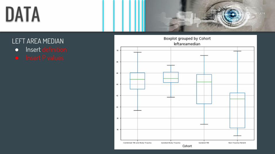
DATALEFT AREA MEDIAN● Insert definition● Insert P values

RIGHT AREA MEDIAN● Insert definition● Insert P values
DATA

RIGHT SKEW TOP● Insert definition● Insert P values
DATA

● Eye tracking can be performed as a means of noninvasive detection and long-term monitoring of elevated ICP (1)
○ Evaluating slight cranial nerve palsies that can be detected by abnormal eye tracking metrics
○ Slight abnormalities in specific metrics might be indicative of a TBI
● Can be performed on anyone who suffered a TBI, whether mild, moderate, or severe
● Does not require a trained examiner, pupil dilation, imaging studies, or invasive procedures (1)
● Data obtained from eye tracking suggests that elevated ICP may have effects at levels lower than those currently considered pathological (1)
Future Directionsof EYE TRACKING

1. Lees DH. Severe Abdominal Crush Injury. British Medical Journal. 1944;2(4382):853-854.2. Samadani, U. et al. Detection of third and sixth cranial nerve palsies with a novel method for eye tracking while watching a short film clip. Research 2, 707–720 (2014).3. Papavramidis TS, Marinis AD, Pliakos I, Kesisoglou I, Papavramidou N. Abdominal compartment syndrome – Intra-abdominal hypertension: Defining, diagnosing, and managing. Journal of
Emergencies, Trauma and Shock. 2011;4(2):279-291. doi:10.4103/0974-2700.82224.4. Sertaridou E, Papaioannou V, Kouliatsis G, Theodorou V, Pneumatikos I. Traumatic asphyxia due to blunt chest trauma: a case report and literature review. Journal of Medical Case
Reports. 2012;6:257. doi:10.1186/1752-1947-6-257.5. Hawthorne, C. & Piper, I. Monitoring of Intracranial Pressure in Patients with Traumatic Brain Injury. Frontiers in Neurology 5, (2014).6. Kirkman, M. & Smith, M. Intracranial pressure monitoring, cerebral perfusion pressure estimation, and ICP/CPP-guided therapy: a standard of care or optional extra after brain injury?
British Journal of Anaesthesia 112, 35–46 (2014).7. Peponis, T. & King, D. R. Intracranial Pressure Monitoring in Severe Traumatic Brain Injury. Oxford Medicine Online (2018). doi:10.1093/med/9780190467654.003.00078. Kolecki, R. et al. Elevated intracranial pressure and reversible eye-tracking changes detected while viewing a film clip. Journal of Neurosurgery 128, 811–818 (2018).9. .Lees, D. H. Severe Abdominal Crush Injury. Bmj 2, 853–854 (1944).
10. Sertaridou, E., Papaioannou, V., Kouliatsis, G., Theodorou, V. & Pneumatikos, I. Traumatic asphyxia due to blunt chest trauma: a case report and literature review. Journal of Medical Case Reports 6, (2012).
11. Peponis, T. & King, D. R. Intracranial Pressure Monitoring in Severe Traumatic Brain Injury. Oxford Medicine Online (2018). doi:10.1093/med/9780190467654.003.000712. Google Search Available at:
https://www.google.com/search?q=oculomotor%2Bnerve%2Bpalsy&tbm=vid&source=lnms&sa=X&ved=0ahUKEwiolJP71p7ZAhVp5oMKHZwyD20Q_AUICygC&biw=1920&bih=949&dpr=1. (Accessed: 2nd March 2018)
13. Abdominal Compartment Syndrome. Practice Essentials, Pathophysiology, Etiology (2017). Available at: https://emedicine.medscape.com/article/829008-overview. (Accessed: 2nd March 2018)
14. Crush injury. MedlinePlus Medical Encyclopedia Available at: https://medlineplus.gov/ency/article/000024.htm. (Accessed: 2nd March 2018)
REFERENCES

SPECIAL THANKS TO
Daniel Rafter, M.D.