Bleeding and Thrombosis hemoptysis, hematemesis, melena, hematuria, and vaginal bleeding. •...
44
1 Chapter 1 Bleeding and Thrombosis BLEEDING FAST FACTS • Description: Bleeding can result from a reduction in platelets, an alteration in clotting fac- tors, a paraneoplastic syndrome, infection, hepatic problems, or a combination of these fac- tors. • Incidence: Bleeding can occur with any type of cancer but especially in patients with advanced cancer and those with hematologic malignancies. • Clinical Manifestations: Signs of bleeding include petechiae, ecchymoses, bruising, epi- staxis, hemoptysis, hematemesis, melena, hematuria, and vaginal bleeding. • Evaluation/Diagnostic Tests: Tests include complete blood count (especially hemoglo- bin and platelet levels) and coagulation tests. • Treatment Modalities: Interventions for managing bleeding include transfusions, vitamin K therapy, vasopressive hormones, and mechanical measures. • Nursing Management: Recognition of early signs and symptoms of bleeding or coagu- lation is key in the nurse’s assessment. Bleeding precautions are implemented for at-risk patients. • Outcome: Patients with cancer are at high risk for bleeding. Early recognition and initia- tion of appropriate treatment are crucial to patient outcomes. Introduction Abnormalities in hemostasis, including bleeding and thrombosis, are common complications in patients with cancer and can greatly affect mor- bidity and mortality (Rodriguez, 2018). Several factors can contribute to the etiology of bleeding and thrombosis, including the disease process and the use of treatment modalities that may affect bleeding or coagulation. These factors include surgical procedures, chemotherapy, biotherapy, radiation therapy, central venous catheters, supportive care with colony-stimulating factors, and newer anticoagulants (Königsbrügge, Pabinger, & Ay, 2014; Rodriguez, 2014). Several tumor-related or treatment-related factors can increase the risk for bleeding by disrupting normal hemostatic mechanisms, including alterations in platelet function or the coagulation pathway. Plate- Colleen M. O’Leary, MSN, RN, AOCNS ® Copyright 2018 by Oncology Nursing Society. All rights reserved.
Transcript of Bleeding and Thrombosis hemoptysis, hematemesis, melena, hematuria, and vaginal bleeding. •...

1
Chapter 1Bleeding and Thrombosis
BLEEDING
FAST FACTS• Description: Bleeding can result from a reduction in platelets, an alteration in clotting fac-
tors, a paraneoplastic syndrome, infection, hepatic problems, or a combination of these fac-tors.
• Incidence: Bleeding can occur with any type of cancer but especially in patients withadvanced cancer and those with hematologic malignancies.
• Clinical Manifestations: Signs of bleeding include petechiae, ecchymoses, bruising, epi-staxis, hemoptysis, hematemesis, melena, hematuria, and vaginal bleeding.
• Evaluation/Diagnostic Tests: Tests include complete blood count (especially hemoglo-bin and platelet levels) and coagulation tests.
• Treatment Modalities: Interventions for managing bleeding include transfusions, vitaminK therapy, vasopressive hormones, and mechanical measures.
• Nursing Management: Recognition of early signs and symptoms of bleeding or coagu-lation is key in the nurse’s assessment. Bleeding precautions are implemented for at-riskpatients.
• Outcome: Patients with cancer are at high risk for bleeding. Early recognition and initia-tion of appropriate treatment are crucial to patient outcomes.
IntroductionAbnormalities in hemostasis, including bleeding and thrombosis, are
common complications in patients with cancer and can greatly affect mor-bidity and mortality (Rodriguez, 2018). Several factors can contribute to the etiology of bleeding and thrombosis, including the disease process and the use of treatment modalities that may affect bleeding or coagulation. These factors include surgical procedures, chemotherapy, biotherapy, radiation therapy, central venous catheters, supportive care with colony-stimulating factors, and newer anticoagulants (Königsbrügge, Pabinger, & Ay, 2014; Rodriguez, 2014). Several tumor-related or treatment-related factors can increase the risk for bleeding by disrupting normal hemostatic mechanisms, including alterations in platelet function or the coagulation pathway. Plate-
Colleen M. O’Leary, MSN, RN, AOCNS®
Copyright 2018 by Oncology Nursing Society. All rights reserved.
2 Understanding and Managing Oncologic Emergencies (Third Edition)
let dysfunction can be a result of the disease itself or medications and ther-apies administered to patients. The presence of infection, liver disease, and the high-viscosity proteins that some tumors express can each alter the coag-ulation pathway, increasing the risk for bleeding (Rodriguez, 2014). The risk for bleeding increases with cancer, but certain malignancies, especially most leukemias, are associated with a higher risk than others. Acute promy-elocytic leukemia carries a particularly high risk for bleeding with as many as 90% of patients developing bleeding complications (Coombs, Tavakkoli, & Tallman, 2015). The incidence of fatal hemorrhage in these patients can be as high as 17.3% (Coombs et al., 2015).
These disturbances can directly affect patient morbidity and mortal-ity and present complex challenges when caring for patients with cancer. Because bleeding poses dire consequences for patients, it is imperative that any management plan includes bleeding prevention. To increase sur-vival rates, rapid recognition, assessment of the precipitating factors, and knowledge of the treatment of bleeding are necessary. Ongoing bleeding can affect patients’ quality of life by causing fatigue and weakness. If left untreated, bleeding may progress to life-threatening hemorrhage. The abil-ity to recognize early signs of bleeding and initiate interventions can dra-matically improve patients’ quality of life and increase their potential for survival. Oncology nurses, both in ambulatory and inpatient settings, play a key role in preventing, assessing, and managing major bleeding events and should be knowledgeable about evidence-based interventions as they evolve.
IncidenceBleeding, in general, can occur with any type of cancer, and there are
no specific incidence rates for this complication. However, bleeding does occur more frequently in individuals with hematologic cancers because these cancers affect the bone marrow, often resulting in thrombocyto-penia—a reduction in the number of circulating platelets. Therefore, the incidence and severity of bleeding in patients with acute leukemia are greater than in patients with solid tumors. Although fatal hemor-rhagic complications occur in 1.5% of patients with solid tumors, more than 50% of patients with acute leukemia experience these complica-tions (Rodriguez, 2018).
Mucin-producing adenocarcinomas, such as those of the lung, breast, stomach, pancreas, and prostate, are more prone to hemostatic abnormal-ities (Escobar, Henke, & Wakefield, 2013). However, these solid tumors are thought to be more commonly associated with disseminated intravascular coagulation (DIC), which is discussed in depth in Chapter 4. Malignancies with a higher incidence of bleeding include large carcinomas of the head and neck, large centrally located lung cancers, acute myeloid leukemia, and chronic myeloid leukemia (Rodriguez, 2018).
Copyright 2018 by Oncology Nursing Society. All rights reserved.

Chapter 1. Bleeding and Thrombosis 3
Risk Factors and EtiologySeveral factors increase the risk of bleeding in patients with cancer. Anti-
neoplastic therapies increase the risk for bleeding by damaging normal tis-sues. Radiation therapy and steroid treatment can cause blood vessels to become fragile and more prone to injury. Chemotherapy and radiation can cause myelosuppression, including thrombocytopenia, which increases the risk of bleeding. Other general risk factors for bleeding in patients with can-cer include fever, sepsis, infection, anticoagulant therapy, impaired platelet function, and adverse effects of medications (Rodriguez, 2014).
Because of the critical role that platelets play in hemostasis and throm-bosis, the platelet count is considered to be the most significant fac-tor for predicting bleeding in a patient with cancer (Rodriguez, 2018). The first reported association between thrombocytopenia and bleeding risk occurred in 1962, when it was demonstrated that hemorrhage rarely occurred in patients with acute leukemia when the platelet count remained higher than 20,000/mm3 (Gaydos, Freirich, & Mantel, 1962). Platelet func-tion can be affected by numerous drugs, particularly aspirin and nonsteroi-dal anti-inflammatory drugs (NSAIDs), such as ibuprofen (Scharf, 2012). Both drug types can drastically reduce platelet aggregation, with the effects of aspirin lasting up to four days after ingestion. A newer class of NSAIDs, the cyclooxygenase-2 inhibitors (commonly known as COX-2 inhibitors), has a lesser effect on platelets than other NSAIDs and may be an option for patients with cancer (Cho, Walker, Williams, & Hasty, 2015).
Antibiotic use can also contribute to bleeding risk by impairing platelet function. Carbenicillin, ticarcillin, and other penicillins impair the aggre-gation of platelets and prolong bleeding time (Scharf, 2012). This plate-let dysfunction is maximized one to three days after administration and can continue for several days after the antibiotic course is completed. Other medications that suppress platelet function include phenothiazines, tricy-clic antidepressants, heparins, cimetidine, thiazide diuretics, statins, plasma expanders, antihistamines, and estrogen. In addition, corticosteroids affect the gastric mucosa and impair wound healing, resulting in the skin becom-ing thin, fragile, and more susceptible to bleeding. However, a systematic review and meta-analysis of the risk of gastrointestinal (GI) bleeding with corticosteroid use showed that the increased risk of bleeding was only sig-nificant for hospitalized patients on corticosteroids and not for those in the ambulatory setting (Narum, Westergren, & Klemp, 2014).
Tumor growth can lead to bleeding through invasion into the surround-ing structures and blood vessels, as well as the bone marrow. Leukemias and lymphomas are the most common cancers associated with bone marrow inva-sion. In these diseases, the malignant cells crowd out the normal cells in the marrow, leading to thrombocytopenia, anemia, and neutropenia. Tumors can also erode and enter mucous membranes and cause bleeding by local tis-sue disruption. Tumors that are close to large blood vessels put patients at risk for a massive hemorrhage. Other risk factors for bleeding in patients with can-
Copyright 2018 by Oncology Nursing Society. All rights reserved.

4 Understanding and Managing Oncologic Emergencies (Third Edition)
cer include fever, infection, uremia, decreased hematocrit, anatomic lesions, endothelial injury, hypoalbuminemia, recent hemorrhage, hyperleukocytosis, anticoagulant therapy, and stem cell transplant (Rodriguez, 2014).
Infection, mainly sepsis, increases the risk of bleeding in several situa-tions. DIC, a possible complication of sepsis, can result in a fatal bleeding event (Levi & van der Poll, 2013). Periods of febrile neutropenia are associ-ated with the greatest bleeding risk. An infection within the GI or genito-urinary tract can irritate or ulcerate the mucosal linings, thereby increasing the risk for bleeding events. Viruses can cause myelosuppression or throm-bocytopenia, and fungal infections, especially in the lungs, can cause fatal hemorrhage (Nucci, Nouér, Cappone, & Anaissie, 2013). Fever has been known to increase the risk of bleeding and potentially affects a patient’s response to a platelet transfusion (Rodriguez, 2014). Vitamin K is necessary for proper coagulation and is absorbed solely from the diet. Malnutrition can lead to an increased risk of bleeding because of a vitamin K deficiency, which can develop as a direct result of a dietary deficiency, biliary obstruc-tion, malabsorption syndromes, liver disease, and anticoagulation therapy (Nguyen-Khoa, Patel, & Mikhail, 2015).
Uremia is another risk factor for bleeding. Uremic bleeding is related to platelet dysfunction, including a reduced platelet count and increased plate-let turnover. In addition, uremia can cause decreased platelet adhesion and aggregation. Uremic toxins, increased production of nitric oxide, prostacy-clin, calcium, and cyclic adenosine monophosphate, along with renal anemia, cause these symptoms (Berns & Coutre, 2017). Bleeding in uremic patients most commonly manifests in the mucocutaneous areas and puncture sites as ecchymosis, purpura, epistaxis, GI and genitourinary bleeding, and subdural hematomas. Excessive bleeding due to uremia may also manifest as cardiovas-cular and neurologic deficits (Alper, Shenava, & Young, 2017).
PathophysiologyNormal control of bleeding (hemostasis) is maintained through a finely
regulated balance between clot formation (coagulation) and clot dissolu-tion (fibrinolysis). Chapter 4 provides a more detailed discussion of this pro-cess. The presence of malignancy disrupts this essential balance and can lead to bleeding through several different mechanisms, including alterations in platelet count and function, activation of the coagulation cascade, disruption in vascular integrity, and the effects of antineoplastic therapies. Patients with cancer often have more than one of these conditions occurring simultane-ously, putting them at increased risk for bleeding (Rodriguez, 2018).
Alone or in combination, local and systemic factors can cause bleeding. Local anatomic causes of bleeding include tumor extension and invasion of blood vessels, whereas systemic causes include bone marrow invasion by tumor cells, bone marrow suppression from chemotherapy or radiation,
Copyright 2018 by Oncology Nursing Society. All rights reserved.

Chapter 1. Bleeding and Thrombosis 5
and the development of DIC (see Chapter 4). Other systemic factors con-tributing to bleeding risk include liver failure; medications, such as anti-coagulants, aspirin, and NSAIDs; and concomitant disease, such as cirrho-sis (Abrams, Shattil, & Bennett, 2016; Levi, Seligsohn, & Kaushansky, 2011; Mitchell, 2015; Rodriguez, 2018). See Table 1-1 for an overview of the etiol-ogy of bleeding.
Platelets and ThrombocytopeniaPlatelets play a crucial role in the process of hemostasis. Platelets are not
true cells, but rather fragments of megakaryocytes, giant cells within the bone marrow integral to the production of platelets. When fragmented, a megakaryocyte can release more than 1,000 platelets. The hormone throm-bopoietin regulates the platelet production process. In the event of an injury or cut that breaks the endothelial layer of a blood vessel, platelets function as first responders to form a clot that seals the injury site and inhibits blood loss (Monroe & Hoffman, 2014). Disorders of platelet function and platelet abnormalities can affect the clotting process and put an individual at risk for severe and possibly fatal bleeding.
Thrombocytopenia is the platelet abnormality most frequently associ-ated with cancer. Several factors are linked with the development of throm-bocytopenia, including reduced platelet production, a change in platelet distribution, increased platelet destruction, vascular dilution of platelets, platelet consumption as seen in DIC, and some drug therapies, including beta-lactam antibiotics, nitrates, beta-blockers, tricyclic antidepressants, and selective serotonin reuptake inhibitors (Gao & Sahai, 2011; Konkle, 2011).
Table 1-1. Factors Contributing to Bleeding in Patients With Cancer
Type Factors
Anatomic factors Local tumor invasionTumor involvement of vascular tissueTumor location near major vesselsHead and neck tumors
Systemic factors Bone marrow involvement by tumorBone marrow suppressionConcomitant disease (e.g., cirrhosis)Disseminated intravascular coagulationLiver failureMedications (e.g., anticoagulants, aspirin, nonsteroidal
anti-inflammatory drugs)
Combination of local and systemic factors
Local tumor invasion and any systemic factors
Note. Based on information from Abrams et al., 2016; Mitchell, 2015; Rodriguez, 2018.
Copyright 2018 by Oncology Nursing Society. All rights reserved.

6 Understanding and Managing Oncologic Emergencies (Third Edition)
Decreased platelet production can occur secondary to direct invasion of the bone marrow by tumor cells or from the myelosuppressive effects of chemotherapy or radiation therapy on the marrow. When a large bur-den of tumor cells overwhelms the normal elements of bone marrow, the resulting thrombocytopenia reflects an overall pancytopenia in which all cell lines are reduced. The occurrence and severity of treatment-associated thrombocytopenia depend on the type of chemotherapy drugs, dosage, and the time between treatments. With radiation therapy, the development of thrombocytopenia depends on the amount of bone marrow encompassed in the treatment fields. The development of thrombocytopenia may be a dose-limiting factor in delivering these treatments and can lead to a bleed-ing event (Rodriguez, 2018).
Thrombocytopenia may arise indirectly in patients with cancer whose spleens have enlarged because of infection, inflammation, autoimmune disor-der, or neoplasm within the spleen. Splenic pooling of platelets has been iden-tified as a cause of thrombocytopenia, with approximately one-third of trans-fused platelets being removed from circulation and sequestered in the spleen (Izak & Bussel, 2014). Splenic enlargement may occur with metastasis to the spleen from cancers of the lung, breast, colon, prostate, and stomach, as well as lymphomas. If the spleen is not enlarged, it is unlikely that existing thrombocy-topenia is the result of splenic trapping of platelets (Rodriguez, 2018).
Thrombocytopenia may develop when platelet destruction increases, a fea-ture of idiopathic thrombocytopenic purpura, an autoimmune disorder in which autoantibodies are formed against the individual’s own platelets. Imma-ture platelets accumulate in the bone marrow while the number of circulating mature platelets diminishes. This presentation most often occurs in patients with lymphomas and may precede clinical diagnosis (Rodriguez, 2018).
Platelet RefractorinessPlatelet refractoriness exists when the platelet level does not increase to
the desired level following a platelet transfusion. It is defined as a one-hour post-transfusion platelet count that fails to increase by an increment of 11,000/mm3 on two consecutive transfusions (Fletcher, DomBourian, & Millward, 2015). Platelet refractoriness has been reported in 30%–50% of patients who receive platelet transfusions (Valsami, Dimitroulis, Gialer-aki, Chimonidou, & Politou, 2015). Two categories of platelet refractoriness exist: nonimmune and immune.
Nonimmune: Causes of nonimmune refractoriness include sepsis, fever, bleeding, splenomegaly, and DIC. Fever, one of the most frequently cited causes, most likely is not an independent factor but rather is associ-ated with underlying infection or sepsis. DIC results in an increased rate of platelet consumption, which can result in refractoriness. Splenomegaly is a well-established cause of poor response to platelet transfusion. Normally, approximately one-third of a person’s platelets are sequestered in the spleen,
Copyright 2018 by Oncology Nursing Society. All rights reserved.

Chapter 1. Bleeding and Thrombosis 7
which is in equilibrium to the circulating platelet pool. In extreme spleno-megaly, up to 90% of the platelets can be sequestered (Wang et al., 2013).
Immune: Refractoriness to platelet transfusion caused by an immune response is known as alloimmunization and has the greatest potential for pre-vention and management. Some platelet antigens, such as human leukocyte antigens (HLAs), are shared by other blood cells and tissues, with HLA-A and HLA-B antigens most likely responsible for alloimmunization. To coun-teract this effect, transfusions of leukoreduced platelets, in which the white blood cells (leukocytes) have been removed from the blood product, can be used to decrease the potential for alloimmunization. Another type of anti-gen, human platelet antigen (HPA), is specific to platelets only. HPAs cause changes in platelet membranes that can stimulate production of antibod-ies once exposed to foreign platelets with different HPAs through platelet transfusions (Curtis & McFarland, 2014).
Clotting Factor DeficienciesClotting factors are the blood components responsible for conversion of
fibrinogen to fibrin, which is needed to form a clot. Clotting factor deficien-cies can contribute to bleeding risk and may occur with certain cancers. Patients with solid tumors, hematologic cancers, myeloproliferative disor-ders, macroglobulinemia, and lymphoproliferative disorders may acquire von Willebrand disease, in which factor VIII and von Willebrand factor pro-coagulant activity is diminished or absent. Patients who present with this syndrome usually have increased bruising, mucosal bleeding, and GI hem-orrhage (Rodriguez, 2018).
Clotting factors I, II, V, VII, IX, and X are synthesized in the liver; there-fore, conditions that compromise liver function, such as infection, chemo-therapy treatment, tumor invasion, and surgery, can contribute to prolonged bleeding times. The liver also clears fibrin degradation products and acti-vated clotting factors from circulation. Thus, impairment of liver function disturbs normal coagulation (Rodriguez, 2018). Therefore, patients with can-cer who undergo extensive surgical procedures and receive large amounts of fresh frozen plasma may become prone to increased bleeding (Rodriguez, 2018). In addition, deficiency of clotting factor III may exist with acute leu-kemias and myelodysplastic syndrome. This is associated with impaired pro-coagulant activities and abnormal platelet function because of alterations in platelet size, shape, and aggregation responses (Rodriguez, 2018).
Clinical ManifestationsBleeding manifests in several ways in patients with cancer. Signs of bleed-
ing without visible hemorrhage include petechiae, ecchymoses, and bruis-ing. This type of bleeding may often be overlooked because it is not thought
Copyright 2018 by Oncology Nursing Society. All rights reserved.

8 Understanding and Managing Oncologic Emergencies (Third Edition)
of as active bleeding and manifests in covert signs. Overt signs of bleed-ing include epistaxis, hemoptysis, hematemesis, melena, hematuria, vaginal bleeding, and bleeding around wounds and vascular access devices (Rodri-guez, 2014). Bleeding can begin slowly and present as oozing but can prog-ress to an acute hemorrhagic event. Even the smallest amount of bleeding can eventually lead to absolute anemia, defined as a reduction in the num-ber or volume of circulating red blood cells.
Anemia is not a disease but rather a symptom of other disorders. If bleed-ing is present in patients with cancer, anemia will most likely develop. Patients with bleeding can show symptoms of anemia, including fatigue, pallor, diz-ziness, irritability, weakness, chest pain, shortness of breath, decreased body temperature, and numbness in the hands and feet. Hypotension and tachy-cardia may be present. Many symptoms of anemia are nonspecific, and a drastic reduction in the red blood cell count may occur before absolute ane-mia is diagnosed. Because there is no easy or practical way to measure abso-lute anemia, the more typical measurement used is accepted anemia, which is defined as a reduction in one or more of the major red blood cell measure-ments, including the hemoglobin concentrate, hematocrit, and red blood cell count (Balentine & Nabili, 2017; Lam, 2017).
More than 400 types of anemia exist and are classified by either etiology or morphology (Schrier, 2016). Etiologic classifications focus on the cause of the anemia. Etiologically, anemia can be caused by a decreased production of red blood cells, as seen with bone marrow suppression; by increased red blood cell destruction, as seen with hemolysis or blood loss; or by lack of hormones, such as erythropoietin to stimulate production of red blood cells.
Morphologic classifications focus on changes in the erythrocyte itself, such as in color, shape, or size (Schrier, 2016). Morphologically, anemias are classified as microcytic, macrocytic, or normocytic. In microcytic ane-mias, the red blood cell is smaller than normal with a mean corpuscular volume (MCV) of less than 80 femtoliters (fL). It is usually accompanied by a decrease in hemoglobin content. The most common types of micro-cytic anemias are thalassemia, iron-deficiency anemia, and anemia of chronic disease (Schrier, 2016). In macrocytic anemias, the red blood cell is larger than normal, with an MCV of greater than 100 fL with accompa-nying abnormal nucleic acid production and abnormal red blood cell mat-uration. Macrocytic anemias most commonly occur with myelodysplastic syndrome and acute leukemia (Schrier, 2016). Normocytic anemias have a normal-sized red blood cell with an MCV of 80–100 fL. Normocytic anemia is seen with endocrine disorders, acute blood loss, myelosuppression, and early iron-deficiency anemia (Schrier, 2016).
Several organ systems are sensitive to the effects of bleeding and anemia. In the renal system, the kidneys respond to decreased blood flow by releasing renin, an enzyme that participates in maintaining blood pressure by stim-ulating salt and water retention, which can lead to edema (Schrier, 2016). The decreased oxygen levels that accompany chronic anemia can cause hair
Copyright 2018 by Oncology Nursing Society. All rights reserved.

Chapter 1. Bleeding and Thrombosis 9
to become thin and gray. In the nervous system, anemia affects nerve fibers by causing degeneration of myelin, leading to loss of nerve fibers. Effects of anemia on the GI system manifest as abdominal pain, nausea, and vomiting. The skin loses elasticity in response to the decreased oxygenation associated with anemia. Table 1-2 provides an overview of the effects of bleeding on selected organ systems. It is important for nurses to instruct patients about the signs and symptoms of anemia and the need for immediate follow-up with their physicians.
Patient AssessmentAssessment should begin with a thorough history. A complete bleeding
history should be obtained, including signs of bleeding such as easy bruis-ing, nosebleeds, gingival bleeding, excessive bleeding with invasive proce-dures, unusually heavy menstrual bleeding, petechiae, and a change in the color of stools or urine. Nurses should question patients about the presence of symptoms associated with anemia, such as fatigue, headache, and weak-ness, and whether they have a history of heavy menstrual bleeding or prior anemia. A medication history should include antibiotics; NSAIDs; chemo-therapy, biologic, and supportive agents; and all over-the-counter medica-tions and supplements. History of acute or viral infections is important. The patient history should also include transfusions; immunologic disorders, such as idiopathic thrombocytopenic purpura; and family history of bleed-ing abnormalities. Patients’ nutritional status needs to be assessed to possi-bly identify any vitamin deficiencies, such vitamin B12 or folic acid deficien-cies (Rodriguez, 2018).
A systematic physical examination to assess for signs of bleeding follows the patient history. Bleeding within the central nervous system can man-ifest as mental status changes, lethargy, restlessness, altered level of con-sciousness, seizures, or coma, as well as changes in neurologic signs (e.g.,
Table 1-2. Effects of Bleeding on Selected Organ Systems
Organ System Cause Effect
Gastrointestinal Decreased oxygenation Abdominal pain, nausea and vomiting
Integumentary Decreased oxygenation Thinning and graying of hair, loss of skin elasticity
Nervous Myelin degeneration Loss of nerve fibers
Renal Kidneys release renin Salt and water retention, edema
Note. Based on information from Schrier, 2016.
Copyright 2018 by Oncology Nursing Society. All rights reserved.

10 Understanding and Managing Oncologic Emergencies (Third Edition)
pupil size, reactivity), motor strength and coordination, and speech. Head-ache also may be present. Examination of the eyes and ears may reveal par-tial visual field loss, increased redness in the eyes, periorbital edema, and subconjunctival hemorrhage. Patients may report blurred vision and eye or ear pain. Examination of the nose, mouth, and throat may reveal pete-chiae on the nasal or oral mucosa, ulceration, gingivitis, or mucous mem-brane bleeding. Epistaxis may be present. Signs of bleeding manifested by the cardiovascular system include changes in vital signs, changes in the color and temperature of all extremities, changes in the peripheral pulses, tachycardia, or hypotension. During examination of the pulmonary sys-tem, it is important to note changes in the respiratory rate and depth, dyspnea, tachypnea, shortness of breath, crackles, wheezing, orthop-nea, hemoptysis, or cyanosis. Examination of the GI system could reveal increased or vague pain, blood around the rectum, tarry stools, and occult blood in the stool or hematemesis. Indications of bleeding in the genito-urinary system present as blood in the urine, dysuria, burning and fre-quency with urination, decreased urine output, and a change in the char-acter or amount of menses. Bleeding in the musculoskeletal system could manifest as warm, tender, swollen joints with diminished mobility. Indica-tions of bleeding in the integumentary system include signs of bruising, petechiae, purpura, ecchymoses, hematomas, pallor or jaundice, and ooz-ing from injection sites, biopsy sites, central lines, catheters, or even naso-gastric tubes (Rodriguez, 2018).
Diagnostic EvaluationMany diagnostic tests are used to determine if bleeding is present.
Hemoccult tests can identify blood in stool, and a urine dipstick typically is used to test for microscopic hematuria. For more active blood loss, mea-surement of the volume of melena or hematemesis should be used (Wilkins, Khan, Nabh, & Schade, 2012). Imaging scans also can be used to assess for internal bleeding. Laboratory values are the most commonly used tools to determine bleeding and coagulation status and include tests to determine platelet counts, bleeding times, D-dimer levels, and levels of specific coag-ulation factors. Hemoglobin and hematocrit values are measured to moni-tor blood loss (Wilkins et al., 2012). Patients with cancer may present with existing anemia and have reduced hemoglobin and hematocrit levels. Sud-den drops in these levels could indicate an acute blood loss and should be treated immediately.
Bleeding time can be measured by calculating the time it takes to stop bleeding from a small incision in the skin. Normal bleeding time varies between one and nine minutes (Rodriguez, 2018). Bleeding time depends on both the number of platelets and how well the blood vessels function, especially the ability to vasoconstrict. Prolonged bleeding time
Copyright 2018 by Oncology Nursing Society. All rights reserved.

Chapter 1. Bleeding and Thrombosis 11
may result from alterations in either of these factors and may occur with thrombocytopenia, tumor infiltration of bone marrow, consumption of platelets in DIC, and use of medications that alter platelet function. Bone marrow aspiration is used to determine the etiology of thrombocytope-nia by assessing whether the number of megakaryocytes in the marrow is normal or altered. If the number of megakaryocytes within the bone marrow is reduced, the cause relates to primary thrombocytopenia or a reduced production of platelets. When the number of megakaryocytes in the bone marrow is normal or elevated, thrombocytopenia is a result of increased uptake or peripheral destruction of platelets (Barsam et al., 2011).
The platelet count, which quantifies the number of platelets in the blood volume, is the best gauge of possible bleeding in a patient with cancer. The Common Terminology Criteria for Adverse Events (National Cancer Insti-tute Cancer Therapy Evaluation Program [NCI CTEP], 2017) grades a decreased platelet count from 1 to 5, with mild thrombocytopenia (grade 2) defined as a platelet count of 50,000–75,000/mm3. Table 1-3 presents thegrading system for decreased platelet count.
After a patient receives a platelet transfusion for thrombocytopenia, the response to the transfusion must be evaluated. The corrected count incre-ment (CCI) is a formula based on a platelet count obtained 10 minutes to 1 hour after transfusion. If the CCI is less than 5,000/mm3 on at least two sequential occasions, the patient is determined to be refractory to platelets (Thiagarajan & Afshar-Kharghan, 2013). Often, CCI is not the most prac-tical means of assessing response because the formula for calculating CCI uses the number of plate-lets transfused, which often is unavailable. There-fore, a more common def-inition uses measurements of platelet recovery and platelet response to indi-cate refractoriness. Plate-let recovery is defined as the increment in platelet count measured 10 minutes to 1 hour following transfusion. Platelet survival is evalu-ated by a platelet count obtained 18–24 hours after transfusion. When evalu-ating both platelet recov-ery and survival, platelet refractoriness is present if the absolute platelet count
Table 1-3. Common Terminology Criteria for Adverse Events Grading of Decreased
Platelet Count
Grade Platelet Levels
1
2
3
4
5
< LLN–75,000/mm3; < LLN–75.0 × 10⁹/L
< 75,000–50,000/mm3; < 75.0–50.0 × 10⁹/L
< 50,000–25,000/mm3; < 50.0–25.0 × 10⁹/L
< 25,000/mm3; < 25.0 × 10⁹/L
–
LLN—lower limit of normalNote. From Common Terminology Criteria for Adverse Events [v.5.0], by National Cancer Institute Cancer Therapy Evalua-tion Program, 2017. Retrieved from https://ctep.cancer.gov/ protocoldevelopment/electronic_applications/docs/CTCAE _v5_quick_reference_5x7.pdf.
Copyright 2018 by Oncology Nursing Society. All rights reserved.

12 Understanding and Managing Oncologic Emergencies (Third Edition)
rises less than 10,000/mm3 after administration of a unit of platelets or if it rapidly returns to pretransfusion levels (Thiagarajan & Afshar-Kharghan, 2013).
Besides laboratory tests, certain radiographic studies, such as computed tomography (CT), magnetic resonance imaging, x-ray, or ultrasound, can be used to determine whether any bleeding is present. Endoscopic exam-inations can be used to assess for bleeding in the upper or lower GI tract but would rarely be used for this purpose because any sort of invasive pro-cedure could also put a patient at an increased risk for bleeding (Travis & Saltzman, 2016).
Treatment ModalitiesProphylaxis
Transfusions of red blood cells, platelets, and plasma have been used effectively in patients with thrombocytopenia to prevent bleeding. Trans-fusions of red blood cells usually are initiated when a patient’s hemoglobin level drops below 8 g/dl, or grade 3 anemia (NCI CTEP, 2017). Currently, no studies indicate the level at which transfusions should be initiated to improve outcomes. However, the guidelines for several chemotherapy regi-mens require the platelet count to be at a certain level prior to administer-ing treatment (Camp-Sorrell, 2016; Crighton et al., 2015).
The transfusion of platelets plays an important role in bleeding pre-vention and management (Yuan & Goldfinger, 2017). Usually one unit of platelets can increase platelet count by 6,000–10,000/mm3, but each case is unique, and each individual may not have the same results. Patients with acute leukemia or solid tumors and those undergoing stem cell trans-plantation should maintain a platelet threshold of 10,000/mm3. Patients scheduled to undergo minor procedures and those with bladder tumors, necrotic tumors, and highly vascular tumors should have a threshold of 20,000/mm3. Patients undergoing any type of invasive procedure should have a platelet threshold of 40,000–50,000/mm3 (Yuan & Goldfinger, 2017). Several factors can influence the effectiveness of platelet transfu-sions, including fever, sepsis, and hypersplenism. Another important fac-tor in the effectiveness of platelet transfusions is the proper storage of platelets to maintain freshness and metabolic activity. After the platelets are obtained, the ideal administration time for maximum effectiveness is within six hours. However, they can be stored for up to five days (Lebois & Josefsson, 2016; Reddoch et al., 2014). Plasma transfusion is generally reserved for patients with coagulation abnormalities who must undergo surgical procedures.
Recombinant colony-stimulating growth factor has been used to reduce the negative hematopoietic effects of chemotherapy and radia-tion therapy by accelerating the recovery period. Recombinant human
Copyright 2018 by Oncology Nursing Society. All rights reserved.

Chapter 1. Bleeding and Thrombosis 13
interleukin-11 (rhIL-11), erythropoietin, and recombinant thrombopoi-etin have been used effectively to stimulate megakaryocyte proliferation. Several studies using rhIL-11 in patients with solid tumors, lymphoma, acute myeloid leukemia, and myelodysplastic syndrome have shown effi-cacy in the prevention of severe thrombocytopenia following myelosup-pressive chemotherapy (Rodriguez, 2018). To achieve maximum ben-efit, rhIL-11 should be initiated within 24 hours after chemotherapy is completed and continued for up to 21 days until the platelet count is greater than 50,000/mm3 (Wyeth Pharmaceuticals Inc., 2011). This treat-ment will increase platelet levels after five to nine days of administration, which should coincide with the expected chemotherapy-induced platelet nadir (Jung et al., 2011). Studies are limited regarding the use of eryth-ropoietin for chemotherapy-induced thrombocytopenia. The administra-tion of erythropoietin increases hemoglobin concentration and reduces the need for red blood cell transfusions in patients with anemia result-ing from chemotherapy. Darbepoetin alfa, a growth factor derived with recombinant DNA technology, stimulates erythropoiesis. Because it has a longer half-life than erythropoietin, darbepoetin alfa can be given once per week, whereas erythropoietin must be administered three times per week (Amgen Inc., 2016). Although studies have shown some benefit to the use of erythropoiesis-stimulating agents, their use is controversial given the increased incidence of life-threatening cardiovascular events in patients with cancer.
TreatmentA variety of interventions can be used to manage bleeding in patients
with cancer. A specific plan designed to treat bleeding should be individu-alized for each patient and depends on many factors, including the under-lying causes, likelihood of reversing or controlling the bleeding etiology, disease status, goals of therapy, presence of comorbidities, and whether the treatment benefits outweigh the risks. If a benefit is expected, specific mea-sures are put in place to control the bleeding. Treatments used to control bleeding include transfusions of blood products, vitamin K therapy, and mechanical measures. Other therapies include radiation treatments, endo-scopic procedures, and surgery, but these often are used as a last resort because of increased bleeding risk.
Transfusions reduce morbidity and death from thrombocytopenia and are likely to prevent and manage bleeding in patients with cancer who are actively bleeding (Yuan & Goldfinger, 2017). With normal splenic pooling, an average of four to six units of platelets are needed to control bleeding (Yuan & Goldfinger, 2017). The traditional management of patients with HLA antibodies is to provide platelets from HLA-matched donors. Unfortu-nately, for many patients, HLA-matched donors are unavailable. In this case, platelets from partially mismatched donors may provide adequate responses.
Copyright 2018 by Oncology Nursing Society. All rights reserved.

14 Understanding and Managing Oncologic Emergencies (Third Edition)
Patients generally respond well to HLA-matched donor platelets if alloim-munization alone is responsible for platelet refractoriness. These platelets should be irradiated to prevent transfusion-associated graft-versus-host dis-ease (Wang et al., 2013).
Red blood cell therapy is usually initiated when a patient’s hematocrit falls below 30% (Rodriguez, 2018). In patients with cancer, decreased red blood cell production typically is caused by the disease process or myelosup-pressive therapy. One unit of red blood cells generally will increase hema-tocrit by 3% and hemoglobin by 1 g/dl in a nonbleeding patient weighing 70 kg. Transfusion of packed red blood cells is used most often because it can provide more than 70% of the hematocrit of whole blood and one-third of the plasma (Rodriguez, 2018), thus minimizing fluid overload issues. As with any therapy, individual patient assessment is taken into consideration, including pulmonary or cardiac issues.
Human plasma, which is derived from whole blood products or plasma-pheresis, is used to correct coagulopathy. Fresh frozen plasma commonly is given when a patient’s international normalized ratio (INR) level is greater than two times baseline with an abnormal partial thromboplastin time level. Plasma can be used in patients who present with shock, severe bleeding, and bleeding associated with an infection, as well as in the management of acute DIC. To obtain an adequate INR, large volumes of fresh frozen plasma would need to be infused. For this reason, it is not the treatment of choice in most cases. Typically, plasma is infused quickly so that the max-imum plasma level is reached before any metabolic changes occur (Rodri-guez, 2018). Another agent, cryoprecipitate (cryo) is used most commonly for replacement of fibrinogen in patients who are bleeding or at increased risk of bleeding, as well as in patients with a consumptive coagulopathy, such as DIC. Cryo is prepared by slowly thawing fresh frozen plasma to form an insoluble precipitate. Factor VIII, fibrinogen, and factor XIII are the major components of cryo. Other proteins in the concentrate include fibronectin, immunoglobulin G, immunoglobulin M, and albumin (Nascimento, Good-nough, & Levy, 2014).
The indirect plasmin inhibitors, tranexamic acid and aminocaproic acid, have also been used to decrease bleeding by reducing fibrinolysis. These agents have been shown to decrease blood loss and subsequent transfusions with no increased risk of venous thromboembolism. These drugs do not help the body to form a clot; rather, they stop the body’s enzymes and plas-min from breaking down a clot. Aminocaproic acid may be taken orally or intravenously, whereas tranexamic acid is only available intravenously (Mon-troy et al., 2017).
Vitamin K is necessary for the hepatic production of several clotting fac-tors, including factors II, VII, IX, and X. Patients who have small bowel dis-ease or resection or biliary obstruction are prone to deficiencies of these clotting factors. Vitamin K therapy is effective if a deficiency of these fac-tors or excessive warfarin therapy is implicated in bleeding in a patient with
Copyright 2018 by Oncology Nursing Society. All rights reserved.

Chapter 1. Bleeding and Thrombosis 15
advanced cancer. The preferred route to administer vitamin K is oral, which reduces the potential for additional bleeding or infection in an already com-promised patient (Hull & Garcia, 2017).
Although not typically recommended for the control of bleeding in patients with cancer, vasopressin has shown effectiveness in controlling lower GI bleeding. Vasopressin acts by causing severe splenic arteriolar constriction, which reduces blood flow, thus aiding in plug formation in the affected vessel. It is less effective in bleeding that is not arteriolar and requires close monitoring in an intensive care unit (Cagir, Chico, Cirin-cione, & Manas, 2017).
Mechanical measures can be used to manage active bleeding, including applying direct steady pressure to the site of bleeding or, if the bleeding site is not directly exposed, inserting a balloon catheter or nasal packing, especially when dealing with epistaxis. It is very important that extreme care is taken when removing or replacing packing to avoid disturbing the clot that has formed (Rodriguez, 2018). If the bleeding is from peripheral phlebotomy sites or central venous catheter sites, hemostatic bioabsorb-able dressings can be applied to stop it (Rodriguez, 2018). Other usable topical agents include absorbable gelatin, collagen, cellulose, fibrin seal-ants, and alginates (Agrawal, Soni, Mittal, & Bhatnagar, 2014; Boateng & Catanzano, 2015).
When minor vascular bleeding caused by damaged capillaries is evi-dent, it is imperative to treat the underlying malignancy. If the patient is anemic, then oral iron supplements will be used most often. Oral sup-plements are safe and can correct anemia within six weeks, but therapy may need to continue for up to six months for the iron stores to be ade-quately replaced (Barragán-Ibañez, Santoyo-Sánchez, & Ramos-Peñafiel, 2016).
Nursing ManagementNurses play a key role in the prevention and management of bleeding in
patients with cancer and must be able to recognize the early signs and symp-toms of bleeding through astute observation and physical assessment. Table 1-4 provides an overview of the clinical assessments and nursing interven-tions in the care of patients who have active bleeding or are at risk for bleed-ing.
Vital signs, hemodynamic status, oxygenation, and fluid status are closely monitored in patients at risk for bleeding. All unnecessary proce-dures should be avoided, including intramuscular injections, subcutaneous injections, rectal temperatures and suppositories, and indwelling catheters. Injections sites could put patients at risk for hematomas and lead to infec-tion. If an injection must be given, the smallest gauge needle should be used and direct pressure applied for several minutes. This is followed by applica-
Copyright 2018 by Oncology Nursing Society. All rights reserved.

16 Understanding and Managing Oncologic Emergencies (Third Edition)
Table 1-4. Nursing Management of Patients With Actual or Potential Bleeding
System Clinical Assessment Nursing Management
(Continued on next page)
Content not available for preview.
Copyright 2018 by Oncology Nursing Society. All rights reserved.

Chapter 1. Bleeding and Thrombosis 17
Table 1-4. Nursing Management of Patients With Actual or Potential Bleeding (Continued)
System Clinical Assessment Nursing Management
(Continued on next page)
Content not available for preview.
Copyright 2018 by Oncology Nursing Society. All rights reserved.

18 Understanding and Managing Oncologic Emergencies (Third Edition)
Table 1-4. Nursing Management of Patients With Actual or Potential Bleeding (Continued)
System Clinical Assessment Nursing Management
CT—computed tomography; DIC—disseminated intravascular coagulationNote. From “Bleeding” (pp. 863–865), by A.L. Rodriguez in C.H. Yarbro, D. Wujcik, and B.H. Gobel (Eds.), Cancer Nursing: Principles and Practice (8th ed.), 2018, Burlington, MA: Jones & Bartlett Learning, www.jblearning.com. Copyright 2018 by Jones & Bartlett Learning. Adapted with permis-sion.
Content not available for preview.
Copyright 2018 by Oncology Nursing Society. All rights reserved.

Chapter 1. Bleeding and Thrombosis 19
tion of a pressure dressing to avoid a hematoma. Cold compresses also can be used to induce vasoconstriction (Rodriguez, 2018).
Nurses must ensure that patients with a risk for bleeding who present with a cough have an antitussive medication ordered. Medications with codeine are recommended to help minimize the induction of bleeding related to coughing. Antiemetics help to avoid nausea and vomiting. Bowel strain from constipation could result in bleeding, so laxatives and stool softeners should be used.
Care should be taken when performing dressing changes. Gentle dress-ing removal; use of nonadherent dressings, moist wound products, and multiple-layer dressings; and minimal dressing changes and packing can reduce bleeding from wounds (Abdelrahman & Newton, 2011).
Patient and Caregiver EducationBecause bleeding is a very common and potentially fatal event in
patients with cancer, it is imperative that nurses instruct patients and care-givers on strategies to help prevent bleeding and what to do if it occurs. Nurses should instruct patients to do an environmental check at home to identify and remove bump and fall risks such as throw rugs, to remove clutter from rooms and pathways, and to ensure the patient wears shoes or slippers at all times to minimize the potential for injury. To maintain good skin integrity, nurses should teach patients to use lotion that pre-vents dryness and breaks in skin and to avoid the use of adhesive tape, which causes skin trauma; only paper tape should be used. The mouth and gums of thrombocytopenic patients are susceptible to injury; there-fore, patients should apply nonpetroleum lubricant to the lips and gums to keep them moist and use a soft-bristled toothbrush to avoid trauma. Patients should avoid substances that can irritate the tissues of the mouth and gums, including hot and spicy foods, alcoholic beverages, and mouth-washes that contain alcohol. To prevent bleeding from the nose, patients should be taught to clean the nostrils with a cotton swab or tissue and to avoid vigorous nose blowing (Healthwise Staff, 2016). The use of saline nose drops and sprays, as well as a small amount of moisturizing ointment, such as petroleum jelly, inside the nostrils, will help to prevent nosebleeds (Healthwise Staff, 2016).
Bleeding events can be very distressing for patients and caregivers, so excellent communication should be maintained with the care team, and a plan should be developed in case an acute bleeding episode takes place. To help reduce psychological distress from bleeding, it is recommended that dark towels be used and a basin be placed next to the patient’s bed to use if bleeding occurs (von Gunten & Buckholz, 2017). Instruction to put patients in a lateral position for comfort and to avoid suffocation in the case of a massive bleed is critical (von Gunten & Buckholz, 2017).
Copyright 2018 by Oncology Nursing Society. All rights reserved.

20 Understanding and Managing Oncologic Emergencies (Third Edition)
THROMBOSIS
FAST FACTS• Description: Patients with cancer are at increased risk for developing blood clots (throm-
boemboli), particularly in the venous circulation. Venous thromboembolism includes bothdeep vein thrombosis and pulmonary embolism.
• Incidence: Annual incidence of venous thromboembolism is five times greater in patientswith cancer than in the general population. Newer therapies, including thalidomide, lenalido-mide, and bevacizumab, are associated with higher rates of venous thromboembolism.
• Clinical Manifestations: – Deep vein thrombosis presents as unilateral swelling of extremity, edema, warmth,localized pain, vein dilation, limb color changes, and pyrexia.
– Pulmonary embolism presents as dyspnea, pleuritic pain, tachypnea, apprehension,and tachycardia.
• Evaluation/Diagnostic Tests: – Deep vein thrombosis can be diagnosed using D-dimer, venous duplex ultrasound, andcomputed tomography scans.
– Pulmonary embolism can be diagnosed using computed tomography scans, pulmonaryangiogram, and ventilation/perfusion scans.
• Treatment Modalities: Prophylaxis includes low-molecular-weight heparin, unfractionatedheparin, factor Xa inhibitors, and mechanical prophylaxis for at-risk nonambulatory hospi-talized patients. Therapy with low-molecular-weight heparin or vitamin K antagonists maycontinue indefinitely for patients with active cancer.
• Nursing Management: Nursing measures are instituted to prevent venous thromboem-bolism in hospitalized patients and those at risk. The disease is the most preventablecause of death in hospitalized patients.
• Outcome: Patients with cancer are at high risk for venous thromboembolism. Early recog-nition and initiation of appropriate treatment are crucial to patient outcome. Those at riskare considered for pharmacologic prophylaxis, balancing the risk of venous thromboem-bolism with the increased risk of bleeding.
IntroductionVenous thromboembolism (VTE), which includes deep vein thrombo-
sis (DVT) and pulmonary embolism (PE), is the most preventable cause of death in patients who are in the hospital or have been recently hospi-talized (International Society on Thrombosis and Haemostasis Steering Committee for World Thrombosis Day, 2014; Jha et al., 2013). An estimated 900,000 people in the United States (1–2 per 1,000) will experience VTE each year (Centers for Disease Control and Prevention [CDC], 2017a). Of those affected by VTE, 60,000–100,000 will die as a result, with 10%–30% dying within one month of occurrence (CDC, 2017a). When left untreated, proximal leg DVT progresses to PE in approximately 50% of cases, with sud-den death being the first symptom in about 25% of those with PE (CDC, 2017a). VTE is reported to be the second leading cause of death in patients with cancer. These patients also have a much higher risk of developing a new DVT or having DVT recur compared to patients who do not have can-cer (Falanga, Russo, & Verzeroli, 2013; Gussoni, Frasson, La Regina, Di Micco, & Monreal, 2013; Walker, Card, West, Crooks, & Grainge, 2013). The
Copyright 2018 by Oncology Nursing Society. All rights reserved.

Chapter 1. Bleeding and Thrombosis 21
first few months following a cancer diagnosis pose the highest risk for DVT recurrence, but the risk persists for many years after the initial DVT (Gus-soni et al., 2013; Timp, Braekkan, Versteeg, & Cannegieter, 2013; Walker et al., 2013). The positive relationship between the development of VTE and underlying cancer is well known (Gheshmy & Carrier, 2016). Sørensen, Mel-lemkjaer, Steffensen, Olsen, and Nielsen first described this finding in 1998, and other landmark studies have since confirmed this relationship (Lee & Levine, 2003; White et al., 2005). Bura et al. (2004) studied 103 patients with DVT and found that 25% were known to have cancer at admission, and a new cancer was diagnosed in 26% of those without known cancer at admis-sion. In addition, 62% of the patients with known cancers and 70% of the patients with newly diagnosed cancers already had metastatic disease. In this study, the odds of cancer were nearly five times higher for patients with idiopathic thrombosis than for those with secondary thrombosis.
VTE has been shown to adversely affect quality of life. Lubberts, Paulino Pereira, Kabrhel, Kuter, and DiGiovanni (2016) completed a meta-analysis on the effects of VTE on quality of life. They reviewed 17 studies that mea-sured mid- and long-term (beyond one year of diagnosis) health-related quality-of-life (HRQOL) factors in patients who experienced either DVT or PE. Their analysis showed that although patients who experienced DVT reported HRQOL similar to population norms, if they experienced post-thrombotic syndrome (chronic pain and swelling in the leg) follow-ing DVT, their quality of life decreased. In addition, patients who experi-enced PE reported decreased HRQOL one year later compared to popula-tion norms. The costs for managing DVT have increased over time in the United States, with length of hospitalization being the primary driver (Fer-nandez, Hogue, Preblick, & Kwong, 2015). Annual costs for management of VTE went from $33,200 using data for 1997–2004 to $36,918 in the period of 2008–2011 (Fernandez et al., 2015). Additionally, the costs for recurrent VTE are greater than initial management costs. Annual costs for recurrent VTE increased from $14,975 in studies from 1997–2001 to $38,591 using data from 2008–2011 (Fernandez et al., 2015).
IncidencePatients with cancer have an estimated fivefold higher annual incidence of
VTE than the general population. It is estimated that 1%–8% of all patients with cancer experience VTE. The Vienna Cancer and Thrombosis Study examined 1,544 patients with a variety of malignancies and found the cumu-lative incidence of VTE to be 7.4% (Riedl et al., 2013). A population-based study conducted to determine the relative risk for VTE in patients with can-cer found a four to seven times increase above the general population (Kho-rana, Francis, Culakova, Kuderer, & Lyman, 2007). However, it is believed that the true incidence is much higher. In more recent studies, 2.5%–10.7%
Copyright 2018 by Oncology Nursing Society. All rights reserved.

22 Understanding and Managing Oncologic Emergencies (Third Edition)
of patients with cancer developed VTEs that were asymptomatic and referred to as incidental VTEs (Gary et al., 2012). Often these incidental VTEs are dis-covered during restaging CT scans. It is important to identify VTEs, as they can lead to an increase in mortality (Connolly, Menapace, Safadjou, Francis, & Khorana, 2013). The reasons for this increased incidence in patients with cancer are uncertain. However, vascular toxicity, particularly thromboem-bolism, is a specific adverse effect of antiangiogenic drugs. Cancer therapy regimens that include immunomodulatory drugs, such as thalidomide and lenalidomide; antiangiogenic drugs, such as bevacizumab; platinum-based chemotherapy, such as carboplatin; and tyrosine kinase inhibitors of vascu-lar endothelial growth factor, such as sunitinib and sorafenib, have reported increased rates of VTE (Connolly & Francis, 2013; Qi, Shen, Tang, & Yao, 2014; Tully et al., 2016; Yu, Chen, Ruan, Chang, & Cheung, 2016).
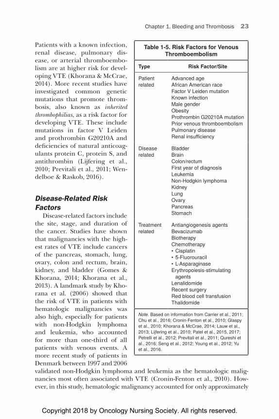
Risk Factors and EtiologyThrombophilia is the general term for a condition where the blood has an
increased tendency to form clots. Two distinct types of thrombosis exist: venous and arterial, with VTE occurring more often than arterial thrombo-embolism (Previtali, Bucciarelli, Passamonti, & Martinelli, 2011). The pres-ence of cancer is a well-established independent risk factor for development of VTE, with almost one-fifth of all new VTE events being associated with active cancer (Mandalà et al., 2012). Epidemiologic studies have shown that the risk for VTE is up to sevenfold higher in patients with cancer than in those without malignancy (Khorana, Dalal, Lin, & Connolly, 2013). In addi-tion to the independent variable of cancer, risk for VTE development can be divided into patient-, disease-, and treatment-related factors. See Table 1-5 for a list of factors associated with increased VTE risk.
Patient-Related Risk FactorsPatient-related factors for VTE include obesity, previous VTE, age, gen-
der, ethnicity, comorbid conditions, and prothrombotic mutations. The most common of these risk factors are obesity and previous VTE, with each occurring in 20%–30% of patients (Chu, Tokumaru, Izumi, & Nakagawa, 2016; Patel, Chun, & Chang, 2017). VTE usually affects people older than age 40, with a 21% increase in risk for those older than age 80 (Patel et al., 2017; Wendelboe & Raskob, 2016). The incidence of VTE increases with age in both genders; however, the overall incidence ratio of VTE in men to women is now 1.2:1, indicating that men have a higher risk of DVT (Patel et al., 2017; Wendelboe & Raskob, 2016). Ethnicity has been reported to play a role in thrombotic risk in patients with breast cancer, whereby Afri-can Americans were at higher risk for VTE and Asians/Pacific Islanders and Hispanics were at lower risk (Patel et al., 2017; Wendelboe & Raskob, 2016).
Copyright 2018 by Oncology Nursing Society. All rights reserved.
Chapter 1. Bleeding and Thrombosis 23
Patients with a known infection, renal disease, pulmonary dis-ease, or arterial thromboembo-lism are at higher risk for devel-oping VTE (Khorana & McCrae, 2014). More recent studies have investigated common genetic mutations that promote throm-bosis, also known as inherited thrombophilias, as a risk factor for developing VTE. These include mutations in factor V Leiden and prothrombin G20210A and deficiencies of natural anticoag-ulants protein C, protein S, and antithrombin (Lijfering et al., 2010; Previtali et al., 2011; Wen-delboe & Raskob, 2016).
Disease-Related Risk Factors
Disease-related factors include the site, stage, and duration of the cancer. Studies have shown that malignancies with the high-est rates of VTE include cancers of the pancreas, stomach, lung, ovary, colon and rectum, brain, kidney, and bladder (Gomes & Khorana, 2014; Khorana et al., 2013). A landmark study by Kho-rana et al. (2006) showed that the risk of VTE in patients with hematologic malignancies was also high, especially for patients with non-Hodgkin lymphoma and leukemia, who accounted for more than one-third of all patients with venous events. A more recent study of patients in Denmark between 1997 and 2006 validated non-Hodgkin lymphoma and leukemia as the hematologic malig-nancies most often associated with VTE (Cronin-Fenton et al., 2010). How-ever, in this study, hematologic malignancy accounted for only approximately
Table 1-5. Risk Factors for Venous Thromboembolism
Type Risk Factor/Site
Patient related
Advanced ageAfrican American raceFactor V Leiden mutationKnown infectionMale genderObesityProthrombin G20210A mutationPrior venous thromboembolismPulmonary diseaseRenal insufficiency
Disease related
BladderBrainColon/rectumFirst year of diagnosisLeukemiaNon-Hodgkin lymphomaKidneyLungOvaryPancreasStomach
Treatment related
Antiangiogenesis agentsBevacizumabBiotherapyChemotherapy• Cisplatin• 5-Fluorouracil• L-AsparaginaseErythropoiesis-stimulating
agentsLenalidomideRecent surgeryRed blood cell transfusionThalidomide
Note. Based on information from Carrier et al., 2011; Chu et al., 2016; Cronin-Fenton et al., 2010; Glaspy et al., 2010; Khorana & McCrae, 2014; Lauw et al., 2013; Lijfering et al., 2010; Patel et al., 2015, 2017; Petrelli et al., 2012; Previtali et al., 2011; Qureshi et al., 2016; Seng et al., 2012; Young et al., 2012; Yu et al., 2016.
Copyright 2018 by Oncology Nursing Society. All rights reserved.

24 Understanding and Managing Oncologic Emergencies (Third Edition)
6% of the patients with VTE. The risk of developing VTE varies over the con-tinuum of the cancer, with the highest risks occurring in the initial period after diagnosis and peak incidence occuring at three months and after devel-opment of metastasis (Khorana & McCrae, 2014; Qureshi et al., 2016; Yu et al., 2012). In addition, the incidence of VTE is highest during the first year of follow-up and decreases significantly over time (Qureshi et al., 2016).
Treatment-Related Risk FactorsTreatment-related risk factors include pharmacologic and nonpharmaco-
logic therapies. Many therapies, including surgery, chemotherapy, biother-apy, and supportive treatments, place patients at greater risk for developing VTE. Hospitalized patients, as well as those who have recently undergone surgery, are also at greater risk (Khorana & McCrae, 2014; Qureshi et al., 2016; Wendelboe & Raskob, 2016). Chemotherapy increases the risk of devel-oping VTE, with studies indicating a 6.5-times increase in risk (Khorana & McCrae, 2014). Chemotherapy agents with the highest risk of VTE are cispl-atin (8%–18%), L-asparaginase (4%–37%) and 5-fluorouracil (15%–17%) (Lauw et al., 2013; Mitchell, 2015; Seng et al., 2012). Studies of cancer regi-mens, including those that incorporate antiangiogenesis therapies, such as thalidomide, lenalidomide, and bevacizumab, have reported very high rates of VTE (Deng, Galanis, & Graham, 2014; Young et al., 2012).
The use of thalidomide in conjunction with chemotherapy or dexameth-asone in patients with multiple myeloma increases the risk of VTE (Deng et al., 2014). In a landmark meta-analysis of 17 randomized controlled tri-als, Gray, Chu, Wu, and Lin (2008) studied the use of thalidomide in 3,977 patients who had multiple myeloma and a variety of solid tumors. The over-all incidence of VTE was 11.7%. Patients treated with thalidomide demon-strated double the risk of VTE compared to controls. This risk was especially high in patients with multiple myeloma, with 15% developing VTE—tri-ple the risk compared with control patients not receiving thalidomide. A more recent systematic review and meta-analysis of 71 studies investigat-ing the rates of VTE in patients with multiple myeloma treated with tha-lidomide or lenalidomide confirmed that the use of thalidomide in con-junction with chemotherapy or dexamethasone increases the risk of VTE in these patients. Thalidomide alone conferred a 1.3% risk of VTE, while the use of thalidomide with dexamethasone increased the risk to 4.1%. When patients received combination thalidomide and dexamethasone along with thromboprophylaxis, the risk of VTE dropped to 2.6% (Carrier, Le Gal, Tay, Wu, & Lee, 2011). The risks of DVT with the use of lenalidomide, a struc-tural analog of thalidomide, have not proved to be significant (Carrier et al., 2011). No studies reported VTE risk with the use of lenalidomide alone. However, the combination of lenalidomide and dexamethasone was associ-ated with a 0.8% risk of VTE (Carrier et al., 2011). Pomalidomide, a newer novel therapy, has shown to have a 3.3% risk of VTE (Li et al., 2017).
Copyright 2018 by Oncology Nursing Society. All rights reserved.

Chapter 1. Bleeding and Thrombosis 25
Bevacizumab, a monoclonal antibody with antiangiogenic properties, has increased survival rates when used in combination with chemotherapy in patients with colorectal or non–squamous cell lung cancers (Ferrara & Adamis, 2016). Historically, findings related to bevacizumab and VTE have been conflicting. However, more recent studies indicate that the addition of bevacizumab to chemotherapy does not significantly increase the risk of VTE (Hurwitz et al., 2011; Patel et al., 2015). Hurwitz et al. (2011) analyzed 10 studies with a total of 6,055 patients with a variety of cancer types. In total, 3,448 patients were treated with bevacizumab and 2,607 received a control treatment that did not include bevacizumab. The unadjusted inci-dence of VTE of all grades was 10.9% in the bevacizumab group and 9.8% in the control group. When looking at grade 3–5 VTEs, the incidence was not statistically significant in both the bevacizumab group (6.4%) and in the control group (6.3%) (Hurwitz et al., 2011).
Patel et al. (2015) randomized a total of 1,008 patients, of whom 503 received treatment with docetaxel, prednisone, and bevacizumab and 505 were treated with docetaxel, prednisone, and a placebo. Results indicated that the addition of bevacizumab increased the incidence of grade 3 and higher arterial thromboembolism from 1.4% in the control group to 3.8% in the bevacizumab-treated group. However, the addition of bevacizumab was not statistically significant when looking at grade 3 and higher VTE inci-dence (Patel et al., 2015).
Petrelli, Cabiddu, Borgonovo, and Barni (2012)reviewed several ran-domized clinical trials to determine the incidence of VTE events asso-ciated with the use of epidermal growth factor receptor (EGFR) inhibi-tors. The monoclonal antibodies cetuximab and panitumumab were also included, while tyrosine kinase inhibitors reviewed included erlotinib and gefitinib. A total of 7,073 patients in 11 trials were included in the review. Overall, the incidence of VTE for patients receiving treatments was 5%, compared to 3.7% in the designated control groups. Additionally, the dif-ference between the incidence of VTE associated with monoclonal anti-bodies (5.9%) was compared to that of the tyrosine kinase inhibitors (2.6%), with the overall incidence of VTE and the comparison between monoclonal antibodies and tyrosine kinase inhibitors both being statisti-cally significant. The meta-analysis revealed a 32% higher risk of develop-ing VTEs with the use of anti-EGFR agents than with controls.
Some supportive therapies can increase a patient’s risk of developing VTE. The use of erythropoiesis-stimulating agents, including epoetin alfa and darbepoetin alfa, has been associated with DVT. A meta-analysis of 44 stud-ies reported that treatment with epoetin alfa or darbepoetin alfa increased the risk of DVT (Glaspy et al., 2010). The use of red blood cell transfusions can also increase the risk of DVT. In patients who received blood trans-fusions, 7.2% developed VTE and 5.2% developed arterial thromboembo-lism, compared to 3.7% and 3%, respectively, of patients who did not receive transfusions (Dubovoy & Engoren, 2016).
Copyright 2018 by Oncology Nursing Society. All rights reserved.

26 Understanding and Managing Oncologic Emergencies (Third Edition)
PathophysiologyCoagulation or clotting is the process whereby blood changes from a liq-
uid form to a gel, forming a clot that potentially will stop bleeding. Many factors contribute to the general prothrombotic state present in patients with cancer. In 1856, Rudolf Virchow described three mechanisms integral to thrombosis formation. Commonly referred to as the Virchow triad, these mechanisms are aberrant blood flow, vascular integrity, and blood compo-nents (Wolberg, Aleman, Leiderman, & Machlus, 2012). Figure 1-1 depicts the Virchow triad. Armand Trousseau elaborated on the cellular compo-nents within blood and was the first to discuss the relationship between malignancy and coagulation (Wolberg et al., 2012).
Blood FlowThe first mechanism involved in coagulation is normal blood flow. The
normal fluidity, or viscosity, of blood depends on a balance between the plasma fluid in the blood and the cellular components. Changes in one or both elements may occur in patients with cancer, resulting in atypical blood flow and an increased prothrombotic risk. Under normal physiologic con-
Figure 1-1. Virchow Triad
Aberrant Blood Flow• Hyperviscosity
– Plasma – Whole blood – Immunoglobulins – Hyperleukocytosis – Increased platelets
• External compression• Immobilization
Vascular Integrity• Tumor invasion• Prothrombin factors
from tumor• Cytokine release
Blood Components• Tissue factor• Plasminogen activator
inhibitor-1• Cancer procoagulant
Note. Based on information from Wolberg et al., 2012.
Copyright 2018 by Oncology Nursing Society. All rights reserved.

Chapter 1. Bleeding and Thrombosis 27
ditions, whole blood viscosity is a function of plasma viscosity, hematocrit, and red cell aggregation. When plasma viscosity increases, as may happen with high levels of plasma proteins or high fibrinogen levels, hyperviscosity of whole blood occurs, contributing to thrombotic risk. The brain, myocar-dium, lungs, and kidneys are especially vulnerable to the development of microthrombi, which most often manifest as headache, visual changes, chest pain, and dyspnea (Rodriguez, 2014).
The role of fibrinogen levels in relation to blood viscosity is controversial. One study using a therapeutic defibrination agent showed that when fibrin-ogen levels were reduced to 5 mg/dl, blood viscosity was greatly reduced; however, it returned to normal when the fibrinogen level was restored to 270 mg/dl, which is within the normal range. This effect on blood viscos-ity could be significant given that patients with most solid cancers, espe-cially renal cell and ovarian cancers, frequently have high fibrinogen levels (Pichler et al., 2013). Comorbid conditions such as infection, which is com-mon in cancer, also can increase fibrinogen levels. In addition, it has long been recognized that high fibrinogen levels increase the risk for cardiovas-cular events.
Immunoglobulins, which are plasma proteins, also affect viscosity and blood flow. Immunoglobulins can cause red cell dysplasia, in which nor-mal red blood cells have reduced ability to change their shape to flow through small spaces, especially in the microvasculature, and red cell aggregation can result. In the presence of high levels of plasma proteins, red cells begin to stack together or create long chains that can be seen in peripheral blood smears. This is known as rouleaux formation (Rosenthal, 2017). Plasma viscosity increases proportionately to the quantity and size of the plasma protein. Hyperviscosity is linked to increased levels of the proteins immunoglobulin M and immunoglobulin A (Hemmingway, Sav-itsky, & Kupas, 2015).
The cellular components of blood also influence the blood’s ability to flow freely. The consequences of high hematocrit levels have been stud-ied with conflicting results. In earlier studies, it was believed that a high hematocrit was a thrombotic risk factor. This led to the recommenda-tion of maintaining a hematocrit level less than 45% (Lehman, 2015). However, another study showed that hematocrit measures taken at the same time as thrombotic events were not associated with increased risk (Hernández-Boluda & Gómez, 2014). More recent studies have confirmed the results of the earlier studies, concluding that a hematocrit greater than 45% is a risk for thrombosis (Braekkan, Mathiesen, Njølstad, Wilsgaard, & Hansen, 2010).
Hyperleukocytosis, a high white blood cell count, is a common finding in acute and chronic leukemias. This can lead to leukostasis, a condition in which white blood cell plugs develop within the microcirculation of the cen-tral nervous and respiratory systems. This is most commonly found in acute myeloid leukemia and acute promyelocytic leukemia because the myelo-
Copyright 2018 by Oncology Nursing Society. All rights reserved.

28 Understanding and Managing Oncologic Emergencies (Third Edition)
blasts are larger and less able to change shape than normal neutrophils (Schiffer & Gurbuxani, 2017). Leukostasis occurs less in lymphoid leuke-mias because the cells are smaller, are more deformable, and have a lower adherence to the vasculature (Schiffer & Gurbuxani, 2017).
Elevated platelet levels have been identified in the pathogenesis of VTE. The Arctic University of Norway (2016) began the Tromsø studies in 1974 to reduce the high mortality from cardiovascular disease in Tromsø, Norway, and eventually expanded to include other disease states. The researchers collected platelet counts from 25,160 patients without cancer in the 1994–1995 study. In a follow-up study, Jensvoll, Blix, Braekkan, and Hansen (2014) found that patients with a platelet count greater than 295 × 109/L (295,000/mm3) developed cancer twice as often than those with platelet counts less than 295 × 109/L.
External compression resulting from tumor growth can also impede blood flow. For example, extrinsic compression from a space-occupying mass in the mediastinum, often a bronchogenic tumor, can cause superior vena cava syndrome. However, intrinsic thrombus formation secondary to the presence of vascular access devices is increasingly associated with inci-dence of superior vena cava syndrome (Drews & Rabkin, 2017). Chapter 11 discusses superior vena cava syndrome in more detail. Thrombosis caused by compression has also been seen in bulky lymphadenopathy that occurs in lymphoma—often caused by enlarged lymph nodes in close proximity to vasculature (Drews & Rabkin, 2017).
Immobilization associated with surgery or hospitalization can result in decreased and impaired blood flow. Patients who undergo oncologic sur-gery have twice the risk of thrombosis because of the underlying cancer and the resulting immobility. Several studies have shown this increased risk following colorectal, lung, breast, and gynecologic cancer surgeries (Clarke-Pearson & Abaid, 2012; De Martino et al., 2012; Rahn et al., 2011).
Vessel IntegrityVascular integrity is breached when the tumor directly invades or extends
into the endothelial cell wall. Tumor cells produce prothrombotic factors that can be a major constituent of the resulting clot, especially when the breached vessel is adjacent to the primary tumor. This most often occurs in the portal vein with hepatocellular carcinoma and in the inferior vena cava and right atrium with renal cell carcinoma (Noguchi, Hori, Nomura, & Tanaka, 2012; Quirk, Kim, Saab, & Lee, 2015).
Cytokines produced by tumor cells play an important role in the forma-tion of thrombi and compromise of vascular integrity. The proximity of tumor cells, endothelial cells, and stromal cells provide the opportunity for cytokine-mediated interactions. Tumor cells produce proinflammatory cyto-kines, including interleukin-1, interleukin-6, and tumor necrosis factor-beta. Once released, these cytokines stimulate tissue factor (TF) expression on
Copyright 2018 by Oncology Nursing Society. All rights reserved.
Chapter 1. Bleeding and Thrombosis 29
monocytes, thereby causing the monocyte to bind to the platelet. At the same time, they downregulate anticoagulant factors, including thrombomodulin and plasminogen activator inhibitor-1 (PAI-1), creating an environment for thrombus formation (Mooberry & Key, 2016; Wolberg et al., 2012).
Blood ComponentsTumor cells produce several factors that can disrupt coagulation and
fibrinolytic systems and lead to a prothrombotic state in cancer, includ-ing TF, cancer procoagulant (CP), and PAI-1. TF is a transmembrane gly-coprotein receptor that initiates the coagulation cascade. When activated, TF assembles several complexes that lead to thrombin production. TF can also be induced by proinflammatory cytokines (as previously discussed) or by vascular trauma. Elevated levels of plasma TF antigen and leukocyte TF have been found in patients with DVT (Yamashita, Alves, Barbaro, & San-toro, 2014).
CP is a cysteine protease procoagulant derived from tumor cells. It works by directly activating clotting factor X. CP has been found in the cells of solid tumors and hematologic cancers. A correlation is suspected between CP and fibrinogen in patients with adenocarcinoma. In acute promyelocytic leukemia, CP is thought to be a downregulated all-trans-retinoic acid treat-ment. It may induce platelet activation via a mechanism similar to thrombin; however, more studies are necessary to clearly understand the role of CP in thrombosis (Donnellan, Kevane, Healey Bird, & Ni Ainle, 2014).
Plasminogen activator is a component of the fibrinolytic system that tumor cells express. Elevated levels of PAI-1 have been linked to increased risk of VTE in patients with and without cancer (Previtali et al., 2011).
It has been suggested that the components of the clotting cascade and the vascular factors associated with them play an important role in tumor progression, invasion, angiogenesis, and metastatic formations (Quail & Joyce, 2013). In addition, some interesting in vitro studies have indicated that anticoagulants, especially low-molecular-weight heparin (LMWH), have antineoplastic properties that can interfere with development of metas-tasis (Bochenek, Püsküllüoğlu, & Krzemieniecki, 2013).
Clinical ManifestationsAlthough VTE has classic clinical manifestations, 50% of the cases of
DVT and up to 75% of cases of PE have no initial observable symptoms (CDC, 2017b; Global World Thrombosis Day Movement, n.d.). PE should be considered in patients who present with the four most frequent signs: dyspnea, pleuritic chest pain, hypoxia, and tachypnea. Less frequent signs are cough, hemoptysis, fever, syncope, diaphoresis, nonpleuritic chest pain, apprehension, rales, wheezing, hypotension, tachycardia, cyanosis, or pleu-
Copyright 2018 by Oncology Nursing Society. All rights reserved.

30 Understanding and Managing Oncologic Emergencies (Third Edition)
ral rub. Massive PE can present with hemodynamic instability or cardiac arrest (Dupras et al., 2013). Recent unilateral swelling and pain above or below the knee without explanatory bone or joint trauma suggests DVT (Dupras et al., 2013). Other signs of DVT include edema, warmth, local-ized pain, dilation of veins, color changes of limb, and pyrexia. Edema may be caused by decreased venous return resulting from the embolism or cap-illary damage producing leakage of intravascular fluid into the surround-ing tissue, distal to the thrombosis site (CDC, 2017b). When the affected limb is warm, it is most likely because of localized venous congestion and accumulation of tissue metabolites. Patients can experience pain in the calf muscle during dorsiflexion of the foot—known as Homans’ sign. However, it is not a very specific finding for VTE. One study found that only 37% of patients with VTE had a positive Homans’ sign, and 21% of patients with-out VTE also had positive Homans’ sign (Anthony, 2013; Grinage & Werner, 2015; Woo & Goertz, 2015). Dilation of a vein causing a palpable cord over a superficial vein is a result of systemic and peripheral venous circulatory sta-sis (Dupras et al., 2013). Initially, pallor of the skin might be the only indi-cator of DVT. In other cases, erythema occurs immediately over the DVT site because of superficial thrombophlebitis. Pyrexia, a systemic increase in body temperature, can be caused by the accumulation of tissue metabolites at the site of the thrombosis.
Upper-extremity DVT has become an increasingly recognized source of morbidity and mortality and represents an estimated 16% of all cases of DVT (Woo & Goertz, 2015). Upper-extremity DVT can occur in the sub-clavian, axillary, or brachial veins with clinical symptoms consisting of edema, arm pain or discoloration, or the development of collateral veins involving the affected arm, neck, or chest wall. PE is an acute complication of upper-extremity DVT and occurs in up to 33% of patients, only half of whom may be symptomatic (Woo & Goertz, 2015).
Patient AssessmentA complete thrombosis history is important, including the age at onset
and the location and results of diagnostic examinations for the patient, as well as family members. A history of VTE in one or more first-degree relatives is particularly notable because it suggests a hereditary defect or increased susceptibility for VTE disease (Bauer & Lip, 2018). A history of recurrent pregnancy loss could also indicate hypercoagulability where arte-rial or venous thrombosis occurs at the site of implantation or in the placen-tal blood vessels (Abu-Heija, 2014).
Assessing for risk of VTE is important because half of the patients with VTE are asymptomatic. Nurses should discuss information with patients regarding conditions that could increase risk, such as recent surgery, trauma, heart fail-ure, and immobility. Discussion of women’s use of oral contraceptive medica-
Copyright 2018 by Oncology Nursing Society. All rights reserved.

Chapter 1. Bleeding and Thrombosis 31
tions should also take place. A complete cancer history, including any previ-ous incidence of VTE, is also necessary because recurrent thrombosis, despite therapeutic anticoagulation, is more common in patients with cancer (Lip & Hull, 2017). In a general physical examination, special attention should be directed to the vascular system, extremities, chest, heart, abdominal organs, and skin (Lip & Hull, 2017). Because many clinical manifestations and exam-ination findings can be nonspecific, further diagnostic testing is required to confirm or exclude the diagnosis of VTE.
Diagnostic EvaluationThe American Academy of Family Physicians, the American College of
Physicians, and the Institute for Clinical Systems Improvement all recom-mend the use of prediction rules to establish pretest probability of VTE (Dupras et al., 2013; Kearon et al., 2016). The Wells prediction rules for DVT and PE are validated tools frequently used to determine the proba-bility of VTE prior to performing additional diagnostic tests. In the Wells test for DVT, patient assessment and symptoms determine the number of points given (Rodger et al., 2014). One point each is scored if the patient has active cancer; paralysis, paresis, or plaster immobilization; previous DVT, or is recently bedridden. In addition, one point each is scored if the patient has localized tenderness, entire leg swelling, calf swelling, pitting edema in affected leg, or collateral superficial veins. Two points are deducted if an alternative diagnosis is just as likely as DVT. A low probability of DVT is defined as a score of 0 or less, intermediate probability as a score of 1–2, and high probability as a score of 3 or greater. In patients with symptoms in both legs, the more symptomatic leg is used for scoring. There is also a prediction rule for PE. In this probability test, a score of 0–1 indicates low probability of PE; a score of 2–6 indicates intermediate probability; and a score of 7 or greater indicates high probability (Geersing et al., 2012).
Historically, when a patient has been shown to have a low pretest proba-bility of VTE, a D-dimer test is the next step. The D-dimer has a high sensi-tivity for the presence of an acute thrombosis. It can be used to exclude VTE with a high sensitivity for negative probability. In other words, patients with low pretest predictability and a negative D-dimer are considered to have DVT ruled out, and no further testing is needed (Dupras et al., 2013; Rod-ger et al., 2014). However, studies confirming the use of D-dimer as predic-tive for VTE have not included large numbers of patients with cancer. One study using the Wells test and D-dimer for diagnosis of VTE stated that lit-tle difference existed between patients with and without cancer. However, in that study, patients with cancer were found to have four times the num-ber of symptomatic VTEs at follow-up compared to patients without cancer. In addition, the number of false positives in patients with cancer was three times higher (National Comprehensive Cancer Network® [NCCN®], 2018).
Copyright 2018 by Oncology Nursing Society. All rights reserved.

32 Understanding and Managing Oncologic Emergencies (Third Edition)
For these reasons, NCCN recommends that further studies be completed involving patients with cancer. For PE, a chest radiograph, arterial blood gases, and electrocardiogram should be performed to rule out other disor-ders. Chest x-rays can show atelectasis or pleural-based infiltrates or effu-sions. Arterial blood gases can show hypoxemia and hypocapnia. Electro-cardiogram can show supraventricular arrhythmia, right axis derivation, or cor pulmonale. If the D-dimer is positive, further testing should be com-pleted, including duplex ultrasound for both VTE and PE and chest CT angiography for PE (NCCN, 2018).
Patients with an intermediate to high pretest predictability for VTE should have a venous duplex ultrasound performed as the first test when DVT is sus-pected (Rodger et al., 2014). D-dimer can be used in addition after negative ultrasound to determine whether further testing is needed. When DVT is suspected and D-dimer is positive despite negative ultrasound, serial ultra-sonography, CT venography, or contrast venography can be performed. Contrast venography has historically been considered the gold standard for accurate diagnosis. However, the drawbacks of this method include cost, dis-comfort to patients, limited availability, requirement of foot vein cannula-tion, use of IV contrast, and secondary emboli (Dupras et al., 2013). Because of these downsides, venography should be reserved for difficult diagnostic cases or to help distinguish between old and new clots.
When PE is suspected in the presence of an intermediate to high pretest predictability, noninvasive pulmonary vascular imaging, including a CT pul-monary angiography (CTPA) and ventilation/perfusion (VQ) scan, is rec-ommended as initial radiologic evaluation (Dupras et al., 2013). Both CTPA and VQ scans have a high degree of sensitivity to diagnose PE; therefore, the choice of imaging modality depends on the availability of the test, the resolu-tion of images obtained, underlying conditions (including renal status), and the experience of the radiologist. Extra care should be taken when using dye in patients with renal insufficiency. In some areas, CTPA has become easier to obtain than VQ scans. In addition, it is more useful in patients with under-lying cardiac disease and chronic obstructive pulmonary disease. When alter-native diagnoses are likely, CTPA is especially beneficial because it can rule out PE and confirm other diagnoses in one test (Dupras et al., 2013).
Treatment ModalitiesProphylaxis
Three professional groups issuing medical guidelines—the Ameri-can College of Chest Physicians (ACCP), the American Society of Clinical Oncology (ASCO), and NCCN—recommend routine thromboprophylaxis for patients with cancer who do not have contraindications to anticoagula-tion (Kahn et al., 2012; Lyman et al., 2015; NCCN, 2018). Contraindications to anticoagulation are those conditions that put a patient at an increased
Copyright 2018 by Oncology Nursing Society. All rights reserved.
Chapter 1. Bleeding and Thrombosis 33
risk for bleeding, including clinically significant active or chronic bleed-ing, recent surgery with a high associated bleeding risk, thrombocytope-nia or platelet dysfunction, a high risk for falls, lumbar puncture, interven-tional spine and pain procedures, and clotting factor abnormalities that lead to prolonged prothrombin time or activated partial thromboplas-tin time (NCCN, 2018). Frequent reevaluation of these contraindications is recommended because they can be temporary in many patients. A risk–benefit analysis is critical when determining whether to administer throm-boprophylaxis to patients. Several studies have confirmed a generally low risk of major bleeding in patients with cancer who receive prophylaxis with LMWH, with no difference in risk of major bleeding noted between unfrac-tionated heparin (UFH) and LMWH (Akl et al., 2014; Lyman et al., 2015; Puymirat et al., 2012, 2013; Ragni, 2012). All three guidelines recommend routine prophylaxis (both pharmacologic and mechanical) for patients with cancer who are undergoing surgical procedures and patients with cancer who are confined to bed with an acute illness. According to the guidelines, all adult hospitalized patients with a diagnosis or suspicion of cancer are at risk for the development of VTE. In the absence of contraindications, they should be considered for pharmacologic prophylaxis with LMWH, UFH, or factor Xa inhibitors with or without the addition of venous compression devices. For those with contraindications, mechanical prophylaxis with the use of venous compression devices, including graduated compression stock-ings and intermittent pneumatic compression and vena cava filters, should be considered (NCCN, 2018). Mechanical prophylaxis should not be used alone except in the presence of contraindications to pharmacologic prophy-laxis. The advantage of venous compression devices is that they can be used in patients with an increased risk for bleeding. However, they can interfere with ambulation and must be worn almost continuously to be effective. The placement of inferior vena cava filters has the advantage of preventing PE in patients at high risk for VTE where anticoagulation is contraindicated. How-ever, vena cava filters do not prevent DVT and have been associated with an increased risk of recurrent DVT. The use of inferior vena cava filters cannot substitute for antithrombotic therapy in patients with cancer and should be used only if there is an absolute contraindication to anticoagulation or fail-ure of anticoagulation (NCCN, 2018). Anticoagulation should be resumed as soon as the clinical picture allows. VTE prophylaxis for at-risk ambulatory patients should also be considered. If the patient has had cancer surgery, especially pelvic or abdominal surgery, VTE prophylaxis is recommended for up to four weeks postoperatively (NCCN, 2018).
TreatmentGuidelines for the treatment of VTE from ACCP, ASCO, and NCCN are
based on the findings of three major studies and differ slightly among each organization (Hull et al., 2006; Lee et al., 2003; Meyer et al., 2002). ACCP
Copyright 2018 by Oncology Nursing Society. All rights reserved.

34 Understanding and Managing Oncologic Emergencies (Third Edition)
guidelines recommend LMWH for the first three to six months of long-term anticoagulant therapy in patients with cancer and DVT. Subsequent anticoag-ulation with vitamin K antagonists or LMWH is recommended until cancer is resolved (Kahn et al., 2012). ASCO guidelines state that LMWH is preferred for the first 5–10 days of anticoagulation and given for at least six months for long-term therapy, but vitamin K antagonists may be used if LMWH is not available (Lyman et al., 2015). Similarly, NCCN (2017)guidelines recommend LMWH for at least three months for treatment of VTE and continuing indefi-nite anticoagulation for patients with active cancer or other persistent risk fac-tors. ACCP guidelines also recommend the use of graduated elastic support stockings in patients who have had symptomatic proximal DVT, beginning as soon as the patient or caregiver is able to apply the stockings and continuing for at least two years (Kahn et al., 2012).
Heparin-Induced ThrombocytopeniaBoth UFH and LMWH are associated with heparin-induced thrombocyto-
penia (HIT), which is an immune-mediated reaction to the presence of hep-arin. It occurs in 2%–3% of patients treated with UFH and less than 1% of patients treated with LMWH (Dupras et al., 2013). Paradoxically, patients with HIT have an increased risk for developing VTE for up to 100 days follow-ing diagnosis. Patients with a history of HIT should not be treated with UFH or LMWH. HIT should be suspected in patients who develop skin lesions at the injection site, have a systemic reaction to a bolus administration of hepa-rin, or experience a greater than 50% decrease in platelet count from base-line laboratory values while receiving heparin (Dupras et al., 2013). Another form, delayed-onset HIT, typically presents as thromboembolic complications one to two weeks after the last dose of LMWH or UFH. These patients usually have mild or moderate thrombocytopenia. If HIT is not suspected, the patient is typically rechallenged with heparin, thus leading to worsening thrombo-sis and thrombocytopenia. If HIT is suspected, heparin therapy should be stopped while antibody testing is completed. In the interim, the patient can be effectively treated with direct thrombin inhibitors. If HIT is suspected in a patient on warfarin therapy, the warfarin should be stopped and reversed with vitamin K therapy and direct thrombin inhibitors (Dupras et al., 2013). While the patient is receiving direct thrombin inhibitors, low-dose therapy with war-farin can be restarted after the platelet count has significantly improved and there is clinical improvement in the thrombosis (Dupras et al., 2013).
Nursing ManagementPreventive measures for VTE for all hospitalized patients are a crucial part
of the nurse’s role. Positioning is an important aspect of prevention. Ensuring the patient avoids prolonged sitting and elevating the legs when in bed helps
Copyright 2018 by Oncology Nursing Society. All rights reserved.

Chapter 1. Bleeding and Thrombosis 35
to promote venous return. Daily assessment of extremities for pain, erythema, and size discrepancy is vital. Proper use of compression devices, incentive spi-rometry, adequate hydration, and early ambulation are encouraged to prevent VTE. The nurse must vigilantly monitor patients who are immobilized or have had their activity restricted for unexplained tachypnea, tachycardia, and rest-lessness. These signs should not be simply attributed to anxiety unless a phys-ical reason has been ruled out. To identify bleeding complications, nurses should pay special attention during anticoagulation. If the nurse observes a change in mental status or new focal neurologic deficits in a patient receiv-ing thrombolytics, intracranial hemorrhage must be eliminated as a possible cause. Figure 1-2 lists nursing interventions related to VTE.
Patient and Caregiver EducationEducation regarding VTE should include information on the clinical
condition itself, as well as treatments implemented and potential lifestyle changes that could reduce a patient’s risk. Treatments that require teach-ing include early and frequent ambulation, the use of an incentive spirom-eter, and proper and timely use of compression devices. When anticoagula-tion therapy is initiated, education regarding the administration and side effects of each medication is required. Patients or caregivers may need to learn to give subcutaneous injections if LMWH is to be continued after dis-charge. Patients and caregivers should also learn signs and symptoms of bleeding, including bleeding precautions, as well as the signs of subsequent or recurrent VTE. Education provided to patients and caregivers is essen-tial for patients to maintain adherence to ongoing anticoagulation ther-apy. Patients continuing warfarin therapy will need to be instructed to limit foods high in vitamin K, such as dark green vegetables and apricots, to pre-
Figure 1-2. Nursing Interventions for Patients at Risk for Venous Thromboembolism
• Instruct patients to do the following: – Avoid prolonged sitting. – Elevate legs when in bed. – Use incentive spirometer. – Ambulate early. – Avoid medications that interfere with coagulation (e.g., aspirin, nonsteroidal anti-inflammatory drugs).
• Assess extremities for pain, erythema, and size discrepancy daily.• Apply mechanical compression devices as ordered.• Ensure adequate hydration.
Note. Based on information from Kahn et al., 2012; Lyman et al., 2015.
Copyright 2018 by Oncology Nursing Society. All rights reserved.

36 Understanding and Managing Oncologic Emergencies (Third Edition)
vent decreased warfarin action. Patients should be taught that their blood counts will need to be monitored to keep their INR between 2 and 3 when on warfarin therapy (U.S. National Library of Medicine PubMed Health, 2017). Because patients with a history of VTE are at greater risk for sub-sequent VTE, they should be instructed to keep well hydrated and stretch every hour during confined travel (Kahn et al., 2012).
Thorough education for patients and caregivers not only can help them to recognize potential problems, but also can help to reduce risk for bleed-ing and recurrent VTE. Good communication, including possible interven-tions, needs to be in place to keep patients and caregivers calm and focused should bleeding or VTE occur.
ConclusionBleeding in patients with cancer may be caused by a variety of under-
lying factors, including the disease process and cancer therapies, all of which can contribute to reducing the quantity and functional quality of platelets and initiating alterations in clotting factors. The first s tep i n managing bleeding is to identify its underlying causes. Determining the type and origin of bleeding will help to direct treatment. Although sev-eral treatments are effective for bleeding, the patient’s condition, goals of treatment, and comorbidities must be considered before deciding on a treatment plan.
Patients with cancer also have an increased risk of developing VTE, which is associated with considerable morbidity and mortality. Hospitalized med-ical and surgical patients with cancer are at an increased risk for VTE and should be considered for pharmacologic prophylaxis if no contraindications are present. Consideration of prophylactic anticoagulation in patients with cancer must always balance the risk of VTE with the increased risk of bleed-ing. Proper patient and family education regarding the risk of bleeding, as well as the signs and symptoms of both bleeding and VTE recurrence, is par-amount in the care of patients with cancer.
The author would like to acknowledge Lorraine Mack, MSN, RN, CNL, OCN®, for her contribution to this chapter that remains unchanged from the second edition of this book.
ReferencesAbdelrahman, T., & Newton H. (2011). Wound dressings: Principles and practice. Surgery,
29, 491–495. doi:10.1016/j.mpsur.2011.06.007Abrams, C.S., Shattil, S.J., & Bennett, J.S. (2016). Acquired qualitative platelet disorders. In K.
Kaushansky, M.A. Lichtman, J.T. Prchal, M.M. Levi, O.W. Press, L.J. Burns, & M. Caliguiri (Eds.), Williams hematology (9th ed., pp. 2073–2096). New York, NY: McGraw-Hill Education.
Copyright 2018 by Oncology Nursing Society. All rights reserved.

Chapter 1. Bleeding and Thrombosis 37
Abu-Heija, A. (2014). Thrombophilia and recurrent pregnancy loss: Is heparin still the drug of choice? Sultan Qaboos University Medical Journal, 14, e26–e36.
Agrawal, P., Soni, S., Mittal, G., & Bhatnagar, A. (2014). Role of polymeric biomaterials as wound healing agents. International Journal of Lower Extremity Wounds, 13, 180–190. doi:10 .1177/1534734614544523
Akl, E.A., Kahale, L.A., Sperati, F., Neumann, I., Labedi, N., Terrenato, I., … Schünemann, H. (2014). Low molecular weight heparin versus unfractionated heparin for periopera-tive thromboprophylaxis in patients with cancer. Cochrane Database of Systematic Reviews,2014(6). doi:10.1002/14651858.CD009447.pub2
Alper, A.B., Jr., Shenava, R.G., & Young, B.A. (2017). Uremia. Retrieved from https://emedicine .medscape.com/article/245296-overview
Amgen Inc. (2016). Aranesp® (darbepoetin alfa) [Package insert]. Thousand Oaks, CA: Author.Anthony, M. (2013). Nursing assessment of deep vein thrombosis. MEDSURG Nursing, 22,
95–123.Arctic University of Norway. (2016). The Tromsø Study. Retrieved from https://en.uit.no
/prosjekter/prosjekt?p_document_id=80172#Balentine, J.R., & Nabili, S.N. (2017). Anemia. Retrieved from http://www.medicinenet
.com/anemia/article.htmBarragán-Ibañez, G., Santoyo-Sánchez, A., & Ramos-Peñafiel, C.O. (2016). Iron deficiency
anemia. Revista Médica del Hospital General de México, 79, 88–97. doi:10.1016/j.hgmx.20 15.06.008
Barsam, S.J., Psaila, B., Forestier, M., Page, L.K., Sloane, P.A., Geyer, J.T., … Bussel, J.B. (2011). Platelet production and platelet destruction: Assessing mechanisms of treatment effect in immune thrombocytopenia. Blood, 117, 5723–5732. doi:10.1182/blood-2010-11-321398
Bauer, K.A., & Lip, G.Y.H. (2018). Overview of the causes of venous thrombosis [Literature review current through March 2018]. Retrieved from https://www.uptodate.com/contents/overview-of-the-causes-of-venous-thrombosis
Berns, J.S., & Coutre, S. (2017, May 18). Platelet dysfunction in uremia [Literature review current through July 2017]. Retrieved from https://www.uptodate.com/contents/ pla telet-dysfunction-in-uremia
Boateng, J., & Catanzano, O. (2015). Advanced therapeutic dressings for effective wound healing—A review. Journal of Pharmaceutical Sciences, 104, 3653–3680. doi:10.1002/jps .24610
Bochenek, J., Püsküllüoğlu, M., & Krzemieniecki, K. (2013). The antineoplastic effect of low-molecular-weight heparins—A literature review. Contemporary Oncology, 17, 6–13. doi:10.5114/wo.2013.33766
Braekkan, S.K., Mathiesen, E.B., Njølstad, I., Wilsgaard, T., & Hansen, J.-B. (2010). Hema-tocrit and risk of venous thromboembolism in a general population. The Tromsø Study. Haematologica, 95, 270–275. doi:10.3324/haematol.2009.008417
Bura, A. Cailleux, N., Bienvenu, B., Léger, P., Bissery, A.L., Boccalon, H., … Emmerich, J. (2004). Incidence and prognosis of cancer associated with bilateral venous thrombosis: A prospective study of 103 patients. Journal of Thrombosis and Haemostasis, 2, 441–444. doi:10.1111/j.1538-7933.2004.00619.x
Cagir, B., Chico, G.F., Cirincione, E., & Manas, K.J. (2017). Lower gastrointestinal bleeding treatment and management. Retrieved from http://emedicine.medscape.com/article /188478-treatment#showall
Camp-Sorrell, D. (2016). Nursing implications of support therapies. In J.K. Itano (Ed.), Core curriculum for oncology nursing (5th ed., pp. 268–281). St. Louis, MO: Elsevier.
Carrier, M., Le Gal, G., Tay, J., Wu, C., & Lee, A.Y. (2011). Rates of venous thromboembolism in multiple myeloma patients undergoing immunomodulatory therapy with thalidomide or lenalidomide: A systmatic review and meta-analysis. Journal of Thrombosis and Haemostasis, 9, 653–663. doi:10.1111/j.1538-7836.2011.04215.x
Centers for Disease Control and Prevention. (2017a). Venous thromboembolism (blood clots): Data and statistics. Retrieved from http://www.cdc.gov/ncbddd/dvt/data.html
Copyright 2018 by Oncology Nursing Society. All rights reserved.

38 Understanding and Managing Oncologic Emergencies (Third Edition)
Centers for Disease Control and Prevention. (2017b). Venous thromboembolism (blood clots): Facts. Retrieved from https://www.cdc.gov/ncbddd/dvt/facts.html
Cho, H., Walker, A., Williams, J., & Hasty, K.A. (2015). Study of osteoarthritis treatment with anti-inflammatory drugs: Cyclooxygenase-2 inhibitor and steroids. BioMed Research International, 2015, 595273. doi:10.1155/2015/595273
Chu, C., Tokumaru, S., Izumi, K., & Nakagawa, K. (2016). Obesity increases risk of anticoagulation reversal failure with prothrombin complex concetrate in those with intracranial hemorrhage. International Journal of Neuroscience, 126, 62–66. doi:10.3109 /00207454.2014.993034
Clarke-Pearson, D.L., & Abaid, L.N. (2012). Prevention of venous thromboembolic events after gynecolgic surgery. Obstetrics and Gynecology, 119, 155–167. doi:10.1097/AOG.0b0 13e31823d389e
Connolly, G.C., & Francis, C.W. (2013). Cancer-associated thrombosis. Hematology: American Society of Hematology Education Program, 2013, 684–691. doi:10.1182/asheducation-2013 .1.684
Connolly, G.C., Menapace, L., Safadjou, S., Francis, C.W., & Khorana, A.A. (2013). Prevalence and clinical significance of incidental and clinically suspected venous thromboembolism in lung cancer patients. Clinical Lung Cancer, 14, 713–718. doi:10.1016/j.cllc.2013.06.003
Coombs, C.C., Tavakkoli, M., & Tallman, M.S. (2015). Acute promyleocytic leukemia: Where did we start, where are we now, and the future. Blood Cancer Journal, 5, e304–e313. doi:10 .1038/bcj.2015.25
Crighton, G.L., Estcourt, L.J., Wood, E.M., Trivella, M., Doree, C., & Stanworth, S. (2015). A therapeutic-only versus prophylactic platelet transfusion strategy for preventing bleeding in patients with haematological disorders after myelosuppressive chemotherapy or stem cell transplantation. Cochrane Database of Systematic Reviews, 2015(9). doi:10.1002/14651 858.CD010981.pub2
Cronin-Fenton, D.P., Søndergaard, F., Pedersen, L.A., Fryzek, J.P., Cetin, K., Acquavella, J., … Sørensen, H.T. (2010). Hospitalisation for venous thromboembolism in cancer patietns and the general population: A population-based cohort study in Denmark, 1997–2006. British Journal of Cancer, 103, 947–953. doi:10.1038/sj.bjc.6605883
Curtis, B.R., & McFarland, J.G. (2014). Human platelet antigens—2013. Vox Sanguinis, 106, 93–102. doi.10.1111/vox.12085
De Martino, R.R., Goodney, P.P., Spangler, E.L., Wallaert, J.B., Corriere, M.A., Rzucidlo, E.M., … Stone, D.H. (2012). Variation in thromboembolic complications among patients undergoing commonly performed cancer operations. Journal of Vascular Surgery, 55,1035–1040e4. doi:10.1016/j.jvs.2011.10.129
Deng, A., Galanis, T., & Graham, M.G. (2014). Venous thromboembolism in cancer patients. Hospital Practice, 42(5), 24–33. doi:10.3810/hp.2014.12.1167
Donnellan, E., Kevane, B., Healey Bird, B.R., & Ni Ainle, F. (2014). Cancer and venous thromboembolic disease: From molecular mechanisms to clinical management. Current Oncology, 21, 134–143. doi:10.3747/co.21.1864
Drews, R., & Rabkin, D.J. (2017, May 5). Malignancy-related superior vena cava syndrome [Literature review current through July 2017]. Retrieved from http://www.uptodate.com /contents/malignancy-related-superior-vena-cava-syndrome
Dubovoy, T., & Engoren, M. (2016). Thrombotic risks in red blood cell transfusions. Seminars in Thrombosis and Hemostasis, 42, 102–111. doi:10.1055/s-0035-1569069
Dupras, D., Bluhm, J., Felty, C., Hansen, C., Johnson, T., Lim, K., … Skeik, N. (2013). Venous thromboembolism diagnosis and treatment. Bloomington, MN: Institute for Clinical Systems Improvement. Retrieved from https://pdfs.semanticscholar.org/3f06/d88a25a6888f86 198fab7c3ed9951cac6a94.pdf
Escobar, G.A., Henke, P.K., & Wakefield, T.W. (2013). Hypercoagulable states. In A. Kumar & K. Ouriel (Eds.), Handbook of endovascular interventions (pp. 527–541). doi:10.1007/978 -1-4614-5013-9_38
Copyright 2018 by Oncology Nursing Society. All rights reserved.

Chapter 1. Bleeding and Thrombosis 39
Falanga, A., Russo, L., & Verzeroli, C. (2013). Mechanisms of thrombosis in cancer. Thrombosis Research, 131(Suppl. 1), S59–S62. doi:10.1016/S0049-3848(13)70024-0
Fernandez, M.M., Hogue, S., Preblick, R., & Kwong, W.J. (2015). Review of the cost of venous thromboembolism. ClinicoEconomics and Outcomes Research, 7, 451–462. doi:10.21 47/CEOR.S85635
Ferrara, N., & Adamis, A.P. (2016). Ten years of anti-vascular endothelial growth factor therapy. Nature Reviews Drug Discovery, 15, 385-403. doi:10.1038/nrd.2015.17
Fletcher, C.H., DomBourian, M.G., & Millward, P.A. (2015). Platelet transfusion for patients with cancer. Cancer Control, 22, 47–51.
Gao, S., & Sahai, S.K. (2011). Hematologic disease. In S. Cohn (Ed.), Perioperative medicine (pp. 261–280). doi:10.1007/978-0-85729-498-2_23
Gary, T., Belaj, K., Steidl, K., Pichler, M., Eisner, F., Stöger, H., … Brodmann, M. (2012). Asymptomatic deep vein thrombosis and superficial vein thrombosis in ambulatory cancer patients: Impact on short-term survival. British Journal of Cancer, 107, 1244–1248. doi:10.1038/bjc.2012.401
Gaydos, L.A., Freireich, E.J., & Mantel, N. (1962). The quantitative relation between platelet count and hemorrhage in patients with acute leukemia. New England Journal of Medicine, 266, 905–909. doi:10.1056/NEJM196205032661802
Geersing, G.-J., Erkens, P.M.G., Lucassen, W.A.M., Büller, H.R., ten Cate, H., Hoes, A.W., … Stoffers, H.E.J.H. (2012). Safe exclusion of pulmonary embolism using the Wells rule and qualitative D-dimer testing in primary care: Prospective cohort study. BMJ, 345, e6564. doi:10.1136/bmj.e6564
Gheshmy, A., & Carrier, M. (2016). Venous thromboembolism and occult cancer: Impact on clinical practice. Thrombosis Research, 140(Suppl. 1), S8–S11. doi:10.1016/S0049-384 8(16)30091-3
Glaspy, J., Crawford, J., Vansteenkiste, J., Henry, D., Rao, S., Bowers, P., … Ludwig, H. (2010). Erythropoiesis-stimulating agents in oncology: A study-level meta-analysis of survival and other safety outcomes. British Journal of Cancer, 102, 301–315. doi:10.1038/sj.bjc .6605498
Global World Thrombosis Day Movement. (n.d.). Know VTE. Retrieved from http://www .worldthrombosisday.org/issue/vte
Gomes, M., & Khorana, A.A. (2014). Risk assessment for thrombosis in cancer. Seminars in Thrombosis and Hemostasis, 40, 319–324. doi:10.1055/s-0034-1370770
Gray, K.N., Chu, D., Wu, S., & Lin, R.Z. (2008). Risk of venous thromboembolism with thalidomide in cancer patients: A systematic review and meta-analysis of randomized controlled trials [Abstract]. Blood, 112, 3820. Retrieved from http://www.bloodjournal .org/content/112/11/3820
Grinage, J.A., & Werner, C.M. (2015). The Homans’ sign for DVT: Use caution with interpretation. Advance Healthcare Network for NPs and PAs. Retrieved from http://nurse -practitioners-and-physician-assistants.advanceweb.com/Features/Articles/The-Homa ns -Sign-for-DVT.aspx
Gussoni, G., Frasson, S., La Regina, M., Di Micco, P., & Monreal, M. (2013). Three-month mortality rate and clinical predictors in patients with venous thromboembolism and cancer. Findings from the RIETE registry. Thrombosis Research, 131, 24–30. doi:10.1016 /j.thromres.2012.10.007
Healthwise Staff. (2016, May 27). Nosebleeds. Retrieved from http://www.cigna .com/in dividualandfamilies/health-and-well-being/hw/medical-topics/nosebleeds -nosbd.html
Hemmingway, T.J., Savitsky, E.A., & Kupas, D.F. (2015, November 23). Hyperviscos-ity syndrome. Retrieved from http://emedicine.medscape.com/article/780258 -overview
Hernández-Boluda, J.C., & Gómez, M. (2014). Target hematologic values in the management of essential thrombocythemia and polycythemia vera. European Journal of Haematology, 94, 4–11. doi:10.1111/ejh.12381
Copyright 2018 by Oncology Nursing Society. All rights reserved.

40 Understanding and Managing Oncologic Emergencies (Third Edition)
Hull, R.D., & Garcia, D.A. (2017, June 16). Management of warfarin-associated bleeding or supratherapeutic INR [Literature review current through July 2017]. Retrieved from http://www.uptodate.com/contents/management-of-warfarin-associated-bleeding-or -supratherapeutic-inr
Hull, R.D., Pineo, G.F., Brant, R.F., Mah, A.F., Burke, N., Dear, R., … Raskob, G. (2006). Long-term low-molecular-weight heparin versus usual care in proximal-vein thrombosis in patients with cancer. American Journal of Medicine, 119, 1062–1072. doi:10.1016/j.am jmed.2006.02.022
Hurwitz, H.I., Saltz, L.B., Van Cutsem, E., Cassidy, J., Wiedemann, J., Sirzén, F., … Rohr, U.-P. (2011). Venous thromboembolic events wth chemotherapy plus bevacizumab: A pooled analysis of patients in randomized phase II and III studies. Journal of Clinical Oncology, 29, 1757–1764. doi:10.1200/JCO.2010.32.3220
International Society on Thrombosis and Haemostasis Steering Committee for World Thrombosis Day. (2014). Thrombosis: A major contributor to the global disease burden. Journal of Thrombosis and Haemostasis, 12, 1580–1590. doi:10.1111/jth.12698
Izak, M., & Bussel, J.B. (2014). Management of thrombocytopenia. F1000 Prime Reports, 6, 45. doi:10.12703/P6-45
Jensvoll, H., Blix, K., Braekkan, S.K., & Hansen, J.-B. (2014). Platelet count measured prior to cancer development is a risk factor for future symptomatic venous thromboembolism: The Tromsø Study. PLOS ONE, 9, e9201. doi:10.1371/journal.pone.0092011
Jha, A.K., Larizgoitia, I., Audrea-Lopez, C., Prasopa-Plaizer, N., Waters, H., & Bates, D.W. (2013). The global burden of unsafe medical care: Analytic modelling of observational studies. BMJ Quality and Safety, 22, 809–815. doi:10.1136/bmjqs-2012-001748
Jung, Y., Ahn, H., Kim, D.-S., Hwang, Y.R., Ho, S.-H., Kim, J.-M., … Kim, S. (2011). Improve-ment of biological and pharmocokinetic features of human interleukin-11 by site-directed mutagenesis. Biochemical and Biophysical Research Communications, 405, 399–404. doi:10.10 16/j.bbrc.2011.01.041
Kahn, S.R., Lim, W., Dunn, A.S., Cushman, M., Dentali, F., Akl, E.A., … Murad, M.H. (2012). Prevention of VTE in nonsurgical patients: Antithrombotic therapy and prevention of thrombosis, 9th ed.: American College of Chest Physicians, Evidence-Based Clinical Practice Guidelines. Chest, 141(Suppl.), e195S–e226S. doi:10.1378/chest.11-2296
Kearon, C., Aki, E.A., Ornelas, J., Blaivas, A., Jimenez, D., Bounameaux, H., … Moores, L. (2016). Antithrombotic therapy for VTE disease: CHEST guideline and expert panel report. Chest, 149, 315–352. doi:10.1016/j.chest.2015.11.026
Khorana, A.A., Dalal, M., Lin, J., & Connolly, G.C. (2013). Incidence and predictors of venous thromboembolism (VTE) among ambulatory high-risk cancer patients undergoing chemotherapy in the United States. Cancer, 119, 648–655. doi:10.1002/cncr.27772
Khorana, A.A., Francis, C.W., Culakova, E., Fisher, R.I., Kuderer, N.M., & Lyman, G.H. (2006). Thromboembolism in hospitalized neutropenic cancer patients. Journal of Clinical Oncology, 24, 484–490. doi:10.1200/JCO.2005.03.8877
Khorana, A.A., Francis, C.W., Culakova, E., Kuderer, N.M., & Lyman, G.H. (2007). Throm-boembolism is a leading cause of death in cancer patients receiving outpatient chemo-therapy. Journal of Thrombosis and Haemostasis, 5, 632–634. doi:10.1111/j.1538-7836.2007 .02374.x
Khorana, A.A., & McCrae, K.R. (2014). Risk stratification strategies for cancer-associated thrombosis: An update. Thrombosis Research, 133(Suppl. 2), S35–S38. doi:10.1016/S0049 -3848(14)50006-0
Königsbrügge, O., Pabinger, I., & Ay, C. (2014). Risk factors for venous thromboembolism in cancer: Novel findings from the Vienna Cancer and Thrombosis Study (CATS). Thrombosis Research, 133(Suppl. 2), S39–S43. doi:10.1016/S0049-3848(14)50007-2
Konkle, B.A. (2011). Acquired disorders of platelet function. Hematology: American Society of Hematology Education Program, 2011, 391–396. doi:10.1182/asheducation-2011.1.391
Lam, P. (2017). Everything you need to know about anemia. Medical News Today. Retrieved from https://medicalnewstoday.com/articles/158800.php
Copyright 2018 by Oncology Nursing Society. All rights reserved.

Chapter 1. Bleeding and Thrombosis 41
Lauw, M.N., van der Holt, B., Middeldorp, S., Meijers, J.C.M., Cornelissen, J.J., Bajetta, M., & Biemond, B.J. (2013). Venous thromboembolism in adults treated for acute lymphoblastic leukaemia: Effect of fresh frozen plasma supplemntation. Thrombosis and Haemostasis, 109, 633–642. doi:10.1160/TH12-11-0845
Lebois, M., & Josefsson, E.C. (2016). Regulation of platelet lifespan by apoptosis. Platelets, 27, 497–504. doi:10.3109/09537104.2016.1161739
Lee, A.Y.Y., & Levine, M.N. (2003). Venous thromboembolism and cancer: Risks and outcomes. Circulation, 107(Suppl. 1), I-17–I-21. doi:10.1161/01.CIR.0000078466.72504.AC
Lee, A.Y.Y., Levine, M.N., Baker, R.I., Bowden, C., Kakkar, A.K., Prins, M., … Gent, M. (2003). Low-molecular-weight heparin versus a coumarin for the prevention of recurrent venous thromboembolism in patients with cancer. New England Journal of Medicine, 349, 146–153. doi:10.1056/NEJMoa025313
Lehman, C.M. (2015). Testing for acquired platelet disorders. In S.T. Bennett, C.M. Lehman, & G.M. Rodgers (Eds.), Laboratory hemostasis: A practical guide for pathologists (2nd ed., pp. 99–110). doi:10.1007/978-3-319-08924-9_7
Levi, M., Seligsohn, U., & Kaushansky, K. (2016). Classification, clinical manifestations, and evaluation of disorder in hemostasis. In K. Kaushansky, M.A. Lichtman, J.T. Prchal, M.M. Levi, O.W. Press, L.J. Burns, & M. Caliguiri (Eds.), Williams hematology (9th ed., pp. 1985–1992). New York, NY: McGraw-Hill Education.
Levi, M., & van der Poll, T. (2013). Disseminated intravascular coagulation: A review for the internist. Internal and Emergency Medicine, 8, 23–32. doi:10.1007/s11739-012-0859-9
Li, W., Garcia, D., Cornell, R.F., Gailani, D., Laubach, J., Maglio, M.E., … Moslehi, J. (2017). Cardiovascular and thrombotic complications of novel multiple myeloma therapies: A review. JAMA Oncology, 3, 980–988. doi:10.1001/jamaoncol.2016.3350
Lijfering, W.M., Middeldorp, S., Veeger, N.J.G.M., Hamulyák, K., Prins, M.H., Büller, H.R., & van der Meer, J. (2010). Risk of recurrent veonous thrombosis in homozygous carriers and double heterozygous carriers of factor V Leiden and prothrombin G20210A. Circulation, 121, 1706–1712. doi:10.1161/CIRCULATIONAHA.109.906347
Lip, G.Y.H., & Hull, R.D. (2017). Overview and treatment of lower extremity deep vein thrombosis (DVT) [Literature review current through June 2017]. Retrieved from http://www.uptodate.com/contents/overview-of-the-treatment-of-lower-extremity-de ep -vein-thrombosis-dvt
Lubberts, B., Paulino Pereira, N.R., Kabrhel, C., Kuter, D.J., & DiGiovanni, C.W. (2016). What is the effect of venous thromboembolism and related complications on patient reported health-related quality of life? Thrombosis and Haemostasis, 116, 417–431. doi:10 .1160/TH16-02-0152
Lyman, G.H., Bohlke, K., Khorana, A.A., Kuderer, N.M., Lee, A.Y., Arcelus, J.I., … Falanga, A. (2015). Venous thromboembolism prophylaxis and treatment in patients with cancer: American Society of Clinical Oncology clinical practice guideline update 2014. Journalof Clinical Oncology, 33, 654–656. doi:10.1200/JCO.2014.59.7351
Mandalà, M., Grosso, F., Vitalini, C., Corradino, I., Sanfilippo, R., Colombini, S., … Marsoni, S. (2012). Venous thromboembolism is a relevant and underestimated adverse event incancer patients treated in phase I studies. British Journal of Cancer, 107, 612–616. doi:10.1038/bjc.2012.325
Meyer, G., Marjanovic, Z., Valcke, J., Lorcerie, B., Gruel, Y., Solal-Celigny, P., … Farge, D. (2002). Comparison of low-molecular-weight heparin and warfarin for the secondary prevention of venous thromboembolism in patients with cancer: A randomized controlled study. Archives of Internal Medicine, 162, 1729–1735. doi:10.1001/archinte.162.15.1729
Mitchell, R.N. (2015). Hemodynamic disorders, thromboembolic disease, and shock. In V. Kumar, A.K. Abbas, & J.C. Aster (Eds.), Robbins and Cotran pathologic basis of disease (9th ed., pp. 113–136). Philadelphia, PA: Elsevier Saunders.
Monroe, D.M., & Hoffman, M. (2014). Theories of blood coagulation: Basic concepts and recent updates. In H.I. Saba & H.R. Roberts (Eds.), Hemostasis and thrombosis: Practical guidelines in clinical management (pp. 1–13). Oxford, United Kingdom: John Wiley & Sons.
Copyright 2018 by Oncology Nursing Society. All rights reserved.

42 Understanding and Managing Oncologic Emergencies (Third Edition)
Montroy, J., Fergusson, N.A., Hutton, B., Lavallée, L.T., Morash, C., Cagiannos, I., … Breau, R.H. (2017). The safety and efficacy of lysine analogues in cancer patients: A systematic review and meta-analysis. Transfusion Medicine Review, 31, 141–148. doi:10.1016/j.tmrv .2017.03.002
Mooberry, M.J., & Key, N.S. (2016). Microparticle analysis in disorders of hemostasis and thrombosis. Cytometry Part A: Journal of the International Society for Advancement of Cytology, 89, 111–122. doi:10.1002/cyto.a.22647
Narum, S., Westergren, T., & Klemp, M. (2014). Corticosteroids and risk of gastrointestinal bleeding: A systematic review and meta-analysis. BMJ Open, 4, e004587. doi:10.1136/bm jopen-2013-004587
Nascimento, B., Goodnough, L.T., & Levy, J.H. (2014). Cryoprecipitate therapy. BJA: British Journal of Anaesthesia, 113, 922–936. doi:10.1093/bja/aeu158
National Cancer Institute Cancer Therapy Evaluation Program. (2017). Common terminology criteria for adverse events [v.5.0]. Retrieved from https://ctep.cancer.gov/protocoldevelopment/electronic_applications/docs/CTCAE_v5_Quick_Reference_5x7 .pdf
National Comprehensive Cancer Network. (2018). NCCN Clinical Practice Guidelines in Oncol-ogy (NCCN Guidelines®): Venous thromboembolic disease [v.1.2018]. Retrieved from http:// www.nccn.org/professionals/physician_gls/pdf/vte.pdf
Nguyen-Khoa, D.T., Patel, P., & Mikhail, M. (2015). Vitamin K deficiency. Retrieved from http://emedicine.medscape.com/article/126354-overview
Noguchi, K., Hori, D., Nomura, Y., & Tanaka, H. (2012). Renal cell carcinoma with tumor–--thrombus extension into the right ventricle. Annals of Vascular Disease, 5, 376–380. doi:10 .3400/avd.cr.11.00067
Nucci, M., Nouér, S.A., Cappone, D., & Anaissie, E. (2013). Early diagnosis of invasive pulmonary aspergillosis in hematologic patients: An opportunity to improve outcomes. Haematologica, 98, 1657–1660. doi:10.3324/haematol.2013.094359
Patel, J.N., Jiang, C., Hertz, D.L., Mulkey, F.A., Owzar, K., Halabi, S., … McLeod, H.L. (2015). Bevacizumab and the risk of arterial and venous thromboembolism in metastatic castration-resistant prostate cancer patients treated on Cancer and Leukemia Group B (CALGB) 90401 (Alliance). Cancer, 121, 1025–1031. doi:10.1002/cncr.29169
Patel, K., Chun, L.J., & Chang, J.S. (2017). Deep venous thrombosis (DVT). Retrieved from http://emedicine.medscape.com/article/1911303-overview
Petrelli, F., Cabiddu, M., Borgonovo, K., & Barni, S. (2012). Risk of venous and arterial thromboembolic events associated with anti-EGFR agents: A meta-analysis of randomized clinical trials. Annals of Oncology, 23, 1672–1679. doi:10.1093/annonc/mdr592
Pichler, M., Hutterer, G.C., Stojakovic, T., Mannweiler, S., Pummer, K., & Zigeuner, R. (2013). High plasma fibinogen level represents an independent negative prognostic factor regarding cancer-specific, metastasis-free, as well as overall survival in a European cohort of non-metastatic renal cell carcinoma patients. British Journal of Cancer, 109, 1123–1129. doi:10.1038/bjc.2013.443
Previtali, E., Bucciarelli, P., Passamonti, S.M., & Martinelli, I. (2011). Risk factors for venous and arterial thrombosis. Blood Transfusion, 9, 120–138. doi:10.2450/2010.0066-10
Puymirat, E., Aïssaoui, N., Collet, J.-P., Chaib, A., Bonnet, J.-L., Bataille, V., … Danchin, N. (2013). Comparison of bleeding complications and one-year survival of low molecular weight heparin versus unfractioned heparin for acute myocardial infarction in elderly patients. The FAST-MI registry. International Journal of Cardiology, 166, 106–110. doi:10 .1016/j.ijcard.2011.10.008
Puymirat, E., Aïssaoui, N., Silvain, J., Bonello, L., Cuisset, T., Motreff, P., … Danchin, N. (2012). Comparison of bleeding complications and 3-year survival with low-molecular-weight heparin versus unfractionated heaparin for acute myocardial infarction: The FAST-MI registry. Archives of Cardiovascular Disease, 105, 347–354. doi:10.1016/j.acvd.20 12.04.002
Qi, W.-X., Shen, Z., Tang, L.-N., & Yao, Y. (2014). Risk of arterial thromboembolic events with vascular endothelial growth factor receptor tyrosine kinase inhibitors: An up-to-date
Copyright 2018 by Oncology Nursing Society. All rights reserved.

Chapter 1. Bleeding and Thrombosis 43
meta-analysis. Critical Reviews in Oncology/Hematology, 92, 71–82. doi:10.1016/j.critrevonc .2014.04.004
Quail, D.F., & Joyce, J.A. (2013). Microenvironmental regulation of tumor progression and metastasis. Nature Medicine, 19, 1423–1437. doi:10.1038/nm.3394
Quirk, M., Kim, Y.H., Saab, S., & Lee, E.W. (2015). Management of hepatocellular carcinoma with portal vein thrombosis. World Journal of Gastroenterology, 21, 3462–3471. doi:10.3748 /wjg.v21.i12.3462
Qureshi, W., Ali, Z., Amjad, W., Alirhayim, Z., Farooq, H., Qadir, S., … Al-Mallah, M.H. (2016). Venous thromboembolism in cancer: An update of treatment and prevention in the era of newer anticoagulants. Frontiers in Cardiovascular Medicine, 3, 24. doi:10.3389/fcvm.20 16.00024
Ragni, M.V. (2012). The bloody problem with VTE prophylaxis. Hematologist, 9(1), 11. Retrieved from http://www.hematology.org/Thehematologist/Diffusion/1193.aspx
Rahn, D.D., Mamik, M.M., Sanses, T.V.D., Matteson, K.A., Aschkenazi, S.O., Washington, B.B., … Sung, V.W. (2011). Venous thromboembolism prophylaxis in gynecologic surgery: A systematic review. Obstetrics and Gynecology, 118, 1111–1125. doi:10.1097/AOG.0b013 e318232a394
Reddoch, K.M., Pidcoke, H.F., Montgomery, R.K., Fedyk, C.G., Aden, J.K., Ramasubramanian, A.K., & Cap, A.P. (2014). Hemostatic function of apheresis platelets stored at 4°C and 22°C. Shock, 41, 54–61. doi:10.1097/SHK.0000000000000082
Riedl, J., Kaider, A., Reitter, E.-M., Marosi, C., Jäger, U., Schwarzinger, I., … Ay, C. (2013). Association of mean platelet volume with risk of venous thromboembolism and mortality in patients with cancer: Results from the Vienna Cancer and Thrombosis Study (CATS). Thrombosis and Haemostasis, 111, 670–678. doi:10.1160/TH13-07-0603
Rodger, M.A., Le Gal, G., Wells, P., Baglin, T., Aujesky, D., Righini, M., … Meyer, G. (2014). Clinical decision rules and D-dimer in venous thromboembolism: Current controversies and future research priorities. Thrombosis Research, 134, 763–768. doi:10.1016/j.throm res .2014.07.031
Rodriguez, A.L. (2014). Bleeding and thrombotic complications. In C.H. Yarbro, D. Wujcik, & B.H. Gobel (Eds.), Cancer symptom management (4th ed., pp. 287–315). Burlington, MA: Jones & Bartlett Learning.
Rodriguez, A.L. (2018). Bleeding. In C.H. Yarbro, D. Wujcik, & B.H. Gobel (Eds.), Cancer nurs-ing: Principles and practice (8th ed., pp. 851–881). Burlington, MA: Jones & Bartlett Learning.
Rosenthal, D.S. (2017, May 3). Evaluation of the peripheral blood smear [Literature review cur-rent through July 2017]. Retrieved from http://www.uptodate.com/contents/evalu ation -of-the-peripheral-blood-smear
Scharf, R.E. (2012). Drugs that affect platelet function. Seminars in Thrombosis and Hemostasis, 38, 865–883. doi:10.1055/s-0032-1328881
Schiffer, C.A., & Gurbuxani, S. (2017, May 24). Classification of acute myeloid leukemia [Literature review current through July 2017]. Retrieved from http://www.uptodate .com/contents/classification-of-acute-myeloid-leukemia
Schrier, S.L. (2016, December 19). Approach to the adult patient with anemia [Literature re-view current through July 2017]. Retrieved http://www.uptodate.com/contents/app roach -to-the-adult-patient-with-anemia
Seng, S., Liu, Z., Chiu, S.K., Proverbs-Singh, T., Sonpavde, G., Choueiri, T.K., … Galsky, M.D. (2012). Risk of venous thromboembolism in patients with cancer treated with cisplatin: A systematic review and meta-analysis. Journal of Clinical Oncology, 30, 4416–4426. doi:10 .1200/JCO.2012.42.4358
Sørensen, H.T., Mellemkjaer, L., Steffensen, F.H., Olsen, J.H., & Nielsen, G.L. (1998). The risk of a diagnosis of cancer after primary deep venous thrombosis or pulmonary embolism. New England Journal of Medicine, 338, 1169–1173. doi:10.1056/NEJM199804 233381701
Thiagarajan, P., & Afshar-Kharghan, V. (2013). Platelet transfusion therapy. Hematology/Oncology Clinics of North America, 27, 629–643. doi:10.1016/j.hoc.2013.03.004
Timp, J.F., Braekkan, S.K., Versteeg, H.H., & Cannegieter, S.C. (2013). Epidemiology of
Copyright 2018 by Oncology Nursing Society. All rights reserved.

44 Understanding and Managing Oncologic Emergencies (Third Edition)
cancer-associated venous thrombosis. Blood, 122, 1712–1723. doi:10.1182/blood-2013 -04-460121
Travis, A.C., & Saltzman, J.R. (2016, October 24). Evaluation of occult gastrointestinal bleeding [Literature review current through July 2017]. Retrieved from http://www.up todate.com/contents/evaluation-of-occult-gastrointestinal-bleeding
Tully, C.M., Apolo, A.B., Zabor, E.C., Regazzi, A.M., Ostrovnaya, I., Furberg, H.F., … Bajorin, D.F. (2016). The high incidence of vascular throboembolic events in patients with metastatic or unresectable urothelial cancer treated with platinum chemotherapy agents. Cancer, 122, 712–721. doi:10.1002/cncr.29801
U.S. National Library of Medicine PubMed Health. (2017). Warfarin (by mouth). Retrieved from https://www.ncbi.nlm.nih.gov/pubmedhealth/PMHT0012678/?report=details
Valsami, S., Dimitroulis, D., Gialeraki, A., Chimonidou, M., & Politou, M. (2015). Current trends in platelet transfusions practice: The role of ABO-RhD and human leukocyte antigen incompatibility. Asian Journal of Transfusion Science, 9, 117–123. doi:10.4103/09 73-6247.162684
von Gunten, C., & Buckholz, G. (2017, June 26). Palliative care: Overview of cough, stridor, and hemoptysis [Literature review current through July 2017]. Retrieved from http:// www.uptodate.com/contents/palliative-care-overview-of-cough-stridor-and-hemoptysis
Walker, A.J., Card, T.R., West, J., Crooks, C., & Grainge, M.J. (2013). Incidence of venous thromboembolism in patients with cancer—A cohort study using linked United Kingdom databases. European Journal of Cancer, 49, 1404–1413. doi:10.1016/j.ejca.2012.10.021
Wang, H.L., Aguilera, C., Knopf, K.B., Chen, T.-M.B., Maslove, D.M., & Kuschner, W.G. (2013). Thrombocytopenia in the intensive care unit. Journal of Intensive Care Medicine, 28, 268–280. doi:10.1177/0885066611431551
Wendelboe, A.M., & Raskob, G.E. (2016). Global burden of thrombosis: Epidemiologic aspects. Circulation Research, 118, 1340–1347. doi:10.1161/CIRCRESAHA.115.306841
White, R.H., Chew, H.K., Zhou, H., Parikh-Patel, A., Harris, D., Harvey, D., & Wun, T. (2005). Incidence of venous thromboembolism in the year before the diagnosis of cancer in 528,693 adults. Archives of Internal Medicine, 165, 1782–1787. doi:10.1001/arc hinte .165.15.1782
Wilkins, T., Khan, N., Nabh, A., & Schade, R.R. (2012). Diagnosis and management of upper gastrointestinal bleeding. American Family Physician, 85, 469–476.
Wolberg, A.S., Aleman, M.M., Leiderman, K., & Machlus, K.R. (2012). Procoagulant activity in hemostasis and thrombosis: Virchow’s Triad revisited. Anesthesia and Analgesia, 114, 275–285. doi:10.1213/ANE.0b013e31823a088c
Woo, K.C., & Goertz, J.K. (2015). Diagnosis and management of deep venous thrombosis in the emergency department. Emergency Medicine Practice, 17, 1–24.
Wyeth Pharmaceuticals Inc. (2011). Neumega® (oprelvekin) [Package insert]. Philadelphia, PA: Author.
Yamashita, K.M., Alves, A.F., Barbaro, K.C., & Santoro, M.L. (2014). Bothrops jararaca venom metalloproteinases are essential for coagulopathy and increase plasma tissue factor levels during envenomation. PLOS Neglected Tropical Diseases, 15, e2814. doi:10.1371/journal.pntd.0002814
Young, A., Chapman, O., Connor, C., Poole, C., Rose, P., & Kakkar, A.K. (2012). Thrombosis and cancer. Nature Reviews Clinical Oncology, 9, 437–449. doi:10.1038/nrclinonc.2012.106
Yu, I., Chen, L., Ruan, J.Y., Chang, J.T., & Cheung, W.Y. (2016). Risk and management of venous thromboembolisms in bevacizumab-treated metastatic colorectal cancer patients. Supportive Care in Cancer, 24, 1199–1208. doi:10.1007/s00520-015-2899-y
Yu, Y.-B., Gau, J.-P., Liu, C.-Y., Yang, M.-H., Chiang, S.-C., Hsu, H.-C., … Chen, T.-J. (2012). A nation-wide analysis of venous thromboembolism in 497,180 cancer patients with the development and validation of a risk-stratification scoring system. Thrombosis and Haemostasis, 108, 225–235. doi:10.1160/TH12-01-0010
Yuan, S., & Goldfinger, D. (2017, April 11). Clinical and laboratory aspects of platelet transfu-sion therapy [Literature review current through July 2017]. Retrieved from http://www .uptodate.com/contents/clinical-and-laboratory-aspects-of-platelet-transfusion-therapy
Copyright 2018 by Oncology Nursing Society. All rights reserved.