Biotranszformáció Drug metabolism - Semmelweis...
50
Metabolism of xenobiotics Dr. Erzsébet Tóth 23. September 2015. Department of Medical Biochemistry Semmelweis University
Transcript of Biotranszformáció Drug metabolism - Semmelweis...

Metabolism of xenobiotics Dr. Erzsébet Tóth
23. September 2015.
Department of Medical Biochemistry Semmelweis University

Biotransformation
Drug metabolism
Metabolism of xenobiotics - testidegen vegyületek átalakítása
Stoffwechselverwandlung – anyagcserésítés

One role of biotransformation: is to transform foreign xenobiotics to become
more hydrophylic, consequently exretable.
Other role of biotransformation: endogenious hormons, neurotransmitters, paracrine
hormons are synthetized and degraded in this process.
English name of the process is drug metabolism or metabolism of xenobiotics,
although there are endogenious substrates, too.
Usually it inactivates the molecules, sometimes molecules are converted to active formes.
It is ancient mechanism (3.5 billion years) found in bacteria, each cell of fungi, plants,
animals, humans.
Several kind of reactions are found in each cells, in cp. mt. ER, nucleus, Golgi.
It does not yield energy, but uses products of common metabolic reactions:
UDP-glucose, NADPH, SAM, PAPS, glutathione, acetyl-CoA, glycine, taurine.
It has 3 phases.

oxidation by: -dehydrogenases (e.g. alcohol to aldehyde)
- flavin-containing monooxygenases (e.g. amines, nitrones, thioesters, thiocabamates, phosphins)
- monoamine oxidase (e.g. deamination of chatecholamines)
- cytochrome P450 (pigment with an absorbance at 450 nm – hemoproteins in CO complex)
reduction by: - aldehyde and ketone reductase (to alcohol)
- azoreductase (to amine)
- quinone reductase (to hydroquinone)
hydrolysis by: - epoxide hydrolase (to vicinal diol)
- carboxylesterases (to carboxyl + alcohol)
- amidases (to acid + amine)
Oxidation reaction types:
alcohol dehydrogenation
aldehyde dehydrogenation
alkyl/acyclic hydroxylation
aromatic hydroxylation
deamination
desulfuration
N-dealkylation
N-hydroxylation
N-oxidation
O-dealkylation
sulphoxidation
reduction:
azo reduction
dehalogenation
disulfide reduction
nitro reduction
N-oxide reduction
sulfoxide reduction

Cytochrome P450 enzyme system
is the most important oxidating enzyme
main family: at least 40% amino acid sequence homology
CYP1A1 isoform (1 protein)
subfamily: at least 55% amino acid sequence homology
numbering of CYP and sources of enzymes:
1 - human, higher animals
51 – lower eukaryotic animals and fungi
71 – plants
101 – bacteria
11 gene family CYP proteins are found in all mammals,
in humans 18 gene family 57 isoforms
All CYP enzymes together can catalize ~60 different kind of reactions.
In human and mammals CYP enzymes are membrane bound in ER (50 isoforms) and mitochondria (7 isoforms).

1-3 gene family members have unthinkably broad and overlapping substrate specificity:
several enzyme can transform the same substrate,
it is possible that to different product,
one enzyme accepts huge amount of substrates,
these enzymes transform plant poisons, medicines, chemicals, polutants
4-51 gene family members are specific, they transform endogenious compounds
Main localization of CYP enzymes:
liver, kidney, lung, intestine, skin (but found probably in each cell)
Genetic polymorphism often occurs: small alterations in amino acid sequence can affect
enzyme activity, stability, substrate specificity, affinity for substrates
leading to differences in medicine’s tolerance, but age, gender (and species)
cause difference, too.
Liver failure leads to toxicity of usual doses of drugs.
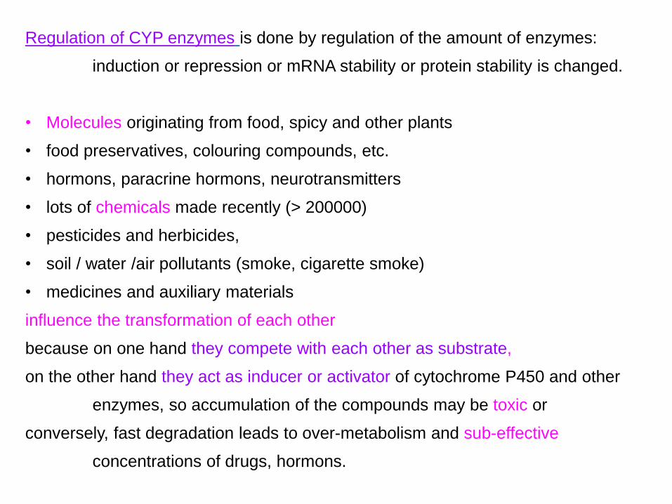
Regulation of CYP enzymes is done by regulation of the amount of enzymes:
induction or repression or mRNA stability or protein stability is changed.
• Molecules originating from food, spicy and other plants
• food preservatives, colouring compounds, etc.
• hormons, paracrine hormons, neurotransmitters
• lots of chemicals made recently (> 200000)
• pesticides and herbicides,
• soil / water /air pollutants (smoke, cigarette smoke)
• medicines and auxiliary materials
influence the transformation of each other
because on one hand they compete with each other as substrate,
on the other hand they act as inducer or activator of cytochrome P450 and other
enzymes, so accumulation of the compounds may be toxic or
conversely, fast degradation leads to over-metabolism and sub-effective
concentrations of drugs, hormons.

Drug interaction caused by enzyme induction
One drug (drug A) binds to transcription factor (PXR here) and they induce the
expression of a drug-metabolizing enzyme (CYP3A here) which will catalyze
the conversion of another drug (drug B) to become more or less active.
DNA

Mechanism of enzyme induction, regulation of
gene expression
• Receptors of lipophylic hormons, medicines, foreign compounds (xenobiotics)
are nuclear transcriptional factors (proteins)
that are bound to promoter or enhancer sequence of DNA
and increase the transcription of mRNA,
therefore more protein is translated from the many mRNA molecules,
this is the enzyme induction.
• The opposite process is the repression, when mRNA transcription is prevented.
• Other mechanism: hydrophilic second messengers e.g. cAMP, cGMP are ligands
of PKA and PKG kinases that phosphorylate and activate transcription factors
to translocate to nucleus and bind to DNA etc.

Cytochrome P450 enzyme system is a monooxygenase: 1 atom of oxygen is built in to the molecule or
called mixed function oxygenase, because the other atom of oxygen forms water
Cytochrome P450 hemoprotein
Substrate-H + O2 + NADPH +H+ → Substrate-OH + H2O + NADP
1. 2.
3.

S-H + O2 + NADPH +H+ → S-OH + H2O + NADP
After hydroxylation:
dealkylation,
desulfuration,
H2O elimination,
aromatization
Other kind of
reactions:
epoxidation,
dehalogenation,
oxidation to keto group
Cytochrome P450 enzyme system can catalyze ~ 60 different type of
reactions, the most frequent one is hydroxylation that can be followed by others
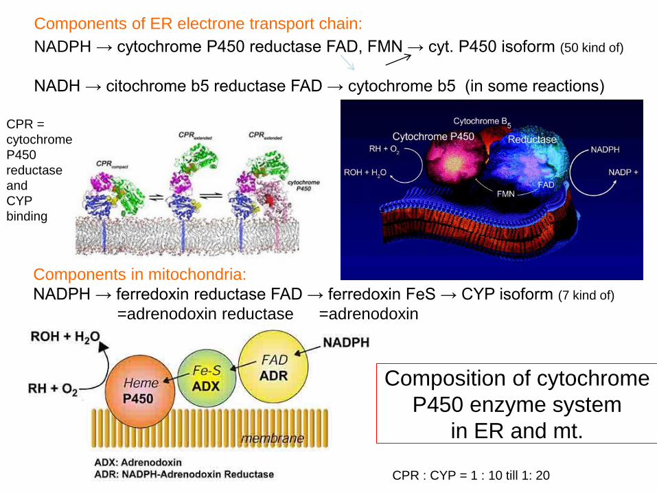
Components in mitochondria:
NADPH → ferredoxin reductase FAD → ferredoxin FeS → CYP isoform (7 kind of)
=adrenodoxin reductase =adrenodoxin
Components of ER electrone transport chain:
NADPH → cytochrome P450 reductase FAD, FMN → cyt. P450 isoform (50 kind of)
NADH → citochrome b5 reductase FAD → cytochrome b5 (in some reactions)
CPR =
cytochrome
P450
reductase
and
CYP
binding
Composition of cytochrome
P450 enzyme system
in ER and mt.
CPR : CYP = 1 : 10 till 1: 20

NADPH FAD FMN
Composition of cytochrome P450 reductase enzyme:
2 electons come from NADPH,
FAD and FMN prosthetic groups can add electrones in stepwise manner
to 1 heme of CYP .

Humans have 18 families of cytochrome P450 genes and 43 subfamilies
CYP1 drug metabolism (3 subfamilies, 3 genes, 1 pseudogene)
CYP2 drug and steroid metabolism (13 subfamilies, 16 genes, 16 pseudogenes)
CYP3 drug metabolism (1 subfamily, 4 genes, 2 pseudogenes)
CYP4 arachidonic acid or fatty acid metabolism (5 subfamilies, 11 genes, 10 pseudogenes)
CYP5 thromboxane A2 synthase (1 subfamily, 1 gene)
CYP7A bile acid biosynthesis 7-alpha hydroxylase of steroid nucleus (1 subfamily member)
CYP7B brain specific form of 7-alpha hydroxylase (1 subfamily member)
CYP8A prostacyclin synthase (1 subfamily member)
CYP8B bile acid biosynthesis (1 subfamily member)
CYP11 steroid biosynthesis (2 subfamilies, 3 genes)
CYP17 steroid biosynthesis (1 subfamily, 1 gene) 17-alpha hydroxylase
CYP19 steroid biosynthesis (1 subfamily, 1 gene) aromatase forms estrogen
CYP20 Unknown function (1 subfamily, 1 gene)
CYP21 steroid biosynthesis (1 subfamily, 1 gene, 1 pseudogene)
CYP24 vitamin D degradation (1 subfamily, 1 gene)
Do not study!

CYP26A retinoic acid hydroxylase important in development (1 subfamily member)
CYP26B probable retinoic acid hydroxylase (1 subfamily member)
CYP26C probable retinoic acid hydroxylase (1 subfamily member)
CYP27A bile acid biosynthesis (1 subfamily member)
CYP27B Vitamin D3 1-alpha hydroxylase activates vitamin D3 (1 subfamily member)
CYP27C Unknown function (1 subfamily member)
CYP39 7 alpha hydroxylation of 24 hydroxy cholesterol (1 subfamily member)
CYP46 cholesterol 24-hydroxylase (1 subfamily member)
CYP51 cholesterol biosynthesis (1 subfamily, 1 gene, 3 pseudogenes)
lanosterol 14-alpha demethylase
Humans have 57 sequenced CYP genes and 58 pseudogenes.
only full length functional genes are named below
1A1, 1A2, 1B1,
2A6, 2A7, 2A13, 2B6, 2C8, 2C9, 2C18, 2C19, 2D6, 2E1, 2F1, 2J2, 2R1, 2S1, 2U1, 2W1,
3A4, 3A5, 3A7, 3A43,
4A11, 4A22, 4B1, 4F2, 4F3, 4F8, 4F11, 4F12, 4F22, 4V2, 4X1, 4Z1
5A1, 7A1, 7B1, 8A1, 8B1, 11A1, 11B1, 11B2, 17, 19, 20, 21A2, 24,
26A1, 26B1, 26C1, 27A1, 27B1, 27C1, 39, 46, 51
Do not study!

Reactions of endogenous CYP substrates:
synthesis of cholesterol and bile acids
CYP51= lanosterol demethylase (ER)
squalene → squalene-epoxide→→ lanosterol → cholesterol
7α-hydroxylase =

Common name Current name
Side-chain cleavage
enzyme; desmolase CYP11A1
3 β-hydroxysteroid
dehydrogenase 3 beta-HSD
17 α-hydroxylase/
17,20 lyase CYP17
21-hydroxylase CYP21A2
11 β-hydroxylase CYP11B1
Aldosterone synthase CYP11B2
Aromatase CYP19 Author: R. A. Bowen
scc = CYP11A1
Synthesis of steroid hormons
Study these structural formulas.

CYP27B1
cholecalciferol = vitamin D3 → 25-OH-cholecalciferol → 1,25-dihydroxicholecalciferol=
liver kidney, bone, placenta calcitriol (hormon)
CYP4=desaturases CYP8A1= prostacyclin synthase
linoleic ac. → arachidonic acid → prostaglandin H2 → prostacyclin
↓CYP5A1= thromboxane synthase
thromboxane
Do not study the formulas

2nd phase: Conjugation reactions
1.) glucuronidation by UDP-glucuronyl transferase
UDP-glucuronidate + drug = drug-glucuronidate + UDP
(OH, NH2, COOH csoport)
UGT1 are formed by alternative splicing – bilirubin, amines, phenols are substrates
UGT2 are formed by different genes – steroids, bile acids, opioids are substrates
It is the most frequent conjugation reaction type. Enzymes are in ER and
cytoplasm of liver, skin, breast, prostate, adipose tissue…
Xenobiotics can induce them.

bilirubin heme degradation not enough induced
product in newborn + glucuronyl transferase in liver
bilirubin-diglucuronide is not produced in liver, it is not secreted to bile,
accumulated free bilirubin in skin, mucous membranes, sclera, it is jaundice = icterus
Deficiency
Free bilirubin = lipophylic,
it binds to albumin in blood,
high amount is toxic.
Bilirubin-monoglucuronide and
bilirubin-diglucuronide are water-soluble,
secreted to bile,
gut bacteria convert them further.
Do not study the figure, only the text!

2.) Sulfatation by sulfotransferase
PAPS + drug = drug-sulfate + PAP
- alcohols, phenols, arylamines are exogenious substrates,
- steroids, heteropolysaccharides, glycolipids, glycoproteins, thyroid hormones are
endogenous substrates, the hormones are inactivated
in cytoplasm and Golgi apparate
Acidic anhydride bond

Conjugation sometimes causes activation:
adrenal cortex-derived androgen:
dihydroepiandrosterone = DHEA
hydroxysteroid sulfotransferase in steroid target cell
dehydroepiandrosterone sulfate active metabolite
Other clinical aspects
17-alfa-etinylestradiol anticontraceptive + rifampicin antituberculotic HSST inducer
sulfonated and noneffective estrogen in liver, pregnancy is possible

3-4.) glutathione conjugation by glutathione S-transferase
+ acetylation by N-acetyltransferase
GSH + drug = drug-S-glutathione → Gly + Glu + drug-S-Cys → N-acetyl-cysteinyl-drug
slow and fast acetylators according to enzyme polymorphism
isoniazid, an antituberculotic agent can be toxic in slow acetylators
GSH = glutathione = γ-glutamyl-cysteinyl-glycine tripeptide
Leukotrienes are formed by leukocytes, they take part
in inflammation. Exceptionally the glutathione
conjugation leads to activation.
Do not study the figure, just the text!
GST is in: ER, mt, px, pl.mb.
GST is induced in tumors, chemotherapy becomes
inefective. GSH pool can be depleted in minutes in liver.

5.) amino acid conjugation by: glycine, taurine, glutamine
Benzoyl-CoA+ Gly = hippurate (way of elimination of N) + CoA
Phenylacetyl-CoA + Gln = phenylacetylglutamine (way of elimination of N) + CoA
chenodeoxycholyl-CoA + taurine = taurochenodeoxycholate primary bile acid + CoA
cholyl-CoA + Gly = glycocholate primary bile acid + CoA
taurocholate

6.) methylation by methyltransferase
dopamine + SAM = methyl-dopamine (inactive) + SAH
by catechol-oximethyltransferase = COMT in catecholamine degradation
noradrenalin + SAM = adrenalin + SAH
by methyltransferase
This is the only one conjugation
reaction when the product is
slightly more lipophylic than the
substrate, but remains water-soluble
enough to become excreted.
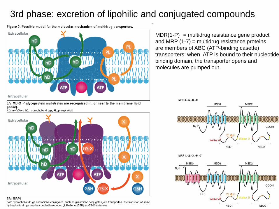
3rd phase: excretion of lipohilic and conjugated compounds
MDR(1-P) = multidrug resistance gene product
and MRP (1-7) = multidrug resistance proteins
are members of ABC (ATP-binding casette)
transporters: when ATP is bound to their nucleotide
binding domain, the transporter opens and
molecules are pumped out.

Metabolism of vitamin D
1,25(OH)2D is the ligand of VDR (vitamin D receptor) transcription factor, it causes induction of many proteins.
CYP27B1:
1-hydroxylase
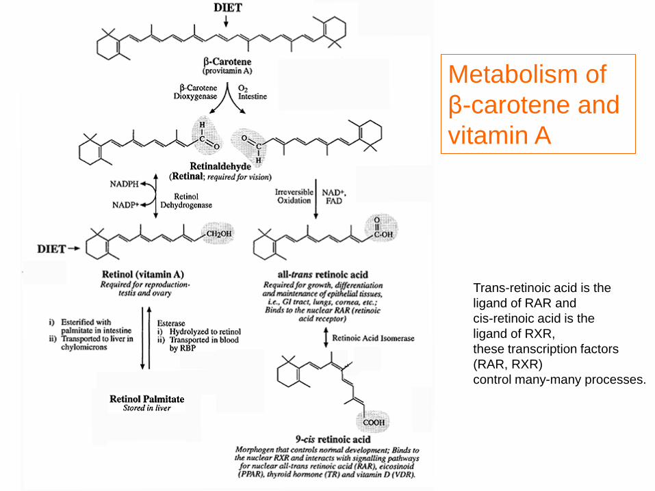
Metabolism of
β-carotene and
vitamin A
Trans-retinoic acid is the
ligand of RAR and
cis-retinoic acid is the
ligand of RXR,
these transcription factors
(RAR, RXR)
control many-many processes.

or cholesterol derivative
or bile acid
or PUFA
or vit. A derivative
or vit. D derivative
or steroid hormon
androgen receptor
estrogen rec.
glucocorticoid rec.
mineralocorticoid rec.
NR = nuclear receptors
NR = nuclear receptor = transcription factor protein that binds ligand
ligand Liver X receptor (lipid synthesis)
Farnesoid X receptor (bile acid synthesis)
Peroxisome proliferation activating receptor
without ligand NR is
in the cytoplasm
without ligand NR is in the nucleus,
it binds to DNA, but it is in repressor
protein complex, so it is inactive

Transformation of medicines and xenobiotics by
CYP enzyme system
Drug metabolizing CYP enzymes 50 % - 3A4 20 % - 2D6 15 % - 2C9, 2C19 15 % - 1A2, 2A6, 2B6 etc.
Regulation of enzyme synthesis is done by induction:
inducer: drug, chemical, pollutant, contaminant, plant compound
inducer binds to any of the following transcriptional factors: CAR, PXR,VDR
that forms a heterodimer with another transcriptional factor: RXR
own ligand binds to RXR: cis-retinoic acid
more mRNA is transcribed,
more CYP isoenzyme is translated,
bigger amount of enzymes catalyze the reactions with bigger velocity,
medicines are converted, degraded faster.

PXR-RXR PXR-RXR or
transcr.fact. CAR-RXR PXR-RXR CAR-RXR or
HNF VDR-RXR CAR-RXR VDR-RXR CYP3A4
DNA chain HNF DR3 ER6 // ER6 ⌐
xenobiotic responsive enhancer module proximal promoter
PXR= pregnane X receptor (inducers: phenobarbital, androstanol)
CAR= constitutive androstane receptor
VDR= vitamin D receptor (inducer: vit. D)
HNF = hepatic nuclear factor DR = direct repeat
RXR = retinoid X receptor ER = everted repeat = inverse direction repeat of sequence
drug = medicine VDR = vitamin D receptor
In mammals several transcription factors regulate the expression of the same gene
Acts as inducer
substrate product
Transcription factors
from gene mRNA
transcription
enzyme protein synthesis

Clinical aspects
Drug interactions, metabolism of medicines, food components and pollutants by the same enzyme
system

Substrates: these are degraded
and compete for the enzyme
Amitriptyline* (Elavil)
Benzodiazepines
Alprazolam (Xanax)
Triazolam (Halcion)
Midazolam (Versed)
Calcium blockers
Carbamazepine (Tegretol)
Cisapride (Propulsid)
Dexamethasone (Decadron)
Erythromycin
Ethinyl estradiol (Estraderm,
Estrace)
Glyburide (Glynase, Micronase)
Imipramine* (Tofranil)
Ketoconazole (Nizoral)
Lovastatin (Mevacor)
Nefazodone (Serzone)
Terfenadine (Seldane)
Astemizole (Hismanal)
Verapamil (Calan, Isoptin)
Sertraline (Zoloft)
Testosterone
Theophylline*
Venlafaxine (Effexor)
Protease inhibitors
Ritonavir (Norvir)
Saquinavir (Invirase)
Indinavir (Crixivan)
Nelfinavir (Viracept)
Inhibitors
Antidepressants
Nefazodone > fluvoxamine (Luvox) > fluoxetine
(Prozac) > sertraline
Paroxetine (Paxil)
Venlafaxine
Azole antifungals
Ketoconazole (Nizoral) > itraconazole (Sporanox)
> fluconazole (Diflucan)
Cimetidine (Tagamet)â�
Clarithromycin (Biaxin)
Diltiazem
Erythromycin
Protease inhibitors
Inducers
Carbamazepine
Dexamethasone
Phenobarbital
Phenytoin (Dilantin)
Rifampin (Rifadin, Rimactane)
CYP3A4
Inhibitors cause slower degradaton
of drug substrates, medicine
concentration remains high, can be toxic.
Inducer drugs cause faster metabolism of
substrate medicines, so medicines will not be
effective enough, they do not reach the
therapeutic concentration.
Localization of CYP3A4:
liver, GI: from esophagus till colon, resp. tract: nose and lung, kidney tubules, skin, blood cells, ovarium, testis Leydig-cells, adrenal gland zona glomerulosa, parathyroid gland, adenohypohysis
Medicines are not required to study in Biochemistry from the figure
One compound can have dual
function: a substrate and inducer
or substrate and repressor either.

CYP2D6 SUBSTRATES AND
INHIBITORS
Substrates
Antidepressants*
Amitriptyline (Elavil)
Clomipramine (Anafranil)
Desipramine (Norpramin)
Doxepin (Adapin, Sinequan)
Fluoxetine (Prozac)
Imipramine (Tofranil)
Nortriptyline (Pamelor)
Paroxetine (Paxil)
Venlafaxine (Effexor)
Antipsychotics
Haloperidol (Haldol)
Perphenazine (Etrafon, Trilafon)
Risperidone (Risperdal)
Thioridazine (Mellaril)
Beta blockers
Metoprolol (Lopressor)
Penbutolol (Levatol)
Propranolol (Inderal)*
Timolol (Blocadren)
Narcotics
Codeine, tramadol (Ultram)
Inhibitors
Antidepressants
Paroxetine > fluoxetine >
sertraline (Zoloft) > fluvoxamine
(Luvox),
Nefazodone (Serzone),
Venlafaxine > clomipramine
(Anafranil) > amitriptyline
Cimetidine (Tagamet)
Fluphenazine (Prolixin)
Antipsychotics
Haloperidol
Perphenazine
Thioridazine
Do not study!
Genetic polymorphism:
5-10% of the Caucasian and African are dificient
in catalytically active CYP2D6.
14-22% of the Asian population lack the active form of
CYP2C19.
One compound can have dual
function: a substrate and inducer
or substrate and repressor either.

Km = 0,2-2 mM
liver, stomach
Km= 8-10 mM
fatty acid oxidation ↓
gluconeogenesis ↓→ blood sugar level ↓
triglyceride synthesis ↑→ fatty liver
protein adducts provoke
immune response
ROS = reactive oxygen species =
O2-, OH˙
O
‖

Alcohol oxidation
by CYP2E1
in endoplasmatic
reticulum
Only this enzyme system
can be induced among the
ethanol degrading
enzymes,
in alcoholists its amount is
significantly increased.
The ethanol is not only a
substrate, but the inducer
of the enzyme, too.
It accelerates its own
metabolism.
CYP2E1 converts the
procarcinogen tobacco
smoke components to
carcinogens.
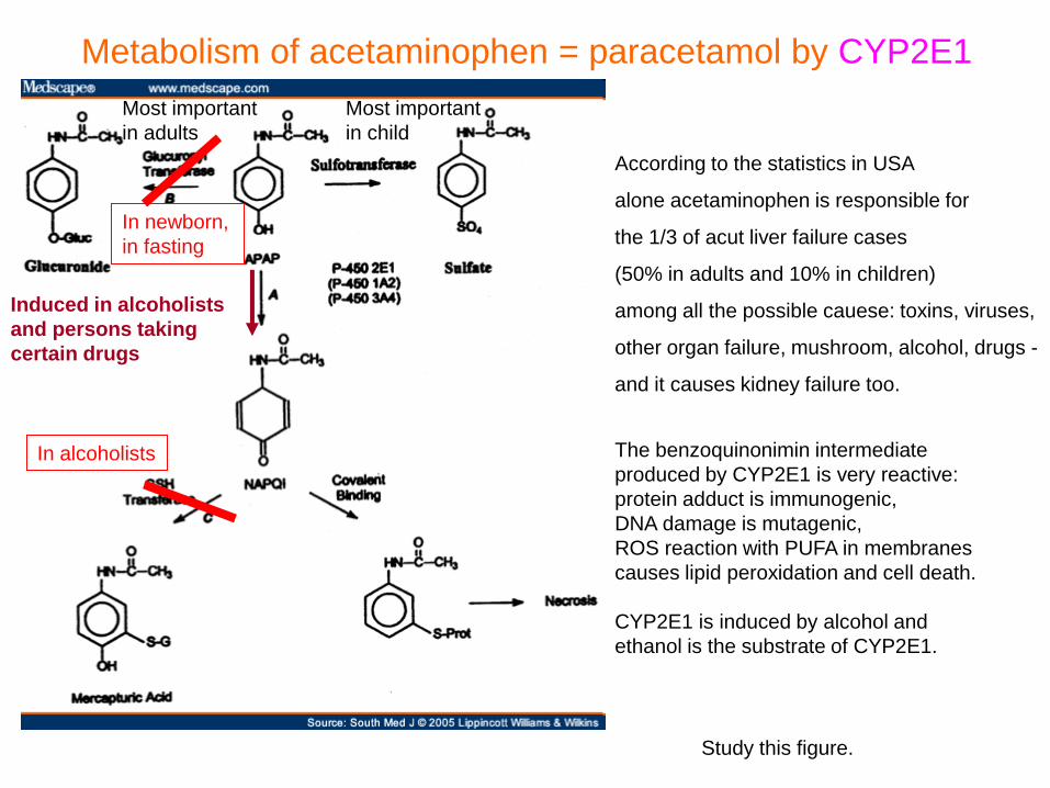
In newborn,
in fasting
In alcoholists
Induced in alcoholists
and persons taking
certain drugs
Most important
in adults
Most important
in child
According to the statistics in USA
alone acetaminophen is responsible for
the 1/3 of acut liver failure cases
(50% in adults and 10% in children)
among all the possible cauese: toxins, viruses,
other organ failure, mushroom, alcohol, drugs -
and it causes kidney failure too.
The benzoquinonimin intermediate
produced by CYP2E1 is very reactive:
protein adduct is immunogenic,
DNA damage is mutagenic,
ROS reaction with PUFA in membranes
causes lipid peroxidation and cell death.
CYP2E1 is induced by alcohol and
ethanol is the substrate of CYP2E1.
Metabolism of acetaminophen = paracetamol by CYP2E1
Study this figure.
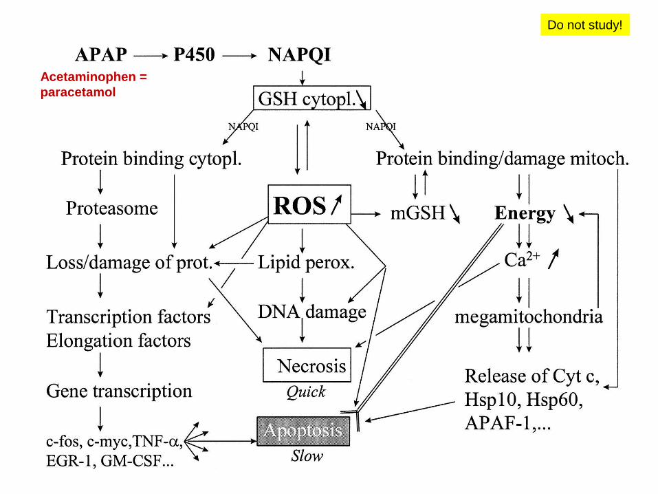
Acetaminophen =
paracetamol
Do not study!

ROS = reactive oxygen species (OH˙, O2˙-,H2O2)
are produced by CYP isoenzymes in ER,
• by mitochondrial electron transport chain,
• in peroxisome during FA oxidation and
• by xanthine oxidase,
• by NADPH oxidase in cytoplasm,
• spontaneously by metal ions.
We have antioxidant, protective enzymes:
o superooxide dismutase, (SOD)
o catalase,
o glutathione peroxidase (GPX)
o thioredoxin (TRR)
o peroxiredoxin
And by antioxidant compounds:
• ascorbate = vitamin C
glutathione = GSH,
vitamin E = α-tocoferol
urate

Relationship between pesticide DDT, hormon
metabolism and BIOTRANSFORMATION enzymes
DDT = dichlordiphenyl-trichloretane is an
insecticide. Insect-killing effect was
discovered in 1934. In world war II the DDT
was used against louse, flea and in tropic
countries against malaria and yellow fever
spreding mosquitoes, against thyphus,
plague = pestilence, colorado-beetle.
Rate of degradation of DDT in soil,
water, plant, animal is 0-5% / year. It is
accumulated in fat and milk. Most, but
not all countries have withdrawn it from
the market, now Mexico, China, India
etc. produce it.
Europe and North America gets DDT back
with Brasil crude coffee bean,
African cocoa seeds and chocolate,
Chineese peanut, Spanish and Greek etc. orange peel.
Most polluted countries: Costa Rica, Zaire, India,
Mexico, Pakistan, China

The effect of DDT (dichlorodiphenyl-trichlorethane) insecticid or its degradation product
DDE in animals and human:
a) induces aromatase → testosteron is converted to estrogen in males (and females)
b) CYP2B and CYP3A enzymes are induced → testosteron hydroxylation and
inactivation is accelerated,
degradation of 70 % of medicines is increased, drugs have no effect
c) sulfotransferase enzyme is induced → testosteron and other steroid hormon
sulfatation and inactivation is faster
d) it has a direct androgen receptor antagonist effect
Because of above mentioned effects in
embryo, the inner gonad formation is
disturebed,
in adult males the androgen is inactivated,
the male is femininized, becomes impotent.
The similar dicofol, endosulfan and
methoxychlor are used in USA, too.
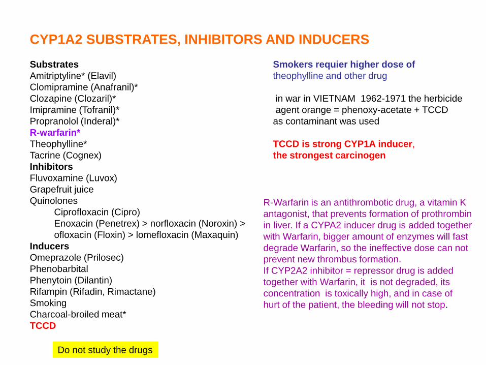
CYP1A2 SUBSTRATES, INHIBITORS AND INDUCERS
Substrates Smokers requier higher dose of
Amitriptyline* (Elavil) theophylline and other drug
Clomipramine (Anafranil)*
Clozapine (Clozaril)* in war in VIETNAM 1962-1971 the herbicide
Imipramine (Tofranil)* agent orange = phenoxy-acetate + TCCD
Propranolol (Inderal)* as contaminant was used
R-warfarin*
Theophylline* TCCD is strong CYP1A inducer,
Tacrine (Cognex) the strongest carcinogen
Inhibitors
Fluvoxamine (Luvox)
Grapefruit juice
Quinolones
Ciprofloxacin (Cipro)
Enoxacin (Penetrex) > norfloxacin (Noroxin) >
ofloxacin (Floxin) > lomefloxacin (Maxaquin)
Inducers
Omeprazole (Prilosec)
Phenobarbital
Phenytoin (Dilantin)
Rifampin (Rifadin, Rimactane)
Smoking
Charcoal-broiled meat*
TCCD
R-Warfarin is an antithrombotic drug, a vitamin K
antagonist, that prevents formation of prothrombin
in liver. If a CYPA2 inducer drug is added together
with Warfarin, bigger amount of enzymes will fast
degrade Warfarin, so the ineffective dose can not
prevent new thrombus formation.
If CYP2A2 inhibitor = repressor drug is added
together with Warfarin, it is not degraded, its
concentration is toxically high, and in case of
hurt of the patient, the bleeding will not stop.
Do not study the drugs

Agent Orange Agent Orange is the code name for one of the herbicides and defoliants used by the U.S. military as part of its
herbicidal warfare program, Operation Ranch Hand, during the Vietnam War from 1961 to 1971. Vietnam
estimates 400,000 people being killed or maimed, and 500,000 children born with birth defects.[1]
A 50:50 mixture of 2,4,5-T and 2,4-D, it was manufactured for the U.S. Department of Defense primarily by
Monsanto Corporation and Dow Chemical. The 2,4,5-T used to produce Agent Orange was later discovered to
be contaminated with 2,3,7,8-tetrachlorodibenzodioxin, an extremely toxic dioxin compound. It was given its
name from the color of the orange-striped 55 US gallon (200 L) barrels in which it was shipped, and was by far
the most widely used of the so-called “Rainbow Herbicides”.[2]
During the Vietnam War, between 1962 and 1971, the United States military sprayed 12,000,000 US gallons
(50,000,000 L) of chemical herbicides and defoliants in Vietnam, eastern Laos and parts of Cambodia, as part
of Operation Ranch Hand.[3] The program’s goal was to defoliate forested and rural land, depriving guerrillas of
cover; another goal was to induce forced draft urbanization, destroying the ability of peasants to support
themselves in the countryside, and forcing them to flee to the U.S. dominated cities, thus depriving the guerrillas
of their rural support base and food supply
180 millió $ kártérítés 45%-át a Monsato fizette a 210 000 veteránnak.
Do not study

Viktor Juscsenko in 2004 and before
Do not study

with metabolizable inducers: with permanently present inducers (TCCD)
aromatic hydrocarbones
dietary plant constituents
tryptophan derivatives TCCD = terachlorodibenzo-p-dioxin (inducers)
↓ ↓
AhR = aromatic hydrocarbone receptor (transcription factor)
↓
forms heterodimer with Arnt = aromatic hydrocarbon receptor nuclear translocator
↓
AhR-Arnt translocation to nucleus and reacts with XREs =xenobiotic response elements =
AhREs = aromatic hydrocarbon responsive elements in the DNA
↓ ↓
biotransformation enzymes induced skin toxicity: chloracne (Viktor Juscsenko)
antioxidant enzymes induced teratogenic response (abnormal emmbryo)
cdk inhibitors induced (cell cycle inhibited) immunosuppression (low immune response)
proapoptotic Bax induced carcinogenesis
IL-2 for T-cells activation of MAPK cascade and
cell proliferation
abnormal hormon metabolism
neurotoxical effect
liver failure
cardiotoxic effect
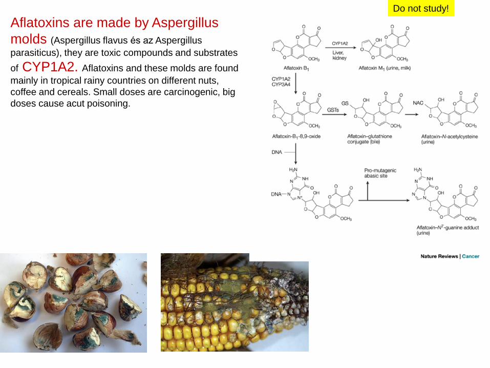
Aflatoxins are made by Aspergillus
molds (Aspergillus flavus és az Aspergillus
parasiticus), they are toxic compounds and substrates
of CYP1A2. Aflatoxins and these molds are found
mainly in tropical rainy countries on different nuts,
coffee and cereals. Small doses are carcinogenic, big
doses cause acut poisoning.
CYP1A2
CYP3A4
o o
o
o o
Do not study!

CYP1A2
This compound is found
in cigarett smoke, too.
Do not study!
PAH = polyaromatic hydrocarbon
Do not study!

aflatoxin
Enzymatic conversion of some selected human carcinogens towards their
ultimate DNA-reactive metabolites. Nature Reviews Cancer 5, 113-125
Benzo-α-pyrene
Do not study!
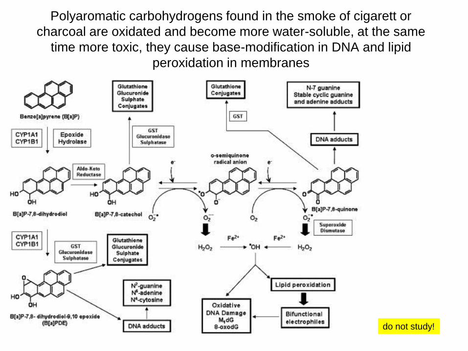
Polyaromatic carbohydrogens found in the smoke of cigarett or
charcoal are oxidated and become more water-soluble, at the same
time more toxic, they cause base-modification in DNA and lipid
peroxidation in membranes
do not study!