Biotransformation modulates the penetration of metallic ...
10
Biotransformation modulates the penetration of metallic nanomaterials across an artificial blood–brain barrier model Zhiling Guo a,1 , Peng Zhang a,1 , Swaroop Chakraborty b , Andrew J Chetwynd a , Fazel Abdolahpur Monikh c , Christopher Stark a , Hanene Ali-Boucetta d , Sandra Wilson e , Iseult Lynch a , and Eugenia Valsami-Jones a,1 a School of Geography, Earth and Environmental Sciences, University of Birmingham, Birmingham B15 2TT, United Kingdom; b Department of Material Engineering, Indian Institute of Technology, Gandhinagar 382355, India; c Department of Environmental and Biological Sciences, University of Eastern Finland, FI-80101 Joensuu, Finland; d Nanomedicine, Drug Delivery and Nanotoxicology Laboratory, The School of Pharmacy, College of Medical and Dental Sciences, University of Birmingham, Birmingham B15 2TT, United Kingdom; and e Sophion Bioscience A/S, Baltorpvej 154, DK-2750 Ballerup, Denmark Edited by Catherine J. Murphy, University of Illinois at Urbana–Champaign, Urbana, IL, and approved May 28, 2021 (received for review March 19, 2021) Understanding the potential of nanomaterials (NMs) to cross the blood–brain barrier (BBB), as a function of their physicochemical properties and subsequent behavior, fate, and adverse effect be- yond that point, is vital for evaluating the neurological effects arising from their unintentional entry into the brain, which is yet to be fully explored. This is not only due to the complex nature of the brain but also the existing analytical limitations for characterization and quantification of NMs in the complex brain environment. By using a fit-for-purpose analytical workflow and an in vitro BBB model, we show that the physiochemical properties of metallic NMs influence their biotransformation in biological matrices, which in turn modulates the transport form, efficiency, amounts, and path- ways of NMs through the BBB and, consequently, their neurotoxicity. The data presented here will support in silico modeling and predic- tion of the neurotoxicity of NMs and facilitate the tailored design of safe NMs. nanomaterials | blood–brain barrier | neurotoxicity | single-particle ICP-MS | synchrotron M ore than a few decades have passed since the application of nanomaterials (NMs) in medicine and since the concept of nanotoxicity was formulated (1). Despite the time lapsed, con- cerns regarding their potential neurotoxicity (2) have yet to be fully addressed, primarily because of challenges in characterization resulting from their unique physicochemical properties. Recent studies found NMs (e.g., ZnO and magnetite NMs) can accumu- late in the brain with distinct morphologies and forms that differ from their pristine counterpart, which can affect the cholinergic neurotransmission and thus brain health (3, 4). Beyond their safety, also important is the potential of NMs to act as carriers for medical interventions to the brain (e.g., to treat neurodegener- ative conditions) (5). To date, our understanding of NM biotransformation in a biological matrix and their subsequent behavior and adverse effects remains limited because of the limitation in analytical techniques capable of tracing the transformation of such small particles in biological samples. This is even more challenging when quanti- fying the cellular uptake of the transformed NMs, because the unique physicochemical properties of NMs (i.e., small size, high specific surface area, high free surface energy, etc.) not only can modulate their biotransformation (6) and consequently their cel- lular uptake but also can directly influence the cellular penetration pathway and intracellular trafficking of NMs (7). This is of para- mount importance for blood–brain barrier (BBB) cells, as the penetration and trafficking pathways could influence the number and forms of NMs entering the brain and possibly exerting ad- verse effects to the brain (e.g., inducing neurotoxicity) (8, 9). For instance, the biotransformation of nanoparticles (e.g., ZnO) in the brain and subbrain region (hippocampus) has been reported (3, 10). Inhaled, ingested, and dermally applied NMs can reach the blood stream (11). From there, there is a possibility that NMs in the bloodstream can cross the BBB and then directly or indirectly impact on the central nervous system (CNS) (12). The BBB is a physical barrier composed of a tightly packed layer of endothelial cells surrounding the brain, which separates the blood from the cerebrospinal fluid, allowing the transfer of oxygen and essential nutrients but preventing the access of most molecules. NMs func- tion at the nanoscale; bind critical transport proteins such as apo- lipoproteins, including the brain transporter apolipoprotein E (13); and thus may have better ability to be transported across the BBB via endocytosis or diffusion (14, 15). Moreover, their biotransfor- mation may dynamically alter their mobility and transport pathways. While emerging human (4) and in vivo (3) evidence suggests that NMs can deposit in brain regions, it is not clear whether these NMs enter the brain through the BBB pathway, what form(s) (i.e., pristine or transformed forms) and quantity of NMs enter the brain, and how the physicochemical properties of NMs modulate this penetration. Moreover, the fate of NMs within and beyond the BBB (i.e., the translocation, deposition, distribution, transformation, and adverse effect during and after crossing the BBB) is largely unknown, pri- marily because of the limited capacity of animal testing and diffi- culties in direct quantitative analysis of NMs within and beyond the Significance Although the brain is protected by a tight physiological guardian named the blood–brain barrier (BBB), deposition of engineered nanomaterials (ENMs) in the brain and consequent neurotoxicity has been reported. To date, it is still unclear whether and how ENMs enter the brain by crossing the BBB. In this study, we found that metallic ENMs transform in the BBB as affected by their shape, size, and intrinsic solubility, which in turn modulates their transport form, efficiency, and pathways through the BBB and, consequently, their neurotoxicity. The library of quantita- tive data on the chemical transformations presented here will support in silico modeling and prediction of the neurotoxicity of NMs and facilitate the tailored design of safe NMs. Author contributions: Z.G., P.Z., S.W., I.L., and E.V.-J. designed research; Z.G., P.Z., S.C., A.J.C., and C.S. performed research; H.A.-B. contributed new reagents/analytic tools; Z.G. and P.Z. analyzed data; and Z.G., P.Z., F.A.M., and I.L. wrote the paper. The authors declare no competing interest. This article is a PNAS Direct Submission. This open access article is distributed under Creative Commons Attribution-NonCommercial- NoDerivatives License 4.0 (CC BY-NC-ND). 1 To whom correspondence may be addressed. Email: [email protected], e.valsamijones@ bham.ac.uk, or [email protected]. This article contains supporting information online at https://www.pnas.org/lookup/suppl/ doi:10.1073/pnas.2105245118/-/DCSupplemental. Published July 6, 2021. PNAS 2021 Vol. 118 No. 28 e2105245118 https://doi.org/10.1073/pnas.2105245118 | 1 of 10 APPLIED PHYSICAL SCIENCES Downloaded by guest on December 10, 2021
Transcript of Biotransformation modulates the penetration of metallic ...

Biotransformation modulates the penetration ofmetallic nanomaterials across an artificialblood–brain barrier modelZhiling Guoa,1
, Peng Zhanga,1, Swaroop Chakrabortyb, Andrew J Chetwynda
, Fazel Abdolahpur Monikhc,Christopher Starka, Hanene Ali-Boucettad, Sandra Wilsone, Iseult Lyncha
, and Eugenia Valsami-Jonesa,1
aSchool of Geography, Earth and Environmental Sciences, University of Birmingham, Birmingham B15 2TT, United Kingdom; bDepartment of MaterialEngineering, Indian Institute of Technology, Gandhinagar 382355, India; cDepartment of Environmental and Biological Sciences, University of EasternFinland, FI-80101 Joensuu, Finland; dNanomedicine, Drug Delivery and Nanotoxicology Laboratory, The School of Pharmacy, College of Medical and DentalSciences, University of Birmingham, Birmingham B15 2TT, United Kingdom; and eSophion Bioscience A/S, Baltorpvej 154, DK-2750 Ballerup, Denmark
Edited by Catherine J. Murphy, University of Illinois at Urbana–Champaign, Urbana, IL, and approved May 28, 2021 (received for review March 19, 2021)
Understanding the potential of nanomaterials (NMs) to cross theblood–brain barrier (BBB), as a function of their physicochemicalproperties and subsequent behavior, fate, and adverse effect be-yond that point, is vital for evaluating the neurological effectsarising from their unintentional entry into the brain, which is yetto be fully explored. This is not only due to the complex nature of thebrain but also the existing analytical limitations for characterizationand quantification of NMs in the complex brain environment. Byusing a fit-for-purpose analytical workflow and an in vitro BBBmodel, we show that the physiochemical properties of metallicNMs influence their biotransformation in biological matrices, whichin turn modulates the transport form, efficiency, amounts, and path-ways of NMs through the BBB and, consequently, their neurotoxicity.The data presented here will support in silico modeling and predic-tion of the neurotoxicity of NMs and facilitate the tailored design ofsafe NMs.
nanomaterials | blood–brain barrier | neurotoxicity | single-particleICP-MS | synchrotron
More than a few decades have passed since the applicationof nanomaterials (NMs) in medicine and since the concept
of nanotoxicity was formulated (1). Despite the time lapsed, con-cerns regarding their potential neurotoxicity (2) have yet to be fullyaddressed, primarily because of challenges in characterizationresulting from their unique physicochemical properties. Recentstudies found NMs (e.g., ZnO and magnetite NMs) can accumu-late in the brain with distinct morphologies and forms that differfrom their pristine counterpart, which can affect the cholinergicneurotransmission and thus brain health (3, 4). Beyond their safety,also important is the potential of NMs to act as carriers formedical interventions to the brain (e.g., to treat neurodegener-ative conditions) (5).To date, our understanding of NM biotransformation in a
biological matrix and their subsequent behavior and adverse effectsremains limited because of the limitation in analytical techniquescapable of tracing the transformation of such small particles inbiological samples. This is even more challenging when quanti-fying the cellular uptake of the transformed NMs, because theunique physicochemical properties of NMs (i.e., small size, highspecific surface area, high free surface energy, etc.) not only canmodulate their biotransformation (6) and consequently their cel-lular uptake but also can directly influence the cellular penetrationpathway and intracellular trafficking of NMs (7). This is of para-mount importance for blood–brain barrier (BBB) cells, as thepenetration and trafficking pathways could influence the numberand forms of NMs entering the brain and possibly exerting ad-verse effects to the brain (e.g., inducing neurotoxicity) (8, 9). Forinstance, the biotransformation of nanoparticles (e.g., ZnO) inthe brain and subbrain region (hippocampus) has been reported(3, 10).
Inhaled, ingested, and dermally applied NMs can reach theblood stream (11). From there, there is a possibility that NMs inthe bloodstream can cross the BBB and then directly or indirectlyimpact on the central nervous system (CNS) (12). The BBB is aphysical barrier composed of a tightly packed layer of endothelialcells surrounding the brain, which separates the blood from thecerebrospinal fluid, allowing the transfer of oxygen and essentialnutrients but preventing the access of most molecules. NMs func-tion at the nanoscale; bind critical transport proteins such as apo-lipoproteins, including the brain transporter apolipoprotein E (13);and thus may have better ability to be transported across the BBBvia endocytosis or diffusion (14, 15). Moreover, their biotransfor-mation may dynamically alter their mobility and transport pathways.While emerging human (4) and in vivo (3) evidence suggests thatNMs can deposit in brain regions, it is not clear whether these NMsenter the brain through the BBB pathway, what form(s) (i.e., pristineor transformed forms) and quantity of NMs enter the brain, and howthe physicochemical properties of NMs modulate this penetration.Moreover, the fate of NMs within and beyond the BBB (i.e., thetranslocation, deposition, distribution, transformation, and adverseeffect during and after crossing the BBB) is largely unknown, pri-marily because of the limited capacity of animal testing and diffi-culties in direct quantitative analysis of NMs within and beyond the
Significance
Although the brain is protected by a tight physiological guardiannamed the blood–brain barrier (BBB), deposition of engineerednanomaterials (ENMs) in the brain and consequent neurotoxicityhas been reported. To date, it is still unclear whether and howENMs enter the brain by crossing the BBB. In this study, wefound that metallic ENMs transform in the BBB as affected bytheir shape, size, and intrinsic solubility, which in turn modulatestheir transport form, efficiency, and pathways through the BBBand, consequently, their neurotoxicity. The library of quantita-tive data on the chemical transformations presented here willsupport in silico modeling and prediction of the neurotoxicity ofNMs and facilitate the tailored design of safe NMs.
Author contributions: Z.G., P.Z., S.W., I.L., and E.V.-J. designed research; Z.G., P.Z., S.C.,A.J.C., and C.S. performed research; H.A.-B. contributed new reagents/analytic tools; Z.G.and P.Z. analyzed data; and Z.G., P.Z., F.A.M., and I.L. wrote the paper.
The authors declare no competing interest.
This article is a PNAS Direct Submission.
This open access article is distributed under Creative Commons Attribution-NonCommercial-NoDerivatives License 4.0 (CC BY-NC-ND).1To whom correspondence may be addressed. Email: [email protected], [email protected], or [email protected].
This article contains supporting information online at https://www.pnas.org/lookup/suppl/doi:10.1073/pnas.2105245118/-/DCSupplemental.
Published July 6, 2021.
PNAS 2021 Vol. 118 No. 28 e2105245118 https://doi.org/10.1073/pnas.2105245118 | 1 of 10
APP
LIED
PHYS
ICAL
SCIENCE
S
Dow
nloa
ded
by g
uest
on
Dec
embe
r 10
, 202
1

barrier at molecular and cellular levels in real time. These knowl-edge gaps highlight the importance of using such approaches toenable a breakthrough in our fundamental understanding of theBBB penetration ability of NMs and the relevance of physico-chemical properties on controlling the path, behavior, and impactsof NMs beyond the BBB.Here, we synthesized a library of metallic NMs with different
particle compositions, sizes, and shapes and evaluated their abilityto penetrate the BBB using a well-established in vitro BBB model,followed by an assessment of their behavior and fate in and be-yond the BBB. To achieve this, a set of state-of-the-art analyticaltechniques, including single-particle inductively coupled plasmamass spectrometry (spICP-MS), synchrotron radiation–based X-rayabsorption fine structure spectroscopy (XAFS), and high spatialresolution (20 nm) scanning transmission X-ray microscopy (STXM),and a series of biological assays were applied. We found thatphysiochemical properties of metallic NMs influenced their bio-transformation in the physiological matrix. Biotransformation, inturn, modulated the transport form, efficiency, amounts, and path-ways of NMs through the BBB and, consequently, the neurotoxicity.For example, spherical Ag (Ag NS) and disk-like Ag (Ag ND) NMsunderwent different dissolution regimes in the physiological mediaand gradual transformation to Ag-sulfur compounds within theBBB, which facilitated paracellular and transcytosis entry path-ways. Our finding showed that by changing the chemical com-position of NMs to ZnO, CeO2, and Fe3O4, the transport efficiencyand amount of particles passing through the BBB change signifi-cantly, even if the shape and the size of the particles are kept thesame. This can be linked to the composition-related biotransfor-mation of the NMs, as CeO2 and Fe3O4 NMs with low dissolutionhave the limited ability to transport across the BBB.
Results and DiscussionNM Transport Analysis across the BBB. Determining the behaviorand fate of NMs, as a function of their physicochemical propertiesat the BBB, is not only vital for evaluating their neurologicaleffects but also for facilitating their application, especially intargeting hard-to-reach sites within the brain when treating braindisease. Despite the valuable contribution of animal models to NMtransport studies, they have limited utility in large-scale screeningstudies, and it remains difficult to conduct mechanistic studies andquantitative analysis at molecular and cellular levels. In vitro BBBmodels, which are cell-based models that can reconstitute the keystructure and function of the human BBB (16), are considered areliable and predictive screening platform for brain-penetratingagents (17). Among all available cell types, human-sourced cellsshow the advantages in enhancing the physiological relevance ofin vitro models to the unique properties of the human BBB, pro-viding more clinically representative data regarding NM transport,uptake, transcytosis, and toxicity (18, 19). A primary human cocul-ture in vitro BBB model has been successfully used to investigate thetransport of NMs (e.g., gold and polystyrene) across the BBB (8, 20).This was also the chosen model for our NM transport studies.The critical barrier function of the BBB endothelium is char-
acterized by low permeability and high expression of BBB-specificproteins such as junction proteins. Here, coculture of humanprimary brain microvascular endothelial cells (HBMECs) withhuman primary astrocytes (HAs) to generate an in vitro BBBmodel (Fig. 1A) led to stronger induction of expression of inter-endothelial tight junction proteins, including vascular endothelial(VE)-cadherin, claudin-5, and zonula occludens-1 (ZO-1) (Fig. 1B),than HBMECs alone. Furthermore, confocal fluorescence micros-copy images demonstrated junctional protein (occludin and ZO-1)distribution at the HBMEC cell–cell borders, which form a tightbarrier (Fig. 1C), demonstrating the reproduction of BBB-specificendothelial characteristics. Coculture with HAs in the BBB modelsignificantly increased the transendothelial electrical resistance(TEER) values of the HBMEC monolayer from 216 to 472 Ω cm2
(Fig. 1D). The model showed size-dependent transport of fluo-rescently labeled dextran across the BBB, with significantly lowerpermeability coefficient (Pe) values than for the endothelial mono-culture model (Fig. 1E). The TEER value and Pe to 70 kD fluores-cein isothiocyanate (FITC)–dextran and 20 kD tetramethylrhodamine(TRITC)-dextran were comparable to previous BBB studies (12,21–23), implying that the in vitro model precisely replicates keyBBB features and functions and can be used for subsequent NMtransport studies.To comprehensively understand the influence of NM physi-
cochemical properties on their ability to pass through the BBBand their subsequent fate, metallic NMs with different compo-sitions, sizes, shapes, and dissolution profiles in physiologicalmedium were synthesized and tested (SI Appendix, Table S1 andFig. S1). This includes three sizes of CeO2 (3 nm CeO2, 7 nmCeO2, and 25 nm CeO2), two sizes of Fe3O4 (18 nm Fe3O4 and35 nm Fe3O4), four shapes of Ag (Ag NS, Ag ND, rod-shaped Ag[Ag NR], and Ag nanowires [Ag NW]), and ZnO (30 nm). NMswere applied at the apical side of the model at final concentrationsof 1 and 2.5 mg/L. After 48 h, medium samples from the basolateralside were taken for measurement, including quantification (elementsand particles) and size determination of NMs (SI Appendix, Fig. S2).In most clinical and environmental studies, mass concentra-
tions of metallic NMs are measured to determine their uptakeand transport. Although informative, this fails to provide sufficientparticle-specific transport information based on which concreteconclusion can be made (e.g., particle size, particle number, par-ticle species, etc.), which are critical for understanding and even-tually predicting the behavior, fate, and long-term effect of NMs inthe brain. Particle agglomeration and transformation may alsooccur following cellular uptake and transport into and through theBBB. Such information cannot be obtained from mass measure-ment alone (24), highlighting that the mass only paradigm haslimitations in providing insights into the behavior of NMs in theBBB. Time-resolved spICP-MS analysis can quantify a singleparticle of metallic NMs at trace amounts in environmental andbiological media, while simultaneously providing information onparticle number concentration, size distribution, and ion con-centration (25). However, the size detection limits of spICP-MSfor particles varied depending on the composition elements. There-fore, not all NMs can be measured using spICP-MS. The size de-tection limit of spICP-MS has been shown to be 10 to 20 nm for Ag,20 to 80 nm for ZnO and Fe3O4, and 10 nm for CeO2 NMs (26).Moreover, spICP-MS calculates particle size based on the assump-tion that particles are spherical (26). Therefore, spICP-MS was onlyused for samples of 25 nm CeO2, 18 nm Fe3O4, 35 nm Fe3O4, AgNS, and ZnO. For other NMs, we used a different sampling strategy,as illustrated in SI Appendix, Fig. S2. In sampling Strategy I, spICP-MS can give both the particle concentration (particle/milliliter) andmass of ions (Mi). The mass of the transported particles (Mp) can becalculated as total element mass (Mt) subtracting the Mi. ForNMs that cannot be measured by spICP-MS, sampling Strategy IIwas used. Assuming there were particles transported, the Mp canbe calculated as Mt subtracting the Mi. The fraction of ions andparticles and the total transport percentage then can be calculated.Among all the tested NMs, ZnO exhibited the highest trans-
port percentage, with 10.5 and 13.4% at 1 and 2.5 mg/L exposureconcentrations, respectively, followed by Ag (Ag ND and AgNS), although with a strong shape dependence (Fig. 2A). Ourfindings showed that Ag NDNMs had a higher transport (4.8%at 1 mg/L and 5.7% at 2.5 mg/L) than Ag NS NMs (2.3% at 1 mg/Land 3.1% at 2.5 mg/L). No transport was observed for Ag NR andAg NW. This could be due to the needle-like shape of the particles,which hinders the prolongation of the cell membrane (e.g., en-docytosis of the particles), as reported for silica nanowires (27).Transport of CeO2 NMs was between 0.08 to ∼0.34%, with thehighest transport being observed for 3 nm CeO2. No transportwas detected for Fe3O4 (Fig. 2A). These findings clearly show
2 of 10 | PNAS Guo et al.https://doi.org/10.1073/pnas.2105245118 Biotransformation modulates the penetration of metallic nanomaterials across an
artificial blood–brain barrier model
Dow
nloa
ded
by g
uest
on
Dec
embe
r 10
, 202
1

that variation in shape, size, and the chemical compositions ofNMs can dramatically influence their penetration through theBBB. This is of paramount importance for the tailored medicalapplication of NMs (e.g., targeted delivery system and bioimaging)and for assessing the possible risks associated with each type ofmetallic NMs. For all NMs, increasing the exposure concentrationto 2.5 mg/L did not cause significant increase in the percentage oftransported elements, suggesting that the turnover rate of the en-docytotic receptors is a rate-limiting step, as shown previously for arange of other human cells (28). It is also likely that the increase inthe concentration of NMs facilitates the agglomeration of NMs,which in turn reduces the transport of the agglomerates across theBBB (15). Another potential explanation is that particle agglom-eration may lead to low dissolution and thus the low transport ofthe released ions.The particle number concentration in the basolateral chamber,
obtained by spICP-MS, differed with particle composition (Fig. 2B).The particle number concentration for ZnO, Ag NS, and 25 nmCeO2 were 3.7 to 4.6 × 10⁴ particles/mL, 0.95 to 1.1 × 10⁴ particles/mL, and 0.4 to 0.9 × 10³ particles/mL, respectively. We could notdetect 18 nm Fe3O4 or 35 nm Fe3O4 in the basolateral chamber,although it was recently reported that magnetite NMs, which areprolific in urban, airborne particulate matter, could be found in thehuman brain (4). This suggests that the magnetite NMs may enterthe brain through other routes, such as the olfactory pathway, rather
than via the BBB. The size distribution of the transported par-ticles was also obtained by spICP-MS, as shown in Fig. 2 C–E(1 mg/L) and SI Appendix, Fig. S3 (2.5 mg/L). The mode of thesize distribution for the transported particles shifted toward alarger size compared to those of the pristine particles. This couldhave two explanations; first, the particles undergo agglomeration,and the agglomerates with a certain size can pass through theBBB, as each of the receptor-mediated pathways has an optimizedcargo size (29, 30). Second, the particles undergo agglomerationduring (i.e., in the vesicles) or after passing through the BBB.As shown in SI Appendix, Fig. S2, the total mass of each ele-
ment, along with its ionic and particulate form in the basolateralchamber, were acquired, and the ratio of particle to ion is shownin Fig. 2F. Most of the transported CeO2 (68 to 99%) was in theform of particles, with the highest ionic fraction found for 3 nmCeO2. In contrast, ions accounted for a much higher proportion(>75%) than particles for ZnO NMs. For Ag NM, the ratio oftransported particles to ions was shape dependent. For Ag NS,more than 64% of the transported Ag was in the form of particles,while for Ag ND most of the transported Ag was ionic (>76%).These results showed that higher ionic fraction was correlatedwith higher total transport percentage, which was confirmed bylinear regression analysis (Fig. 2G) (R2 = 0.7570). This impliesthat the dissolution of partially soluble NMs can contribute to
Fig. 1. In vitro BBB model. (A) In vitro BBB model using a coculture of HBMECs and HAs. In the apical chamber, HBMECs were cultured on a transwell insert.At the basolateral side, HAs were cultured in a 12-well plate. After the model setup, nanomaterials (NMs) were added apically, and the NM accumulation in,and transport across, the BBB was assessed after 48 h (see Materials and Methods for detailed information). (B) Western blot and densitometry analysis of VE-cadherin, claudin-5, and ZO-1 protein expression in endothelial monoculture (EC) and the in vitro BBB system (BBB). Blots were probed with anti-GAPDH/actinantibodies as a control for equal loading of cell lysates. (C) Fluorescence images showing the distribution of tight junction markers, ZO-1 and occludin (red),between HBMEC cells. Nuclei were stained with DAPI (blue). (D) TEER measurements across the HBMEC monolayer grown on the transwell insert membranewith (BBB) and without (EC) coculture with HAs. (E) Pe calculated from the diffusion of 70 kD FITC-dextran, 20 kD TRITC-dextran, 3 kD TXR-dextran, andfluorescein sodium salt through the HBMEC monoculture (EC) or HBMECs and HAs coculture model (BBB). Data are presented as mean ± SD (n = 6). All imagesare representative ones from at least three biological and three technical replicates. *P < 0.05 and **P < 0.01.
Guo et al. PNAS | 3 of 10Biotransformation modulates the penetration of metallic nanomaterials across an artificialblood–brain barrier model
https://doi.org/10.1073/pnas.2105245118
APP
LIED
PHYS
ICAL
SCIENCE
S
Dow
nloa
ded
by g
uest
on
Dec
embe
r 10
, 202
1

the transport of NMs across BBB. To confirm this, we measuredthe dissolution of NMs in the culture medium.
NM Passage across the BBB Is Associated with Particle Dissolution.The dissolution of all NMs in water was much lower than inendothelial cell growth medium 2 (EGM-2) medium (Fig. 3 A–D).Among all the NMs, ZnO showed the highest dissolution (∼45%)in cell medium after 48 h incubation, followed by Ag (3 to 26.5%)(Fig. 3 A and B). Different shapes of Ag showed different disso-lution in the following order: Ag ND > Ag NS > Ag NR and AgNW (Fig. 3A). The exposed crystal facets highly affect the re-activity (e.g., dissolution) of NMs. Ag ND was characterized withhighly reactive (111) facet as the basal plane and high levels ofsurface defects, thus presenting high reactivity (31), which con-tributed to the higher dissolution of Ag ND than other shaped Ag
NMs. In comparison, Ag NS with a similar size showed lowerdissolution in the medium, suggesting that shape had a profoundeffect on the dissolution of Ag NMs, as reported previously (32).Ag NR and Ag NW with high aspect ratios and relatively largersizes showed a low dissolution. Compared to ZnO and Ag, the dis-solution of Fe3O4 and CeO2 NMs was quite low in the cell culturemedium (1.5 to 2.0% for Fe3O4 and 0.2 to 0.9% for CeO2) (Fig. 3 Cand D). This is in accordance with previous reports that CeO2 NMsare relatively stable in environmental and biological surroundings(33). In addition, size effects on dissolution were observed for Fe3O4and CeO2 NMs, with smaller-sized NMs showing a little higher ionrelease (Fig. 3 C and D). This could be due to the relatively higherparticle numbers and, consequently, a higher total surface areaof smaller-sized NMs available for ion release. Further linear re-gression analysis showed that the NM passage through the BBB
Fig. 2. NM transport across the BBB. (A) Percentage of total transported elements in the basolateral chamber for different NMs at 1 and 2.5 mg/L (parts permillion). (B) Particle number concentration (particle/milliliter) in the basolateral medium. (C–E) Size distribution of transported particles at 1 mg/L (parts permillion): Ag NS (C), 25 nm CeO2 (D), and ZnO (E). S1, S2, and S3 indicate three sample replicates. The vertical dashed lines indicate the average sizes of thepristine NMs. (F) Ratio of particle versus ion in the total transported elements. The upper columns are the ratios of particles, and the lower columns are theratio of ions. (G) Linear regression analysis showing that the percentage of transported elements correlated positively with the mass fraction of ions, n = 3.The x-axis indicates the transported ions (percentage) for different NMs, and the y-axis indicates the corresponding total transported elements (percentage)for each NM. N/A indicates that no element or particle were detected either by the ICP-MS or spICP-MS method.
4 of 10 | PNAS Guo et al.https://doi.org/10.1073/pnas.2105245118 Biotransformation modulates the penetration of metallic nanomaterials across an
artificial blood–brain barrier model
Dow
nloa
ded
by g
uest
on
Dec
embe
r 10
, 202
1

was positively associated with the NM dissolution in the cellmedium (Fig. 3E; R2 = 0.9439) but not in water (R2 = 0.07786).
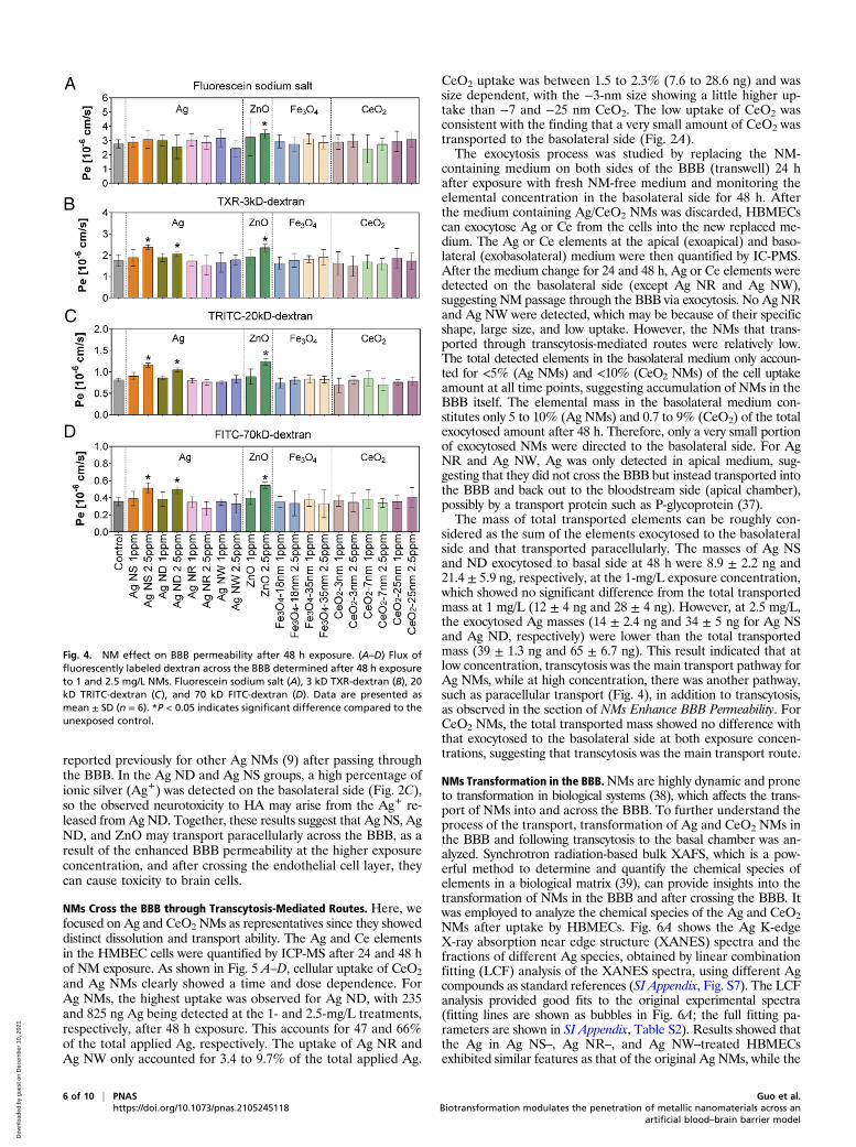
NMs Enhance BBB Permeability. Transport of compounds from bloodto brain occurs via two possible routes (i.e., passive diffusion andactive transport). For NMs, the main transport route is consideredto be the transcytosis process, which commences NM uptake byendocytosis, followed by intracellular vesicular trafficking andexocytosis to the basolateral side (19). Although the passive trans-port of compounds is limited because of the physiological nature ofthe BBB and its tight junctions, in cases in which the BBB per-meability was enhanced, through damage for example, NMs canalso cross the BBB between the cells in a process called paracellulartransport and access the brain (34, 35). We examined the impact ofNMs on BBB permeability after 48 h exposure by testing the fluxof 70 kD FITC-dextran, 20 kD TRITC-dextran, 3 kD Texas Red(TXR)-dextran, and fluorescein sodium salt across the barrierafter it had been exposed to NMs. Results showed that the Pe to70 kD FITC-dextran, 20 kD TRITC-dextran, and 3 kD TXR-dextran were all increased after exposure to 2.5 mg/L Ag NS, AgND, and ZnO. Pe of fluorescein sodium salt did not show a signif-icant change, except for ZnO (Fig. 4). Other NMs at either con-centration did not affect the BBB permeability. Next, we examinedthe toxicity of NMs to the HBMECs. As shown in SI Appendix, Fig.S4, all the NMs showed no effects on the cell viability after 24 and48 h exposure at 1 mg/L, while the viability was decreased by 12.6
and 23.7%, respectively, in the Ag NS and Ag ND group at 2.5 mg/L, after 48 h of exposure. The cell viability was decreased by 15.8%after 48 h of exposure to ZnONMs. Cell viability in the 3-nm CeO2treatment was increased, which is consistent with previous reportthat CeO2 has a role in neuroprotection because of its antioxidativeenzyme-mimicking activity, which can capture the reactive oxygenspecies, thus protecting the cells (36). Fe3O4 NMs of different sizesand CeO2 of the other two sizes (−7 nm and 25 nm) showed noeffects on the cell viability at 48 h. The malondialdehyde (MDA)content in HBMECs was increased when exposed to 2.5 mg/L AgNS, Ag ND, and ZnO NMs for 48 h (SI Appendix, Fig. S5), indi-cating some cellular damage. Together, these data indicate that thehigher concentration (2.5 mg/L) of Ag NS, Ag ND, and ZnO NMsmay impair the cell growth and cause cell membrane damage,which can lead to the enhanced BBB permeability and thus leadto NM access to the brain observed. The BBB plays a vital role inthe maintenance of brain homeostasis and the physiological environ-ment of the CNS by strictly restricting the passage of various chemicalsubstances and foreign molecules from the brain vasculature into thebrain. The impaired BBB integrity will compromise the health of theCNS and increase the permeability of foreign substances includingNMs (e.g., Ag and ZnO) from the peripheral blood into the brain,which may eventually cause damage to the brain (neurotoxicity).HA cell viability in the BBB was also decreased in the Ag ND
and Ag NS treatments at 2.5 mg/L after 48 h (SI Appendix, Fig. S6),indicating that Ag ND and Ag NS exert neurotoxicity, as also
Fig. 3. NM passage through the BBB is associated with NM dissolution in the medium. (A–D) Dissolution (percentage) of NMs in deionized water and EGM-2medium after 48 h of incubation. Ag (A), ZnO (B), CeO2 (C), and Fe3O4 (D) NMs. (E) Linear regression analysis showing that the percentage of transportedelements correlated positively with the NM dissolution in the EGM-2 medium (R2 = 0.9439) but not in water (R2 = 0.07786), n = 6. The x-axis indicates the totaltransported elements (percentage) for different NMs, and the y-axis indicates the corresponding dissolution (percentage) in water or EGM-2 medium foreach NM.
Guo et al. PNAS | 5 of 10Biotransformation modulates the penetration of metallic nanomaterials across an artificialblood–brain barrier model
https://doi.org/10.1073/pnas.2105245118
APP
LIED
PHYS
ICAL
SCIENCE
S
Dow
nloa
ded
by g
uest
on
Dec
embe
r 10
, 202
1
reported previously for other Ag NMs (9) after passing throughthe BBB. In the Ag ND and Ag NS groups, a high percentage ofionic silver (Ag+) was detected on the basolateral side (Fig. 2C),so the observed neurotoxicity to HA may arise from the Ag+ re-leased from Ag ND. Together, these results suggest that Ag NS, AgND, and ZnO may transport paracellularly across the BBB, as aresult of the enhanced BBB permeability at the higher exposureconcentration, and after crossing the endothelial cell layer, theycan cause toxicity to brain cells.
NMs Cross the BBB through Transcytosis-Mediated Routes. Here, wefocused on Ag and CeO2 NMs as representatives since they showeddistinct dissolution and transport ability. The Ag and Ce elementsin the HMBEC cells were quantified by ICP-MS after 24 and 48 hof NM exposure. As shown in Fig. 5 A–D, cellular uptake of CeO2and Ag NMs clearly showed a time and dose dependence. ForAg NMs, the highest uptake was observed for Ag ND, with 235and 825 ng Ag being detected at the 1- and 2.5-mg/L treatments,respectively, after 48 h exposure. This accounts for 47 and 66%of the total applied Ag, respectively. The uptake of Ag NR andAg NW only accounted for 3.4 to 9.7% of the total applied Ag.
CeO2 uptake was between 1.5 to 2.3% (7.6 to 28.6 ng) and wassize dependent, with the −3-nm size showing a little higher up-take than −7 and −25 nm CeO2. The low uptake of CeO2 wasconsistent with the finding that a very small amount of CeO2 wastransported to the basolateral side (Fig. 2A).The exocytosis process was studied by replacing the NM-
containing medium on both sides of the BBB (transwell) 24 hafter exposure with fresh NM-free medium and monitoring theelemental concentration in the basolateral side for 48 h. Afterthe medium containing Ag/CeO2 NMs was discarded, HBMECscan exocytose Ag or Ce from the cells into the new replaced me-dium. The Ag or Ce elements at the apical (exoapical) and baso-lateral (exobasolateral) medium were then quantified by IC-PMS.After the medium change for 24 and 48 h, Ag or Ce elements weredetected on the basolateral side (except Ag NR and Ag NW),suggesting NM passage through the BBB via exocytosis. No Ag NRand Ag NW were detected, which may be because of their specificshape, large size, and low uptake. However, the NMs that trans-ported through transcytosis-mediated routes were relatively low.The total detected elements in the basolateral medium only accoun-ted for <5% (Ag NMs) and <10% (CeO2 NMs) of the cell uptakeamount at all time points, suggesting accumulation of NMs in theBBB itself. The elemental mass in the basolateral medium con-stitutes only 5 to 10% (Ag NMs) and 0.7 to 9% (CeO2) of the totalexocytosed amount after 48 h. Therefore, only a very small portionof exocytosed NMs were directed to the basolateral side. For AgNR and Ag NW, Ag was only detected in apical medium, sug-gesting that they did not cross the BBB but instead transported intothe BBB and back out to the bloodstream side (apical chamber),possibly by a transport protein such as P-glycoprotein (37).The mass of total transported elements can be roughly con-
sidered as the sum of the elements exocytosed to the basolateralside and that transported paracellularly. The masses of Ag NSand ND exocytosed to basal side at 48 h were 8.9 ± 2.2 ng and21.4 ± 5.9 ng, respectively, at the 1-mg/L exposure concentration,which showed no significant difference from the total transportedmass at 1 mg/L (12 ± 4 ng and 28 ± 4 ng). However, at 2.5 mg/L,the exocytosed Ag masses (14 ± 2.4 ng and 34 ± 5 ng for Ag NSand Ag ND, respectively) were lower than the total transportedmass (39 ± 1.3 ng and 65 ± 6.7 ng). This result indicated that atlow concentration, transcytosis was the main transport pathway forAg NMs, while at high concentration, there was another pathway,such as paracellular transport (Fig. 4), in addition to transcytosis,as observed in the section of NMs Enhance BBB Permeability. ForCeO2 NMs, the total transported mass showed no difference withthat exocytosed to the basolateral side at both exposure concen-trations, suggesting that transcytosis was the main transport route.
NMs Transformation in the BBB.NMs are highly dynamic and proneto transformation in biological systems (38), which affects the trans-port of NMs into and across the BBB. To further understand theprocess of the transport, transformation of Ag and CeO2 NMs inthe BBB and following transcytosis to the basal chamber was an-alyzed. Synchrotron radiation-based bulk XAFS, which is a pow-erful method to determine and quantify the chemical species ofelements in a biological matrix (39), can provide insights into thetransformation of NMs in the BBB and after crossing the BBB. Itwas employed to analyze the chemical species of the Ag and CeO2NMs after uptake by HBMECs. Fig. 6A shows the Ag K-edgeX-ray absorption near edge structure (XANES) spectra and thefractions of different Ag species, obtained by linear combinationfitting (LCF) analysis of the XANES spectra, using different Agcompounds as standard references (SI Appendix, Fig. S7). The LCFanalysis provided good fits to the original experimental spectra(fitting lines are shown as bubbles in Fig. 6A; the full fitting pa-rameters are shown in SI Appendix, Table S2). Results showed thatthe Ag in Ag NS–, Ag NR–, and Ag NW–treated HBMECsexhibited similar features as that of the original Ag NMs, while the
Fig. 4. NM effect on BBB permeability after 48 h exposure. (A–D) Flux offluorescently labeled dextran across the BBB determined after 48 h exposureto 1 and 2.5 mg/L NMs. Fluorescein sodium salt (A), 3 kD TXR-dextran (B), 20kD TRITC-dextran (C), and 70 kD FITC-dextran (D). Data are presented asmean ± SD (n = 6). *P < 0.05 indicates significant difference compared to theunexposed control.
6 of 10 | PNAS Guo et al.https://doi.org/10.1073/pnas.2105245118 Biotransformation modulates the penetration of metallic nanomaterials across an
artificial blood–brain barrier model
Dow
nloa
ded
by g
uest
on
Dec
embe
r 10
, 202
1

Ag in Ag ND–treated HBMECs exhibited features similar to Ag-cysteine, a representative Ag-sulfur–binding species (Fig. 6A). LCFanalysis revealed that the dominant Ag species was still the particleform in Ag NS– (82.1%), Ag NR– (93.6%), and Ag NW– (95.1%)treated HBMECs, with a small proportion being transformed toAg-cysteine (Fig. 6B). In comparison, in Ag ND–treated HBMECs,nearly 40% of the Ag had transformed into Ag-cysteine. A similarphenomenon was reported in THP-1 cells, in which Ag NMs grad-ually transformed to Ag-sulfur compounds during the uptake andexocytosis processes (40). In cells, there are abundant peptides,proteins, and antioxidant molecules, which are composed of sulfur-containing cysteine, cystine, and methionine moieties. Silver hasa high affinity for sulfur, so upon entering the cell, Ag NMs en-counter various organic substances that promote the release of Ag+
ions, which may coordinate with these moieties by binding withsulfur. Also, the Ag+ can induce the accumulation of reactive oxygenspecies, which oxidize the surface of Ag NMs and further promotethe release of Ag+ (40).The transformation of Ag NMs was further shown by mapping
the Ag chemical species in situ in Ag ND– and Ag NS–treatedHBMECs using STXM. STXM provided the in situ distributionof the Ag species, as shown by the phase mapping (Fig. 6C). Cluster1 was identified as Ag species by the L-edge near edge X-ray ab-sorption fine structure (NEXAFS) spectra extracted from the phasemapping. As seen from the optical density image (gray images) andthe phase mapping, Ag was observed in the cells in both Ag ND
and Ag NS groups. The chemical species of the Ag clusters in AgND and Ag NS groups were identified as a mixture of particulateAg and Ag-cysteine, as shown by fitting of the NEXAFS spectrausing Ag reference compounds (Fig. 6C). There was 62 and 38% ofparticulate Ag and Ag-cysteine in the Ag ND group and 74 and26% in Ag NS group, respectively. The results are in accordancewith that obtained by bulk XAFS, confirming that transformationof Ag NMs in cells occurred, with more transformation being ob-served for Ag ND because of its higher solubility, compared to theother Ag NM shapes (Fig. 3A).The Ce in 3 nm CeO2-, 7 nm CeO2-, and 25 nm CeO2-treated
HBMECs was mainly present as CeO2 (Fig. 6D). The transformedspecies, Ce acetates, which originate from the binding of Ce3+ withcarboxyl groups, only accounted for 0 to 4.3%. Although thetransformation of CeO2 NMs was minimal, the percentage of Ceacetates showed a size dependence, with the smallest sized CeO2showing more transformation (Fig. 6E).We also determined the chemical species of Ag and CeO2
NMs in the solids collected after 48 h of incubation in EGM-2medium (SI Appendix, Figs. S8 and S9). Results showed that forAg NMs the solids remained mainly as particulate Ag, with only3 and 8% in the form of AgCl for Ag NS and Ag ND, respectively.For Ag NR, Ag NW, and CeO2 NMs, the solids were 100% Ag orCeO2 particles after incubation, suggesting that no solid productsformed. Together with the dissolution data (Fig. 3), the resultssuggest that Ag NMs might be taken up by cell in the form of both
Fig. 5. Cellular uptake and exocytosis of Ag and Ce in HBMECs 24 and 48 h after replacing the NM-containing medium with fresh NM-free medium. (A)Exposure to 1 mg/L Ag NMs. (B) Exposure to 2.5 mg/L Ag NMs. (C) Exposure to 1 mg/L CeO2 NMs. (D) Exposure to 2.5 mg/L CeO2 NMs. Open circles indicateelemental mass in HBMECs; the diamonds indicate the total mass transported to the basolateral chamber; and orange and green bars indicate the elementsthat were exocytosed to the apical and basolateral sides, respectively. The inner image is a magnification of the dashed rectangle area in each panel.
Guo et al. PNAS | 7 of 10Biotransformation modulates the penetration of metallic nanomaterials across an artificialblood–brain barrier model
https://doi.org/10.1073/pnas.2105245118
APP
LIED
PHYS
ICAL
SCIENCE
S
Dow
nloa
ded
by g
uest
on
Dec
embe
r 10
, 202
1

particles and ions, while CeO2 NMs are mainly taken up asparticles. Although AgCl was detected in the solids of Ag NS andAg ND, it was not detected in the cells (Fig. 6), which agrees withprevious study by Wang et al. that Ag-cysteine and particulateAg are the main species in cells (40).We also collected cocultured HA samples from the Ag ND
and Ag NS exposed groups to examine whether we can detect AgXAFS spectra. Interestingly, we were able to get the Ag spectrain HAs from both groups (SI Appendix, Fig. S10). LCF analysisrevealed that 22 and 46% of Ag presented as Ag-cysteine in theAg NS and Ag ND groups, respectively, with the rest being par-ticles (Fig. 6G). The fractions of Ag-cysteine in HAs were highercompared with that observed in HBMECs, indicating that more
transformation occurred during the Ag NM transport. Theseresults also confirm that Ag was also internalized by HAs at thebasolateral side after crossing through the BBB, which poses arisk to neuronal cells of the brain. It should be noted that in vivothe BBB contains endothelial cells of the capillary wall, astrocyteend-feet ensheathing the capillary, and pericytes embedded in thecapillary basement membrane. Based on our results that bio-transformation occurred in both endothelial cells and astrocytes, itis also possible that NMs can be taken up by pericytes and undergobiotransformation after crossing the BBB.In summary, we examined the behavior, fate, and adverse ef-
fects of several metal-based NMs in an in vitro BBB model andfound that Ag and ZnO NMs, which are widely and largely used
Fig. 6. NM transformation in the HBMECs of the BBB. (A) Ag K-edge XANES spectra of the HBMECs in the BBB model. The solid lines indicate the originalspectra. The bubble lines indicate the fitting obtained from LCF analysis. (B) The fractions of the Ag species obtained by LCF analysis. (C) STXM images of AgND and Ag NS in HBMECs. The gray images (optical density images) were the transmission images of the cells. Clusters 1 to 3 indicate the distribution(clustering) of the three different compositions obtained by cluster analysis. The STXM image (the phase mapping) was the overlay of the cluster images. AgL-edge NEXAFS spectra were subtracted from the STXM image. (D) Ce K-edge XANES spectra in the HBMECs in the BBB model. (E) The fractions of the Cespecies obtained by LCF analysis.
8 of 10 | PNAS Guo et al.https://doi.org/10.1073/pnas.2105245118 Biotransformation modulates the penetration of metallic nanomaterials across an
artificial blood–brain barrier model
Dow
nloa
ded
by g
uest
on
Dec
embe
r 10
, 202
1

in various daily consumer products and healthcare products, havethe potential to cross the BBB and enter the brain in the form ofboth particles and dissolved ions, depending on the physiochemicalproperties of NMs, either through the paracellular space by mod-ulating the BBB permeability or through transcytosis-mediatedroutes. Moreover, they can also adversely affect the health of as-trocyte cells after entering the brain in the pristine or transformedform. While significant uptake of NMs into the BBB was observed,very little was transcytosed to the basolateral (brain) side, withsignificant amounts being recycled back to the apical (bloodstream)side (Fig. 7). Paracellular transport was only observed at the higherconcentration tested (2.5 mg/L) and was associated with membranedamage and NM dissolution. XANES and STXM provided im-portant insights into the form of the Ag and Ce internalized by theBBB and transcytosed to the basolateral (brain) side. The datapresented in our study provide important insights into NMs in-teractions with, and transport through and from, the BBB and willfacilitate the tailored design of NMs with the desired bioeffectsand the development of in silico models to predict NMs uptake,transformation, and transport across the BBB.
Materials and MethodsCell Culture. HBMECs (H-6023, 2BScientific) and HAs (CC-2565, Lonza) wereincubated at 37 °C in a water-saturated, 5% CO2 incubator. Both primarycells were used between passages 3 and 5 for all experiments.
NMs. NMs are homemade or purchased. Details of the synthesis and char-acterization are described in SI Appendix. For the transport experiment, theconcentrated NM stock solution was centrifuged to remove any ions thathad been released during storage. The solutions were then immediatelyaliquoted. One aliquot was taken to measure the nanoparticle concentra-tion using ICP-MS; the others were frozen by liquid nitrogen and storedat −80 °C. The frozen aliquots were taken upon use, and the exposure mediawere prepared by diluting the stock in EGM-2 medium, based on the ICP-MSdetermined concentration. With this centrifuge step, all the ions in the stockare removed before cellular exposure.
BBB Model Setup. The BBB model was composed of a coculture system, con-taining an insert with HBMECs grown on the polyester membrane at the
luminal side and a 12-well plate at the bottom with Has at the abluminal side(Fig. 1A). In addition to the coculture system, inserts without cells or medium,inserts with EGM-2 medium only, or inserts with HBMECs alone were setup ascomparisons and controls. The BBB model was verified by testing the expres-sion of VE-cadherin, claudin-5, and ZO-1, using immunocytochemistry and theWestern blot method, and the permeability using fluorescently labeledchemicals. TEER was measured using an Endohm-12G chamber and an Epi-thelial Volt/Ohm Meter (World Precision Instruments). Detailed information isdescribed in SI Appendix.
NM Transport Study. Detailed information of all NMs tested are shown in SIAppendix, Table S1. At day 9, differentiation medium in the luminal side ofthe inserts was replaced with EGM-2 medium containing NMs with finalconcentrations of 1 and 2.5 mg/L. The medium in the 12-well plates wasreplaced with fresh EGM-2 medium. Following 24 and 48 h of Ag andCeO2 NM exposure, HBMEC cells were collected for element analysis by ICP-MS. Medium from the 12-well plates was sampled after 48 h for spICP-MS orICP-MS measurements (SI Appendix, Tables S3 and S4) to analyze thetransported particles and ions, respectively, as well as the total elementalmass (SI Appendix, Fig. S2). HBMECs and HAs were collected for XAFS orSTXM analysis. The integrity of the barrier after NM exposure was measured.
NM Toxicity to HBMECs and HAs. The toxicity of NMs to the HBMECs and HAsafter adding NMs to the apical side was tested after 24 and 48 h NM ap-plication to the BBB model using the cell counting kit-8 assay (Abcam)according to the manufacturer’s instructions. MDA content was measured asa lipid peroxidation marker using a lipid peroxidation (MDA) assay kit(Abcam) according to the manufacturer’s instructions.
Dissolution of NMs. Dissolution of all NMs in deionized water and EGM-2 me-dium was assessed by measuring the ions released into the solution. Briefly, NMsuspensions were prepared and mixed in deionized water or EGM-2 medium at1 and 2.5 mg/L at 37 °C for 48 h. Samples were centrifuged at 11,000 × g for15 min. The supernatants were collected and diluted with 2% nitric acids forICP-MS analysis, as described in section of NM Transport Study. The pellets werecollected to analyze the Ag and Ce species in the solids by XAFS.
Ag and CeO2 NM Uptake and Exocytosis in HBMECs. The BBBmodelwas setup, asmentioned in section of BBB Model Setup. At day 9, NMs’ (Ag NS, Ag NR, AgNW, Ag ND, 3 nm CeO2, 7 nm CeO2, and 25 nm CeO2) suspensions were addedto the luminal side of the inserts with the final concentrations of 1 and 2.5 mg/L.
Fig. 7. A schematic diagram of the uptake, biotransformation, transport, and fate of representative NMs, Ag and CeO2 NM, in and across the BBB. A sig-nificant uptake of Ag ND and Ag NS into the BBB was observed; however, very little was transcytosed to the basolateral (brain) side of the BBB model, withsignificant amounts being recycled back to the apical (bloodstream) side and limited retention in the BBB cells. Paracellular transport was only observed at thehigher concentration tested and was associated with membrane damage and NM dissolution. Very limited uptake into or transport across the BBB of CeO2
NMs was observed, which correlates with their low solubility.
Guo et al. PNAS | 9 of 10Biotransformation modulates the penetration of metallic nanomaterials across an artificialblood–brain barrier model
https://doi.org/10.1073/pnas.2105245118
APP
LIED
PHYS
ICAL
SCIENCE
S
Dow
nloa
ded
by g
uest
on
Dec
embe
r 10
, 202
1

After 24 h of exposure, NM suspension from the luminal side of inserts and themedium in the basolateral chamber were removed, followed by a rinsing ofthe cells five times with 1× phosphate-buffered saline. Then, cells were furthercultured for 24 and 48 h in fresh (NM free) EGM-2 medium. The medium in theapical and basolateral sides were then collected for ICP-MS analysis.
Ag and CeO2 NM Transformation. The transformation of NMs following uptakeinto, and transport through, the BBB was examined by synchrotron-basedXAFS techniques and in situ soft STXM, as described in SI Appendix.
Statistical Analysis. Data are expressed as mean ± SD and were analyzed withGraphPad InStat software (GraphPad Software, Inc.) and OriginPro 9.1.Student’s t test was applied for comparison between control and differenttreatments. One-way ANOVA was used for the comparison of differentgroups. Differences are considered as statistically significant at values of P <0.05 and P < 0.01.
Data Availability.All study data are included in the article and/or SI Appendix.
ACKNOWLEDGMENTS. This work was supported by the Horizon 2020 (H2020)Marie Skłodowska-Curie Individual Fellowships research program (NanoBBB,Grant Agreement 798505, Z.G. Fellow; NanoLabels, Grant Agreement 750455,P.Z. Fellow). Additional support from the H2020 European Union research in-frastructure for nanosafety project NanoCommons (Grant Agreement 731032),for data management and the Horizon 2020 project ACEnano (Grant H2020-NMBP-2016-720952), and for analytical support are acknowledged. This workwas carried out with the support of Diamond Light Source instrument STXM(beamline I08, proposal SP20567-2) and instrument core X-ray absorption spec-troscopy (beamline B18, proposal SP20204-2). We thank Giannantonio Cibinand Diego Gianolio at the B18 beamline for their support during XANES ex-periments and Tohru Araki and Burkhard Kaulich at I08 beamline for theirsupport during STXM experiments. Rachel Smith from Public Health England isacknowledged for early discussion of project design.
1. E. Valsami-Jones, I. Lynch, NANOSAFETY. How safe are nanomaterials? Science 350,388–389 (2015).
2. Z. Guo et al., Elucidating the mechanism of the surface functionalization dependentneurotoxicity of graphene family nanomaterials. Nanoscale 12, 18600–18605 (2020).
3. Z. Guo et al., Intranasal exposure to ZnO nanoparticles induces alterations in cho-linergic neurotransmission in rat brain. Nano Today 35, 100977 (2020).
4. B. A. Maher et al., Magnetite pollution nanoparticles in the human brain. Proc. Natl.Acad. Sci. U.S.A. 113, 10797–10801 (2016).
5. R. Meenambal, M. M. Srinivas Bharath, Nanocarriers for effective nutraceutical de-livery to the brain. Neurochem. Int. 140, 104851 (2020).
6. H. Wang et al., Toxicity, bioaccumulation, and biotransformation of silver nano-particles in marine organisms. Environ. Sci. Technol. 48, 13711–13717 (2014).
7. P. Foroozandeh, A. A. Aziz, Insight into cellular uptake and intracellular trafficking ofnanoparticles. Nanoscale Res. Lett. 13, 339 (2018).
8. M. N. Raghnaill et al., Paracrine signalling of inflammatory cytokines from an in vitroblood brain barrier model upon exposure to polymeric nanoparticles. Analyst 139,923–930 (2014).
9. N. Repar et al., Silver nanoparticles induce neurotoxicity in a human embryonic stemcell-derived neuron and astrocyte network. Nanotoxicology 12, 104–116 (2018).
10. Z. Guo, P. Zhang, H. Q. Xie, B. Zhao, I. Lynch, First in vivo evidence for compromisedbrain energy metabolism upon intranasal exposure to ZnO nanoparticles. Environ. Sci.Technol. Lett. 7, 315–322 (2020).
11. V. De Matteis, Exposure to inorganic nanoparticles: Routes of entry, immune re-sponse, biodistribution and in vitro/in vivo toxicity evaluation. Toxics 5, 29 (2017).
12. S. I. Ahn et al., Microengineered human blood-brain barrier platform for under-standing nanoparticle transport mechanisms. Nat. Commun. 11, 175 (2020).
13. J. Kreuter et al., Apolipoprotein-mediated transport of nanoparticle-bound drugsacross the blood-brain barrier. J. Drug Target. 10, 317–325 (2002).
14. S. Bhaskar et al., Multifunctional nanocarriers for diagnostics, drug delivery andtargeted treatment across blood-brain barrier: Perspectives on tracking and neuro-imaging. Part. Fibre Toxicol. 7, 3 (2010).
15. M. N. Ragnaill et al., Internal benchmarking of a human blood-brain barrier cellmodel for screening of nanoparticle uptake and transcytosis. Eur. J. Pharm. Biopharm.77, 360–367 (2011).
16. C.-F. Cho et al., Blood-brain-barrier spheroids as an in vitro screening platform forbrain-penetrating agents. Nat. Commun. 8, 15623 (2017).
17. S. Bagchi et al., In-vitro blood-brain barrier models for drug screening and perme-ation studies: An overview. Drug Des. Devel. Ther. 13, 3591–3605 (2019).
18. M. Campisi et al., 3D self-organized microvascular model of the human blood-brainbarrier with endothelial cells, pericytes and astrocytes. Biomaterials 180, 117–129(2018).
19. D. J. Mc Carthy, M. Malhotra, A. M. O’Mahony, J. F. Cryan, C. M. O’Driscoll, Nano-particles and the blood-brain barrier: Advancing from in-vitro models towards ther-apeutic significance. Pharm. Res. 32, 1161–1185 (2015).
20. S. A. Jensen et al., Spherical nucleic acid nanoparticle conjugates as an RNAi-basedtherapy for glioblastoma. Sci. Transl. Med. 5, 209ra152 (2013).
21. P. M. D. Watson et al., Modelling the endothelial blood-CNS barriers: A method forthe production of robust in vitro models of the rat blood-brain barrier and blood-spinal cord barrier. BMC Neurosci. 14, 59 (2013).
22. V. Siddharthan, Y. V. Kim, S. Liu, K. S. Kim, Human astrocytes/astrocyte-conditionedmedium and shear stress enhance the barrier properties of human brain microvas-cular endothelial cells. Brain Res. 1147, 39–50 (2007).
23. E. S. Lippmann et al., Derivation of blood-brain barrier endothelial cells from humanpluripotent stem cells. Nat. Biotechnol. 30, 783–791 (2012).
24. F. Abdolahpur Monikh et al., A dose metrics perspective on the association of goldnanomaterials with algal cells. Environ. Sci. Technol. Lett. 6, 732–738 (2019).
25. F. Abdolahpur Monikh et al., Method for extraction and quantification of metal-based nanoparticles in biological media: Number-based biodistribution and bio-concentration. Environ. Sci. Technol. 53, 946–953 (2019).
26. S. Lee et al., Nanoparticle size detection limits by single particle ICP-MS for 40 ele-ments. Environ. Sci. Technol. 48, 10291–10300 (2014).
27. J. F. Zimmerman et al., Cellular uptake and dynamics of unlabeled freestanding siliconnanowires. Sci. Adv. 2, e1601039 (2016).
28. E. J. Guggenheim, J. Z. Rappoport, I. Lynch, Mechanisms for cellular uptake ofnanosized clinical MRI contrast agents. Nanotoxicology 14, 504–532 (2020).
29. E. J. Guggenheim et al., Refining in vitro models for nanomaterial exposure to cellsand tissues. NanoImpact 10, 121–142 (2018).
30. H. Hillaireau, P. Couvreur, Nanocarriers’ entry into the cell: Relevance to drug deliv-ery. Cell. Mol. Life Sci. 66, 2873–2896 (2009).
31. S. George et al., Surface defects on plate-shaped silver nanoparticles contribute to itshazard potential in a fish gill cell line and zebrafish embryos. ACS Nano 6, 3745–3759(2012).
32. C. Graf et al., Shape-dependent dissolution and cellular uptake of silver nanoparticles.Langmuir 34, 1506–1519 (2018).
33. T. Xia et al., Comparison of the mechanism of toxicity of zinc oxide and cerium oxidenanoparticles based on dissolution and oxidative stress properties. ACS Nano 2,2121–2134 (2008).
34. H. Meng, W. Leong, K. W. Leong, C. Chen, Y. Zhao, Walking the line: The fate ofnanomaterials at biological barriers. Biomaterials 174, 41–53 (2018).
35. C. Buzea, I. I. Pacheco, K. Robbie, Nanomaterials and nanoparticles: Sources andtoxicity. Biointerphases 2, MR17–MR71 (2007).
36. B. A. Rzigalinski, C. S. Carfagna, M. Ehrich, Cerium oxide nanoparticles in neuro-protection and considerations for efficacy and safety. Wiley Interdiscip. Rev.Nanomed. Nanobiotechnol. 9, e1444 (2017).
37. G. Siegel, R. W. Albers, S. Brady, D. Price, Eds., Basic Neurochemistry: Molecular,Cellular, and Medical Aspects, (Elsevier, 7th Ed., 2005).
38. G. V. Lowry, K. B. Gregory, S. C. Apte, J. R. Lead, Transformations of nanomaterials inthe environment. Environ. Sci. Technol. 13, 6893–6899 (, 2012).
39. P. Zhang et al., Biotransformation of ceria nanoparticles in cucumber plants. ACSNano 6, 9943–9950 (2012).
40. L. Wang et al., Use of synchrotron radiation-analytical techniques to reveal chemicalorigin of silver-nanoparticle cytotoxicity. ACS Nano 9, 6532–6547 (2015).
10 of 10 | PNAS Guo et al.https://doi.org/10.1073/pnas.2105245118 Biotransformation modulates the penetration of metallic nanomaterials across an
artificial blood–brain barrier model
Dow
nloa
ded
by g
uest
on
Dec
embe
r 10
, 202
1