Biotin carboxyl carrier protein and carboxyltransferase
13
University of Nebraska - Lincoln DigitalCommons@University of Nebraska - Lincoln Biochemistry -- Faculty Publications Biochemistry, Department of 1996 Biotin carboxyl carrier protein and carboxyltransferase subunits of the multi-subunit form of acetyl-CoA carboxylase from Brassica napus: cloning and analysis of expression during oilseed rape embryogenesis Kieran M. Elborough University of Durham Robert Winz University of Durham Ranjit K . Deka Brassica and Oilseeds Research Department, John Innes Institute, Norwich Jonathan E. Markham University of Nebraska-Lincoln, [email protected] Andrew J. White University of Durham is Article is brought to you for free and open access by the Biochemistry, Department of at DigitalCommons@University of Nebraska - Lincoln. It has been accepted for inclusion in Biochemistry -- Faculty Publications by an authorized administrator of DigitalCommons@University of Nebraska - Lincoln. Elborough, Kieran M.; Winz, Robert; Deka, Ranjit K.; Markham, Jonathan E.; White, Andrew J.; Rawsthorne, Stephen; and Slabas, Antoni R., "Biotin carboxyl carrier protein and carboxyltransferase subunits of the multi-subunit form of acetyl-CoA carboxylase from Brassica napus: cloning and analysis of expression during oilseed rape embryogenesis" (1996). Biochemistry -- Faculty Publications. 94. hp://digitalcommons.unl.edu/biochemfacpub/94
Transcript of Biotin carboxyl carrier protein and carboxyltransferase

University of Nebraska - LincolnDigitalCommons@University of Nebraska - Lincoln
Biochemistry -- Faculty Publications Biochemistry, Department of
1996
Biotin carboxyl carrier protein andcarboxyltransferase subunits of the multi-subunitform of acetyl-CoA carboxylase from Brassicanapus: cloning and analysis of expression duringoilseed rape embryogenesisKieran M. ElboroughUniversity of Durham
Robert WinzUniversity of Durham
Ranjit K. DekaBrassica and Oilseeds Research Department, John Innes Institute, Norwich
Jonathan E. MarkhamUniversity of Nebraska-Lincoln, [email protected]
Andrew J. WhiteUniversity of Durham
This Article is brought to you for free and open access by the Biochemistry, Department of at DigitalCommons@University of Nebraska - Lincoln. Ithas been accepted for inclusion in Biochemistry -- Faculty Publications by an authorized administrator of DigitalCommons@University of Nebraska -Lincoln.
Elborough, Kieran M.; Winz, Robert; Deka, Ranjit K.; Markham, Jonathan E.; White, Andrew J.; Rawsthorne, Stephen; and Slabas,Antoni R., "Biotin carboxyl carrier protein and carboxyltransferase subunits of the multi-subunit form of acetyl-CoA carboxylase fromBrassica napus: cloning and analysis of expression during oilseed rape embryogenesis" (1996). Biochemistry -- Faculty Publications. 94.http://digitalcommons.unl.edu/biochemfacpub/94

See next page for additional authors
Follow this and additional works at: http://digitalcommons.unl.edu/biochemfacpub
Part of the Biochemistry, Biophysics, and Structural Biology Commons

AuthorsKieran M. Elborough, Robert Winz, Ranjit K. Deka, Jonathan E. Markham, Andrew J. White, StephenRawsthorne, and Antoni R. Slabas
This article is available at DigitalCommons@University of Nebraska - Lincoln: http://digitalcommons.unl.edu/biochemfacpub/94

Biochem. J. (1996) 315, 103–112 (Printed in Great Britain) 103
Biotin carboxyl carrier protein and carboxyltransferase subunits of themulti-subunit form of acetyl-CoA carboxylase from Brassica napus : cloningand analysis of expression during oilseed rape embryogenesisKieran M. ELBOROUGH*†, Robert WINZ*‡, Ranjit K. DEKAs§, Jonathan E. MARKHAM*, Andrew J. WHITE*,Stephen RAWSTHORNEs and Antoni R. SLABAS**Lipid Molecular Biology Group, Biological Sciences Department, University of Durham, South Road, Durham DH1 3LE, U.K., andsBrassica and Oilseeds Research Department, John Innes Institute, Colney Lane, Norwich NR4 7UJ, U.K.
In the oilseed rape Brassica napus there are two forms of acetyl-
CoA carboxylase (ACCase). As in other dicotyledonous plants
there is a type I ACCase, the single polypeptide 220 kDa form,
and a type II multi-subunit complex analogous to that of
Escherichia coli and Anabaena. This paper describes the cloning
and characterization of a plant biotin carboxyl carrier protein
(BCCP) from the type II ACCase complex that shows 61%
identity}79% similarity with Anabaena BCCP at the amino acid
level. Six classes of nuclear encoded oilseed rape BCCP cDNA
were cloned, two of which contained the entire coding region.
The BCCP sequences allowed the assignment of function to two
previously unassigned Arabidopsis expressed sequence tag (EST)
sequences. We also report the cloning of a second type II
INTRODUCTION
Plant acetyl-CoA carboxylase (ACCase; EC 6.4.1.3) is one of the
pivotal enzymes of fatty acid biosynthesis in both leaf and seed
tissue [1]. It catalyses the ATP-dependent carboxylation of acetyl-
CoA to form malonyl-CoA and is thought to be an important
regulatory step in de no�o fatty acid synthesis in chloroplasts [2].
Its central role reflects its importance as a target for the
commercial aryloxyphenoxypropionate-type herbicides used
presently [3]. The herbicides’ action differentiates between the
two forms of ACCase found in dicotyledonous plants [4].
All the ACCases so far studied have a biotin moiety, covalently
bound to a lysine residue, that is thought to act as a molecular
arm to pass the carboxy group between the subunit active sites
[5]. However, the molecular organization of the enzyme differs
according to the source of the enzyme. The mammalian form
(type I) is a large multifunctional polypeptide [6–8] of at least
200 kDa made up of three fused domains [biotin carboxylase
(BC), biotin carboxylase carrier protein (BCCP), and the carbo-
xyltransferase (CT)]. The protein represents one of the four
biotinylated enzymes in mammalian cells and one of three in
yeast [9,10]. In contrast, the Escherichia coli form (ACCase type
II) consists of a polypeptide complex [11–13] consisting of BC
dimer, BCCP dimer and CT tetramer (α#β#) subunits. These are
encoded by four separate genes, which in E. coli are named accC,
Abbreviations used: ACCase, acetyl-CoA carboxylase; BC, biotin carboxylase; BCCP, biotin carboxyl carrier protein ; βCT, β-carboxyltransferasesubunit of type II ACCase; EST, expressed sequence tag; MCCase, methylcrotonyl-CoA carboxylase; ORF, open reading frame; d, days ; UTR,untranslated region.
† To whom correspondence should be addressed.‡ Present address : Department of Biochemistry, University of Otago, P.O. Box 56, Dunedin, New Zealand.§ Present address : Plant Biology Division, S. R. Noble Foundation, P.O. Box 2180, Ardmore, OK 73402, U.S.A.The DNA sequences presented in this paper have been submitted to the EMBL/Genbank data libraries under accession numbers X90727–X90732
and Z50868.
ACCase component from oilseed rape, the β-carboxyltransferase
subunit (βCT), which is chloroplast-encoded. Northern analysis
showed that although the relative levels of BCCP and βCT
mRNA differed between different oilseed rape tissues, their
temporal patterns of expression were identical during embryo
development. At the protein level, expression of BCCP during
embryo development was studied by Western blotting, using
affinity-purified anti-biotin polyclonal sera. With this technique
a 35 kDa protein thought to be BCCP was shown to reside
within the chloroplast. This analysis also permitted us to view the
differential expression of several unidentified biotinylated pro-
teins during embryogenesis.
accB, accA and accD respectively. The BCCP subunit is bio-
tinylated in �i�o but, in contrast with BCCP from mammals and
yeast, represents the only biotinylated protein in E. coli [10]. In
higher plants at least six biotinylated proteins are present in
crude extracts [14].
Initially, plant ACCase was thought to be of the type I form,
as indicated by the isolation of both plant}photosynthetic algal
ACCase protein and cDNA}genomic clones indicative of the
220 kDa form [15–25]. The early understanding of ACCase in
plants as being solely of the mammalian-like type I form was in
retrospect over-simplistic. As far back as 1972 Kannagara and
Stumpf reported a smaller plant ACCase indicative of type II
[26]. In addition, functional ACCases similar to the multi-
subunit complexes in E. coli were observed in spinach chloro-
plasts, barley chloroplasts and avocado plastids [26–29]. How-
ever, until recently these observations were largely discounted
because ACCase I was shown to be highly susceptible to protease
degradation [30], and most reports studied a form of at least
220 kDa.
In 1992 the sequencing of the accD gene, encoding the β-
subunit of the carboxyltransferase (βCT) subunit of the E. coli
ACCase [13], enabled a putative function to be assigned to an
open reading frame (ORF) reported in the chloroplast genome of
several plant species. The ORFs included pea [31,32], tobacco
[33], the parasitic plants Cuscuta reflexa (G. Haberhausen;

104 K. M. Elborough and others
EMBL accession number X69803) and Epifagus �irginiana [34],
Pinus thunbergii [35], Marchantia polymorpha [36], Physcomitrella
patens [37] and Angiopteris lygodiifolia [38]. Despite the knowl-
edge of these homologous ORFs, much less was known about
their expression and the association between their predicted
products and ACCase activity. Indeed, the pea chloroplast DNA
ORF was first designated as a putative DNA binding protein
containing a zinc-finger motif [39]. The transcriptional analysis
of the accD homologues in plants has so far been confined to pea,
where it has been shown to be complex as a result of multiple
transcription start sites and co-transcription with genes both 5«and 3« [31,39].
The realization that there were two forms of ACCase in
dicotyledonous plants was brought about by Li and Cronan [40]
initially, and more exhaustive studies followed soon afterwards
[41,42]. Antibodies generated to an oligopeptide derived from the
pea βCT sequence were used to immunoprecipitate an 87 kDa
accD-like protein from pea leaf chloroplast extract [42]. This led
to the loss of ACCase activity from the extract. The protein
antigen co-precipitated with two other proteins of 91 and 35 kDa,
suggestive of a complex in �i�o [42]. These polypeptide subunits
were shown to be organized into a large complex similar to that
of E. coli ACCase. The 35 kDa protein was biotinylated in �i�o
and was located in the chloroplast, highly indicative of its being
BCCP, whereas the 91 kDa associated protein was thought to be
the BC subunit. A recent study by Shorrosh et al. [25] described
the cloning and characterization of the tobacco BC subunit from
type II ACCase.
With two forms of ACCase present in dicotyledonous plants
the possibility arose that only one or both types might play a part
in de no�o lipid synthesis. In pea leaves it has been shown that
both types are present, but in different cellular and subcellular
compartments [41]. The larger type I is found in the cytosol of
epidermal cells whereas type II is in the chloroplasts of mesophyll
cells. It has been suggested that because plant ACCase I is
induced by UV irradiation and fungal elicitors, its role is mainly
biosynthesis of flavonoids and waxes [43,44] through a fatty acid
chain extension activity. The chloroplast is thought to be the site
for de no�o fatty acid synthesis in mesophyll cells [45] and BCCP
resides within the chloroplast. It is therefore reasonable to
suppose that a type II Brassica napus ACCase would be mainly
associated with de no�o lipid synthesis.
As part of the Arabidopsis genome project, random cDNA
clones are continually being partly sequenced and compared with
the available databases [46]. If no significant similarity is shown
the clone remains unassigned. Although Arabidopsis provides an
ideal model for plant genetic manipulation it is not suitable as an
agricultural crop. Oilseed rape is the major oil-bearing crop in
Europe and is amenable to genetic engineering. Several groups
are working on the manipulation of its fatty acid content [47] and
on the production of alternative products such as polyhydroxy-
butyrate [48].
Because both BC [25] and βCT [31–38,42] have been cloned
from different plants, a full understanding of the type II ACCase
requires a full-sized ORF BCCP subunit cDNA clone. We report
here the successful use of an EST sequence to clone B. napus
BCCP cDNAs. The BCCP sequence data obtained allowed us to
assign an identity to two previously unassigned ESTs i.e. 5«BCCP gene. Using degenerative primers we also cloned a second
member of the ACCase II complex, the chloroplast-encoded
βCT. Both the BCCP and βCT oilseed rape clones were used as
a tool for the first comparative study of the RNA expression of
two type II ACCase subunits during oilseed rape embryo
development. Western analysis of biotinylated protein levels
during oilseed rape embryogenesis allowed us to view the
temporal expression of BCCP and other unidentified biotinylated
proteins.
MATERIALS AND METHODS
cDNA library construction
The cDNA library used was generated by using poly(A)+ mRNA
isolated by the method of Logemann et al. [49] from mid-
cotyledon-stage developing embryos of B. napus cv. Jet neuf.
First-strand synthesis was performed with poly(dT) oligo-
nucleotide primers in accordance with themanufacturers’ instruc-
tions (Amersham International). The resulting cDNAs generated
were cloned into the EcoRI}XhoI sites of λ-ZAP II as recom-
mended by the manufacturers (Stratagene). The host bacterium
used was XL-1 Blue (Stratagene).
Strategy for βCT cloning and sequencing
Degenerate oligonucleotide primers were designed against the
conserved peptide motifs MDEDMV and FGMLGD (residues
293–298 and 432–437 respectively in the tobacco protein) found
in the predicted amino acid sequences of all the published accD
homologues. These oligonucleotides were used with PCR to
amplify a single product of approx. 440 bp from (i) B. napus
chloroplast DNA, (ii) plasmid psbLa containing a 4.9 kb HindIII
insert of Arabidopsis thaliana chloroplast DNA shown previously
to encode an accD homologue (as judged by partial sequence
analysis ; W. Schuster, unpublished work), and (iii) total DNA
extracted from B. napus (results not shown). The PCR product
from total B. napus DNA was cloned into the XbaI}BamHI sites
of pBluescript (pβCT1) and sequenced. Sequence analysis showed
that pβCT1 encoded an accD homologue. On the basis of
amplification of a PCR product of identical size from both
chloroplast DNA and total DNA, the insert from pβCT1 was
used as a probe to screen a total genomic library of B. napus cv.
Jet neuf. Several clones were isolated. A HindIII fragment of
approx. 5 kb was present in several overlapping genomic clones
and was recognized by pβCT1. The HindIII fragment from one
clone was subcloned into pBS (pβCT2) and sequenced 5« and 3«from within the coding region identified in pβCT1 by using
overlapping primers.
Probe preparation and labelling
The cDNA probes used for screening the rape libraries were
generated by appropriate restriction endonuclease digestion of
cDNA containing plasmids. The DNA fragment required was
separated from vector DNA by TAE agarose electrophoresis and
isolated by using the GeneClean II kit (Bio 101) or by freezing
and ultrafiltration.
The probe (200–300 ng) was either radiolabelled with [α-$#P]dCTP with the Megaprime kit as recommended by the
manufacturers (Amersham International) to a level of
5¬10* c.p.m.}µg or 3« end labelled with Klenow polymerase.
Unincorporated label was removed by using Biospin chromato-
graphy columns (Bio-Rad).
Library screening
For the rape poly(dT)-primed cDNA libraries 150000 plaque-
forming units were screened as previously described [20]. The
filters were hybridized at 65 °C and washed four times with
2¬SSC, 0.1% SDS within 30 min at 65 °C. Plasmid rescue of
cDNA clones was carried out as described in the Stratagene
protocol for in �i�o excision of pSK from λ-ZAP II.

105Plant multi-subunit acetyl-CoA carboxylase
Sequencing of DNA clones
Sequencing was performed with an Applied Biosystems Inc.
373A DNA sequencer (Ms. J. Bartley, Durham University
Sequencing Service). Both forward and reverse primers (®21m13
and M13RP1) were used initially for all clones. Further sequen-
cing was performed with a combination of forward and reverse
primers and synthesized oligonucleotide primers. Primers were
made by using a 381A DNA synthesizer (Applied Biosystems
Inc.). Computer analysis of DNA sequence was performed with
the SEQNET GCG package from the SERC facility at
Daresbury, and DNA Strider shareware [50].
Northern blot analysis
Total RNA (for βCT blot) and poly(A)+ mRNA (for BCCP blot)
was prepared from 5 g of young leaf, 5 g of root and 5 g of
embryos harvested at 15, 22, 29, 36, 42 and 49 d after anthesis,
with the recommended procedure (Pharmacia mRNA purifi-
cation kit). Poly(A)+ mRNA (1–5 µg) and total RNA (10 µg)
were loaded onto a 1% (w}v) formamide}formaldehyde agarose
gel for electrophoresis. The Northern blot procedure was per-
formed as described previously [20].
B. napus protein extracts
Embryos were removed from seeds of B. napus at defined days
after flowering and stored in liquid nitrogen until required. Ten
embryos from different days after flowering were used for protein
extraction. Embryos were ground in liquid nitrogen with a pestle
and mortar, weighed and kept frozen in 15 ml polypropylene
tubes. Ten volumes of boiling extraction buffer [50 mM Tris,
1 mM EDTA, 0.5 mM DTT, 10% (v}v) glycerol, 1 mM PMSF,
0.5% Triton X-100 and 2% (w}v) SDS] were added, vortexed
and placed in a vigorously boiling water bath for 20 min. The
extracts were cooled and spun at 10000 g. The supernatant from
between the fat layer and the pellet was removed and used for
SDS}PAGE analysis. Root and leaf extracts were also prepared
by this method.
Chloroplasts were isolated from 10-day-old rape seedlings by
pelleting through a 40% Percoll layer (a gift from Dr. I. M.
Evans).
Western blot analysis
SDS}PAGE was performed with 12% gels. Equal loading of all
protein extracts were made. Biotinylated SDS molecular mass
markers (Sigma) were also loaded. The protein was transferred
to nitrocellulose (Hybond C extra, Amersham) by wet-blotting in
25 mM Tris, 192 mM glycine, 0.038% SDS, 20% (v}v) methanol
at 40 mA for 16 h. The nitrocellulose was then exposed to 2%
(w}v) SDS at 100 °C and allowed to cool. After extensive
washing with Milli-Q water and PBS-T [137 mM NaCl, 2.7 mM
KCl, 10 mM Na#HPO
%, 1.75 mM KH
#PO
%, 0.1% (v}v) Tween-
20, pH 7.4] the nitrocellulose was blocked in 3% (w}v) BSA in
PBS-T containing 0.02% azide for 2 h. This and all subsequent
steps were performed at 4 °C. The nitrocellulose was incubated
with a 1:250 dilution of affinity-purified sheep anti-biotin anti-
bodies for 2 h in 1% BSA inPBS-T containing 0.02% thimerosal.
After being washed three times for 10 min with PBS-T, the
membrane was incubated for a further 2 h with donkey anti-
sheep IgG conjugated with alkaline phosphatase (Serotec) in 1%
BSA in PBS-T containing 0.02% thimerosal. The membrane was
washed a further three times for 10 min with PBS-T. The blot
was developed with BCIP}NBT for approx. 5 min at room
temperature until the background began to develop, at which
time the membrane was transferred to distilled water for extensive
washing.
RESULTS
BCCP cDNA cloning
ACCase catalyses a pivotal step in fatty acid synthesis. Isolation
of the ACCase II genes from oilseed rape is therefore desirable
for the manipulation of fatty acid synthesis in the crop B. napus.
To clone the BCCP subunit from the multi-subunit form of
ACCase we required a probe. Previous data from this laboratory
had shown that Arabidopsis DNA sequences are highly homo-
logous with those of rape (results not shown). As a first step we
analysed the Arabidopsis EST database for any E. coli BCCP-
like sequences. A 410 bp sequence entry was identified that
showed similarity to the accB gene (BCCP subunit) from E. coli.
It contained a homologous biotin-binding domain sequence but
its ORF was shorter than that of E. coli because a large part of
the clone was 3« untranslated region (UTR). The EST cDNA
clone (EST-ID:ATTS1191, GenBank-ID:Z25714) was obtained
from the Ohio State Genetic Resource Centre.
Having identified a heterologous probe we used it to screen
150000 plaque-forming units from both leaf- and embryo-derived
poly(dT)-primed mRNA libraries from B. napus cv. Jet neuf. The
primary round of leaf library screening yielded no hybridizing
plaques. However, with the embryo library 24 positive plaques
were identified, which were taken through three rounds of
screening to plaque purity. Complete sequencing of several
clones revealed six different classes (Figure 1) typified by pBP1
(842 bp), pBP2 (421 bp), pBP3 (925 bp), pBP4 (1008 bp), pBP6
(1133 bp) and pBP7 (720 bp), which all had significant similarity
to other bacterial and algal BCCP sequences. The six classes fell
into two groups based on their 3« UTR and ORF similarity
(group 1, pBP1 and pBP2; group 2, pBP3, pBP4, pBP6 and
pBP7). The percentage derived amino acid sequence similarity}identity between pBP1 and pBP2 (subgroup 1) was
89.4%}83.3%, pBP4 and pBP6 (representatives of subgroup 2)
85.7%}80.1%, and pBP1 and pBP4 (representatives of sub-
groups 1 and 2) 65.9%}50.8%. Other inter- and intra-group
comparisons showed very similar figures, further demonstrating
the existence of the two subgroups. The Arabidopsis EST cDNA
(EST-ID:ATTS1191) used to isolate the clones showed similarity
to all the clones in the ORF region but only to the group 1 clones
in the 3« UTR region. The pBP2 insert was 79% identical at the
DNA level, throughout the whole length, with the EST cDNA
sequence. Analysis of the two longest clones pBP4}pBP6 identi-
fied a putative translational start methionine codon, which
showed 100% homology with the consensus sequence for plants
AACAAUGGC [51]. In addition both clones had classical
poly(A) tails at their 3« ends. Both features together suggest that
pBP4 and pBP6 are full-length clones with respect to the coding
region. Computer analysis, based on the method of von Heijne
[52], of the sequence identified a putative transit peptide sequence
cleavage site that is conserved between both full-length ORF
clones, pBP4 and pBP6.
Comparison of the derived amino acid sequence from the
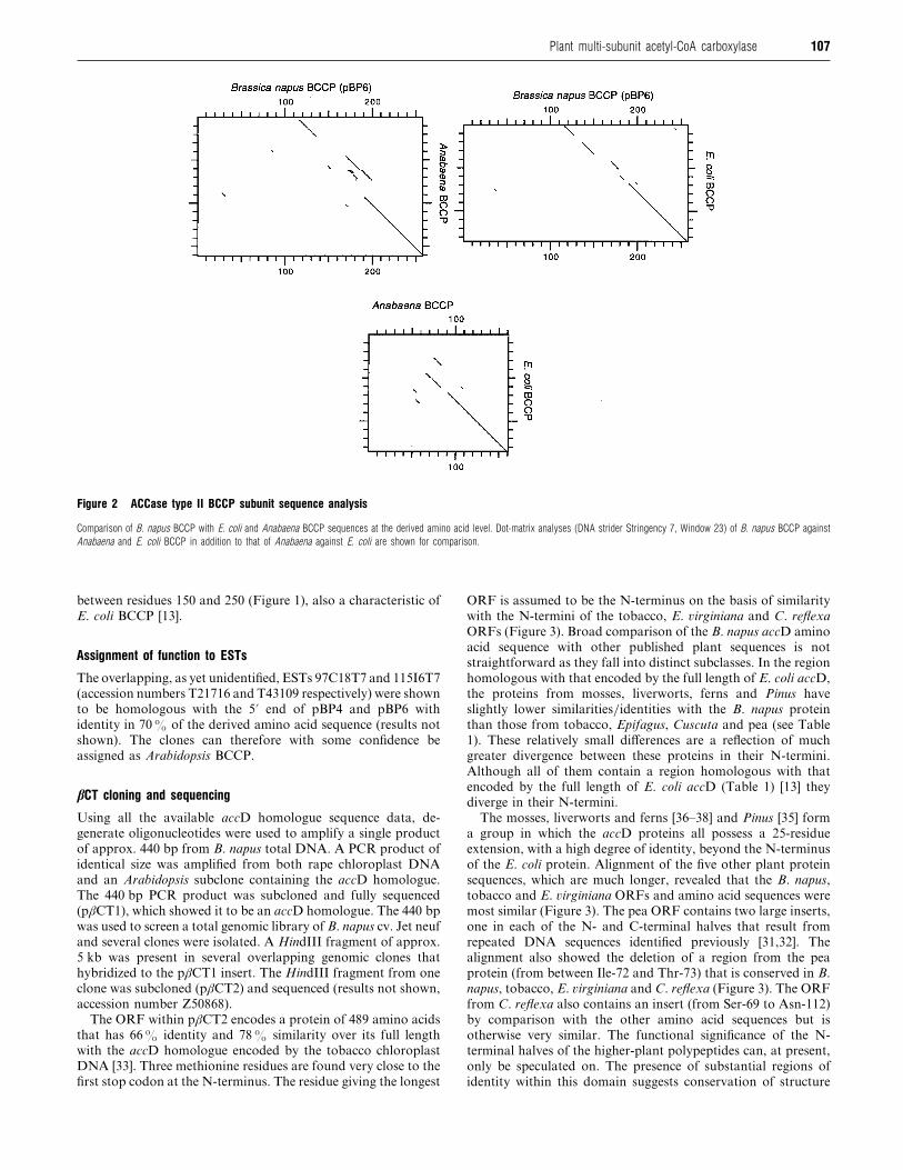
longest clone (pBP6) with that of Anabaena and E. coli BCCPs
shows that the similarity between the proteins is primarily at
their N- and C-termini (Figure 2). A comparison of Anabaena
and E. coli BCCPs is also presented pictorially to demonstrate
the known divergences (Figure 2). The direct amino acid GAP
comparison (BBSRC Computing Facility, Daresbury) with the
E. coli BCCP shows similarity throughout the length of the E.
coli protein with the longest continuous stretch occurring around
the biotinylation site (Figure 2). It has been shown previously
106 K. M. Elborough and others
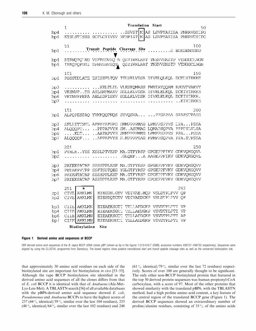
Figure 1 Derived amino acid sequences of BCCP
ORF-derived amino acid sequences of the B. napus BCCP cDNA clones pBP (shown as bp in the figure) 1/2/3/4/6/7 (EMBL accession numbers X90727–X90732 respectively). Sequences were
aligned by using the CLUSTAL programme from Daresbury. The boxed regions show putative translational start and transit peptide cleavage sites as well as the conserved biotinylation site.
that approximately 30 amino acid residues on each side of the
biotinylated site are important for biotinylation in �i�o [53–55].
Although the rape BCCP biotinylation site identified in the
derived amino acid sequences of all the clones differs from that
of E. coli BCCP it is identical with that of Anabaena (Ala-Met-
Lys-Leu-Met). A TBLASTN search [56] of all available databases
with the pBP6-derived amino acid sequence showed E. coli,
Pseudomonas and Anabaena BCCPs to have the highest scores of
237 (44% identical}59% similar over the last 104 residues), 255
(48% identical}64% similar over the last 102 residues) and 248
(61% identical}79% similar over the last 72 residues) respect-
ively. Scores of over 100 are generally thought to be significant.
The only other non-BCCP biotinylated protein that featured in
the top 50 derived protein sequences was human propionyl-CoA
carboxylase, with a score of 97. Most of the other proteins that
showed similarity with the translated pBP6, with the TBLASTN
method, had a high proline amino acid content, a key feature of
the central region of the translated BCCP gene (Figure 1). The
derived BCCP sequences showed an extraordinary number of
proline}alanine residues, consisting of 35% of the amino acids
107Plant multi-subunit acetyl-CoA carboxylase
Figure 2 ACCase type II BCCP subunit sequence analysis
Comparison of B. napus BCCP with E. coli and Anabaena BCCP sequences at the derived amino acid level. Dot-matrix analyses (DNA strider Stringency 7, Window 23) of B. napus BCCP against
Anabaena and E. coli BCCP in addition to that of Anabaena against E. coli are shown for comparison.
between residues 150 and 250 (Figure 1), also a characteristic of
E. coli BCCP [13].
Assignment of function to ESTs
The overlapping, as yet unidentified, ESTs 97C18T7 and 115I6T7
(accession numbers T21716 and T43109 respectively) were shown
to be homologous with the 5« end of pBP4 and pBP6 with
identity in 70% of the derived amino acid sequence (results not
shown). The clones can therefore with some confidence be
assigned as Arabidopsis BCCP.
βCT cloning and sequencing
Using all the available accD homologue sequence data, de-
generate oligonucleotides were used to amplify a single product
of approx. 440 bp from B. napus total DNA. A PCR product of
identical size was amplified from both rape chloroplast DNA
and an Arabidopsis subclone containing the accD homologue.
The 440 bp PCR product was subcloned and fully sequenced
(pβCT1), which showed it to be an accD homologue. The 440 bp
was used to screen a total genomic library of B. napus cv. Jet neuf
and several clones were isolated. A HindIII fragment of approx.
5 kb was present in several overlapping genomic clones that
hybridized to the pβCT1 insert. The HindIII fragment from one
clone was subcloned (pβCT2) and sequenced (results not shown,
accession number Z50868).
The ORF within pβCT2 encodes a protein of 489 amino acids
that has 66% identity and 78% similarity over its full length
with the accD homologue encoded by the tobacco chloroplast
DNA [33]. Three methionine residues are found very close to the
first stop codon at the N-terminus. The residue giving the longest
ORF is assumed to be the N-terminus on the basis of similarity
with the N-termini of the tobacco, E. �irginiana and C. reflexa
ORFs (Figure 3). Broad comparison of the B. napus accD amino
acid sequence with other published plant sequences is not
straightforward as they fall into distinct subclasses. In the region
homologous with that encoded by the full length of E. coli accD,
the proteins from mosses, liverworts, ferns and Pinus have
slightly lower similarities}identities with the B. napus protein
than those from tobacco, Epifagus, Cuscuta and pea (see Table
1). These relatively small differences are a reflection of much
greater divergence between these proteins in their N-termini.
Although all of them contain a region homologous with that
encoded by the full length of E. coli accD (Table 1) [13] they
diverge in their N-termini.
The mosses, liverworts and ferns [36–38] and Pinus [35] form
a group in which the accD proteins all possess a 25-residue
extension, with a high degree of identity, beyond the N-terminus
of the E. coli protein. Alignment of the five other plant protein
sequences, which are much longer, revealed that the B. napus,
tobacco and E. �irginiana ORFs and amino acid sequences were
most similar (Figure 3). The pea ORF contains two large inserts,
one in each of the N- and C-terminal halves that result from
repeated DNA sequences identified previously [31,32]. The
alignment also showed the deletion of a region from the pea
protein (from between Ile-72 and Thr-73) that is conserved in B.
napus, tobacco, E. �irginiana and C. reflexa (Figure 3). The ORF
from C. reflexa also contains an insert (from Ser-69 to Asn-112)
by comparison with the other amino acid sequences but is
otherwise very similar. The functional significance of the N-
terminal halves of the higher-plant polypeptides can, at present,
only be speculated on. The presence of substantial regions of
identity within this domain suggests conservation of structure

108 K. M. Elborough and others
Figure 3 ACCase type II β-carboxyltransferase subunit sequence analysis
Amino acid alignment of the N-terminal halves of the predicted higher-plant polypeptides with similarity to the βCT subunit of ACCase [40]. Sequences were aligned by using the PILEUP and
LINEUP programs and were taken from pea [31], tobacco [42], Epifagus virginiana [4] and Cuscuta reflexa (G. Haberhausen ; EMBL accession number X69803). Identical residues are boxed.
and}or function. A search of the translation products from the
EMBL Database with the conserved polypeptide motifs found
within this region failed to identify any significant relationship
with any other polypeptides. B. napus βCT is exceptional within
the C-terminal region of the accD homologues. The zinc-finger
motif previously identified [31,32] is found as CXXCX"&
CXXC
(where X is any amino acid) in all sequences, including that of E.
coli, except for B. napus where the central domain contained only
12 residues (Figure 3). Apart from this difference and the insert
present in pea, the C-terminal halves of all the accD homologues
are remarkably similar (Table 1).
Northern blot analysis of BCCP and βCT expression
Storage lipid synthesis occurs in B. napus seed with a maximal
rate of production during the middle stage of embryogenesis.
Turnham and Northcote [56] showed that ACCase enzymatic
activity in rape embryos followed the rate of lipid deposition;
more recently Kang et al. [57], using different extraction tech-
niques, showed that ACCase activity increased before lipid
deposition. In both cases the activity decreased before the
maximal accumulation of lipid occurred. To study the mRNA
expression of the BCCP genes, which are thought to be involved
in de no�o storage lipid synthesis, we screened a developmental
Northern blot. Each lane on the blot had an equal amount of
root, leaf or embryo poly(A)+ mRNA from different stages of B.
napus embryogenesis. The blot shown in Figure 4A was generated
by screening with the pBP7 insert and was identical when all six
cDNA types were used on separate occasions (results not shown).
During embryogenesis, BCCP mRNA expression (1.2 kb full
length) rose steeply, peaking during the middle portion of
embryogenesis. Maximal mRNA expression occurred at 54 d,
after which the level of BCCP mRNA dropped markedly. This
profile mimicked that of the other B. napus fatty acid synthesis
components enoyl reductase, β-keto reductase and ACCase I
[20]. Although there was a notable amount of BCCP mRNA
present in root there was a comparatively low level in leaf. The
integrated phosphoimager data showed that the level of BCCP
mRNA expression in 54-day embryos is 26 times that of leaves,
which is itself only twice that of background.

109Plant multi-subunit acetyl-CoA carboxylase
Table 1 Comparison of the predicted amino acid sequence from B. napuswith those of all other published sequences with similarity to the βCT subunitof ACCase [40]
The comparison was made by using the BESTFIT program and is restricted to residues
220–489 of the B. napus sequence. To enable sensible comparison the insert in the pea
sequence (residues 234–394) has been deleted. Sequences are taken from the references
described in Figure 3 and also : Angiopteris lygodiifolia [38], Marchantia polymorpha [36],
Physcomitrella patens [37] and Black pine [35].
B. napus
Species βCT identity (%) βCT similarity (%)
Tobacco 79 86
E. virginiana 77 87
C. reflexa 72 83
Pea 68 81
M. polymorpha 69 79
Black pine 67 77
P. patens 66 76
A. lygodiifolia 66 76
E. coli 41 62
The B. napus chloroplastic genome-encoded βCT subunit was
used in a Northern blot against total RNA from the same batch
of whole seeds used for the BCCP Northern data (see Figure 4A).
The cDNA insert hybridized to several bands of 2.4, 1.6, 1.3 and
1.1 kb (Figure 4B), which were expressed proportionally through-
out embryogenesis, the strongest association being with a band
of approx. 2.4 kb. Because all the bands are proportional we
made the assumption that they all contain the βCT gene.
Northern analysis of pea βCT was complicated in a similar way
Figure 4 Northern blot analysis of ACCase type II BCCP and βCT subunit expression during rapeseed embryogenesis
(A) Northern analysis with the BCCP cDNA (pBP7 insert) as a probe : 1 µg of poly(A)+ RNA from embryos 38, 44, 54, 57 and 68 d after anthesis and from root and young leaf was used for
the blot. Hybridization and washing conditions were as described in the Materials and methods section. Phosphorimaging screens were exposed for 2 h. Molecular mass markers were revealed
by ethidium bromide/UV. (B) Northern analysis with the rape β-carboxyltransferase domain DNA : 10 µg of total RNA from the same batch of embryos as in (A) was used for the blot. Hybridization
and washing conditions were the same as in (A). The hybridizing bands were revealed by autoradiography with an exposure time of 7 d.
as a result ofmultiple transcription start sites and co-transcription
with genes both 5« and 3« [31,39]. Nagano et al. [31] showed that
three transcripts with sizes of 5.0, 2.8 and 2.4 kb contained the
entire ORF of accD and ORFs of both trnQ and psaI genes. The
blot also shows (Figure 4B) that in leaf material there is one extra
hybridizing RNA species not present in embryo tissue (2.7 kb)
and a band visible in embryos (2.6 kb) not present in leaves.
It can be seen from a comparison of Figures 4A and 4B that
the rape βCT mRNA expression was very similar to that of
BCCP during embryogenesis. Because both proteins are thought
to associate in �i�o to form part of the type II ACCase complex,
this was perhaps to be expected. The peak of expression occurred
at 54 d after anthesis, followed by the characteristic decrease in
expression. However, in contrast with BCCP the relative amount
of βCT expression in leaf was higher with an embryo: leaf ratio
of 7:2.
Anti-biotin Western blot of rape embryos
To gain a real insight into BCCP expression it was important to
study protein levels as well as mRNA. In the absence of specific
BCCP antibodies, and because BCCP is biotinylated in �i�o,
biotin-specific antiserum was used for this purpose. To generate
biotin-specific antibodies, keyhole limpet haemocyanin was
coated with biotin and used as an antigen in rabbits. To minimize
false background signals in a Western blot the antibodies
generated were affinity-purified. This was achieved by column
chromatography on a biotin-agarose matrix. The generated
antibodies were more specific and had a higher titre than those
available commercially.
Previous work describing the prokaryotic BCCP studies
showed that the proline}alanine-rich region of E. coli BCCP
confers an SDS gel anomaly [13]. The protein runs at 35% less
110 K. M. Elborough and others
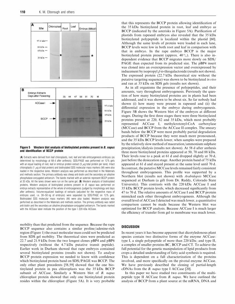
Figure 5 Western blot analysis of biotinylated proteins present in B. napusand identification of BCCP protein
(A) Extracts were derived from leaf chloroplasts, root, leaf and late embryogenesis embryos (as
determined by morphology at 68 d after anthesis). SDS/PAGE was performed on 12% gels
with an equal loading of root, leaf or embryo protein extract (5 µg total protein per lane). Intact
chloroplasts (1 µg of total protein) and biotinylated SDS molecular mass markers (M) were also
loaded in the respective lanes. Western analysis was performed as described in the Materials
and methods section. The primary antibody was sheep anti-biotin and the secondary an alkaline
phosphatase-conjugated antiserum. The bands marked with an asterisk represent BCCP protein
at 35 kDa. All the lanes shown were run on the same gel. (B) Western analysis of biotinylated
proteins. Western analysis of biotinylated proteins present in B. napus was performed on
embryo extracts representative of the whole of embryogenesis (judged by morphology and days
after anthesis). Volume-equalized loadings of extracts (adjusted for the respective mass of
embryos used, i.e. 30–50 ng of embryos) were separated by SDS/PAGE on 12% gels.
Biotinylated SDS molecular mass markers (M) were also loaded. Western analysis was
performed as described in the Materials and methods section. The primary antibody was rabbit
anti-biotin and the secondary an alkaline phosphatase-conjugated antiserum. The bands marked
with the ACCase label indicate the position of the type I 220 kDa ACCase.
mobility than that predicted from the sequence. Because the rape
BCCP sequence also contains a similar proline}adenine-rich
region (Figure 1) the exact molecular mass could not be predicted
from SDS gel mobility. The theoretical sizes were predicted as
22.7 and 21.9 kDa from the two longest clones pBP4 and pBP6
respectively (without the 4.7 kDa putative transit peptide).
Earlier work in Durham showed that rape embryos contained
several biotinylated proteins (results not shown). To analyse
BCCP protein expression we needed to know with confidence
which biotinylated protein band on SDS}PAGE was BCCP. The
only other plant precedents [58,59] showed that the one bio-
tinylated protein in pea chloroplasts was the 35 kDa BCCP
subunit of ACCase. Similarly a Western blot of B. napus
chloroplast protein showed that only one biotinylated protein
resides within the chloroplast (Figure 5A). It is very probable
that this represents the BCCP protein allowing identification of
the 35 kDa biotinylated protein in root, leaf and embryo as
BCCP (indicated by the asterisks in Figure 5A). Purification of
plastids from rapeseed embryos also revealed that the 35 kDa
biotinylated polypeptide is localized within the plastid [60].
Although the same levels of protein were loaded in each lane,
BCCP levels were low in both root and leaf in comparison with
that in embryo. In the rape embryo BCCP is the major
biotinylated protein present (approx. 40%). There is also in-
dependent evidence that BCCP migrates more slowly on SDS}PAGE than expected from its predicted size. The pBP4 insert
was cloned into an overexpression vector and overexpressed by
inducement by isopropyl β--thiogalactoside (results not shown).
The expressed protein (22.7 kDa theoretical size without the
putative targeting sequence) was shown to be biotinylated in �i�o
and ran at 33 kDa on SDS gels (results not shown).
As in all organisms the presence of polypeptides, and their
amounts, vary throughout embryogenesis. Previously the ques-
tion of how many biotinylated proteins are in plants had been
addressed, and it was shown to be about six. So far nobody had
shown (i) how many were present in rapeseed and (ii) the
differential expression in the embryo during embryogenesis.
Figure 5B shows the Western blot of the embryos at different
stages. During the first three stages there were three biotinylated
proteins present at 220, 82 and 35 kDa, which most probably
represented ACCase I, methylcrotonyl-CoA carboxylase
(MCCase) and BCCP from the ACCase II complex. The smeary
bands below the BCCP were most probably partial degradation
products of BCCP because they were much more pronounced,
and the 35 kDa BCCP levels lower, when samples were prepared
by the relatively slow method of maceration}ammonium sulphate
precipitation}dialysis (results not shown). At 58 d after anthesis
three more biotinylated proteins appeared at 50, 70 and 88 kDa.
Their levels rose to a peak at 61 d and dropped slightly at 70 d,
just before the desiccation stage. Another protein band of 75 kDa
appeared at 61 d and stayed present at the same level until 70 d.
In contrast, the putativeMCCase levels remained fairly consistent
throughout embryogenesis. This profile was supported by a
Northern blot (results not shown) with Arabidopsis MCCase
sequenced at Durham (a gift from Dr. Reddy, Colorado State
University). This contrasts with the 220 kDa ACCase I and
35 kDa BCCP protein levels, which decreased significantly from
47 to 70 d. The relative amounts of ACCase I and BCCP protein
mimicked each other throughout embryogenesis. Although the
overall level of ACCase I detected was much lower, a quantitative
comparison cannot be made because the Western blot was
optimized for BCCP analysis. Because ACCase I is much larger
the efficiency of transfer from gel to membrane was much lower.
DISCUSSION
In recent years it has become apparent that dicotyledonous plant
species contain two distinctive forms of the enzyme ACCase:
type I, a single polypeptide of more than 220 kDa; and type II,
a complex of smaller proteins BC, BCCP and CT. To achieve the
full potential for the genetic manipulation of lipid products from
crops a detailed understanding of fatty acid synthesis is required.
This is dependent on a full characterization of the proteins
involved, and more specifically on the pivotal enzyme ACCase.
We have previously described the cloning of partial-length
cDNAs from the B. napus type I ACCase [20].
In this paper we have studied two constituents of the multi-
peptide type II ACCase from B. napus. We have outlined the
analysis of BCCP from a plant source at the mRNA, DNA and
111Plant multi-subunit acetyl-CoA carboxylase
protein level and have described the cloning of several cDNAs.
In addition we have described the first cloning and character-
ization of the chloroplast-encoded B. napus βCT cDNA. We
isolated six distinct BCCP clones, of which two contained full-
length ORFs encoding theoretical proteins of 22.7 and 21.9 kDa.
The B. napus BCCP is therefore derived from a multi-gene family
of at least six members. The size of the coding region was
significantly greater than that of E. coli or Anabaena but
correlated with the Northern data showing the full length to be
1.2 kb. On analysis of the pBP4 and pBP6 sequence the cDNAs
revealed that they contained a putative 4.7 kDa 40 amino acid
transit peptide at the 5« end. The UTR and ORF sequence
similarity showed that the cDNAs fall loosely into two different
groups, group 1 (pBP1}2) and group 2 (pBP3}4}6}7). All six
showed a strong degree of similarity to each other (at the 3« ends
of the coding regions) around the biotinylation site. This simi-
larity also extended to the known prokaryotic BCCP sequences
but showed no similarity to type I ACCase sequences. The
biotinylation site, although divergent from that of E. coli BCCP,
is identical with that for the Anabaena BCCP. The divergent 3«UTRs of BCCP will permit the isolation of the corresponding six
different promoter sequences. This study has shown that temporal
expression is highly regulated and almost identical with other
fatty acid synthesis genes that we have isolated. Using a
comparison of all the promoter sequences from BCCP, and other
fatty acid synthesis genes available in our laboratory, we might
be able to identify specific sequences representing binding sites
for the key time-specific transcription factors that control their
expression.
By using the cDNAs obtained for two components of type II
ACCase we were able to compare their expression directly at the
RNA level. The βCT and BCCP mRNA expression levels during
embryogenesis were very similar. Previous work has shown that
rape type I ACCase and several other lipid synthesis genes are
expressed in a very similar manner [20]. However, the Northern
analysis of the βCT was complicated by the presence of several
hybridizing bands. This was also observed by Sasaki et al. in
their analysis of pea leaf βCT [42]. Given that the βCT primary
transcript size is 2.4 kb in B. napus and yet the ORF is less than
1.5 kb long, there may be another ORF on the transcript. In the
chloroplast DNAs of several other species such as pea the accD
gene is followed closely (by less than 180 bp) by the psaI gene,
which has a very small ORF (120 bp in pea [39]). As in pea there
may be several transcription start signals at the 5« end of the
ORFs that may all be used, giving rise to several bands seen on
the Northern analysis.
The Western blot data that we present here show that rape leaf
chloroplasts contain the biotinylated 35 kDa BCCP protein,
which in turn implies that the putative transit peptide sequence
is chloroplastic. In addition because leaf-to-embryo BCCP pro-
tein ratios are higher than that for mRNA we can only assume
that either protein turnover is much greater in embryos or the
translational control is much less prevalent in the leaf. The
protein was larger on Western analysis than the theoretical size
expected but the cDNA sequence showed that, like E. coli BCCP,
the central region was rich in alanine and proline. This particular
sequence characteristic has been shown previously to reduce the
mobility of proteins on SDS}PAGE, i.e. the E. coli BCCP runs
as a 22 kDa band in contrast with its 17 kDa theoretical size. The
rape BCCP similarity to E. coli BCCP also extends to the E. coli
BCCP N-terminus. It has been shown before that the N-terminal
region represents the site for protein–protein interaction [61],
whereas the proline–alanine central region is thought to act as a
flexible arm to allow the carboxy group to move between subunit
active sites. The BCCP genes will allow the construction of
antisense constructs to study the function of type II ACCases in
�i�o. We have already generated antisense type I ACCase
transgenic rape plants, which in combination with BCCP anti-
sense plants will allow us to differentiate between the roles of
both types of ACCase within dicotyledonous species.
Using the anti-biotin antibodies generated for this study we
analysed all the biotinylated proteins during rapeseed devel-
opment. The major biotinylated protein in the embryo was the
BCCP, which showed expression correlating to its mRNA
expression. This is in marked contrast with that in pea where the
BCCP has not been detected in the embryo [62]. This may reflect
the different major storage products used by the two plants. Peas
have only 2% of their mass as oil whereas rape has approx.
45%. With embryo morphology as a marker, the peak of BCCP
expression occurred simultaneously to that of maximal lipid
synthesis, after which expression was markedly reduced. We
obtained enough affinity-purified antibody to generate an anti-
biotin immunochromatography column. This could be used to
eventually purify the BCCP from seed extracts for protein
analysis.
Several other biotinylated proteins were identified, some of
which can be tentatively attributed to type I ACCase and
MCCase. Without functional analysis the identification of the
other proteins was impossible but previous work detected
pyruvate carboxylase activity in embryo extracts, a biotinylated
enzyme (results not shown). Some of the bands might also
represent the rape equivalent of the biotin-binding protein in pea
embryos [62] and geranoyl-CoA carboxylase recently partly
purified from maize [63].
Recent research has isolated the biotin carboxylase [49] and
previous research the β-carboxyltransferase subunit clones
[31–38,42] from different plant sources. In this report we have
described the first isolation of plant full-sized BCCP and in
addition a βCT clone from the same species. With the isolation
of the α-carboxyltransferase subunit and the purification of
expressed gene products the stage will be set for the full
characterization of type II ACCase from plants at the protein
level. Because the subunits are thought to form a complex in �i�o
by protein–protein interactions it may be important to study the
subunits from the same plant source. In isolating two of the three
or four subunit types we have gone some way to achieving this
in the agriculturally relevant crop B. napus.
We thank Jane Bird at Zeneca Pharmaceuticals for generation of antibodies, MartaEvans for the gift of intact chloroplasts and Dr. Wolfgang Schuster (IGF, Berlin) forthe gift of plasmid psbLa. This work was supported under the AFRC Link scheme‘Crops for industrial use ’ project no. E15 (Durham) and through BBSRC grant-in-aidto J. I. C. R.W. was supported by SERC grant no. GR-H06910 and R.K.D. by BBSRCgrant no. PG208/623 (PMB).
REFERENCES
1 Slabas, A. R. and Fawcett, T. (1992) Plant Mol. Biol. 19, 169–191
2 Post Beittenmiller, D., Roughan, G. and Ohlrogge, J. B. (1992) Plant Physiol. 100,923–930
3 Gronwald, J. W. (1994) Biochem. Soc. Trans. 22, 616–621
4 Walker, K. A., Ridley, S. M., Lewis, T. and Harwood, J. L. (1990) Phytochemistry 29,3743–3747
5 Samols, D., Thornton, C. G., Mrtif, V. L., Kumar, G. K., Haase, C. and Wood, H. G.
(1988) J. Biol. Chem. 263, 6461–6464
6 Al-Feel, W., Chirala, S. S. and Wakil, S. J. (1992) Proc. Natl. Acad. Sci. U.S.A. 89,4534–4538
7 Lopez-Casillas, F., Bai, D.-H, Luo, X., Kong, I.-S., Hermodson, M. S. and Kim, K. H.
(1988) Proc. Natl. Acad. Sci. U.S.A. 85, 5784–5788
8 Takai, T., Yokoyama, C., Wada, K. and Tanabe, T. (1988) J. Biol. Chem. 263,2651–2657
9 Chandler, C. S. and Ballard, F. (1988) Biochem. J. 251, 749–755
10 Fall, R. R. (1979) Methods Enzymol. 67, 390–398

112 K. M. Elborough and others
11 Guchhait, R. B., Polakis, S. E., Dimroth, P., Stoll, E., Moss, J. and Lane, M. D. (1974)
J. Biol. Chem. 249, 6633–6645
12 Li, S.-J. and Cronan, J. E. (1992) J. Biol. Chem. 267, 16841–16847
13 Li, S.-J. and Cronan, J. E. (1992) J. Biol. Chem. 267, 855–863
14 Wurtele, E. S. and Nikolau, B. J. (1990) Arch. Biochem. Biophys. 278, 179–186
15 Ashton, A. R., Jenkins, C. L. D. and Whitfeld, P. R. (1994) Plant. Mol. Biol. 24,35–49
16 Bettey, M, Ireland, R. J. and Smith, A. M. (1992) J. Plant Physiol. 140, 513–520
17 Charles, D. J. and Cherry, J. H. (1986) Phytochemistry 25, 1067–1071
18 Egin-Bu$ hler, B. and Ebel, J. (1983) Eur. J. Biochem. 133, 335–339
19 Egin-Bu$ hler, B., Loyal, R. and Ebel, J. (1980) Arch. Biochem. Biophys. 203, 90–100
20 Elborough, K. M., Simon, J. W., Swinhoe, R., Ashton, A. R. and Slabas, A. R. (1994)
Plant Mol. Biol. 24, 21–34
21 Gornicki, P. and Haselkorn, R. (1993) Plant Mol. Biol. 22, 547–552
22 Hellyer, A., Bambridge, H. E. and Slabas, A. R. (1986) Biochem. Soc. Trans. 14,565–568
23 Roessler, K. R., Shorrosh, B. S. and Ohlrogge, J. B. (1994) Plant Physiol. 105,611–617
24 Roessler, P. G. and Ohlrogge, J. B. (1993) J. Biol. Chem. 268, 19254–19259
25 Shorrosh, B. S., Roesler, K. R., Shintani, D., van de Loo, F. J. and Ohlrogge, J. B.
(1995) Plant Physiol. 108, 805–812
26 Kannagara, C. G. and Stumpf, P. K. (1972) Arch. Biochem. Biophys. 152, 83–91
27 Kannagara, C. G. and Jensen, C. J. (1975) Eur. J. Biochem. 54, 25–30
28 Mohan, S. B. and Kekwick, R. G. O. (1980) Biochem. J. 187, 667–676
29 Reitzel, L. and Nielsen, N. C. (1976) Eur. J. Biochem. 65, 131–138
30 Slabas, A. R. and Hellyer, A. (1985) Plant Sci. 39, 177–182
31 Nagano, Y., Matsuno, R. and Sasaki, Y. (1991) Curr. Genet. 20, 431–436
32 Smith, A. G., Wilson, R. M., Kaethner, T. M., Willey, D. L. and Gray, J. C. (1991)
Curr. Genet. 19, 403–410
33 Shinozaki, K., Ohme, M., Tanaka, M., Wakasugi, T., Hayashida, N., Matsubayashi, T.,
Zaita, N., Chunwongse, J., Obokata, J., Yamaguchi-Shinozaki, K., Ohto, C., Torazawa,
Meng, B. Y., Sugati, M., Deno, H., Kamogashira, T., Yamada, K., Kusuda, J., Takaiwa,
F., Kato, A., Tohdoh, N., Shimada, H. and Sugiura, M. (1986) EMBO J. 5,2043–2049
34 Wolfe, K. H., Morden, C. W. and Palmer, J. D. (1992) Proc. Natl. Acad. Sci. U.S.A.
89, 10648–10652
35 Tsudzuki, J., Nakashima, K., Tsudzuki, T., Hiratsuka, J., Shibata, M., Wakasugi, T.
and Sugiura, M. (1992) Mol. Gen. Genet. 232, 206–214
36 Ohyma, K., Fukuzawa, H., Kohchi, T., Shirai, H., Sano, T., Sano, S., Umesono, K.,
Shiki, Y., Takeuchi, M., Chang, Z., Aota, S. I., Inokuchi, H. and Ozeki, H. (1986)
Nature (London) 322, 572–574
Received 18 August 1995/13 November 1995 ; accepted 16 November 1995
37 Kasten, B., Wehe, M., Reski, R. and Abel, W. O. (1991) Nucleic Acids Res. 19, 5074
38 Yoshinaga, K., Kubota, Y., Ishii, T. and Wada, K. (1992) Plant Mol. Biol. 18, 79–82
39 Sasaki, Y., Nagano, Y., Morioka, S., Ishikawa, H. and Matsuno, R. (1989) Nucleic
Acids Res. 17, 6217–6227
40 Li, S.-J. and Cronan, J. E. (1992) Plant Mol. Biol. 20, 759–761
41 Alban, C., Baldet, P. and Douce, R. (1994) Biochem. J. 300, 557–565
42 Sasaki, Y., Kazuhiko, H., Yukiko, S., Yukio, N., Iwao, F. and Ryuichi, M. (1993) J.
Biol. Chem. 268, 25118–25123
43 Ebel, J. and Hahlbrock, K. (1977) Eur. J. Biochem. 75, 201–209
44 Shorrosh, B. S., Dixon, R. A. and Ohlrogge, J. B. (1994) Proc. Natl. Acad. Sci. U.S.A.
91, 4323–4327
45 Ohlrogge, J. B., Kuhn, D. N. and Stumpf, P. K. (1979) Proc. Natl. Acad. Sci. U.S.A.
76, 1194–1198
46 Newman, T., Debruijn, F. J., Green, P., Keegstra, K., Kende, H., Mcintosh, L.,
Ohlrogge, J., Raikhel, N., Somerville, S., Thomashow, M., Retzel, E. and Somerville,
C. (1994) Plant Physiol. 106, 1241–1255
47 Slabas, A. R., Simon, J. W. and Elborough, K. M. (1995) Inform 6, 159–166
48 Smith, E., White, K. A., Holt, D. S., Fentem, P. A. and Bright, S. W. J. (1994) in
Abstracts of the 4th International Congress on Plant Molecular Biology. Abstract
1955
49 Logemann, J., Schell, J. and Willmitzer, L. (1987) Anal. Biochem. 163, 16–20
50 Marck, C. (1988) Nucleic Acids Res. 16, 1829–1836
51 Lu$ tcke, H. A., Chow, K. C., Mickel, F. S., Moss, K. A., Kern, H. F. and Scheele, G. A.
(1987) EMBO J. 6, 43–48
52 von Heijne, G. (1986) Nucleic Acids Res. 14, 4683–4690
53 Cronan, J. E. (1990) J. Biol. Chem. 265, 10327–10333
54 Murtif, V. L. and Samols, D. (1987) J. Biol. Chem. 262, 11813–11816
55 Reed, K. E. and Cronan, J. E. (1991) J. Biol. Chem. 266, 11425–11428
56 Altschul, S. F., Gish, W., Miller, W., Myers, E. W., Lipman, D. J. (1990) J. Mol. Biol.
215, 403-410
57 Turnham, E. and Northcote, D. H. (1983) Biochem. J. 212, 223–229
58 Kang, F., Ridout, C. J., Morgan, C. L. and Rawsthorne, S. (1994) Planta 193,320–325
59 Konishi, T. and Sasaki, Y. (1994) Proc. Natl. Acad. Sci. U.S.A. 91, 3598–3601
60 Kang, F. (1995) Ph.D. Thesis, University of East Anglia, Norwich.
61 Fall, R. R. and Vagelos, P. R. (1973) J. Biol. Chem. 248, 2078–2088
62 Duval, M., Job, C., Alban, C., Douce, R. and Job, D. (1994) Biochem. J. 299,141–150
63 Caffrey, J. J., Chen, Y., Diez, T., Guan, X., Huang, J.-Y., McKean, A. L., Song, J.,
Shang, X., Wang, X., Weaver, L. M., Wurtele, E. S. and Nikolau, B. J. (1995) in Plant
Lipid Metabolism (Kader, J.-C. and Mazliak, P., eds.), pp. 49–51, Kluwer Academic
Publishers, Netherlands