
BioTek Synergy HT Multi-Mode Microplate Reader -...
17
BioTek Synergy HT Multi-Mode Microplate Reader Bergen County Technical Schools Biotechnology Lab Room 224A
Transcript of BioTek Synergy HT Multi-Mode Microplate Reader -...

BioTek Synergy HT
Multi-Mode Microplate Reader
Bergen County Technical Schools Biotechnology Lab
Room 224A

BioTek Synergy HT Multi-Mode Microplate Reader Information Sheet
The BioTek Synergy HT Multi-Mode Microplate Reader is a single-channel
absorbance, fluorescence, and luminescence microplate reader for research and
development and in vitro diagnostic use. The instrument essentially functions as a
spectrophotometer, which measures intensity as a function of the color, or more
specifically, the wavelength of light. The instrument has applications that would provide
useful for multiple disciplines, from Biology, to Chemistry and Nanotechnology.
The Synergy HT supports a wide range of labware, with many common
microplate formats already preconfigured and ready for use in the software, ranging
from 6 well to 384 well plates. .
The Gen5 Software configures and controls all measurement, protocols, and
actions performed by the Synergy HT. The software supports detection for absorbance,
luminescence, fluorescence intensity, fluorescence polarization, and time-resolved
fluorescence (TRF) measurements. This instrument has been suggested to be used for
studies involved with apoptosis, fluorescent molecule binding, cell proliferation and
viability, immunoassays (such as ELISA).
The software allows the user to establish unique protocols to analyze different
sample plates. Samples are analyzed in the instrument, and the results stored on the
computer’s hard drive. The data from the experiments are able to be viewed and
analyzed in the Gen5 program, as well as in Excel by exporting the data.

1. Turn on the BioTek Synergy HT Multi-Mode Microplate Reader by
BioTek Synergy HT Multi-Mode Microplate Reader Quick Start Guide
flipping up the Power Switch on the front of the instrument.
a. The light on the power switch will turn green.
b. Allow the instrument to perform its start-up procedure before
proceeding. When the instrument stops making noise, it is
ready to use.
2. On the desktop, double click the Gen5 Icon.
1. Open the Filter Panel on the front of the instrument (see Instrument Info Sheet).
Changing Filters
Cartridges containing the filters needed for all absorbance and fluorescence
experiments should be left in the instrument. The filter cartridge only needs to be
exchanged for luminescence experiments. Changing filters should only be done under
the supervision of the instructor.
Note: DO NOT open the panel or change filters if the power is on.
2. Unscrew the Emission Filter Cartridge and remove it from the instrument.
3. Take the Blank Cartridge, and insert the Blocker Plug into filter space 1. Lock it
in place with the provided C-clip Filter Retainer (see Instrument Info Sheet).
4. Insert the blank cartridge with the blocker into the instrument, and tighten the
screw to lock it in place.
5. In Gen5, go to SystemReader Configuration, highlight the Synergy
Reader, and click the View/Modify Button.

6. On the next screen, click the Setup Button.
7. Click the Fluorescence / Luminescence Tab.
a. Write down the values that are found under Emission. These will need to
be replaced after the protocol is run.
b. Under Center, change the value of Filter 1 to Plug, and the rest to Hole.
c. Click Send Values to send the information to the instrument.
8. After the protocol is run on the microplates, and the instrument is shut down,
remove the Blank Cartridge from the instrument and replace it with the
Emission Filter Cartridge.
Note: DO NOT open the panel or change filters if the power is on.
9. Repeat Steps 5 - 7 and replace the original values from the Emission Filter.
1. The Gen5 program will open to a blank screen. From this
Setting up a Protocol
screen, click File New Protocol on the Tool Bar to set
up a new protocol for an experiment.
Note: If a protocol is already made for the
experiment, proceed to the section Running a Protocol on a Sample Plate.

2. A screen will appear with icons along the Tool Bar, and a list of options along the
left side.
3. Double click Procedure on the list, or click the
Procedure Icon on the Tool Bar.
4. In the new screen, the procedure for the microplate will be created.
a. Select Plate Type from the
Dropdown Menu, and select the
type of plate being used for the experiment.
b. Determine if the plate needs to be shaken prior to reading, if the
temperature needs to be set for the instrument, or if multiple readings
need to be taken over a period of time.
i. Clicking the Shake Button brings up a screen that allows a shake
step to be added. Click OK.

ii. Clicking the Delay Button delays the run prior to
reading or performing another action. Click OK.
iii. Clicking the Kinetic Button allows multiple readings to be taken
over a period of time (determined by the user). Click OK.
iv. Clicking the Set Temperature Button allows the internal
temperature of the instrument to be adjusted for the read. Click OK.
c. Click the Read Button to enter the specific information about the type of
read that is to be performed on the plate.

i. In Detection Method, select if
Absorbance, Fluorescence, or
Luminescence should be used on
the plate.
Note: The following options will vary slightly depending on
the method that is chosen.
ii. Select the Read type that should be performed.
iii. Select the Number of Wavelengths or Filter Sets that should be
used, and select the wavelengths to be used.
Note: Use this if the samples in the plate should be
compared using two or more different absorbance or
fluorescence settings.
iv. Click OK.
d. The steps may be reordered in the procedure by
clicking and dragging one step above or below
another step in the list.

e. To ensure that the instrument can perform the
selected procedure, click Validate.
i. If the procedure is ok, the program says
that the procedure is valid.
ii. If there is a problem with the order of
the procedure, the program will direct the user to where to make
changes.
f. Once the validation is ok, click the OK Button.
5. Double click Plate Layout on the list or click the Plate Layout Button on the Tool Bar.
6. A new screen will pop up that will allow the wells in the plate to be defined.
a. First, select Standard from the Type
Dropdown Menu.
b. Click the (…) button next to the
Concentration area of the plate layout.

i. A screen pops up, and the concentrations of the standards can be
entered in the appropriate boxes.
ii. Clicking the Incr. Button allows the concentration to be increased
by the increment entered in the box, and added
by clicking next to each standard.
iii. Clicking the Fact. Button allows the concentration to be increased
buy a factor of whatever value is entered in the
box, and added by clicking next to each standard.
iv. Click OK once all standard concentrations are entered.
c. If there are replicates of the wells, select how many
replicates there are in the Replicates box.
Note: The Auto Select Button will have Next ID selected.
This causes the standard number to increase by 1 after
each click on a well.
d. Select if the Filling should be Vertical or Horizontal in the plate.
e. Click and drag the mouse across the cells that contain the standard. The
cells will fill with the standard’s number, and the concentration that was
added.

f. Select the next type of wells to fill in the plate
from the Type Dropdown Menu. Options are
Sample, Empty, Blank, Sample Control, and
Assay Control.
g. Repeat steps a – e for each type of sample or control until the entire plate
is identified.
Note: For types such as Sample and the Controls, in addition to
being able to enter a concentration by clicking the (…) Button, a
Dilution Value can also be entered.
h. Once all the cells in the plate have been identified, click
the OK Button.
7. This is enough information to run the protocol. If no formulas are to be applied to
the samples before the run, continue to 10, and then the section Running a Protocol on a Sample Plate.
8. To add formulas and calculations to the plates prior
to the run, double click Data Reduction on the list,
or click the Data Reduction Button on the Tool Bar.
Note: This is most useful when multiple wavelengths are being run on a
plate, and when comparisons of the data from the different runs are to be
analyzed after the run.

a. A window will pop up, with options for Transformation, Curve Analysis,
Cutoff, and Validation.
b. Enter the appropriate formulas in the desired tabs, and then click OK.
9. The protocol is now ready to be run on a plate. Click the Save Button on the Tool Bar to save the protocol.
a. Name the protocol with information such as the user’s name, sample type,
etc., and include the date the procedure was created.
1. Start a New Experiment by going to File New Experiment, or by
Running a Protocol on a Sample Plate
clicking the New Experiment Button on the Tool Bar.
2. Select the desired protocol from the list on the screen that pops up. Click OK.
3. A screen will appear with information similar to the procedure setup.
a. Verify the information in the protocol by clicking the (+) Button next to Protocol in the list on the left side of the
screen. The Procedure and Plate Layout can be examined to
ensure that all information is correct before running the plate.
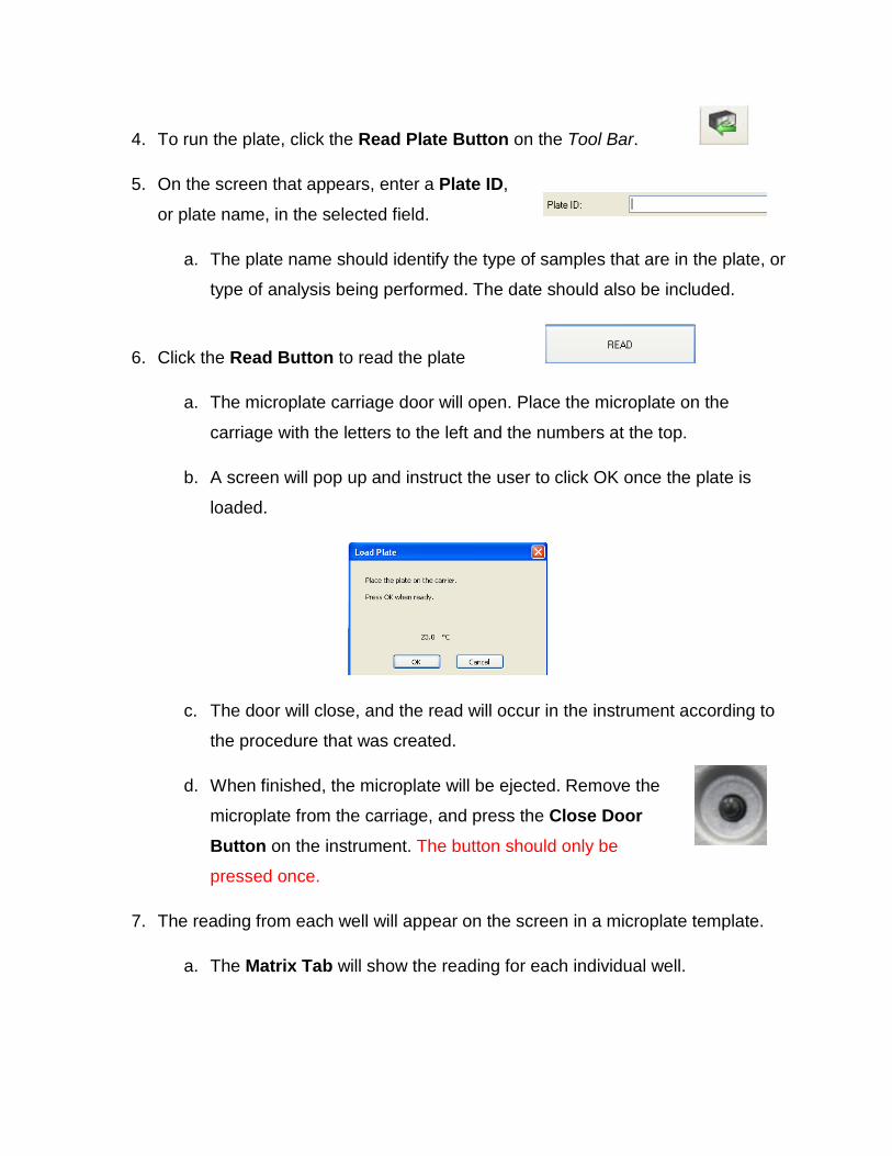
4. To run the plate, click the Read Plate Button on the Tool Bar.
5. On the screen that appears, enter a Plate ID,
or plate name, in the selected field.
a. The plate name should identify the type of samples that are in the plate, or
type of analysis being performed. The date should also be included.
6. Click the Read Button to read the plate
a. The microplate carriage door will open. Place the microplate on the
carriage with the letters to the left and the numbers at the top.
b. A screen will pop up and instruct the user to click OK once the plate is
loaded.
c. The door will close, and the read will occur in the instrument according to
the procedure that was created.
d. When finished, the microplate will be ejected. Remove the
microplate from the carriage, and press the Close Door Button on the instrument. The button should only be
pressed once.
7. The reading from each well will appear on the screen in a microplate template.
a. The Matrix Tab will show the reading for each individual well.

b. The Statistics Tab allows the data to be viewed according to the type of
well.
i. This tab also gives the mean and standard deviation of each
different sample if there were replicates selected in the plate.
8. The data can now be Exported to Excel by clicking the Excel Button in the Plate Data Window.
a. Clicking the Excel Button in the Matrix Tab will export the data in all the
wells, and show the data in Excel in a format that looks like the microplate.

b. Clicking the Excel Button in the Statistics Tab only exports the data on
the screen. To export data from each of the different well types, the Excel
button must be pressed when each one is selected on the screen.
1. Once finished with the experiment and the data, click the Save
Saving Data and Shutting Down the Instrument
Button on the Tool Bar to save the results of the run.
a. Name the experiment and include the date that the plate was run.
(Ex. StudentX_ELISAresults_4-29-09).
2. Shut Down the Gen5 program by clicking the (X) Button at the top
right of the screen.
3. Make sure that the microplate was removed from the
instrument, and turn the instrument off by pressing the Power Switch on the front of the instrument.
a. The green light on the power button will turn off.
4. Make sure the station is neat before leaving the instrument!

1. Samples should be handled according to good laboratory procedures and methods in order to prevent accidents.
BioTek Synergy HT Multi-Mode Microplate Reader Safety Sheet
2. Dispose of all waste solutions according to waste disposal procedures.
3. Do not remove any panels or cords from the instrument to avoid electrical shock.
4. The instrument has moving parts:
a. Do not attempt to exchange microplates or filters cartridges while the instrument is operating.
b. Keep the work area around the instrument clear to avoid obstruction of the moving parts.
c. When the microplate carriage is open, only place a microplate in the carriage to avoid breaking the instrument.
d. Be aware of clothing and body parts when instrument doors are opening and closing.
5. Cleaning the workstation around the instrument is necessary. Never attempt to clean any internal spaces of the instrument.
6. Measurement values may be affected by extraneous particles (such as dust) in the microplate wells. A clean work area is necessary for an accurate reading.
7. Food and drinks should not be placed on or near the instrument.
8. If any liquid should fall near the instrument, do not operate the instrument. Fluid seepage into internal components creates a potential shock hazard, and can cause the instrument to not work properly.
9. Running unauthorized programs or changing preferences on the instrument’s computer is not allowed. Laptops are provided for personal use and internet access. Do not plug memory sticks in the instrument’s computer unless instructed to do so by the instructor.

BioTek Synergy HT Multi-Mode Microplate Reader Instrument Information Sheet
Synergy HT Multi-Mode Microplate Reader Overview:
1. Microplate Carrier Eject Button
2. Power On / Off Switch
3. Microplate Carrier
4. Filter Panel Door (See next page)

Filter Panel Overview:
1. Excitation Filter Wheel Cartridge (Remains in instrument)
2. Excitation Filter Wheel Screw
3. Emission Filter Wheel Cartridge (Remains in instrument)
i. Cartridge can be replaced with an empty cartridge containing 7 and 8.
4. Emission Filter Wheel Screw
5. Fluorescence Lamp Assembly
6. Filter Wheel with 4 filter openings
7. Blocker Plug for luminescence studies
8. C-clip Filter Retainer


![Synergy - BioTek Instruments Brochure... · Synergy 2 & Synergy H4. Built for Drug Discovery Applications. Life Science Research Drug Discovery “We found that each of these [com-petitive]](https://static.fdocuments.net/doc/165x107/5b365dc47f8b9a7e4b8e45dd/synergy-biotek-instruments-brochure-synergy-2-synergy-h4-built-for-drug.jpg)
![Research Article Coccoloba alnifolia Leaf Extract as a Potential … · 2020. 10. 10. · measured at 765nm (BioTek Epoch Microplate, California, CA, USA) [17]. 2.4.3. Total Soluble](https://static.fdocuments.net/doc/165x107/61039079da0154192a317b59/research-article-coccoloba-alnifolia-leaf-extract-as-a-potential-2020-10-10.jpg)