biosystematics of four species of euphorbia l. grown in...
191
Republic of Iraq Ministry of Higher Education And Scientific Research University of Baghdad College of Science Biology Department biosystematics of four species of euphorbia l. grown in baghdad university campus- jadiriyah A thesis Submitted to the College of Science University of Baghdad As partial fulfillment of the Requirements for the Degree of Doctor of Philosophy in Biology By silva a. yakoub zokian M.Sc. Biology-College of Science University of Baghdad 2006 Supervised By Prof. Al-Musawi Ali H.,Ph.D Assist.Prof. AlJibouri Abidaljasim M., Ph.D 2011A.C. 1432 H.
Transcript of biosystematics of four species of euphorbia l. grown in...

Republic of Iraq Ministry of Higher Education And Scientific Research University of Baghdad College of Science Biology Department
biosystematics of four species of
euphorbia l. grown in baghdad
university campus- jadiriyah
A thesis Submitted to the College of Science
University of Baghdad As partial fulfillment of the Requirements for the Degree of
Doctor of Philosophy in Biology By
silva a. yakoub zokian
M.Sc. Biology-College of Science University of Baghdad
2006
Supervised By
Prof. Al-Musawi Ali H.,Ph.DAssist.Prof. AlJibouri Abidaljasim M., Ph.D
2011A.C. 1432 H.

جمهورية العراق وزارة التعليم العالي والبحث العلمي
كلية العلوم/جامعة بغداد قسم علوم الحياة
التصنيف الحياتي الربعة انواع من الجنس
Euphorbia L. -النامية في مجمع جامعة بغداد الجادرية
اطروحة جامعة بغداد - كلية العلوم مقدمة إلى
وهي جزء من متطلبات نيل درجة دكتوراه فلسفة في علوم الحياة/ نبات
من قبل
سيلفا انرتانيك يعقوب زوكيان
جامعة بغداد- كلية العلوم -ماجستيرفي علوم الحياة 2006
شرافءبا
علي حسين الموسوي أ.م.د. عبد الجاسم محيسن الجبوري أ. د.

هـ1432 م2011
Chapter One Introduction & Literature Review
1
chapter one
introduction & literature review 1- introduction
The history of plant systematic-the biological classification of plants-
stretches from the work of ancient Greek to modern evolutionary biologists.
As a field of science, plant systematic came into being only slowly, early
plant lore usually being treated as part of the study of medicine. Later,
classification and description were driven by natural history and natural
theology. Until the advent of the theory of evolution, nearly all classification
was based on the scala naturae. The professionalization of botany in the 18th
and 19th century marked a shift toward more holistic classification methods,
eventually based on evolutionary relationships. A major influence on plant
systematic was the theory of evolution (Charles Darwin published origin of
species in 1859), resulting in the aim to group plants by their phylogenetic
relationships. To this added the interest in plant anatomy, aided by the use of
the light microscope and the rise of chemistry, allowing the analysis of
secondary metabolites ( www.en.wikipedia.org).
The Arabs in the early times gave much attention to the study of
plants of the Arabian Peninsula, North Africa and Spain from different
angles especially for their uses in medicine. Mention needs to be made in
this connection of Eben Sina or Avicenna (980-1037), Ghafiqy (1160), and
Eben Al-Baithar (1248).
The study of the plants of Iraq as a modern science may be started by later
part of eighteenth century and after A. Michaux(1782); Oliver Bruguiers

Chapter One Introduction & Literature Review
2
(1795); Aucher-Eloy(1835); Col.Chenseney(1836); Theodore Kotschy
(1841); Noe(1851) and Haussknecht(1865-1867) worked on the plants of
Arabia which have been incorporated in Boissier’s Flora Orientalis(1876).
Further J. Bornmu ller(1892-1893); Oppenheimer(1893); F. Nbe’lek(1909);
H.F.V. Handel-Mazzetti(1910); C.Pau and C.Vicioso(1910); E.Guest
(1929); A.Eig(1929-1946), Z.Zohary(1933) and Col.Meinertzhagen (1934)
amongst others made extensive collections of Iraqi plants. But it is worth
mentioning that most of their collections are not deposited In Iraqi herbaria
but remain scattered in different herbaria of the continent and elsewhere.
E.Guest began to establish the nucleus of a herbarium of Iraq-plants in the
year 1929. Further collections have been added to the herbarium by many
workers later (Al-Rawi, 1964).
Systematics is important in providing a foundation of information
about the tremendous diversity of life. Virtually all fields of biology are
depend on the correct taxonomic determination of a given study organism,
which relies on formal description, Identification, naming, and classification.
Systematics is also an integrative and unifying science. One of the fun
aspects of systematic is that it may utilize data from all fields of Biology:
Morphology, Anatomy, Embryology/Development, Ultra structure,
Paleontology, Ecology, Geography, Chemistry, Physiology, Genetics,
Karyology, and cell/ Molecular biology (Simpson, 2006). While scientists
have agreed for some time that a functional and objective classification
system must reflect actual evolutionary processes and genetic relationships,
the technological means for creating such a system did not exist until
recently. In the 1990s DNA technology saw immense progress, resulting in

Chapter One Introduction & Literature Review
3
unprecedented accumulation of DNA sequence data from various genes
present in compartments of plant cells. The genus Euphorbia L. is one of the largest and most complex genera of
flowering plants. High morphological plasticity and diversity of this genus
make taxonomical studies on Euphorbia attractive for botanists.
The species of Euphorbia have their own economic value and hence
contribute to the floristic wealth of tropical and subtropical countries of the
world. This genus is also well reputed for the production of valuable
secondary metabolites like alkaloids, flavonoids and terpenes in nature.
Local Euphorbia species are quite rich, and have not been studied yet.
There are about 44 species of Euphorbia in Iraqi flora (Radcliff-Smith,
1980), and more than four species just in University of Baghdad Campus in
Jadiriyah.
The aims of this study: 1- Capable of providing a comprehensive monograph of genus
Euphorbia through appropriate field exploration, study of herbarium
and living collections from fields and gardens of University of
Baghdad Campus -Jadiriyah.
2- Identify the morphological characters.
3- Identify the anatomical characters
4- Study the habitat and geographic distribution in Iraq.
5- Identify the species of Euphorbia by investigation for proteins by
using the technique of protein electrophoresis.
6- Identify the species of Euphorbia by using molecular analysis tool
(the technique of RAPID- PCR).

Chapter One Introduction & Literature Review
4
7- Investigation for the response of the species of Euphorbia for callus
induction by using the technique of tissue culture under different
parameters in vitro.

Chapter One Introduction & Literature Review
5
2- literature reviews
2.1 systematic position of the family
Euphorbiaceae family is one of the largest families of plants .The
Euphorbiaceae was first adequately delimited as a natural group of plants by
A.L. de Jussieu in 1789 (Bruce and Perry, 1943, Simpson, 2006, Takhtajan,
2009). Since this time many contributions have been made to the
classification, phylogeny, morphology, and anatomy of the group. Though
the family has been known for many years, considerable differences of
opinion still exist as to the number of genera and species involved. Estimates
of the number of species in the family vary from 3000 to 8000 (Bruce and
Perry, 1943); reach’s to 8910 species in flora of China (Bingtao et al., 2008).
The family is closely related to the Geraniales by structure of the
gynoecium, although widely separated from other families in the order by
the amount of reduction in the most of its flowers. Small combines the
Euphorbiaceae and the Callitrichaceae into a separate order - Euphorbiales.
This order placed between the Polygalales and the Sapindales, with the
Geraniales immediately preceding this group (Heywood, 1978; Radcliff-
Smith, 1980; Webster, 1994; Judd et al., 1999).
The family is characterized as follows: Flowers hypogynous,
actinomorphic, mostly unisexual; perianth rarely double, usually simple or
wanting; androecium 1-∞; ovary of 3 carpels, trilocular, with 1 or 2
suspended ovules in each cell; micropyle directed upwards and outwards,
and covered with a fleshy outgrowth (caruncle). Fruit almost invariably a
schizocarp-capsule, splitting into carpels, often elastically (Heywood, 1978;
Judd et al., 1999; Singh, 2006; Takhtajan, 2009).

Chapter One Introduction & Literature Review
6
As we said the Euphorbiaceae are recognized as one of the largest
families of the dicotyledons. They are relatively natural group, although
showing many lines of evolution. The impression exists among many plants
mention that the Euphorbiaceae have a general preference for semi-desert or
desert regions, but a survey of the geographic distribution of the family
proves this to be erroneous . Its members live in the varied habitats, in many
different areas of the tropical and temperate world, and exhibit considerable
diversity in growth types. Because of this great diversity of form and habitat,
the family is of special interest. The Euphorbiaceae is of further interest due
to the great diversity of chromosome numbers and chromosome sizes both
between and within so called natural groups. Certain tribes, or other
taxonomic groups, appear cytologically to be natural groups in that one
basic chromosome number is found in each, while in other group nearly all
the basic numbers present in the family are found, suggesting that the unit is
merely descriptive and artificial and not phyletic (Bruce and Perry, 1943).
The Euphorbiaceae consists of trees, shrubs, herbs but are rarely
woody climbers. The larger genera are: Euphorbia (about 2,000 species),
Croton (700 species), Phyllanthus (500 species), Acalypha (430 species),
Jatropha (175 species), Manihot (170 species) (Aworinde et al.,2009).
Additionally Judd et al. (1999) stated more major genera like
Glochidion (300 species), Macaranga (250 species), Antidesma (150
species), and Tragia (150 species).
According to the most recent molecular research Euphorbiaceae is a
complex family previously comprising five subfamilies: the Acalyphoideae,
the Crotonodeae, the Euphorbiodeae, the Phyllanthoideae and the
Oldfieldioideae. The first three are uni-ovulate sub-families while the two

Chapter One Introduction & Literature Review
7
last one are bi-ovulate (Heywood, 1978; Radcliff-Smith, 1980; Takhtajan,
2009). The Euphorbiaceae has been split into five families: the three uni-
ovulate subfamilies have become the Euphorbiaceae in the strict sense, with
the tribe Galearieae in the Acalyphoideae forming the most of the family
Pandaceae. The bi-ovulate subfamily Phyllanthoideae has become the family
Phyllanthaceae, with the tribe Drypeteae as family Putranjivaceae and, the
tribe Centroplaceae part of the Pandaceae. The other bi-ovulate subfamily
Oldfieldioideae has become the Picrodendraceae (Heywood, 1978; Webster,
1994; Tokuoka and Tobe, 1995; Takhtajan, 2009). Euphorbioideae divided
to five tribes:
Tribe Euphorbieae
Tribe Hippomaneae
Tribe Hureae
Tribe Pachystomateae
Tribe Stomatocalyceae
(Heywood, 1978; Webster, 1994; Llamas, 2003; Mwine and Van Damme,
2011).
Webster suggested that “Phllanthoideae” are the primitive group from
which the other subfamilies are derived. They are almost surely
paraphyletic, and are characterized by having two ovules per locule, usually
alternate leaves, nonspiny pollen, and nonarillate seeds. Oldfieldioideae also
have two ovules per locule, and may be monophyletic on the basis of their
spiny pollen. Members of Euphorbiaceae having only one ovule per locule
probably form clade, which has been divided into three subfamilies by
Webster above: the Acalyphoideae (Acalypha, Alchornea, Tragia, Ricinus,
and relatives), which lack latex; and Crorotonoideae (Croton, Manihot,

Chapter One Introduction & Literature Review
8
Jatropha, Codiaeum, Aleurites, Cnidoscolus, etc.) and Euphorbiodeae
(Hippomane, Hura, Euphorbia, Gymnanthes, Stillingia, Sapium, etc.), both
of which have latex. The Contonoideae have distinctive polyporate pollen,
often stellate, peltate, or branched hairs, and colored to white, noncaustic
sap, while Euphorbiodeae have tricolporate pollen, simple hairs, and white,
often caustic sap. Euphorbioideae contain the large tribe Euphorbieae
(mainly Euphorbia, which includes Poinsettia, Chamaesyce, etc.), which are
considered monophyletic on the basis of their inflorescences, which are
cyathia. The carpellate flower is surrounded by numerous staminate flowers
(each of which is reduced to a single stamen) within a cuplike structure
formed from a highly reduced cymose inflorescence and associated bracts.
One to five nectar glands, sometimes with petal-like appendages, are
associated with the cuplike axis of each cyathium (Willis, 1973; Judd et al.,
1999; Prenner and Rudall, 2007). Most Euphorbiaceae are insect-pollinated
(flies, bees, wasps, and butterflies), with nectar providing the floral
attractant, yet some are probably pollinated by birds, bats, or other amimals.
Acalypha, Ricinus, and Alchornea, among others, are wind-pollinated.
Outcrossing is promoted by maturation of carpellate flowers before
staminate ones. Most have elastic schizocarps. The large fruits of hura (Hura
crepitans) or hevea (Hevea brasiliensis) are able to explosively eject their
seeds, shooting them several meters. Some are secondarily water-dispersed,
while those with oily arils are sometimes secondarily dispersed by ants.
Some taxa have fleshy arils (or indehiscent fleshy fruits) and are dispersed
by birds (Judd et al., 1999).

Chapter One Introduction & Literature Review
9
2.2 systematic position of the genus
The genus Euphorbia is one of the largest, most complex and diverse
groups of flowering plants on earth. It contains at least 2000 species
(Heywood, 1978; Radcliff-Smith, 1980; Judd et al., 1999; Pritchard, 2003).
Many of the species are known as "spurges." They all produce a mostly
white latex which they exude when cut, and this sap is often toxic. There are
many herbaceous spurges, especially in temperate zones worldwide, but the
genus is best known for its many succulent species, some of which appear
very similar to cacti. Succulent euphorbias are most diverse in southern and
eastern Africa and Madagascar, but they also occur in tropical Asia and the
Americas (Pritchard, 2003; Heywood, 1978; Takhtajan, 2009). All flowers
in the Euphorbiaceae are unisexual (either male or female only), and they are
often very small in size. In Euphorbia, the flowers are reduced even more
and then aggregated into an inflorescence or cluster of flowers known as a
"cyathium" (plural cyathia). This feature is present in every species of the
genus but nowhere else in the plant kingdom. Whereas most other large
genera of plants differ in features of the flowers themselves, Euphorbia
varies instead in features of the cyathium, which can show amazing
modifications in different groups within the genus (Pritchard, 2003;
Heywood, 1978).

Chapter One Introduction & Literature Review
10
FIGURE (1): Typical Flower of Euphorbia
(Diagram from www.Euphorbiaceae.Org (PBI))
The main defining feature of the cyathium is the floral envelope or
involucre that surrounds each group of flowers. The involucre almost always
has one or more special glands attached to it, most often on the upper rim,
and these glands and their appendages vary greatly in size and shape. There
may be specialized leaves called cyathophylls or cyathial leaves that
surround the cyathium and give an overall flower-like appearance to the
whole complex inflorescence. Inside the involucre are the flowers, usually
with a number of extremely simplified male flowers consisting of a single
anther, filament, joint and pedicel. Generally there is a single female flower
in the center consisting of a pedicel, a three-lobed ovary, and no petals or
sepals associated with it (Radcliff-Smith, 1980; Simpson, 2006; Takhtajan,
2009) Figure (1).

Chapter One Introduction & Literature Review
11
FIGURE (2): Cyathium of Euphorbia
(Diagram from www.Euphorbiaceae.Org (PBI))
With this basic model of the cyathium, many modifications upon it have
evolved in the genus, as well as in the aggregation of cyathia into higher
order units (Heywood, 1978; Radcliff-Smith, 1980; Webster, 1994; Judd et
al., 1999) Figure (2).
2.3 fruits and seeds
Fruits of Euphorbia are capsules that typically split open explosively
when ripe. There are potentially three seeds per capsule, and there is a wide
variety of size, shape, and surface features of the seeds and capsules. Seeds
of some species have a fleshy appendage called the caruncle above the point
of attachment to the central column of the fruit (Mangaly et al., 1979).

Chapter One Introduction & Literature Review
12
Structure of the seed coat is characteristic for the family and does not
provide evidence for a polyphylectic origin of the family (Webster, 1994).
2.4 diversity of life forms
The variety of habits or life forms is one of the most salient features of
Euphorbia. There are many annual or perennial herbs, and these tend to
retain leaves through their active growing periods. At its simplest, in a
number of species in the Chamaescyce lineage, the plant will germinate,
dichotomously branch, flower, fruit, and die in a matter of weeks. There are
also leafy shrubs and trees as members of species that can reach 20 meters
high. A large portion of Euphorbias, however, are succulent, with thickened,
photosynthetic stems and very ephemeral leaves if present at all. Many
succulents are in turn thorny, and some have well developed underground
tubers (Radcliff-Smith, 1980, Pritchard, 2003).
2.5 systematics and classification
The understanding of the relationships of Euphorbia has been bolstered
by comparative DNA sequence data from many species, and these results
support a broad view of the genus that includes a number of groups that
were formerly recognized as different genera, such as Chamaesyce,
Monadenium, Pedilanthus, and Poinsettia. The most current information
places Euphorbia species into four distinct monophyletic groups or clades .
Steinmann & Porter (2002) has examined different regions from the three
plant genomes (nuclear, chloroplast, and mitochondrial), and this shows
clear support for the following relationships among the four main clades of

Chapter One Introduction & Literature Review
13
Euphorbia: clade B (subgenus Esula) is the sister group to clade A
(subgenus Rhizanthium), and that is in turn sister to both clades C and D
(subgenus Euphorbia and subgenus Chamaesyce) Figure (3).
FIGURE (3) Major Clades of Euphorbiaceae
Diagram from www.Euphorbiaceae.Org (PBI))
2.6 origin of the botanical (latin) name of
euphorbia
King Juba of Mauritania (today’s Morocco) is credited as the first person
to discover a succulent Euphorbia and give the genus its name. Somewhere
between 25 BC and 18 AC, he discovered a plant in the Atlas mountains: it
was most likely E.officinarum or E.resinifera, King Juba named the plant
after his doctor whose name was Euphorbus, the meaning of this word being
“well fed”, the king comparing his fat flashy doctor with the plant
(Pritchard, 2003; Gledhill, 2008). Non- succulent species had been known

Chapter One Introduction & Literature Review
14
back in ancient Greek times as “Titbymalus”. The two names existed side by
side until 1583 when a botanical link was made by Andrea Cesalpino, then
in 1753 Linnaeus listed both under one name “Euphorbia”. Today
“Titbymalas” still exists as section or subgroup of Euphorbia (Radcliff-
Smith, 1980; Pritchard, 2003; Simpson, 2006)
2.7 meaning of the common name "spurge"
Many of the herbaceous, leafy species of Euphorbia are commonly called
"spurges". This word derives from the old French word espurgier (Latin
expurgare), which means "to purge." The sap of many herbaceous
Euphorbia species have traditionally been used as a purgative, or laxative
(www.Euphorbiaceae.Org). Radcliff-Smith (1980) stated that Bailey (1939)
points out that Euphorbia is sometimes improperly referred to as Milkweed
and that the name spurge, though often applied to the genus as a whole,
cover more correctly the smaller herbaceous species. In our own territory
several general name, covering a number of different and unrelated plants
with milky juice, have frequently been noted for the weedy vegetal and ruder
species of Euphorbia which occur in fields, gardens and waste places in
lower Iraq (E. puples, E. granulata, E. chaemaesyse, E. prostrate, E.
petiolata, E. helioscopia): UM AL-HALIB ام الحليب, HALIB حليب and
ALBAINA البينة or LUBAINA لبينة (all colloquial names denoting “milk”).
Similarly, in the north, names embodying SHIR شير (‘milk”, kurd.) have
been noted for several species in the mountains - SHIR KITIK شير كتك
(“cat‘s milk”, Kurd.) for E.macroclada in Rowanduz district, SHIR-I MAR
or SHIR MAR شير مار (“snake‘s milk”, Kurd. ) at Amadiya. Another

Chapter One Introduction & Literature Review
15
common name in the north for plants with bitter milky juice is KUJILK
sometimes KHUZHILKA, SHIR KHUZHILK) خريلكةor KHURHILKA كجيلك
etc., Kurd). Ibn Al-Bitar indeed mentions the name FORBIYUN فربيون
(Euphorbium) and quotes an extract from Ibn Radwan who speaks of
LABAN AS-SUDA as a gum exported from (”negress‘s milk“) لبن الصودة
Morocco which resolves tumors and has other medicinal properties. This he
says is obtained from the plant known as RAQIB ASH-SHAMS راقب الشمس
(“sun observer,” in Greek “helioscopia”), suggesting that the Sun Spurge
(E.helioscopia) which contains euphorbium has long been used medicinally
(Radcliffe-Smith, 1980). Additionally the common names of E.helioscopia
are: SUN SPURGE, EUPHORBE REVEILLE-MATIN فربيون الشمس
(Chemaly and Chemaly, 2007). Also it is known as KHANNAIQ-AD-
DIJDJ ,Al-Rawi and Chakravarty) عص الكلبة US-AL-KALBA ,خنيق الدجاج
1964).
While E.peplus known as: PETTY SPERG, EUPHORBE DES VIGNES
.(Chemaly and Chemaly, 2007) فرفخ
E.hirta known as the AUSTRALIAN ASTHEMA HERB or
QUEENSLAND ASTHEMA WEED, CAT‘S HAIR, HAIRY SPURGE
(Ekpo and Pretorius, 2007).
There are many local names for particular species of Euphorbia.
"POINSETTIA" is the much-used common name for Euphorbia
pulcherrima cultivars BINT AL-QINSIL بنت القنصل , harking back to when
the genus Poinsettia was used for this and related species. "CROWN OF
THORNE" is the English common name for Euphrobia milii cultivars.

Chapter One Introduction & Literature Review
16
"MILKBUSH" or "PENCIL CACTUS"are both used for the much-cultivated
Euphorbia tirucalli.
Some other names used for different species of Euphorbia include
"SNOW-ON-THE-MOUNTAIN", "MEDUSA‘S HEAD", "MEXICAN
FIRE PLANT", and "SCARLET PLUME" (www.Euphorbiaceae.Org ).
2.8 previous work on euphorbia
The last complete monograph of Euphorbia was the treatment by
Boissier (1862) in de Candolle's Prodromus, in which 740 species were
recognized (www.Euphorbiaceae.Org).
Handel Mazzetti (1910) had described 4 species of Euphorbia with their
geographical distribution in Iraq and Syria. Guest (1933) described 4 species
with mentioning to their cultivating time and their colloquial names in Iraq
(our studied species E.helioscopia was one of them). In the same year (1933)
Post reported 44 species of Euphorbia for flora of Syria, Palestine, and Sinai
(our studied species E.helioscopia, E.granulata and E. peplus were among
them). Zohary in (1950) mentioned (25) species (E.helioscopia was among
them). Rechinger in (1959) stated about 36 species in a description for flora
of Syria and Lebanon. But in (1964) Rechinger described 25 species and
their geographical distribution in Iraq. Rechinger and Schiman-Czeika (1964) described the family of Euphorbiaceae in flora of Iran and reported
98 species, 29 species of them are distributed in Iraq. Additionally Al- Rawi
(1964) reported about 39 species distributed in Iraq (E.helioscopia,
E.granulata and E.peplus were among them). Migahid (1978) reported 14
species in flora of Saudi Aarabia (E.helioscopia, and E.granulata were
Chapter One Introduction & Literature Review
17
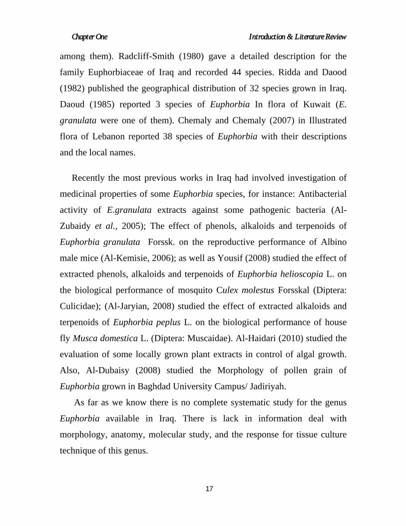
among them). Radcliff-Smith (1980) gave a detailed description for the
family Euphorbiaceae of Iraq and recorded 44 species. Ridda and Daood
(1982) published the geographical distribution of 32 species grown in Iraq.
Daoud (1985) reported 3 species of Euphorbia In flora of Kuwait (E.
granulata were one of them). Chemaly and Chemaly (2007) in Illustrated
flora of Lebanon reported 38 species of Euphorbia with their descriptions
and the local names.
Recently the most previous works in Iraq had involved investigation of
medicinal properties of some Euphorbia species, for instance: Antibacterial
activity of E.granulata extracts against some pathogenic bacteria (Al-
Zubaidy et al., 2005); The effect of phenols, alkaloids and terpenoids of
Euphorbia granulata Forssk. on the reproductive performance of Albino
male mice (Al-Kemisie, 2006); as well as Yousif (2008) studied the effect of
extracted phenols, alkaloids and terpenoids of Euphorbia helioscopia L. on
the biological performance of mosquito Culex molestus Forsskal (Diptera:
Culicidae); (Al-Jaryian, 2008) studied the effect of extracted alkaloids and
terpenoids of Euphorbia peplus L. on the biological performance of house
fly Musca domestica L. (Diptera: Muscaidae). Al-Haidari (2010) studied the evaluation of some locally grown plant extracts in control of algal growth.
Also, Al-Dubaisy (2008) studied the Morphology of pollen grain of
Euphorbia grown in Baghdad University Campus/ Jadiriyah.
As far as we know there is no complete systematic study for the genus
Euphorbia available in Iraq. There is lack in information deal with
morphology, anatomy, molecular study, and the response for tissue culture
technique of this genus.

Chapter One Introduction & Literature Review
18
2.9 chemical contents and medicinal uses
Euphorbia has a reputation for being incredibly toxic plants. Some of the
Euphorbia are used in folk medicine to cure skin diseases, gonorrhea,
migraines, intestinal parasites, and warts. The genus Euphorbia has been the
source of large number of biological active compounds. Tannins, flavonoids,
unsaturated sterols/triterpenes, carbohydrates, lactones and proteins/amino
acids were reported as major active constituents of some Euphorbia species.
A variety of diterpenoids with antibacterial, anticancer, prostaglandin E2-
inhibitory, antifeedant, anti-HIV, and analgesic activity have also been
isolated from different Euphorbia species. They include jatrophane, ingenol,
and myrsinane diterpenoids . These diterpenoids have been reported to act in
diverse ways; they have been found to be skin- irritants, tumor-promoters. In
addition to anti-tumor activity, several species of this genus have been
investigated for their immunomodulatory activity and some immunotoxic,
immunosuppressive and immunostimulatory effects have been reported.
These broad range and diversity of biological activities in the Euphorbia
genus, perhaps due to the presence of various components with different
modes of action in the plants (Sayed, 1980 ; Amirghofran et al., 2008;
Gyuris et al., 2009; Noori et al., 2009; Kumar et al., 2010).
On the other hand Ressler (1985) reported that the latex of
Euphorbiaceae cause keratoconjunctivitis. Phorbol esters are considered to
be responsible for the toxicity of the latex e.g. in the case of E.helioscopia.
Over seventy jatrophane, modified jatrophane, segetane, pepluane, and
paraliane diterpenoids, fifty of them reported for the first time, were
extracted, purified and characterized from E.dendroides, E.characias,

Chapter One Introduction & Literature Review
19
E.peplus, E.helioscopia, and E.papalias. These compounds showed
interesting pharmacological activities (Corea et al., 2008).
Chemical investigation of E.peplus revealed almost identical profiles of
secondary metabolites affording ingenanes, jatrophanes, and tetracyclic
diterpene with new carbon skeleton for the name pepluane is proposed.
(Jakupovic et al., 1998). In a study for Mucsi et al. (2001) cytotoxicities and
anti-herpes simplex activities of nine diterpenes isolated from Euphorbia
species were determined. From a pro-inflammatory active extract of
E.peplus two new diterpine polyester based on the pepluane and
jatrophanes skeletons were isolated. Ingenol 3-angelate, which was obtained
for the first time from this plant, is an irritant toxin with high activity
(Hohmann et al., 2000). Non- polar component of the latex of E.peplus was
found to contain nobtusifoliol, cycloartenol, 24-methylenecycloartaenol,
lanosterol, and 24-methylenelanosterol in the free and esterified triterpene
alcohol fractions (Giner et al., 2000). As well as Williams (2005) stated that
inginol 3-angelate (PEP005) is diterpene ester isolated from E.peplus used as
a home remedy for skin cancer. PEP005 is a novel anticancer agent was
previously shown to modulate protein kinase C (PKC), resulting in
antiproliferative and proapoptotic effects in several human cancer cell lines
(Serova et al., 2008). Additionally, Weedon and Chick (1976) reported that
the sap of the E.peplus used as a home treatment for warts and basal cell
carcinomas and documented its successful use on a biopsy-proven basal cell
carcinoma. Also, Al-Haidari (2010) stated that the growth of algae
Anabaena cylindrica, Nostoc commune, and Microcystis aeruginosa were
stimulated by phenolic compounds extracted only from flower and leaf of
local E.peplus, while alkaloid extracts had no effect against studied algae.

Chapter One Introduction & Literature Review
20
Sharawy and AL-Shammari (2009) stated the toxic principles of
E.granulata include terpenes, triterpenols, glycosides, and phenols present in
the milky juice, all parts of these plants or the milky latex can cause an
inflammation or irritation when in contact with the skin. If eaten by animals,
the plants may cause vomiting and severe purgation, the same as for
E.peplus. On the other hand the feeding of lactating goats on usual green
fodder, contaminated with E.helioscopia or E.nubica, results in poisoning of
the dams as well as their kids. General sings of toxicity were emaciation,
depression, shedding body hair, arching of back, and possible death. The
goat also suffered from deterioration of renal function. Necrosis of epithelial
cells of kidney tubules were noticed. Considerable degenerative changes
were also observed in heart and lung. The pathophysiological appearances
indicate that by feeding on the E.peplus reported previously. Such
intoxication most likely is done to irritant and hyperphysiogenic diterpene
ester (DTE) toxins. Usually present in the aerial part of Euphoeria species
and well known as tumor promoters in mouse skin. As discussed previously
in lactating goat feed on fodder contaminated with E.peplus, tumor
promoters of the DTE type may enter the human food chain via this source
of milk (Nawito et al., 2000).
Leaf extract of E. granulata is effective against the pathogenic bacteria
(Klebsiella pnemoniae, Salmonella typhi, and Staphylococcus epidermidis);
there is little reference to the use of plant in medicine but its latex is locally
applied against poisonous bites (Salamah et al., 1989).
Pohl et al., (1975) had isolated flavonol glycoside from
E.helioscopia. Irritant diterpine ester toxins were isolated from E.helioscopia
and E.nubica which contaminate the green fodder of livestock in Egypt. Six

Chapter One Introduction & Literature Review
21
short-to medium-chain polyfunctional diterpene esters of the ingenane type,
generally containing unsaturated acids were obtained. These compounds
considered to be more or less active tumor promoters, i.e., conditional (non-
genotoxic) cancerogens (Zayed et al., 2001).
Methanol and chloroform extracts of E.helioscopia and other Saudi
Arabian Euphorbiales have molluscicidal activity against the snail
Biomphalaria pfeifferi (Al-Zanbagi et al., 2000). While Salamah et al.,
(1989) further stated that the leaf extract of E.helioscopia is specifically
lethal to the bacteria Salmonella typhi.
E. hirta is used in the treatment of gastrointestinal disorders, bronchial
and respiratory diseases and in conjunctivitis. Hypotensive and tonic
properties are also reported in E.hirta. The aqueous extract exhibits
anxiolytic, analgesic, antipyretic, and anti-inflammatory activities. The stem
sap is used in the treatment of eyelid styles and a leaf poultice is used on
swelling and boils. Extracts of E.hirta have been found to show anticancer
activity. The aqueous extract of the herb strongly reduced the release of
prostaglandins. The aqueous extract also inhibits aflatoxin contamination in
rice, wheat, maize, and mustard crops. Methanolic extract of leaves have
antifungal and antibacterial activities (Rao et al., 2010; Kumar et al., 2010).
Decoction of dry herbs is used for skin diseases. Decoction of fresh herbs is
used as gargle for the treatment of thrush. Root decoction also beneficial for
nursing mothers deficient in milk. Roots are also used for snake bites. The
polyphenolic extract of E. hirta has antiamoebic and antispasmodic activity.
Quercitrin, a flavanoid glycoside, isolated from the herb showed an
antidiarrheal activity. It is reported to have of whole plant shows

Chapter One Introduction & Literature Review
22
hypoglycemic activity in rats. It has a sedative effect on the genito-urinary
tract (Lanhers et al.,1993; Kumar et al.,2010). The study of Adedapo et al.
(2005) has clearly explained that the aqueous crude extracts of E.hirta after
its administration into dogs produced a significant increase in PCV (Packed
Cell Volume), RBC(total erythrocytes count), Hb(Hemoglobin
concentration, Total WBC (Leucocyte count) and lymphocyte counts. The
fecal egg counts also showed a remarkable and significant reduction in the
levels of the identified helminths. Thus E. hirta could serve as antihelmintic
agent. Liu et al. (2007) explained that bioassay-guided fractionation of
methanolic extracts of E.hirta aerial parts led to the isolation of flavanol
glycosides afzelin, myricitrin, the two compounds showed proliferation
inhibition of Plasmodium falciparum, on the other hand they exhibited little
cytotoxic property against human epidermoid carcinoma KB cells. Johnson
et al. (1999) proved the diuretic effect of the E.hirta leaf extract in rats using
diuretic drugs. The positive results validate the traditional use of the plant as
diuretic agent. Ogueke et al.(2007) stated that the ethanolic extract of E.hirta
is hematologically not toxic to rats, as well as the antimicrobial activities
were believed to be due to the tannins, alkaloids, and flavonoids which were
identified in the extract. The results are significant in the health care delivery
system and justifies the use of the plant in the treatment of sores-boils,
wounds and control of dysentery and diarrhea.

Chapter Two Morphological studies
23
Chapter two
Morphological studies
1- Introduction
Morphological characters are features of external form or
appearance. They currently provide most of the characters used for
practical plant identification and many for hypothesizing phylogenetic
relationships. These features have been used for a longer time than
anatomic or molecular evidence and have constituted the primary
source of taxonomic evidence since the beginnings of plant
systematic. Morphological characters are easily observed and find
practical use in keys and descriptions. Phylogenetically informative
characters may be found in all parts of the plant, both vegetative and
reproductive (Judd et al., 1999; Simpson, 2006).
Euphorbiaceae is one of the largest families of flowering plants.
It is widespread throughout the globe, strongly represented in tropics.
There are seven genera in Iraq (4 native, 2 naturalized and now
subspontaneous, and 1 quite recently introduced). Euphorbia is the
largest genus cosmopolitan with some 2000 species (Radcliff-Smith,
1980).
There are 44 species in Iraq most of them native, but including
a few naturalized species and 2-3 were introduced as an ornamental
species (Radcliff-Smith, 1980). High morphological plasticity and
diversity of this genus in Iraq, as well as few studies on the genus

Chapter Two Morphological studies
24
make taxonomical studies on Euphorbia attractive for many botanists.
Taxonomic and phylogenic significance of morphological features of
Euphorbia are emphasized by many (Rechinger, 1959; Rechinger,
1964; Rechinger and Czeika, 1964; Bentham and Hooker, 1965;
Mangaly et al., 1979; Radcliff-Smith, 1980; Pahlevani, 2007;
Aworinde et al., 2009).
In the present study an attempt has been made to collect as
much information as possible about four species of Euphorbia. With
the aim of providing useful taxonomic data that would give further
insight into proper classification and identification.
2- Materials and Methods
Plant materials of E.helioscopia, E.peplus, E.granulata and
E.hirta, used for this investigation were obtained from Baghdad
University Herbarium (BUH), and newly collected specimens from
different parts of University of Baghdad were studied and identified
using corresponding scientific papers and the flora of Iraq (Radcliff-
Smith, 1980). Field collection were conducted between the flowering
and fructifying throughout the years (2008-2010). Photographs were
taken from fields by using digital camera (model Sony Cyber-Shot T
700) as well as fitted on dissecting microscope. These photographs
were useful for identification and differentiation of morphological
features.

Chapter Two Morphological studies
25
2.1 Observation of Characters
The macromorphological characters were assessed on stems,
mature leaves, trichomes, cyathia and seeds at comparative position.
Morphological characters of stems include stem length, width,
shape, color and stem types (the mode of branching).
Morphological characters were assessed on mature leaves
include leaf apex, margin, shape, leaf surface, and leaf base, as well
as leaf length, width at the widest point, petiole length, petiole width
and blade. As well as the presence of the trichomes.
The species have been studied for cyathial characteristics
including number of type of cyathia, involucres shapes and
diameters, lobe shapes, numbers, colors and shapes of glands as
well as the apexes of glands.
Mature seeds of Euphorbia were collected from plants growing
in the University of Baghdad campus and studied to understand their
general morphology, which included seed shape, diameter, color and
surface configuration in addition to the presence of curuncle.

Chapter Two Morphological studies
26
3- Results and Discussion
3.1 Habit and Duration
Euphorbia species are herbs in Iraq, unarmed or spinous, with
a milky juice. Our native species are all herbaceous, small herbs on
the plaines and in the mountains shrub-like herbs with woody
rootstocks. Many species of Euphorbia have a xerophytic habit and
grow in very dry places (and are usually cactus like in some cultivated
species). However they are readily distinguishable from cactus, even
when not in flower by the presence of latex, figure (4).
The species E.helioscopia, E.peplus and E.hirta are annual
herbs, where as E.granulata is perennial herb, with erect or
ascending or prostrate, simple or branched stem, the whole life cycle
from seed germination to seed production requires (2-6) months,
plate (1).
E.helioscopia, E.peplus and E.hirta grow in disturbed habitats,
while E.granulata is thermophilous plant with desert habitat.

Chapter Two Morphological studies
27
FIGURE (4) Latex of Euphorbia.
Euphorbia helioscopia
Euphorbia peplus
PLATE (1-a): Species of Euphorbia in nature.

Chapter Two Morphological studies
28
Euphorbia granulata
Euphorbia hirta

Chapter Two Morphological studies
29
PLATE (1-b): Species of Euphorbia in nature.
3.2 Vegetative parts Morphology
3.2.1 Stems
The stems of Euphorbia species have taxonomic importance. It
was found in this study that the mode of branching, color, length,
thickness, presence of stipules and trichomes are characters of
taxonomic values as shown in table (2-1).
Stems are erect, ascending or prostrate, simple or ramified,
single or with ascending branches near base. Stems of E.helioscopia
have scars at the base where the leaves have fallen, also
E.helioscopia and E.peplus are glabrous (sometimes E.helioscopia
may has unicellular hairs in upper part of stem) characterized by
absence of stipules. Stems of E.granulata are un-branched,
occasionally branched at the end, usually woody at base, many from
base, ascending or prostrate, internodes conspicuous (has distinct
swollen nodes), pilose on one side of the stem and characterized by
presence of stipules. E.hirta stems branched from the middle or
above (usually few branched), ascending to erect or prostrate, with
mixture of long yellow – brown multicellular hairs and much shorter
white hairs, in addition to presence of distinct hairy stipules as shown
in plate (2).
Our field observations in University of Baghdad campus-
Jadiriyah revealed occurrence of two ecotypes for E.helioscopia and

Chapter Two Morphological studies
30
E.hirta, one erect and the other prostrate in relation to the moisture
level of the soil and the degree of biotic factors.

Chapter Two Morphological studies
31
E.hirta is erect and grows in more shady areas, has more
active growth of one or two branches is more useful, since the plant
has to struggle for the maximum availability of sun light, and therefore
all or most of the extra-axillary branches disappear.
Mangaly et al. (1979) reported that the erect ecotype of E.hirta
predominates during the rainy season, whereas the prostrate one
predominates during winter months and exhibits higher reproductive
potentialities under extreme biotic disturbances.

Chapter Two Morphological studies
32

Chapter Two Morphological studies
33
3.2.2 Leaves
Leaves are simple, alternate and opposite, stipules absent or
membranous, petiole absent or present ( short or long ), obovate to
spatulate or subelliptic or lanceolate-oblong, apex rounded, obtuse or
acute, margin entire or dentate, base cuneate or oblique or obliquely
rounded, color pale to yellowish green or green to red, glabrous or
hairy as shown in table ( 2-2 ). The leaf size shows considerable
variation within the studied species. The largest size recorded in
E.helioscopia and the smallest in E.granulata. Also, we observed in
E.hirta leaves that hairs of abaxial side is more abundant and longer
than the hairs of adaxial side.
Inflorescence of E.peplus is a terminal pseudoumbel, rays
irregulary branched, total branches few; primary involucral leaves 3 or
4, short; cyathophylls 2, similar to normal leaves. While E.helioscopia
Inflorescence a compound pseudumbel, usually rather compound;
primary invollucral leaves 5, yellowish green, obovate-oblong 3-4 ×
0.8-1.4 cm, progressively shorter; cyathophylls 2, obovate, base
rounded, margin dentate, apex rounded.

Chapter Two Morphological studies
34

Chapter Two Morphological studies
35
3.2.3 Trichomes
E.peplus is glabrous; while E.helioscopia is glabrous or
sparsely hairy in the upper part (unicellular hairs). E.granulata has
multicellular hairs on one side of the stem as seen in plate (2);
however E.hirta is with mixture long yellow-brown multicellular hairs
and much shorter white hairs as shown in plate (2) and figure (5).
.
FIGURE (5) Multicellular gland-like trichomes in E.hirta
(100X).

Chapter Two Morphological studies
36
E.helioscopia E.peplus
E.hirta E.granulata
PLATE (2): Stems and trichomes in species of Euphorbia.
a- Trichomes in E.helioscopia, one side trichomes in E.granulata, mixture of long yellow and shorter white hairs in E.hirta.
b- Hairy stipules.
a
b
a
b
a

Chapter Two Morphological studies
37
3.3 reproductive parts Morphology
3.3.1 Cyathia
The cyathial characteristics have shown well differences.
Cyathia of the four species were subsessile, axillary or dense. The
shape of involucres were cuplike, campanulate or turbinate. Lobes
were rounded, subtruncate or triangular- ovate, glabrous or pilose. All
the species studied had four glands, with appendages - such as
E.granulata and E.hirta - or without appendages -such as
E.helioscopia and E.peplus which varied from the rest of the species
by having two horned glands, as well as the color of glands differ
from green, pale brown to red, also the shape of glands varies
between crescent shape, disk like shape, irregular and rounded to
transversely elliptic as shown in table (2-3) and plate (3). Also E.hirta
differs in having multicellular uniseriate rugose hairs on all parts of
the cyathium except style and stigma, in the meantime E.granulata
has hairs only on ledges of cyathium. We believe that this is an
important characteristic for these species.

Chapter Two Morphological studies
38

Chapter Two Morphological studies
39
-A- -B- -C-
E.helioscopia
E.peplus

Chapter Two Morphological studies
40
E.granulata
E.hirta
PLATE (3): The Cyathia of Euphorbia Species.
A: Cyathium B: female flower C: C.S. of immature fruit (Bars ═ 1.0 mm)
3.3.2 The Male and Female Flowers
The study of male and female flowers aid to the differentiation of

Chapter Two Morphological studies
41
the four species of Euphorbia as shown in table (2-4), Figure (6). The
pedicel of the stamen is longer than the filament, despite the different
dimensions and forms of the anthers and the variation in numbers of
male flowers, splitting of anthers is horizontal in all studied species. In
floras there are no real numbers for male flowers of studied species.
According to flora of China, numbers of male flowers are many in
E.helioscopia, E.peplus and E.granulata, whilst numbers for male
flowers in E.hirta are 4 or 5 (Bingtao et al., 2008); this result differs
from our finding recorded in table (2-4).
FIGURE (6): Cyathia of Euphorbia. A. Cyathium in E.peplus , B. Mature opened cyathium showing sex organs,
C. Cyathium in E.hirta a- male flower b- 2-horned gland
(Bars ═ 1.0 mm)
A B
a b
C
a

Chapter Two Morphological studies
42
3.3.3 Seeds.
The taxonomic value of the seeds of Euphorbiaceae had long
been recognized (Mangaly et al., 1979; Webster, 1994; Tokuoka and

Chapter Two Morphological studies
43
Tobe, 1995). The morphological characters of seeds for the four
species of Euphorbia shown in table (2-5). Seeds shapes were
ovoid-angulate, ovoid, tetragonal or subglobose-tetragonal. The
seeds size shows considerable variation within the species studied.
The largest diameter recorded in E.helioscopia (1.3-0.8) mm and the
smallest in E.hirta (0.8-0.5) mm. As well as seeds had different colors
and surface configurations such as gray - micropores, brown -
reticulated, brown - adaxially grooved and brown to reddish smooth
seeds. Besides the seed often shows a fleshy outgrowth from the
integuments-known as caruncle-that often functions as an
inducement for animal (usually ants) dispersal. The presence and
type of caruncle had taxonomically importance, which separated the
species studied for into groups, one related to E.helioscopia and
E.peplus that had white sessile caruncle, and the other belongs to
E.granulata and E.hirta which were ecarunculate (without caruncle),
plate (4). Webster, (1994) as well as Tokuoka and Tobe (1995)
explained that seeds are useful for comparison between and within
subfamilies.

Chapter Two Morphological studies
44

Chapter Two Morphological studies
45
E.helioscopia E.peplus E.granulata E.hirta
PLATE (4) Seed of species of Euphorbia.
A - Seed in ventral view B - Seed in dorsal view
(Bars ═ 1.0 mm)
These morphological studies have revealed important stable
taxonomic characteristics that can be used as a key in Euphorbia
species distinction, such as the presence of hairy stipules,
Multicellular gland-like trichomes in both species E.granulata and
E.hirta; Also the presence of swollen nodules and the distribution of
hairs on one side of stem in E.granulata. In addition to the variation
of cyathia, male and female flowers in Euphorbia species that were
studied for the first time in addition to the characteristics of seeds.
-A-
-B-

Chapter three Anatomical studies
44
chapter three
anatomical studies
1- introduction
The role of anatomical data in traditional taxonomy has been long
recognized since the variations within the species, genera or family are
usually reflection of anatomical features as well. The anatomical features of
roots, stems, leaf epidermis, stomata, trichomes and other characters are
useful anatomical tools (Judd et al., 1999; Simpson, 2006; Ahmad et al.,
2010). Anatomical features are of a particular value to scientists who need to
identify small scraps of plant material. There has been a remarkable
evolution in the past 50 years or so in the investigation of vascular plant
anatomy and its uses in classification (Ahmad et al., 2010).
The anatomical structure of Euphorbiaceae exhibits a wide range of
variations in correlation with the diversity of habit, and no important
character occurs throughout the numerous tribes into which the family is
divided (Metcalfe and Chalk, 1950).
The variable literatures of the anatomy of the Euphorbia species for
vegetative organs far as we know is quite rich (Metcalfe and Chalk, 1950;
Kakkar and Paliwal, 1972; Gales and Toma, 2006; Gales et al., 2008b; Jafari
and Nasseh, 2009; Ahmad et al., 2010). But the local literatures of the field
is poor and includes no study exclusively on the structure of the Euphorbia
species.

Chapter three Anatomical studies
45
So comparative internal structure study on Euphorbia was carried
out for the first time. In the present study, vegetative organs (stem and leaf)
anatomy in cross section were investigated; so as epidermis, stomata,
trichomes were studied.
2- materials and methods
Fresh samples of E.helioscopia, E.peplus, E.granulate and E.hirta
used for this investigation were collected throughout the years (2008-2010),
from different parts of University of Baghdad campus fields. The sample
materials were subjected to analysis (stem and leaf) have been cross-
sectioned by hand, so slides of stems and slides of both abaxial and adaxial
sides of leaves were prepared and observed under light microscope.
Microphotographs were taken by using digital camera (model Sony Cyber-
Shot T 700) fitted on light microscope. These micrographs were useful for
identification and differentiation of cell of vegetative organs on the base of
microscopic features.

Chapter three Anatomical studies
46
3- results and discussion
3.1 the stem
Cross section of the stem variable in shape in Euphorbia species, and
generally has a circular shape as seen in figure (7) and plate (5).
a- Epidermis
Epidermis of Euphorbia stem is uniseriate, but it is biseriate in E.peplus
and with red color because of Anthocyanin presence. Epidermis thickness in
E.helioscopia and in E.peplus were about 20-25 µm, while in E.granulata
15-20 µm and in E.hirta were about 45-50 µm. The epidermal cells seem to
be isodiametric. The cuticle layer is distinct over the epidermis reached
about 2.5 µm.
b- Cortex
Cortex of Euphorbia stems is distinctly formed in the species studied,
and with 8-10 rows of cells in E.helioscopia and E.peplus, but with about 5-
6 rows in E.granulata and E.hirta. These cells are rich in chloroplasts,
therefore it is chlorenchyma. Chlorenchyma of E.granulata and E.hirta
constitute the whole cortex, but with three outer rows in E.peplus , in time
that the cortex of E.helioscopia lack chloroplast. The chloroplast in E.hirta
present in the cortex, phloem, xylem and within 1-2 layers of the pith cells,
were as in E.granulata chloroplasts presence continue deeply within the
pith centre. Thickness of the cortex in E.helioscopia and E.peplus were

Chapter three Anatomical studies
47
about 200-225 µm, while in E.granulata 90-100 µm and in E.hirta was
about 300-320 µm.
C- Central Cylinder (Stele)
Euphorbia has got wavy central cylinder in the species E.granulata
because of the bundle cap which prolongated and projected along the
phloem positions. This waving may be initiated by two ways:
1- Variations of the vascular bundles positions, so that there are internal
vascular bundles and external ones which are alternated.
2- Variations among bundle cap diameters at each two following bundle
caps so that one is projected out and the other in.
The central cylinder in the other Euphorbia species is slightly wavy,
wide and with peripheral position because of their similar size and regular
positions of the bundle caps.
Tracheary elements of wood resembled by vessels and trachieds, which
are in radial rows in the species studied. Xylem parenchyma is distinct
within the wood. Wood arms projected clearly toward the pith, and they are
about 22-25 in E.helioscopia, 20-25 in E.peplus15-18 in E.granulata and
about 24-30 in E.hirta. Wood arms (xylary arms) may be single, double or
triples. The mean thickness of wood is about 100-150µm in E.helioscopia
and E.peplus, 50-65µm in E.granulata and about 250-300 µm in E.hirta
The phloem is in external position and usually surrounded by thick
fibrous tissue which resemble the bundle caps. The sieve elements and other
cells of phloem distributed regularly between the bundle caps and xylem,

Chapter three Anatomical studies
48
and there are few fibers among them as in E.granulata and E.hirta, or these
cell elements present as an islands between the bundle cap fibers, and
segregated from each other as in E.helioscopia and E.peplus. The mean of
phloem thickness in species studied was about 15-20 µm.
All Euphorbia stems have wide pith except E.granulata which has got
pith moderate in size. The pith of most species studied has a distinct gap or
central cavity at maturation stage. The pith cells are big, with thin walls and
distinct intercellular spaces. The pith may reach in diameter up to 300 µm
as in E.helioscopia and E.peplus , 150-200 µm E.granulata and reached
900-1000 µm in E.hirta. Pith cells increase in size toward the centre, and
these cells are similar in shape (usually spherical) and polyhedrals. Pith
often becomes hollow in mature stage. Also laticiferes are distinct within
the pith and variable in size.

Chapter three Anatomical studies
49
FIGURE (7): Cross Section in E.helioscopia Stem (100X). a- Epidermis, b-Cortex, c-Phloem, d-Xylem, e-Pith
a
b
c
d
e

Chapter three Anatomical studies
50
E.helioscopia (100X) E.peplus (100X)
E.granulata (100X) E.hirta (100X)
PLATE (5) Cross Section in Stems of Species of Euphorbia.

Chapter three Anatomical studies
51
3.2 the leaf
a- Epidermis
The epidermis is uniseriate, regular, thin walled, usually similar
in diameters and covered with thin cuticle layer in all the species
studied.
b- Mesophyll
Mesophyll is differentiated into palisade layer and spongy layer
in E.granulata and E.hirta, but it is undifferentiated in E.helioscopia
and E.peplus, so in the latter case, mesophyll is composed of
parenchyma cells which are variable in size. The palisade layer in
E.granulata and E.hirta is on the adaxial side of the leaf and
composed of 2-rows of (elongated at right angles to the epidermis)
cells. Because of that, these plants are xerophytes, the palisade layer
is increased into two rows so that obtaining of the energy can be
suitable for the photosynthesis. The spongy layer is in a good growth
in general, and their cells are spherical and subspherical with big and
small intercellular spaces. The spongy layer thickness is different
around the midrib region, compared with other parts, so it has 2-6
rows of cells. Laticifers are present in the middle part of the
mesophyll and usually between palisade layer and spongy layer, as it
is clear in leaf vertical sections, figure (8) and plate (6-a,b).

Chapter three Anatomical studies
52
C- Vascular bundles
Vascular bundle shape can be seen in cross section as a sub-
circular, and these bundles occupied third part of the section in the
species studied. The xylem elements initiated perfectly and composed
of many straight rows of mainly vessels. The xylem elements
surrounded internally by variable parenchyma cells. These
parenchyma cells are different in size, so that vascular bundle may be
enclosed completely by parenchyma cells. Vascular bundle phloem
elements are abundant and occupied a good part of the vascular
bundle as a semicircle shape.
FIGURE (8) Vertical Section in Leaf Blade of E.hirta (100X). a- Upper epidermis, b- Palisade parenchyma, c- Bundle- sheat, d- Xylem, e- Phloem, f- Laticifer, g- Spongy parenchyma, h- Lower epidermis
c
b a
d e
f
g
c
h

Chapter three Anatomical studies
53
E.helioscopia (100X)
E.peplus (100X)
PLATE (6-a): Vertical Section in Leaves of the Species of Euphorbia.

Chapter three Anatomical studies
54
E.granulata (100X)
E.hirta (100X)
PLATE (6-b): Vertical Section in Leaves of the Species of Euphorbia.

Chapter three Anatomical studies
55
3.3 the foliar lamina
The epidermal cells on adaxial side may be circular, rectangular or
polygonal in outline. Cells with feebly undulated walls seen in
E.helioscopia, straight to feebly sinuous walls in E.peplus, undulated in E.
granulata and highly undulated E hirta on abaxial side as shown in plate (7-
a,b).
Raju and Rao (2008) studied the variation in the structure and
development of foliar stomata in the Euphorbiaceae and found that the
epidermal cells are polygonal, trapezoidal or variously elongated in different
direction and diffusely arranged. The epidermal anticlinal walls are straight,
arched or sinuous. The occurrence of curved walls in species studied agreed
with the suggestion of Stace (1965) that curved wall is a mesomorphic
character and that environmental conditions such as humidity play a
significant role in determining the pattern of anticlinal cell walls (Aworinde
et al., 2009).
a. Stomatal Complex
The studied species of Euphorbia possess amphistomatic leaves bearing
anomocytic and anisocytic stomatal complexes, as well as paracytic stomatal
complexes seen on adaxial side as shown in plate (7-a,b and 8).
Generally stomatal complexes occur only on the lower surface
(hypostomatic leaves). But in studied species they are located in both abaxial
and adaxial surfaces. Metcalfe and Chalk (1950) stated the presence of
amphistomatic leaves in some species of Euphorbia.

Chapter three Anatomical studies
56
Freire et al., (2005) found anomocytic stomatal complexes in E.peplus.
Whereas, Kakkar and Paliwal (1972) stated that various species of
Euphorbia have been found to possess anomo-, aniso-, para-, and cyclocytic
types of stomatal complexes or they may have even a single subsidiary cell,
as well as some species may show combinations of two or three different
types of stomatal complexes on the same leaf surface.

Chapter three Anatomical studies
57
-A- -B-
E.helioscopia (100X)
E.peplus (100X)
PLATE (7-a) Stomatal Complex of Species of Euphorbia. A- adaxial side, B- abaxial side

Chapter three Anatomical studies
58
-A- -B-
E.granulata (100X)
E.hirta (100X)
PLATE (7-b) Stomatal Complex of Species of Euphorbia. A- adaxial side, B- abaxial side

Chapter three Anatomical studies
59
E.granulata (100X)
E.hirta (100X)
E.hirta (100X) PLATE (8) Adaxial Side of E.granulata and E.hirta Leaf.
a-Stellated epidermal cells b-Convex epidermal cells
a b
a
a

Chapter three Anatomical studies
60
b- Trichomes
Unicellular and multicellular, uniseriate trichomes occur in the four
species investigated. E.granulata and E.hirta differ from the others in having
multicellular uniseriate rugose hairs on all vegetative parts and on the
cyathium except style and stigma, figure (9). Further distinction can also be
made on the basis of multicellular gland-like trichomes. Tichomes that
present on leaves of E.granulata and E.hirta are surrounded by stellated
arranged epidermal cells ranged between 12-14 cells around the base of each
hair in E.hirta, and around 7-8 cells inserted at the base of each hair in
E.granulata, as shown in plate (8). The epidermal cells appear convex in
surface appearance, so each epidermal cell can be distinguished from those
of neighboring as shown in plate (8). Metcalfe and Chalk (1950) hold that
the trichome frequency and size are environmentally controlled, while Stace
(1965) believes that hairs are constant in a species when present and showed
a constant range of form and distribution useful in diagnosis.
Trichome of E.granulata (400X) Trichome of E.hirta (400X)
FIGURE (9): Thrichomes of Euphorbia.

Chapter three Anatomical studies
61
c- Laticifers
Anatomical investigation pointed out that the laticifers are present in all
the vegetative organs of all species studied. Laticifers present constant
characters (non-articulated, ramified, polygonal shape in transverse section,
cellulosic wall thicker than of adjacent cells).
In the aerial stem, laticifers are present in the internal zone, in the phloem
periphery, usually between or in the thickness of the cordons of
sclerenchymatous fibers and in the primary phloem, as shown in figure (10).
In the leaf, laticifers are localized in the medium nervure (generally, in close
proximity of the phloem of median vascular bundle) as well as in the
mesophyll, figure (11).
These findings agreed with Gales and Toma (2007), as well as Jafari and
Nasseh (2009) results. Also Metcalfe and Chalk (1950) reported that
laticifers vessels occur chiefly in the phloem of both stem and leaf, at the
margin of the pith and the primary cortex. As well as they sometimes extend
into the mesophyll. Laticiferous cells, not always clearly distinguish from
laticiferous vessels as it mentioned in most literatures. In mature plants
laticifers occur in the pith and primary cortex of the axis as well as in the
veins and sometimes free in the mesophyll of the leaf.
Laticifers generally contains milky latex in living material, but becoming
brown or grey in herbarium specimens.
In present study we found that latex of species of Euphorbia is containing
rod-or bone-shaped starch grains as shown in figure (11), these results
agreed with Metcalfe and Chalk (1950), and Evert (2006) statements.

Chapter three Anatomical studies
62
FIGURE (10): Laticifers in Species of Euphorbia (100X). The arrows showing Laticifer above phloem and in pith
FIGURE (11): Rod-Shaped Starch Grains in Latex of Euphorbia
(400X).

Chapter four Ecology & Geographical Distribution
63
chapter four
ecology and geographical
distribution
1. introduction
The data on geographical distribution have always been important to
determine whether taxa are sympatric or allopatric when assessing their
status or the barriers to gene - exchange which exist between them.
The identification of areas of diversity can help in understanding the
course of evolution within a genus, while, information on the habitat
occupied by the species can provide added insights, since there is evidence,
from several groups, of progressive evolution from stable, mesic
environments to disturbed and more xeric conditions (Al-Musawi, 1979).
It is obviously known that the plant morphology is influenced by
geographical and environmental agents therefore the members of Euphorbia
species show a high variation in their appearance under different
environmental conditions.

Chapter four Ecology & Geographical Distribution
64
2. materials and methods
The information on the geographical distribution of Euphorbia species
has been assembled from the herbarium specimens that have been examined,
as well as from literature citations and personal field observations.
Every specimen examined had its geographical and habitat data recorded
on an index card. Specimens that were collected from Jadiriyah campus,
dried and labeled with information: Scientific name, Date of collection,
Locality, Altitude, Soil, Habitat, Abundance, etc..
Also the data based on the lists of Iraqi plants were published by: Handel-
Mazzetti, 1910; Nabelek, 1929; Zohary, 1950; Al-Rawi, 1964; Ridda and
Daoud, 1982; and on some Floras such as: Flora of Syria, Pal., Sin. (Post,
1933); Flora of Lowland Iraq (Rechinger, 1964); Flora Iranica (Rechinger
and Schiman-Czeika, 1964); (Radcliffe-Smith, 1980).
The Map of Physiographic regions and Districts of Iraq printed from
Flora of Iraq (Guest, 1966) with some modifications figure (12).
3. results and discussion
3.1 ecology and geographical distribution
This part of the investigation was allocated to study the geographical
distribution of the studied Euphorbia and recording some ecological notes
which are helpful to isolate the species.
E. helioscopia is common in the lower forest zone and steppe region of
Iraq, and on the alluvial plain region: (DWD), Haditha, Baghdadi highway
on the road side in cultivated fields, Khan Baghdadi, Ramadi province

Chapter four Ecology & Geographical Distribution
65
grown in cultivation; (MJS), Jabal Sinjar, Kursi, grown in limestone, rocky
soil. grooves; (FUJ), around Mosul city; (MAM), Aqra city on the side of
water fall in rocky grooves; (FAR), Arbil province on roads, limestone
ledges and on stream side slopes; (MRO), Shaglawa and Gendian,
Rawandus on slopes in rocky soil; (MSU), North side of Darbandikhan mt.,
Deyala province in open oak forest, Sulaimaniya distributed in heavy clay
soil of hillsides; (FKI) in several places as weeds; (FPF), North of Jaloula in
sandy gravel on mountain sides. Distribution continuous to the middle of
Iraq (LEA)on road sides and fields; (LCA), Khan Baghdadi, Ramadi
province and in north of Baghdad, in orchards near river side, also we found
a big robust specimen which had been collected from Jadiriya and Karada in
a field near the river, from Baghdad University campus, near water stream,
Kut Al-Hai. To the south (LSM) grown on roads and fields; (SDS), southern
desert, Basra province grown under palmetto, Abu- Alkhassib.
The results of our field observations in Baghdad University Campus
revealed that E. helioscopia is well grown on road sides, watered fields and
it recorded the highest length near water pipes.
General distribution of E.helioscopia : Almost throughout Europe,
Cyprus, Aegean isles, Syria, Lebanon, Palestine, Jordan, Egypt, Saudi
Arabia, Turkey, Caucasus, Iran, W. Pakistan, Afghanistan, N. India, China,
C. Asia, N. Africa (Morocco, Algeria, Libya). Introduced in to N. America
and other parts of the world .
Habitat of E. helioscopia : In the mountains and upland plains, on a cliff,
on grassland, in waste places, gardens and fields, a common weed in
gardens, orchards, fields and wasteland on the irrigated alluvial plain.
Flowering in January, March, April, fructifying in April and May.

Chapter four Ecology & Geographical Distribution
66
E.peplus is quite common on the irrigated alluvial plain : (DWD), Ana,
Ramadi province, near Euphrates river, 50 Km. North of Rutba grown on
rocks ,outcrops fields, hill side, Habaniyh and Diyala, as a weed among
vegetables; (DLJ); (FNI); (MSU), Darbandikhan (2000)Km; (FPF) in fields
as weeds; (LEA), Baquba; (LCA), Khan Bani Saad, Abu-Ghraib west of
Baghdad, Jadiriyah in University of Baghdad distributed in fields; (LSM) on
roads and wastes areas ; (SDS), Shatt Hamdan, Basra Liwa grown under
palmetto.
This species has very good abundance and well distribution in Baghdad
University Campus, it is grown everywhere on roads, fields, wastes areas,
near water pipes and it is often companied by E.helioscopia.
General distribution of E.peplus : Widespread in Europe, Aegean Isles,
Cyprus, Syria, Lebanon, Palestine, Sinia, Egypt, Arabia, Kuwait, Turkey,
Iran, W. Pakistan, Asia, N. Africa (Morocco, Algeria, Libya). Introduced
into China, Japan, N. and tropical America and most other parts of the
world.
Habitat of E. peplus : Ruderal weed in date grooves, orchards, gardens,
generally on irrigated alluvial soil and near urban centers; flowering in
January - March, fructifying in February - May.
E. granulata has occasional distribution in the northern sectors of the
desert region of Iraq : (DWD)Western Desert, 55Km West of Ramadi,
depression in sandy gravel desert, South east of Rutba, in Haswa desert;
(DLJ), about 10 Km South of Beiji , on the top of sandy hill; (MJS), North
of Jabal Sinjar grown on road sides; (MSU), North East of Dawana, grown
in very saline plain; (FKI) in many places; (FPF) in fields and open areas;

Chapter four Ecology & Geographical Distribution
67
(LEA) in some fields as a weeds; (LCA), about 20 Km West Falluja on
sandy planes and in Baghdad grown in several places.
E.granulata has well distribution in University of Baghdad Campus in dry
and watered fields, as well as on rock grooves, rarely seen on roads, and it
may stay all over the year if there is enough water and nutrition. Field
observations revealed that this species may companied by E.chamaesyce
which has similar appearance, hence sometimes cultivators confuse between
them.
General distribution of E.granulata : Syria, Palestine, Jordan, Sinai,
Egypt, Kuwait, Bahrain, Caucasus, Iran, W. Pakistan, Afghanistan, N. India,
C. Asia, N. Africa (Morocco, Algeria, Libya), tropical Africa.
Habitat of E.granulata: In the desert, on sandy or gravel soil,
gypsiferous slopes, roadside depressions, stony canal banks, irrigated
alluvium, fields and gardens, sometimes slightly saline depressions;
flowering and fructifying throughout the year.
E.hirta is rare in Iraq only found in one locality on the alluvial plain in the
desert region: (LCA), Jadiriyah and Karada in Baghdad as a weed in
irrigated gardens and cultivation (LEA). As well as this species has limited
distribution in Baghdad University campus.
General distribution of E.hirta : Lebanon, Palestine, Arabia, India,
China, Japan, Malaya. Originally a native of Mexico and other parts of
tropical America, now found throughout most of the tropics and subtropics
of both hemispheres.
Habitat of E. hirta : Weed in irrigated areas and cultivation; flowering in
October, fructifying in October and November.

Chapter four Ecology & Geographical Distribution
68
M- MOUNTAIN REGION MAM Amadiya District
MRO Rowanduz District
MSU Sulaimaniya District
MJS Jabal Sinjar District
F- UPPER PLAINS AND FOOTHILLS REGION FUJ Upper Jaziera District
FNI Nieneveh District
FAR Arbil District
FKI Kirkuk District
FPF Persian Foothills District
D- LOWER PLATEU REGION DLJ Lower Jaziera District
DGA Ghurfa-Adhaim District
DWD Westren Desert District
DSD Southren Desert District
L-LOWER MESOPOTAMIAN REGION LEA Eastern Alluvial Plain District
LCA Central Alluvial Plain District
LSM Southern Marsh District
LBA Basra Estuarine District
Physiographic Regions and Districts of Iraq (Guest, 1966).

Chapter four Ecology & Geographical Distribution
69
: E.helioscopia, : E.peplus, : E. granulata, : E.hirta
FIGURE (12) Physiographic regions and Districts Map of Iraq.
(From Guest, 1966, with some modifications)
w
N
FUJ
GORDAN
0

Chapter four Ecology & Geographical Distribution
70
It appears from above that the species E.helioscopia, E.peplus and
E.hirta are annual herbs, where as E.granulata is perennial herb and they
mostly distributed in desert and alluvial plain region of Iraq.
Rabia, et al., 2008 mentioned that E.granulata is perennial or annual herb,
while Rechinger, 1964; Radcliffe-Smith, 1980; Jafari and Nasseh, 2009
recorded it as an annual.
The most annual species were forming dominance on area and spreading
in the large area. The possible reason could be the availability of plentiful
moisture (as that in studied area of Baghdad University campus-Jadiriyah).
Furthermore, the common species found were more competitive due to rapid
growth than the rest of species (Rabia et al., 2008).
In Iraq E.helioscopia and E.peplus are wider distributed than E.granulata
and E.hirta.
In Baghdad University campus the most distributed species were E.peplus,
E.helioscopia, and E. granulata, whereas E.hirta was seen just in few areas.
Rechinger and Schiman-Czeika (1964) had listed E.helioscopia, E.peplus,
E.granulata in flora Iranica without any mention about their distribution in
Iraq.
Euphorbia species have morphological plasticity and diversity in Iraq and
neighboring countries. The chief interest centers on those species that inhabit
very dry places and have consequently a xerophytic habit (Willis, 1973).
This study revealed that E.peplus is quite common on the irrigated alluvial
plain, in moisture and shaded places; this result disagrees with Radcliffe-
Smith (1980) statement that E.peplus distributed in the desert region of Iraq.

Chapter four Ecology & Geographical Distribution
71
From herbarium investigation we noticed that in recent years E.granulata
has well distribution in watered areas, gardens and fields as a weed in the
middle region of Iraq in addition to the northern sectors of the desert region.
E.granulata has allelopathic effects of other weeds (such as with Cynodon
dactylon). Besides weeds are important in cultivated fields since they not
only compete with the crops, but may exhibit allelopathy. Farmers generally
leave litter to organic matter and thereby releasing minerals during its
decaying. On the other hand providing habitats for microorganisms, the litter
enhances the porosity and water-holding of soil (Hussain, 1980).
The active principles in the seeds and foliage of E.helioscopia are not
affected by drying, as well as it accumulates boron and may enrich compost
to which the weed is added (Salisbury, 1961).
In many parts E.helioscopia litters irrigation fields of grain crops, less
often vegetable crops. It occurs frequently, but with low abundance. The
plant also litters tilled crops and spring grain in all areas. This plant did not
name as a main weed. The species is also found in anthropogenic
landscapes, i.e., in garbage places, along roads, railways, and on fallow
lands (Nikitin, 1983).
E. hirta is a very common plant of the wild that can grow in almost all
types of habitat. It can grow in various types of positions in sandy soil and
even in pure sand mixed with little nutrients. It grows abundantly on rubble,
old building sites, at the margins of streams etc. where water logging does
not occur. Euphorbia hirta cannot grow properly in polluted area. So it can
also be taken as a pollution indicator (www.Ecosensorium.org.).
Pollination is principally brought about by insects or especially by ants
Metcalfe and Chalk (1950). Seeds get dispersed through simple mechanical
process. Though a number of seeds get destroyed and eaten up by birds and

Chapter four Ecology & Geographical Distribution
72
insects, it does not cause any impact on the general population density of the
plant due to abundance of its seeds, figure (13).
FIGURE (13) Member of Aphidae Insect Live and Feed on
E.helioscopia (Distinct pollen on insect leg).
There is lack of precise geographical distribution for the species E. hirta
in flora of Iraq and in Baghdad University Herbarium (BUH) specimens.
There is just two local specimens for E. hirta in BUH (No.99126 and
No.49093) collected from University of Baghdad campus-Jadiriyah fields by
Al-Dubaisy; Actually after examination we found that specimen has
incorrect naming E.densa in time it is E.hirta not E.densa.
In recent years E.hirta may have well distribution in many regions of Iraq
because it has active growth in Baghdad as well as in neighboring countries;
and because of its abundance pollination.
Depending on our observation species has appeared in Iraqi area within
the three past decades.

Chapter four Ecology & Geographical Distribution
73
From the results of herbarium and field observations we noticed that the
species of Euphorbia exhibit variations -ecotypes- when grown under
different conditions of light intensity and soil moisture and these results
agree with Metcalfe and Chalk (1950); Mangaly et al. (1979) findings.
The plant abundance, leaves and stems size as well as plant height varies
from year to year in relation with rainy seasons and the presence of water
sources like rivers, streams, water pipes, sewages, etc.
Euphorbia species assume striking adaptation to dry situations. Many
species are horticultural interest due to their ornamental nature. Some have
been found useful for reclamation of waste lands for their excellent
adaptability to extreme conditions of life. Their decomposed body also
enriches the soil by supplying organic matter (Chakravarty, 1976; Hussain,
1980).

Chapter five Protein analysis
74
chapter five
protein analysis
1-introduction
Many plants and cultivars can be distinguished morphologically.
Vegetative and floral morphology does not always provide a clear basis for
identification. Numerous biochemical and chemical analytical procedures
have been used in chemotaxonomic studies, often these procedures are used
in conjunction with traditional taxonomic interpretations for comparisons at,
or above the species level. Electrophoretic separation of proteins and
isozymes has been a widely used and valuable technique in the field of
biochemical systematics. Many chemotaxonomic studies have used gel
electrophoresis of proteins to study the relationships between species,
hybrids and interspecific hybridsens (Werner and Sink, 1977; Al-Jibouri and
Dham, 1989; Jensen et al., 1994; De Freitas et al., 2010). Proteins and
isozymes are under genetic control, being the consequence of nucleotide
sequence at the gene level. Thus, morphological differences between
cultivars not environmentally manifested nor caused by histogenic
rearrangement should have a biochemical basis reflected as differences in
protein structure (Werner and Sink, 1977).
Electrophoresis is a technique for measuring the rate and direction of
movement of organic molecules (in this case, proteins) in response to an
electric field. The rate and direction of protein movement in a starch or an
agar gel will depend on the protein's net surface charge, size, and shape.
Proteins can then be stained, resulting in a series of bands in the gel. Those

Chapter five Protein analysis
75
proteins that migrate to the same place in a slab of agar gel and yield similar
banding patterns when stained are considered to represent homologous
proteins. The banding patterns resulting after electrophoresis of seed or
pollen proteins (or of specific types of enzymes) can be compared, and the
presence or absence of particular bands used as taxonomic characters (Judd
et al., 1999).
In this study there was an investigation for proteins by using the
technique of electrophoresis to identify the species of Euphorbia.

Chapter five Protein analysis
76
2-materials and methods
2.1 materials
2.1.1 equipments and devices
Table (5-1) illustrates all equipments and devices used in this study.
TABLE (5-1): The apparatus used in the study.
Apparatus Names Company and Origin Autoclave Express –Japan Cold centrifuge Eppendorf-Germany Deep freeze Sanyo-Japan Distiller Controls-England Polyacrylamide Gel Electrophoresis apparatus
Consort-Belgium
pH-Meter Bio Red – Italy Power supply Consort- German Refrigerator Ishtar-Iraq Sensitive electronic Balance Sartorius-Germany Magnetic stirrer Scientific Industries-USA Water bath Memmert – Germany

Chapter five Protein analysis
77
2.1.2 chemicals and biological materials
TABLE (5-2): illustrates all Chemicals and Biological Materials used in
this study.
TABLE (5-2): Chemicals and Biological Materials.
Chemicals and Biological Materials Company and origin
Tris-Hcl BDH – England
Tris-Base BDH – England Ammonium persulfate BDH – England N,N,N,N Tetra methyl ethylene diamine (TEMED)
BDH – England
Acrylamide powder BDH – England Bis acrylamide BDH – England Glycine BDH – England Hydrochloric acid BDH – England Phosphoric acid BDH – England Methanol Alcohol BUH ــ England Perchloric acid BUH ــ England Trichloroacetic acid (TCA) BUH ــ England Acetic acid Fluka ــ Germany Sodium dedocyl sulfate (SDS) Fluka ــ Germany Bromophenol Blue powder Sigma ــ USA Coomassi brilliant blue R-250 Riedel de Haën Germany

Chapter five Protein analysis
78
2.1.3 extraction buffer
This solution prepared by dissolving 0.985 gm of Tris-HCl, 2gm of Sodium dodecyl sulfate (SDS), 10 ml Glycerol, and 10 ml α-
mercaptoethanol in distilled water. The pH was adjusted to 8.0 and the
volume was completed to 100 ml by distilled water (Dzayee, 2002).
2.1.4 gel electrophoresis
Solutions were prepared according to Garfin (1990), as described below.
2.1.4.1 Resolving gel buffer solution:
This solution prepared by dissolving 18.2 gm of Tris-Base in 80 ml
distilled water. The pH was adjusted to 8.8 by 1M HCl solution and the
volume was completed to 100 ml by distilled water.
2.1.4.2 Stacking gel buffer:
This solution prepared by dissolving 6 gm of Tris-Base in a volume of
distilled water. The pH was adjusted to 6.8 by using 1M HCl, and the
volume was completed to 100 ml by distilled water.
2.1.4.3 Sodium dodecyl sulfate solution (10% SDS):
prepared by dissolving 10 gm of SDS in a volume of distilled water,
and the volume was completed to 100 ml by distilled water, to give 10%
SDS solution.

Chapter five Protein analysis
79
2.1.4.4 Reservoir buffer solution:
This solution prepared by dissolving 3 gm of Tris-Base with 14.4 gm
glycine in 800 ml distilled water. The pH was adjusted to 8.3 by 1M HCl,
then 10 ml of 10% SDS solution was added and the volume was completed
to 1000 ml by distilled water.
2.1.4.5 Acrylamide bisacrylamide solution:
prepared by dissolving 30 gm of Acrylamide and 0.8 gm
bisacrylamide in 80 ml distilled water, and the volume was completed to 100
ml by distilled water, kept in dark bottle.
2.1.4.6 Ammonium persulfate solution:
Prepared fresh by dissolving 1 gm of ammonium persulfate in 10 ml
distilled water, to gave 10% ammonium persulfate solution.
2.1.4.7 N,N,N,N Tetra Methyl Ethylene Diamine (TEMED)
This solution prepared fresh. It composed of 40% methanol and 10%
trichloroacetic acid (TCA). It was prepared by dissolving 100 gm of TCA in
500 ml distilled water, 400 ml of methanol was added, and the volume was
completed to 1000 ml by distilled water.
2.1.4.8 Bromophenol blue:
A dye prepared by dissolving 0.5 gm of bromophenol blue in 100 ml
distilled water, mixed until completely dissolved.

Chapter five Protein analysis
80
2.1.4.9 Coomassie Brilliant Blue R-250:
This stain prepared as follows: 10 ml 70% perchloric acid (PCA)
diluted to 200 ml by distilled water. 0.8 gm of dye was dissolved in it; the
solution was mixed for 1 hour then filtered and kept in dark bottle.
2.1.4.10 Fixing Solution:
This solution prepared by mixing 10 ml of trichloroacetic acid (TCA)
with 40 ml of methanol, and volume was completed to 100 ml by distilled
water.
2.1.4.11 De-staining solution:
This solution composed of 40% methanol and 10% acetic acid. It was
prepared by mixing acetic acid, methanol and distilled water in a ratio of
1:4:5, respectively.
2.1.4.12 Preparation of the Resolving Gel:
The (10%) resolving (separating) gel prepared by mixing 5 ml of 30%
acrylamide bisacrylamide, 6.3 ml distilled water, 3.75 ml resolving gel
buffer, 0.1 ml SDS, 150 µl of 10% Ammonium persulfste and 15 µl of
TEMED were added to solution and mixed gently. Using a Pasteur pipette,
the resolving gel was transferred to polyacrylamide gel electrophoresis slab.
Using another pipette, the top of the gel was covered with isobutyl alcohol.
The gel was then allowed to polymerize for 1 hour at room temperature
(25ᵒ C).

Chapter five Protein analysis
81
2.1.4.13 Preparation of the stacking gel:
Stacking gel was prepared by mixing 0.65 ml of 30% Acrylamide
solution, 1.6 ml of stacking gel buffer, 4.2 ml of distilled water, and 0.1 ml
of 10% SDS solution. 67µl Ammonium persulfate solution and 6.7 µl of
TEMED were added then mixed gently. The stacking gel was transferred
slowly over the resolving; the top of the gel was covered with isobutyl
alcohol. The gel was then allowed to polymerize for 1 hour at room
temperature (25ᵒ C).
2.1.4.14 Standard Protein Solution (Markers)
The protein markers Tris-Glycine 2-4 % markers (10-180 KDa) were
prepared according to the manufacturer instructions.
2.2 methods
2.2.1 protein extraction buffer from dry seeds of
the species of euphorbia
Seed samples 0.2 gm were homogenized with 3 ml of extraction
buffer in a chilled pestle and mortar at 4ᵒC. the homogenate was
centrifuged in a refrigerated centrifuge at 14,000×g for 10 minute. The
supernatants were stored in small aliquots at -85ᵒC for SDS-PAGE.
Supernatant samples were mixed with equal volumes of solublizing buffer
62.5 mM Tris-HCl, pH 6.8,20% (W/V) glycerol, 2%(W/V)SDS, 5% (V/V)
2-mercaptoethanol and 0.01%bromophenol blue and heated for 4 minute at
95ᵒC, cooled on ice before loading on 10% polyacrylamide slab gels.

Chapter five Protein analysis
82
2.2.2 preparation of samples of protein
The samples were prepared by adding 15µl of extracted proteins to 5
µl of loading dye, then was transferred to water bath 65ᵒ C for 10 minute
before loading to the slab gel.
2.2.3 polyacrylamide gel electrophoresis of
species of euphorbia
In order to determine the molecular weight of the proteins obtained
from Euphorbia species, protein polyacrylamide gel electrophoresis was
performed (Garfin, 1990) for total protein as follows:
The slab gel was submerged in the reservoir buffer and 20µl of
sample protein solution of each species was loaded on the gel surface
respectively. The power supply was connected and run. Current was 25 Am.,
Voltage was 125 Volt, Power was 300 W and the total time was 120
minutes. Then the polyacrylamide gels were removed from the slab gel and
placed separately in plate. Gels were immersed in fixing solution for 30
minutes, fixing solution was then poured off and the gels were immersed in
the staining solution (Coomassie Brilliant Blue R-250) for 3 hours. Then
staining solution poured off and the gel was immersed in the de-staining
solution to remove the unbound stain. The de-staining process continued
until blue band of protein were obtained. The gel was stored in 7% acetic
acid solution.

Chapter five Protein analysis
83
To determine the molecular weight of the appearing protein bands, the
distance of the bromophenol blue dye transfer from the top of the gel to the
center of the dye band was measured. Also the distance from the top of the
gel to the center of the separated standard protein bands were measured.
The relative mobility (Rm) was calculated for each protein according to the
following equation:
Relative mobility (Rm) = Distance of Protein / Distance of Bromophenol
Blue Dye
The relation between Rm and log molecular weight of the standard
proteins was plotted.
The molecular weight of the appearing protein bands were determined
by projecting the Rm values of the corresponding bands on the standard
curve of the standard proteins.

Chapter five Protein analysis
84
3- results and discussion
In this investigation we tried to make a comparative study for protein
profile. The banding pattern of proteins were different for the four studied
species of Euphorbia, such differences included the number of bands,
relative mobility value (Rm), molecular weight and intensity on the gel as
shown in figure (14).
FIGURE (14) Polyacrylamide Gel (SDS/PAGE) patterns of extractable seed proteins of the four studied species of Euphorbia. Bands were fractionated by electrophoresis on Polyacrylamide Gel (SDS/PAGE) (Current 25 Amp.,125 V, 300 W, 120 min.) and visualized by Coomasi Brilliant Blue R-250 staining
From left M: Markers, 1: E.granulata 2: E.hirta 3: E.helioscopia 4: E.peplus
The total number of protein bands ranged between (9-10) bands, with
molecular weight ranging from (10.00 to 93.33) KDa. These differences are
obvious in table (5-3).
M
1 2 3 4 KDa 180
130
75
48
35
28
10

Chapter five Protein analysis
85
TABLE (5-3): The total number of protein bands with molecular
weight of proteins of the four species of Euphorbia.
Species E.granulata E.hirta E.helioscopia E.peplus
The Bands
number with
Molecular
Weight in Kilo
Daltons
2 - - 10 10 3 10 12.58 10.71 10.71 4 11.75 16.98 19.95 12.58 5 14.13 24.54 23.98 19.95 6 27.54 35.48 31.62 21.87 7 36.30 54.95 36.30 27.54 8 54.95 69.18 41.68 41.68 9 63.11 91.20 54.95 70.79 10 - - 83.17 93.32
Total No. of Bands 9 9 10 10 *Colored cells indicate for bands with similar molecular weights
The total number of protein bands of E.granulata were (7) bands
with molecular weight ranged between (10.00-63.11)KDa, and (2) bands
with molecular weight less than the minimum molecular weight of marker
protein. As well as E.hirta has (8) bands as a total number with molecular
weight ranged between (10.00-91.20)KDa, and (1) band with molecular
weight less than the minimum molecular weight of marker protein. The total
number of protein bands for E.helioscopia were (9) bands with molecular
weight ranged between (10.00-83.18)KDa. Also E.peplus has (9) bands with
molecular weight ranged between (10.00-93.33)KDa. Both species have (1)
band with molecular weight less than the minimum molecular weight of
marker protein.
The banding pattern revealed the presence of bands with similar molecular
weight among the species studied. The molecular weight of the second
bands of E.helioscopia, E.peplus and E.granulata are equal 10KDa; the

Chapter five Protein analysis
86
third band of E.hirta and the forth band of E.peplus have the same
molecular weight 12.58 KDa; the sixth band of E.granulata and the seventh
band of E.peplus have the same molecular weight 27.54 KDa; Also the
seventh bands of E.granulata and E.helioscopia, in addition to the eighth
bands of both E.helioscopia and E.peplus have similar molecular weight
reached to 36.30 KDa and 41.68 KDa, respectively. Finally the eighth band
of E.granulata and the ninth band of E.helioscopia have the same molecular
weight 54.95 KDa.
The banding patterns of the same protein (a protein with the same
function) may be different in different species; because over evolutionary
time, the DNA of the two species accumulates differences due to mutation.
The proteins in the two types of organisms (which are encoded by the DNA)
may have slightly different sequences, which accounts for different banding
patterns on the gels, but still retain the same or similar functions.
(Barraclough et al., 2004).
Each band represents specific protein of known molecular weight.
Since the protein is gene product (Scandalios, 1969; Al-Jibouri, 1988;
Hussain et al., 1989; DeFreitas et al., 2010), we can conclude, that there are
genetic differences between the studied species, these genetic variations are
confirmed by the phenotypic differences in plants as well as on the
molecular level (DNA). However the similar or close bands may show the
genetic convergence between these species.

Chapter five Protein analysis
87
This study can be significant in the relation to classification,
comparative studies between species, cultivars and determination of the
purity of mutant genotypes (Al-Jibouri, 1988; Al-Jibouri and Dham, 1989;
Hussain et al., 1989; Judd et al., 1999; DeFreitas et al., 2010).

Chapter six Molecular systematic
88
chapter six
molecular systematic study
1-introduction
One of the most exciting developments in the past decade has been the
application of nucleic acid data to problems in systematics.
The term molecular systematic is used to mean macromolecular
systematic- the use of DNA and RNA to infer relationships among
organisms. Molecular data have revolutionized our view of phylogenetic
relationships, although not for the reasons initially suggested. Early
proponents of molecular systemaics claimed that molecular data were more
likely to reflect the true phylogeny than morphological data, ostensibly
because they reflected gene-level changes, which were thought to be less
subject to convergence and parallelism than were morphological traits. This
early assurance appears to be wrong, and molecular data are in fact subject
to most of the same problems that morphological data are. The big
difference is that there are simply many more molecular characters available,
and their interpretation is generally easier. In many cases, molecular data
have supported the morphology of groups that were recognized on
morphological grounds. More importantly, molecular data often have
allowed systematists to choose among competing hypotheses of
relationships (Judd et al., 1999).

Chapter six Molecular systematic
89
As we said genetic identification can be performed by examining
morphological or phenotypical characteristics but such characteristics are
affected by environmental conditions. However, DNA based techniques
allow scanning the genome directly without being environmental affected.
Random amplified polymorphic DNA (RAPD), which is polymerase
chain reaction (PCR) based, was developed by Williams, et al. (1990) and
Welsh and McClelland (1990). Today genetic variety or similarity can be
revealed in short time and easily, and the population can be examined
rapidly through RAPD- PCR (Sesli and Yegenoglu, 2010).
In this study there was an attempt to identify the species of Euphorbia by
the technique of RAPD- PCR.

Chapter six Molecular systematic
90
2-materials and methods
2.1 materials
2.1.1 equipments and devices
Table (6-1) illustrates all equipments and devices used in this study.
TABLE (6-1): The apparatus used in the study.
Apparatus Names Company and Origin
Autoclave Express -Japan Cold microfuge Eppendorf-Germany Deep freeze Sanyo-Japan Distiller Controls-England Electrophoresis unit Consort-Belgium Gel documentation system Consort-Belgium Germination Cabinet Hoffman-USA Hot plate magnetic stirrer IKA-USA Laminar air flow hood Techne- UK Magnetic stirrer Scientific Industries-USA Master cycler personal Eppendorf-Germany Microcentrifuge Eppendorf- Germany Microwave Oven LG -Korea pH-Meter Bio Red – Italy Power supply Consort- German Refrigerator Ishtar-Iraq Sensitive electronic Balance Sartorius-Germany Spectrophotometer Shimadzu- Japan Ultra violet transilluminator Consort-Germany Vortex Stuart Scientific-UK Water bath Memmert – Germany

Chapter six Molecular systematic
91
2.1.2 chemicals and biological materials
Table (6-2) illustrates all Chemicals and Biological Materials used in this
study.
TABLE (6-2): Chemicals and Biological Materials.
Chemicals and Biological
Materials Company and origin
Absolute ethyl alcohol BDH-England Agarose Promega-USA Ammonium Acetate (CH3COONH4) Thomas Bakar-India Bench top PCR markers (50-1000bp) Promega- USA Boric acid Fluka – Switzerland Bromophenol Blue Sigma-USA Chloroform BDH-England CTAB(cetyltrimethyl ammonium bromide) Riedel-de Haën Ethanol Alcohol Hazard-UK Ethidium bromide Promega- USA EDTA(ethylene diaminetetra acetate) BDH-England Go Taq®Green master mix Promega – USA Glycerol Fluka – Switzerland Hydrochloric acid BDH – England High Pure GMO Sample Preparation Kit Roche – Germany Isoamyl Alcohol Thomas Bakar-India Isoprobanol BDH-England
(1) Kb DNA Ladder (250-10000)bp Promega – USA
Na2EDTA Riedel-de Haën Sodium chloride (NaCL) BDH-England Sodium hydroxide (NaOH) Fluka – Switzerland Tris-Base Thomas Bakar-India TBE-buffer (10x) Promega – USA

Chapter six Molecular systematic
92
2.1.3 solutions used in agarose gel electro-
phoresis
2.1.3.1 Tris-Borate (TBE) Buffer
(0.89M Tris-Base; 0.88M Boric acid; 20 mM EDTA, pH 8.0)
To prepare 10 X TBE solutions, the components used as following:
108 gm of Tris-Base, 55gm of Boric acid, 40ml of 0.5 M EDTA (pH=8.0)
in an appropriate amount of D.W, pH was adjusted to 7.8 and volume
completed to 1 liter with D.W. The solution was sterilized by autoclave and
stored at room temperature (Sambrook et al., 1989).
2.1.3.2 Loading Buffer It was prepared by dissolving 0.25gm Bromophenol blue dye in 50ml
D.W, 30ml glycerol was added, volume completed with D.W to 100ml
(Sambrook et al., 1989).
2.1.3.3 Ethidium Bromide Dye (10 mg/ml)
Prepared by dissolving 1gm of Ethidium Bromide in 100ml of a sterile
D.W, and kept in dark bottle (Maniatis et al., 1982). Ethidium is a powerful
mutagen; gloves and mask were worn during weighing and through all steps
of handling.
2.1.3.4 Molecular Weight Markers The DNA markers (Bench top PCR markers 50-1000bp and 1 Kb DNA
Ladder 250-10000bp were prepared according to the manufacturer
instructions.

Chapter six Molecular systematic
93
2.2 methods
2.2.1 dna extraction from dry seeds of euphorbia
The DNA was extracted from dry seeds by using commercial kit, High
Pure GMO Sample Preparation Kit, that was provided by (Roche –
Germany).
2.2.2 estimation of the dna concentration by the
spectrophotometer
Five microliters (µl) of each sample were added to 495µl of D.W and
mixed well to determine the DNA concentration and its purity by using the
Spectrophotometer.
A spectrophotometer was used to measure the optical density (O.D.) at
wave length of 260nm and 280nm. An O.D of 1 corresponds to
approximately 50µg/ml for double stranded DNA (Sambrook et al., 1989).
The concentration of DNA was calculated according to the formula:-
DNA concentration (µg/ml) = O.D 260 nm ´ 50 ´ Dilution factor
The spectrophotometer was used also to estimate the DNA purity ratio
according to this formula:-
DNA purity ratio = O.D 260 nm / O.D 280 nm
This ratio was used to detect nucleic acid contamination in protein
preparations. DNA quality can be also assessed by simply analyzing the
DNA by agarose gel electrophoresis (Maniatis et al., 1982).

Chapter six Molecular systematic
94
2.2.3 agarose gel electrophoresis
Agarose gels in different concentrations were used 0.8% for extracted
DNA, and 1.2% for visual checking to separate DNA fragments, of RAPD
product. Gels were run horizontally in 0.5X TBE buffer.
Electrophoresis buffer was added to cover the gel and run for 2 hours at
5V/cm. Agarose gels were stained with ethidium bromide 0.5mg/ml for 20-
30 minutes. DNA bands were visualized by U.V transilluminator at 365 nm
wavelength (Maniatis et al., 1982). A gel documentation system was used to
document the observed bands.
2.2.4 rapd-pcr analysis of genomic dna of
euphorbia species
1- Randomly primers:
Nine random sequence decamer primers were used, synthesis by (Alpha
DNA-Canada) from different series (A, C, D, P and R) in a lyophilized form
and were dissolved in sterile distilled water to give a final concentration of
10 pmol/µl as recommended by provider. The primers used and their
sequences are listed in table (6-3).

Chapter six Molecular systematic
95
2- Go Taq®Green Master Mix (2X):
Go Taq®Green Master Mix is a ready to use mixture that contains Taq
DNA polymerase, MgCl2, pure deoxynucleotides (dNTPs), reaction buffer
and two dyes (blue and yellow) that allow monitoring of progress during
electrophoresis, with concentration 2X. Go Taq®Green Master Mix was
provided by (Promega-USA).
Amplification was performed on ice in aseptic conditions in laminar air
flow using 0.2 ml tight cap Eppendorf tubes.
A negative control reaction in each PCR experiment was set up
containing all components of the reaction without template DNA so that any
contaminating DNA present in the reaction would be amplified and detected
on agarose gel.
2.2.4.1 The Protocol of RAPD-PCR
PCR was performed with a protocol includes the following:
v PCR Primers: The random PCR primers as indicated in table (6-3).
v PCR mix: About 12.5 µl of the PCR ready mix (Go Taq®Green
Master Mix) was added when the final reaction volume was 25µl to
obtain a final concentration 1X as recommended by provider and
sterile distilled water was used to achieve a total volume of 25 µl after
added each of primers and DNA template.

Chapter six Molecular systematic
96
TABLE (6-3): The names of the random primers used in the study
and their sequences(Ahmadikhah and Alvi, 2009).
(Ahmadikhah and Alvi, 2009)
v Amplification reaction Amplification of random fragments of genomic DNA was preformed
with the following master amplification reaction.
RAPD-PCR master mix (final reaction volume = 25 µl)
Genomic DNA 2µl (50ng/µl) + mix 23µl.
No. Primer'sname Sequence 5'------ 3' 1 A07 GAAACGGGTG 2 A08 GTGACGTAGG 3 A13 CAGCACCCAC 4 C05 GATGACCGCC 5 D20 ACCCGGTCAC 6 P06 TCGGCGGTTC 7 P07 CTGCATCGTG 8 R02 GTCCTCGTGT 9 R03 ACGGTTCCAC
Material's concentration and manufacturer
Final concentration
Volume for (1) tube
D.W ______ 9.5ml Promega Green Mix ( 2X)
1X 12.5ml
Primer (10 pmol/ml) 10 pmol/ml
1ml Total reaction volume
23ml

Chapter six Molecular systematic
97
v PCR Program
The amplification program was run as follow:
Time: 5 min Temp.: 94°C Initial denaturation
No. of cycles = 45 cycles
Time: 1 min Temp.: 94°C Denaturation
Time: 1 min Temp.: 36°C Annealing
Time: 2 min Temp.: 72°C Extension
Time: 10 min Temp.: 72°C Final extension
Approximately 20µl of PCR amplified products were separated by
electrophoresis in 1.2 % agarose gels (2 hr, 5V/cm, 0.5X Tris-borate buffer).
Gels stained with ethidium bromide, PCR products were visualized by U.V
transilluminator and then were imaged by gel documentation system
(Hashemi et al., 2009).
The amplified products usually consist of 1-10 discrete bands and may
reach to 15 bands, the size of RAPD-PCR products estimated by comparing
with the marker 1Kb DNA ladder 250-10,000 bp.

Chapter six Molecular systematic
98
3. results and discussion
3.1 dna extraction from dry seeds of euphorbia
species
The extraction of genomic DNA from dry seeds of Euphorbia spp. using
commercial kit produced good quality and high purity of intact DNA to use
in the RAPD-PCR analysis. The DNA yield of E.peplus, E.helioscopia, E.
granulata and E.hirta were (11.5, 18.5, 9.5, 14.0) μg per mg, respectively, of
dry seeds powder, while the purity of the extracted DNA were (1.3, 1.6, 1.3,
1.4), respectively. The integrity of the extracted DNA checked by agarose
gel 0.8 %.
Molecular biological studies of plants, such as the PCR techniques,
require pure DNA (Kang and Yang, 2004; Ahmadikhah and Alvi, 2009).
One of the advantages of the PCR techniques is the rapid DNA analysis of
many plant samples using small quantities of DNA.
The DNA samples extracted from seeds were very stable and could be
stored in (4 to -80) °C for a long time without degradation, so it could be
used in further studies (Ahmadikhah and Alvi, 2009).
3.2 rapd-pcr analysis
RAPD-PCR technique was used to reveal DNA polymorphism in DNA
of the studied Euphorbia spp. in order to search for the sources of
differences that could be used as a DNA marker represent the Euphorbia
spp.

Chapter six Molecular systematic
99
The primers used in this study were selected randomly. Nine primers had
been tested with same DNA samples under optimum conditions.
The primers were classified into three groups according to results
obtained. The first group, gave no amplified products and this group include
(P07). Similar results were reported in different studies and a number of
random primers were scored as non amplification producing primers (Al-
Judy, 2004; Sujatha, et al, 2005; Younan, 2010; Sesli and Yegenoglu, 2010).
The second group, that gave results in term of amplification and
polymorphism, including (A13, C05 and D20).
The third group which include (A08, A07, R02, R03 and P06) primers
gave amplification and polymorphism of the genomic DNA for some
species, while no amplification was detected with other species.
The reasons of failure of these primers to amplify genomic DNA may be
absence of suitable priming site for these primers on template DNA (Devos
and Gale, 1992).
The analysis of PCR amplified DNA fragments relies on several bases
including the absence or presence of bands, differences in molecular weight;
also, there were distinct divergence in intensity of the bands, but in this
study, it was not taken into account because the presence of obvious
differences in total number of bands and their molecular weight among
species studied.
Levels of polymorphisms were generated in this study among the four
species of Euphorbia and also some primers generate unique bands that
could be used as a DNA marker to distinguish between the local species of
Euphorbia. In some instances, the reasons behind DNA polymorphism
among samples may be due to a single base changes in genomic DNA.
Other sources of polymorphisms may include deletions of a priming site,

Chapter six Molecular systematic
100
insertions that render priming sites too distant to support amplification, or
insertions that change the size of a DNA segment without preventing its
amplification (Williams et al., 1990). Furthermore it had been reported that
single nucleotide changes in a primer sequence caused a complete change in
the pattern of amplified DNA segments.
v Primer A13 PCR results of primer A13, that amplified genomic DNA of species
studied of Euphorbia shown (33) bands as a total number of bands. These
total number of bands were distributed into (19) main bands, there were
polymorphic bands. The range of bands between (3-12) bands, E.granulata
produced the lower number of bands only 3 bands, while E.helioscopia
produced the highest number of bands (12) bands as shown in figure (15).
Primer A13 generated eight unique bands, the second, third, tenth and
nineteenth bands with molecular weight of about (2712)bp, (2402)bp,
(1283)bp and (527)bp respectively, which distinguished E. peplus. The other
unique bands were the first , fourth, eighth and eighteenth bands with
molecular weight of about (2955)bp, (2213)bp, (1500)bp and (618)bp ,
respectively which differentiated E.helioscopia from other species of
Euphorbia as shown in table (6-4).

Chapter six Molecular systematic
101
FIGURE (15): Agarose gel electrophoresis of RAPD-PCR reaction for random primer A13 for DNA samples of Euphorbia species. Bands were fractionated by electrophoresis on a 1.2% agarose gel (2hr, 5V/cm, 0.5X Tris-borate buffer) and visualized under UV light by ethidium bromide staining.
M: 1 Kb ladder. NC: negative control. Lanes: 1. E.peplus, 2. E.helioscopia, 3. E.granulata, 4. E.hirta
NC
1
4 2 3 M
pb
2500
1000
750
500
250
1500
2000
3000

Chapter six Molecular systematic
102
TABLE (6-4): The polymorphic, monomorphic and unique bands with
their molecular weight for primer A13.
1: present of band 0: absent of band
: Unique bands : Polymorphic bands
Lanes: 1. E.peplus, 2. E.helioscopia, 3. E.granulata, 4. E.hirta
v Primer C05
Euphorbia four spp. Band M.wt in bp No.
4 3 2 1 0 0 1 0 2955 1 0 0 0 1 2712 2 0 0 0 1 2402 3 0 0 1 0 2213 4 1 0 1 0 2650 5 1 0 0 1 1810 6 0 0 1 1 16450 7 0 0 1 0 1500 8 1 1 0 1 1400 9 0 0 0 1 1283 10 0 0 1 1 1090 11 1 0 0 1 970 12 0 1 1 1 865 13 0 1 1 0 814 14 0 0 1 1 775 15 1 0 1 0 750 16 1 0 1 0 690 17 0 0 1 0 618 18 0 0 0 1 527 19 6 3 12 11 Total number of band

Chapter six Molecular systematic
103
Genomic DNA of Euphorbia spp. was amplified by using primer C05 and
the results included, a total number of bands were (30) distributed into (17)
main bands. Out of (30) bands, (8) bands were polymorphic, ranging in
molecular weight of (439-1569)bp, and one band were monomorphic with
molecular weight a bout of (690)bp (Table 6-5). The present monomorphic
band in result of PCR reaction means there was share DNA fragment in
genomic of all four species of Euphorbia. Primer C05 produced bands in
range (6-9) bands, E.peplus produced the lowest number of bands (6) bands,
while E.hirta had the highest number of bands that produced (9) bands as in
figure (16).
Primer C05 generated eight unique bands as shown in table (6-5),
E.peplus had one unique band which was the third band with molecular
weight of about (1225)bp, as well as E.helioscopia distinguished by one
unique band, the fourteenth band with molecular weight (492)bp, while
E.granulata was differentiated by three unique bands, the fourth, ninth and
sixteenth bands, a molecular weight of about (1169)bp,(871)bp and (401)bp,
respectively. Also E.hirta distinguished by three unique bands, the second,
tenth and seventeenth bands a molecular weight of approximately (1321)bp,
(825)bp and (376)bp, respectively.

Chapter six Molecular systematic
104
FIGURE (16): Agarose gel electrophoresis of RAPD-PCR reaction for random primer C05 for DNA samples of Euphorbia species. Bands were fractionated by electrophoresis on a 1.2% agarose gel (2hr, 5V/cm, 0.5X Tris-borate buffer) and visualized under UV light by ethidium bromide staining.
M: 1 Kb ladder. NC: negative control. Lanes: 1. E.peplus, 2. E.helioscopia, 3. E.granulata, 4. E.hirta
250
3000
500
750
1000
1500
2000 2500
M NC 2 3 1 4
pb

Chapter six Molecular systematic
105
TABLE (6-5): The polymorphic, monomorphic and unique bands with their molecular weight for primer C05.
Euphorbia species Bands M.Wt in bp No.
4 3 2 1 1 0 1 1 1569 1 1 0 0 0 1321 2 0 0 0 1 1225 3 0 1 0 0 1169 4 1 0 0 1 1090 5 1 0 1 0 1031 6 0 1 0 1 960 7 1 0 1 0 910 8 0 1 0 0 871 9 1 0 0 0 825 10 0 1 1 0 560 11 1 1 1 1 690 12 1 1 0 0 605 13 0 0 1 0 492 14 0 1 1 1 439 15 0 1 0 0 401 16 1 0 0 0 376 17
9 8 7 6 Total number of bands
1: present of band 0: absent of band
: Unique bands : Monomorphic bands : Polymorphic bands
Lanes: 1. E.peplus, 2. E.helioscopia, 3. E.granulata, 4. E.hirta

Chapter six Molecular systematic
106
v Primer D20 The results of PCR reaction of primer D20, that reacted with genomic
DNA of the species of Euphorbia, appeared (31) bands as a total number of
bands, distributed into (16) main bands, of which (21) bands were
polymorphic bands, the range of their molecular weight were between (241-
2476)bp (Table 6-6), and there were one monomorphic band with size of
about (431)bp. E.helioscopia produced the highest number of bands (13)
bands compared with E. granulata that produced (3) bands.
This primer generated six unique bands (Table 6-6). The first, sixth
thirteenth bands with molecular weight of approximately (2476)bp, (980)bp,
and (391)bp, respectively, distinguished E.helioscopia .The other unique
bands were the second, fourteenth and sixteenth bands with molecular
weight of about (2240)bp, (339)bp, and (250)bp, respectively ,that
distinguished E.peplus as in figure (17).

Chapter six Molecular systematic
107
FIGURE (17): Agarose gel electrophoresis of RAPD-PCR reaction for random primer D20 for DNA samples of Euphorbia species. Bands were fractionated by electrophoresis on a 1.2% agarose gel (2hr, 5V/cm, 0.5X Tris-borate buffer) and visualized under UV light by ethidium bromide staining.
M: 1 Kb ladder. NC: negative control. Lanes: 1. E.peplus, 2. E.helioscopia, 3. E.granulata, 4. E.hirta
250
500
1500
2000 2500 3000
1000
750
M 1 NC 2 3 4
pb

Chapter six Molecular systematic
108
TABLE (6-6): The polymorphic, monomorphic and unique bands with their molecular weight for primer D20.
1: present of band 0: absent of band
: Unique bands : Monomorphic bands : Polymorphic bands
Lanes: 1. E.peplus, 2. E.helioscopia, 3. E.granulata, 4. E.hirta
Euphorbia species Bands M.Wt in bp
No. 4 3 2 1 0 0 1 0 2476 1 0 0 0 1 2240 2 1 0 1 0 1500 3 0 0 1 1 1300 4 0 0 1 1 1100 5 0 0 1 0 980 6 1 0 1 0 880 7 1 0 1 0 790 8 1 0 1 1 678 9 1 0 1 0 590 10 0 1 1 1 520 11 1 1 1 1 431 12 0 0 1 0 391 13 0 0 0 1 339 14 1 1 1 0 275 15 0 0 0 1 250 16
7 3 13 8 Total number of bands

Chapter six Molecular systematic
109
v Primer A08 PCR results of primer A08, shown (9) bands as a total number of bands
for the two species E.helioscopia and E.peplus. All bands were unique, the
size of these bands ranged from (500-2000)bp,
This primer produced amplification products with range (5) bands of the
first species and (4) bands of the second as shown in figure (18) which
include the molecular weight of amplified bands, their presence or absence,
and the unique bands.
FIGURE (18): Agarose gel electrophoresis of RAPD-PCR reaction for random
primer A08 for DNA samples of Euphorbia species. Bands were fractionated by electrophoresis on a 1.2% agarose gel (2hr, 5V/cm, 0.5X Tris-borate buffer) and visualized under UV light by ethidium bromide staining.
M. 1 Kb ladder. NC: negative control. Lanes: 1. (E.peplus), 2. (E.helioscopia), 3. (E. granulata), 4. (E.hirta),
2500
250
750
500
1000
1500
3000
2000
3
pb 2
M 1
NC 4

Chapter six Molecular systematic
110
v Primers A07, R02, R03 and P06 The genomic DNA of the two species of E.granulata and E.helioscopia
were amplified by using the primers A07, R02, R03 and P06, while the
genomic DNA of E.peplus and E.hirta had no amplification.
The results of primer A07 were (8) bands as a total number of bands, the
size of these bands had ranged between (500-2100) bp. This primer
produced amplification products with range (3) bands for E.granulata and
(5) bands for E.helioscopia.
The genomic DNA of these two species was amplified by using primer
R02, the results that appeared were (7) bands as a total number of bands,
size of bands were ranged between (545-1400) bp. This primer gave
amplification products with range (4) bands for E.granulata and (3) bands
for E.helioscopia.
While the results of primer R03 included, total number of bands for the
two spp., were (12) bands, size of bands were ranged between (350-2200)
bp. This primer gave amplification products with range of (9) bands for E.
granulata and (3) bands for E.helioscopia .
The primer P06 amplified genomic DNA of E.granulata and E.
helioscopia. The total number were (2) bands, the size of the first band were
about (2400) bp for E.granulata and the second band were about (1000) bp
for E. helioscopia.
The figure (19- A, B, C, D) below for the four primers include the molecular
weight of amplified bands, their presence or absence, and the unique bands.

Chapter six Molecular systematic
111
(A) Primer A07 (B) Primer R02
(A) Primer A07 (B) Primer R02 (C) Primer R03 (D) Primer P06 FIGURE (19): Agarose gel electrophoresis of RAPD-PCR reaction for random
primers A07, R02, R03 and P07 for DNA samples of Euphorbia species. Bands were fractionated by electrophoresis on a 1.2% agarose gel (2hr, 5V/cm, 0.5X Tris-borate buffer) and visualized under UV light by ethidium bromide staining.
M. 1 Kb ladder. NC: negative control. Lanes: 1. E.peplus, 2. E.helioscopia, 3. E.granulata, 4. E.hirta
1 2 3 4 NC M
1 4 NC 3 2 M
1 NC 2 M 4 3 1 2 3 4 M NC 2 1 3 4 NC M

Chapter six Molecular systematic
112
RAPD-PCR analyses based on the second group of primers (A13, C05
and D20) because it gave results in term of amplification and polymorphism
for the species of Euphorbia as seen in table (6-7).
The second group of primers produced a total of 52 main bands across the
four species, table (6-8). Of these 52 PCR products generated 3.85% (2
bands) were monomorphic across the studied species. The remaining 50
bands (96.2% of the total products scored) were polymorphic among the
species studied, this means that there is high difference among the genotypes
of the species of Euphorbia.
A total of 50 (96.2%) polymorphic bands were observed ranging from 8
to 11 bands. The primer A13 gave the highest number of polymorphic bands
(11), while the minimum number of polymorphic bands (8) by using C05
primer. The average number of polymorphic bands per primer among the
species was (9.3).
Polymorphism of each primer was calculated as the percentage of
polymorphic bands to the number of total main bands produced by the
designated primer. The obtained high polymorphism rate indicates a high
genetic diversity.
The number of bands generated by each primer varied, A13 generated
maximum number of bands (33) while R02 amplified minimum number of
bands (2). The variation in the number of bands amplified by different
primers influenced by variable factors such as primer structure, template
quantity and less number of annealing sites in the genome (Kernodle et al.,
1993).
Visual examination of electrophoresis gels and analysis of banding
patterns confirmed that E.peplus and E.helioscopia had high degree of

Chapter six Molecular systematic
113
similarity appeared in the pattern of DNA with most of primers compared
with other species, but there were clear differences among them specially in
term of unique bands. While E.hirta and E.granulata had less similarity
pattern of DNA.
TABLE (6-7): The species of Euphorbia and the primers that appeared the unique bands, the number and molecular weight of these bands.
No. Species name Primer Molecular weight
of unique bands Unique bands
number
1 E. peplus
A13
2712 2nd 2402 3rd 1283 10th 527 19th
C05 1225 3rd
D20 2240 2nd 339 14th 241 16th
2 E. helioscopia
A13
2955 1st 2213 4th 1500 8th 618 18th
C05 492 14th
D20 2476 1st 980 6th 391 13th
3 E. granulata C05
1169 4th 871 9th 401 16th
4 E.hirta C05
1321 2nd
825 10th
376 17th

Chapter six Molecular systematic
114
TABLE (6-8): Distinct characteristic of random primers included in the study: primer's name, total number of bands, number of polymorphic bands and percentage of polymorphism in species of Euphorbia.
No. Primer Total number of main bands
Number of polymorphic bands
Polymorphism %
1 A13 19 11 84%
2 C05 17 8 47%
3 D20 16 9 56%
Total 52 28 ___
The RAPD assay generated specific products in all of the species studied.
These may be used as DNA fingerprints for species identification. It would
be of immense use for the establishment of proprietary rights and the
determination of species purity. On the other hand, RAPD markers had been
useful as the first step to produce a genetic map in plants with unknown or
much or less known genetic series (Sesli and Yegenoglu, 2010). On the
other hand these results confirm the isolation of the four species of
Euphorbia from each other obviously; as well as distinction of E.peplus and
E.helioscopia from the other two species E.hirta and E.granulata, and this
true corresponds to the morphological features of these species. These
results may be applied in isolation of similar species that could not be
isolated by using other qualities and characteristic features.
This is one of the goals of this biosystematic study which can be applied
such methods in the diagnosis of the rest of Euphorbia species existing in
Iraq.

Chapter seven Plant tissue culture
115
chapter seven
plant tissue culture
1-introduction
Plant tissue culture techniques are essential to many types of academic
inquiry, as well as to many applied aspects of plant science. In the past, plant
tissue culture techniques have been used in academic investigations of
totipotency and the roles of hormones in cytodifferentiation and
organogenesis. Also tissue cultures are required in the formation of somatic
haploid embryos from which homogyous plants can be generated. Thus,
tissue culture techniques have been, and still are, prominent in academic and
applied plant science (Mineo, 1990). Researchers are constantly exploring
new evidence for natural resources. A lot of economical factors are based on
our resources which make a transition from one element to another very
difficult.
` The members of Euphorbiaceae are valuable source of different kinds of
useful products like dyes, edible tubers, oil crops, furniture, agricultural
implements, ornamental plants, pharmacological products, rubber, timber
and aesthetic items. Micropropagation is an alternative mean of propagation
that can be employed in conservation of the flora in relatively shorter time.
Tissue culture is useful for multiplying and conserving the species, which
are difficult to regenerate by conservation methods and save them from
extinction. Cryopreservation of germplasm would help in maintaining the
genetic diversity of the endangered population. Improved cell and tissue
culture technologies would help in producing the active compounds in vitro

Chapter seven Plant tissue culture
116
with better productivities without cutting down the natural resources. There is sufficient progress at research level to suggest that the tissue
culture of Euphorbiaceae can and should be further developed (Rajesh et al.,
2009). In this study we have made an investigation for the response of the
species of Euphorbia for callus induction by using nodule and leaf explants
grown in the dark and in the light condition to reveal the reflection of the
genotypes in vitro and to state a biotechnical sort to preserve the rare and
endangered plants.
2- materials and methods
2.1 materials
2.1.1 equipments and devices
The following equipments and device were used throughout the
experimental work:
Apparatus Company and Origin Autoclave Karl ــGermany Distiller GFL ــ Germany Electric balance Mettler ــ Switzerland Hot plate with magnetic stirrer Gallenkamp ــ England Laminar air flow cabinet ESCO ــ Singapore Micropipettes Slamid ــ Germany Oven Gallenkamp ــ English pH-meter Meter Gmbh-teledo ــ England Refrigerator Ishtar ــ Iraq Sensitive balance Delta range ــ Switzerland Vortex Buchi ــ Switzerland

Chapter seven Plant tissue culture
117
2.1.2 plant material
The plant material (mature leaves and stems) of the four species of
Euphorbia was obtained from Baghdad University gardens for developing
tissue culture protocol for study the callus growth in response to different
concentrations of plant growth regulators, light and dark , then selecting the
best conditions for callus induction.
2.2. methods
2.2.1 sterilization of explants
Mature leaf and stem explants were excised, rinsed with tap water for 30
min, then transferred to a laminar air flow-cabinet where rinsed with sodium
hypochlorite 2% NaOCl for 15 min (Yang et al., 2009, Al-Naqshabandy,
2010), followed by three rinses in sterile distilled water. The nodal explants
were trimmed 1cm at the base and cultured with the cut surface in contact
with medium surface. Leaves were cut into 1cm2 segments and placed with
the basal side in contact with the medium.
2.2.2 preparation of culture medium
Culture medium consisted of MS (Murashige and Skoog, 1962)
(Table 7-1), salts and vitamins, 30 g/l sucrose and 9 g/l agar. For callus
initiation nodal and leaf explants were cultured on medium supplemented
with 1mg/l Cytokinin BA (Benzyl adinine) and Auxin 2,4-
Diclorophenoxyacetic acid at different concentrations individually. The pH
was adjusted to (5.8) using NaOH or HCl (1 N), then 9 g/l of the agar type
(agar-agar) was added to the medium, placed on a hot plate magnetic stirrer
until boiling, then aliquots of 20 ml were dispensed into (8×6) cm culture

Chapter seven Plant tissue culture
118
vessels. Then autoclaved at 121 ºC under 1.04 Kg/cm² pressure, for 15 min.
The medium was left at room temperature to cool and become ready to
culture explants.
TABLE (7-1): Composition of Murashige and Skoog medium (1962).
Weight (mg/l) Chemical formula Components Stock (1) Macronutrients
1650 NH4NO3 Ammonium nitrate
1900 KNO3 Potassium nitrate
440 CaCl2.2H2O Calcium chloride.2H2O
370 MgSO4.7H2O Magnesium sulphate.7H2O
170 KH2PO4 Potassium phosphate monobasic
Stock (2) Micronutrients 6.20 H3BO3 Boric acid
0.83 KI Potassium iodide
22.30 MnSO4.4H2O Manganese sulphate.4H2O
8.60 ZnSO4.7H2O Zinc sulphate.7H2O
0.25 Na2MoO4.2H2O Molybdic acid (sodium salt).2H2O
0.025 CuSO4.5H2O Cupric sulphate.5H2O
0.025 CoCl2.6H2O Cobalt chloride.6H2O
Stock (3) Chelated Iron
37.3 Na2-EDTA.2H2O Sodium ethylene diamine tetra acetate
27.8 FeSO4.7H2O Ferrous sulfate.7H2O
Stock (4) Vitamins and organics
0.1 Cl2H17ClN4OS.HCl Thiamine.HCl (B1)
0.5 C8H11NO3.HCl Nicotinic acid (free acid) (B3)
0.5 C6H5NO2 Pyridoxine.HCl (B6)
2.0 C2H5NO2 Glycine (free base)
100 C6H6(OH)6 Myoinositol
30000 C12H22O11 Sucrose

Chapter seven Plant tissue culture
119
To prepare one liter of MS medium we need:
Stock (1) Macro 100 ml
Stock (2) Micro 10 ml Stock (3) Fe-EDTA 10 ml Stock (4) Vitamins 1 ml Inositol 100 mg Sucrose 30 gm Agar 9 gm
2.2.3 plant growth regulators
Different concentrations of the Auxin 2,4-D (0,0.5, 1, 1.5, 2) mg/l and
the Cytokinin BA 1 mg/l were prepared and added to the culture medium as
required before autoclaving.
2.2.4 incubation of cultures
Surface sterilized leaf and stem explants 1cm in diameter were
inoculated into the culture vessels under aseptic conditions. Cultures were
divided for two groups .the first group cultures were incubated at 25± 2 ºC
under 16 hr photoperiod, white fluorescent lights 40 µmolQ/ m2 s. The
second group were incubated at 25± 2ºC under dark. Each treatment
consisted of three replicates.
2.2.5 initiation of callus cultures
Observation on number of sprouted initiated callus were recorded
after 2, 4, 6 and 8 weeks of culture on the primary medium.

Chapter seven Plant tissue culture
120
2.2.6 maintenance of callus cultures
Eight weeks callus was removed from the explants using forceps and
scalpel, then pieces weighing about 50 mg were subcultured onto fresh
medium supplemented with different concentrations of the 2,4-D (0, 0.4, 0.8,
1.2, 1.6 ) mg/l and the BA 1mg/l. The data were recorded after four weeks.
Callus fresh weight was determined using sensitive balance, and then oven
dried at 40 ºC for 24 hrs for callus dry weight measurements.
2.2.7 statistical analysis The observation of experiment were analyzed by statistical analysis
system – SAS (2004) was used to study the effect of Concentration of 2,4-D
and Euphorbia species (factorial experiment) in study traits. The least
significant difference (LSD) test was used to significant compare between
means.

Chapter seven Plant tissue culture
121
3. results and discussion
The most important factors contributing the induction of callus and
plant regeneration are: the explants type, medium formulation and growth
regulators (Buyukalaca and Mairtuna 1996). Callus induction in different
plants have been achieved using a variety of media ranging from relatively
dilute - White's medium (1963) to a more concentrated formulations of
Gamborg et al. (1968), Schenk and Hildebrandt (1972), as well as
Murashige and Skoog (MS) medium (1962); However, over 70% successful
cases have used MS salts or its derivatives (Evans et al. 1981). Among the
plant growth regulators, generally auxin is known to be essential for the
induction of somatic embryogenesis and 2,4-D is the most commonly used
auxin (Ammirato 1983). The results of the present study revealed the
different response of the species of Euphorbia for tissue culture technique
under different parameters.
3.1 the response for callus induction
a- Callus induction from nodule explants of Euphorbia species
cultured on MS medium supplemented with different
concentrations of 2,4-D incubated in dark and light conditions Results of table (7-2) show different Callus induction among Euphorbia
spp. in control treatment and MS medium supplemented with different
concentrations of 2,4-D incubated in dark and light conditions.
The control treatment (without 2,4-D) of E.helioscopia was induced 25%
in third and fourth two weeks in light condition only, while the other species

Chapter seven Plant tissue culture
122
failed to produce callus from nodule explants on MS medium incubated in
dark and light conditions.
In the first two weeks, nodules incubated in dark condition showed
responses for callus induction in E.peplus reached 25% and 75 % on MS
supplemented with 1.5 and 2 mg/l 2,4-D, respectively. While in light
condition the response reached 25%, 50% and 50 % on MS medium with 1,
1.5 and 2 mg/l of 2,4-D, respectively. As well as in E.hirta reached 50 % on
MS with 1.5 mg/l 2,4-D.
The second two weeks showed responses for callus induction in all
species of Euphorbia. The maximum callus induction of nodules incubated
in dark was recorded in E.peplus reached 100% on MS supplemented with 2
mg/ml 2,4-D, and in E.hirta reached 75% on MS with 1and 2 mg/l 2,4-D.
As well as in E.granulata and E.helioscopia reached 75% on MS with 1.5
mg/l 2,4-D. Whereas in light condition the response of E.hirta reached
100% on MS supplemented with 2 mg/l. In E.peplus reached 75% on MS
with 1, 1.5 and 2 mg/l. While in E.granulata reached 75% on MS with 1.5
mg/l and in E.helioscopia reached 50% on MS supplemented with 1 mg/l
2,4-D.
The third two weeks the maximum callus induction of nodules incubated
in dark was recorded in E.peplus 100% on MS with 2 mg/l 2,4-D. While in
E.hirta reached 75% on MS supplemented with all concentrations of 2,4-D.
In E.granulata reached 75% on MS with 1.5 mg/l 2,4-D, and in
E.helioscopia reached 75% on MS with 0.5 mg/l. However the maximum
callus induction of nodules in light were recorded in E.peplus 100% on MS
supplemented with 1, 1.5 and 2 mg/l 2,4-D. In the meantime E.hirta reached
75% on MS supplemented with all concentrations of 2,4-D. In E.granulata

Chapter seven Plant tissue culture
123
reached 75% on MS with 1.5 and 2 mg/l 2,4-D and in E.helioscopia reached
75% on MS with 1 mg/l 2,4-D.
The final two weeks the response for callus induction in dark condition in
E.hirta reached 75% in all concentrations of 2,4-D. In E.peplus reached to
100% on MS with 0.5 and 2 mg/l 2,4-D. In E.granulata recorded 75%
callus induction on MS with 1, 1.5and 2 mg/l 2,4-D. While E.helioscopia
recorded 75% on MS with 0.5 mg/l 2,4-D. However the light condition was
better for the response of E.hirta reached 100% in all concentrations of 2,4-
D. Also, in E.peplus reached 100% on MS supplemented with 1, 1.5 and 2
mg/l 2,4-D. In E.granulata reached 75% on MS with 1.5 and 2 mg/l 2,4-D
and in E.helioscopia reached 75% on MS with 1 mg/l 2,4-D.
From above we can conclude that E.hirta prefers light condition while
the other species prefer dark condition for best callus induction.

Chapter seven Plant tissue culture
124

Chapter seven Plant tissue culture
125
b- Callus induction on MS medium supplemented with 1mg/l
BA and different concentrations of 2,4-D from leaf explants of
Euphorbia species incubated in dark and light conditions The results in table (7-3) explain the response of leaf explants of
Euphorbia spp. for callus induction. All studied species failed to produce callus from leaf explants on MS
medium incubated in dark and light conditions in control treatment (without
2,4-D) on period of eight weeks..
In the first two weeks, E.peplus showed first response for callus
induction compared with other species reached 75% on MS supplemented
with 0.5 mg/ml 2,4-D in leaf explants incubated in dark condition only,
while other species failed to produce callus.
The second two weeks showed maximum callus induction on leaves
incubated in dark in E.peplus reached 100% on MS supplemented with 0.5
mg/l 2,4-D. While E.hirta recoded 25% on MS with 1 and 2 mg/l 2,4-D.
The maximum responses for callus induction incubated in light were
recorded in E.peplus 75% on MS supplemented with 1 and 1.5 mg/l 2,4-D.
While E.hirta recorded 75% on MS with 1.5 mg/l 2,4-D. In E.granulata
reached 50% on MS with 2 mg/l 2,4-D and in E.helioscopia reached 50% on
MS with 1 mg/l 2,4-D.
The third two weeks the maximum callus induction of leaf explants
incubated in dark was recorded in E.peplus 100% on MS with 0.5,and 2
mg/l 2,4-D.While in E.hirta reached 75% on MS with 1.5 and 2 mg/ml 2,4-
D. In E.granulata reached 75% on MS with 1.5 and 2 mg/l 2,4-D, and in
E.helioscopia reached 25% on MS with 0.5, 1.5 and 2 mg/l 2,4-D.

Chapter seven Plant tissue culture
126
However the maximum responses for callus induction incubated in light
were recorded in E.peplus and E.hirta 100% on MS with 1.5 mg/l 2,4-D.
In E.granulata reached 75% on MS with 1.5 and 2 mg/l 2,4-D and in
E.helioscopia reached 50% on MS with 1 and 1.5 mg/l 2,4-D.
The final two weeks the maximum response for callus induction of leaves
incubated in dark reached in E.hirta 100% on MS supplemented with 1.5
and 2 mg/l 2,4-D. As well as in E.peplus recorded 100% on MS with 0.5,
1.5 and 2 mg/l of 2,4-D. While E.granulata recorded 75% callus induction
on MS with 1.5 and 2 mg/l 2,4-D. E.helioscopia recorded 25% on MS with
0.5, 1.5 and 2 mg/l 2,4-D. In light condition the response of leaves of
E.hirta reached 100% on MS with 1 and 1.5 mg/l of 2,4-D. As well as in
E.peplus reached 100% on MS with 1.5 and mg/l 2,4-D.
In E.granulata reached 75% on MS with 1.5 and 2 mg/l 2,4-D. However
E.helioscopia recorded 50% on MS with 1 and 1.5 mg/l 2,4-D.
From the results above we can conclude that leaf explants of E.hirta and
E.peplus have highest responses for callus induction among the other species
in dark and light conditions, while E.helioscopia has the lowest response for
callus induction. As well as the nodule explants were better than leaf
explants in the base of response for callus induction in dark and light
condition.
A semi-compact and pale-yellow callus was seen on nodule and leaf
explants of studied species incubated in dark conditions after eight weeks
(Figure 20). In the meantime a semi-compact pale-green callus was seen on
nodule and leaf explants of studied species incubated in light conditions after
eight weeks (Figure 21).

Chapter seven Plant tissue culture
127

Chapter seven Plant tissue culture
128
FIFURE (20) Callus induction on MS medium supplemented with
2,4-D in nodule and leaf explants of E.hirta incubated in dark
after 60 days. a- Nodule explants b- Leaf explants
FIFURE (21) Callus induction on MS medium supplemented with
2,4-D in nodule and leaf explants of E.peplus incubated in light
after 60 days. a- Nodule explants b- Leaf explants
a b
a b

Chapter seven Plant tissue culture
129
3.2 callus production
3.2.1 Callus production and the fresh weight from nodule and
leaf explants of Euphorbia species incubated in dark Calli were produced on nodule explants taken from Euphorbia species
and cultured on MS medium with different concentrations of 2,4-D.
Results in Table (7-4) explain the response of nodule explants of the species
of Euphorbia for callus production incubated in dark. The mean of fresh
weight on nodule explants of E.hirta and E.peplus were significantly
increased compared with the other species, reached 1.174 mg and 1.156 mg
respectively. While E.helioscopia recorded the minimum mean of fresh
weight on nodule explants reached 0.496 mg and showed significant
differences from other species.
The concentrations of 2,4-D also showed significant differences in the
maximum mean of callus fresh weight on nodule explants reached 1.200 mg
and 1.060 at the concentration of 0.4 mg/l and 0.8 mg/l, respectively. While
the minimum mean of callus fresh weight on nodule explants were recorded
in control treatment reached 0.230 mg and showed significant differences
from other concentrations of 2,4-D.
The interaction between the species of Euphorbia and 2,4-D
concentration recorded the maximum callus fresh weight in E.peplus
reached 1.77 mg callus fresh weight with 0.4 mg/l 2,4-D.

Chapter seven Plant tissue culture
130
Table (7-4): Effect of 2,4-D and species of Euphorbia on callus fresh weight
produced from nodule explants cultured on MS medium supplemented with
different concentrations of 2,4-D incubated in dark condition.
Conc. of 2,4-D mg/l
Callus fresh weight (mg)
E.helioscopia E. peplus E.granulata E.hirta Mean
0 0.000 0.000 0.000 0.920 0.230 0.4 0.800 1.770 1.260 0.970 1.200 0.8 1.680 0.850 0.580 1.140 1.060 1.2 0.000 1.630 1.000 1.210 0.960 1.6 0.000 1.530 0.640 1.630 0.950
Mean 0.496 1.156 0.696 1.174 --
LSD (P≤0.05)
2,4-D : 0.136 * Euphorbia species: 0.121 * 2,4-D x Euphorbia species: 0.272 *
*Significant
On the other hand, different results were observed with leaf explants.
Table (7-5) explains the response of leaf explants of the species of
Euphorbia for callus production incubated in dark. The mean of fresh weight
significantly increased in E.hirta compared with the other species, reached
1.03 mg of mean callus fresh weight on leaf explants. While E.peplus
recorded the minimum mean of fresh weight on leaf explants reached 0.370
mg and showed significant differences from the other species.
The concentrations of 2,4-D also showed significant differences which
recorded maximum mean of callus fresh weight on leaf explants reached
0.92 mg at the concentration 0.8 mg/l and showed significant differences
from all concentrations except 1.6 mg/l 2,4-D. While the minimum mean of
callus fresh weight on leaf explants was recorded on control treatment
reached to 0.220 mg and showed significant differences from other
concentrations.

Chapter seven Plant tissue culture
131
The interaction between the species of Euphorbia and 2,4-D
concentration was significant, the maximum callus fresh weight on leaf
explants in E.peplus and E.hirta reached 1.550, 1.510 mg callus fresh weight
with 1.6 and 0.6 mg/l 2,4-D, respectively and showed significant differences
from all combinations.
Table (7-5): Effect of 2,4-D and species of Euphorbia on callus fresh weight
produced from Leaf explants cultured on MS medium supplemented with different
concentrations of 2,4-D incubated in dark condition
Conc. of 2,4-D mg/l
Callus fresh weight (mg) E.helioscopia E. peplus E.granulata E.hirta Mean
0 0.000 0.000 0.000 0.870 0.220 0.4 0.450 0.000 1.010 1.510 0.740 0.8 1.290 0.000 1.240 1.150 0.920 1.2 0.630 0.300 1.090 0.770 0.700 1.6 0.000 1.550 0.890 0.860 0.830
Mean 0.470 0.370 0.850 1.030 -- LSD
(P≤0.05) 2,4-D : 0.116 * Euphorbia species: 0.l04 *
2,4.D x Euphorbia species: 0.233 * *Significant 3.2.2 Callus production and the fresh weight from nodal and
leaf explants of Euphorbia species incubated in light
The results of callus production and the fresh weight of Euphorbia species
from nodule explants in light were revealed in table (7-6). The mean of fresh
weight significantly increased in E.hirta compared with the other species,
reached 1.64 mg of mean callus fresh weight on nodule explants. While
E.peplus recorded the minimum mean of fresh weight on nodule explants

Chapter seven Plant tissue culture
132
reached 0.390 mg and showed significant differences from other species
except E.helioscopia.
The concentrations of 2,4-D showed significant differences, reached
1.14 mg of mean callus fresh weight on nodule explants at the concentration
of 1.6 mg/l. While the minimum mean of callus fresh weight on nodule
explants were recorded in control treatment and showed significant
differences from other concentrations of 2,4-D.
The interaction between the species of Euphorbia and 2,4-D
concentration recorded the maximum callus fresh weight on nodule explants
in E.hirta reached 2.70 mg and 2.530 mg callus fresh weight with 1.6 mg/l
2,4-D and 1.2 mg/l 2,4-D, respectively, and showed significant differences
from all other combinations.
Table (7-6): Effect of 2,4-D and species of Euphorbia on callus fresh weight
produced from nodule explants cultured on MS medium supplemented with
different concentrations of 2,4-D incubated in light condition.
Conc. of 2,4-D mg/l
Callus fresh weight (mg) E.helioscopia E. peplus E.granulata E.hirta Mean
0 0.000 0.000 0.000 0.000 0.000 0.4 0.730 0.520 0.820 1.550 0.905 0.8 0.650 0.410 1.020 1.450 0.882 1.2 0.570 0.550 0.650 2.530 1.075 1.6 0.580 0.470 0.810 2.700 1.140
Mean 0.506 0.390 0.660 1.646 -- LSD
(P≤0.05) 2,4-D : 0.188 * Euphorbia species: 0.168 *
2,4-D x Euphorbia species: 0.376 *
*Significant

Chapter seven Plant tissue culture
133
While table (7-7) Show the response of leaf explants of the species of
Euphorbia for callus production incubated in light. The mean of fresh
weight significantly increased in E.hirta compared with the other species,
reached 0.890 mg of mean callus fresh weight on leaf explants.
Whereas, E.peplus recorded the minimum mean of fresh weight on
leaf explants reached 0.360 mg and showed significant differences from
other species.
The concentrations of 2,4-D also showed significant differences,
reached 1.00 mg of mean callus fresh weight on leaf explants at the
concentration of 0.8 mg/l. While the minimum mean of callus fresh weight
on leaf explants were recorded in control treatment and showed significant
differences from other concentrations of 2,4-D.
The interaction between the species of Euphorbia and 2,4-D
concentration recorded the maximum callus fresh weight on leaf explants in
E.helioscopia reached 1.750 mg with 0.8 mg/l 2,4-D and showed significant
differences from all other combinations.

Chapter seven Plant tissue culture
134
Table (7-7): Effect of 2,4-D and species of Euphorbia on callus fresh weight
produced from Leaf explants cultured on MS medium supplemented with different
concentrations of 2,4-D incubated in light condition.
Conc. of 2,4-D mg/l
Callus fresh weight (mg) E.helioscopia E. peplus E.granulata E.hirta Mean
0 0.00 0.000 0.000 0.000 0.000 0.4 0.770 0.280 1.150 0.980 0.790 0.8 1.750 0.490 0.790 0.960 1.000 1.2 0.700 0.390 0.910 1.180 0.800 1.6 0.000 0.660 0.880 1.310 0.710
Mean 0.640 0.360 0.750 0.890 -- LSD
(P≤0.05) 2,4-D : 0.107 * Euphorbia species: 0.096 *
2,4-D x Euphorbia species: 0.214 *
*Significant 3.2.3 The dry weight of callus from nodule and leaf explants of
Euphorbia species incubated in dark The species of Euphorbia showed significant differences in nodules
explants callus dry weight incubated in dark as seen in Table (7-8)
The maximum mean of dry weight showed significant differences in
E.peplus compared with E.helioscopia and E.granulata species, which
reached 0.044 mg. While E.granulata recorded the minimum mean of dry
weight on nodule explants reached 0.021 mg and showed significant
differences from E.peplus and E.hirta.
The concentrations of 2,4-D showed significant differences, reached
0.047 mg of mean callus dry weight on nodule explants at the concentration
of 0.4 mg/l. While the minimum mean was recorded in control treatment
0.008 mg and showed significant differences from the other concentrations.

Chapter seven Plant tissue culture
135
The interaction between the species of Euphorbia and 2,4-D
concentration recorded the maximum callus dry weight in E.peplus reached
0.083 mg with 1.2 mg/l 2,4-D and showed significant differences from most
combinations.
Table (7-8): Effect of 2,4-D and species of Euphorbia on callus dry weight produced
from nodule explants cultured on MS medium supplemented with different
concentrations of incubated in dark condition.
Conc. of 2,4-D mg/l
Callus dry weight (mg) E.helioscopia E. peplus E.granulata E.hirta Mean
0 0.000 0.000 0.000 0.034 0.008 0.4 0.060 0.045 0.032 0.050 0.047 0.8 0.073 0.038 0.024 0.032 0.042 1.2 0.000 0.083 0.023 0.026 0.033 1.6 0.000 0.052 0.027 0.054 0.033
Mean 0.026 0.044 0.021 0.039 -- LSD
(P≤0.05) 2,4-D : 0.009 Euphorbia species: 0.008 *
2,4-D x Euphorbia species: 0.019 * *Significant
Table (7-9) explains the dry weight of callus from leaf explants incubated
in dark. The species of Euphorbia showed significant differences in leaf
callus dry weight which significantly increased in E.helioscopia and
E.granulata compared with the other species, reached in both 0.028 mg of
mean callus dry weight of leaf explants and showed significant differences
from the other species except E.peplus which recorded the minimum mean
of dry weight on leaf explants reached 0.014 mg and showed significant
differences from other species.
The mean of concentrations of 2,4-D showed significant differences,
the maximum mean of callus dry weight on leaf explants reached 0.034 mg

Chapter seven Plant tissue culture
136
at the concentration of 0.8 mg/l. While the minimum mean was recorded in
control treatment 0.007 mg and showed no significant differences from the
other concentrations.
The interaction between the species of Euphorbia and 2,4-D
concentration recorded the maximum callus fresh weight in E.hileoscopia
reached 0.075 mg with 0.8 mg/l 2,4-D and showed significant differences
from the other combination.
Table (7-9): Effect of 2,4-D and species of Euphorbia on callus dry weight produced
from Leaf explants cultured on MS medium supplemented with different
concentrations of 2,4-D incubated in dark condition.
Conc. of 2,4-D mg/l
Callus dry weight (mg) E.helioscopia E. peplus E.granulata E.hirta Mean
0 0.000 0.000 0.000 0.031 0.007 0.4 0.033 0.000 0.039 0.043 0.028 0.8 0.075 0.000 0.036 0.025 0.034 1.2 0.035 0.027 0.036 0.017 0.029 1.6 0.000 0.046 0.030 0.018 0.023
Mean 0.028 0.014 0.028 0.027 -- LSD
(P≤0.05) 2,4-D : 0.004 * Euphorbia species: 0.004 *
2,4-D x Euphorbia species: 0.009 *
*Significant 3.2.4 The dry weight of callus from nodule and leaf explants of
Euphorbia species incubated in light The measurements of nodule explants callus dry weight revealed high
differences among the species as shown in table (7-10).
The maximum mean of callus dry weight was significantly increased in
E.hirta compared with the other species, reached 0.052 mg of mean callus
dry weight of nodule explants and showed significant differences from other

Chapter seven Plant tissue culture
137
species. While the minimum mean was recorded in E.peplus reached 0.020
mg and showed significant differences from other species.
The concentrations of 2,4-D showed significant differences , reached
0.041 mg of mean dry weight at the concentration of 1.6 mg/l and differs
significantly from all other combinations. While the minimum mean was
recorded in control treatment 0.011 mg and also showed significant
differences from the other concentrations. The interaction between the
species of Euphorbia and 2,4-D concentration recorded the maximum callus
dry weight in E.hirta reached 0.074 mg with 1.2 mg/l 2,4-D and showed
significant differences from most combinations.
Table (7-10): Effect of 2,4-D and species of Euphorbia on callus dry weight produced
from nodule explants cultured on MS medium supplemented with different
concentrations of 2,4-D incubated in light condition.
Conc. of 2,4-D mg/l
Callus dry weight (mg) E.helioscopia E.peplus E.granulata E. hirta Mean
0 0.000 0.000 0.000 0.043 0.011 0.4 0.047 0.032 0.023 0.033 0.034 0.8 0.038 0.017 0.033 0.043 0.033 1.2 0.031 0.021 0.023 0.074 0.037 1.6 0.055 0.021 0.021 0.068 0.041
Mean 0.034 0.018 0.020 0.052 -- LSD
(P≤0.05) 2,4-D : 0.005 * Euphorbia species: 0.005 *
2,4-D x Euphorbia species: 0.011 * *Significant

Chapter seven Plant tissue culture
138
However Table (7-11) explains the mean of leaf explants callus dry
weight which significantly increased in E.helioscopia, reached 0.040 mg and
showed significant differences from other species. While the minimum mean
was recorded in E.peplus and E.hirta reached in both 0.021 mg and showed
significant differences from other species.
The concentrations of 2,4-D also showed significant differences
reached 0.043 mg of mean callus dry weight on leaf explants at the
concentration of 0.8mg/l. whereas the minimum mean was recorded in
control treatment and showed significant differences from the other
concentrations.
The interaction between the species of Euphorbia and 2,4-D concentration
recorded the maximum callus dry weight in E.helioscopia reached 0.090 mg
with 0.8 mg/l 2,4-D and showed significant differences from other
combinations.
Table (7-11): Effect of 2,4-D and species of Euphorbia on callus dry weight produced
from Leaf explants cultured on MS medium supplemented with different
concentrations of 2,4-D incubated in light condition.
*Significant
Conc. of 2,4-D Mg/ml
Callus fresh dry (mg/ml) E.helioscopia E. peplus E.granulata E.hirta Mean
0 0.000 0.000 0.000 0.000 0.000 0.4 0.065 0.029 0.042 0.022 0.039 0.8 0.090 0.033 0.030 0.017 0.043 1.2 0.047 0.015 0.033 0.022 0.029 1.6 0.000 0.029 0.028 0.045 0.025
Mean 0.0400 0.021 0.026 0.021 -- LSD (P≤0.05)
2,4-D: 0.027 Euphorbia species : 0.005* 2,4-D x Euphorbia species: 0.0114 *

Chapter seven Plant tissue culture
139
A loose granuliform and yellow to pale green callus was seen on nodule and
leaf explants of E.helioscopia, E.peplus and E.granulata incubated in light
and dark conditions after four weeks, plate (9). The cullus texture and color
of E.helioscopia agreed with Yang et al. (2009) that further stated “callus
turn brown when the concatenation of 2,4-D rose to 4.0 mg/l”.
While a loose granuliform and pale yellow to pale brown callus was
seen on nodule and leaf explants of E.hirta incubated in light and dark
conditions after four weeks (Plate 10). Also, it is obvious that the calli
incubated in dark have more humidity in all species studies.

Chapter seven Plant tissue culture
140
PLATE (9) Callus production on MS medium supplemented with 0.4 mg/l 2,4-D in nodule and leaf explants of E.peplus incubated in light
and dark conditions after 30 days. a- Callus of nodule explant incubated in light
b- Callus of leaf explant incubated in light
c- Callus of nodule explant incubated in dark
d- Callus of leaf explant incubated in dark
a b
c d

Chapter seven Plant tissue culture
141
PLATE (10) Callus production on MS medium supplemented with 0.4 mg/l 2,4-D in nodule and leaf explants of E.hirta incubated in
light and dark conditions after 30 days. a- Callus of nodule explant incubated in light
b- Callus of leaf explant incubated in light
c- Callus of nodule explant incubated in dark
d- Callus of leaf explant incubated in dark
a b
c d

Chapter seven Plant tissue culture
142
On the other hand, the results of diagram (1) indicated to the presence of
differences between the mean of fresh weight of vegetative parts (nodule and
leaf) of the species of Euphorbia initiated on MS medium for callus
production, incubated in dark and in light conditions. In addition to
presence of differences among the four studied species of Euphorbia in the
base of vegetative parts.
E.helioscopia showed no significant differences in callus dry weight
from nodule and leaf explants either those incubated in dark or those
incubated in light conditions. The maximum mean of callus fresh weight
was 0.644 mg on leaf explants incubated in light condition. While the
response of E.peplus for callus production showed significant differences
between vegetative parts and incubation conditions, the maximum mean of
fresh weight reached 1.156 mg on nodule incubated in dark conditions and
significantly differed from other vegetative parts which recorded no
significant differences. E.granulata just like E.helioscopia showed no
significant differences in callus production, the maximum mean of callus
fresh weight reached 0.856 mg on leaf explants incubated in dark condition.
But E.hirta showed significant differences in callus induction. The
maximum mean of callus fresh weight recorded 1.646 mg on nodule
explants incubated in light condition and showed significant differences
from other vegetative parts incubated in dark or light conditions.
From diagram (1) we can reveal that E.hirta has the highest response for
callus production, while E.helioscopia has the lowest response for callus
production among the studied species. As well as the highest callus
production achieved with E.peplus nodule explants incubated in dark
conditions, and E.hirta nodule explants incubated in light conditions that
recorded 1.156 mg and 1.646 mg, respectively.

Chapter seven Plant tissue culture
143
While, the results of diagram (2) showed presence of differences between
the mean of dry weight of vegetative parts (nodule and leaf) of the species of
Euphorbia initiated on MS medium for callus induction, incubated in dark
and light conditions. In addition to presence differences among studied
species of Euphorbia in the base of vegetative parts.
E.helioscopia showed no significant differences in callus induction from
nodule and leaf explants either those incubated in dark or those incubated in
light. The maximum mean of dry weight for callus production reached 0.040
mg on leaf explants incubated in light condition. While the response of
E.peplus for callus production showed high significant differences between
vegetative parts and among incubation conditions, the maximum mean of
dry weight reached 0.043 mg on nodule incubated in dark conditions and
0
0.2
0.4
0.6
0.8
1
1.2
1.4
1.6
1.8
E.helioscopia E.peplus E.granulata E.hirta
Cal
lus f
resh
wei
ght (
mg)
Species of Euphorbia
Diagram (1) Fresh weight of callus produced from nodule and leaf explants of species of Euphorbia cultured on MS medium supplemented
with different concentrations of 2,4-D incubated in dark and light conditions.
Nodule explants grown in dark
Leaf explants grown in dark
Nodule explants grown in light
Leaf explants grown in light
1.646
1.156
E.hirta LSD (P≤0.05): 0.367*
E.peplus LSD (P≤0.05): 0.452*

Chapter seven Plant tissue culture
144
significantly differed from other vegetative parts which recorded no
significant differences. E.granulata showed no significant differences in
callus dry weight, the maximum mean of dry weight for callus production
reached 0.028 mg on leaf explants incubated in dark condition. But E.hirta
showed high significant differences in callus dry weight. The maximum
mean of dry weight for callus production recorded 0.052 mg on nodule
explants incubated in light condition and showed significant differences
from other vegetative parts incubated in dark or light conditions.
In addition the diagram (2) showed that E.hirta has the highest response
for callus induction, while E.granulata has the lowest response for callus
induction among studied species. As well as the highest callus production
achieved with E.peplus nodule explants incubated in dark conditions, and
E.hirta nodule explants incubated in light conditions that recorded 0.043 mg
and 0.052 mg, respectively.

Chapter seven Plant tissue culture
145
According to the results of tissue culture stated above, we can conclude
that there are significant differences within studied species on the base of
response to callus induction. E.hirta achieved the highest response for callus
induction reached (75-100) % in period eight weeks according to the
concentration of 2,4-D supplemented to MS medium. While E.helioscopia
achieved the lowest response for callus induction reached (25-50) % in the
period eight weeks according to the concentration of 2,4-D supplemented to
MS medium. This response revealed on the fresh and dry weight of callus
production of the studied species. Also these results revealed on callus
production in the second experiment specially on E.peplus nodule explants
incubated in dark and E.hirta nodule explants incubated in light that
achieved highest callus production compared with the other species.
0
0.01
0.02
0.03
0.04
0.05
0.06
E.helioscopia E.peplus E.granulata E.hirta
Cal
lus d
ry w
eigh
t (m
g)
Species of Euphorbia
Diagram (2) Dry weight of callus produced from nodule and leaf explants of species of Euphorbia cultured on MS medium
supplemented with different concentrations of 2,4-D incubated in dark and light conditions.
Nodul explants grown in dark
Leaf explants grown in dark
Nodule explants grown in light
Leaf explants grown in light
E.peplus LSD (P≤ 0.05): 0.014*
E.hirta LSD (P≤ 0.05): 0.011*
0.052
0.043

Chapter seven Plant tissue culture
146
Yang et al. (2009) stated that the highest frequencies of callus induction
in E.helioscopia observed on MS media supplemented with 3.0 mg/l 2,4-D.
The differences of these results may be due to the differences in
genotypes of studied species in the base of the response to callus induction
and production, as well as due to endogenous concentration of growth
hormones.
Similar results were reported in different studies (Hameed, 2001;
Aljibouri et al., 2005; Yang et al., 2009; Aljibouri et al., 2010).
Previous results reveal the significant differences in the response of the
four species for callus induction in different conditions. These differences
can be used as secondary supported characteristics among the species
studied, in addition to the other characteristics used in distinction and
identification. Also we can use these results in this section in segregation
and identification of species which have very close or similar characteristics,
in all species of plant within this genus of this family or any other species
taxa. Furthermore, in this study we stated a technique that may be used by
any taxonomist to preserve and regenerate plants (unavailable in studied area
or preserve important rare or endangered plants), as well as this technique
may be used in studies of ontogeny.

Chapter eight General Discussion
147
chapter eight
general discusson
In the preceding chapters of this thesis detailed information has been
provided on the variation in morphology, anatomy, ecology and distribution
of the four species of the genus Euphorbia grown in University of Baghdad
campus. In addition, data of protein electrophoresis on polyacrylamide gel
and RAPD-PCR analysis, as well as investigation of the response of species
studied to callus induction as a new taxonomic tool. These results permit a
clear indication of the systematic position of the four species of Euphorbia.
Latex production is a major feature of the family Euphorbiaceae and a
useful field character. Latex usually signals to toxicity and this is true for this
family. Two floral features provide good recognition characters for the
family; unisexual flowers and tricarpellate gynoecium with each carpel
containing a single seed. The latter produces a three-lobed ovary and fruit
that, when present on pistillate flower, is a very key character. In addition to
the seed type (carunculate or ecarunculate) and configuration which are good
characteristics for the intergeneric and interspecfic taxa of the family to be
able to recognize by sight (www.Euphorbiaceae.org).
According to the flora of China (Bingtao et al., 2008) and the
molecular phylogenetic of Frajman and Scho nswetter (2011) E.helioscopia
and E.peplus belong to subgenus Esula, a group of annual to perennial herbs
and shrubs. Stems often little branched, often hollow, often dying after
flowering, leaves usually alternate; stipules absent; leaf blade symmetrical,

Chapter eight General Discussion
148
usually persistent. Inflorescence usually a terminal pseudoumbel, sometimes
compound, and also with axillary cymes from uppermost axils forming
cylindric thyrse. Cyathia subtended by cyathophylls loger than cyathia,
mostly green, occasionally colored; glands usually 4, sometimes 5, simple
with 2 horns. Seeds with or without caruncle.
However E.granulata and E.hirta belong to subgenus Chamaesyce a
group of herbs or shrubs. Main stem branched, lateral stem usually many.
Leaves opposite; stipules membranous; leaf blade oblique. Cyathia lateral at
nodes, sometimes gathered in to terminal inflorescence by reduction of
subtending leaves, in cymes or solitary; cyathophylls inconspicuous; glands
with pink or white petal like appendages. Seeds not carunculate.
In this first attempt to assess the overall variations in the studied
species of Euphorbia the following characters have been found to be of
greatest value in delimiting the species recognized in the systematic
treatment: 1- Leaf morphology (shape, size, color, presence of trichomes,
presence of petiole and leaf arrangement); 2- Stem morphology (shape, size,
color, presence of stipules, presence of trichomes, type of stem and type of
branching); 3- morphology of cyathium (shape, size, appendages, shape and
color of glands); 4- morphology of stamens; 5- morphology of stigma; 6-
Seed morphology (shape, size, color, configuration and presence of
caruncle). All these morphological characteristics confirm the segregation of
the species studied to the subgenus Esula and Chamaesyce as explained in
flora of China.

Chapter eight General Discussion
149
The stem of E.granulata is pilose on one side, this characteristic found in
local species and as far as we know it is reported for the first time in this
work.
The anatomical characters of vegetative parts of Euphorbia species have
a taxonomic importance. The variation of epidermis and cortex thickness of
the stem; the presence of chlorenchyma in whole cortex of E.granulata and
E.hirta; the presence of wavy central cylinder in the species E.granulata, and
the width of the pith, are characters of taxonomic values, according to our
knowledge it is studied for first time. Metcalfe and Chalk (1950) pointed out
to the presence of terminal tracheids in species of Euphorbia; as well as
sclerenchyma said to be absent from around the veins. The present study has
not noticed or investigated such characters.
In addition, the variations in leaf texture of the species studied have a
taxonomic importance. The mesophyll is differentiated into palisade layer
and spongy layer in E.granulata and E.hirta, but it is undifferentiated in
E.helioscopia and E.peplus (i.e. dorsi-ventral mesophyll). This confirming
the results of Jafari and Nasseh (2009) about E.helioscopia, but disagreed
about E.granulata. Also, they stated in their study that isolateral and dorsi-
ventral mesophyll were in mesophytic and xerophytic species. It sounds the
variation in anatomy characters of studied species is related to ecologic
factors. Cutler et al. (2007) explained, because some leaves lack distinction
of layers and others have very marked layers, the mesophyll can be used as
an aid to identification. It cannot often be used as a guide to taxonomic
position of a plant, but within a group of related plants there may be close
similarities of arrangement. Furthermore, the environmental variations will

Chapter eight General Discussion
150
not alter arrangements that are rigidly controlled by the genome. Palisade
cells can be present next to the upper or lower surface, or both. There are,
however, striking changes that can occur to the layers themselves. Also,
because in some plants the leaves growing in bright light may be thicker and
have more layers of palisade cells than those leaves that have developed in
the shade, this is not sound diagnostic character and is clearly an effect of the
environment (Cutler et al.,2007). This explanation confirms our anatomical
results in relation to habitat of the species studied.
The anatomical study of leaf epidermis revealed several interesting
epidermal features some which have not previously been studied. The surface
of the blade presents several types. Cells with feebly undulated walls seen in
E.helioscopia and E. peplus, undulated in E. granulata and highly undulated
in E.hirta. The undulating pattern has an important taxonomic value
(Metclafe and Chalk, 1950; Evert, 2006; Cutler et al., 2007). Ahmad et al.
(2010) had studied the taxonomic diversity in epidermal cells of some sub-
tropical plant species, and stated that the type of epidermal cells in
E.helioscopia are tetra to penta and hexagonal only on adaxial side and its
walls are wavy on abaxial side. Also, Raju et al. (2008) find that the
epidermal cells in Euphorbiaceae are polygonal or elongated in different
directions and diffusely arranged. The epidermal anticlinal walls are straight,
arched or sinuous.
Leaves are amphistomatic bearing anomocytic, anisocytic and diacytic
stomatal complexes in all the species studied. The mode of stomatal
development has been found to be identical in a given species. Kakkar and
Paliwal (1972) had made a study of epidermis in 150 species of Euphorbia.

Chapter eight General Discussion
151
Stomata of anomo-, para-, anisocytic types have been recorded; anomocytic
type being most common. Often, more than one type of stomata may occur
on the same leaf surface. Besides, they stated that E.peplus has anomocytic
stomatal type, E.granulata has anomocytic, anisocytic and paracytic stomatal
type whereas E.hirta has anomocytic, para-, anomo- and dia-cytic stomatal
type.
In the mean time, Kandalkar et al. (2009) and Aworinde et al. (2009) pointed
out to the presence of anomocytic type of stomatal complex in E.hirta, which
agreed with our findings. Aworinde et al.(2009) stated that leaf epidermal
characters such as pattern of epidermal cells, types of stomata and presence
of trichomes are constant in some species of Euphorbia and variable in
others, and thus of great significant in understanding the relation between and
within species. Moreover, the stomatal types found on the adaxial surface
may differ from those on the abaxial surface of the same species. In spite of
fact that, vegetative and floral characters are markedly modified in relation to
the habitat and pollination mechanism (Kapil and Bhatnagar, 1994).
There is another valuable character, E.granulata and E.hirta differs from
the others in having multicellular uniseriate rugose hairs (trichomes) on all
parts of the cyathium except style and stigma, as well as on leaves and stems,
with special ornamental epidermis. The hair base is surrounded by stellated
arranged epidermal cells ranged between 12-14 cells around the base of each
hair in E.hirta and 7-8 cells around the base of each hair in E.granulata. The
epidermal cells appear convex in surface appearance. As we know, these
results are stated for first time in our work. Although trichomes vary widely
in structure within families and smaller groups of plants, they are sometimes

Chapter eight General Discussion
152
remarkably uniform in a given taxon and have long been used for taxonomic
purposes (Evert, 2006; Cutler et al., 2007).
Evert (2006) explained that plants growing in arid habitats tend to have
hairier leaves than similar plants from more mesic habitats. Studies of arid-
land plants indicate that increase in leaf hairiness reduces the transpiration
rate by (1) increasing the reflection of solar radiation, which lowers leaf
temperatures, and (2) increasing the boundary layer (the layer of still air
through which water vapor must diffuse). Moreover the basal or stalk cells of
the trichomes of at least some xeromorphic leaves are completely cutinized,
precluding apoplastic water flow into the trichomes. Many air plants utilize
foliar trichomes for the absorption of water and minerals (Evert, 2006). The
greatest value of hairs is in identification, that is, they have high diagnostic
value. They are constant in a species when present, or show a constant range
of form (Metclafe and Chalk, 1950). Consequently, small fragments of leaf
with hairs can often be matched with known material. Examination of hair
types can help in quality control, for example of dried herbs, like mint
(Mentha) where cheaper substitutes may have been added. Also, in some
families individual can be defined on the form of their hairs alone (Cutler et
al., 2007).
Laticifers present constant characters and has important taxonomic value
for Euphorbiaceae (Metcalfe and Chalk, 1950; Webster, 1994; Evert, 2006;
Cutler et al., 2007; Biesboer and Mahlberg, 2008). Laticifers are present in
all vegetative organs of all the species studied. They are undoubtedly served
as systems to sequester toxic secondary metabolites, which may function as
protection against herbivores (Raven et al., 2005). Systematic comparative

Chapter eight General Discussion
153
studies of laticifers are scarce, and possible phylogenetic significance of the
variation in the degree of their specialization has not yet been revealed, in
spite of laticifers have been object of intensive study since the early days of
plant anatomy for instance De Bary (1884) and Sperlich (1939) studies
(Evert, 2006). Starch grains occur in laticifers of some genera of
Euphorbiaceae (Metcalfe and Chalk, 1950, Mahlberg and Assi, 2002; Evert,
2006; Biesboer and Mahlberg, 2008) and assume various forms -rod, needle-
like, osteoid, discoid, and intermediate forms- and may become very large.
Metcalfe and Chalk (1950) pointed out to the presence of rod-or bone shaped
in species of Euphorbia, Hippomane, Hura, and Pedilanthus. This agreed
with our results.
Gales et al. (2008a) had studied the morphology and anatomy of the fruit and
seed development of E.helioscopia which agreed with our findings in
morphological aspects of the ovary. Additionally they explained that the one
layered epidermis of the ovary wall shows xeromorphic characters. But it is
worth to mention that the morphological view of seeds is incorrect, they
presented the seed view of E.peplus instead of E.helioscopia.
Despite the diversity of Euphorbia species, their inflorescence has been a
homogeneous organization, whose rendering during the time have launched
to contradictory hypothesis. Tournefort (1700), Linnaeus (1753), Payer
(1857) and Baillon (1858) concluded that the true status is that of a single
hermaphroditic flower. Le Maout (1842) adduces a new interpretation of this
curious “flower”, named cyathium by Warming (1912), the notion being a
long time discussed (Gales et al., 2008).

Chapter eight General Discussion
154
Mishra and Sahu (1983) have studied the cyathial characteristics and hair
types of some prostrate Euphorbias common in India including E.hirta. The
morphological results of current study agreed with their findings in the base
of cyathial characteristics and hair types of E.hirta, also they reported that the
study of trichome types and their organographic distribution on the cyathium
provide and aid to the differentiation of the species.
Also the species of Euphorbia exhibit variations -ecotypes- when grown
under different conditions of light intensity and soil moisture as mentioned in
several literatures (Metcalfe and Chalk, 1950; Mangaly et al., 1979). We
found the occurrence of two ecotypes in species E.helioscopia and E.hirta,
one erect and the other prostrate in agreement with Ramakrishan (1960) and
Mangaly et al.(1979).
Scientists have agreed for some time that a functional and objective
classification system must reflect actual evolutionary processes and genetic
relationships, the technological means for creating such a system did not exist
until recently (www.en.Wikipedia.org).
The banding pattern that forms in an experiment like protein
polyacrylamide gel electrophoresis is distinctive for each set of proteins of
species studied. The total number of protein bands of the studied species of
Euphorbia ranged between (9-10) bands, with molecular weight ranging from
(10.00 to 93.33) KDa. These results confirm the segregation of the four
species clearly, as well as indicates to the similarity between E.helioscopia
and E.peplus that belong to subgenus Esula, and the similarity between
E.granulata and E.hirta that belong to the subgenus Chamaesyce.

Chapter eight General Discussion
155
In fact, comparison of the same protein in different species can show
different banding patterns in the different species (Werner and Sink, 1977;
Al-Jibouri and Dham, 1989; Jensen et al., 1994). Using this sort of
distinguishing information, scientists can be helped to identify to what plant
species an individual plant belongs. Protein is gene product, so this explain
the genetic differences between the studied species. These genetic variations
are confirmed by the phenotypic results among the differences of species; as
well as explain similarity in certain features. Jensen et al. (1994) reported that
the classification dilemma illustrates the need for additional taxonomic and
phylogenetic criteria to improve our understanding of the systematic of
Euphorbiaceae; it is worth to mention that several posteriori statements have
been supported by the evidence seed proteins have provided in a variety of
systematic studies such as, the studies of Cristofolini, 1980; Fairbrothers,
1983; Jensen and Fairbrothers, 1983.
The genetic identification can be performed by examining morphological
or phenotypical characteristics but such characteristics are affected by
environmental conditions. However, DNA based techniques allow scanning
the genome directly without being environmental affected. Today genetic
variety or similarity can be revealed in short time and easily, and the
population can be examined rapidly through RAPD- PCR technique. In
present study, the results of RAPD- PCR confirm the isolation of the four
species of Euphorbia from each other obviously. Because even closely
related individuals may show some sequence variation that may determine
potential primer sites, these different individuals will show different
amplification products (Simpson, 2006).

Chapter eight General Discussion
156
It was concluded that RAPD markers could be used to gain information
about genetic similarities or differences that are not evident from pedigree
information (Demeke and Adams, 1994). In many cases, molecular data have
supported the monophyly of groups that were recognized on morphological
ground. More importantly, molecular data often have allowed systematists to
choose among competing hypotheses of relationships. In other cases,
molecular data have allowed the placement of taxa whose relationships were
known to be problematic (Judd et al., 1999). Zimmermann et al. (2010)
summarize the molecular evidence about the phylogenetic relationships
within Euphorbia, their results support the hypothesis that Euphorbia
evolved in Africa from progenitors of subgenus Esula. In the meantime
Frajman and Scho nswetter (2011) stated that the majority of Euphorbia
belongs to subgenus Esula, including about 500 taxa, and the phylogenetic
relationships among its constituents remain poorly understood; they have
sampled DNA sequences from about 100 European taxa of subgenus Esula in
order to infer its phylogenetic history. The data support monophyly of
subgenus Esula. As well as, character state reconstruction illustrates that the
annual life form developed independently several times in different clades of
subgenus Esula from perennial ancestors.
Scientific research in the field of purely taxonomic research through to the
technical applications aims to conservation for all plants groups, and their
pragmatic applications in the floriculture, herbal and medicinal plant
industries. In present investigation the response for callus induction in the
studied species of Euphorbia showed significant differences according to
different parameters: (1)The species, (2) concentrations of the auxin 2,4-D
(3) type of explants and (4) incubation conditions. The results of this

Chapter eight General Discussion
157
investigation pointed to the segregation of the studied species on the base of
genotypes. Vardja and Vardja (2001) inferred that all cells of the plants
normally carry the same genetic information, but the morphogenic responses
vary according to the spatial and temporal distribution of the cells and their
physiological and developmental stages. As well as, the genetic make-up,
varied endogenous concentrations of growth hormones and response of the
genotype to different concentrations of growth hormones play a key role.
We believe that the response for callus induction is a reflection of species
genotype; so this behavior may be used as one of secondary distinction tools
among the species studied. As well as micropropagation can be used
to conserve rare or endangered plant species, in addition to the other
applications. Kondamudi et al. (2009) explained the importance of
Euphorbiaceae and their economic value and hence contribute to the floristic
wealth of tropical and subtropical countries of the world. The family
comprises a number of endemic and endangered taxa. However the in vitro
tissue culture studies are confined only to few genera of aesthetic, medicinal,
timber yielding, rubber yielding, dye yielding, cottage industries, ornamental
and food crops.
It is obvious from the above and referring to data reported further
studies are necessary, including both field and laboratory work, but it is
hoped that this work has provided information leading to above scenario
which gives a firm basis for future investigations.

Chapter nine Conclusions & Recommendations
158
conclusions
1- Morphological study of the vegetative parts revealed several interesting
taxonomic characteristics. The Presence of petiole, stipules and trichomes
are important taxonomic characteristics for stems and leaves. Also the
cyathium reveals obvious characteristics in all the species studied. The
taxonomic value of the seeds are clearly recognized. The largest seed
diameter recorded in E.helioscopia. Seeds have different colors and
distinct configurations. The presence and type of caruncle have
taxonomic importance too.
2- Anatomical study reveals constant taxonomical characteristics such as:
* Presence of constitute chlorenchyma in whole cortex of E.granulata
and E.hirta stems, three outer rows in E.peplus, in time E.helioscopia
lack chlorenchyma.
* Presence of distinct wavy central cylinder in E.granulata.
* Presence of differentiated mesophyll into palisade layer and spongy
layer in E.granulata and E.hirta; and undifferentiated in E.helioscopia
and E.peplus.
* Presence of distinct leaf pattern of epidermal cells. Presence of
anomocytic, anisocytic and paracytic stomatal complexes, as well as
presence of trichomes which are significant constant characteristics in
species studied.

Chapter nine Conclusions & Recommendations
159
3- From an environmental perspective E.helioscopia showed a wider range
of distribution, while E.hirta showed such confined distribution in Iraq
and at the University of Baghdad Campus, with xerophytic and
mesophytic habitat. E.helioscopia, E.peplus and E.hirta are nnual herbs,
whereas E.granulata is perennial.
4- The banding pattern of protein electrophoresis reveals the isolation of the
four species of Euphorbia, in addition to converge E.helioscopia with
E.peplus, and E.hirta with E.granulata.
5- RAPD-PCR technique confirms the isolation of the four species of
Euphorbia obviously. The analyses based on three primers A13, C05 and
D20 that gave results in term of amplification and polymorphism. Primer
A13 produced the highest percent of genetic polymorphism compared
with primer C05. These primers could be used as markers distinguishing
the studied Euphorbia species.
6- E.hirta achieved the highest response for callus induction reached (75-
100)%, whereas E.helioscopia achieved the lowest response for callus
induction reached (25-50) %. Also, E.peplus nodule explants incubated in
dark and E.hirta nodule explants incubated in light achieved the highest
callus production compared with the other species studied. These
differences give more confirm to morphological, anatomical and
molecular characteristics.

Chapter nine Conclusions & Recommendations
160
recommendations
1- Investigation of ecology and geographical distribution for all species of
Euphorbia in Iraq and try to find out new taxa.
2- Comparative studies are needed for all taxa of the genus Euphorbia
grown in Iraq including all parts of plant.
3- Expansion in study of detailed morphological, anatomical and pollen
grain features of species of Euphorbia by using electron microscope.
4- Conduct chemical investigation by using the technique of HPLC or GC
for all Iraqi species.
5- Using wider range of universal primers to find if there is any genetic
relationship among the studied species as well as among the other
species of Euphorbia.
6- Adoption of DNA markers and molecular techniques in futuristic
taxonomical studies.
7- Adoption of the technique of tissue culture as a part of biosystematical
studies that can be used as secondary supported characteristics.
Additionally its importance in studies of ontogeny and conserve rare
or endangered plant species.

Refferences
161
references
a
· Adedapo, A., Shabi, O. and Adedokun, O. (2005). Anthelmintic Efficacy of The
Aqueous Crude Extract of Euphorbia hirta Linn. In Nigerian Dogs. Veterinarski Arhiv.
Vol. 75(1), pp. 39-47.
· Ahmad, K., Khan, M., Ahmad, M., Shaheen N. and Abdul Nazir, A. (2010).
Taxonomic Diversity in Epidermal Cells of Some Sub-Tropical Plant Species.
International Journal of Agriculture and Biology. Vol.12, pp. 115-118.
· Ahmadikhah, A. and Alvi, M. (2009). A cold-inducible modifier QTL affecting
fertility restoration of WA-CMS in rice. Int. J. Genetic Molecular Biology. Vol. 1(5), pp.
089-093.
· Al-Dubaisy, E. (2008). Morphological study of pollen grain In Wild Dicotyledones
Species Grown in Baghdad University Campus/ Jadiriyah. M.Sc. Thesis, College of
Science, Baghdad University.
· Al-Haidari, A. (2010). The Evaluation Of Some Locally Grown Plant Extracts In
Control Of Algal Growth. M.Sc. Thesis, College of Science, Baghdad University.
· Al-Jaryian R. (2008). The Effect of Extracted Alkaloids and Terpenoids of Euphorbia
peplus L. On The Biological Performance of House Fly Musca domestica L. (Diptera:
Muscaidea). M.Sc. Thesis, College of Science, Baghdad University.
· Al-Jibouri, A. (1988). Pollen Isozyme Analysis of Male Cultivars of Date Palm
Phoenix dactylifera. The Journal of Date Palm. Vol. 9(2), pp.341-358.
· Al-Jibouri, A. and Dham, K. (1989). Isozymes Differences in Barley Mutants. Proc.
Of 6th International. Congress of SABRAO, pp.805-808.
· Al-Jibouri, A., Sulaiman, A. and Dallal, R. (2005). Tissue Culture Technique and
Gamma Irradiation Used in Evaluation of Five Genotypes of Bread Wheat to Salinity
tolerance. Iraqi Journal of Science and Technology. Vol.2(2), pp.24-35.

Refferences
162
· Al-Jibouri, A., Altahan, A., Alani, S. and Mahmoud, S. (2010). Effect of Explants
and the Growth Regulators 2,4-D and Kinetin on Callus Induction of Three Sunflower
Genotypes In Vitro. Iraqi Journal of Science and Technology. Vol.9(3), pp. 502-517.
· Al-Judy, N. (2004). Detecting of DNA Fingerprints and Genetic Relationship Analysis
in Local and Improved Rice (Oryza sativa L.) Varieties in Iraq. A Ph.D. thesis, College
of Science, Baghdad University.
· Al-Kemisie, R. (2006). The Effect of Phenols, Alkaloids and Terpenoids of Euphorbia
granulata Forssk. on The Reproductive Performance of Albino Male Mice. M.Sc.
Thesis, College of Science, Baghdad University.
· Al-Musawi, A. (1979). A Systematic Study of the Genus Hyoscyamus (Solanaceae).
Ph.D. Thesis, University of Reading. UK.
· Al-Naqshabandy, S. (2010). Production of Pyrethrins in Tissue Cultures of Pyrethrum
(Chrysanthemum cinerariaefolium). M.Sc. Thesis, College of Science, Baghdad
University.
· Al-Rawi, A.(1964). Wild Plants of Iraq with Their Distribution. Tech. Bull. No.14.
Directorate General of Agriculture. Baghdad.
· Al-Rawi, A. and Chakravarty, H. (1964). Medicinal Plants of Iraq. Ministry Of
Agriculture and Irrigation. State Board for Agriculture and Water Resources Research.
National Herbarium of Iraq. Baghdad.
· Al-Zanbagi, N., Banaja, A. and Barrett, J. (2000). Molluscicidal Activity of Some
Saudi Arabian Euphorbiales Against the Snail Biomphalaria pfeifferi. Journal of
Ethnopharmacol. Vol.70(2), pp. 119-134.
· Al-Zubaidi, F., Adnan, S. and Al-Okaily, L. (2005). Antibacterial Activity of
E.granulata Extracts Against Some Pathogenic Bacteria. Medical Journal of Babilon.
Vol. 2(3), pp. 351-354.
· Amirghofran, Z., Azadmehr A., Bahmani, M. and Javidnia, K. (2008). Stimulatory
Effects Of Euphorbia cheiradenia On Cell Mediated Immunity and Humoral Antibody
Synthesis. Journal of Immunology. Vol.5(2), pp. 115-123.

Refferences
163
· Ammirato, P. (1983). Embryogenesis. In: Handbook of plant cell culture. Vol. 1, Evans
D., Sharp W., Ammirato and Yamada (Eds.), MacMillan Publishing Co., New York.
pp.82-123.
· Aworinde, D., Nwoye, D., Jayeola, A., Olagoke, A. and Ogundele, A. (2009).
Taxonomic Significance of Foliar Epidermis in Some Members Of Euphorbiaceae
Family In Nigeria. Journal of Botany Vol.4(1), pp. 17-28.
b
· Barraclough, D., Obenland, D., Laing, W. and Carroll, T. (2004). A General Method
for Two-Dimensional Protein Electrophoresis of Fruit Samples. Journal of Postharvest
Biology and Technology. Vol. 32, pp. 175–181.
· Bentham, G. and Hooker, J. (1965). Genera Plantarum. Vol.3. Face simile reprint. pp.
239-258.
· Biesboer, D. and Mahlberg, P. (2008). Laticifer Starch Grain Morphology and
Laticifer Evolution in Euphorbia (Euphorbiaceae). Nodric Journal Vol. 1(3), pp.447-
457.
· Bingtao, L., Huaxing, Q. Jin-Shuang, M., Huza, Z., Michael, G., Gilbert, E., Stefan,
D., Petra, H., Lynn, J., Marig, V. and Gordon, D. (2008). Flora of China. Vol. 11, pp.
163.
· Bruce, A. and Perry (1943). Chromosome Number and Phylogenetic Relationships in
the Euphorbiaceae. American Journal of Botany. Vol. 30(7), pp. 527-543.
· Buyukalaca and Mairtuna (1996). Somatic Embryogenesis and Plant Regeneration of
Pepper in Liquid Media. Journal of Plant Cell Tissue Culture and Organogenesis. Vol.
46, pp. 227-235.
c
· Chakravarty, H. (1976). Plant Wealth of Iraq. Vol.1. Ministry of Agriculture and
Agrarian Reform. Iraq.

Refferences
164
· Chemaly and Chemaly (2007). Illustrated Flora of Lebanon, 2600 wild Flowers.
National Council for Scientific Research CNRS. Beirut, Lebanon.
· Corea, G., Dipietro, A., Fattorusso, E. and Lanzotti, V. (2008). Jatrophane diterpenes
from Euphorbia spp. As Modulaters of Multidrug Resistance in Cancer Therapy. Journal
of Phytochemistry Reviews. Vol. 8(2), pp.431-447.
· Culter,D., Botha, T. and Stevenson, D. (2007). Plant Anatomy an Applied Approach.
Blackwell Publishing.
d
· Daoud, H. (1985). Flora of Kuwait. Dicotyledoneae. Kuwait University. Vol.1, pp. 60-
62.
· De Freitas, C., De Souza, D., Araujo, L. and Ramos, M. (2010). Anti-Oxidative and
Proteolytic activities and Protein Profile of Laticifer Cells of Ctyptostegia grandiflora,
Plumeria rubra and Euphorbia tirucalli. Journal of Brazilian Society of Plant
Physiology. Vol. 22(1), pp. 11-22.
· Demeke, T. and Adams, R.P. (1994). The Effect of Plant Polysaccharides and Buffer
Additives of PCR. Journal of Biotechniques. 12: 332-334.
· Devos, K. and Gale, M. (1992). The Use of Random Amplified Polymorphic DNA
Markers in Wheat. Journal of Applied Genetics. Vol. 84, pp. 567-572.
· Dzayee, A. (2002). Study of Different Salinity Levels and Gamma Irradiation Effect of
Some Cellular Contents of Calli for Five Genotypesof Triticum aestivum L. in vitro. A
Ph.D. thesis, College of Science, Al- Mustansiriyah University.
e
· Ekpo, O. and Pretorius, E. (2007). Asthma, Euphorbia hirta and Its Anti-
Inflammatory Properties. South African Journal of Science. Vol. 130, pp. 201-202.

Refferences
165
· Evans, D., Sharp, W. and Flick, C. (1981). Growth and Behavior of Cell Cultures:
Embryogenesis and Organogenesis. In: Plant tissue culture, Thorp TA (ED.), Academic
Press, New York, pp. 45-113.
· Evert, R. (2006). Esau’s Plant Anatomy; Meristems, Cells, and Tissues of Plant Body:
There Structure, Function, and Development. 3ed edition. Wiley Interscience
Publication.
f
· Frajman, B. and Scho nswetter, P. (2011). Giants and Dwarf: Molecular Phylogenies
Reveal Multiple Origins of Annual Spurges within Euphorbia subgenus Esula.
Molecular Phylogenetics and evolution. Elsevier Publications.
· Freire, E., Aramloarri, A., Bayon, N., Sancho, G., Urtubey, E.; Monti, C., Novoa,
M. and Colares, M. (2005). Epidermal characteristics of Toxic Plants for Cattle from
The Salado River Basin (Buenos Aires, Argentina). Boletin De La Sociedad Argentina
De Botanica. Vol.40, pp.3-4.
g
· Gales, R. and Toma, C. (2006). Histo-Anatomical Data of Some Euphorbia Species
from the Romanian Flora. Analele Stiintifice Ale Universitatii “Al.I.Cuza” Iasi, Faculty
of Biology, B-Dul Carol I No.11, Romania.
· Gales, R., Toma, C. and Ivanescu, L. (2008a). Morphology and Anatomy of The Fruit
and Seed in Development of Euphorbia helioscopia (Euphorbiaceae Juss.). Studia
Universitatis “Vasile Goldis” Sera Stiintele Vietii (Life Sciences Series). Vol. 18
Romania.
· Gales, R.; Toma, C. and Ivanescu, L. (2008b). Morphological And Histo-Anatomical
Aspects Regarding the Floral Morphogenesis in Euphorbia cyparissiasL.(Euphorbiaceae
Juss.). Analele Stiintifice ale Universitatii“Al.I. Cuza” Iasi, Tomul LIV, Fasc.2, s.IIa.
Biologie Vegetala.
Refferences
166
· Garfin, D. (1990). Purification Procedure: Electrophoretic Methods. Methods in
Enzymology. Vol.182, pp.425-441.
· Giner, J., Berkowitz, J., andersson, T. (2000). Nonpolar Component of The Latex of
Euphorbia peplus. Journal of Natural Products. Vol. 63(2), pp. 267-276.
· Gledhill, D. (2008). The Name of Plants. 4th edition. Cambridge University Press, UK.
· Guest, E. (1933). Plants and Plants Products with Their Colloquial Name in Iraq.
Ministry Of Agriculture Reform. Iraq-Baghdad. Printed at the Government Press. pp.33-
34.
· Guest, E. (1966). Flora of Iraq. Ministry Of Agriculture Reform. Iraq-Baghdad. Vol. 1.
· Gyuris, A., szlavik, L., Minarovits, J., Vasas, A., Molnar, J. and Hohmann, J.
(2009). Antiviral Activities of Extracts of Euphorbia hirta against HIV-1, HIV-2 and
SIV. In Vivo. International Journal of Experimental and Clinical Pathophysiology and
Drug Research. Vol.23(3), pp. 429-432.
h
· Hameed, M. (2001). Propagation of Some Date Palm (Phoenix dactylifera L.) Cultivars
through Tissue culture. Ph.D. Thesis, University of Baghdad, College of Agriculture.
· Hashemi, S., Mirmohammadi-Maibody, S., Nematzadeh, G. and Arzani, A. (2009).
Identification of Rice Hybrids Using Microsatellite and RAPD Markers. African Journal
of Biotechnology. Vol. 8(10), pp. 2094-2101.
· Handel-Mazzetti, H. (1910). Pteridophyta Und Anthophyta Aus Mesopotamien Und
Kurdistan Sowie Syrien Und Prinkipo. Wissenschaftliche Ergebnisse Der Expedition
nach Mesopotamien.
· Heywood, V. (1978). Flowering Plants of The World. Oxford University Press. pp. 185-
187.
· Hohmann, J., Evanics, F., Berta, L. and Bartok, T. (2000). Diterpenoids from
Euphorbia peplus. Journal of Planta Medicinals. Vol. 66(3), pp.291-295.
· Hussain, A.; Scanlon, M.; Juliano, B. and Bushuk, W. (1989). Discrimination of Rice
Cultivars by Polyacrylamide Gel Electrophoresis and High-Performance Liquid

Refferences
167
Chromatography. Journal of Cereal Chemistry. American Association of Cereal
Chemists, Inc. Vol. 66(4), pp. 353-356.
· Hussain, F. (1980). Allelopathic Effects of Pakistani Weeds: Euphorbia granulata
Forssk. Journal of Ecologia (Berl.). Vol. 45, pp. 267-269.
j
· Jafari, A. and Nasseh, Y. (2009). An Internal Structure Investigation on Euphorbia L.
species In North-East of Iran. Asian Journal of Plant Sciences. Vol. 8(1), pp.86-88.
· Jakupovic, J., Morgenstern, T., Bittner, M. and Silva, M. (1998). Diterpenes From
Euphorbia peplus. Journal of Phytochemistry. Vol. 47(8), pp. 1601-1609.
· Jensen, U., Bauer, I., Nitschke, M. (1994). Annals of Missouri Botanical Garden. Vol.
81(2), pp.160-179.
· Johnson, P., Abdurahman, E., Tiam, E., Abdo-Aguye, I. and Hussaini, I. (1999).
Euphorbia hirta Leaf Extracts Increase Urine Output and Electrolytes in Rats. Journal of
Ethnopharmacol. Vol. 65(1), pp. 63-72.
· Judd, W., Cambell, C., Kellog, E. and Stevens, P. (1999). Plant Systematic. A
Phylogenetic Approach. Sinauer Associates, Inc. Publishers Suderland, Massachusetts
USA.
k
· Kakkar, L. and Paliwal,G. (1972). Studies On The Leaf Anatomy of Euphorbia. Part
of Senior Auther’s Thesis Entitled “Studies of The Leaf Anatomy of Euphorbia
Complex” approved By The University of Delhi for the Degree of Ph.D. Vol.40(1).
· Kandalkar, A., Manjunath, K., Sholapur, H., Patel, A. and Darade, S. (2009).
Phytochemical and Pharmacognostic Evolution of Euphorbia hirta Linn. Leaves. Journal
of Pharmacy Research. vol.2 (3), pp. 349-352.
· Kang, T.J. and Yang, M.S. (2004). Rapid and Reliable Extraction of Genomic DNA
from Various Wild-Type And Transgenic Plants. Journal of Biotechnology. Vol.4, p.
20.

Refferences
168
· Kapil, R. and Bhatnagar,A. (1994). The contribution of embryology to the systematic
of the Euphorbiaceae. Annals of Missouri Botanical Garden. Vol.81, pp. 145-159.
· Kernodle, S., Cannon, R. and Scandalios, J. (1993). Concentration Of Primer And
Template Qualitatively Affects Product In RAPD-PCR. Journal of Biotechniques. Vol.1,
pp. 362-364.
· Kumar, S., Malhotra, R. and Kumar D. (2010). Euphorbia hirta: its Chemistry,
Traditional and Medicinal Uses and Pharmacological Activities. Journal of
Pharmacognosy Reviews. Vol.4(7).
l
· Lanhers, M., Fleurentin, J., Mortier, F., Misslin, R. and Cablion, P. (1993).
Behavioral And Neurotropic Effect of An Aqua’s extract of Euphorbia hirta L.
(Euphorbiaceae). Medicaments et Aliments: L’approche Ethnopharmacologique.
· Liu, Y., Murakami, N., Ji, H., Abreu, P. and Zhang, S. (2007). Antimalarial Flavonol
Glycosides from Euphorbia hirta. Journal of Informa Health Care Pharmaceutical
Biology. Vol. 45(4), pp. 278-281.
· Llamas, K. (2003). A Guide to Identification and Cultivation Text And Photography.
Timber Press Potland. Cambridge.
m
· Mangaly, J.; Swarupanandan, K. and Madhusoodan, P. (1979). Seed And Seedling
Morphology of two Species of Euphorbia hirta (Euphorbiaceae). Journal of Indian
Academic Science. Vol.88(2), pp. 87-94.
· Maniatis, T., Fritsch, E. and Sambrook, J. (1982). Molecular Cloning: A Laboratory
Manual. Cold Spring Harbor Laboratory. Cold Spring Harbor, N.Y.
· Metcalfe, C. and Chalk, L. (1950). Anatomy of The Dicotyledons Leaves, Stems And
Wood in Relation To Taxonomy With Notes On Economic Uses. Vol. 2. Clarendon
Press, Oxford.

Refferences
169
· Migahid, A. (1978). Flora of Saudi Arabia 2ed Edition Revised And Illustrated. Riyadh
University Publication.Vol. 1.
· Mineo, L. (1990). Plant Tissue Culture Technique. In: Tested Studies For Laboratory
Education (ABLE).
· Mucsi, T., Molnar, J. and Redei, D. (2001). Cytotoxicities And Anti-Herpes Simplex
Virus Activities of Diterpenes Isolated From Euphorbia peplus. Journal Of Planta
Medicinals. Vol.67(7), pp. 672-676.
· Murashige, T. and Skoog, F.(1962). A Revised Medium for Rapid Growth and
Bioassays with Tobacco Tissue Culture. Journal of Plant Physiology. Vol.15: 473-497.
· Mwine, T. and Van Damme P.(2011). Why do Euphorbiaceae Tick as Medicinal
Plants? A review Of Euphorbiaceae Family and Its Medicinal Features. Journal of
Medicinal Plants Research. Vol.5(5), pp.652-662.
n
· Nabelek, F. (1929). Plantarum Collectarum Enumeratio. Publications De La Faculte
Des Sciences De L’univiersite. Masaryk. pp. 16-20.
· Narbona, E., Ortiz, P. and Arista M. (2002). Functional Andromonoecy in Euphorbia
(euphorbiaceae). Journals of Oxford. Annals of Botany. Vol. 89(5), pp.571-577.
· Nawito, M., Ahmed, Y., Shalaby, S., Nada, A., Salah, M., Zayed, D. and Hecker, E.
(2000). Toxicologic and Pathophysiologic Observations in Lactating Goats and Their
Suckling Kids Fed on The Irritant Herbs Euphorbia nubica and Euphorbia helioscopia.
In: Dietary Cancer Risk from Conditional Cancerogens (Tumor Promoters) In Produce
of Livestock Fed On Species of Spurge (Euphorbiaceae). Journal of Cancer Research
and Clinical Oncology. Vol.127(1), pp. 34-39.
· Nikitin, V. (1983). Weed Plants of The USSR Flora. Leningrad: Nauka. pp.454.
· Noori, M., Chereghani, A. and Kaveh, M. (2009). Flavonoids of 17 Species of
Euphorbia (Euphorbiaceae) In Iran. Journal of Toxicological and Environmental
Chemistry. Vol. 9(4), pp. 631-641.

Refferences
170
o
· Ogueke, C., Ogbulie, J., Okoli, I. and Anyanwu, B. (2007). Antibacterial Activities
and Toxicological Potentials of Crude Ethanolic Extracts of Euphorbia hirta. Journal of
American Science. Vol.(3), pp.11-16.
p
· Pahlevani, A. (2007). Notes on Some Species of the Genus Euphorbia in Iran. Journal
of Rostaniha. Vol.8(2), pp. 89-103.
· Pohl, R., Janistyn, B. and Nahrstedt, A. (1975). Flavonol Glycosides From Euphorbia
helioscopia, Euphoebia stricta, Euphorbia verrucosa and Euphorbia dulcis. Journal of
Planta Medicine. Vol. 27(4), pp.301-304.
· Post, G. (1933). Flora of Syria Palestine and Sinnai. American Press. Beirut. Vol.2, pp.
491-509.
· Prenner, G. and Rudall, P. (2007). Comparative ontogeny of the cyathium in
Euphorbia (Euphorbiaceae) and its allies: exploring the organ–flower–inflorescence
boundary. American Journal of Botany. Vol.94(10), pp. 1612-1629.
· Pritchard, A. (2003). Introduction to the Euphorbiaceae Cuctus and Co. Libri. Tradate
(VA), Italy.
r
· Rabia, N., Sabila, B., Azra, N., Rahmatullah, Q., Rabia, A., Abdulkhaliq, Ch. and
Zahid, A. (2008). Weed Flora Of Pirmehr Ali Shah Arid Agriculture University
Rawalpindi: Winter Aspect. Journal of Pakistan Weed Science Research. Vol. 14(1-2),
pp. 55-72.
· Radcliffe-Smith, A. (1980). Family Euphorbiaceae. In: Flora of Iraq Townsend, C.
and Guest, E. (Eds). Vol. 4. Ministry of Agriculture and Agrarian Reform. Baghdad.

Refferences
171
· Rajesh, K., Murthy, K. and Pullaiah, T. (2009). Euphorbiaceae- A Critical Review on
Plant Tissue Culture. Journal of Tropical and Subtropical Agroecosystems. Vol.10(3),
pp. 313-335.
· Raju, S. and Rao, P. (2008). Variation in the development of Foliar Stomata in The
Euphorbiaceae. Botanical Journal of Linnaeus Society. Vol.74, pp.69-97.
· Ramakrishnan, P. (1960). Studies in The Autecology of E.hirta L.. Journal of Indian
Botany Society. Vol. 39, pp. 455-473.
· Rao, K., Karthik, L., Elumalai, E., Srinivasan, K. and Kumar, G. (2010).
Antibacterial and Antifungal Activity of Euphorbia hirta L. Leaves: A comparative
Study. Journal of Pharmacy Research. Vol. 3(3), pp.548-549.
· Raven, P.; Evert, R. and Eichhorn, S. (2005). Biology of Plants, 7th ed. Freeman, New
York.
· Rechinger, K. (1959). Zur Flora Von Syrien, Libanon undAngremenden Turkischen
Gebieten. In: Arkiv for Botanik. S. Almqvist and Wiksell, Stockholm.
· Rechinger, K. (1964). Flora of Low Land Iraq. J. Cramer. Germany.
· Rechinger, K. and Schiman-Czeika H. (1964). Flora Des Iranischen. Hochlands Und
Der Umrahenden Gebirge. Persien, Afghanistan, Teitle Von West-Pakistan, Nord-Iraq,
Azerbaidjan, Turkmenistan. Akademische Druck Verlagsanstalt. Graz-Austria.
· Ridda, T. and Daood, W. (1982). Geographical Distribution Of Wild Vascular Plants
Of Iraq. National Herbarium of Iraq.
· Rossler, G. (1985). Euphorbia Keratoconjunctivitis. Klin Monatsbl Augenheilkd. Vol.
186(5), pp. 380-381.
s
· Salamah, A., Hassan, H. and Nassar, T. (1989). Antibacterial Wild Flowering Plants
in Saudi Arabia. Journal of King Saud University, Riyadh. Vol. 1(1-2), pp. 5-19.
· Salisbury, E. (1961). Weeds and Undisturbed Soil. Journal of Ecology. Vol.10, pp. 133-
143.

Refferences
172
· Sambrook, J., Fritsch, E. and Maniatis, T. (1989). Molecular Cloning: A Laboratory
Manual. Cold Spring Harbor Laboratory Press, Cold Spring Harbor, N.Y.
· Sayed, M. (1980). Traditional Medicine in Health Care. Journal of Ethnopharmacol.
Vol.2(1), pp. 19-22.
· Scandalios, A. (1969). Genetic Control of Multiple Molecular Forms of Enzymes in
Plants. A Review. Journal of Biochemistry Genetics. Vol.3, pp. 37-39.
· Serova, M., Ghoul, A., Benhadji, K., Faiver, S., Tourneau, C., Cvitovic, E., Lokiec,
F., Lord, J., Ogbourne, S., Calvo, F. and Raymond, E. (2008). Effect Of Protein
Kinase C Modulation By PEP005, A Novel Ingenol Angelate, On Mitogen-Activated
Protein Kinase and Phosphatidylinositol 3-Kinase Signaling In Cancer Cells. Journal of
Molecular Cancer Therapeutics. Vol. 7(4), pp. 915 - 937.
· Sesli, M. and Yegenoglu, E. (2010). Comparison of Manzanilla and Wild Type Olives
by RAPD-PCR Analysis. African Journal of Biotechnology. Vol. 9(7), pp. 986-990.
· Sharawy, S. and Al-Shammari, A. (2009). Checklist of Poisonous Plants and animals
in Aja Mountain, Ha’il Region, Saudi Arabia. Australian Journal of Basic and Applied
Science. Vol.3(3), pp. 2217-2225.
· Simpson, M. (2006). Plant Systematics. Elsevier Academic Press. Canada.
· Singh, G. (2006). Diversity of Seed Plants and Their Systematics (Diversity of
Flowering Plants). SBD Khalsa College, New Delhi.
· Stace, C. (1965). Cuticular Studies as an Aid to Plant Taxonomy Bull.Botany. Vol.4,
pp.3-78.
· Steinmann, V. and Porter, J. (2002). Phylogenetic Relationships in Euphorbieae
(Euphorbiaceae) Based on ITS And ndhf Sequence Data. Annals of the Missouri
Botanical Garden. Vol.89(4), pp. 453-490.
· Sujatha, M., Makkar, H. and Becker, K. (2005). Shoot Bud Proliferation From
Axillary Nodes and Leaf Sections Of Non-Toxic Jatropha curcas L.. Journal of Plant
Growth Regulation. Vol. 47, pp. 83-90.

Refferences
173
t
· Takhtajan, A. (2009). Flowering Plants 2nd edition. Springer Publishing.
· Tokuoka, T. and Tobe, H. (1995). Embryology and Systematics of Euphorbiaceae
Sens. Lat.: A Review and Perspective. Journal of Plant Research. Vol.108, pp. 97-106.
w
· Webster, G. (1994). Classification of the Euphorbiaceae. Annals of The Missouri
Botanical Garden. Vol. 81(1), pp. 3-32.
· Weedon, D. and Chick, K. (1976). Home Treatment of Basil Cell Carcenoma. Journal
of Medicine. Australia. Vol. 1(24), p. 928.
· Welsh, J. and McClelland, M. (1990). Fingerprinting Genomes Using PCR with
Arbitrary Primers. Nucleic Acids Research. Vol. 18(24), pp. 7213-7218.
· Werner, D. and Sink, K. (1977). Identification of Poinsettia Cultivars By
Electrophoretic Analysis Of Proteins And Peroxidase. Journal of Heredity. Vol. 68, pp.
35-40.
· Williams, J., Kubelik, A., Livak, K., Rafalski, J.and Tingey, S. (1990). DNA
Polymorphisms Amplified By Arbitrary Primers Are Useful As Genetic Markers.
Nucleic Acid Research. Vol. 18(22), pp. 6531-6535.
· Williams, R. (2005). Searching For Anticancer Natural Production from The Rainforest
Plants Of Suriname And Madagascar. Ph.D. Thesis in Chemistry. Virginia Polytechnic
Institute and State University. Blacksburg, Virginia.
· Willis, J. (1973). A Dictionary of the Flowering Plants and Ferns. Cambridge University
Press.
y
· Yang, Z., Chen, G., Li, Y. and Chen, J. (2009). Characterization of callus formation in
leaf of E.helioscopia. African Journal of Plant Science. Vol.3(6), pp.122-126.

Refferences
174
· Younan, H. (2010). Identification of Several Rice genotypes (Oryza sativa) Using
DNA Markers Based Methods. M.Sc. Thesis. College of Science, Baghdad University.
· Yousif, H. (2008). The Effect of Phenols, Alkaloids and Terpenoids of Euphorbia
helioscopia L. On the Biological Performance of Mosquito Culex molestus Forsskal
(Diptera; Culicidae). M.Sc. Thesis. College of Science, Baghdad University.
z
· Zayed, S., Farghaly, M., Soliman, S., Gotta, H., Sorg, B. and Hecher, E. (2001). The
Herbs Euphorbia nubica and Euphorbia helioscopia Contaminating Fodder of
Livestock. In: dietary Cancer Risk from Conditional Cancerogens (Tumor Promoters) In
Produce of Livestock Fed on Species Of Spurge (Euphorbiaceae). Journal of Cancer
Research and Clinical Oncology. Vol. 127(1), pp. 40-47.
· Zimmermann, N., Ritz, C. and Hellwig, F. (2010). Further Support for The
Phylogenetic Relationships within Euphorbia L. (Euphorbiaceae) from nrITS and trnL-
trnF IGS Sequence Data. Journal of Plant Systematic Evolution. Vol.286, pp.39-58.
· Zohary, M. (1950). The flora Of Iraq and Its Phytogeographical Subdivisions. Bull. No.
31. Directorate General of Agriculture. Ministry of Economic. Baghdad.
internet references
· http://www.Ecosensorium.org
Euphorbia hirta: A pollution Indicator of Diverse Habitats.
· http://www.Euphorbiaceae.org
Euph. Planetary Biodiversity Inventory Project (PBI).
· http://www.en.Wikipedia.org
History of plant systematic.

31
Table: ( 2-1 ) Morphological characters of stems of studied species of Euphorbia
species Stem Length (cm)
Stem Width (cm)
Stem Shape
Stem Color
Stem Type Type of branching
E.helioscopia (15-70) 28 (0.6-1.8) 0.8 cylindrical green to red
erect or ascending
single or branched at base
E.peplus (10-35) 20 (0.3-0.5) 0.35 cylindrical pale green erect or ascending
single or branched at base
E.granulata (6-20) 12 (0.1-0.2) 0.15 cylindrical pale brown
ascending or prostrate
usually un-branched, occasionally branched
at end
E.hirta (30-75) 40 (0.3-0.5) 0.33 cylindrical green to red
ascending to erect, or prostrate
branched from middle or both

33
Table : ( 2-2 ) Morphological characters of the leaves of studied species of Euphorbia
spp. Apex Margin shape Base Trichome color Arrange-ment
leaf length (cm)
leaf width (cm)
petiole length (cm)
petiole width (cm)
E. helioscopia rounded dentate obovate to spatulate cuneate _ yellowish
green alternate (1.5-4.5) 3.0
(0.7-1.8) 1.5
absent absent
E. peplus rounded entire obovate to spatulate
cuneate below _ pale
green alternate (1.0-2) 1.2
(0.5-1.0) 0.7
absent absent
E. granulata obovate
or oblong
entire subelliptic obliquely rounded
₊ green to red opposite (0.3-0.6)
0.45 (0.2-0.4)
0.25 0.1 0.09
E. hirta acute
entire or few
serrate below middle
lanceolate-oblong oblique ₊₊ green to
red opposite (2.5-3.5) 2.8
(0.3-1.6) 0.8
0.35 0.2

37
Table: (2-3) Morphological characters of the cyathia of studied species of Euphorbia
SPP. type of cyathium
shape of involucre
shape of lobes
no. of glands appendages color of
gland shape of
gland apex of gland
E.helioscopia Sub-sessile campanulate 2.5×2 mm
rounded smooth and
glabrous pilose at margin
4 no appendages bright green
Disk-like shortly concave
no horned
E.peplus Sub-sessile cuplike 1×1 mm
rounded, ciliate 4 no
appendages bright green crescent-shaped
2- horned
E.granulata
single, axillary
peduncle exist
turbinate 1.5×1.5 -2
mm
Sub-truncate white pilose,
marginal lobes 5
4
white & unequal narrow
adaxially
pale brown irregular
concave or pepand
no horned
E.hirta
dens head-like
pedunclate cymes at
upper nodes
campanulate 1×1 mm
triangular-ovate, pilose,
marginal lobes 5
4
white to reddish
margin entire to slightly undulate
red
rounded to transversely
elliptic, center
slightly sunken
no horned

40
Table: (2-4) Morphological characters of male and female flowers of studied species of Euphorbia
species
no. of male
flower
diameter of
anther (mm
)
diameter of
filament (m
m)
color of filam
ent
length of pedicel (m
m)
ovary shape &
diameter (m
m)
length of ovary pedical (m
m)
no. of style
length of style (m
m)
branches of style
no. of stigma
stigma shape &
color
E.helioscopia (5-15) 9 0.2×0.1 0.2×0.01 white 0.8 3 lobed (3×3) 1 3 0.4 2 6
round, pale
yellow
E.peplus (5-15) 9 0.2×0.1 0.2×0.01 white 1 3 lobed (1.5×1.6) 2.8 3 0.2 2 6
round, pale
yellow
E.granulata ( 4-10) 6 0.03×0.01 0.02×0.01 white 0.06 3 lobed (1.2×1.4) 3.5 3 0.02 2 6 discoid,
red
E.hirta ( 9-12) 10 0.025×0.01 0.04×0.01 white 0.5 3 lobed (1×1) 1.5 3 0.35 2 6 discoid,
red

42
Table: ( 2-5) Morphological characters of seeds of studied species of Euphorbia
species Seed shape Seed diameter (mm) Seed color & configuration Seed caruncle
E.helioscopia ovoid 2.0×1.5 dark brown,reticulately wrinkled compressed, white, sessile
E.peplus ovoid-angulate 1.3×0.8 gray or gray white, each surface with 3 -5 micropores
peltate, yellow - white, sessile
E.granulata Subglobose-tetragonal 0.8×0.5 browen to reddish, wavy wrinkled absent
E.hirta tetragonal 1.5×1.0 gray, adaxially grooved, smooth absent

43

Summary
The present study has dealt with many biosystematic aspects
for four species of Euphorbia L. grown in University of Baghdad
campus-Jadiriyah. The species were compared to the adoption of
field and herbarium specimens.
A detailed morphological study of the stems, leaves, cyathia
and seeds is presented, and revealed several interesting taxonomic
characteristics, some of which have not previously been studied in
Iraq. The Presence of petiole, stipules and trichomes are important
taxonomic characteristics for stems and leaves. The largest leaf size
recorded in E.helioscopia L. and the smallest in E.granulata Forssk..
Also the cyathium reveals obvious characteristics in all the species
studied. The taxonomic value of the seeds are clearly recognized.
The largest seed diameter recorded in E.helioscopia and the
smallest in E.hirta L.. Also, Seeds have different colors and distinct
configurations. The presence and type of caruncle have taxonomic
importance too, E.helioscopia and E.peplus L. have white sessile
caruncle, while E.granulata and E.hirta are ecarunculate.
Anatomical studies reveals constant taxonomical
characteristics such as in the stems, the chlorenchyma of E.granulata
and E.hirta constitute the whole cortex of the stems, but with three
outer rows in E.peplus, in time that the cortex of E.helioscopia lack
chloroplast and presence of distinct wavy central cylinder in
E.granulata. Leaf anatomy was strongly supported in species

segregation, so that mesophyll is differentiated into palisade layer
and spongy layer in E.granulata and E.hirta, but it is undifferentiated
in E.helioscopia and E.peplus. Leaf pattern of epidermal cells and
presence of trichomes are significant constant characteristics in
species studied. From an environmental perspective has been
studying the habitat and the distribution of the herbarium species in
Iraq and at the University of Baghdad Campus, and a map was
prepared for distributions of the four species on the provinces.
E.helioscopia showed a wider range of distribution, while E.hirta
showed such confined distribution. The chief interest focus in those
species that inhabit very dry places and have consequently a
xerophytic habit. E.helioscopia, E.peplus and E.hirta are annual
herbs, whereas E.granulata is perennial herbs and they are mostly
distributed in desert and alluvial plane region of Iraq.
From chemical and genetic perspective there were studies to
perform a protein electrophoresis using polyacrylamide gel
electrophoresis for the total protein extracted from dry seeds of
species studied. The total number of protein bands were (9 and 10)
bands, with molecular weight ranged between (10.00-93.32)KDa. The
banding pattern reveals great significance in understanding the
relationships among species. Also there was an attempt to identify
the four species of Euphorbia and find the genetic polymorphism
among them by using DNA markers in Polymerase Chain Reaction
(PCR) technique. Total genomic DNA of species studied was
extracted from dry seeds by using commercial kit. Molecular analysis
was performed by using nine random markers in Random Amplified

Polymorphic DNA (RAPD-PCR) technique. RAPD-PCR analyses
based on three primers A13, C05 and D20) that gave results in term
of amplification and polymorphisim for the four species studied. The
genetic polymorphisms value of each primer was determined and
ranged between (47-84%), primer A13 produced the highest percent
of genetic polymorphism compared with primer C05. (RAPD-PCR)
technique confirm the isolation of the four species of Euphorbia
obviously.
To complete the molecular studies on the species there was an
attempt to investigate the response of the species for callus induction
on Murashige and Skoog's (MS) medium supplemented with different
concentrations of auxin hormone 2,4-Dichlorophenoxy acetic acid
(2,4-D) by using nodule and leaf explants incubated in dark and light
conditions. E.hirta achieved the highest response for callus induction
reached (75-100) % , whereas E.helioscopia achieved the lowest
response for callus induction reached (25-50) %. Additionally
E.peplus nodule explants incubated in dark and E.hirta nodule
explants incubated in light achieved the highest callus production
compared with the other species studied. These differences give
more confirm to morphological, anatomical and molecular
characteristics, and can be used as a secondary supported
characteristics used in distinction and identification of Euphorbia
species.

الخالصة
أنواع من الجنس تعاملت الدراسة الحالية مع العديد من الجوانب التصنيفية الحياتية الربعة
Euphorbia النامية في مجمع جامعة بغداد-الجادرية، حيث جرت مقارنة االنواع باالعتماد على
العينات الحقلية و المعشبية.
قدمت دراسة مظهرية مفصلة للسيقان و االوراق والنورة الكاسية و البذور، بعضها لم
تدرس مسبقا في العراق. وجدت الدراسة ان للسويقات و االذينات و الشعيرات خصائص هامة
اكبر حجم للورقة، والنوع .E.helioscopia L بالنسبة للسيقان واالوراق. سجل النوع
E.hirta L. اصغرها. كما بينت النورة الكاسية صفات مميزة في جميع االنواع التي شملتها
الدراسة. وكانت القيمة التصنيفية للبذور واضحة تماما، اذ كان لها الوان وزخارف مميزة. وكانت
اصغرها، وكان .E.granulata Forsskاكبرها حجما، وبذور E.helioscopia بذور
و L. E.peplus اهمية تصنيفية مميزة ايضا للنوعينCaruncleللجسم االسفنجي
E.helioscopia حيث تكون جالسة sessile بيضاء اللون، بينما تكون مفقودة في النوعين
E.granulata و E.hirta .
اظهرت الدراسة التشريحية خصائص تصنيفية ثابتة في السيقان اذ تشكل الكلورنكيما في
E.peplus القشرة باكملها، لكنها تشكل ثالثة صفوف في E.hirta و E.granulataالنوعين
فضال عن وجود اسطوانة مركزية E.helioscopia.، وتكون عديمة البالستيدات في النوع
. اعطى تشريح الورقة دعما قويا لعزل االنواع اذ تكون E.granulataمتموجة واضحة في

E.granulata طبقة الميزوفيل متمايزة الى طبقة عمادية و طبقة اسفنجية في النوعين
. ووجد ان انماط E.peplus و E.helioscopia ، وغير متمايزة في النوعين E.hirtaو
البشرة ووجود الشعيرات هي خصائص ثابتة مهمة في االنواع التي شملتها الدراسة.
من الناحية البيئية تم مسح للعينات المعشبية ودراسة انتشارها في العراق و في حرم جامعة
بغداد، وأعدت خارطة لتوزيع االنواع على المقاطعات . وتركز االهتمام على االنواع التي تستوطن
هي E.hirta وE.peplus و E.helioscopiaالمناطق الجافة جدا خاصة الصحراوية. االنواع
هي اعشاب معمرة، تنتشر على االغلب في المنطقة E.granulataاعشاب حولية في حين
الصحراوية و منطقة السهل الرسوبي في العراق.
من الناحية الكيميائية والجينية تم اجراء الترحيل الكهربائي للبروتين باستخدام هالم البولي
بالنسبة للبروتين الكلي المستخلص من polyacrylamide gel electrophoresis اكريالميد
حزم عند 10 و9البذور الجافة لالنواع التي شملتها الدراسة. وكان العدد االجمالي لحزم البروتين
) كيلو دالتون. كما يظهر نمط توزيع الحزم اهمية 10.00-39.32 ( وزن جزيئي يتراوح ما بين
، Euphorbiaكبيرة في فهم العالقة بين االنواع فضال عن محاولة تشخيص االنواع االربعة لل
المعتمدة على تقنية انزيم DNA markersوايجاد التباينات الوراثية باستخدام مؤشرات الدنا
وتم استخالص الدنا Polymerase Chain Reaction (PCR) . بلمرة الدنا المتسلسل
. Kit من البذور الجافة باستخدام العدة الخاصة بالعزل Total genomic DNAالمجيني الكلي
Random Amplified تقنية اجري التحليل الجزيئي باستخدام تسع بادئات عشوائية باستخدام
Polymorphic DNA(RAPD-PCR) اعتمد التحليل الجزيئي العشوائي على ثالث بادئات .
التي اعطت بدورها نتائجا للتضاعفات وتباينات وراثية لالنواع االربعة D20 وC05 و A13هي
التي شملتها الدراسة. وتم تحديد قيمة التباين الوراثي التي انتجتها البادئات الوراثية وتراوحت بين
. اكدت C05 اكبر عدد من الحزم المتباينة مقارنة مع البادئ A13 % . وقد حقق البادئ 47-84
بشكل واضح تماما. Euphorbia انعزال االنواع االربعة للجنسRAPDتقنية ال
ألستكمال الدراسات الجزيئية لالنواع كانت هناك محاولة للتحري عن استجابة االنواع
المجهز بتراكيز Murashige and Skoog (MS)على وسط Callus الستحداث الكالس
ستخدام نبيتات با2,4-Dichlorophenoxy acetic acid (D-2,4)مختلفة من منظم النمو

حقق المحضنة في ظروف الظالم والضوء. Stem and leaf explantsالساق والورقة
E.hirtaفي حين حقق %،100-75 اعلى استجابة للكالس E.helioscopia اقل استجابة
المحضنة في ظروف الظالم، ونبيتات E.peplus%. كما اعطت نبيتات الساق للنوع 25-50
المحضنة في ظروف الضوء اعلى انتاجية للكالس مقارنة مع االنواع E.hirtaالساق للنوع
االخرى. هذه النتائج تعطي مزيدا من التاكيد على الصفات المظهرية والتشريحية والجزيئية ويمكن
. Euphorbiaاستخدامها بوصفها صفات ثانوية ساندة في عزل وتشخيص انواع ال