Biology for Prelims Exam
385
Sci.01.Bio.Cell.pdf Sci.02.Bio.Carbohydrates.pdf Sci.03.Bio.Proteins.pdf Sci.04.Bio.Vitamins.pdf Sci.05.Bio.DNA, RNA.pdf Sci.06.Bio.Fats.pdf Sci.07.Bio.Mitosis.pdf Sci.08.Bio.Meosis.pdf Sci.09.Bio.Inheritence.pdf Sci.10.Bio.Human Genome Project.pdf Sci.11.Bio.Gen etic Disorders.p df Sci.12.Bio.Diseases-Microbes.pdf Sci.13.Bio.Bene fical Microbes.pd f Sci.13.Bio.Tissues.pdf Sci.14.Bio.Evolution.pdf Sci.15.Bio.Classification.pdf Sci.16.Bio.Plant Parts.pdf Sci.17.Bio.Plantae.pdf Sci.18.Bio.Plant Tissues.pdf Sci.19.Bio.Plant Nutrition.pdf Sci.20.Bio.Plant Reproductio n.pdf Sci.21.Bio.Animal Classification.p df Sci.22.Bio.Vertebrata.pdf Sci.22.Biotech.pdf Sci.HB.01.Digestive.pdf Sci.HB.02.Respiratory.pdf Sci.HB.03.Excretory.pdf Sci.HB.04.Reproductive.pdf Sci.HB.05.Skeletal.pdf
-
Upload
anjali-mohan -
Category
Documents
-
view
219 -
download
0
Transcript of Biology for Prelims Exam
8/16/2019 Biology for Prelims Exam
http://slidepdf.com/reader/full/biology-for-prelims-exam 1/384
i.01.Bio.Cell.pdf
i.02.Bio.Carbohydrates.pdf
i.03.Bio.Proteins.pdf
i.04.Bio.Vitamins.pdf
i.05.Bio.DNA, RNA.pdf
i.06.Bio.Fats.pdf
i.07.Bio.Mitosis.pdf
i.08.Bio.Meosis.pdf
i.09.Bio.Inheritence.pdf
i.10.Bio.Human Genome Project.pdf
i.11.Bio.Genetic Disorders.pdf
i.12.Bio.Diseases-Microbes.pdf
i.13.Bio.Benefical Microbes.pdf
i.13.Bio.Tissues.pdf
i.14.Bio.Evolution.pdf
i.15.Bio.Classification.pdf
i.16.Bio.Plant Parts.pdf
i.17.Bio.Plantae.pdf
i.18.Bio.Plant Tissues.pdf
i.19.Bio.Plant Nutrition.pdf
i.20.Bio.Plant Reproduction.pdf
i.21.Bio.Animal Classification.pdf
i.22.Bio.Vertebrata.pdf
i.22.Biotech.pdf
i.HB.01.Digestive.pdf
i.HB.02.Respiratory.pdf
i.HB.03.Excretory.pdf
i.HB.04.Reproductive.pdf
i.HB.05.Skeletal.pdf

8/16/2019 Biology for Prelims Exam
http://slidepdf.com/reader/full/biology-for-prelims-exam 2/384
8/16/2019 Biology for Prelims Exam
http://slidepdf.com/reader/full/biology-for-prelims-exam 3/384
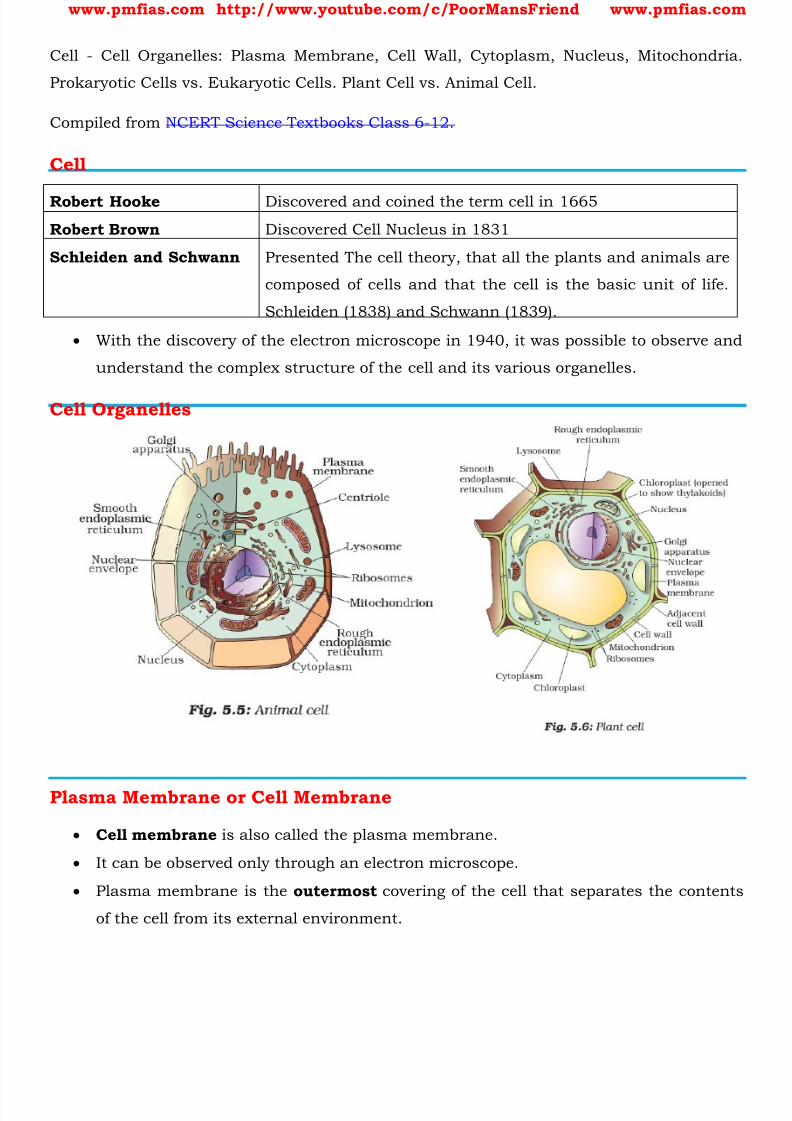
www.pmfias.com http://www.youtube.com/c/PoorMansFriend www.pmfias.com
Cell - Cell Organelles: Plasma Membrane, Cell Wall, Cytoplasm, Nucleus, Mitochondria.
Prokaryotic Cells vs. Eukaryotic Cells. Plant Cell vs. Animal Cell.
Compiled from NCERT Science Textbooks Class 6-12.
Cell
Robert Hooke Discovered and coined the term cell in 1665
Robert Brown Discovered Cell Nucleus in 1831
Schleiden and Schwann Presented The cell theory, that all the plants and animals are
composed of cells and that the cell is the basic unit of life.
Schleiden (1838) and Schwann (1839).
With the discovery of the electron microscope in 1940, it was possible to observe and
understand the complex structure of the cell and its various organelles.
Cell Organelles
Plasma Membrane or Cell Membrane
Cell membrane is also called the plasma membrane.
It can be observed only through an electron microscope.
Plasma membrane is the outermost covering of the cell that separates the contents
of the cell from its external environment.

8/16/2019 Biology for Prelims Exam
http://slidepdf.com/reader/full/biology-for-prelims-exam 4/384
www.pmfias.com http://www.youtube.com/c/PoorMansFriend www.pmfias.com
Endocytosis
The plasma membrane is flexible and is made up of organic molecules called lipids
and proteins .
The flexibility of the cell membrane also enables the cell to engulf in food and other
material from its external environment. Such processes are known as endocytosis
(endo → internal; cyto → of a cell). Amoeba acquires its food through such processes.
Diffusion
Plasma membrane is a selectively permeable membrane [The plasma membrane is
porous and allows the movement of substances or materials both inward and
outward].
Some substances like carbon dioxide or oxygen can move across the cell membrane
by a process called diffusion [spontaneous movement of a substance from a region ofhigh concentration (hypertonic solution) to a region where its concentration is low
(hypotonic solution)].
Thus, diffusion plays an important role in gaseous exchange between the cells as well
as the cell and its external environment.
Osmosis
Water also obeys the law of diffusion. The movement of water molecules through a
selectively permeable membrane is called osmosis.
Osmosis is the passage of water from a region of high water concentration through a
semi-permeable membrane to a region of low water concentration. Thus, osmosis is
a special case of diffusion through a selectively permeable membrane.
Unicellular freshwater organisms and most plant cells tend to gain water through
osmosis. Absorption of water by plant roots is also an example of osmosis.
Thus, diffusion is important in exchange of gases and water in the life of a cell. In
additions to this, the cell also obtains nutrition from its environment.
Different molecules move in and out of the cell through a type of transport requiring
use of energy in the form of ATP.
Reverse Osmosis (RO)
Reverse osmosis (RO) is a water purification technology that uses a semipermeable
membrane to remove larger particles from drinking water.

8/16/2019 Biology for Prelims Exam
http://slidepdf.com/reader/full/biology-for-prelims-exam 5/384
www.pmfias.com http://www.youtube.com/c/PoorMansFriend www.pmfias.com
In reverse osmosis, an applied pressure is used to overcome osmotic pressure.
Reverse Osmosis is a phenomenon where pure water flows from a dilute solution
[hypotonic] through a semi permeable membrane to a higher concentrated solution
[hypertonic].
Semi permeable means that the membrane will allow small molecules and ions to
pass through it but acts as a barrier to larger molecules or dissolved substances.
Cell Wall
Cell wall is absent in animals .
Plant cells, in addition to the plasma membrane, have another rigid outer covering
called the cell wall. The cell wall lies outside the plasma membrane.
The plant cell wall is mainly composed of cellulose . Cellulose is a complex substance
and provides structural strength to plants.
Plasmolysis
When a living plant cell loses water through osmosis there is shrinkage or
contraction of the contents of the cell away from the cell wall. This phenomenon is
known as plasmolysis (plasma → fluid; lysis → disintegration, decomposition).
Only living cells , and not dead cells, are able to absorb water by osmosis. Cell walls
permit the cells of plants, fungi and bacteria to withstand very dilute [hypotonic]
external media without shrinkage.
In such media the cells tend to lose water by osmosis. The cell shrinks, building up
pressure against the cell wall. The wall exerts an equal pressure against the
shrunken cell.
Cell wall also prevents the bursting of cells when the cells are surrounded by a
hypertonic medium (medium of high concentration).
In such media the cells tend to gain water by osmosis. The cell swells, building up
pressure against the cell wall. The wall exerts an equal pressure against the swollen
cell.
Because of their walls, plant cells can withstand much greater changes in the
surrounding medium than animal cells.
Cytoplasm
It is the jelly-like substance present between the cell membrane and the nucleus .

8/16/2019 Biology for Prelims Exam
http://slidepdf.com/reader/full/biology-for-prelims-exam 6/384
www.pmfias.com http://www.youtube.com/c/PoorMansFriend www.pmfias.com
The cytoplasm is the fluid content inside the plasma membrane.
It also contains many specialized cell organelles [ mitochondria, golgi bodies,
ribosomes, etc ].
Each of these organelles performs a specific function for the cell.
Cell organelles are enclosed by membranes .
The significance of membranes can be illustrated with the example of viruses. Viruses lack any membranes and hence do not show characteristics of life until
they enter a living body and use its cell machinery to multiply.
Nucleus
It is an important component of the living cell.
It is generally spherical and located in the center of the cell.
It can be stained and seen easily with the help of a microscope.
Nucleus is separated from the cytoplasm by a double layered membrane called the
nuclear membrane .
This membrane is also porous and allows the movement of materials between the
cytoplasm and the inside of the nucleus [diffusion].
With a microscope of higher magnification, we can see a smaller spherical body in the
nucleus. It is called the nucleolus .
In addition, nucleus contains thread-like structures called chromosomes . Thesecarry genes and help in inheritance or transfer of characters from the parents to the
offspring. The chromosomes can be seen only when the cell divides .
Gene is a unit of inheritance in living organisms. It controls the transfer of a
hereditary characteristic from parents to offspring. This means that your parents
pass some of their characteristics on to you.
Nucleus, in addition to its role in inheritance, acts as control center of the activities
of the cell.
The entire content of a living cell is known as protoplasm [cytoplasm + nucleus] . It
includes the cytoplasm and the nucleus. Protoplasm is called the living substance of
the cell.
The nucleus of the bacterial cell is not well organized like the cells of multicellular
organisms. There is no nuclear membrane .

8/16/2019 Biology for Prelims Exam
http://slidepdf.com/reader/full/biology-for-prelims-exam 7/384
www.pmfias.com http://www.youtube.com/c/PoorMansFriend www.pmfias.com
Every cell has a membrane around it to keep its own contents separate from the
external environment.
Large and complex cells, including cells from multicellular organisms, need a lot of
chemical activities to support their complicated structure and function.
To keep these activities of different kinds separate from each other, these cells use
membrane- bound little structures (or ‘organelles’) within themselves.
Chromosomes
The nucleus contains chromosomes, which are visible as rod-shaped structures only
when the cell is about to divide.
Chromosomes contain information for inheritance of features from parents to next
generation in the form of DNA (deoxyribo nucleic acid) molecules.
Chromosomes are composed of DNA and Protein .
DNA molecules contain the information necessary for constructing and organizing
cells. Functional segments of dna are called genes .
In a cell which is not dividing, this dna is present as part of chromatin material
Chromatin material is visible as entangled mass of thread like structures. Whenever
the cell is about to divide, the chromatin material gets organised into
chromosomes .
The nucleus plays a central role in cellular reproduction , the process by which a
single cell divides and forms two new cells.
It also plays a crucial part, along with the environment, in determining the way the
cell will develop and what form it will exhibit at maturity, by directing the chemical
activities of the cell.
Prokaryotic Cells vs. Eukaryotic Cells
Organisms whose cells lack a nuclear membrane , are called prokaryotes (pro =
primitive or primary; karyote ≈karyon = nucleus).
Organisms with cells having a nuclear membrane are called eukaryotes.
Prokaryotic cells also lack most of the other cytoplasmic organelles present in
eukaryotic cells.
Many of the functions of such organelles are also performed by poorly organised
parts of the cytoplasm .

8/16/2019 Biology for Prelims Exam
http://slidepdf.com/reader/full/biology-for-prelims-exam 8/384
www.pmfias.com http://www.youtube.com/c/PoorMansFriend www.pmfias.com
The chlorophyll in photosynthetic prokaryotic bacteria is associated with
membranous vesicles (bag like structures) but not with plastids as in eukaryotic
cells.
Prokaryotes → defined nuclear region, the membrane-bound cell organelles are absent.
Eukaryotic Cells → have nuclear membrane as well as membrane-enclosed organelles.
Prokaryotes Eukaryotes
Organisms Monera: Eubacteria and
Archebacteria
Protists, Fungi, Plants
and Animals
Meaning of name Pro = before
Karyon = nucleus
Eu = after
Karyon = nucleus
Evolution 3.5 billion years ago (older type
of cell)
1.5 billion years ago
Uni-/multicellular Unicellular (less
complex)
Multicellular (more complex)
Cell wall almost all have cell walls
(murein)
fungi and plants (cellulose and
chitin): none in animals
Organelles usually none many different ones with
specialized functions
Metabolism anaerobic and aerobic: diverse mostly aerobicGenetic
material
single circular double stranded
DNA
complex chromosomes usually
in pairs; each with a single
double stranded DNA molecule
and associated proteins
contained in a nucleus
Location of genetic Nucleoid region Nucleus

8/16/2019 Biology for Prelims Exam
http://slidepdf.com/reader/full/biology-for-prelims-exam 9/384
www.pmfias.com http://www.youtube.com/c/PoorMansFriend www.pmfias.com
information
Mode of
division
binary fission mostly; budding mitosis and meiosis using a
spindle: followed by
cytokinesis
Nucleoid
In some organisms like bacteria, the nuclear region of the cell may be poorly defined
due to the absence of a nuclear membrane . Such an undefined nuclear region
containing only nucleic acids is called a nucleoid.
Vacuoles
Empty structure in the cytoplasm is called vacuole. It could be single and big as in
an onion cell (plant cell). Cheek cells (animal cells) have smaller vacuoles.
Large vacuoles are common in plant cells. Vacuoles in animal cells are muchsmaller.
Vacuoles are storage sacs for solid or liquid contents.
The central vacuole of some plant cells may occupy 50-90% of the cell volume.
In plant cells vacuoles are full of cell sap and provide turgidity [swollen and
distended or congested] and rigidity to the cell.
Many substances of importance in the life of the plant cell are stored in vacuoles.
These include amino acids, sugars, various organic acids and some proteins. In single-celled organisms like amoeba, the food vacuole contains the food items that
the amoeba has consumed.
In some unicellular organisms, specialized vacuoles also play important roles in
expelling excess water and some wastes from the cell
Endoplasmic Reticulum (ER)
The endoplasmic reticulum (ER) is a large network of membrane-bound tubes and
sheets. It looks like long tubules or round or long bags (vesicles).
The ER membrane is similar in structure to the plasma membrane.
There are two types of ER –– rough endoplasmic reticulum (RER) and smooth
endoplasmic reticulum (SER) .
Rough Endoplasmic Reticulum RER – Ribosomes

8/16/2019 Biology for Prelims Exam
http://slidepdf.com/reader/full/biology-for-prelims-exam 10/384
www.pmfias.com http://www.youtube.com/c/PoorMansFriend www.pmfias.com
RER looks rough under a microscope because it has particles called ribosomes
attached to its surface.
The ribosomes, which are present in all active cells, are the sites of protein
manufacture.
The manufactured proteins are then sent to various places in the cell depending on
need, using the ER.
Smooth Endoplasmic Reticulum SER
The SER helps in the manufacture of fat molecules, or lipids , important for cell
function.
Functions of Endoplasmic Reticulum (ER)
Some of these proteins and lipids help in building the cell membrane. This process is
known as membrane biogenesis .
Some other proteins and lipids function as enzymes and hormones .
Although the ER varies greatly in appearance in different cells, it always forms a
network system.
Thus, one function of the ER is to serve as channels for the transport of materials
(especially proteins) between various regions of the cytoplasm or between the
cytoplasm and the nucleus.
The ER also functions as a cytoplasmic framework providing a surface for some of
the biochemical activities of the cell.
In the liver cells of the group of animals called vertebrates, SER plays a crucial
role in detoxifying many poisons and drugs .
Golgi Apparatus or Golgi Complex
The golgi apparatus consists of a system of membrane-bound vesicles arranged
approximately parallel to each other in stacks called cisterns . These membranes often have connections with the membranes of ER and therefore
constitute another portion of a complex cellular membrane system.
The material synthesized near the ER is packaged and dispatched to various targets
inside and outside the cell through the golgi apparatus.
Its functions include the storage, modification and packaging of products in
vesicles.
8/16/2019 Biology for Prelims Exam
http://slidepdf.com/reader/full/biology-for-prelims-exam 11/384
www.pmfias.com http://www.youtube.com/c/PoorMansFriend www.pmfias.com
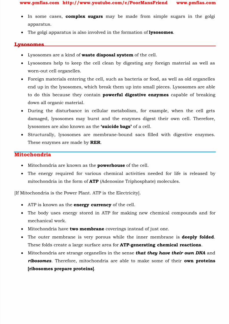
In some cases, complex sugars may be made from simple sugars in the golgi
apparatus.
The golgi apparatus is also involved in the formation of lysosomes .
Lysosomes
Lysosomes are a kind of waste disposal system of the cell.
Lysosomes help to keep the cell clean by digesting any foreign material as well as
worn-out cell organelles.
Foreign materials entering the cell, such as bacteria or food, as well as old organelles
end up in the lysosomes, which break them up into small pieces. Lysosomes are able
to do this because they contain powerful digestive enzymes capable of breaking
down all organic material.
During the disturbance in cellular metabolism, for example, when the cell gets
damaged, lysosomes may burst and the enzymes digest their own cell. Therefore,
lysosomes are also known as the ‘suicide bags’ of a cell.
Structurally, lysosomes are membrane-bound sacs filled with digestive enzymes.
These enzymes are made by RER .
Mitochondria
Mitochondria are known as the powerhouse of the cell.
The energy required for various chemical activities needed for life is released by
mitochondria in the form of ATP (Adenosine Triphosphate) molecules.
[If Mitochondria is the Power Plant. ATP is the Electricity].
ATP is known as the energy currency of the cell.
The body uses energy stored in ATP for making new chemical compounds and for
mechanical work.
Mitochondria have two membrane coverings instead of just one.
The outer membrane is very porous while the inner membrane is deeply folded
These folds create a large surface area for ATP-generating chemical reactions .
Mitochondria are strange organelles in the sense that they have their own DNA and
ribosomes . Therefore, mitochondria are able to make some of their own proteins
[ribosomes prepare proteins] .

8/16/2019 Biology for Prelims Exam
http://slidepdf.com/reader/full/biology-for-prelims-exam 12/384
www.pmfias.com http://www.youtube.com/c/PoorMansFriend www.pmfias.com
Plastids
You might have noticed several small colored bodies in the cytoplasm of the cells of
Tradescantia leaf. They are scattered in the cytoplasm of the leaf cells. These are
called plastids.
They are of different colours . Some of them contain green pigment called
chlorophyll . Green coloured plastids are called chloroplasts . They provide green
colour to the leaves.
Chloroplasts are important for photosynthesis in plants.
Chloroplasts also contain various yellow or orange pigments in addition to
chlorophyll.
Plastids are present only in plant cells . There are two types of plastids –
chromoplasts (coloured plastids) and leucoplasts (white or colourless plastids) .
Leucoplasts are primarily organelles in which materials such as starch, oils and
protein granules are stored.
The internal organization of the plastids consists of numerous membrane layers
embedded in a material called the stroma.
Plastids are similar to mitochondria in external structure. Like the mitochondria,
plastids also have their own dna and ribosomes .
Summary
Each cell acquires its structure and ability to function because of the organization of
its membrane and organelles in specific ways. The cell thus has a basic structural
organization. This helps the cells to perform functions like respiration, obtaining
nutrition, and clearing of waste material, or forming new proteins. Thus, the cell is
the fundamental structural unit of living organisms. It is also the basic functional
unit of life.
Cells are enclosed by a plasma membrane composed of lipids and proteins .
The presence of the cell wall enables the cells of plants, fungi and bacteria to exist in
hypotonic media without bursting.
The ER functions both as a passage way for intracellular transport and as a
manufacturing surface.
The golgi apparatus consists of stacks of membrane-bound vesicles that function in
the storage, modification and packaging of substances manufactured in the cell.
8/16/2019 Biology for Prelims Exam
http://slidepdf.com/reader/full/biology-for-prelims-exam 13/384
www.pmfias.com http://www.youtube.com/c/PoorMansFriend www.pmfias.com
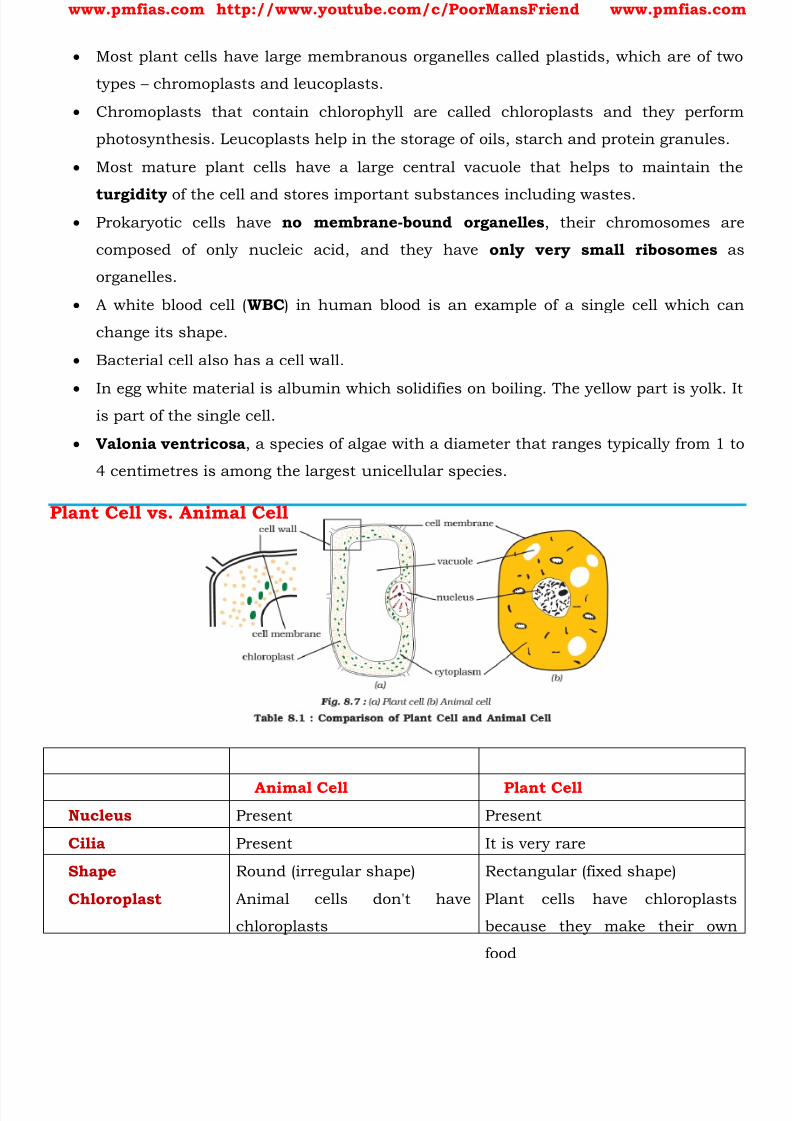
Most plant cells have large membranous organelles called plastids, which are of two
types – chromoplasts and leucoplasts.
Chromoplasts that contain chlorophyll are called chloroplasts and they perform
photosynthesis. Leucoplasts help in the storage of oils, starch and protein granules.
Most mature plant cells have a large central vacuole that helps to maintain the
turgidity of the cell and stores important substances including wastes. Prokaryotic cells have no membrane-bound organelles , their chromosomes are
composed of only nucleic acid, and they have only very small ribosomes as
organelles.
A white blood cell ( WBC ) in human blood is an example of a single cell which can
change its shape.
Bacterial cell also has a cell wall.
In egg white material is albumin which solidifies on boiling. The yellow part is yolk. Itis part of the single cell.
Valonia ventricosa , a species of algae with a diameter that ranges typically from 1 to
4 centimetres is among the largest unicellular species.
Plant Cell vs. Animal Cell
Animal Cell Plant CellNucleus Present Present
Cilia Present It is very rare
Shape Round (irregular shape) Rectangular (fixed shape)
Chloroplast Animal cells don't have
chloroplasts
Plant cells have chloroplasts
because they make their own
food
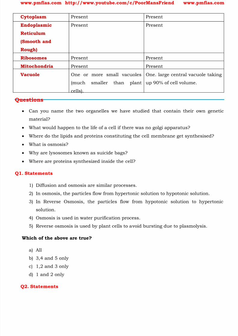
8/16/2019 Biology for Prelims Exam
http://slidepdf.com/reader/full/biology-for-prelims-exam 14/384
www.pmfias.com http://www.youtube.com/c/PoorMansFriend www.pmfias.com
Cytoplasm Present Present
Endoplasmic
Reticulum
(Smooth and
Rough)
Present Present
Ribosomes Present PresentMitochondria Present Present
Vacuole One or more small vacuoles
(much smaller than plant
cells).
One. large central vacuole taking
up 90% of cell volume.
Questions
Can you name the two organelles we have studied that contain their own genetic
material? What would happen to the life of a cell if there was no golgi apparatus?
Where do the lipids and proteins constituting the cell membrane get synthesised?
What is osmosis?
Why are lysosomes known as suicide bags?
Where are proteins synthesized inside the cell?
Q1. Statements
1) Diffusion and osmosis are similar processes.
2) In osmosis, the particles flow from hypertonic solution to hypotonic solution.
3) In Reverse Osmosis, the particles flow from hypotonic solution to hypertonic
solution.
4) Osmosis is used in water purification process.
5) Reverse osmosis is used by plant cells to avoid bursting due to plasmolysis.
Which of the above are true?
a) All
b) 3,4 and 5 only
c) 1,2 and 3 only
d) 1 and 2 only
Q2. Statements

8/16/2019 Biology for Prelims Exam
http://slidepdf.com/reader/full/biology-for-prelims-exam 15/384
www.pmfias.com http://www.youtube.com/c/PoorMansFriend www.pmfias.com
1) Protoplasm = Cytoplasm + Nucleus + Plasma Membrane
2) Osmosis happens in dead cells as well.
3) Bacteria have cell walls.
4) Virus are non-living substances.
5) Animals have no cell walls and vacuoles.
Which of the above are true?
a) All
b) 3,4 only
c) 2, 3 and 5 only
d) 1, 3 and 4 only
Answers
Q1 → C
Q2 → B
Any doubts? Leave a comment..
8/16/2019 Biology for Prelims Exam
http://slidepdf.com/reader/full/biology-for-prelims-exam 16/384
www.pmfias.com http://www.youtube.com/c/PoorMansFriend www.pmfias.com
Biomolecules – Carbohydrates – Monosaccharides: Glucose, Fructose; Disaccharides:
Sucrose, Lactose; Oligosaccharides and Polysaccharides: Starch, Cellulose, Glycogen.
Biomolecule
A biomolecule [biological molecule] is any molecule that is present in living organisms
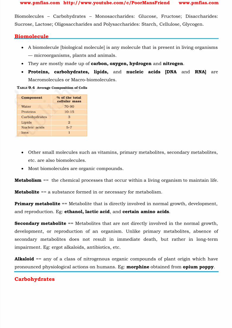
–– microorganisms, plants and animals. They are mostly made up of carbon, oxygen, hydrogen and nitrogen .
Proteins, carbohydrates, lipids, and nucleic acids [DNA and RNA] are
Macromolecules or Macro-biomolecules.
Other small molecules such as vitamins, primary metabolites, secondary metabolites,
etc. are also biomolecules.
Most biomolecules are organic compounds.
Metabolism == the chemical processes that occur within a living organism to maintain life.
Metabolite == a substance formed in or necessary for metabolism.
Primary metabolite == Metabolite that is directly involved in normal growth, development,
and reproduction. Eg: ethanol, lactic acid , and certain amino acids .
Secondary metabolite == Metabolites that are not directly involved in the normal growth,
development, or reproduction of an organism. Unlike primary metabolites, absence of
secondary metabolites does not result in immediate death, but rather in long-term
impairment. Eg: ergot alkaloids, antibiotics, etc.
Alkaloid == any of a class of nitrogenous organic compounds of plant origin which have
pronounced physiological actions on humans. Eg: morphine obtained from opium poppy .
Carbohydrates
8/16/2019 Biology for Prelims Exam
http://slidepdf.com/reader/full/biology-for-prelims-exam 17/384
www.pmfias.com http://www.youtube.com/c/PoorMansFriend www.pmfias.com
Carbohydrates are one of the most important biomolecules that forms a major part of
the living things.
Carbohydrates are primarily produced by plants and form a very large group of
naturally occurring organic compounds.
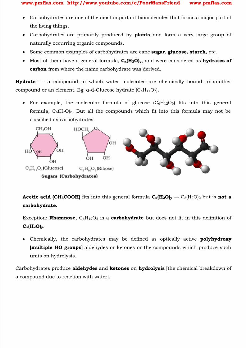
Some common examples of carbohydrates are cane sugar, glucose, starch, etc.
Most of them have a general formula, C x (H 2 O) y , and were considered as hydrates ofcarbon from where the name carbohydrate was derived.
Hydrate == a compound in which water molecules are chemically bound to another
compound or an element. Eg: α -d-Glucose hydrate (C 6 H 14 O 7 ).
For example, the molecular formula of glucose (C 6H 12 O 6 ) fits into this general
formula, C 6 (H2O) 6 . But all the compounds which fit into this formula may not be
classified as carbohydrates.
Acetic acid (CH 3 COOH) fits into this general formula C x (H 2 O) y → C2 (H2O)2 but is not a
carbohydrate.
Exception: Rhamnose , C 6H 12 O 5 is a carbohydrate but does not fit in this definition of
C x (H 2 O)y .
Chemically, the carbohydrates may be defined as optically active polyhydroxy
[multiple HO groups] aldehydes or ketones or the compounds which produce such
units on hydrolysis.
Carbohydrates produce aldehydes and ketones on hydrolysis [the chemical breakdown of
a compound due to reaction with water].
8/16/2019 Biology for Prelims Exam
http://slidepdf.com/reader/full/biology-for-prelims-exam 18/384
www.pmfias.com http://www.youtube.com/c/PoorMansFriend www.pmfias.com
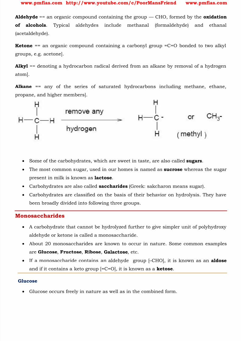
Aldehyde == an organic compound containing the group — CHO, formed by the oxidation
of alcohols . Typical aldehydes include methanal (formaldehyde) and ethanal
(acetaldehyde).
Ketone == an organic compound containing a carbonyl group =C=O bonded to two alkyl
groups, e.g. acetone].
Alkyl == denoting a hydrocarbon radical derived from an alkane by removal of a hydrogen
atom].
Alkane == any of the series of saturated hydrocarbons including methane, ethane,
propane, and higher members].
Some of the carbohydrates, which are sweet in taste, are also called sugars .
The most common sugar, used in our homes is named as sucrose whereas the sugar
present in milk is known as lactose . Carbohydrates are also called saccharides (Greek: sakcharon means sugar).
Carbohydrates are classified on the basis of their behavior on hydrolysis. They have
been broadly divided into following three groups.
Monosaccharides
A carbohydrate that cannot be hydrolyzed further to give simpler unit of polyhydroxy
aldehyde or ketone is called a monosaccharide.
About 20 monosaccharides are known to occur in nature. Some common examples
are Glucose , Fructose , Ribose , Galactose , etc.
If a monosaccharide contains an aldehyde group [ – CHO], it is known as an aldose
and if it contains a keto group [=C=O], it is known as a ketose .
Glucose
Glucose occurs freely in nature as well as in the combined form.
8/16/2019 Biology for Prelims Exam
http://slidepdf.com/reader/full/biology-for-prelims-exam 19/384
www.pmfias.com http://www.youtube.com/c/PoorMansFriend www.pmfias.com
It is present in sweet fruits and honey . Ripe grapes also contain glucose in large
amounts.
Glucose is an aldohexose [ An aldohexose is a hexose with an aldehyde group on one
end ] and is also known as dextrose . It is the monomer of many of the larger
carbohydrates, namely starch, cellulose .
Aldohexose == An aldohexose is a hexose with an aldehyde group on one end.
Aldehyde group [ – CHO]
Hexose == any of the class of simple sugars whose molecules contain six carbon atoms
(e.g. glucose)
It is probably the most abundant organic compound on earth.
Glucose is found to exist in two different crystalline forms which are named as α and
β.
Such isomers, i.e., α -form and β -form, are called anomers .
Fructose
Fructose is an important ketohexose . It is obtained along with glucose by the
hydrolysis of disaccharide, sucrose .
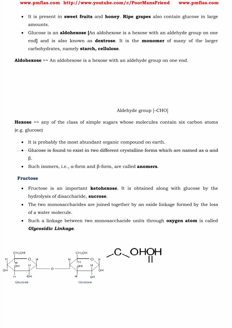
The two monosaccharides are joined together by an oxide linkage formed by the loss
of a water molecule.
Such a linkage between two monosaccharide units through oxygen atom is called
Glycosidic Linkage .
8/16/2019 Biology for Prelims Exam
http://slidepdf.com/reader/full/biology-for-prelims-exam 20/384
www.pmfias.com http://www.youtube.com/c/PoorMansFriend www.pmfias.com
Ribose
The ribose β -D-ribofuranose forms part of the backbone of RNA. It is related to
deoxyribose, which is found in DNA.
Galactose
Galactose is a monosaccharide. When combined with glucose (monosaccharide),through a condensation reaction, the result is the disaccharide lactose .
The hydrolysis of lactose to glucose and galactose is catalyzed by the enzymes
lactase and β -galactosidase .
Oligosaccharides
Carbohydrates that yield two to ten monosaccharide units, on hydrolysis, are called
oligosaccharides.
They are further classified as disaccharides , trisaccharides , tetrasaccharides , etc.,
depending upon the number of monosaccharides, they provide on hydrolysis.
Amongst these the most common are disaccharides .
The two monosaccharide units obtained on hydrolysis of a disaccharide may be same
or different.
For example, sucrose on hydrolysis gives one molecule each of glucose and fructose
whereas maltose gives two molecules of glucose only.
Sucrose == Glucose + Fructose
Maltose == Glucose + Glucose
Lactose == Glucose + Galactose
Sucrose
One of the common disaccharides is sucrose which on hydrolysis gives equimolar
mixture of glucose and fructose.
Maltose
Another disaccharide, maltose is composed of two α -D-glucose units
Lactose

8/16/2019 Biology for Prelims Exam
http://slidepdf.com/reader/full/biology-for-prelims-exam 21/384
www.pmfias.com http://www.youtube.com/c/PoorMansFriend www.pmfias.com
It is more commonly known as milk sugar since this disaccharide is found in milk
It is composed of β -D- galactose and β -D-glucose.
Polysaccharides
Carbohydrates which yield a large number of monosaccharide units on hydrolysis are
called polysaccharides. Some common examples are Starch, Cellulose, Glycogen, Gums, etc.
Polysaccharides are long chains of sugars . Polysaccharides are not sweet in taste,
hence they are also called non-sugars .
They are threads (literally a cotton thread) containing different monosaccharides as
building blocks.
For example, Cellulose is a polymeric polysaccharide consisting of only one type of
monosaccharide i.e., Glucose . Cellulose is a homopolymer. Starch is a variant of this
but present as a store house of energy in plant tissues.
Animals have another variant called Glycogen .
Inulin is a polymer of fructose .
Plant cell walls are made of cellulose. Paper made from plant pulp and cotton fibre is
cellulosic . There are more complex polysaccharides in nature.
Exoskeletons of arthropods, for example, have a complex polysaccharide called
chitin. These complex polysaccharides are mostly homopolymers.
Starch
Polysaccharides contain a large number of monosaccharide units joined together by
glycosidic linkages .
These are the most commonly encountered carbohydrates in nature.
They mainly act as the food storage or structural materials.
Starch is the main storage polysaccharide of plants.
It is the most important dietary source for human beings.
High content of starch is found in cereals, roots, tubers and some vegetables.
It is a polymer of α -glucose and consists of two components — Amylose and
Amylopectin .
Amylose is water soluble polysaccharide which constitutes about 15-20% of starch.
Amylopectin is water insoluble polysaccharide which constitutes about 80- 85% of
starch.

8/16/2019 Biology for Prelims Exam
http://slidepdf.com/reader/full/biology-for-prelims-exam 22/384
www.pmfias.com http://www.youtube.com/c/PoorMansFriend www.pmfias.com
Cellulose
Cellulose occurs exclusively in plants and it is the most abundant organic
substance in plant kingdom.
It is a predominant constituent of cell wall of plant cells.
Cellulose is a straight chain polysaccharide composed only of β -D-glucose units .
Glycogen
The carbohydrates are stored in animal body as Glycogen.
It is also known as animal starch because its structure is similar to amylopectin and
is rather more highly branched.
It is present in liver, muscles and brain .
Glycogen is also found in yeast and fungi.
When the body needs glucose, enzymes break the glycogen down to glucose.
Importance of Carbohydrates
Carbohydrates are essential for life in both plants and animals.
They form a major portion of our food. Honey has been used for a long time as an
instant source of energy in ayurvedic system of medicine.
Carbohydrates are used as storage molecules as starch in plants and glycogen in
animals.
Cell wall of bacteria and plants is made up of cellulose which is a carbohydrate.
We build furniture, etc. from cellulose in the form of wood and clothe ourselves with
cellulose in the form of cotton fibre .
They provide raw materials for many important industries like textiles, paper,
lacquers and breweries.
Summary
Carbohydrates are optically active polyhydroxy aldehydes or ketones or molecules
which provide such units on hydrolysis.
They are broadly classified into three groups — monosaccharides, disaccharides and
polysaccharides.
Glucose, the most important source of energy for mammals, is obtained by the
digestion of starch.

8/16/2019 Biology for Prelims Exam
http://slidepdf.com/reader/full/biology-for-prelims-exam 23/384
www.pmfias.com http://www.youtube.com/c/PoorMansFriend www.pmfias.com
Monosaccharides are held together by glycosidic linkages to form disaccharides or
polysaccharides.
8/16/2019 Biology for Prelims Exam
http://slidepdf.com/reader/full/biology-for-prelims-exam 24/384
www.pmfias.com http://www.youtube.com/c/PoorMansFriend www.pmfias.com
General Science for UPSC: Amino Acids – Proteins – Structure of Proteins, Fibrous proteins,
Globular proteins, Role of Proteins. Enzymes, Factors Affecting Enzyme Activity.
Amino Acids
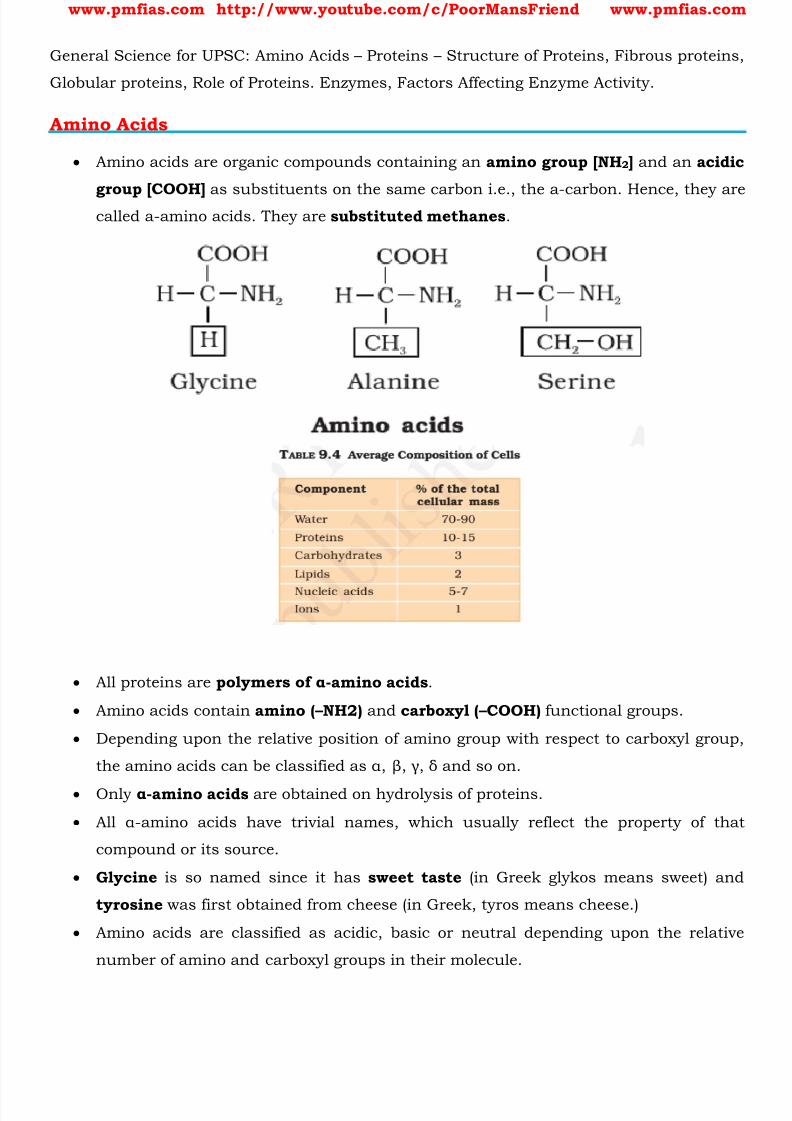
Amino acids are organic compounds containing an amino group [NH 2 ] and an acidic
group [COOH] as substituents on the same carbon i.e., the a-carbon. Hence, they arecalled a-amino acids. They are substituted methanes .
All proteins are polymers of α -amino acids .
Amino acids contain amino ( – NH2) and carboxyl ( – COOH) functional groups.
Depending upon the relative position of amino group with respect to carboxyl group,
the amino acids can be classified as α, β, γ, δ and so on.
Only α -amino acids are obtained on hydrolysis of proteins.
All α -amino acids have trivial names, which usually reflect the property of that
compound or its source.
Glycine is so named since it has sweet taste (in Greek glykos means sweet) and
tyrosine was first obtained from cheese (in Greek, tyros means cheese.)
Amino acids are classified as acidic, basic or neutral depending upon the relative
number of amino and carboxyl groups in their molecule.

8/16/2019 Biology for Prelims Exam
http://slidepdf.com/reader/full/biology-for-prelims-exam 25/384
www.pmfias.com http://www.youtube.com/c/PoorMansFriend www.pmfias.com
1. Equal number of amino and carboxyl groups makes it neutral;
2. more number of amino than carboxyl groups makes it basic and
3. more carboxyl groups as compared to amino groups makes it acidic.
The amino acids, which can be synthesized in the body, are known as nonessential
amino acids .
On the other hand, those which cannot be synthesized in the body and must beobtained through diet, are known as essential amino acids.
Amino acids are usually colorless, crystalline solids. These are water-soluble , high
melting solids and behave like salts rather than simple amines or carboxylic acids.
This behavior is due to the presence of both acidic (carboxyl group) and basic
(amino group) groups in the same molecule.
In aqueous solution, the carboxyl group can lose a proton and amino group can
accept a proton, giving rise to a dipolar ion known as zwitter ion . This is neutral butcontains both positive and negative charges.
In zwitter ionic form, amino acids show amphoteric behavior as they react both with
acids and bases.
Except glycine , all other naturally occurring α -amino acids are optically active
since the α -carbon atom is asymmetric.
Optically Active: https://www.youtube.com/watch?v=gBELxxGbzKk
Proteins
Proteins are the most abundant biomolecules of the living system.
Chief sources of proteins are milk, cheese, pulses, peanuts, fish, meat, etc.
They occur in every part of the body and form the fundamental basis of structure and
functions of life.
They are also required for growth and maintenance of body.
The word protein is derived from G reek word, “proteios” which means primary or ofprime importance.
Proteins are polypeptides .
[Peptide == a compound consisting of two or more amino acids linked in a chain].
Proteins are linear chains of amino acids linked by peptide bonds .
Each protein is a polymer of amino acids .
8/16/2019 Biology for Prelims Exam
http://slidepdf.com/reader/full/biology-for-prelims-exam 26/384
www.pmfias.com http://www.youtube.com/c/PoorMansFriend www.pmfias.com
[Monomer == a molecule that can be bonded to other identical molecules to form a
polymer].
Dietary proteins are the source of essential amino acids .
Therefore, amino acids can be essential or non-essential.
[Non-Essential Amino Acids == Amino Acids that our body can make].
[Essential Amino Acids == We get them through our diet/food].
Collagen is the most abundant protein in animal world.
Ribulose bisphosphate Carboxylase-Oxygenase (RuBisCO) is the most abundant
protein in the whole of the biosphere.
Structure of Proteins
You have already read that proteins are the polymers of α -amino acids and they are
connected to each other by peptide bond or peptide linkage .
Chemically, peptide linkage is an amide [an organic compound containing the group -
C(O)NH2] formed between – COOH group and – NH 2 group.
The reaction between two molecules of similar or different amino acids, proceeds
through the combination of the amino group of one molecule with the carboxyl
group of the other .
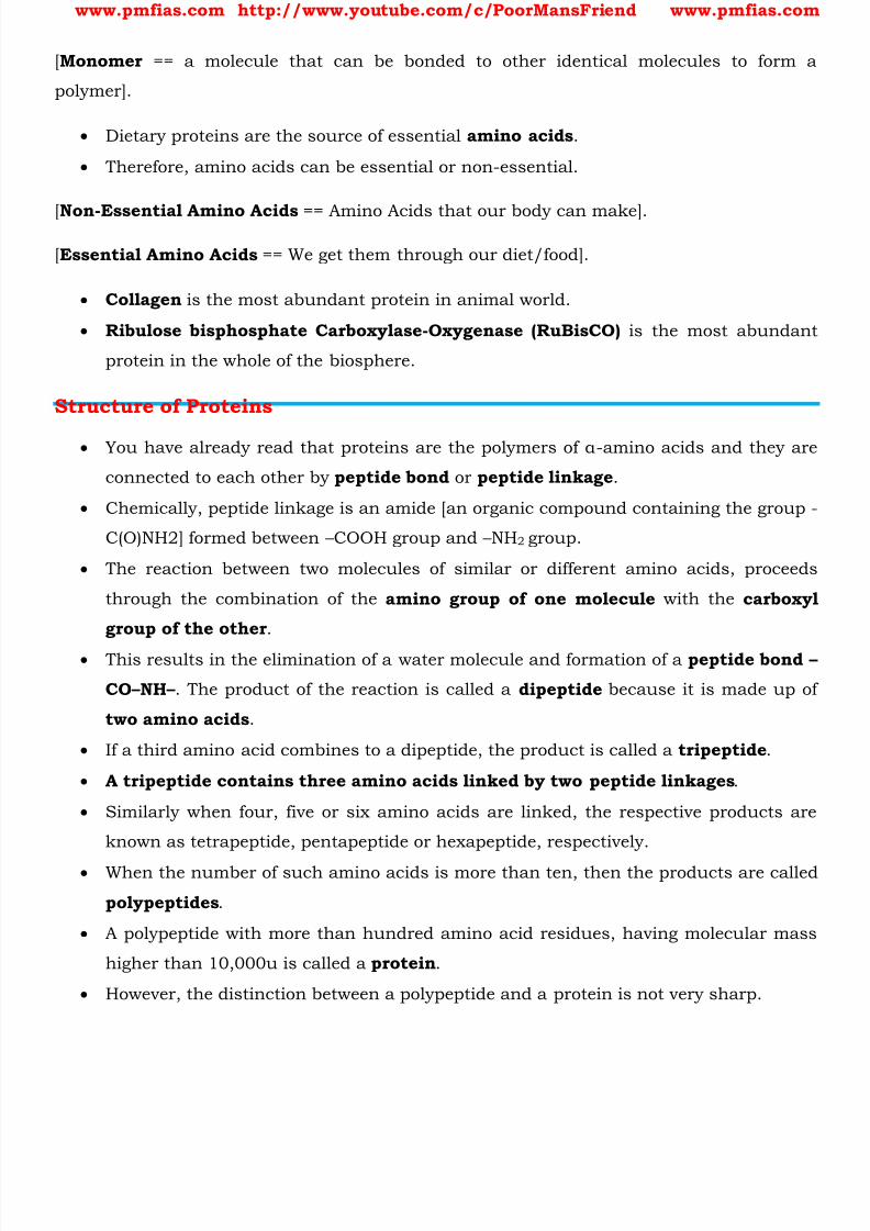
This results in the elimination of a water molecule and formation of a peptide bond –
CO – NH – . The product of the reaction is called a dipeptide because it is made up of
two amino acids .
If a third amino acid combines to a dipeptide, the product is called a tripeptide .
A tripeptide contains three amino acids linked by two peptide linkages .
Similarly when four, five or six amino acids are linked, the respective products are
known as tetrapeptide, pentapeptide or hexapeptide, respectively.
When the number of such amino acids is more than ten, then the products are called
polypeptides .
A polypeptide with more than hundred amino acid residues, having molecular mass
higher than 10,000u is called a protein .
However, the distinction between a polypeptide and a protein is not very sharp.
8/16/2019 Biology for Prelims Exam
http://slidepdf.com/reader/full/biology-for-prelims-exam 27/384
www.pmfias.com http://www.youtube.com/c/PoorMansFriend www.pmfias.com
Polypeptides with fewer amino acids are likely to be called proteins if they ordinarily
have a well-defined conformation of a protein such as insulin which contains 51
amino acids .
Proteins can be classified into two types on the basis of their molecular shape:
Fibrous Proteins and Globular proteins .
Fibrous proteins
When the polypeptide chains run parallel and are held together by hydrogen and
disulphide bonds, then fibre – like structure is formed.
Such proteins are generally insoluble in water . Some common examples are keratin
(present in hair, wool, silk) and myosin (present in muscles), etc.
Globular proteins
This structure results when the chains of polypeptides coil around to give a spherical
shape .
These are usually soluble in water . Insulin and albumin s are the common examples
of globular proteins.
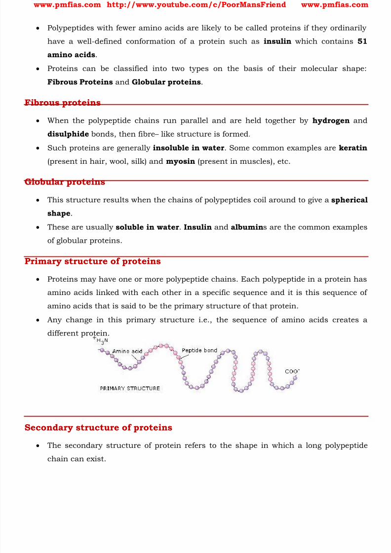
Primary structure of proteins
Proteins may have one or more polypeptide chains. Each polypeptide in a protein has
amino acids linked with each other in a specific sequence and it is this sequence of
amino acids that is said to be the primary structure of that protein.
Any change in this primary structure i.e., the sequence of amino acids creates a
different protein.
Secondary structure of proteins
The secondary structure of protein refers to the shape in which a long polypeptide
chain can exist.

8/16/2019 Biology for Prelims Exam
http://slidepdf.com/reader/full/biology-for-prelims-exam 28/384
www.pmfias.com http://www.youtube.com/c/PoorMansFriend www.pmfias.com
Protein found in a biological system with a unique three-dimensional structure and
biological activity is called a native protein .
When a protein in its native form, is subjected to physical change like change in
temperature or chemical change like change in pH, the hydrogen bonds are
disturbed. Due to this, globules unfold and helix get uncoiled and protein loses its
biological activity . This is called denaturation of protein .
During denaturation 2° and 3° structures are destroyed but 1º structure remains
intact. The coagulation of egg white on boiling is a common example of
denaturation. Another example is curdling of milk which is caused due to the
formation of lactic acid by the bacteria present in milk.
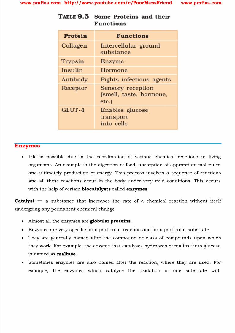
Role of Proteins
1. Some transport nutrients across cell membrane,2. some fight infectious organisms,
3. some are hormones,
4. some are enzymes, etc.
8/16/2019 Biology for Prelims Exam
http://slidepdf.com/reader/full/biology-for-prelims-exam 29/384
www.pmfias.com http://www.youtube.com/c/PoorMansFriend www.pmfias.com
Enzymes
Life is possible due to the coordination of various chemical reactions in living
organisms. An example is the digestion of food, absorption of appropriate molecules
and ultimately production of energy. This process involves a sequence of reactionsand all these reactions occur in the body under very mild conditions. This occurs
with the help of certain biocatalysts called enzymes .
Catalyst == a substance that increases the rate of a chemical reaction without itself
undergoing any permanent chemical change.
Almost all the enzymes are globular proteins .
Enzymes are very specific for a particular reaction and for a particular substrate.
They are generally named after the compound or class of compounds upon which
they work. For example, the enzyme that catalyses hydrolysis of maltose into glucose
is named as maltase .
Sometimes enzymes are also named after the reaction, where they are used. For
example, the enzymes which catalyse the oxidation of one substrate with
8/16/2019 Biology for Prelims Exam
http://slidepdf.com/reader/full/biology-for-prelims-exam 30/384
www.pmfias.com http://www.youtube.com/c/PoorMansFriend www.pmfias.com
simultaneous reduction of another substrate are named as oxidoreductase enzymes.
The ending of the name of an enzyme is -ase .
Almost all enzymes are proteins .
There are some nucleic acids that behave like enzymes. These are called ribozymes .
An enzyme like any protein has a primary structure, i.e., amino acid sequence of the
protein. Enzyme catalysts differ from inorganic catalysts in many ways. Inorganic catalysts
work efficiently at high temperatures and high pressures, while enzymes get damaged
at high temperatures (say above 40°C).
However, enzymes isolated from organisms who normally live under extremely high
temperatures (e.g., hot vents and sulphur springs), are stable and retain their
catalytic power even at high temperatures (up to 80°-90°C). Thermal stability is thus
an important quality of such enzymes isolated from thermophilic organisms.
Thermophile == a bacterium or other microorganism that grows best at high
temperatures (above 45°C).
Factors Affecting Enzyme Activity
The activity of an enzyme can be affected by a change in the conditions which can
alter the structure of the protein. These include temperature, pH, change in
substrate concentration or binding of specific chemicals that regulate its activity.
Temperature and pH
Enzymes generally function in a narrow range of temperature and pH.
Each enzyme shows its highest activity at a particular temperature and pH called the
optimum temperature and optimum pH .
Activity declines both below and above the optimum value.
Low temperature preserves the enzyme in a temporarily inactive state whereas high
temperature destroys enzymatic activity because proteins are denatured by heat .
Concentration of Substrate
With the increase in substrate concentration, the velocity of the enzymatic reaction
rises at first. The reaction ultimately reaches a maximum velocity (Vmax) which is
not exceeded by any further rise in concentration of the substrate. This is because
the enzyme molecules are fewer than the substrate molecules and after saturation of
8/16/2019 Biology for Prelims Exam
http://slidepdf.com/reader/full/biology-for-prelims-exam 31/384
www.pmfias.com http://www.youtube.com/c/PoorMansFriend www.pmfias.com
these molecules, there are no free enzyme molecules to bind with the additional
substrate molecules.
The activity of an enzyme is also sensitive to the presence of specific chemicals that
bind to the enzyme. When the binding of the chemical shuts off enzyme activity, the
process is called inhibition and the chemical is called an inhibitor .
When the inhibitor closely resembles the substrate in its molecular structure andinhibits the activity of the enzyme, it is known as competitive inhibitor .
Summary
Proteins are the polymers of about twenty different α -amino acids which are linked by
peptide bonds .
Ten amino acids are called essential amino acids because they cannot be synthesised
by our body, hence must be provided through diet.
Proteins perform various structural and dynamic functions in the organisms.
Proteins which contain only α -amino acids are called simple proteins .
The secondary or tertiary structure of proteins get disturbed on change of pH or
temperature and they are not able to perform their functions. This is called
denaturation of proteins .
Enzymes are biocatalysts which speed up the reactions in biosystems. They are very
specific and selective in their action and chemically all enzymes are proteins.
8/16/2019 Biology for Prelims Exam
http://slidepdf.com/reader/full/biology-for-prelims-exam 32/384
www.pmfias.com http://www.youtube.com/c/PoorMansFriend www.pmfias.com
Primary and Secondary Metabolites, Vitamins, Deficiency Diseases, Micronutrients –
Vitamins and Minerals, Food Sources of Vitamins and Minerals.
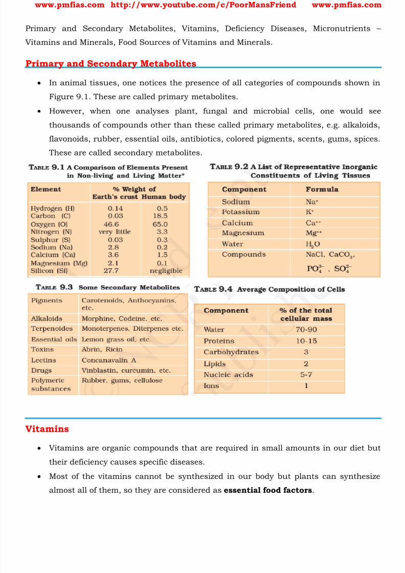
Primary and Secondary Metabolites
In animal tissues, one notices the presence of all categories of compounds shown in
Figure 9.1. These are called primary metabolites. However, when one analyses plant, fungal and microbial cells, one would see
thousands of compounds other than these called primary metabolites, e.g. alkaloids,
flavonoids, rubber, essential oils, antibiotics, colored pigments, scents, gums, spices.
These are called secondary metabolites.
Vitamins
Vitamins are organic compounds that are required in small amounts in our diet but
their deficiency causes specific diseases.
Most of the vitamins cannot be synthesized in our body but plants can synthesize
almost all of them, so they are considered as essential food factors .

8/16/2019 Biology for Prelims Exam
http://slidepdf.com/reader/full/biology-for-prelims-exam 33/384
www.pmfias.com http://www.youtube.com/c/PoorMansFriend www.pmfias.com
However, the bacteria of the gut can produce some of the vitamins required by us.
All the vitamins are generally available in our diet. Different vitamins belong to
various chemical classes and it is difficult to define them on the basis of structure.
They are generally regarded as organic compounds required in the diet in small
amounts to perform specific biological functions for normal maintenance of
optimum growth and health of the organism. Vitamins are designated by alphabets A, B, C, D, etc. Some of them are further
named as sub-groups e.g. B1, B2, B6, B12, etc.
Vitamin A keeps our skin and eyes healthy.
Vitamin C helps body to fight against many diseases. Vitamin C gets easily destroyed
by heat during cooking.
Vitamin D helps our body to use calcium for bones and teeth.
Excess of vitamins is also harmful and vitamin pills should not be taken without theadvice of doctor.
The term “Vitamine” was coined from the word vital + amine since the earlier
identified compounds had amino groups.
Later work showed that most of them did not contain amino groups, so the letter ‘e’
was dropped and the term vitamin is used these days.
Vitamins are classified into two groups depending upon their solubility in water or
fat.
Fat soluble vitamins
Vitamins which are soluble in fat and oils but insoluble in water are kept in this
group. These are vitamins A, D, E and K . They are stored in liver and adipose (fat
storing) tissues .
Water soluble vitamins
B group vitamins and vitamin C are soluble in water so they are grouped together.
Water soluble vitamins must be supplied regularly in diet because they are readily
excreted in urine and cannot be stored (except vitamin B12) in our body.
Deficiency Diseases
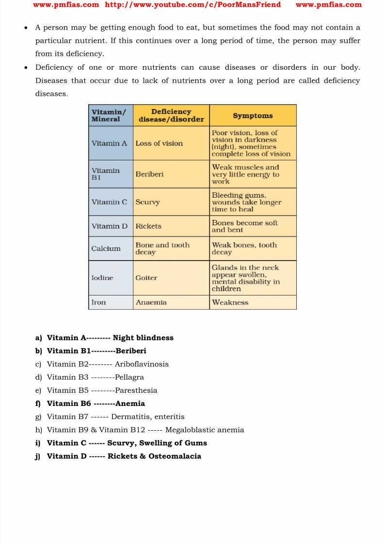
8/16/2019 Biology for Prelims Exam
http://slidepdf.com/reader/full/biology-for-prelims-exam 34/384
www.pmfias.com http://www.youtube.com/c/PoorMansFriend www.pmfias.com
A person may be getting enough food to eat, but sometimes the food may not contain a
particular nutrient. If this continues over a long period of time, the person may suffer
from its deficiency.
Deficiency of one or more nutrients can cause diseases or disorders in our body.
Diseases that occur due to lack of nutrients over a long period are called deficiency
diseases.
a) Vitamin A--------- Night blindness
b) Vitamin B1---------Beriberi
c) Vitamin B2-------- Ariboflavinosis
d)
Vitamin B3 --------Pellagrae) Vitamin B5 --------Paresthesia
f) Vitamin B6 --------Anemia
g) Vitamin B7 ------ Dermatitis, enteritis
h) Vitamin B9 & Vitamin B12 ----- Megaloblastic anemia
i) Vitamin C ------ Scurvy, Swelling of Gums
j) Vitamin D ------ Rickets & Osteomalacia

8/16/2019 Biology for Prelims Exam
http://slidepdf.com/reader/full/biology-for-prelims-exam 35/384
www.pmfias.com http://www.youtube.com/c/PoorMansFriend www.pmfias.com
k) Vitamin E ------ Less Fertility
l) Vitamin K ------ Non-Clotting of Blood.
Micronutrients – Vitamins and Minerals
https://www.dsm.com/content/dam/dsm/cworld/en_US/documents/what-are-
micronutrients.pdf Micronutrients, as opposed to macronutrients (protein, carbohydrates and fat), are
comprised of vitamins and minerals which are required in small quantities to
ensure normal metabolism, growth and physical well ‐being.
Vitamins
These are essential organic nutrients, most of which are not made in the body, or
only in insufficient amounts, and are mainly obtained through food.
When their intake is inadequate, vitamin deficiency disorders are the consequence.
Although vitamins are only present and required in minute quantities, compared to
the macronutrients, they are as vital to health and need to be considered when
determining nutrition security.
Each of the 13 vitamins known today have specific functions in the body: vitamin A,
provitamin A (Beta carotene), vitamin B1, vitamin B2, vitamin B6, vitamin B12,
biotin, vitamin C, vitamin D, vitamin E, folic acid, vitamin K, niacin and
pantothenic acid.
Minerals
These are inorganic nutrients that also play a key role in ensuring health and well ‐
being.
They include the trace elements copper, iodine, iron, manganese, selenium and
zinc together with the macro elements calcium, magnesium, potassium and
sodium .
Five Important Micronutrients
As with vitamins, minerals they are found in small quantities within the body and
they are obtained from a wide variety of foods.
No single food contains all of the vitamins and minerals we need and, therefore, a
balanced and varied diet is necessary for an adequate intake.
8/16/2019 Biology for Prelims Exam
http://slidepdf.com/reader/full/biology-for-prelims-exam 36/384
www.pmfias.com http://www.youtube.com/c/PoorMansFriend www.pmfias.com
Of course, we already know a huge amount about how these work, and the
importance they have in normal human growth and development.
Based on this, an Expert Panel of nutritionist, NGOs and development agencies
indentified five micronutrients such as those below in their priority group:
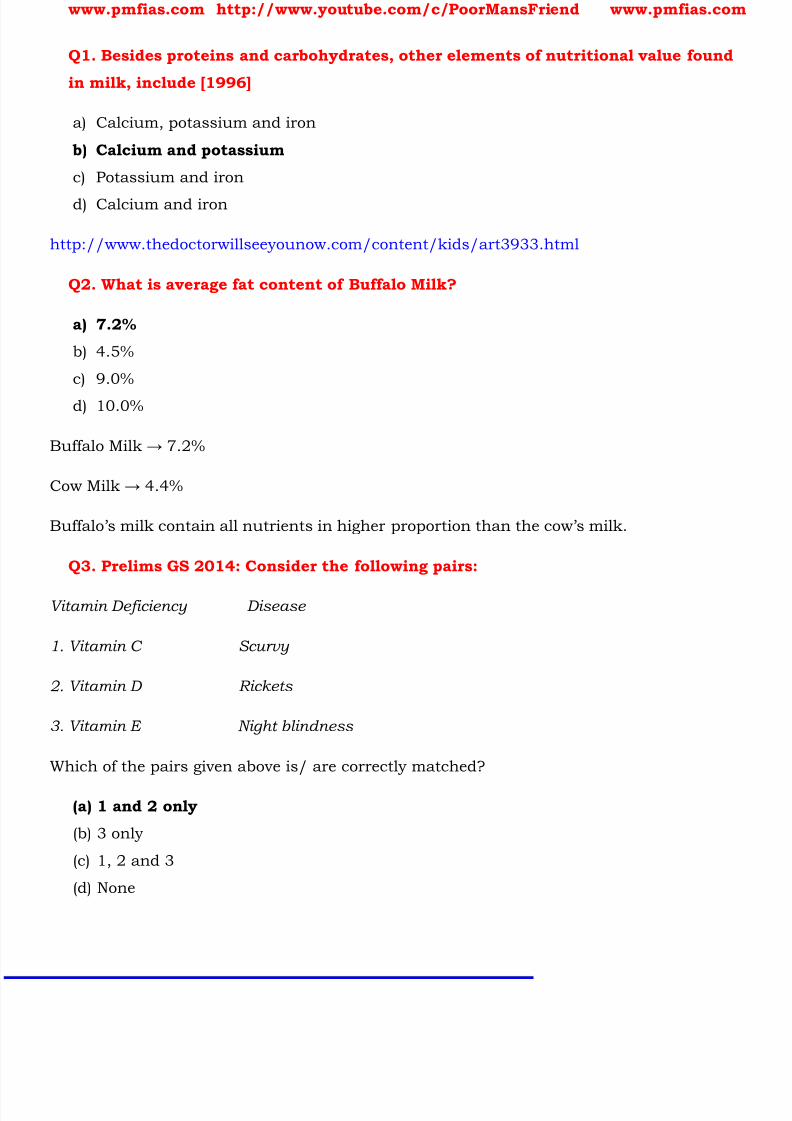
Vitamin A
This vital micronutrient is found in a range of different foods including carrots,
spinach, broccoli, milk, egg, liver and fish.
It plays an essential role in vision (lack of Vitamin A is a common cause of
blindness), reproduction and growth, and the functioning of a healthy immune
system (it plays a key role in the development of white blood cells).
Worldwide about 5 million children under the age of five are affected by
xerophthalmia , a serious eye disorder caused by vitamin A deficiency.
These children are at risk of becoming blind and are more likely to die of common
childhood diseases.
Folate (folic acid)
This is a generic term for a group of B vitamins including folic acid and naturally
occurring folates.
Folic acid is a synthetic folate compound used in vitamin supplements and fortified
food because of its increased stability.
Folates are found in egg, dairy products, asparagus, orange juice, dark green leafy
vegetables, beans and brown bread.
They play a key role in the metabolism of amino acids and the production of
proteins , the synthesis of nucleic acid (the molecules that carry genetic
information in the cells), and the formation of blood cells .
Iodine
Seaweed and fish are rich sources but in many countries the addition of iodine(known as iodization) to salt is an important source.
Iodine is one of the most important elements required by the developing foetus due
to its effect on brain development .
Iodine also serves a number of other important functions especially in the
production of hormones.
Goitre is a visible sign of severe iodine deficiency.
8/16/2019 Biology for Prelims Exam
http://slidepdf.com/reader/full/biology-for-prelims-exam 37/384
www.pmfias.com http://www.youtube.com/c/PoorMansFriend www.pmfias.com
Iron
Iron has a number of key functions within the body. It acts as a carrier for oxygen
from the lungs to the body’s tissues – it does so in the form of hemoglobin – and it
also integral to the working of various tissues through the role that it plays in
enzymatic reactions.
Iron deficiency ultimately leads to iron deficiency anemia , the most common cause
of anemia, a condition in which the blood lacks healthy red bloods cells required to
carry oxygen, and which results in morbidity and death.
Iron deficiency is the most widespread health problem in the world, impairing normal
mental development in 40 ‐60% of infants in the developing world.
Iron ‐rich foods include lentils, red meat, poultry, fish, lentils, leaf vegetables and
chick ‐peas.
Zinc
Found in a range of foodstuffs including liver, eggs, nuts, cereals and seafood.
The absence of zinc is associated with a number of conditions including, short
stature, anemia, impaired healing of wounds, poor gonadal function, and
impaired cognitive and motor function.
It can also lead to appetite disorders, as well as contributing to the increased
severity and incidence of diarrhea and pneumonia .
The most important effect of zinc deficiency is its impact on children’s resistance to
infectious diseases including the risk of infection , the recurrence of infections and
the severity of infection. This is well document in the case of diarrhoea. Zinc nutrition
is therefore an important determinant of mortality in children.
Food Sources of Vitamins and Minerals
8/16/2019 Biology for Prelims Exam
http://slidepdf.com/reader/full/biology-for-prelims-exam 38/384
www.pmfias.com http://www.youtube.com/c/PoorMansFriend www.pmfias.com
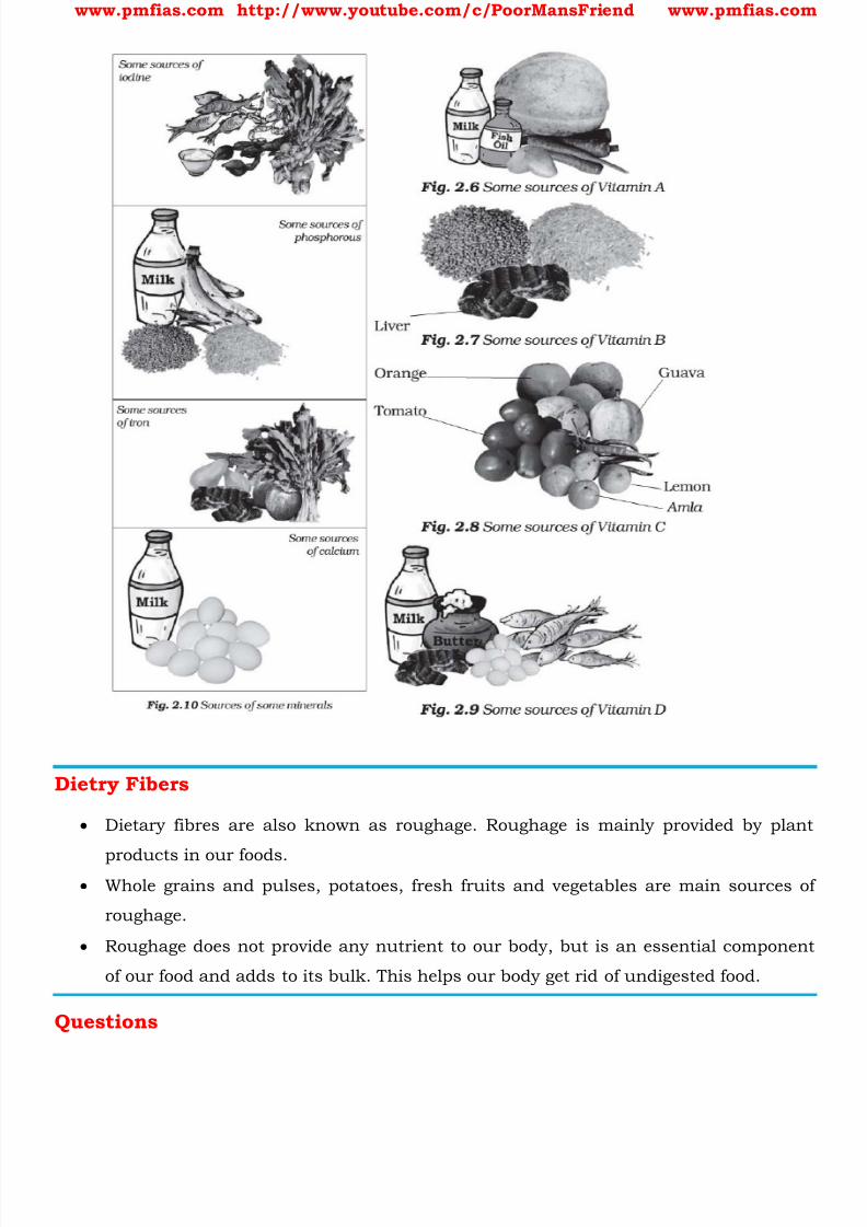
Dietry Fibers
Dietary fibres are also known as roughage. Roughage is mainly provided by plant
products in our foods. Whole grains and pulses, potatoes, fresh fruits and vegetables are main sources of
roughage.
Roughage does not provide any nutrient to our body, but is an essential component
of our food and adds to its bulk. This helps our body get rid of undigested food.
Questions
8/16/2019 Biology for Prelims Exam
http://slidepdf.com/reader/full/biology-for-prelims-exam 39/384
www.pmfias.com http://www.youtube.com/c/PoorMansFriend www.pmfias.com
Q1. Besides proteins and carbohydrates, other elements of nutritional value found
in milk, include [1996]
a) Calcium, potassium and iron
b) Calcium and potassium
c) Potassium and iron
d) Calcium and iron
http://www.thedoctorwillseeyounow.com/content/kids/art3933.html
Q2. What is average fat content of Buffalo Milk?
a) 7.2%
b) 4.5%
c) 9.0%
d) 10.0%
Buffalo Milk → 7.2%
Cow Milk → 4.4%
Buffalo’s milk contain all nutrients in higher proportion than the cow’s milk.
Q3. Prelims GS 2014: Consider the following pairs:
Vitamin Deficiency Disease
1. Vitamin C Scurvy
2. Vitamin D Rickets
3. Vitamin E Night blindness
Which of the pairs given above is/ are correctly matched?
(a) 1 and 2 only
(b) 3 only
(c) 1, 2 and 3
(d) None

8/16/2019 Biology for Prelims Exam
http://slidepdf.com/reader/full/biology-for-prelims-exam 40/384
www.pmfias.com http://www.youtube.com/c/PoorMansFriend www.pmfias.com
8/16/2019 Biology for Prelims Exam
http://slidepdf.com/reader/full/biology-for-prelims-exam 41/384
www.pmfias.com http://www.youtube.com/c/PoorMansFriend www.pmfias.com
DNA and RNA | Deoxyribonucleic Acid | Ribonucleic Acid – Chromosomes, Nucleotide and
Nucleoside, Nucleic acids, DNA and RNA, Recombinant DNA.
Nucleus
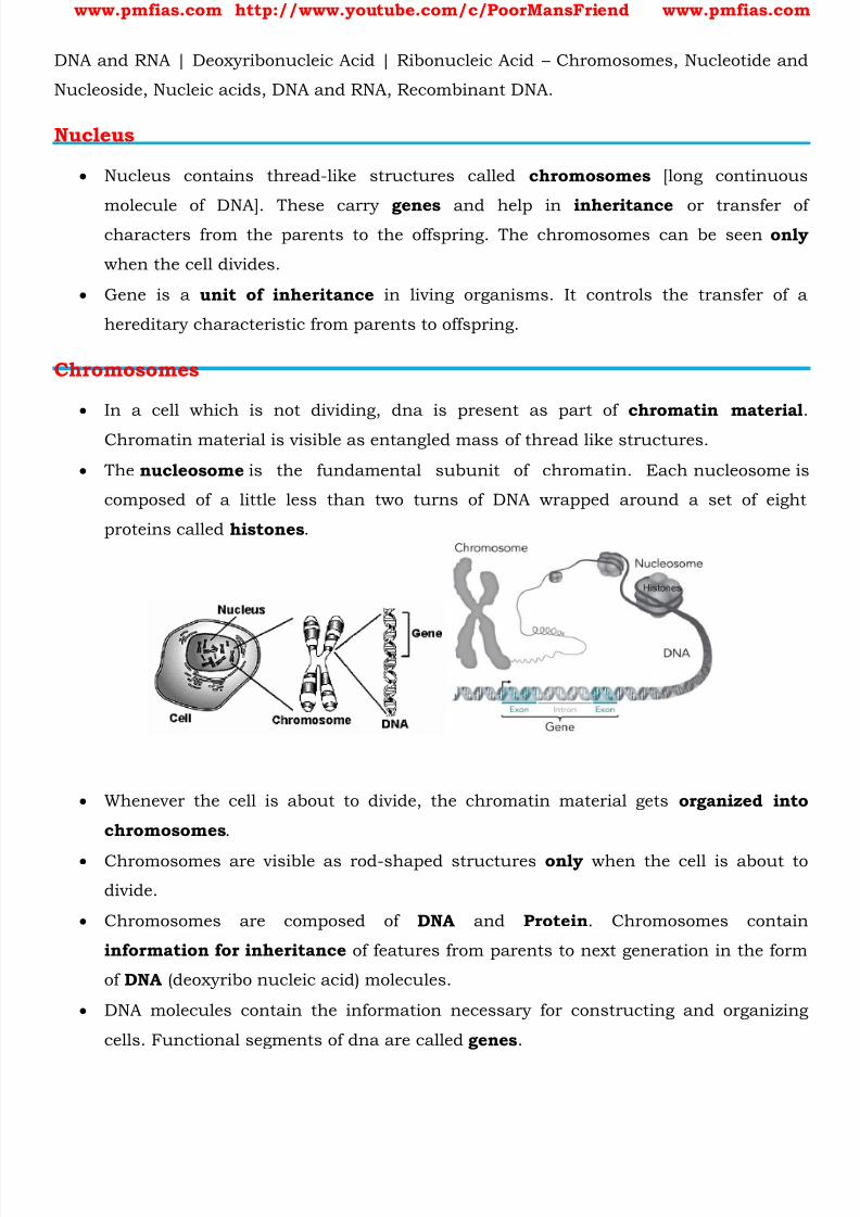
Nucleus contains thread-like structures called chromosomes [long continuous
molecule of DNA]. These carry genes and help in inheritance or transfer ofcharacters from the parents to the offspring. The chromosomes can be seen only
when the cell divides.
Gene is a unit of inheritance in living organisms. It controls the transfer of a
hereditary characteristic from parents to offspring.
Chromosomes
In a cell which is not dividing, dna is present as part of chromatin material
Chromatin material is visible as entangled mass of thread like structures.
The nucleosome is the fundamental subunit of chromatin. Each nucleosome is
composed of a little less than two turns of DNA wrapped around a set of eight
proteins called histones .
Whenever the cell is about to divide, the chromatin material gets organized into
chromosomes .
Chromosomes are visible as rod-shaped structures only when the cell is about to
divide.
Chromosomes are composed of DNA and Protein . Chromosomes contain
information for inheritance of features from parents to next generation in the form
of DNA (deoxyribo nucleic acid) molecules.
DNA molecules contain the information necessary for constructing and organizing
cells. Functional segments of dna are called genes .
8/16/2019 Biology for Prelims Exam
http://slidepdf.com/reader/full/biology-for-prelims-exam 42/384
www.pmfias.com http://www.youtube.com/c/PoorMansFriend www.pmfias.com
Nucleotide and Nucleoside
Living organisms have a number of carbon compounds in which heterocyclic rings
can be found.
When heterocyclic rings are attached to a sugar , they are called nucleosides .
If a phosphate group is also found esterified to the sugar they are called
nucleotides .
Nucleic acids like DNA and RNA consist of nucleotides only.
Heterocyclic Rings == A heterocyclic compound or ring structure is a cyclic compound
that has atoms of at least two different elements as members of its ring(s).
Ester == An organic compound made by replacing the hydrogen of an acid by an alkyl or
other organic group.
Nucleic Acids
Nucleic acid is a macromolecule that is found in the acid insoluble fraction of any
living tissue.
Together with polysaccharides and polypeptides these comprise the true
macromolecular fraction of any living tissue or cell.
For nucleic acids, the building block is a nucleotide, i.e. nucleic acids are polymers
of nucleotides. Since nucleic acids are long chain polymers of nucleotides, they are also called
polynucleotides .
The nucleotides are joined to one another in a chain by covalent bonds between the
sugar of one nucleotide and the phosphate of the next, resulting in an alternating
sugar-phosphate backbone .
A nucleotide has three chemically distinct components. One is a heterocyclic
compound , the second is a monosaccharide and the third a phosphoric acid orphosphate .
The sugar found in polynucleotides is either ribose (a monosaccharide pentose) or
deoxyribose .
The heterocyclic compounds in nucleic acids are the nitrogenous bases named
Adenine, Guanine, Uracil, Cytosine, and Thymine .

8/16/2019 Biology for Prelims Exam
http://slidepdf.com/reader/full/biology-for-prelims-exam 43/384
www.pmfias.com http://www.youtube.com/c/PoorMansFriend www.pmfias.com
DNA and RNA
Every generation of each and every species resembles its ancestors in many ways.
How are these characteristics transmitted from one generation to the next?
It has been observed that nucleus of a living cell is responsible for this transmission
of inherent characters, also called heredity .
The particles in nucleus of the cell, responsible for heredity, are called chromosomes
which are made up of proteins and another type of biomolecules called nucleicacids .
Nucleic acids are responsible for the transfer of characters from parents to off
springs. There are two types of nucleic acids — DNA and RNA.
A nucleic acid containing deoxyribose is called deoxyribonucleic acid (DNA) while that
which contains ribose is called ribonucleic acid (RNA) .
Both DNA and RNA contain Adenine, Guanine and Cytosine . The fourth base is
Thymine in DNA and Uracil in RNA .
The structure of DNA is a double strand [helix] whereas RNA is a single strand
molecule.
Hydrogen bonds bind the nitrogenous bases of the two separate polynucleotide
strands to make double-stranded DNA.

8/16/2019 Biology for Prelims Exam
http://slidepdf.com/reader/full/biology-for-prelims-exam 44/384
www.pmfias.com http://www.youtube.com/c/PoorMansFriend www.pmfias.com
The DNA backbone is resistant to cleavage, and both strands of the double-stranded
structure store the same biological information. Biological information is replicated
as the two strands are separated.
Within cells, DNA is organized into long structures called chromosomes . During cell
division these chromosomes are duplicated in the process of DNA replication ,
providing each cell its own complete set of chromosomes. Eukaryotic organisms (animals, plants, fungi, and protists) store most of their DNA
inside the cell nucleus and some of their DNA in organelles, such as mitochondria
or chloroplasts .
In contrast, prokaryotes (bacteria and archaea) store their DNA only in the
cytoplasm .

8/16/2019 Biology for Prelims Exam
http://slidepdf.com/reader/full/biology-for-prelims-exam 45/384
www.pmfias.com http://www.youtube.com/c/PoorMansFriend www.pmfias.com
DNA is the chemical basis of heredity and have the coded message for proteins to
be synthesized in the cell.
There are three types of RNA — mRNA, rRNA and tRNA which actually carry out the
protein synthesis in the cell based on the coded message for proteins provided by
DNA.
Ribonucleic acid (RNA) is implicated in various biological roles in coding, decoding,regulation, and expression of genes.
Cellular organisms use messenger RNA (mRNA) to convey genetic information that
directs synthesis of specific proteins.
Many viruses encode their genetic information using an RNA genome. Example: HIV
virus used this technique to proliferate within human body.
Biological Functions of Nucleic Acids – DNA and RNA
DNA is the chemical basis of heredity and may be regarded as the reserve of genetic
information .
DNA is exclusively responsible for maintaining the identity of different species of
organisms over millions of years.
A DNA molecule is capable of self-duplication during cell division and identical DNA
strands are transferred to daughter cells.
Another important function of nucleic acids is the protein synthesis in the cell.
Actually, the proteins are synthesized by various RNA molecules in the cell but the
message for the synthesis of a particular protein is present in DNA .
DNA Fingerprinting
It is known that every individual has unique fingerprints. These occur at the tips of
the fingers and have been used for identification for a long time but these can be
altered by surgery.
A sequence of bases on DNA is also unique for a person and information regarding
this is called DNA fingerprinting. It is same for every cell and cannot be altered by
any known treatment.
DNA fingerprinting is now used (i) in forensic laboratories for identification of
criminals. (ii) to determine paternity of an individual. (iii) to identify the dead bodies
in any accident by comparing the DNA’s of parents or children. (iv) to i dentify racial
groups to rewrite biological evolution.
8/16/2019 Biology for Prelims Exam
http://slidepdf.com/reader/full/biology-for-prelims-exam 46/384
www.pmfias.com http://www.youtube.com/c/PoorMansFriend www.pmfias.com
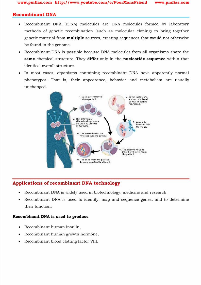
Recombinant DNA
Recombinant DNA (rDNA) molecules are DNA molecules formed by laboratory
methods of genetic recombination (such as molecular cloning) to bring together
genetic material from multiple sources, creating sequences that would not otherwise
be found in the genome.
Recombinant DNA is possible because DNA molecules from all organisms share the
same chemical structure. They differ only in the nucleotide sequence within that
identical overall structure.
In most cases, organisms containing recombinant DNA have apparently normal
phenotypes. That is, their appearance, behavior and metabolism are usually
unchanged.
Applications of recombinant DNA technology
Recombinant DNA is widely used in biotechnology, medicine and research.
Recombinant DNA is used to identify, map and sequence genes, and to determinetheir function.
Recombinant DNA is used to produce
Recombinant human insulin,
Recombinant human growth hormone,
Recombinant blood clotting factor VIII,

8/16/2019 Biology for Prelims Exam
http://slidepdf.com/reader/full/biology-for-prelims-exam 47/384

8/16/2019 Biology for Prelims Exam
http://slidepdf.com/reader/full/biology-for-prelims-exam 48/384
www.pmfias.com http://www.youtube.com/c/PoorMansFriend www.pmfias.com
Fats - Lipid, Fatty Acid, Saturated fat, Unsaturated fat. Healthy Fats – Omega-3 and
Omega-6, Monounsaturated and Polyunsaturated. Unhealthy Fats – Saturated Fat and
Trans Fat.
Fat
Fat is one of the three main macronutrients: fat, carbohydrate, and protein.
Fat is a major source of energy and helps your body absorb vitamins.
Fat has the most calories compared to any other nutrient. Controlling fat intake is
one of the most important steps in losing or maintaining weight and preventing or
delaying type 2 diabetes.
Fats, also known as triglycerides , are esters of three fatty acid chains and the
alcohol glycerol .
Fats are solids at room temperature. Oil refers to a fat with unsaturated fatty acid
chains that is liquid at room temperature.
Fats, like other lipids, are generally insoluble in water .
Lipid
A lipid is chemically defined as a substance that is insoluble in water and soluble in
alcohol and chloroform.
Lipids are an important component of living cells. Together with carbohydrates and
proteins, lipids are the main constituents of plant and animal cells.
Cholesterol and triglycerides are lipids. Lipid is not necessarily a triglyceride.
Glycerol is a simple sugar alcohol compound. A triglyceride is an ester derived from
glycerol and three fatty acids (tri + glyceride)
Triglycerides are the main constituent of body fat in humans and animals, as well
as vegetable fat.
8/16/2019 Biology for Prelims Exam
http://slidepdf.com/reader/full/biology-for-prelims-exam 49/384
www.pmfias.com http://www.youtube.com/c/PoorMansFriend www.pmfias.com
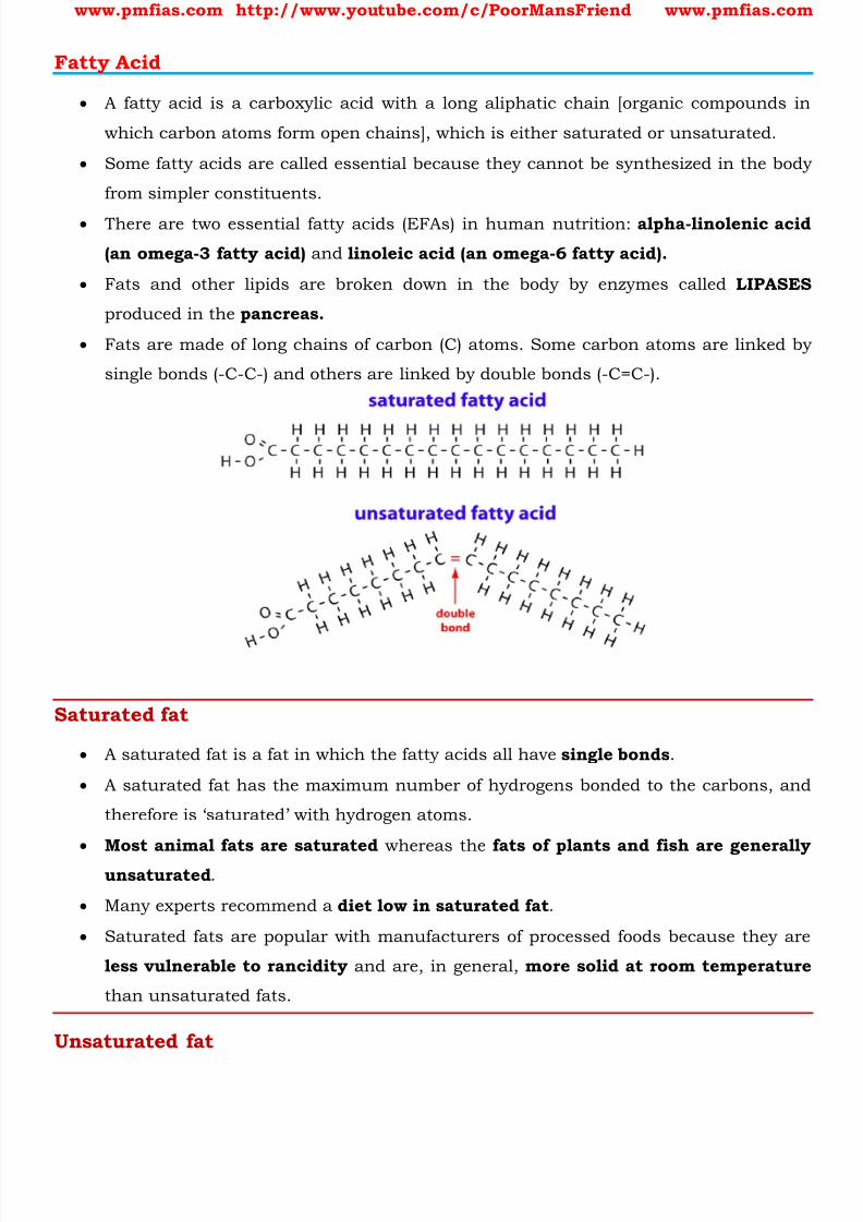
Fatty Acid
A fatty acid is a carboxylic acid with a long aliphatic chain [organic compounds in
which carbon atoms form open chains], which is either saturated or unsaturated.
Some fatty acids are called essential because they cannot be synthesized in the body
from simpler constituents.
There are two essential fatty acids (EFAs) in human nutrition: alpha-linolenic acid
(an omega-3 fatty acid) and linoleic acid (an omega-6 fatty acid).
Fats and other lipids are broken down in the body by enzymes called LIPASES
produced in the pancreas.
Fats are made of long chains of carbon (C) atoms. Some carbon atoms are linked by
single bonds (-C-C-) and others are linked by double bonds (-C=C-).
Saturated fat
A saturated fat is a fat in which the fatty acids all have single bonds .
A saturated fat has the maximum number of hydrogens bonded to the carbons, and
therefore is ‘saturated’ with hydrogen atoms.
Most animal fats are saturated whereas the fats of plants and fish are generally
unsaturated .
Many experts recommend a diet low in saturated fat .
Saturated fats are popular with manufacturers of processed foods because they are
less vulnerable to rancidity and are, in general, more solid at room temperature
than unsaturated fats.
Unsaturated fat
8/16/2019 Biology for Prelims Exam
http://slidepdf.com/reader/full/biology-for-prelims-exam 50/384
www.pmfias.com http://www.youtube.com/c/PoorMansFriend www.pmfias.com
An unsaturated fat is a fat or fatty acid in which there is at least one double bond
within the fatty acid chain.
Where double bonds are formed, hydrogen atoms are eliminated.
In cellular metabolism, unsaturated fat molecules contain somewhat less energy (i.e.,
fewer calories) than an equivalent amount of saturated fat.
The greater the degree of unsaturation in a fatty acid (i.e., the more double bonds inthe fatty acid) the more vulnerable it is to rancidity [lipid oxidation][rusting of fats].
Antioxidants can protect unsaturated fat from lipid oxidation.
Healthy Fats – Omega-3 and Omega-6, Monounsaturated andPolyunsaturated
The main types of “healthy” fats are monounsaturated , polyunsaturated, alpha-
linolenic acid (an omega-3 fatty acid) and linoleic acid (an omega-6 fatty acid) .
The fat is termed “monounsaturated” if there is one double bond, and
“polyunsaturated” if there are two or more double bonds.
Omega-3 and Omega-6 fatty acids are heart healthy fats and can help in lowering
high triglyceride values in blood. They are found in fish, soybean products, Walnuts
etc.
Both of these fatty acids are needed for growth and repair, but can also be used to
make other fatty acids.
The omega-3 and omega-6 are fatty acids are both polyunsaturated. The difference is
in where the first of the double bonds occurs.
Both omega-3 (ω-3) and omega- 6 (ω -6) fatty acids are important components of cell
membranes.
There is increasing support for omega-3 fatty acids in protecting against fatal heart
disease and it is known that they have anti-inflammatory effects .
There is also growing interest in the role of omega-3 fatty acids in the prevention of
diabetes and certain types of cancer.
Monounsaturated and polyunsaturated fat are considered “heart healthy” and can
help with improving cholesterol when used in place of unhealthy fats.
Some sources of these fats include almonds, cashews, pecans, peanuts, pine nuts,
pumpkin, sesame seeds, sunflower seeds, Olive oil and olives, vegetable oils (such as
sunflower, safflower, corn, soybean, and cottonseed).
8/16/2019 Biology for Prelims Exam
http://slidepdf.com/reader/full/biology-for-prelims-exam 51/384
www.pmfias.com http://www.youtube.com/c/PoorMansFriend www.pmfias.com
Unhealthy Fats – Saturated Fat and Trans Fat
The main types of “unhealthy” fats are saturated and trans-fat .
Saturated fats are primarily found in foods that come from animals, such as meat
and dairy.
Saturated fats are unhealthy because they increase LDL (“bad” cholesterol) levels in
your body and increase your risk for heart disease.
Many saturated fats are “solid” fats that you can see, such as the fat in meat. Other
sources of saturated fats include high-fat cheeses, high-fat cuts of meat, butter, Ice
cream, palm and coconut oils, etc..
Trans fats, or trans-unsaturated fatty acids, trans fatty acids, are a type of
unsaturated fats that are uncommon in nature.
Trans fat is simply liquid oils turned into solid fats during food processing. There is
also a small amount of trans fat that occurs naturally in some meat and dairy
products, but those found in processed foods tend to be the most harmful to your
health.
Trans fats are worse than saturated fats. They increase LDL (“bad” cholesterol) and
decreasing HDL (“healthy” cholesterol).
Trans fatty acids are used as preservative in packaged food items. Foods containing
trans- fat are usually labeled as “ partially hydrogenated ”.
Partially hydrogenated oil is less likely to spoil, so foods made with it have a longershelf life.
Trans fats are easy to use, inexpensive to produce and last a long time. Trans fats
give foods a desirable taste and texture.
Q1. Statements:
1) Trans fats are considered beneficial for the human body
2) Double bond chemistry of the fat molecules in Trans fats causes a Plaque formation3) Omega-3 fatty acids are considered healthier than the saturated fatty acids
Codes:
a) 1 & 3
b) 2 & 3
c) 1 & 2

8/16/2019 Biology for Prelims Exam
http://slidepdf.com/reader/full/biology-for-prelims-exam 52/384
www.pmfias.com http://www.youtube.com/c/PoorMansFriend www.pmfias.com
d) 1, 2, 3
Ans. B
Adipose tissue
In animals, adipose, or fatty tissue with adipose cells is the body's means of storing
fat derived from the diet and from liver metabolism.
Under stress conditions, adipose cells degrade their stored fat to supply fatty acids
and also glycerol.
These metabolic activities are regulated by several hormones (e.g., insulin, glucagon
and epinephrine).
Metabolic Basis for Living
Metabolic pathways can lead to a more complex structure from a simpler structure(for example, acetic acid becomes cholesterol ) or lead to a simpler structure from a
complex structure (for example, glucose becomes lactic acid in our skeletal
muscle ).
The former cases are called biosynthetic pathways or anabolic pathways . The latter
constitute degradation and hence are called catabolic pathways .
Anabolic pathways, as expected, consume energy . Assembly of a protein from amino
acids requires energy input. On the other hand, catabolic pathways lead to the release of energy. For example,
when glucose is degraded to lactic acid in our skeletal muscle, energy is liberated.
This metabolic pathway from glucose to lactic acid which occurs in 10 metabolic
steps is called glycolysis .
Living organisms have learnt to trap this energy liberated during degradation and
store it in the form of chemical bonds.
As and when needed, this bond energy is utilized for biosynthetic, osmotic andmechanical work that we perform.
The most important form of energy currency in living systems is the bond energy in a
chemical called adenosine triphosphate (ATP) .
http://www.cdc.gov/diabetes/prevention/pdf/postcurriculum_session2.pdf
8/16/2019 Biology for Prelims Exam
http://slidepdf.com/reader/full/biology-for-prelims-exam 53/384
www.pmfias.com http://www.youtube.com/c/PoorMansFriend www.pmfias.com
Cell Cycle, Cell Division, Phases of Cell Cycle: Interphase, Mitosis: Prophase,
Prometaphase, Metaphase, Anaphase, Telophase. Cytokinesis. Significance of Mitosis.
Cell Cycle and Cell Division
During the division of a cell, DNA replication and cell growth takes place.
DNA: DNA and RNA | Recombinant DNA
All these processes, i.e., cell division, DNA replication, and cell growth have to take place
in a coordinated way to ensure correct division and formation of progeny (offspring)
cells containing intact genomes (the complete set of genetic material of an organism).
The sequence of events by which a cell duplicates its genome, synthesizes the other
constituents of the cell and eventually divides into two daughter cells is termed cell
cycle.
Although cell growth (in terms of cytoplasmic increase) is a continuous process, DNA
synthesis occurs only during one specific stage in the cell cycle.
The replicated chromosomes (DNA) are then distributed to daughter nuclei by a
complex series of events during cell division . These events are themselves under
genetic control [DNA] .
Cell Cycle – Phases of Cell Cycle
A typical eukaryotic cell divides once in approximately every 24 hours .
Eukaryotic vs Prokaryotic Cells: Eukaryotic vs. Prokaryotic Cells|Plant Cell vs. Animal
Cell
However, this duration of cell cycle can vary from organism to organism and also from
cell type to cell type.
Yeast for example, can progress through the cell cycle in only about 90 minutes.
Basic Phases of Cell Cycle – Interphase and M Phase or Mitosis
Interphase == Phase between two successive M phases.
M Phase [Mitosis phase] == Actual cell division or Mitosis .

8/16/2019 Biology for Prelims Exam
http://slidepdf.com/reader/full/biology-for-prelims-exam 54/384
www.pmfias.com http://www.youtube.com/c/PoorMansFriend www.pmfias.com
In the 24 hour average duration of cell cycle of a human cell, cell division proper lasts
for only about an hour . The interphase lasts more than 95% of the duration of cell
cycle.
The M Phase or Mitosis starts with the nuclear division or karyokinesis [separation of
daughter chromosomes].
It usually ends with division of cytoplasm [ cytokinesis ]. Interphase is called the resting phase .
It is the time during which the cell is preparing for division by undergoing both cell
growth and DNA replication .
Interphase
The interphase is divided into three further phases.
1. G1 phase (Gap 1)
2. S phase (Synthesis)
3. G2 phase (Gap 2)
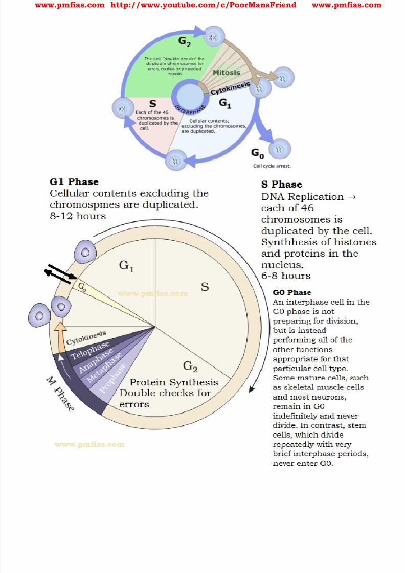
8/16/2019 Biology for Prelims Exam
http://slidepdf.com/reader/full/biology-for-prelims-exam 55/384
www.pmfias.com http://www.youtube.com/c/PoorMansFriend www.pmfias.com

8/16/2019 Biology for Prelims Exam
http://slidepdf.com/reader/full/biology-for-prelims-exam 56/384
www.pmfias.com http://www.youtube.com/c/PoorMansFriend www.pmfias.com
http://www.highlands.edu/academics/divisions/scipe/biology/faculty/harnden/2121/images/cellcycle.jpg
G1 phase
G1 phase == interval between mitosis and beginning of DNA replication [initiation of
DNA replication].
http://www.highlands.edu/academics/divisions/scipe/biology/faculty/harnden/2121/images/cellcycle.jpg
http://www.highlands.edu/academics/divisions/scipe/biology/faculty/harnden/2121/images/cellcycle.jpg
http://www.highlands.edu/academics/divisions/scipe/biology/faculty/harnden/2121/images/cellcycle.jpg

8/16/2019 Biology for Prelims Exam
http://slidepdf.com/reader/full/biology-for-prelims-exam 57/384
www.pmfias.com http://www.youtube.com/c/PoorMansFriend www.pmfias.com
During G1 phase the cell is metabolically active and continuously grows but does not
replicate its DNA.
S or synthesis phase
S or synthesis phase == DNA synthesis or replication takes place .
During this time the amount of DNA per cell doubles .
If the initial amount of DNA is denoted as 2C then it increases to 4C.
However, there is no increase in the chromosome number ; if the cell had diploid or
2n number of chromosomes at G1, even after s phase the number of chromosomes
remains the same, i.e., 2n.
In animal cells, during the S phase, DNA replication begins in the nucleus , and the
centriole duplicates in the cytoplasm.
G2 phase
During the G2 phase, proteins are synthesized in preparation for mitosis while cell
growth continues.
In the S and G2 phases the new DNA molecules formed are not distinct but intertwined.
Onion root tip cell has 16 chromosomes in each cell. Can you tell how many chromosomes
will the cell have at G1 phase, after S phase, and after M phase?
Also, what will be the DNA content of the cells at G1, after S and at G2, if the content after
M phase is 2C?
Quiescent stage (G0)
Some cells in the adult animals do not appear to exhibit division (e.g., heart cells )
and many other cells divide only occasionally, as needed to replace cells that have been
lost because of injury or cell death.
These cells that do not divide further exit G1 phase to enter an inactive stage calledquiescent stage (G0) of the cell cycle.
Cells in this stage remain metabolically active but no longer proliferate unless called
on to do so depending on the requirement of the organism.
Mitosis Phase or M Phase

8/16/2019 Biology for Prelims Exam
http://slidepdf.com/reader/full/biology-for-prelims-exam 58/384
www.pmfias.com http://www.youtube.com/c/PoorMansFriend www.pmfias.com
This is the most dramatic period of the cell cycle, involving a major reorganization of
virtually all components of the cell.
Since the number of chromosomes in the parent and progeny cells is the same , it is
also called as equational division .
Though for convenience mitosis has been divided into four stages of nuclear division, it
is very essential to understand that cell division is a progressive process and very clear-cut lines cannot be drawn between various stages.
Mitosis is the process in which a eukaryotic cell nucleus splits in two , followed by
division of the parent cell into two daughter cells .
The word “mitosis” means “threads,” and it refers to the threadlike appearance of
chromosomes as the cell prepares to divide.
Early microscopists were the first to observe these structures, and they also noted the
appearance of a specialized network of microtubules during mitosis. These tubules, collectively known as the spindle fibres , extend from structures called
centrosomes — with one centrosome located at each of the opposite ends, or poles, of a
cell.
As mitosis progresses, the microtubules [spindle fibres] attach to the chromosomes,
which have already duplicated their DNA and aligned across the center of the cell.
The spindle tubules then shorten and move toward the poles of the cell. As they move,
they pull the one copy of each chromosome with them to opposite poles of the cell.
This process ensures that each daughter cell will contain one exact copy of the parent
cell DNA.
Mitosis consists of five morphologically distinct phases:
1. prophase,
2. prometaphase,
3. metaphase,
4. anaphase, and
5. telophase.
Each phase involves characteristic steps in the process of chromosome alignment and
separation.
Once mitosis is complete, the entire cell divides in two by way of the process called
cytokinesis.
In animals, mitotic cell division is only seen in the diploid somatic cells .

8/16/2019 Biology for Prelims Exam
http://slidepdf.com/reader/full/biology-for-prelims-exam 59/384
www.pmfias.com http://www.youtube.com/c/PoorMansFriend www.pmfias.com
But plants can show mitotic divisions in both haploid and diploid cells.
[Diploid == containing two complete sets of chromosomes, one from each parent].
[Haploid == only one set of chromosomes from one of the parent].
[Somatic == the parts of an organism other than the reproductive cells ].
Prophase

8/16/2019 Biology for Prelims Exam
http://slidepdf.com/reader/full/biology-for-prelims-exam 60/384

8/16/2019 Biology for Prelims Exam
http://slidepdf.com/reader/full/biology-for-prelims-exam 61/384
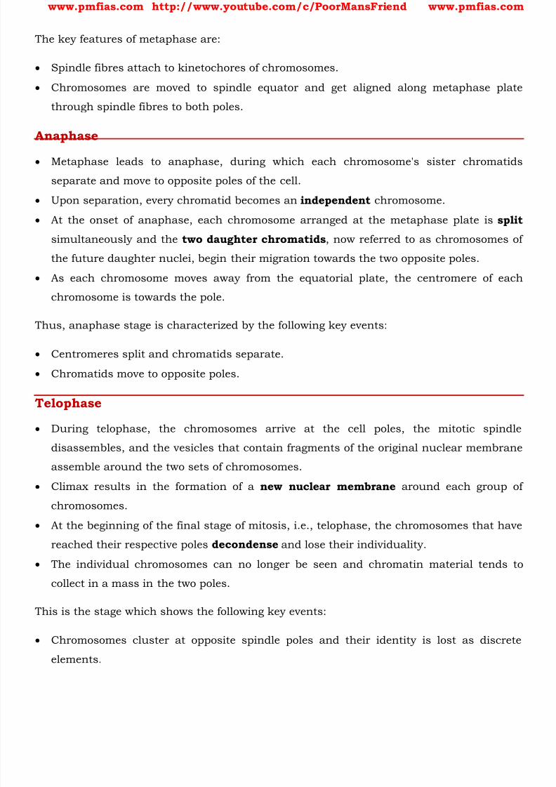
8/16/2019 Biology for Prelims Exam
http://slidepdf.com/reader/full/biology-for-prelims-exam 62/384
www.pmfias.com http://www.youtube.com/c/PoorMansFriend www.pmfias.com
The key features of metaphase are:
Spindle fibres attach to kinetochores of chromosomes.
Chromosomes are moved to spindle equator and get aligned along metaphase plate
through spindle fibres to both poles.
Anaphase Metaphase leads to anaphase, during which each chromosome's sister chromatids
separate and move to opposite poles of the cell.
Upon separation, every chromatid becomes an independent chromosome.
At the onset of anaphase, each chromosome arranged at the metaphase plate is split
simultaneously and the two daughter chromatids , now referred to as chromosomes of
the future daughter nuclei, begin their migration towards the two opposite poles.
As each chromosome moves away from the equatorial plate, the centromere of each
chromosome is towards the pole.
Thus, anaphase stage is characterized by the following key events:
Centromeres split and chromatids separate.
Chromatids move to opposite poles.
Telophase
During telophase, the chromosomes arrive at the cell poles, the mitotic spindle
disassembles, and the vesicles that contain fragments of the original nuclear membrane
assemble around the two sets of chromosomes.
Climax results in the formation of a new nuclear membrane around each group of
chromosomes.
At the beginning of the final stage of mitosis, i.e., telophase, the chromosomes that have
reached their respective poles decondense and lose their individuality.
The individual chromosomes can no longer be seen and chromatin material tends to
collect in a mass in the two poles.
This is the stage which shows the following key events:
Chromosomes cluster at opposite spindle poles and their identity is lost as discrete
elements.
8/16/2019 Biology for Prelims Exam
http://slidepdf.com/reader/full/biology-for-prelims-exam 63/384
www.pmfias.com http://www.youtube.com/c/PoorMansFriend www.pmfias.com
Nuclear envelope assembles around the chromosome clusters.
Nucleolus, golgi complex and ER reform.
Cell Organelles: Cell Organelles
Cytokinesis – Actual Cell Division
Cytokinesis is the physical process that finally splits the parent cell into two identical
daughter cells.
Mitosis is the process of nuclear division , which occurs just prior to cell division, or
cytokinesis.
Mitosis accomplishes not only the segregation of duplicated chromosomes into daughter
nuclei (karyokinesis), but the cell itself is divided into two daughter cells by a separate
process called cytokinesis at the end of which cell division is complete.
In an animal cell, this is achieved by the appearance of a furrow in the plasma
membrane. The furrow gradually deepens and ultimately joins in the center dividing the
cell cytoplasm into two.
Plant cells however, are enclosed by a relatively inextensible cell wall, therefore they
undergo cytokinesis by a different mechanism.
In plant cells, wall formation starts in the center of the cell and grows outward to meet
the existing lateral walls.
The formation of the new cell wall begins with the formation of a simple precursor, calledthe cell-plate.
At the time of cytoplasmic division, organelles like mitochondria and plastids get
distributed between the two daughter cells.
In some organisms karyokinesis is not followed by cytokinesis as a result of which
multinucleate condition arises leading to the formation of syncytium (e.g., liquid
endosperm in coconut).

8/16/2019 Biology for Prelims Exam
http://slidepdf.com/reader/full/biology-for-prelims-exam 64/384
www.pmfias.com http://www.youtube.com/c/PoorMansFriend www.pmfias.com
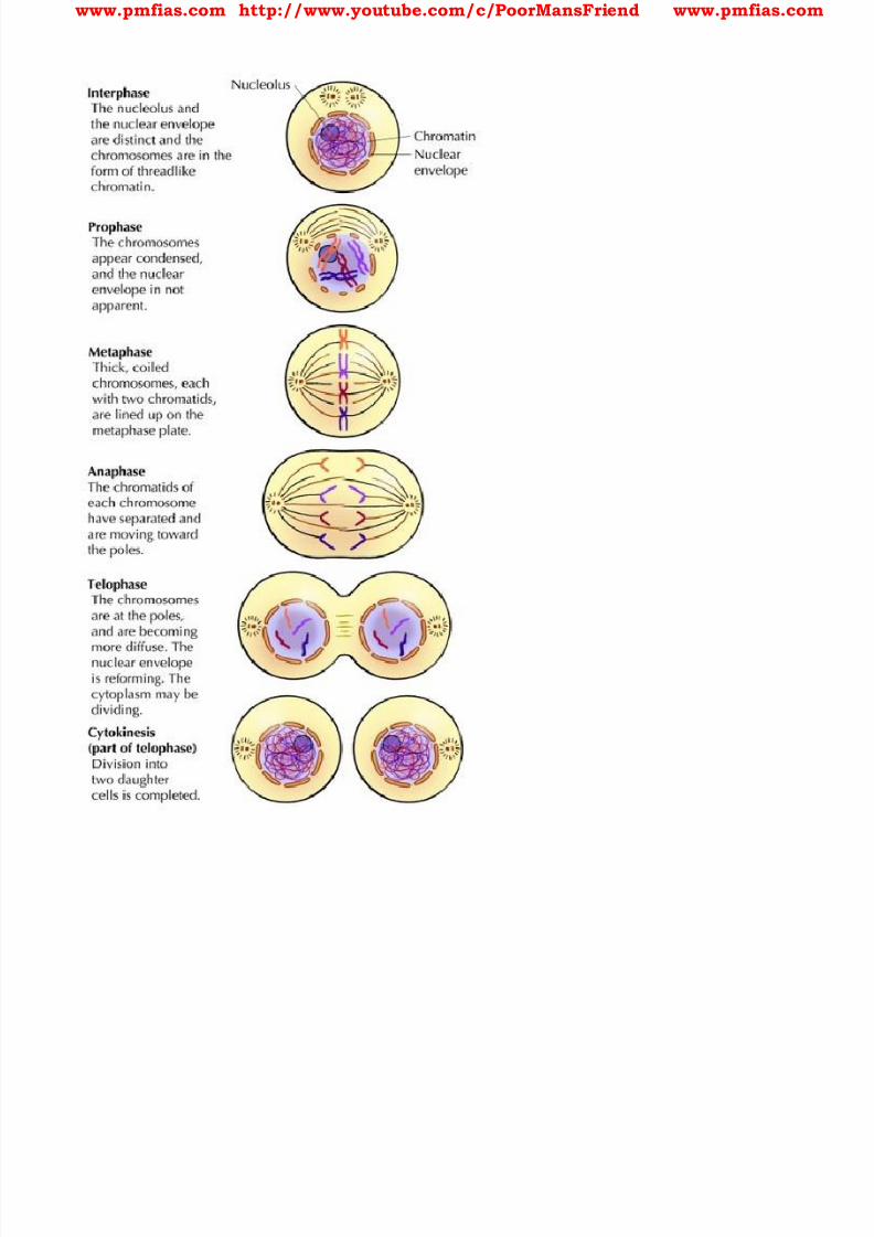
8/16/2019 Biology for Prelims Exam
http://slidepdf.com/reader/full/biology-for-prelims-exam 65/384
www.pmfias.com http://www.youtube.com/c/PoorMansFriend www.pmfias.com
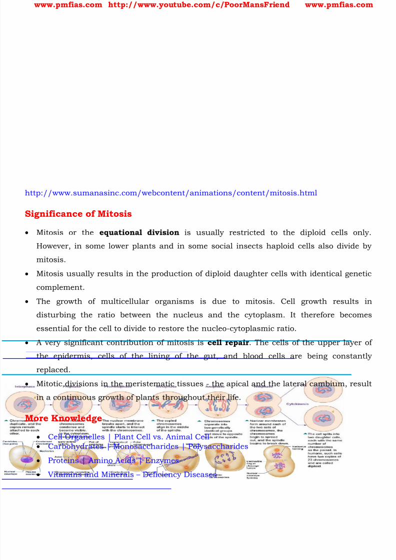
8/16/2019 Biology for Prelims Exam
http://slidepdf.com/reader/full/biology-for-prelims-exam 66/384
www.pmfias.com http://www.youtube.com/c/PoorMansFriend www.pmfias.com
http://www.sumanasinc.com/webcontent/animations/content/mitosis.html
Significance of Mitosis
Mitosis or the equational division is usually restricted to the diploid cells only.
However, in some lower plants and in some social insects haploid cells also divide by
mitosis.
Mitosis usually results in the production of diploid daughter cells with identical genetic
complement.
The growth of multicellular organisms is due to mitosis. Cell growth results in
disturbing the ratio between the nucleus and the cytoplasm. It therefore becomes
essential for the cell to divide to restore the nucleo-cytoplasmic ratio.
A very significant contribution of mitosis is cell repair . The cells of the upper layer of
the epidermis, cells of the lining of the gut, and blood cells are being constantly
replaced.
Mitotic divisions in the meristematic tissues - the apical and the lateral cambium, result
in a continuous growth of plants throughout their life.
More Knowledge
Cell Organelles | Plant Cell vs. Animal Cell Carbohydrates | Monosaccharides | Polysaccharides
Proteins | Amino Acids | Enzymes
Vitamins and Minerals – Deficiency Diseases

8/16/2019 Biology for Prelims Exam
http://slidepdf.com/reader/full/biology-for-prelims-exam 67/384
www.pmfias.com http://www.youtube.com/c/PoorMansFriend www.pmfias.com
Fats | Healthy Fats and Unhealthy Fats
Animal Tissues – Epithelium, Connective Tissues
Human Digestive System | Digestive Glands
Respiratory System – NCERT General Science
Endocrine Glands and Hormones
Human Neural System | Human Brain Muscular and Skeletal System
Nucleic acids – DNA and RNA | Recombinant DNA
8/16/2019 Biology for Prelims Exam
http://slidepdf.com/reader/full/biology-for-prelims-exam 68/384
www.pmfias.com http://www.youtube.com/c/PoorMansFriend www.pmfias.com
Meiosis – Meiotic Cell Division – Production of Gametes – Meiosis I and Meiosis II,
Significance of Meiosis. Mitosis – Meiosis Comparison.
Meiosis
Meiosis is a specialized kind of cell division that reduces the chromosome number
by half and results in the production of haploid daughter cells . The production of offspring by sexual reproduction includes the fusion of two
gametes [sperm and ovum], each with a complete haploid set of chromosomes.
These haploid gametes come into existence due to meiosis [meiosis type of cell
division]. The gametes are formed from specialized diploid cells .
[Diploid == containing two complete sets of chromosomes, one from each parent].
[Haploid == only one set of chromosomes from one of the parent].
Meiosis ensures the production of haploid phase in the life cycle of sexually
reproducing organisms whereas fertilization restores the diploid phase.
Meiotic events can be grouped under the following phases

8/16/2019 Biology for Prelims Exam
http://slidepdf.com/reader/full/biology-for-prelims-exam 69/384
www.pmfias.com http://www.youtube.com/c/PoorMansFriend www.pmfias.com

8/16/2019 Biology for Prelims Exam
http://slidepdf.com/reader/full/biology-for-prelims-exam 70/384
www.pmfias.com http://www.youtube.com/c/PoorMansFriend www.pmfias.com
http://www.ib.bioninja.com.au/higher-level/topic-10-genetics/101-meiosis.html
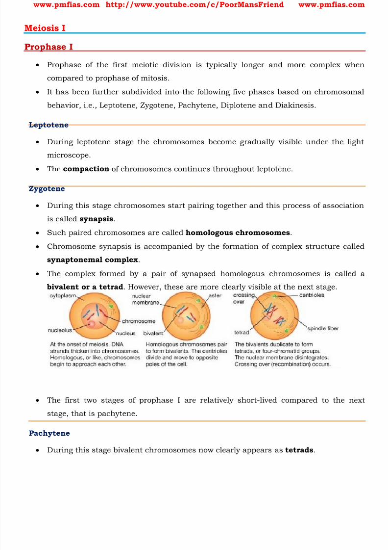
8/16/2019 Biology for Prelims Exam
http://slidepdf.com/reader/full/biology-for-prelims-exam 71/384
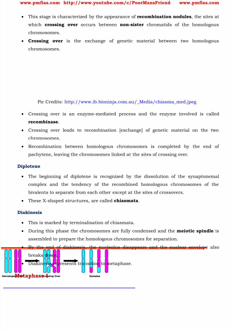
8/16/2019 Biology for Prelims Exam
http://slidepdf.com/reader/full/biology-for-prelims-exam 72/384
www.pmfias.com http://www.youtube.com/c/PoorMansFriend www.pmfias.com
This stage is characterized by the appearance of recombination nodules , the sites at
which crossing over occurs between non-sister chromatids of the homologous
chromosomes.
Crossing over is the exchange of genetic material between two homologous
chromosomes.
Pic Credits: http://www.ib.bioninja.com.au/_Media/chiasma_med.jpeg
Crossing over is an enzyme-mediated process and the enzyme involved is calledrecombinase .
Crossing over leads to recombination [exchange] of genetic material on the two
chromosomes.
Recombination between homologous chromosomes is completed by the end of
pachytene, leaving the chromosomes linked at the sites of crossing over.
Diplotene
The beginning of diplotene is recognized by the dissolution of the synaptonemal
complex and the tendency of the recombined homologous chromosomes of the
bivalents to separate from each other except at the sites of crossovers.
These X-shaped structures, are called chiasmata .
Diakinesis
This is marked by terminalisation of chiasmata.
During this phase the chromosomes are fully condensed and the meiotic spindle isassembled to prepare the homologous chromosomes for separation.
By the end of diakinesis, the nucleolus disappears and the nuclear envelope also
breaks down.
Diakinesis represents transition to metaphase.
Metaphase I
8/16/2019 Biology for Prelims Exam
http://slidepdf.com/reader/full/biology-for-prelims-exam 73/384
www.pmfias.com http://www.youtube.com/c/PoorMansFriend www.pmfias.com
The bivalent chromosomes align on the equatorial plate. The microtubules from the
opposite poles of the spindle attach to the pair of homologous chromosomes.
Anaphase I
The homologous chromosomes separate, while sister chromatids remain associated
at their centromeres.
Telophase I
The nuclear membrane and nucleolus reappear, cytokinesis follows and this is called
as diad of cells .
Interkinesis
The stage between the two meiotic divisions is called interkinesis and is generally
short lived.
Interkinesis is followed by prophase II, a much simpler prophase than prophase I.
Meiosis II
Prophase II
Meiosis II is initiated immediately after cytokinesis, usually before the chromosomes
have fully elongated.
In contrast to meiosis I, meiosis II resembles a normal mitosis .
The nuclear membrane disappears by the end of prophase II. The chromosomes again
become compact.
Metaphase II
At this stage the chromosomes align at the equator and the microtubules from
opposite poles of the spindle get attached to the kinetochores of sister chromatids.
Anaphase II
It begins with the simultaneous splitting of the centromere of each chromosome
(which was holding the sister chromatids together), allowing them to move toward
opposite poles of the cell.
Telophase II
8/16/2019 Biology for Prelims Exam
http://slidepdf.com/reader/full/biology-for-prelims-exam 74/384
www.pmfias.com http://www.youtube.com/c/PoorMansFriend www.pmfias.com
Meiosis ends with telophase II, in which the two groups of chromosomes once again
get enclosed by a nuclear envelope; cytokinesis follows resulting in the formation of
tetrad of cells i.e., four haploid daughter cells .
Significance of Meiosis
Meiosis is the mechanism by which conservation of specific chromosome number ofeach species is achieved across generations in sexually reproducing organisms, even
though the process, per se, paradoxically, results in reduction of chromosome
number by half.
It also increases the genetic variability in the population of organisms from one
generation to the next. Variations are very important for the process of evolution.
Mitosis – Meiosis Comparison
Cell cycle is divided into two phases called (i) Interphase – a period of preparation for
cell division, and (ii) Mitosis (M phase) – the actual period of cell division.
Interphase is further subdivided into G1, S and G2.
G1 phase is the period when the cell grows and carries out normal metabolism. Most
of the organelle duplication also occurs during this phase.
S phase marks the phase of DNA replication and chromosome duplication.
G2 phase is the period of cytoplasmic growth .
Mitosis is also divided into four stages namely prophase, metaphase, anaphase and
telophase.
1. Prophase == Chromosomes condense [chromatids to chromosomes], centrioles move
to the opposite poles, nuclear envelope and the nucleolus disappear and the spindle
fibres start appearing.
2. Metaphase == alignment of chromosomes at the equatorial plate.
3. Anaphase == centromeres divide, chromatids start moving towards the two opposite
poles.
4. Telophase == chromosomal elongation starts, nucleolus and the nuclear membrane
reappear.
5. Cytokinesis == cytoplasmic division.
Mitosis thus, is the equational division in which the chromosome number of the
parent is conserved in the daughter cell.
8/16/2019 Biology for Prelims Exam
http://slidepdf.com/reader/full/biology-for-prelims-exam 75/384
www.pmfias.com http://www.youtube.com/c/PoorMansFriend www.pmfias.com
In contrast to mitosis, meiosis occurs in the diploid cells , which are destined to form
gametes .
It is called the reduction division since it reduces the chromosome number by
half while making the gametes.
In sexual reproduction when the two gametes fuse the chromosome number is
restored to the value in the parent. Meiosis is divided into two phases – meiosis I and meiosis II .
In the first meiotic division the homologous chromosomes pair to form bivalents,
and undergo crossing over.
Meiosis I has a long prophase.
Prophase I == leptotene, zygotene, pachytene, diplotene and diakinesis.
Metaphase I == bivalents arrange on the equatorial plate.
Anaphase I == homologous chromosomes move to the opposite poles with both theirchromatids. Each pole receives half the chromosome number of the parent cell.
Telophase I == nuclear membrane and nucleolus reappear.
Meiosis II is similar to mitosis.
During anaphase II the sister chromatids separate.
Thus at the end of meiosis four haploid cells are formed.
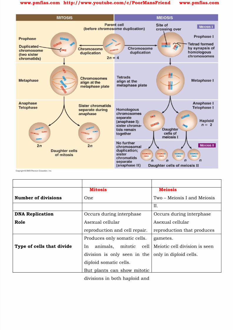
8/16/2019 Biology for Prelims Exam
http://slidepdf.com/reader/full/biology-for-prelims-exam 76/384
www.pmfias.com http://www.youtube.com/c/PoorMansFriend www.pmfias.com
Mitosis Meiosis
Number of divisions One Two – Meiosis I and Meiosis
II.
DNA Replication Occurs during interphase Occurs during interphase
Role Asexual cellular
reproduction and cell repair.Produces only somatic cells.
Asexual cellular
reproduction that producesgametes.
Type of cells that divide In animals, mitotic cell
division is only seen in the
diploid somatic cells.
But plants can show mitotic
divisions in both haploid and
Meiotic cell division is seen
only in diploid cells.

8/16/2019 Biology for Prelims Exam
http://slidepdf.com/reader/full/biology-for-prelims-exam 77/384
www.pmfias.com http://www.youtube.com/c/PoorMansFriend www.pmfias.com
diploid cells.
Cell Cycle Both cell division and cell
cycle.
It is only cell division and not
cell cycle.
Daughter cells produced Two diploid daughter cells
(2n) that are genetically
identical to the parent cell.
Four haploid daughter cells
(n) containing half the
number of chromosome asthe parent cell.
8/16/2019 Biology for Prelims Exam
http://slidepdf.com/reader/full/biology-for-prelims-exam 78/384
www.pmfias.com http://www.youtube.com/c/PoorMansFriend www.pmfias.com
Mendel’s Laws of Inheritance – Law of Dominance, Law of Segregation. Incomplete
Dominance, Co-dominance, Inheritance of Two Genes, Law of Independent Assortment.
Source|Credits|Picture Credits: NCERT Science Textbooks Class 6-12.
Mendel’s Experiments on Inheritance
Inheritance is the process by which characters are passed on from parent to progeny; it
is the basis of heredity. Variation is the degree by which progeny differ from their
parents.
It was only during the mid-nineteenth century that a major headway was made in the
understanding of inheritance.
Gregor Mendel , conducted hybridisation experiments on garden peas for seven years
(1856-1863) and proposed the laws of inheritance in living organisms.
Mendel conducted such artificial pollination/cross pollination experiments using severaltrue-breeding pea lines.
[Truebreeding line == A truebreeding line is one that, having undergone continuous self-
pollination , shows the stable trait inheritance and expression for several generations].
Mendel investigated characters in the garden pea plant that were manifested as two
opposing traits, e.g., tall or dwarf plants, yellow or green seeds etc.
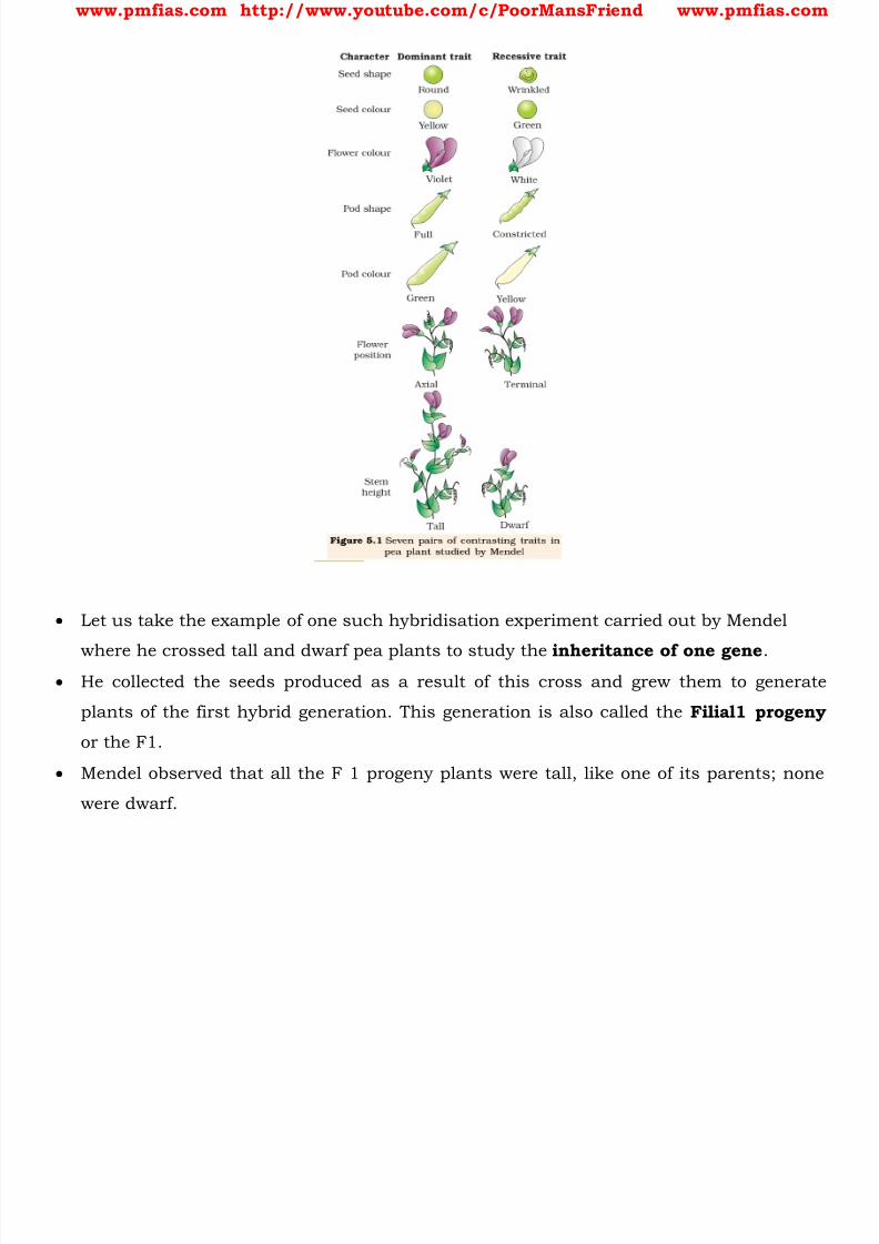
8/16/2019 Biology for Prelims Exam
http://slidepdf.com/reader/full/biology-for-prelims-exam 79/384
www.pmfias.com http://www.youtube.com/c/PoorMansFriend www.pmfias.com
Let us take the example of one such hybridisation experiment carried out by Mendel
where he crossed tall and dwarf pea plants to study the inheritance of one gene . He collected the seeds produced as a result of this cross and grew them to generate
plants of the first hybrid generation. This generation is also called the Filial1 progeny
or the F1.
Mendel observed that all the F 1 progeny plants were tall, like one of its parents; none
were dwarf.

8/16/2019 Biology for Prelims Exam
http://slidepdf.com/reader/full/biology-for-prelims-exam 80/384
www.pmfias.com http://www.youtube.com/c/PoorMansFriend www.pmfias.com
He made similar observations for the other pairs of traits – he found that the F 1 always
resembled either one of the parents, and that the trait of the other parent was not seen
in them. Mendel then self-pollinated the tall F1 plants and to his surprise found that in the Filial
2 generation some of the offspring were ‘dwarf’; the character that was not seen in the F
1 generation was now expressed.
The proportion of plants that were dwarf were 1/4th of the F 2 plants while 3/4th of the
F2 plants were tall.
The tall and dwarf traits were identical to their parental type and did not show any
blending, that is all the offspring were either tall or dwarf, none were of in betweenheight.
Similar results were obtained with the other traits that he studied: only one of the
parental traits was expressed in the F1 generation while at the F 2 stage both the traits
were expressed in the proportion 3:1 . The contrasting traits did not show any blending
at either F1 or F2 stage.
Factors – Genes
8/16/2019 Biology for Prelims Exam
http://slidepdf.com/reader/full/biology-for-prelims-exam 81/384
www.pmfias.com http://www.youtube.com/c/PoorMansFriend www.pmfias.com
Based on these observations, Mendel proposed that something was being stably passed
down, unchanged, from parent to offspring through the gametes, over successive
generations. He called these things as ‘factors’ .
Now we call the ‘factors’ as genes . Genes, therefore, are the units of inheritance . They
contain the information that is required to express a particular trait in an organism.
Pair of Alleles - Homozygous and Heterozygous
Genes which code for a pair of contrasting traits are known as alleles , i.e., they are
slightly different forms of the same gene.
If we use alphabetical symbols for each gene, then the capital letter is used for the trait
expressed at the F 1 stage and the small alphabet for the other trait.
For example, in case of the character of height, T is used for the Tall trait and t for the
‘dwarf’, and T and t are alleles of each other. Hence, in plants the pair of alleles for
height would be TT, Tt or tt.
Genotype and Phenotype == TT and tt are called the genotype of the plant while the
descriptive terms tall and dwarf are the phenotype .
Mendel proposed that in a true breeding, tall or dwarf pea variety the allelic pair of genes
for height are identical or homozygous , TT and tt, respectively. On the other hand, the
allelic pair Tt is heterozygous .
From the observation that the recessive parental trait is expressed without any blending
in the F2 generation, we can infer that, when the tall and dwarf plant produce gametes,
by the process of meiosis , the alleles of the parental pair separate or segregate from
each other and only one allele is transmitted to a gamete .
This segregation of alleles is a random process and so there is a 50 per cent chance of a
gamete containing either allele, as has been verified by the results of the crossings. In
this way the gametes of the tall TT plants have the allele T and the gametes of the dwarf
tt plants have the allele t.
During fertilisation the two alleles, T from one parent say, through the pollen, and t from
the other parent, then through the egg, are united to produce zygotes that have one T
allele and one t allele. In other words the hybrids have Tt. Since these hybrids contain
alleles which express contrasting traits, the plants are heterozygous .
Dominant and Recessive Factor

8/16/2019 Biology for Prelims Exam
http://slidepdf.com/reader/full/biology-for-prelims-exam 82/384
www.pmfias.com http://www.youtube.com/c/PoorMansFriend www.pmfias.com
What twould be the phenotype of a plant that had a genotype Tt? As Mendel found the
phenotype of the F1heterozygote Tt to be exactly like the TT parent in appearance, he
proposed that in a pair of dissimilar factors, one dominates the other (as in the F1) and
hence is called the dominant factor while the other factor is recessive .
In this case T (for tallness) is dominant over t (for dwarfness), that is recessive. He
observed identical behaviour for all the other characters/trait-pairs that he studied. It is convenient (and logical) to use the capital and lower case of an alphabetical symbol
to remember this concept of dominance and recessiveness.
Alleles can be similar as in the case of homozygotes TT and tt or can be dissimilar as in
the case of the heterozygote Tt.
Since the Tt plant is heterozygous for genes controlling one character (height), it is a
monohybrid and the cross between TT and tt is a monohybrid cross .
Punnett Square
The production of gametes by the parents, the formation of the zygotes, the F1 and F2
plants can be understood from a diagram called Punnett Square as shown in Figure
below.
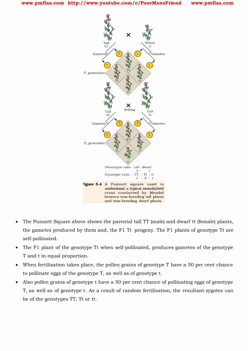
8/16/2019 Biology for Prelims Exam
http://slidepdf.com/reader/full/biology-for-prelims-exam 83/384
www.pmfias.com http://www.youtube.com/c/PoorMansFriend www.pmfias.com
The Punnett Square above shows the parental tall TT (male) and dwarf tt (female) plants,
the gametes produced by them and, the F1 Tt progeny. The F1 plants of genotype Tt are
self-pollinated.
The F1 plant of the genotype Tt when self-pollinated, produces gametes of the genotype
T and t in equal proportion.
When fertilisation takes place, the pollen grains of genotype T have a 50 per cent chance
to pollinate eggs of the genotype T, as well as of genotype t.
Also pollen grains of genotype t have a 50 per cent chance of pollinating eggs of genotype
T, as well as of genotype t. As a result of random fertilisation, the resultant zygotes can
be of the genotypes TT, Tt or tt.

8/16/2019 Biology for Prelims Exam
http://slidepdf.com/reader/full/biology-for-prelims-exam 84/384
www.pmfias.com http://www.youtube.com/c/PoorMansFriend www.pmfias.com
From the Punnett square it is easily seen that 1/4th of the random fertilisations lead to
TT, 1/2 lead to Tt and 1/4th to tt.
Though the F1 have a genotype of Tt, but the phenotypic character seen is ‘tall’.
At F2, 3/4th of the plants are tall, where some of them are TT while others are Tt.
Test Cross
Externally it is not possible to distinguish between the plants with the genotypes TT and
Tt. Hence, within the genopyti c pair Tt only one character ‘T’ tall is expressed. Hence the
character T or ‘tall’ is said to dominate over the other allele t or ‘dwarf’ character.
It is thus due to this dominance of one character over the other that all the F 1 are tall
(though the genotype is Tt) and in the F2 3/4th of the plants are tall (though
genotypically 1/2 are Tt and only 1/4th are TT).
This leads to a phenotypic ratio of 3/4th tall : (1/4 TT + 1/2 Tt) and 1/4th tt, i.e., a 3:1
ratio, but a genotypic ratio of 1:2:1.
The genotypic ratios can be calculated by simply looking at the phenotype of a dominant
trait but it is not possible to know the genotypic composition .
That is, for example, whether a tall plant from F1 or F 2 has TT or Tt composition,
cannot be predicted.
Therefore, to determine the genotype of a tall plant at F2, Mendel crossed the tall plant
from F2 with a dwarf plant. This he called a test cross .
8/16/2019 Biology for Prelims Exam
http://slidepdf.com/reader/full/biology-for-prelims-exam 85/384
www.pmfias.com http://www.youtube.com/c/PoorMansFriend www.pmfias.com
In a typical test cross an organism (pea plants here) showing a dominant phenotype (and
whose genotype is to be determined) is crossed with the recessive parent.
The progenies of such a cross can easily be analysed to predict the genotype of the test
organism.
Figure shows the results of typical test cross where violet colour flower (W) is dominant
over white colour flower (w).
Mendel’s Laws o f Inheritance
Based on his observations on monohybrid crosses Mendel proposed two general rules to
consolidate his understanding of inheritance in monohybrid crosses.
Today these rules are called the Principles or Laws of Inheritance : the First Law or
Law of Dominance and the Second Law or Law of Segregation .
First Law or Law of Dominance
1. Characters are controlled by discrete units called factors .
2. Factors occur in pairs . [pair of alleles]
3. In a dissimilar pair of factors one member of the pair dominates ( dominant ) the other
(recessive ).
The law of dominance is used to explain the expression of only one of the parental
characters in a monohybrid cross [Mendel’s Experiments on Inheritance] in the F1 and
the expression of both in the F 2. It also explains the proportion of 3:1 obtained at the
F2.
Second Law or Law of Segregation
This law is based on the fact that the alleles do not show any blending and that both
the characters are recovered as such in the F 2 generation though one of these is not
seen at the F1 stage.
Though the parents contain two alleles during gamete formation, the factors or alleles ofa pair segregate from each other such that a gamete receives only one of the two
factors [either dominant or recessive] .
Of course, a homozygous parent produces all gametes that are similar while a
heterozygous one produces two kinds of gametes each having one allele with equal
proportion.
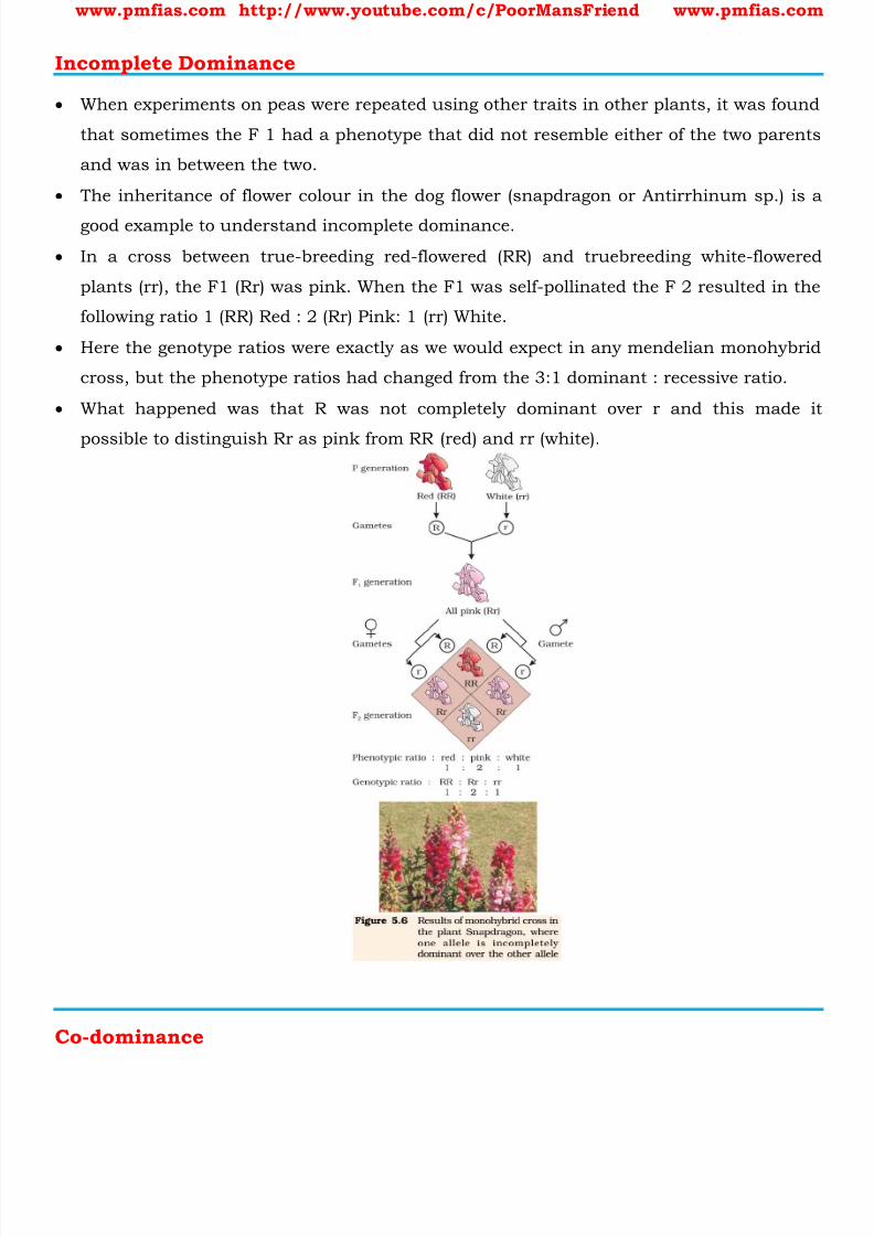
8/16/2019 Biology for Prelims Exam
http://slidepdf.com/reader/full/biology-for-prelims-exam 86/384
www.pmfias.com http://www.youtube.com/c/PoorMansFriend www.pmfias.com
Incomplete Dominance
When experiments on peas were repeated using other traits in other plants, it was found
that sometimes the F 1 had a phenotype that did not resemble either of the two parents
and was in between the two.
The inheritance of flower colour in the dog flower (snapdragon or Antirrhinum sp.) is a
good example to understand incomplete dominance.
In a cross between true-breeding red-flowered (RR) and truebreeding white-flowered
plants (rr), the F1 (Rr) was pink. When the F1 was self-pollinated the F 2 resulted in the
following ratio 1 (RR) Red : 2 (Rr) Pink: 1 (rr) White.
Here the genotype ratios were exactly as we would expect in any mendelian monohybrid
cross, but the phenotype ratios had changed from the 3:1 dominant : recessive ratio.
What happened was that R was not completely dominant over r and this made it
possible to distinguish Rr as pink from RR (red) and rr (white).
Co-dominance

8/16/2019 Biology for Prelims Exam
http://slidepdf.com/reader/full/biology-for-prelims-exam 87/384

8/16/2019 Biology for Prelims Exam
http://slidepdf.com/reader/full/biology-for-prelims-exam 88/384
www.pmfias.com http://www.youtube.com/c/PoorMansFriend www.pmfias.com
Here there are 6 Genotypes and 4 Phenotypes [A, B, AB and O].
Inheritance of Two Genes
Mendel also worked with and crossed pea plants that differed in two characters, as is
seen in the cross between a pea plant that has seeds with yellow colour and round
shape and one that had seeds of green colour and wrinkled shape. Yellow colour was dominant over green and round shape dominant over wrinkled.
Let us use the genotypic symbols Y for dominant yellow seed colour and y for recessive
green seed colour, R for round shaped seeds and r for wrinkled seed shape.
The genotype of the parents can then be written as RRYY and rryy. The cross between
the two plants can be written down as in Figure showing the genotypes of the parent
plants.
The gametes RY and ry unite on fertilisation to produce the F1 hybrid RrYy.
When Mendel self hybridised the F1 plants he found that 3/4th of F2 plants had yellow
seeds and 1/4th had green.
The yellow and green colour segregated in a 3:1 ratio. Round and wrinkled seed shape
also segregated in a 3:1 ratio; just like in a monohybrid cross.

8/16/2019 Biology for Prelims Exam
http://slidepdf.com/reader/full/biology-for-prelims-exam 89/384
www.pmfias.com http://www.youtube.com/c/PoorMansFriend www.pmfias.com
Law of Independent Assortment
In the dihybrid cross, the phenotypes round, yellow; wrinkled, yellow; round, green and
wrinkled, green appeared in the ratio 9:3:3:1. Such a ratio was observed for several pairs of characters that Mendel studied. The ratio
of 9:3:3:1 can be derived as a combination series of 3 yellow: 1 green, with 3 round : 1
wrinkled. This derivation can be written as follows: (3 Round : 1 Wrinkled) (3 Yellow : 1
Green) = 9 Round, Yellow : 3 Wrinkled, Yellow: 3 Round, Green : 1 Wrinkled, Green.

8/16/2019 Biology for Prelims Exam
http://slidepdf.com/reader/full/biology-for-prelims-exam 90/384
www.pmfias.com http://www.youtube.com/c/PoorMansFriend www.pmfias.com
Based upon such observations on dihybrid crosses (crosses between plants differing in
two traits) Mendel proposed a second set of generalisations that we call Mendel’s Law
of Independent Assortment .
The law states that ‘when two pairs of traits are combined in a hybrid, segre gation
of one pair of characters is independent of the other pair of characters’ .
You can verify the law using The Punnett square above [Inheritance of Two Genes –dihybrid cross].
Summary
Genetics is a branch of biology which deals with principles of inheritance and its
practices.
Progeny resembling the parents in morphological and physiological features has
attracted the attention of many biologists.
Mendel was the first to study this phenomenon systematically. While studying the
pattern of inheritance in pea plants of contrasting characters, Mendel proposed the
principles of inheritance, which are today referred to as ‘Mendel’s Laws of Inheritance’.
He proposed that the ‘factors’ (later named as genes) regulating the characters are found
in pairs known as alleles.
He observed that the expression of the characters in the offspring follow a definite
pattern in different – first generations (F1), second (F2) and so on. Some characters are
dominant over others.
The dominant characters are expressed when factors are in heterozygous condition (Law
of Dominance). The recessive characters are only expressed in homozygous conditions.
The characters never blend in heterozygous condition.
A recessive character that was not expressed in heterozygous condition may be
expressed again when it becomes homozygous. Hence, characters segregate while
formation of gametes (Law of Segregation).
Not all characters show true dominance. Some characters show incomplete, and some
show co-dominance.
When Mendel studied the inheritance of two characters together, it was found that the
factors independently assort and combine in all permutations and combinations (Law of
Independent Assortment).
Different combinations of gametes are theoretically represented in a square tabular form
known as ‘Punnett Square’.

8/16/2019 Biology for Prelims Exam
http://slidepdf.com/reader/full/biology-for-prelims-exam 91/384
www.pmfias.com http://www.youtube.com/c/PoorMansFriend www.pmfias.com
The factors (now known as gene) on chromosomes regulating the characters are called
the genotype and the physical expression of the characters is called phenotype.

8/16/2019 Biology for Prelims Exam
http://slidepdf.com/reader/full/biology-for-prelims-exam 92/384
www.pmfias.com http://www.youtube.com/c/PoorMansFriend www.pmfias.com
Chromosomal Theory of Inheritance - Linkage and Recombination - Human Genome Project
- Applications and Benefits of Human Genome Project.
Chromosomal Theory of Inheritance
Due to advancements in microscopy during 1900s, scientists were able to observe cell
division. This led to the discovery of structures in the nucleus that appeared to doubleand divide just before each cell division. These were called chromosomes (colored
bodies, as they were visualised by staining).
By 1902, the chromosome movement during meiosis had been worked out. The behavior
of chromosomes was parallel to the behavior of genes and used biologists used
chromosome movement to explain Mendel’s laws.
[Mitosis (equational division) and meiosis (reduction division) were explained in previous
posts].
The important things to remember are that chromosomes as well as genes occur in
pairs. The two alleles of a gene pair are located on homologous sites on homologous
chromosomes.
During Anaphase of meiosis I, the two chromosome pairs can align at the metaphase
plate independently of each other.
To understand this, compare the chromosomes of four different colour in the left and
right columns. In the left column (Possibility I) orange and green is segregating together.
But in the right hand column (Possibility II) the orange chromosome is segregating with
the red chromosomes.
Sutton and Boveri argued that the pairing and separation of a pair of chromosomes
would lead to the segregation of a pair of factors they carried. Sutton united the
knowledge of chromosomal segregation with Mendelian principles and called it the
chromosomal theory of inheritance .
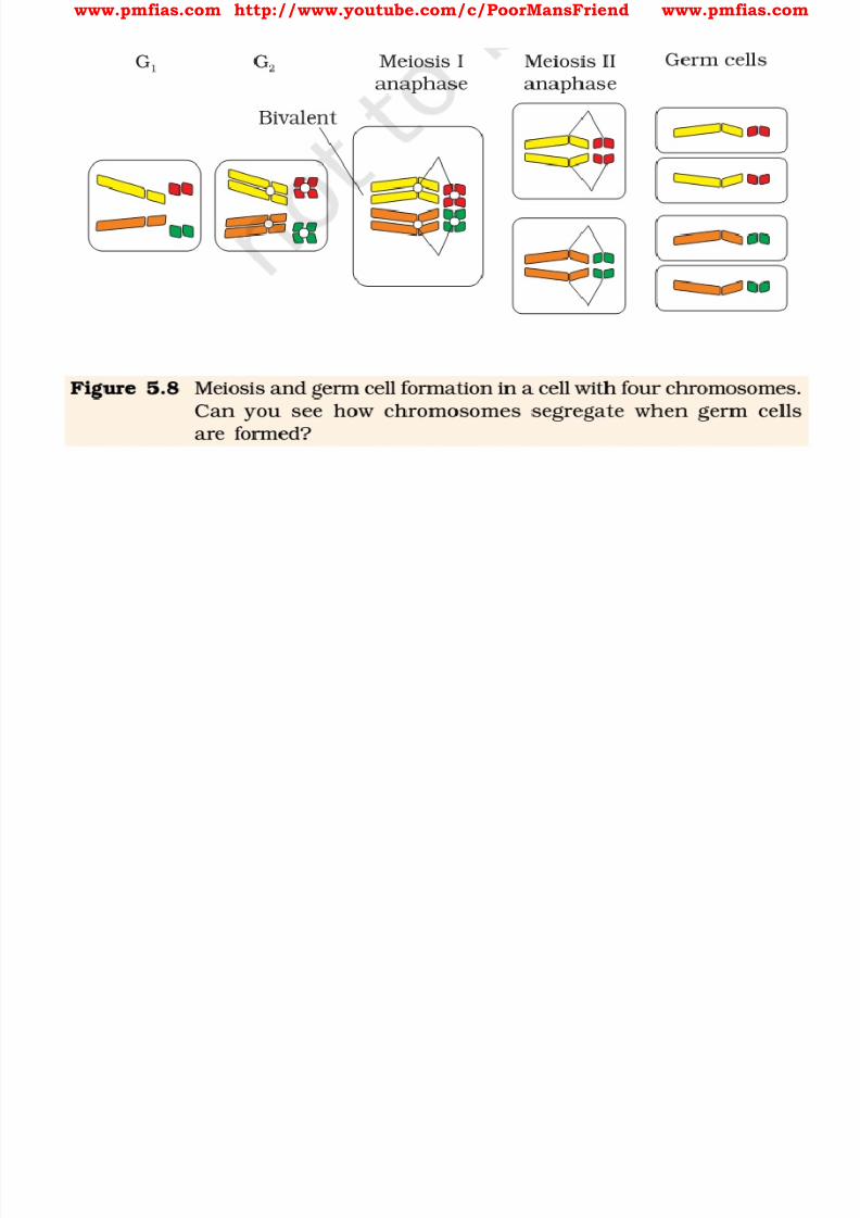
8/16/2019 Biology for Prelims Exam
http://slidepdf.com/reader/full/biology-for-prelims-exam 93/384
www.pmfias.com http://www.youtube.com/c/PoorMansFriend www.pmfias.com
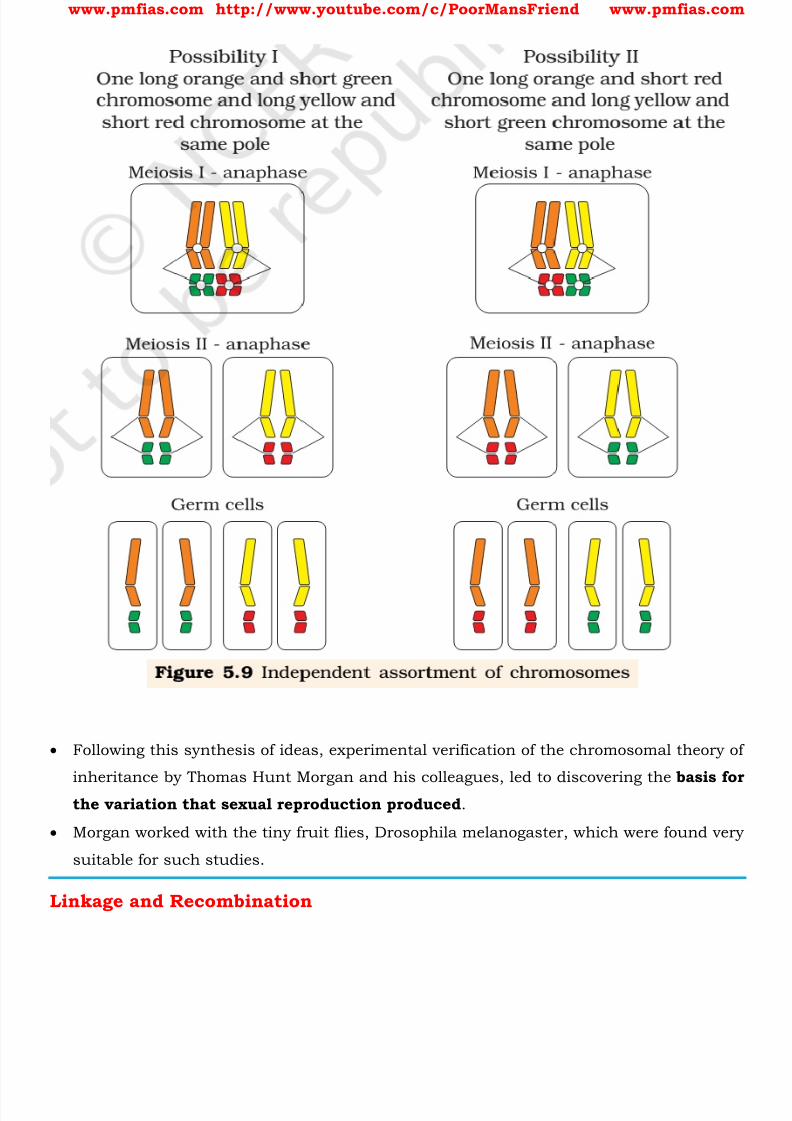
8/16/2019 Biology for Prelims Exam
http://slidepdf.com/reader/full/biology-for-prelims-exam 94/384
www.pmfias.com http://www.youtube.com/c/PoorMansFriend www.pmfias.com
Following this synthesis of ideas, experimental verification of the chromosomal theory of
inheritance by Thomas Hunt Morgan and his colleagues, led to discovering the basis for
the variation that sexual reproduction produced .
Morgan worked with the tiny fruit flies, Drosophila melanogaster, which were found very
suitable for such studies.
Linkage and Recombination

8/16/2019 Biology for Prelims Exam
http://slidepdf.com/reader/full/biology-for-prelims-exam 95/384
www.pmfias.com http://www.youtube.com/c/PoorMansFriend www.pmfias.com
Morgan carried out several dihybrid crosses in Drosophila to study genes that were sex-
linked. The crosses were similar to the dihybrid crosses carried out by Mendel in peas.
For example Morgan hybridised yellow-bodied, white-eyed females to brown-bodied, red-
eyed males and intercrossed their F 1 progeny. He observed that the two genes did not
segregate independently of each other and the F 2 ratio deviated very significantly from
the 9:3:3:1 ratio (expected when the two genes are independent). Morgan and his group knew that the genes were located on the X chromosome and saw
quickly that when the two genes in a dihybrid cross were situated on the same
chromosome, the proportion of parental gene combinations were much higher than the
non-parental type.
Morgan attributed this due to the physical association or linkage of the two genes
and coined the term linkage to describe this physical association of genes on a
chromosome and the term recombination to describe the generation of non-parental gene combinations .
Alfred Sturtevant used the frequency of recombination between gene pairs on the same
chromosome as a measure of the distance between genes and ‘mapped’ their position on
the chromosome.
Today genetic maps are extensively used as a starting point in the sequencing of whole
genomes as was done in the case of the Human Genome Sequencing Project.
Human Genome Project
Human Genome Project is a publicly funded international collaborative scientific
research project aimed at determining the sequence of chemical base pairs which
make up human DNA, and of identifying and mapping all of the genes of the human
genome.
[A base pair (bp) is a unit consisting of two nucleobases bound to each other by hydrogen
bonds . They form the building blocks of the DNA double helix, and contribute to the foldedstructure of both DNA and RNA.]
Human Genome Project was formally launched in 1990, and finally declared complete in
2003.
The "genome" of any given individual is unique; mapping "the human genome" involves
sequencing multiple variations of each gene.

8/16/2019 Biology for Prelims Exam
http://slidepdf.com/reader/full/biology-for-prelims-exam 96/384
www.pmfias.com http://www.youtube.com/c/PoorMansFriend www.pmfias.com
All our genes together are known as our ‘genome.’ The HGP has revealed that there are
probably about 20,500 human genes .
Applications and Benefits of Human Genome Project
Can help us
1. understand diseases including: genotyping of specific viruses to direct appropriate
treatment,
2. in identification of mutations linked to different forms of cancer,
3. understand the design of medication and more accurate prediction of their effects,
4. in advancement of forensic applied sciences, biofuels and other energy applications,
agriculture, animal husbandry, etc..
5. understand evolution much more accurately.
Another proposed benefit is the commercial development of genomics research related toDNA based products, a multibillion-dollar industry.
Summary
After knowing that the genes are located on the chromosomes, a good correlation was
drawn between Mendel’s laws : segregation and assortment of chromosomes during
meiosis. The Mendel’s laws were extended in the form of ‘Chromosomal Theory of
Inheritance’.
Later, it was found that Mendel’s law of independent assortment does not hold true for
the genes that were located on the same chromosomes. These genes were called as
‘linked genes’.
Closely located genes assorted together, and distantly located genes, due to
recombination, assorted independently. Linkage maps, therefore, corresponded to
arrangement of genes on a chromosome.
Many genes were linked to sexes also, and called as sex-linked genes. The two sexes
(male and female) were found to have a set of chromosomes which were common, and
another set which was different.
The chromosomes which were different in two sexes were named as sex chromosomes.
The remaining set was named as autosomes. In humans, a normal female has 22 pairs
of autosomes and a pair of sex chromosomes (XX).

8/16/2019 Biology for Prelims Exam
http://slidepdf.com/reader/full/biology-for-prelims-exam 97/384
www.pmfias.com http://www.youtube.com/c/PoorMansFriend www.pmfias.com
Human Genome Project is a collaborative scientific research project aimed at identifying
and mapping all of the genes of the human genome.
Human genome project is helpful in wide ranging biotechnology applications.
8/16/2019 Biology for Prelims Exam
http://slidepdf.com/reader/full/biology-for-prelims-exam 98/384
www.pmfias.com http://www.youtube.com/c/PoorMansFriend www.pmfias.com
Sex Determination, Genetic Disorders - Pedigree Analysis, Mutation, Mendelian Disorders:
Haemophilia, Sickle-cell anaemia, Phenylketonuria. Chromosomal disorders: Down’s
Syndrome, Klinefelter’s Syndrome, Turner’s Syndrome.
Sex Determination
Henking (1891) could trace a specific nuclear structure all through spermatogenesis in afew insects, and it was also observed by him that 50 per cent of the sperm received this
structure after spermatogenesis, whereas the other 50 per cent sperm did not receive it.
Henking gave a name to this structure as the X body but he could not explain its
significance.
Further investigations by other scientists led to the conclusion that the ‘X body’ of
Henking was in fact a chromosome and that is why it was given the name X-
chromosome.
It was also observed that in a large number of insects the mechanism of sex
determination is of the XO type, i.e., all eggs bear an additional X-chromosome besides
the other chromosomes (autosomes). On the other hand, some of the sperms bear the X-
chromosome whereas some do not.
Eggs fertilized by sperm having an X-chromosome become females and, those fertilized
by sperms that do not have an X-chromosome become males.
Due to the involvement of the X-chromosome in the determination of sex, it was
designated to be the sex chromosome , and the rest of the chromosomes were named as
autosomes .
Grasshopper is an example of XO type of sex determination in which the males have
only one X-chromosome besides the autosomes, whereas females have a pair of X-
chromosomes.
These observations led to the investigation of a number of species to understand the
mechanism of sex determination.
In a number of other insects and mammals including man, XY type of sex determination
is seen where both male and female have same number of chromosomes.

8/16/2019 Biology for Prelims Exam
http://slidepdf.com/reader/full/biology-for-prelims-exam 99/384
www.pmfias.com http://www.youtube.com/c/PoorMansFriend www.pmfias.com
Among the males an X-chromosome is present but its counterpart is distinctly smaller
and called the Y-chromosome. Females, however, have a pair of X-chromosomes.
Both males and females bear same number of autosomes . Hence, the males have
autosomes plus XY, while female have autosomes plus XX.
[one pair of sex chromosome (XX or XY) + 22 pairs of autosomes == total 23 pairs of
chromosomes in a human cell nucleus]
In human beings and in Drosophila the males have one X and one Y chromosome,
whereas females have a pair of X-chromosomes besides autosomes.
In the above description you have studied about two types of sex determining
mechanisms, i.e., XO type and XY type. But in both cases males produce two different
types of gametes, (a) either with or without X-chromosome or (b) some gametes with X-
8/16/2019 Biology for Prelims Exam
http://slidepdf.com/reader/full/biology-for-prelims-exam 100/384
www.pmfias.com http://www.youtube.com/c/PoorMansFriend www.pmfias.com
chromosome and some with Y-chromosome. Such types of sex determination
mechanism is designated to be the example of male heterogamety .
In some other organisms, e.g., birds, a different mechanism of sex determination is
observed. In this case the total number of chromosome is same in both males and
females. But two different types of gametes in terms of the sex chromosomes, are
produced by females, i.e., female heterogamety . In order to have a distinction with the mechanism of sex determination described earlier,
the two different sex chromosomes of a female bird has been designated to be the Z and
W chromosomes. In these organisms the females have one Z and one W chromosome,
whereas males have a pair of Z-chromosomes besides the autosomes.
Sex Determination in Humans
It has already been mentioned that the sex determining mechanism in case of humans
is XY type. Out of 23 pairs of chromosomes present, 22 pairs are exactly same in both
males and females; these are the autosomes .
A pair of X-chromosomes are present in the female, whereas the presence of an X and Y
chromosome are determinant of the male characteristic.
During spermatogenesis among males, two types of gametes are produced. 50 per cent
of the total sperm produced carry the X-chromosome and the rest 50 per cent has Y-
chromosome besides the autosomes.
Females, however, produce only one type of ovum with an X-chromosome. There is an
equal probability of fertilization of the ovum with the sperm carrying either X or Y
chromosome.
In case the ovum fertilizes with a sperm carrying X-chromosome the zygote develops into
a female (XX) and the fertilization of ovum with Y-chromosome carrying sperm results
into a male offspring.
Thus, it is evident that it is the genetic makeup of the sperm that determines the sex of
the child. It is also evident that in each pregnancy there is always 50 per cent
probability of either a male or a female child.
It is unfortunate that in our society women are blamed for giving birth to female children
and have been ostracized and ill-treated because of this false notion.
Genetic Disorders
Pedigree Analysis
8/16/2019 Biology for Prelims Exam
http://slidepdf.com/reader/full/biology-for-prelims-exam 101/384
www.pmfias.com http://www.youtube.com/c/PoorMansFriend www.pmfias.com
The idea that disorders are inherited has been prevailing in the human society since
long. This was based on the heritability of certain characteristic features in families.
After the rediscovery of Mendel’s work the practice of analyzing inheritance pattern of
traits in human beings began.
Since it is evident that control crosses that can be performed in pea plant or some
other organisms, are not possible in case of human beings, study of the family historyabout inheritance of a particular trait provides an alternative.
Such an analysis of traits in a several of generations of a family is called the pedigree
analysis .
In the pedigree analysis the inheritance of a particular trait is represented in the family
tree over generations.
In human genetics, pedigree study provides a strong tool, which is utilized to trace the
inheritance of a specific trait, abnormality or disease. Each and every feature in any organism is controlled by one or the other gene located on
the DNA present in the chromosome. DNA is the carrier of genetic information. It is
hence transmitted from one generation to the other without any change or alteration.
However, changes or alteration do take place occasionally. Such an alteration or change
in the genetic material is referred to as mutation .
A number of disorders in human beings have been found to be associated with the
inheritance of changed or altered genes or chromosomes.
Mutation
Mutation is a phenomenon which results in alteration of DNA sequences and
consequently results in changes in the genotype and the phenotype of an organism.
In addition to recombination, mutation is another phenomenon that leads to variation
in DNA.
DNA helix runs continuously from one end to the other in each chromatid, in a highly
supercoiled form. Therefore loss (deletions) or gain (insertion/duplication) of a
segment of DNA, result in alteration in chromosomes.
Since genes are known to be located on chromosomes, alteration in chromosomes
results in abnormalities or aberrations . Chromosomal aberrations are commonly
observed in cancer cells .
8/16/2019 Biology for Prelims Exam
http://slidepdf.com/reader/full/biology-for-prelims-exam 102/384
8/16/2019 Biology for Prelims Exam
http://slidepdf.com/reader/full/biology-for-prelims-exam 103/384
www.pmfias.com http://www.youtube.com/c/PoorMansFriend www.pmfias.com
The possibility of a female becoming a haemophilic is extremely rare because mother of
such a female has to be at least carrier and the father should be haemophilic (unviable
in the later stage of life).
The family pedigree of Queen Victoria shows a number of haemophilic descendents as
she was a carrier of the disease.
Sickle-Cell Anaemia
This is an autosome linked recessive trait that can be transmitted from parents to the
offspring when both the partners are carrier for the gene (or heterozygous). The disease
is controlled by a single pair of allele, HbA and HbS.
Out of the three possible genotypes only homozygous individuals for HbS (HbSHbS)
show the diseased phenotype.
Heterozygous (HbAHbS) individuals appear apparently unaffected but they are carrier of
the disease as there is 50 per cent probability of transmission of the mutant gene to the
progeny, thus exhibiting sickle-cell trait.
The defect is caused by the substitution of Glutamic acid (Glu) by Valine (Val) at the
sixth position of the beta globin chain of the haemoglobin molecule .
The substitution of amino acid in the globin protein results due to the single base
substitution at the sixth codon of the beta globin gene from GAG to GUG.
The mutant haemoglobin molecule undergoes polymerization under low oxygen tension
causing the change in the shape of the RBC from biconcave disc to elongated sickle like
structure.
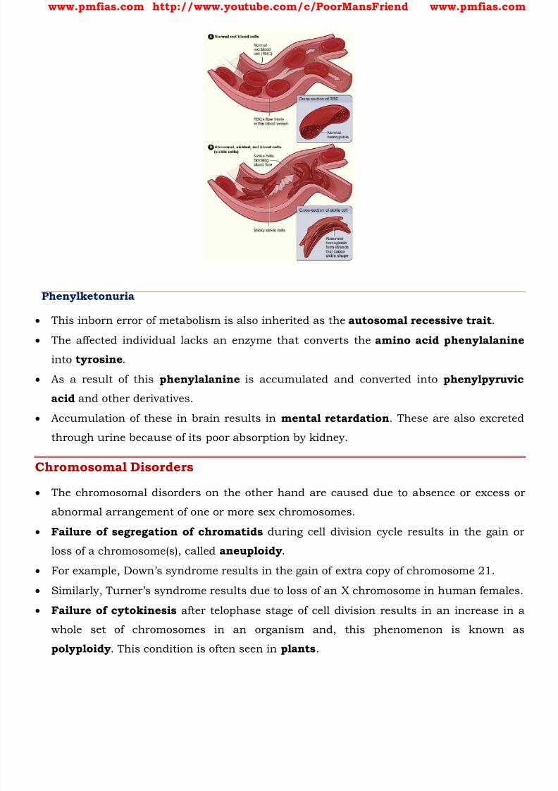
8/16/2019 Biology for Prelims Exam
http://slidepdf.com/reader/full/biology-for-prelims-exam 104/384
www.pmfias.com http://www.youtube.com/c/PoorMansFriend www.pmfias.com
Phenylketonuria
This inborn error of metabolism is also inherited as the autosomal recessive trait .
The affected individual lacks an enzyme that converts the amino acid phenylalanine
into tyrosine .
As a result of this phenylalanine is accumulated and converted into phenylpyruvic
acid and other derivatives.
Accumulation of these in brain results in mental retardation . These are also excreted
through urine because of its poor absorption by kidney.
Chromosomal Disorders
The chromosomal disorders on the other hand are caused due to absence or excess or
abnormal arrangement of one or more sex chromosomes.
Failure of segregation of chromatids during cell division cycle results in the gain or
loss of a chromosome(s), called aneuploidy . For example, Down’s syndrome results in the gain of extra copy of chromosome 21.
Similarly, Turner’s syndrome results due to loss of an X chromosome in human females.
Failure of cytokinesis after telophase stage of cell division results in an increase in a
whole set of chromosomes in an organism and, this phenomenon is known as
polyploidy . This condition is often seen in plants .
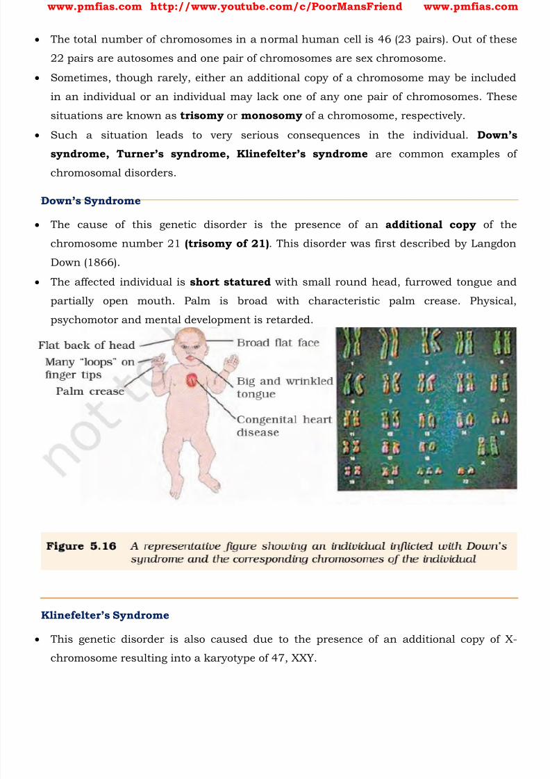
8/16/2019 Biology for Prelims Exam
http://slidepdf.com/reader/full/biology-for-prelims-exam 105/384
www.pmfias.com http://www.youtube.com/c/PoorMansFriend www.pmfias.com
The total number of chromosomes in a normal human cell is 46 (23 pairs). Out of these
22 pairs are autosomes and one pair of chromosomes are sex chromosome.
Sometimes, though rarely, either an additional copy of a chromosome may be included
in an individual or an individual may lack one of any one pair of chromosomes. These
situations are known as trisomy or monosomy of a chromosome, respectively.
Such a situation leads to very serious consequences in the individual. Down’ssyndrome, Turner’s syndrome, Klinefelter’s syndrome are common examples of
chromosomal disorders.
Down’s Syndrome
The cause of this genetic disorder is the presence of an additional copy of the
chromosome number 21 (trisomy of 21) . This disorder was first described by Langdon
Down (1866).
The affected individual is short statured with small round head, furrowed tongue and
partially open mouth. Palm is broad with characteristic palm crease. Physical,
psychomotor and mental development is retarded.
Klinefelter’s Syndrome
This genetic disorder is also caused due to the presence of an additional copy of X-
chromosome resulting into a karyotype of 47, XXY.
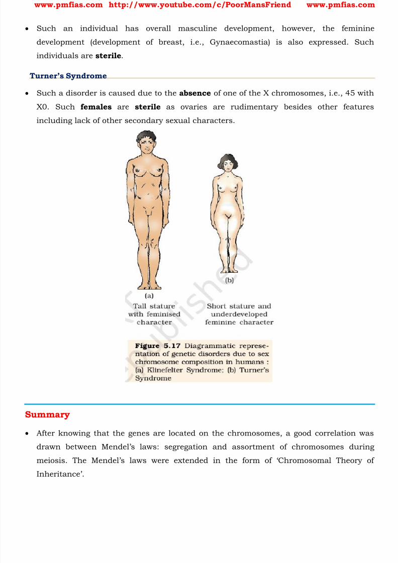
8/16/2019 Biology for Prelims Exam
http://slidepdf.com/reader/full/biology-for-prelims-exam 106/384
www.pmfias.com http://www.youtube.com/c/PoorMansFriend www.pmfias.com
Such an individual has overall masculine development, however, the feminine
development (development of breast, i.e., Gynaecomastia) is also expressed. Such
individuals are sterile .
Turner’s Syndrome
Such a disorder is caused due to the absence of one of the X chromosomes, i.e., 45 with
X0. Such females are sterile as ovaries are rudimentary besides other features
including lack of other secondary sexual characters.
Summary
After knowing that the genes are located on the chromosomes, a good correlation was
drawn between Mendel’s laws : segregation and assortment of chromosomes during
meiosis. The Mendel’s laws were extended in the form of ‘Chromosomal Theory of
Inheritance’.

8/16/2019 Biology for Prelims Exam
http://slidepdf.com/reader/full/biology-for-prelims-exam 107/384
www.pmfias.com http://www.youtube.com/c/PoorMansFriend www.pmfias.com
Later, it was found that Mendel’s law of independent assortment does not hold true for
the genes that were located on the same chromosomes. These genes were called as
‘linked genes’.
Closely located genes assorted together, and distantly located genes, due to
recombination, assorted independently. Linkage maps, therefore, corresponded to
arrangement of genes on a chromosome. Many genes were linked to sexes also, and called as sex-linked genes. The two sexes
(male and female) were found to have a set of chromosomes which were common, and
another set which was different.
The chromosomes which were different in two sexes were named as sex chromosomes.
The remaining set was named as autosomes. In humans, a normal female has 22 pairs
of autosomes and a pair of sex chromosomes (XX).
A male has 22 pairs of autosomes and a pair of sex chromosome as XY. In chicken, sexchromosomes in male are ZZ, and in females are ZW.
Mutation is defined as change in the genetic material. A point mutation is a change of a
single base pair in DNA.
Sickle-cell anemia is caused due to change of one base in the gene coding for beta-chain
of hemoglobin.
Inheritable mutations can be studied by generating a pedigree of a family.
Some mutations involve changes in whole set of chromosomes (polyploidy) or change in
a subset of chromosome number (aneuploidy). This helped in understanding the
mutational basis of genetic disorders.
Down’s syndrome is due to trisomy of chromosome 21, wh ere there is an extra copy of
chromosome 21 and consequently the total number of chromosome becomes 47.
In Turner’s syndrome, one X chromosome is missing and the sex chromosome is as XO,
and in Klinefelter’s syndrome, the condition is XXY. These can be ea sily studied by
analysis of Karyotypes.
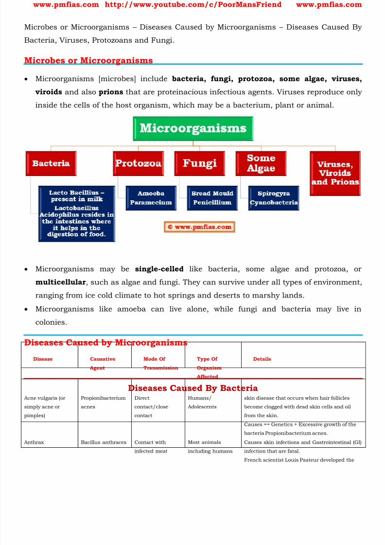
8/16/2019 Biology for Prelims Exam
http://slidepdf.com/reader/full/biology-for-prelims-exam 108/384
www.pmfias.com http://www.youtube.com/c/PoorMansFriend www.pmfias.com
Microbes or Microorganisms – Diseases Caused by Microorganisms – Diseases Caused By
Bacteria, Viruses, Protozoans and Fungi.
Microbes or Microorganisms
Microorganisms [microbes] include bacteria, fungi, protozoa, some algae, viruses,
viroids and also prions that are proteinacious infectious agents. Viruses reproduce onlyinside the cells of the host organism, which may be a bacterium, plant or animal.
Microorganisms may be single-celled like bacteria, some algae and protozoa, or
multicellular , such as algae and fungi. They can survive under all types of environment,
ranging from ice cold climate to hot springs and deserts to marshy lands.
Microorganisms like amoeba can live alone, while fungi and bacteria may live in
colonies.
Diseases Caused by Microorganisms
Disease Causative
Agent
Mode Of
Transmission
Type Of
Organism
Affected
Details
Diseases Caused By Bacteria
Acne vulgaris (or
simply acne or
pimples)
Propionibacterium
acnes
Direct
contact/close
contact
Humans/
Adolescents
skin disease that occurs when hair follicles
become clogged with dead skin cells and oil
from the skin.
Causes == Genetics + Excessive growth of the
bacteria Propionibacterium acnes.
Anthrax Bacillus anthraces Contact with
infected meat
Most animals
including humans
Causes skin infections and Gastrointestinal (GI)
infection that are fatal.
French scientist Louis Pasteur developed the

8/16/2019 Biology for Prelims Exam
http://slidepdf.com/reader/full/biology-for-prelims-exam 109/384
www.pmfias.com http://www.youtube.com/c/PoorMansFriend www.pmfias.com
first effective vaccine in 1881.
Cholera Vibrio cholerae Water/food Humans Effects small intestine. The classic symptom is
large amounts of watery diarrhea that lasts a
few days. Vomiting and muscle cramps may
also occur. Diarrhea can be so severe that itleads within hours to severe dehydration and
electrolyte imbalance.
Citrus Canker Xanthomonas
axonopodis
Air Citrus fruit plants Infection causes lesions on the leaves, stems,
and fruit of citrus trees.
Diptheria Corynebacterium
diphtheriae
Air/direct contact Humans Symptoms: sore throat and fever. The neck may
swell in part due to large lymph nodes.
Complications may include myocarditis,
inflammation of nerves, kidney problems, and
bleeding problems due to low blood platelets.
Myocarditis may result in an abnormal heart
rate and inflammation of the nerves may resultin paralysis.
Pneumonia
[caused by bacteria
or viruses]
Streptococcus
pneumoniae and
Haemophilus
influenzae
Air borne droplets
of sneeze
Humans Pneumonia is an inflammatory condition of the
lung affecting primarily the microscopic air
sacs known as alveoli.
Peptic ulcers Helicobacter pylori Humans Ulcers in the lining of stomach and starting
part of small intestine
Plague Yersinia pestis Air/ direct contact Humans Unhygienic conditions is the main cause. [You
know why Surat is one of the cleanest cities in
India?]
The symptoms of plague depend on the
concentrated areas of infection in each person:
bubonic plague in lymph nodes, septicemic
plague in blood vessels, pneumonic plague in
lungs.
Tuberculosis Mycobacterium Air Humans Tuberculosis generally affects the lungs, but
8/16/2019 Biology for Prelims Exam
http://slidepdf.com/reader/full/biology-for-prelims-exam 110/384
www.pmfias.com http://www.youtube.com/c/PoorMansFriend www.pmfias.com
tuberculosis can also affect other parts of the body. Most
infections do not have symptoms, known as
latent tuberculosis. About 10% of latent
infections eventually progresses to active
disease which, if left untreated, kills about half
of those infected. The classic symptoms of
active TB are a chronic cough with blood-tinged
sputum, fever, night sweats, and weight loss.
Typhoid Salmonella typhi Water Humans Often there is a gradual onset of a high fever
over several days. Weakness, abdominal pain,
constipation, and headaches also commonly
occur.
Diseases Caused By Viruses
AIDS Human
Immunodeficiency
Virus (HIV)
Blood exchange Humans and
primates
Severely weakens immunity and makes way for
a number of other pathogens.
Chicken Pox varicella zoster
virus (VZV)
Air/contact Humans Chickenpox, also known as varicella, is a highly
contagious disease. The disease results in a
characteristic skin rash that forms small, itchyblisters. Less severe than small pox. Almost
eradicated after the invention of vaccination.
Small Pox Variola
major and Variola
minor
Air/contact/water Humans One of the highly dreaded diseases that is
highly contagious.
Almost eradicated after the invention of
vaccination.
Chikungunya Chikungunya viru
s
Aedes mosquitoes,
such as A. aegypti
and A. albopictus
Causes severe joint pains. Animal reservoirs of
the virus include monkeys, birds, cattle, and
rodents. This is in contrast to dengue, for
which primates are the only hosts
Cold, influenza (flu)and most coughs
Rhino viruses Air borne dropletsof sneeze
Humans Summer are hostile for the virus. Most commonduring winter months.
Dengue fever Flavivirus Female Aedes
mosquito
Humans high fever, headache, vomiting, muscle and
joint pains, and a characteristic skin rash.
In a small proportion of cases, the disease
develops into the life-threatening dengue
hemorrhagic fever, resulting in bleeding, low
levels of blood platelets and blood plasma
leakage, or into dengue shock syndrome, where
dangerously low blood pressure occurs.
Ebola Ebola virus Animal to man Humans and Some
Animals
Ebola infection shows a sudden onset of the
disease resulting initially in flu-like symptoms:
fever, chills and malaise.
As the disease progresses, it results in multi-
system involvements indicated by the person
experiencing lethargy, nausea, vomiting,
diarrhoea and headache.
Foot and Mouth
Disease
Picornavirus[genu
s Aphthovirus]
Close-contact
animal-to-animal
spread
Animals Serious problem to animal farming in India.
Hepatitis B hepatitis B virus Blood Exchange, Humans Affects the liver . Acute as well as chronic.
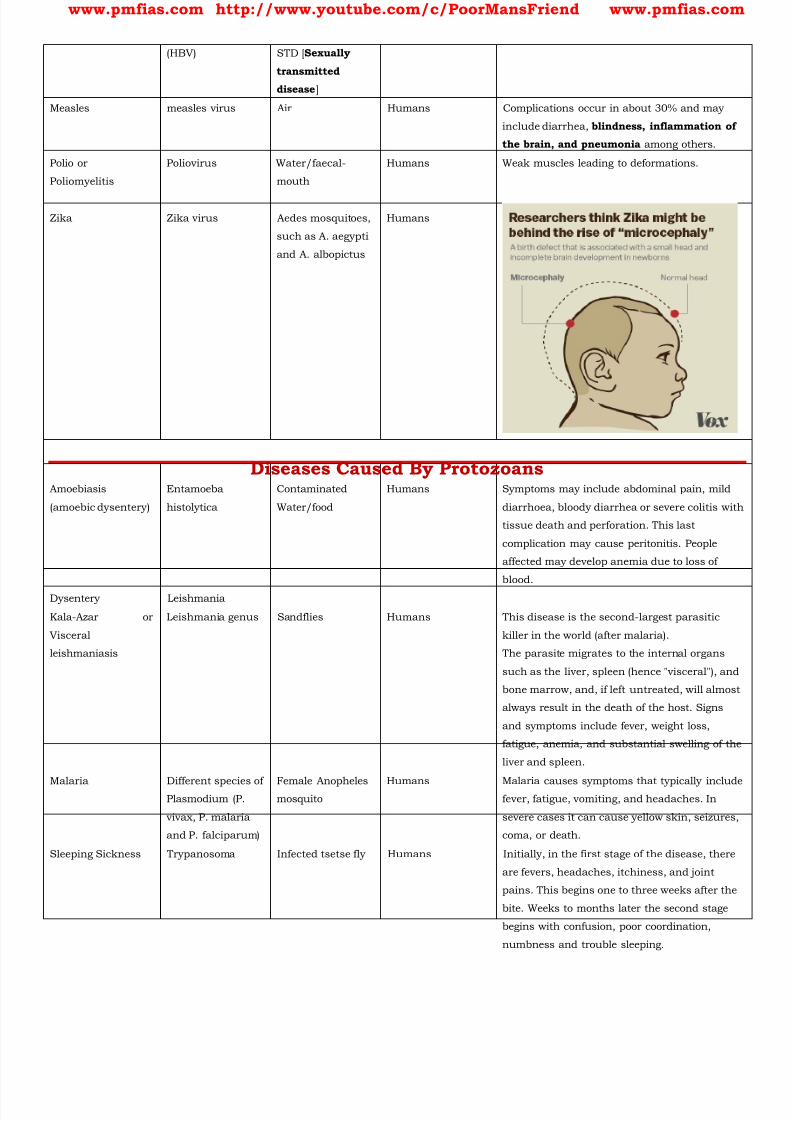
8/16/2019 Biology for Prelims Exam
http://slidepdf.com/reader/full/biology-for-prelims-exam 111/384
www.pmfias.com http://www.youtube.com/c/PoorMansFriend www.pmfias.com
(HBV) STD [ Sexually
transmitted
disease ]
Measles measles virus Air Humans Complications occur in about 30% and may
include diarrhea, blindness, inflammation of
the brain, and pneumonia among others.
Polio or
Poliomyelitis
Poliovirus Water/faecal-
mouth
Humans Weak muscles leading to deformations.
Zika Zika virus Aedes mosquitoes,
such as A. aegypti
and A. albopictus
Humans
Diseases Caused By ProtozoansAmoebiasis
(amoebic dysentery)
Entamoeba
histolytica
Contaminated
Water/food
Humans Symptoms may include abdominal pain, mild
diarrhoea, bloody diarrhea or severe colitis with
tissue death and perforation. This last
complication may cause peritonitis. People
affected may develop anemia due to loss of
blood.
Dysentery Leishmania
Kala-Azar or
Visceral
leishmaniasis
Leishmania genus Sandflies Humans This disease is the second-largest parasitic
killer in the world (after malaria).
The parasite migrates to the internal organs
such as the liver, spleen (hence "visceral"), and
bone marrow, and, if left untreated, will almost
always result in the death of the host. Signs
and symptoms include fever, weight loss,
fatigue, anemia, and substantial swelling of the
liver and spleen.
Malaria Different species ofPlasmodium (P.
vivax, P. malaria
and P. falciparum)
Female Anophelesmosquito
Humans Malaria causes symptoms that typically includefever, fatigue, vomiting, and headaches. In
severe cases it can cause yellow skin, seizures,
coma, or death.
Sleeping Sickness Trypanosoma Infected tsetse fly Humans Initially, in the first stage of the disease, there
are fevers, headaches, itchiness, and joint
pains. This begins one to three weeks after the
bite. Weeks to months later the second stage
begins with confusion, poor coordination,
numbness and trouble sleeping.

8/16/2019 Biology for Prelims Exam
http://slidepdf.com/reader/full/biology-for-prelims-exam 112/384
www.pmfias.com http://www.youtube.com/c/PoorMansFriend www.pmfias.com
Yellow Vein Mosaic
of Okra [Ladies
finger]
Bhendi yellow vein
mosaic virus
Insect Okra plant Okra are dwarfed, malformed.
Diseases Caused By Fungi
Ringworms Fungi belonging to
the genera Micr
Trichophyton and
Epidermophyton
Skin-skin contact Humans The fungi that cause parasitic infection,
collectively dermatophytes, feed on keratin, the
material found in the outer layer of skin, hair,
and nails.
Rust of wheat Puccinia rust
fungus
Air/seeds Wheat and other
crops
Wheat leaf rust is a fungal disease that affects
wheat, barley and rye stems, leaves and grains.
In temperate zones it is destructive on winter
wheat because the pathogen. overwinters.
Infections can lead up to 20% yield loss
exacerbated by dying leaves which fertilize the
fungus.

8/16/2019 Biology for Prelims Exam
http://slidepdf.com/reader/full/biology-for-prelims-exam 113/384
www.pmfias.com http://www.youtube.com/c/PoorMansFriend www.pmfias.com
Useful Microbes – Microbes In Human Welfare: Microbes in the production of Antibiotics,
Vaccines, Curd, Fermented beverages, Biogas, Biofertilisers etc.
Useful Microorganisms – Microorganisms In Human Welfare
Microorganisms include bacteria, fungi, protozoa, some algae, viruses, viroids and also
prions. Microorganisms may be single-celled like bacteria, some algae and protozoa, ormulticellular, such as algae and fungi.
Some microorganisms are harmful [pathogens] as they cause various diseases in
humans. A list of common diseases caused by microorganisms are given in the previous
post [Diseases Caused by Microorganisms].
Some other microorganisms are beneficial and contribute to human welfare. This post is
about such useful microorganisms [useful microbes].
Antibiotics and Vaccination Whenever you fall ill the doctor may give you some antibiotic tablets, capsules or
injections such as of penicillin. The source of these medicines is microorganisms.
These medicines kill or stop the growth of the disease-causing microorganisms. Such
medicines are called ANTIBIOTICS .
These days a number of antibiotics are being produced from bacteria and fungi
Streptomycin, tetracycline and erythromycin are some of the commonly known
antibiotics which are made from fungi and bacteria.
In 1929, Alexander Fleming was working on a culture of disease-causing bacteria
[Staphylococci ]. Suddenly he found the spores of a little green mould [ Penicillium
notatum ] in one of his culture plates. He observed that the presence of mould prevented
the growth of bacteria. In fact, it also killed many of these bacteria. From this the mould
penicillin was prepared.
Antibiotics have greatly improved our capacity to treat deadly diseases such as plague,
whooping cough, diphtheria and leprosy, which used to kill millions all over the globe.
Today, we cannot imagine a world without antibiotics.
Antibiotics taken unnecessarily may kill the beneficial bacteria in the body.
Antibiotics, however, are not effective against cold and flu as these are caused by
viruses .
When a disease-carrying microbe enters our body, the body produces antibodies to fight
the invader. The body also remembers how to fight the microbe if it enters again. So, if
8/16/2019 Biology for Prelims Exam
http://slidepdf.com/reader/full/biology-for-prelims-exam 114/384
www.pmfias.com http://www.youtube.com/c/PoorMansFriend www.pmfias.com
dead or weakened microbes are introduced in a healthy body, the body fights and kills
them by producing suitable antibodies. The antibodies remain in the body and we are
protected from the disease-causing microbes. This is how a vaccine works.
Several diseases, including cholera, tuberculosis, smallpox and hepatitis can be
prevented by vaccination. Edward Jenner discovered the vaccine for smallpox in 1798.
Chemicals, Enzymes and other Bioactive Molecules
Microbes are also used for commercial and industrial production of certain chemicals
like organic acids, alcohols and enzymes . Examples of acid producers are
1. Aspergillus niger (a fungus) of citric acid
2. Acetobacter aceti (a bacterium) of acetic acid
3. Clostridium butylicum (a bacterium) of butyric acid
4. Lactobacillus (a bacterium) of lactic acid .
Yeast (Saccharomyces cerevisiae) is used for commercial production of ethanol .
Lipases are used in detergent formulations and are helpful in removing oily stains
from the laundry.
You must have noticed that bottled fruit juices bought from the market are clearer as
compared to those made at home. This is because the bottled juices are clarified by the
use of pectinases and proteases .
Streptokinase produced by the bacterium Streptococcus and modified by genetic
engineering is used as a ‘clot buster’ for removing clots from the blood vessels of
patients who have undergone myocardial infraction leading to heart attack.
Another bioactive molecule, cyclosporin A , that is used as an immunosuppressive agent
in organ-transplant patients, is produced by the fungus Tnchoderma polysporum .
Statins produced by the yeast Monascus purpureus have been commercialized as
blood-cholesterol lowering agents. It acts by competitively inhibiting the enzyme
responsible for synthesis of cholesterol.
Milk to Curd
Micro-organisms such as Lactobacillus and others commonly called lactic acid
bacteria (LAB) grow in milk and convert it to curd.
During growth, the LAB produce acids that coagulate and partially digest the milk
proteins.
8/16/2019 Biology for Prelims Exam
http://slidepdf.com/reader/full/biology-for-prelims-exam 115/384
www.pmfias.com http://www.youtube.com/c/PoorMansFriend www.pmfias.com
A small amount of curd added to the fresh milk as inoculum or starter contain millions
of LAB, which at suitable temperatures multiply, thus converting milk to curd, which
also improves its nutritional quality by increasing VITAMIN B12 [helps in the
synthesis of DNA and RBC (red blood cells). Vitamin B12 deficiency causes Anemia,
severe damage to nervous system etc.] .
In our stomach too, the LAB play very beneficial role in checking disease-causingmicrobes.
Microbes and Fermentation
Sugar is converted into alcohol by yeast. This process of conversion of sugar into alcohol
is known as fermentation . Louis Pasteur discovered fermentation in 1857.
The dough, which is used for making foods such as dosa and idli is fermented by
bacteria . The puffed-up appearance of dough is due to the production of CO2 gas which
is released by the microbes.
Similarly the dough, which is used for making bread, is fermented using baker’s yeast
(Saccharomyces cerevisiae ).
A number of traditional drinks and foods are also made by fermentation by the
microbes. ‘Toddy’, a traditional drink of some parts of southern India is made by
fermenting sap from palms.
Microbes are also used to ferment fish, soyabean and bamboo- shoots to make foods.
Cheese, is one of the oldest food items in which microbes were used. The large holes in
‘Swiss cheese’ are due to production of a large amount of CO2 by a bacterium named
Propionibacterium sharmanii .
Fermented Beverages
Microbes especially yeasts have been used from time immemorial for the production of
beverages like wine, beer, whisky, brandy or rum.
For this purpose the same yeast Saccharomyces cerevisiae used for bread-making and
commonly called brewer’s yeast , is used for fermenting malted cereals and fruit juices,
to produce ethanol .
Depending on the type of the raw material used for fermentation and the type of
processing (with or without distillation) different types of alcoholic drinks are obtained.
Wine and beer are produced without distillation whereas whisky, brandy and rum are
produced by distillation of the fermented broth.

8/16/2019 Biology for Prelims Exam
http://slidepdf.com/reader/full/biology-for-prelims-exam 116/384
www.pmfias.com http://www.youtube.com/c/PoorMansFriend www.pmfias.com
Microbes in sewage treatment
Sewage contains large amounts of organic matter and microbes. Many of which are
pathogenic.
Treatment of waste water is done by the primary sludge, and the supernatant forms the
effluent. The effluent from the primary settling tank is taken for secondary treatment.
The primary effluent is passed into large aeration tanks where it is constantly agitated
mechanically and air is pumped into it. This allows vigorous growth of useful aerobic
microbes into flocs (masses of bacteria associated with fungal filaments to form mesh
like structures).
While growing, these microbes consume the major part of the organic matter in the
effluent. This significantly reduces the BOD (biochemical oxygen demand) of the
effluent.
BOD refers to the amount of the oxygen that would be consumed if all the organic
matter in one liter of water were oxidized by bacteria.
The sewage water is treated till the BOD is reduced. BOD is a measure of the organic
matter present in the water . The greater the BOD of waste water, more is its polluting
potential.
Once the BOD of sewage or waste water is reduced significantly, the effluent is then
pa ssed into a settling tank where the bacterial ‘flocs’ are allowed to sediment. This
sediment is called activated sludge . A small part of the activated sludge is pumped back into the aeration tank to serve as
the inoculum or starter.
The remaining major part of the sludge is pumped into large tanks called anaerobic
sludge digesters . Here, other kinds of bacteria, which grow anaerobically, digest the
bacteria and the fungi in the sludge.
During this digestion, bacteria produce a mixture of gases such as methane, hydrogen
sulphide and carbon dioxide . These gases form biogas and can be used as source ofenergy as it is inflammable.
The effluent from the secondary treatment plant is generally released into natural water
bodies like rivers and streams.
Microbes in production of biogas

8/16/2019 Biology for Prelims Exam
http://slidepdf.com/reader/full/biology-for-prelims-exam 117/384
www.pmfias.com http://www.youtube.com/c/PoorMansFriend www.pmfias.com
Biogas is a mixture of gases (containing predominantly methane ) produced by the
microbial activity and which may be used as fuel.
Certain bacteria, which grow anaerobically on cellulosic material, produce large
amount of methane [greenhouse gas] along with CO2 and H2 . These bacteria are
collectively called methanogens , and one such common bacterium is
Methanobacterium . These bacteria are commonly found in the anaerobic sludge duringsewage treatment.
These bacteria are also present in the rumen (a part of stomach) of cattle. A lot of
cellulosic material present in the food of cattle is also present in the rumen. In rumen,
these bacteria help in the breakdown of cellulose and play an important role in the
nutrition of cattle. Thus, the excreta (dung) of cattle, commonly called gobar, is rich in
these bacteria. Dung can be used for generation of biogas, commonly called gobar gas
[Humans cannot digest cellulose. Hence their faecal waste cannot produce methane].
Microbes as biocontrol agents
Biocontrol refers to the use of biological methods for controlling plant diseases and
pests. Biological agents are a better alternative to weedicides and pesticides.
Biological control of pests and diseases
In agriculture, there is a method of controlling pests that relies on natural predation
rather than introduced chemicals. A key belief of the organic farmer is that biodiversity furthers health. The more variety a
landscape has, the more sustainable it is. The organic farmer, therefore, works to create
a system where the insects that are sometimes called pests are not eradicated, but
instead are kept at manageable levels by a complex system of checks and balances
within a living and vibrant ecosystem.
Con trary to the ‘conventional’ farming practices which often use chemical methods to
kill both useful and harmful life forms indiscriminately, this is a holistic approach thatseeks to develop an understanding of the webs of interaction between the myriad of
organisms that constitute the field fauna and flora.
The organic farmer holds the view that the eradication of the creatures that are often
described as pests is not only possible, but also undesirable, for without them the
beneficial predatory and parasitic insects which depend upon them as food or hosts

8/16/2019 Biology for Prelims Exam
http://slidepdf.com/reader/full/biology-for-prelims-exam 118/384
www.pmfias.com http://www.youtube.com/c/PoorMansFriend www.pmfias.com
would not be able to survive. Thus, the use of biocontrol measures will greatly reduce
our dependence on toxic chemicals and pesticides.
An important part of the biological farming approach is to become familiar with the
various life forms that inhabit the field, predators as well as pests, and also their life
cycles, patterns of feeding and the habitats that they prefer. This will help develop
appropriate means of biocontrol. The very familiar beetle with red and black markings - the Ladybird , and Dragonflies
are useful to get rid of aphids and mosquitoes, respectively.
An example of microbial biocontrol agents that can be Introduced in order to control
butterfly caterpillars is the bacteria Bacillus thuringiensis (often written as Bt).
These are available in sachets as dried spores which are mixed with water and sprayed
onto vulnerable plants such as brassicas and fruit trees, where these are eaten by the
insect larvae. In the gut of the larvae, the toxin is released and the larvae get killed. Thebacterial disease will kill the caterpillars, but leave other insects unharmed.
Because of the development of methods of genetic engineering in the last decade or so,
the scientists have introduced B. thuringiensis toxin genes into plants. Such plants are
resistant to attack by insect pests. Bt-cotton is one such example, which is being
cultivated in some states of our country.
A biological control being developed for use in the treatment of plant disease is the
fungus Trichoderma. Trichoderma species are free-living fungi that are very common in
the root ecosystems. They are effective biocontrol agents of several plant pathogens.
Baculoviruses are pathogens that attack insects and other arthropods. They have been
shown to have no negative impacts on plants, mammals, birds, fish or even on non-
target insects.
Microbes as biofertilisers
Biofertilisers are organisms that enrich the nutrient quality of the soil. The main sources
of biofertilisers are bacteria, fungi and cyanobacteria .
You may be knowing about the nodules on the roots of leguminous plants formed by the
symbiotic association of Rhizobium . These bacteria fix atmospheric nitrogen into
organic forms, which is used by the plant as nutrient.
Other bacteria can fix atmospheric nitrogen while free-living in the soil (examples
Azospirillum and Azotobacter ), thus enriching the nitrogen content of the soil.
8/16/2019 Biology for Prelims Exam
http://slidepdf.com/reader/full/biology-for-prelims-exam 119/384
www.pmfias.com http://www.youtube.com/c/PoorMansFriend www.pmfias.com
Fungi are also known to form symbiotic associations with plants ( mycorrhiza ). Many
members of the genus Glomus form mycorrhiza. The fungal symbiont in these
associations absorbs phosphorus from soil and passes it to the plant.
Plants having such associations show other benefits also, such as resistance to root-
borne pathogens, tolerance to salinity and drought, and an overall increase in plant
growth and development. Cyanobacteria are autotrophic microbes widely distributed in aquatic and terrestrial
environments many of which can fix atmospheric nitrogen , e.g. Anabaena, Nostoc,
Oscillatona, etc.
In paddy fields, cyanobacteria serve as an important biofertiliser. Blue green algae also
add organic matter to the soil and increase its fertility.
Preservatives
Salts and edible oils are the common chemicals generally used to check the growth of
microorganisms. Therefore they are called preservatives . We add salt or acid
preservatives to pickles to prevent the attack of microbes. Sodium benzoate and
sodium metabisulphite are common preservatives. These are also used in the jams and
squashes to check their spoilage.
Common salt has been used to preserve meat and fish for ages. Meat and fish are
covered with dry salt to check the growth of bacteria. Salting is also used to preserve
amla, raw mangoes, tamarind, etc.
Jams, jellies and squashes are preserved by sugar. Sugar reduces the moisture content
which inhibits the growth of bacteria which spoil food.
Use of oil and vinegar prevents spoilage of pickles because bacteria cannot live in such
an environment. Vegetables, fruits, fish and meat are often preserved by this method.
Pasteurized milk can be consumed without boiling as it is free from harmful microbes.
The milk is heated to about 70 0 C for 15 to 30 seconds and then suddenly chilled and
stored. By doing so, it prevents the growth of microbes. This process was discovered by
Louis Pasteur . It is called pasteurization .
Summary
Microbes are a very important component of life on earth. Not all microbes are
pathogenic. Many microbes are very useful to human beings.
We use microbes and microbially derived products almost every day.

8/16/2019 Biology for Prelims Exam
http://slidepdf.com/reader/full/biology-for-prelims-exam 120/384
www.pmfias.com http://www.youtube.com/c/PoorMansFriend www.pmfias.com
Bacteria called lactic acid bacteria (LAB) grow in milk to convert it into curd.
The dough, which is used to make bread, is fermented by yeast called Saccharomyces
cerevisiae.
Certain dishes such as idli and dosa, are made from dough fermented by microbes.
Bacteria and fungi are used to impart particular texture, taste and flavor to cheese.
Microbes are used to produce industrial products like lactic acid, acetic acid andalcohol, which are used in a variety of processes in the industry.
Antibiotics like penicillins produced by useful microbes are used to kill disease-causing
harmful microbes.
Antibiotics have played a major role in controlling infectious diseases like diphtheria,
whooping cough and pneumonia.
For more than a hundred years, microbes are being used to treat sewage (waste water)
by the process of activated sludge formation and this helps in recycling of water innature.
Methanogens produce methane (biogas) while degrading plant waste.
Biogas produced by microbes is used as a source of energy in rural areas.
Microbes can also be used to kill harmful pests, a process called as biocontrol.
The biocontrol measures help us to avoid heavy use of toxic pesticides for controlling
pests.
Source: NCERT Science Textbooks Class 6-12

8/16/2019 Biology for Prelims Exam
http://slidepdf.com/reader/full/biology-for-prelims-exam 121/384
www.pmfias.com http://www.youtube.com/c/PoorMansFriend www.pmfias.com
Animal Tissues - Epithelial Tissue: Simple Epithelium and Compound Epithelium,
Connective Tissue, Muscular Tissue and Nervous Tissue.
Animal Tissues
Blood and muscles are both examples of tissues found in our body. On the basis of
the functions they perform we can think of different types of animal tissues, such asepithelial tissue, connective tissue, muscular tissue and nervous tissue .
Blood is a type of connective tissue, and muscle forms muscular tissue.
Epithelial Tissue
The covering or protective tissues in the animal body are epithelial tissues.
Epithelium covers most organs and cavities within the body.
It also forms a barrier to keep different body systems separate.
The skin , the lining of the mouth , the lining of blood vessels , lung alveoli and
kidney tubules are all made of epithelial tissue.
Epithelial tissue cells are tightly packed and form a continuous sheet.
They have only a small amount of cementing material between them and almost no
intercellular spaces.
Obviously, anything entering or leaving the body must cross at least one layer of
epithelium.
As a result cells of various epithelia play an important role in regulating the
exchange of materials between the body and the external environment and also
between different parts of the body.
Regardless of the type, all epithelium is usually separated from the underlying tissue
by an extracellular fibrous basement membrane.
There are two types of epithelial tissues namely simple epithelium and compound
epithelium.
Simple Epithelium
Simple epithelium is composed of a single layer of cells and functions as a lining for
body cavities, ducts, and tubes .

8/16/2019 Biology for Prelims Exam
http://slidepdf.com/reader/full/biology-for-prelims-exam 122/384
www.pmfias.com http://www.youtube.com/c/PoorMansFriend www.pmfias.com
Squamous Epithelium
The squamous epithelium is made of a single thin layer of flattened cells with
irregular boundaries. They are found in the walls of blood vessels and air sacs of lungs and are involved in
functions like forming a diffusion boundary .
Different epithelia show differing structures that correlate with their unique
functions. For example, in cells lining blood vessels or lung alveoli, where
transportation of substances occurs through a selectively Permeable surface, there
is a simple flat kind of epithelium. This is called the simple Squamous epithelium.
8/16/2019 Biology for Prelims Exam
http://slidepdf.com/reader/full/biology-for-prelims-exam 123/384
www.pmfias.com http://www.youtube.com/c/PoorMansFriend www.pmfias.com
Simple squamous epithelial cells are extremely thin and flat and form a delicate
lining.
The oesophagus and the lining of the mouth are also covered with squamous
epithelium.
Stratified Squamous Epithelium
The skin, which protects the body, is made of squamous epithelium.
Skin epithelial cells are arranged in many layers to prevent wear and tear.
Since they are arranged in a pattern of layers, the epithelium is called stratified
squamous epithelium.
Ciliated Columnar Epithelium
The columnar epithelium is composed of a single layer of tall and slender cells. Their
nuclei are located at the base.
Where absorption and secretion occur, as in the inner lining of the intestine, tall
epithelial cells are present.
In the respiratory tract, the columnar epithelial tissue also has cilia, which are hair-
like projections on the outer surfaces of epithelial cells. These cilia can move , and
their movement pushes the mucus forward to clear it. This type of epithelium is thus
ciliated columnar epithelium.
They are mainly present in the inner surface of hollow organs like bronchioles and
fallopian tubes.
Cuboidal Epithelium
The cuboidal epithelium is composed of a single layer of cube-like cells. This is
commonly found in ducts of glands and tubular parts of nephrons in kidneys and its
main functions are secretion and absorption .
Cuboidal epithelium (with cube-shaped cells) forms the lining of kidney tubules and
ducts of salivary glands, where it provides mechanical support .
Glandular Epithelium
Epithelial cells often acquire additional specialization as gland cells , which can
secrete substances at the epithelial surface.
Sometimes a portion of the epithelial tissue folds inward, and a multicellular gland is
formed. This is glandular epithelium.

8/16/2019 Biology for Prelims Exam
http://slidepdf.com/reader/full/biology-for-prelims-exam 124/384
www.pmfias.com http://www.youtube.com/c/PoorMansFriend www.pmfias.com
Some of the columnar or cuboidal cells get specialized for secretion and are called
glandular epithelium. They are mainly of two types: unicellular, consisting of isolated
glandular cells ( goblet cells of the alimentary canal), and multicellular, consisting of
cluster of cells ( salivary gland ).
On the basis of the mode of pouring of their secretions, glands are divided into two
categories namely EXOCRINE and ENDOCRINE glands. Exocrine glands secrete mucus, saliva, earwax, oil, milk, digestive enzymes and other
cell products. These products are released through ducts or tubes.
In contrast, endocrine glands do not have ducts . Their products called hormones
are secreted directly into the fluid bathing the gland.
Compound Epithelium
The compound epithelium consists of two or more cell layers and has protective
function as it does in our skin.
Compound epithelium is made of more than one layer ( multi-layered ) of cells and
thus has a limited role in secretion and absorption . Their main function is to
provide protection against chemical and mechanical stresses.
They cover the dry surface of the skin, the moist surface of buccal cavity, pharynx,
inner lining of ducts of salivary glands and of pancreatic ducts.
All cells in epithelium are held together with little intercellular material. In nearly all
animal tissues, specialized junctions provide both structural and functional links
between its individual cells.
Three types of cell junctions are found in the epithelium and other tissues. These are
called as tight, adhering and gap junctions .
Tight junctions help to stop substances from leaking across a tissue. Adhering
junctions perform cementing to keep neighboring cells together. Gap junctions
facilitate the cells to communicate with each other by connecting the cytoplasm of
adjoining cells, for rapid transfer of ions, small molecules and sometimes big
molecules.
Connective Tissue
Connective tissues are most abundant and widely distributed in the body of complex
animals. They are named connective tissues because of their special function of
linking and supporting other tissues/organs of the body.

8/16/2019 Biology for Prelims Exam
http://slidepdf.com/reader/full/biology-for-prelims-exam 125/384
www.pmfias.com http://www.youtube.com/c/PoorMansFriend www.pmfias.com
They range from soft connective tissues to specialized types, which include cartilage,
bone, adipose, and blood.
In all connective tissues except blood, the cells secrete fibres of structural proteins
called collagen or elastin .
The fibres provide strength, elasticity and flexibility to the tissue. These cells also
secrete modified polysaccharides, which accumulate between cells and fibres and actas matrix (ground substance).
Connective tissues are classified into three types: (i) Loose connective tissue, (ii)
Dense connective tissue and (iii) Specialized connective tissue.

8/16/2019 Biology for Prelims Exam
http://slidepdf.com/reader/full/biology-for-prelims-exam 126/384
www.pmfias.com http://www.youtube.com/c/PoorMansFriend www.pmfias.com
Loose Connective Tissue
Loose connective tissue has cells and fibres loosely arranged in a semi-fluid ground
substance, for example, areolar tissue present beneath the skin.
Often it serves as a support framework for epithelium . It contains fibroblasts (cells
that produce and secrete fibres), macrophages [a large phagocytic cell found in
stationary form in the tissues or as a mobile white blood cell, especially at sites of
infection] and mast cells [a cell found in connective tissue and releasing histamine
and other substances during inflammatory and allergic reactions].
Adipose tissue is a type of loose connective tissue located mainly beneath the skin.
The cells of this tissue are specialized to store fats . The excess of nutrients which are
not used immediately are converted into fats and are stored in this tissue.
Dense Connective Tissue
Fibres and fibroblasts are compactly packed in the dense connective tissues.
Orientation of fibres show a regular or irregular pattern and are called dense regular
and dense irregular tissues.
In the dense regular connective tissues, the collagen fibres are present in rows
between many parallel bundles of fibres. Tendons , which attach skeletal muscles to
bones and ligaments which attach one bone to another are examples of this tissue.
Dense irregular connective tissue has fibroblasts and many fibres (mostly collagen)that are oriented differently. This tissue is present in the skin.
Specialized Connective Tissue – Cartilage, Bones, Blood, Areolar
Cartilage, bones and blood are various types of specialized connective tissues.
The intercellular material of cartilage is solid and pliable and resists compression.
Cells of this tissue ( chondrocytes ) are enclosed in small cavities within the matrix
secreted by them.
Most of the cartilages in vertebrate embryos are replaced by bones in adults.
Cartilage is present in the tip of nose, outer ear joints, trachea, larynx, between
adjacent bones of the vertebral column, limbs and hands in adults.
Bone cells are embedded in a hard matrix that is composed of calcium and
phosphorus compounds.

8/16/2019 Biology for Prelims Exam
http://slidepdf.com/reader/full/biology-for-prelims-exam 127/384
www.pmfias.com http://www.youtube.com/c/PoorMansFriend www.pmfias.com
Bones have a hard and non-pliable ground substance rich in calcium salts and
collagen fibres which give bone its strength. It is the main tissue that provides
structural frame to the body. Bones support and protect softer tissues and organs.
The bone cells ( osteocytes ) are present in the spaces called lacunae. The bone
marrow in some bones is the site of production of blood cells.
Two bones can be connected to each other by another type of connective tissue calledthe ligament . This tissue is very elastic. It has considerable strength. Ligaments
contain very little matrix. Tendons connect bones to muscles and are another type of
connective tissue. Tendons are fibrous tissue with great strength but limited
flexibility.
Blood is a fluid connective tissue containing plasma, red blood cells (RBC), white
blood cells (WBC) and platelets . It is the main circulating fluid that helps in the
transport of various substances. Areolar connective tissue is found between the skin and muscles, around blood
vessels and nerves and in the bone marrow.
It fills the space inside the organs, supports internal organs and helps in repair of
tissues.
Muscular Tissue
Each muscle is made of many long, cylindrical fibres arranged in parallel arrays.
These fibres are composed of numerous fine fibrils, called myofibrils .
Muscle fibres contract (shorten) in response to stimulation, then relax (lengthen) and
return to their uncontracted state in a coordinated fashion. Muscles contain special
proteins called contractile proteins , which contract and relax to cause movement.
Muscles are of three types, skeletal, smooth, and cardiac .
Skeletal Muscle Tissue – Voluntary Muscles
We can move some muscles by conscious will. Such muscles are called voluntary
muscles.
These muscles are also called skeletal muscles as they are mostly attached to bones
and help in body movement.
Under the microscope, these muscles show alternate light and dark bands or
striations. As a result, they are also called striated muscles. The cells of this tissue
are long, cylindrical, unbranched and multinucleate (having many nuclei).

8/16/2019 Biology for Prelims Exam
http://slidepdf.com/reader/full/biology-for-prelims-exam 128/384
www.pmfias.com http://www.youtube.com/c/PoorMansFriend www.pmfias.com
Skeletal muscle tissue is closely attached to skeletal bones. In a typical muscle such
as the biceps, striated (striped) skeletal muscle fibres are bundled together in a
parallel fashion. A sheath of tough connective tissue encloses several bundles of such
muscle fibres.
Smooth Muscle Tissue – Involuntary Muscles
The movement of food in the alimentary canal or the contraction and relaxation of
blood vessels are involuntary movements. We cannot really start them or stop them
simply by wanting to do so! Smooth muscles or involuntary muscles control such
movements.
They are also found in the iris of the eye, in ureters and in the bronchi of the lungs.
The cells are long with pointed ends (spindle-shaped) and uninucleate (having a
single nucleus). They are also called unstriated muscles.
The smooth muscle fibres taper at both ends (fusiform, spindle-shaped) and do not
show striations. Cell junctions hold them together and they are bundled together in a
connective tissue sheath. The wall of internal organs such as the blood vessels,
stomach and intestine contains this type of muscle tissue.
Cardiac Muscle Tissue – Involuntary Muscles
The muscles of the heart show rhythmic contraction and relaxation throughout life.
These involuntary muscles are called cardiac muscles. Heart muscle cells are
cylindrical, branched and uninucleate.
Cardiac muscle tissue is a contractile tissue present only in the heart . Cell junctions
fuse the plasma membranes of cardiac muscle cells and make them stick together.
Communication junctions (intercalated discs) at some fusion points allow the cells to
contract as a unit, i.e., when one cell receives a signal to contract, its neighbors are
also stimulated to contract.
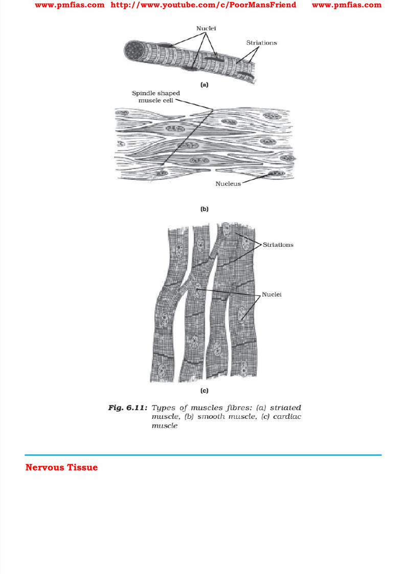
8/16/2019 Biology for Prelims Exam
http://slidepdf.com/reader/full/biology-for-prelims-exam 129/384
www.pmfias.com http://www.youtube.com/c/PoorMansFriend www.pmfias.com
Nervous Tissue
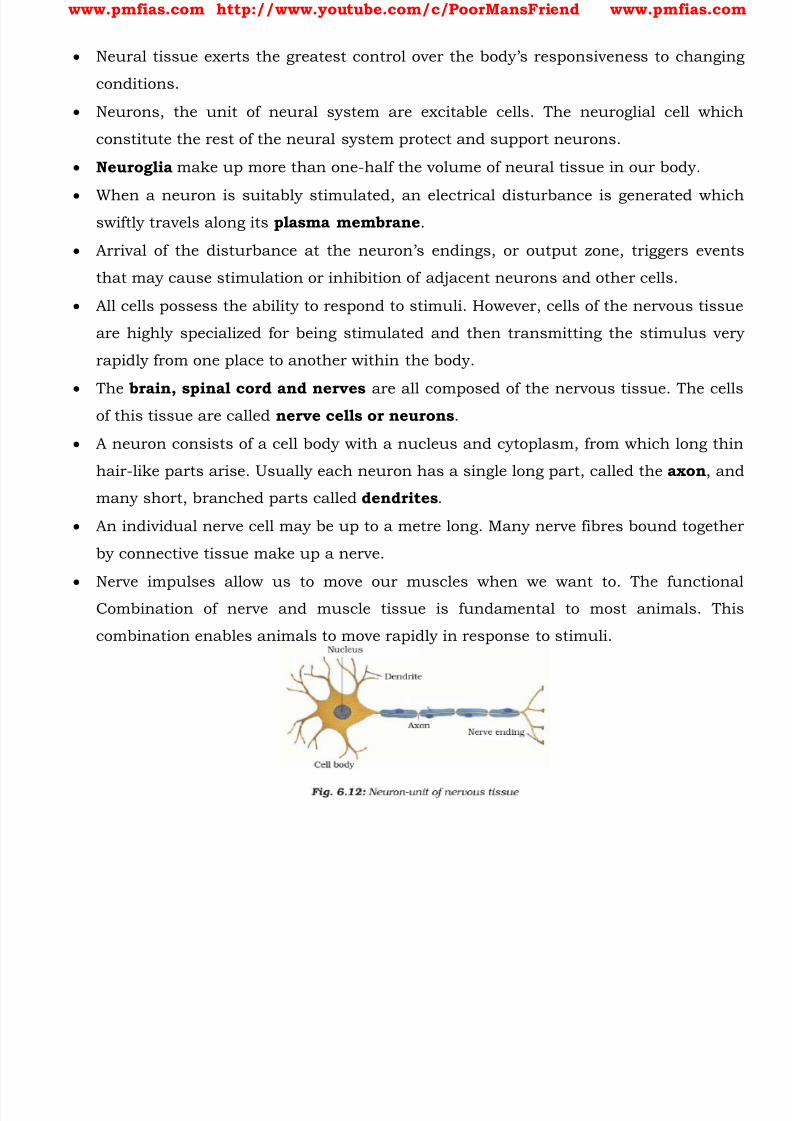
8/16/2019 Biology for Prelims Exam
http://slidepdf.com/reader/full/biology-for-prelims-exam 130/384
www.pmfias.com http://www.youtube.com/c/PoorMansFriend www.pmfias.com
Neural tissue exerts the greatest control over the body’s responsivenes s to changing
conditions.
Neurons, the unit of neural system are excitable cells. The neuroglial cell which
constitute the rest of the neural system protect and support neurons.
Neuroglia make up more than one-half the volume of neural tissue in our body.
When a neuron is suitably stimulated, an electrical disturbance is generated whichswiftly travels along its plasma membrane .
Arrival of the disturbance at the neuron’s endings, or output zone, triggers events
that may cause stimulation or inhibition of adjacent neurons and other cells.
All cells possess the ability to respond to stimuli. However, cells of the nervous tissue
are highly specialized for being stimulated and then transmitting the stimulus very
rapidly from one place to another within the body.
The brain, spinal cord and nerves are all composed of the nervous tissue. The cellsof this tissue are called nerve cells or neurons .
A neuron consists of a cell body with a nucleus and cytoplasm, from which long thin
hair-like parts arise. Usually each neuron has a single long part, called the axon , and
many short, branched parts called dendrites .
An individual nerve cell may be up to a metre long. Many nerve fibres bound together
by connective tissue make up a nerve.
Nerve impulses allow us to move our muscles when we want to. The functionalCombination of nerve and muscle tissue is fundamental to most animals. This
combination enables animals to move rapidly in response to stimuli.
8/16/2019 Biology for Prelims Exam
http://slidepdf.com/reader/full/biology-for-prelims-exam 131/384
www.pmfias.com http://www.youtube.com/c/PoorMansFriend www.pmfias.com
Origin of Life on Earth - Evolution of Life on Earth - Adaptive Radiation - Biological
Evolution - Origin and Evolution of Man – Timeline of Evolution.
Origin Of Life on Earth
The universe is very old - almost 13 billion years old . Huge clusters of galaxies
comprise the universe. The Big Bang theory attempts to explain to us the origin of universe. It talks of a
singular huge explosion unimaginable in physical terms.
The universe expanded and hence, the temperature came down. Hydrogen and Helium
formed sometime later.
The gases condensed under gravitation and formed the galaxies of the present day
universe.
In the solar system of the milky way galaxy, earth was supposed to have been formed
about 4.5 billion years back.
There was no atmosphere on early earth. Methane, carbondioxide and ammonia released
from molten mass covered the surface.
The UV rays from the sun brokeup water into Hydrogen and Oxygen and the lighter H2
escaped. Oxygen combined with ammonia and methane to form water, CO2 and
others.
The ozone layer was formed. As earth cooled, the water vapor fell as rain, to fill all the
depressions and form oceans.
Life appeared 500 million years after the formation of earth, i.e., almost four billion
years back. Some scientists believe that the life came from outerspace.
The first non-cellular forms of life could have originated 3 billion years back. They would
have been giant molecules (RNA, Protein, Polysaccharides, etc.). These capsules
reproduced their molecules perhaps.
The first cellular form of life did not possibly originate till about 2000 million years ago.
These were probably single-cells. All life forms were in water environment only.
The version of a biogenesis, i.e., the first form of life arose slowly through evolutionary
forces from non-living molecules is accepted by majority.
However, once formed, how the first cellular forms of life could have evolved into the
complex biodiversity of today is the fascinating story that will be discussed below.
Evolution of Life on Earth
8/16/2019 Biology for Prelims Exam
http://slidepdf.com/reader/full/biology-for-prelims-exam 132/384
www.pmfias.com http://www.youtube.com/c/PoorMansFriend www.pmfias.com
Evolutionary Biology is the study of history of life forms on earth.
Homology indicates common ancestry. In the context of biology, homology is the
existence of shared ancestry between a pair of structures, or genes, in different species.
A common example of homologous structures in evolutionary biology are the wings of
bats and the arms of primates.
Homology is based on divergent evolution whereas Analogy refers to a situationexactly opposite [ convergent evolution ].
Wings of butterfly and of birds look alike. They are not anatomically similar structures
though they perform similar functions.
Hence, analogous structures are a result of convergent evolution - different structures
evolving for the same function and hence having similarity.
Other examples of analogy are the eye of the octopus and of mammals or the flippers of
Penguins and Dolphins. One can say that it is the similar habitat that has resulted in selection of similar
adaptive features in different groups of organisms but toward the same function: Sweet
potato (root modification) and potato (stem modification) is another example for
analogy.
Q1. Which one of the following is a modified stem? [1996]
a) Carrot
b) Sweet potato
c) Coconut
d) Potato
Carrot → Modified root
Coconut → Modified seed
Adaptive Radiation During his journey, Charles Darwin went to Galapagos Islands. There he observed an
amazing diversity of creatures.
Of particular interest, small black birds later called Darwin’s Finches amazed him.
He realized that there were many varieties of finches in the same island. All the varieties,
he conjectured, evolved on the island itself.

8/16/2019 Biology for Prelims Exam
http://slidepdf.com/reader/full/biology-for-prelims-exam 133/384
www.pmfias.com http://www.youtube.com/c/PoorMansFriend www.pmfias.com
From the original seed-eating features, many other forms with altered beaks arose,
enabling them to become insectivorous and vegetarian finches. This process of evolution
of different species in a given geographical area starting from a point and literally
radiating to other areas of geography (habitats) is called adaptive radiation.
Biological evolution
The essence of Darwinian theory about evolution is natural selection . The rate of
appearance of new forms is linked to the life cycle or the life span.
Microbes that divide fast have the ability to multiply and become millions of individuals
within hours.
A colony of bacteria (say A) growing on a given medium has built- in variation in terms
of ability to utilise a feed component.
A change in the medium composition would bring out only that part of the population
(say B) that can survive under the new conditions.
In due course of time this variant population outgrows the others and appears as new
species. This would happen within days.
For the same thing to happen in a fish or fowl would take million of years as life spans of
these animals are in years.
Hence, there must be a genetic basis for getting selected and to evolve .
Another way of saying the same thing is that some organisms are better adapted to
survive in an otherwise hostile environment. Adaptive ability is inherited. It has a
genetic basis . Fitness is the end result of the ability to adapt and get selected by
nature.
A Brief Account of Evolution
About 2000 million years ago (mya) the first cellular forms of life appeared on earth.
The mechanism of how non-cellular aggregates of giant macromolecules could evolve
into cells with membranous envelop is not known.
Some of these cells had the ability to release O2. The reaction could have been similar to
the light reaction in photosynthesis where water is split with the help of solar energy
captured and channelized by appropriate light harvesting pigments.
Slowly single-celled organisms became multi-cellular life forms. By the time of 500
mya, invertebrates were formed and active. Jawless fish probably evolved around 350
mya. Sea weeds and few plants existed probably around 320 mya.

8/16/2019 Biology for Prelims Exam
http://slidepdf.com/reader/full/biology-for-prelims-exam 134/384

8/16/2019 Biology for Prelims Exam
http://slidepdf.com/reader/full/biology-for-prelims-exam 135/384
www.pmfias.com http://www.youtube.com/c/PoorMansFriend www.pmfias.com
(c) Salamander-Python-Kangaroo
(d) Frog-Crab-Prawn
Answer: Evolution == Single cellular → Multicellular → Fishes → Amphibians → Reptiles →
Birds → Mammals.
(a) Octopus (Mollusc) – Dolphins and Whales (Mammals) – Shark (Fish)
(b) Pangolin (Mammal – always in news as it is an endangered one – its meat is
consumed in some South-East Asian countries) – Tortoise (Reptile) – Hawk (Bird)
(c) Salamander (Amphibian) – Python (Reptile) – Kangaroo (Mammal)
(d) Frog (Amphibian) – Crab (Crustaceans) – Prawn (Crustaceans)

8/16/2019 Biology for Prelims Exam
http://slidepdf.com/reader/full/biology-for-prelims-exam 136/384
www.pmfias.com http://www.youtube.com/c/PoorMansFriend www.pmfias.com

8/16/2019 Biology for Prelims Exam
http://slidepdf.com/reader/full/biology-for-prelims-exam 137/384
www.pmfias.com http://www.youtube.com/c/PoorMansFriend www.pmfias.com
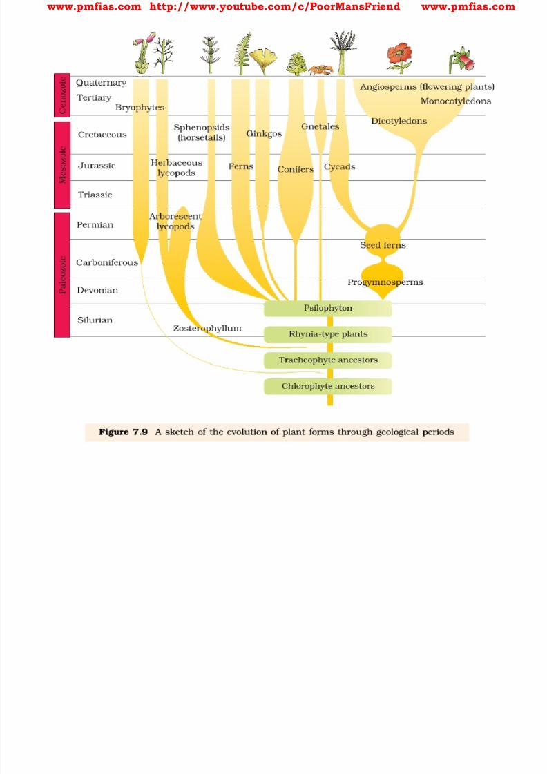
8/16/2019 Biology for Prelims Exam
http://slidepdf.com/reader/full/biology-for-prelims-exam 138/384
www.pmfias.com http://www.youtube.com/c/PoorMansFriend www.pmfias.com
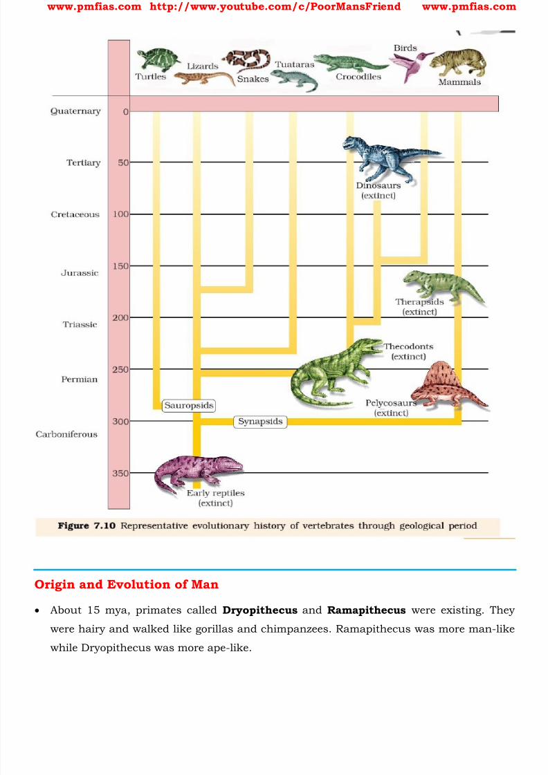
8/16/2019 Biology for Prelims Exam
http://slidepdf.com/reader/full/biology-for-prelims-exam 139/384
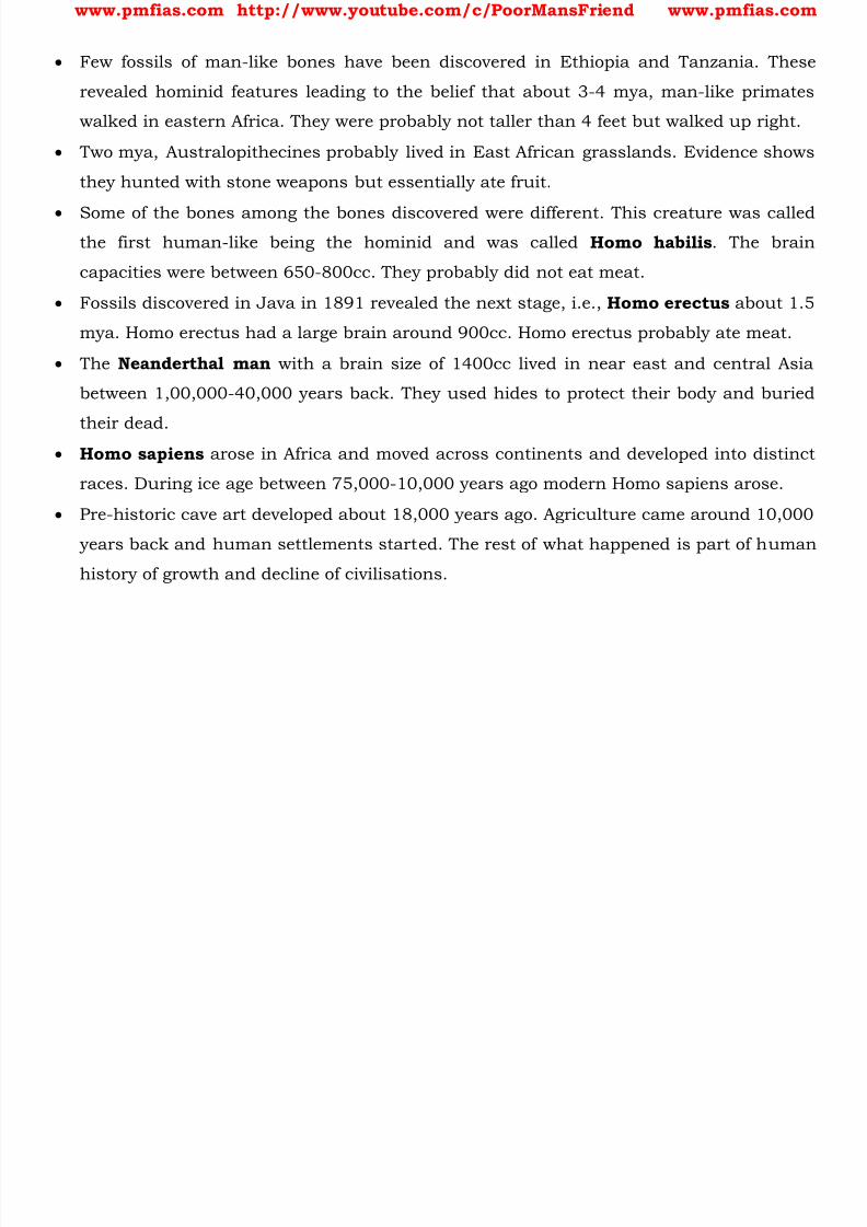
8/16/2019 Biology for Prelims Exam
http://slidepdf.com/reader/full/biology-for-prelims-exam 140/384
www.pmfias.com http://www.youtube.com/c/PoorMansFriend www.pmfias.com
Few fossils of man-like bones have been discovered in Ethiopia and Tanzania. These
revealed hominid features leading to the belief that about 3-4 mya, man-like primates
walked in eastern Africa. They were probably not taller than 4 feet but walked up right.
Two mya, Australopithecines probably lived in East African grasslands. Evidence shows
they hunted with stone weapons but essentially ate fruit.
Some of the bones among the bones discovered were different. This creature was calledthe first human-like being the hominid and was called Homo habilis . The brain
capacities were between 650-800cc. They probably did not eat meat.
Fossils discovered in Java in 1891 revealed the next stage, i.e., Homo erectus about 1.5
mya. Homo erectus had a large brain around 900cc. Homo erectus probably ate meat.
The Neanderthal man with a brain size of 1400cc lived in near east and central Asia
between 1,00,000-40,000 years back. They used hides to protect their body and buried
their dead. Homo sapiens arose in Africa and moved across continents and developed into distinct
races. During ice age between 75,000-10,000 years ago modern Homo sapiens arose.
Pre-historic cave art developed about 18,000 years ago. Agriculture came around 10,000
years back and human settlements started. The rest of what happened is part of human
history of growth and decline of civilisations.
8/16/2019 Biology for Prelims Exam
http://slidepdf.com/reader/full/biology-for-prelims-exam 141/384
www.pmfias.com http://www.youtube.com/c/PoorMansFriend www.pmfias.com
Biological Classification: Monera, Protista, Fungi, Plantae, Animalia and, Viruses, Viroids
and Lichens. Biodiversity, Classification of Biodiversity, Taxonomic Categories.
Biodiversity
Classification of life forms will be closely related to their evolution . Charles Darwin first
described this idea of evolution in 1859 in his book, The Origin of Species . The number of species that are known and described range between 1.7-1.8 million.
Rough estimates state that there are about ten million species on the planet. This refers
to biodiversity or the number and types of organisms present on earth.
The warm and humid tropical regions of the earth, between the tropic of cancer and
the tropic of capricorn, are rich in diversity of plant and animal life. This is called the
region of megadiversity .
Of the biodiversity of the planet, more than half is concentrated in a few countries
within tropics.
In alphabetical order, the 17 megadiverse countries are:
1. Australia
2. Brazil
3. China
4. Colombia
5. Democratic Republic of the Congo
6. Ecuador
7. India
8. Indonesia
9. Madagascar
10. Malaysia
11. Mexico
12. Papua New Guinea13. Peru
14. Philippines
15. South Africa
16. United States
17. Venezuela
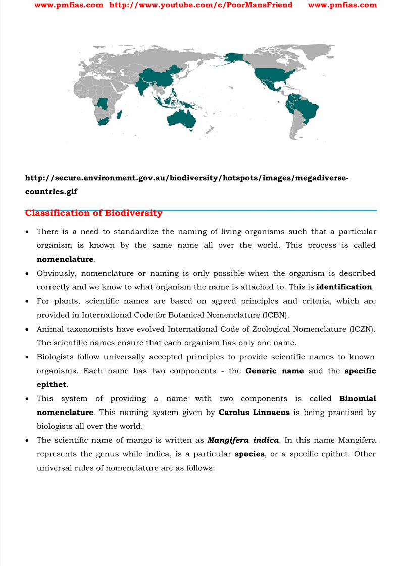
8/16/2019 Biology for Prelims Exam
http://slidepdf.com/reader/full/biology-for-prelims-exam 142/384
www.pmfias.com http://www.youtube.com/c/PoorMansFriend www.pmfias.com
http://secure.environment.gov.au/biodiversity/hotspots/images/megadiverse-
countries.gif
Classification of Biodiversity
There is a need to standardize the naming of living organisms such that a particular
organism is known by the same name all over the world. This process is called
nomenclature .
Obviously, nomenclature or naming is only possible when the organism is described
correctly and we know to what organism the name is attached to. This is identification .
For plants, scientific names are based on agreed principles and criteria, which are
provided in International Code for Botanical Nomenclature (ICBN).
Animal taxonomists have evolved International Code of Zoological Nomenclature (ICZN).
The scientific names ensure that each organism has only one name.
Biologists follow universally accepted principles to provide scientific names to known
organisms. Each name has two components - the Generic name and the specific
epithet .
This system of providing a name with two components is called Binomial
nomenclature . This naming system given by Carolus Linnaeus is being practised by
biologists all over the world.
The scientific name of mango is written as Mangifera indica . In this name Mangifera
represents the genus while indica, is a particular species , or a specific epithet. Other
universal rules of nomenclature are as follows:
8/16/2019 Biology for Prelims Exam
http://slidepdf.com/reader/full/biology-for-prelims-exam 143/384
www.pmfias.com http://www.youtube.com/c/PoorMansFriend www.pmfias.com
1) Biological names are generally in Latin and written in italics . They are Latinised or
derived from Latin irrespective of their origin.
2) The first word in a biological name represents the genus while the second component
denotes the specific epithet.
3) Both the words in a biological name, when handwritten, are separately underlined, or
printed in italics to indicate their Latin origin . The first word denoting the genus starts with a capital letter while the specific epithet
starts with a small letter. it can be illustrated with the example of Mangifera indica .
Name of the author appears after the specific epithet, i.e., at the end of the biological
name and is written in an abbreviated form, e.g., Mangifera indica Linn . It indicates that
this species was first described by Linnaeus.
Since it is nearly impossible to study all the living organisms, it is necessary to devise
some means to make this possible. This process is classification . Classification is the process by which anything is grouped into convenient categories
based on some easily observable characters.
The scientific term for these categories is taxa . Here you must recognise that taxa can
indicate categories at very different levels. ‘Plants’ - also form a taxa. ‘Wheat’ is also a
taxa. Similarly, ‘animals’, ‘mammals’, ‘dogs’ are all taxa - but you know that a dog is a
mammal and mammals are animals. Therefore, ‘animals’, ‘mammals’ and ‘dogs’
represent taxa at different levels.
Hence, based on characteristics, all living organisms can be classified into different taxa.
This process of classification is taxonomy .
External and internal structure, along with the structure of cell, process and ecological
information of organisms are essential and form the basis of modern taxonomic studies.
Hence, characterisation, identification, classification and nomenclature are the
processes that are basic to taxonomy.
Human beings were, since long, not only interested in knowing more about different
kinds of organisms and their diversities, but also the relationships among them. This
branch of study was referred to as systematics .
The word systematics is derived from the Latin word ‘systema’ which means systematic
arrangement of organisms. Linnaeus used Systema Naturae as the title of his
publication.
The scope of systematics was later enlarged to include identification, nomenclature and
classification.
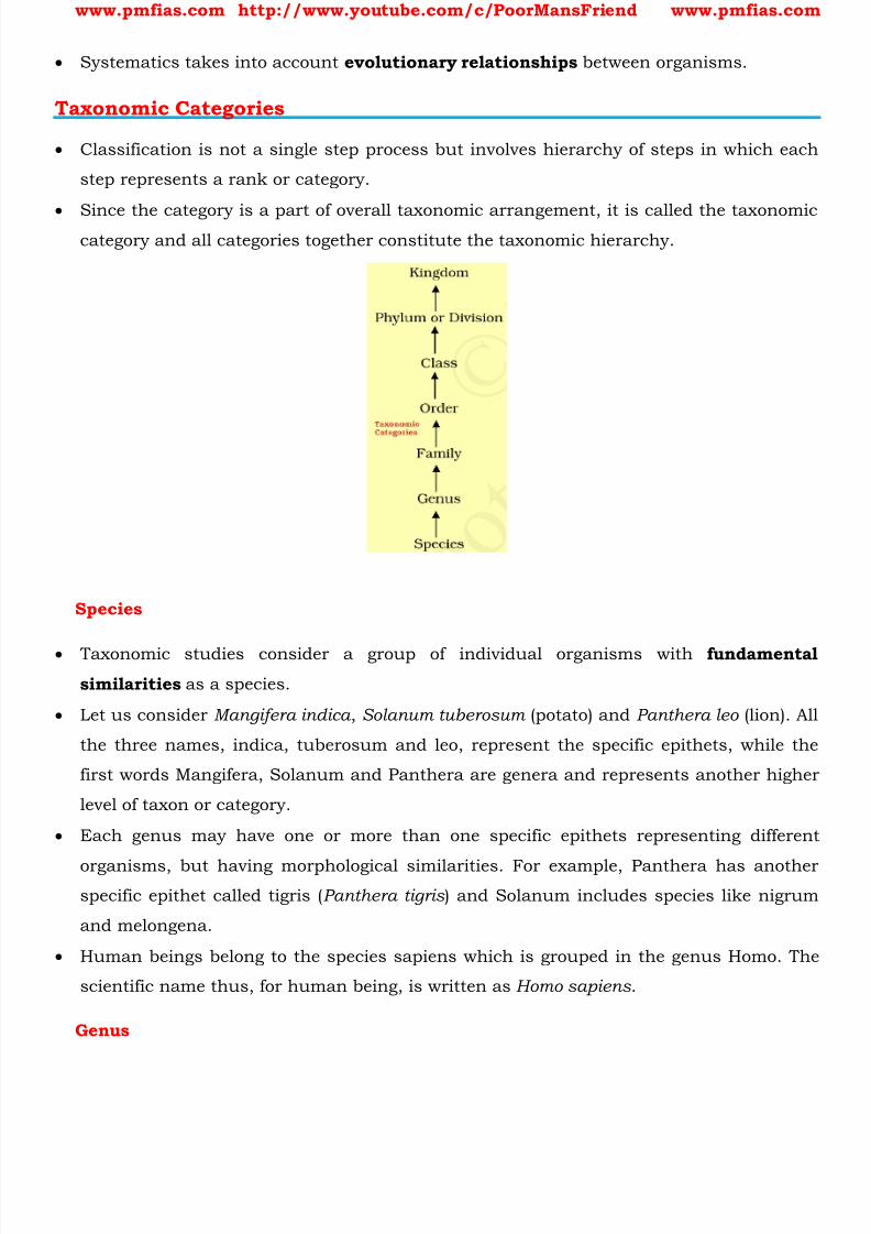
8/16/2019 Biology for Prelims Exam
http://slidepdf.com/reader/full/biology-for-prelims-exam 144/384
www.pmfias.com http://www.youtube.com/c/PoorMansFriend www.pmfias.com
Systematics takes into account evolutionary relationships between organisms.
Taxonomic Categories
Classification is not a single step process but involves hierarchy of steps in which each
step represents a rank or category.
Since the category is a part of overall taxonomic arrangement, it is called the taxonomiccategory and all categories together constitute the taxonomic hierarchy.
Species
Taxonomic studies consider a group of individual organisms with fundamental
similarities as a species.
Let us consider Mangifera indica , Solanum tuberosum (potato) and Panthera leo (lion). All
the three names, indica, tuberosum and leo, represent the specific epithets, while the
first words Mangifera, Solanum and Panthera are genera and represents another higher
level of taxon or category.
Each genus may have one or more than one specific epithets representing different
organisms, but having morphological similarities. For example, Panthera has anotherspecific epithet called tigris ( Panthera tigris ) and Solanum includes species like nigrum
and melongena.
Human beings belong to the species sapiens which is grouped in the genus Homo. The
scientific name thus, for human being, is written as Homo sapiens .
Genus
8/16/2019 Biology for Prelims Exam
http://slidepdf.com/reader/full/biology-for-prelims-exam 145/384
www.pmfias.com http://www.youtube.com/c/PoorMansFriend www.pmfias.com
Genus comprises a group of related species which has more characters in common in
comparison to species of other genera.
We can say that genera are aggregates of closely related species. For example, potato
and brinjal are two different species but both belong to the genus Solanum.
Lion ( Panthera leo ), leopard ( P. pardus ) and tiger ( P. tigris ) with several common features,
are all species of the genus Panthera. This genus differs from another genus Felis whichincludes cats.
Family
The next category, Family, has a group of related genera with still less number of
similarities as compared to genus and species.
Families are characterised on the basis of both vegetative and reproductive features of
plant species. Among animals for example, genus Panthera, comprising lion, tiger, leopard is put along
with genus, Felis (cats) in the family
Felidae. Similarly, if you observe the features of a cat and a dog, you will find some
similarities and some differences as well. They are separated into two different families -
Felidae and Canidae, respectively.
Order
You have seen earlier that categories like species, genus and families are based on a
number of similar characters. Generally, order and other higher taxonomic categories
are identified based on the aggregates of characters.
Class
This category includes related orders.
Phylum
Classes comprising animals like fishes, amphibians, reptiles, birds along with mammals
constitute the next higher category called Phylum.
Kingdom
All animals belonging to various phyla are assigned to the highest category called
Kingdom Animalia in the classification system of animals.

8/16/2019 Biology for Prelims Exam
http://slidepdf.com/reader/full/biology-for-prelims-exam 146/384
www.pmfias.com http://www.youtube.com/c/PoorMansFriend www.pmfias.com
The Kingdom Plantae, on the other hand, is distinct, and comprises all plants from
various divisions. Henceforth, we will refer to these two groups as animal and plant
kingdoms.
Taxonomical Aids
Herbarium
Herbarium is a store house of collected plant specimens that are dried, pressed and
preserved on sheets. Further, these sheets are arranged according to a universally
accepted system of classification.
The herbarium sheets also carry a label providing information about date and place of
collection, English, local and botanical names, family, collector’s name, etc.
Herbaria also serve as quick referral systems in taxonomical studies.
Botanical Gardens
These specialized gardens have collections of living plants for reference.
The famous botanical gardens are at Kew (England), Indian Botanical Garden, Howrah
(India) and at National Botanical Research Institute, Lucknow (India) .
Museum
Museums have collections of preserved plant and animal specimens for study andreference. Specimens are preserved in the containers or jars in preservative solutions.
Zoological Parks
These are the places where wild animals are kept in protected environments under
human care and which enable us to learn about their food habits and behavior.
Key
Key is used for identification of plants and animals based on the similarities and
dissimilarities.
The keys are based on the contrasting characters generally in a pair called couplet.
Flora, manuals, monographs and catalogues are some other means of recording
descriptions.
Biological Classification

8/16/2019 Biology for Prelims Exam
http://slidepdf.com/reader/full/biology-for-prelims-exam 147/384
www.pmfias.com http://www.youtube.com/c/PoorMansFriend www.pmfias.com
In Linnaeus' time a Two Kingdom system of classification with Plantae and Animalia
kingdoms was developed.
This system did not distinguish between the eukaryotes and prokaryotes , unicellular
and multicellular organisms and photosynthetic (green algae) and non-
photosynthetic (fungi) organisms.
Classification of organisms into plants and animals was easily done and was easy tounderstand, but, a large number of organisms did not fall into either category. Hence
the two kingdom classification used for a long time was found inadequate.
Biologists, such as Ernst Haeckel (1894), Robert Whittaker (1959) and Carl Woese
(1977) have tried to classify all living organisms into broad categories, called kingdoms.
The classification Whittaker proposed has five kingdoms and is widely used:
1. Monera,
2.
Protista,3. Fungi,
4. Plantae and
5. Animalia.
More details about each kingdom in the next posts
8/16/2019 Biology for Prelims Exam
http://slidepdf.com/reader/full/biology-for-prelims-exam 148/384
www.pmfias.com http://www.youtube.com/c/PoorMansFriend www.pmfias.com
The main criteria for classification used by him include cell structure, thallus
organisation, mode of nutrition, reproduction etc.
It brought together the prokaryotic bacteria and the blue green algae with other
groups which were eukaryotic .
It also grouped together the unicellular organisms and the multicellular ones.
The classification did not differentiate between the heterotrophic group - fungi, and theautotrophic green plants, though they also showed a characteristic difference in their
walls composition - the fungi had chitin in their walls while the green plants had a
cellulosic cell wall.
When such characteristics were considered, the fungi were placed in a separate kingdom
- Kingdom Fungi.
All prokaryotic organisms were grouped together under Kingdom Monera and the
unicellular eukaryotic organisms were placed in Kingdom Protista . Kingdom Protista has brought together Chlamydomonas, Chlorella (earlier placed in
Algae within Plants and both having cell walls) with Paramoecium and Amoeba (which
were earlier placed in the animal kingdom which lack cell wall).
At present the biological classification includes:
1. Kingdom Monera
2. Kingdom Protista
3. Kingdom Fungi
4. Kingdom Plantae
5. Kingdom Animalia
6. Viruses, Viroids and Lichens
Further classification is done by naming the sub-groups at various levels as given in the
following scheme: KPC OF GS
1. Kingdom,
2. Phylum (For Animals) / Division (For Plants),
3. Class,
4. Order,
5. Family,
6. Genus,
7. Species.
8/16/2019 Biology for Prelims Exam
http://slidepdf.com/reader/full/biology-for-prelims-exam 149/384
www.pmfias.com http://www.youtube.com/c/PoorMansFriend www.pmfias.com
Biological Classification – Kingdom Monera, Kingdom Protista, Kingdom Fungi, Kingdom
Plantae, Kingdom Animalia, Viruses, Viroids and Lichens.
Biological Classification of Plants and Animals
Biological classification of plants and animals was first proposed by Aristotle on the
basis of simple morphological characters. Linnaeus later classified all living organisms into two kingdoms - Plantae and Animalia.
Whittaker proposed an elaborate five kingdom classification - Monera, Protista, Fungi,
Plantae and Animalia .
The main criteria of the five kingdom classification were cell structure, body
organisation, mode of nutrition and reproduction, and phylogenetic relationships
[evolutionary development and diversification of a species ].
At present, the biological classification includes:
1. Kingdom Monera
2. Kingdom Protista
3. Kingdom Fungi
4. Kingdom Plantae
5. Kingdom Animalia
6. Viruses, Viroids and Lichens
In the five kingdom classification, bacteria are included in Kingdom Monera.
Kingdom Protista includes all single-celled eukaryotes such as Chrysophytes,
Dinoflagellates, Euglenoids, Slime-moulds and Protozoans.
Members of Kingdom Fungi show a great diversity in structures and habitat. Most fungi
are saprophytic in their mode of nutrition.
The plantae includes all eukaryotic chlorophyll-containing organisms. Algae, bryophytes,
pteridophytes, gymnosperms and angiosperms are included in this group.
The heterotrophic eukaryotic, multicellular organisms lacking a cell wall are included in
the Kingdom Animalia.
Some acellular organisms like viruses and viroids as well as the lichens are not included
in the five kingdom system of classification.

8/16/2019 Biology for Prelims Exam
http://slidepdf.com/reader/full/biology-for-prelims-exam 150/384
www.pmfias.com http://www.youtube.com/c/PoorMansFriend www.pmfias.com
Monera
The organisms in this group are
1. prokaryotes == do not have a defined nucleus or organelles { Prokaryotic Cells vs.
Eukaryotic Cells }.
2. unicellular == do any of them show multi-cellular body designs.
This group includes all bacteria. Some well-known bacteria include blue-green algae or
cyanobacteria [have cell walls], and mycoplasma [doesn’t possess a Cell Wall ].
They are the most abundant micro-organisms and live in extreme habitats.
Some of them have cell walls [bacteria] while some do not [ mycoplasma ].
The mode of nutrition of organisms in this group can be either by synthesizing their own
food ( autotrophic ) or getting it from the environment ( heterotrophic ). Many of them live
in or on other organisms as parasites .
Bacteria are grouped under four categories based on their shape
1) the spherical Coccus
2) the rod-shaped Bacillus
3) the comma-shaped Vibrium
4) the spiral Spirillum
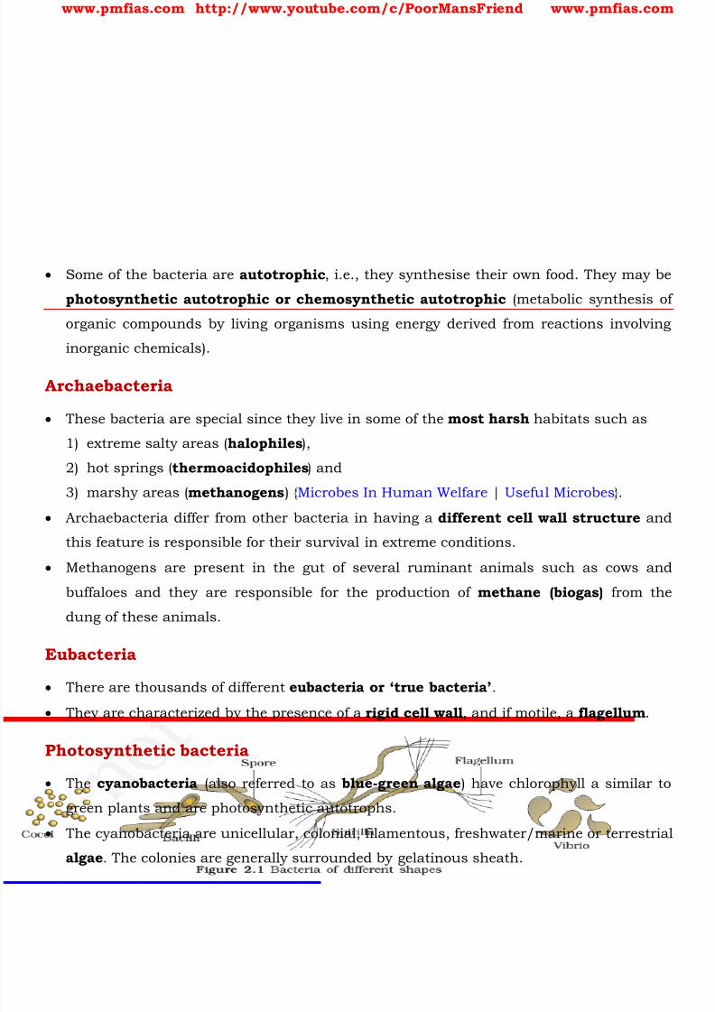
8/16/2019 Biology for Prelims Exam
http://slidepdf.com/reader/full/biology-for-prelims-exam 151/384
www.pmfias.com http://www.youtube.com/c/PoorMansFriend www.pmfias.com
Some of the bacteria are autotrophic , i.e., they synthesise their own food. They may be
photosynthetic autotrophic or chemosynthetic autotrophic (metabolic synthesis of
organic compounds by living organisms using energy derived from reactions involving
inorganic chemicals).
Archaebacteria
These bacteria are special since they live in some of the most harsh habitats such as
1) extreme salty areas ( halophiles ),
2) hot springs ( thermoacidophiles ) and
3) marshy areas ( methanogens ) { Microbes In Human Welfare | Useful Microbes }.
Archaebacteria differ from other bacteria in having a different cell wall structure and
this feature is responsible for their survival in extreme conditions.
Methanogens are present in the gut of several ruminant animals such as cows and
buffaloes and they are responsible for the production of methane (biogas) from the
dung of these animals.
Eubacteria
There are thousands of different eubacteria or ‘true bacteria’ .
They are characterized by the presence of a rigid cell wall , and if motile, a flagellum .
Photosynthetic bacteria
The cyanobacteria (also referred to as blue-green algae ) have chlorophyll a similar to
green plants and are photosynthetic autotrophs.
The cyanobacteria are unicellular, colonial, filamentous, freshwater/marine or terrestrial
algae . The colonies are generally surrounded by gelatinous sheath.

8/16/2019 Biology for Prelims Exam
http://slidepdf.com/reader/full/biology-for-prelims-exam 152/384
www.pmfias.com http://www.youtube.com/c/PoorMansFriend www.pmfias.com
They often form blooms [algal blooms] in polluted water bodies.
Some of these organisms can fix atmospheric nitrogen in specialized cells called
heterocysts , e.g., Nostoc and Anabaena .
Chemosynthetic bacteria
Chemosynthetic autotrophic bacteria oxidise various inorganic substances such asnitrates, nitrites and ammonia and use the released energy for their ATP production.
They play a great role in recycling nutrients like nitrogen, phosphorous, iron and
sulphur.
Heterotrophic bacteria
Heterotrophic bacteria are the most abundant in nature. The majority are important
decomposers .
Many of them have a significant impact on human affairs. They are helpful in making
curd from milk, production of antibiotics, fixing nitrogen in legume roots, etc
{ Microbes In Human Welfare | Useful Microbes }.
Some are pathogens causing damage to human beings, crops, farm animals and pets.
Cholera, typhoid, tetanus, citrus canker are well known diseases caused by different
bacteria { Diseases Caused by Microorganisms , Diseases | Acute, Chronic,
Communicable Diseases }.
Reproduction
Bacteria reproduce mainly by fission .
Sometimes, under unfavorable conditions, they produce spores .
They also reproduce by a sort of sexual reproduction by adopting a primitive type of
DNA transfer from one bacterium to the other.
Mycoplasma
The Mycoplasma are organisms that completely lack a cell wall .
They are the smallest living cells known and can survive without oxygen .
Many mycoplasma are pathogenic in animals and plants.
Protista
8/16/2019 Biology for Prelims Exam
http://slidepdf.com/reader/full/biology-for-prelims-exam 153/384
www.pmfias.com http://www.youtube.com/c/PoorMansFriend www.pmfias.com
All single-celled eukaryotes are placed under Protista [Prokaryotic Cells vs. Eukaryotic
Cells ].
Boundaries of this kingdom are not well defined. This kingdom forms a link with the
others dealing with plants, animals and fungi.
In this group we include Chrysophytes, Dinoflagellates, Euglenoids, Slime moulds
and Protozoans . Examples are unicellular algae, diatoms and protozoans . Their mode of nutrition can be autotrophic or heterotrophic .
Members of Protista are primarily aquatic . Some have flagella or cilia that helps in
movement.
Protists reproduce asexually and sexually by a process involving cell fusion and zygote
formation.
Chrysophytes
This group includes diatoms and golden algae (desmids) .
Most of them are photosynthetic . Diatoms are the chief ‘producers’ in the oceans.
They are found in fresh water as well as in marine environments. They are microscopic
and float passively in water currents ( plankton ).
In diatoms the cell walls form two thin overlapping shells. The walls are embedded with
silica and thus the walls are indestructible. Thus, diatoms have left behind large amount
of cell wall deposits in their habitat; this accumulation over billions of years is referred
to as ‘diatomaceous earth’ . Being gritty this soil is used in polishing, filtration of oils
and syrups.
Dinoflagellates
These organisms are mostly marine and photosynthetic .
They appear yellow, green, brown, blue or red depending on the main pigments present
in their cells.
The cell wall has stiff cellulose plates on the outer surface.
Most of them have two flagella ; one lies longitudinally and the other transversely in a
furrow between the wall plates.
Very often, red dinoflagellates (Example: Gonyaulax) undergo such rapid multiplication
that they make the sea appear red (red tides) .
Toxins released by such large numbers may even kill other marine animals such as
fishes.

8/16/2019 Biology for Prelims Exam
http://slidepdf.com/reader/full/biology-for-prelims-exam 154/384
www.pmfias.com http://www.youtube.com/c/PoorMansFriend www.pmfias.com
Euglenoids
Majority of them are fresh water organisms found in stagnant water.
Instead of a cell wall, they have a protein rich layer called pellicle which makes their
body flexible.
They have two flagella, a short and a long one.
Though they are photosynthetic in the presence of sunlight, when deprived of sunlight
they behave like heterotrophs by predating on other smaller organisms.
Interestingly, the pigments of euglenoids are identical to those present in higher plants.
Example: Euglena.
Slime Moulds
Slime moulds are saprophytic protists .
The body moves along decaying twigs and leaves engulfing organic material. Under suitable conditions, they form an aggregation called plasmodium which may
grow and spread over several feet.
During unfavorable conditions, the plasmodium differentiates and forms fruiting bodies
bearing spores at their tips. The spores possess true walls . They are extremely resistant
and survive for many years, even under adverse conditions. The spores are dispersed by
air currents.
Protozoans
All protozoans are heterotrophs and live as predators or parasites . They are believed to
be primitive relatives of animals . There are four major groups of protozoans.
Amoeboid protozoans
These organisms live in fresh water, sea water or moist soil.
They move and capture their prey by putting out pseudopodia (false feet) as in
Amoeba .
Marine forms have silica shells on their surface. Some of them such as Entamoeba are
parasites.
Flagellated protozoans
The members of this group are either free-living or parasitic. They have flagella .
The parasitic forms cause diseases such as sleeping sickness . Example: Trypanosoma .
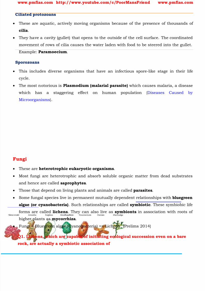
8/16/2019 Biology for Prelims Exam
http://slidepdf.com/reader/full/biology-for-prelims-exam 155/384
www.pmfias.com http://www.youtube.com/c/PoorMansFriend www.pmfias.com
Ciliated protozoans
These are aquatic, actively moving organisms because of the presence of thousands of
cilia .
They have a cavity (gullet) that opens to the outside of the cell surface. The coordinated
movement of rows of cilia causes the water laden with food to be steered into the gullet.
Example: Paramoecium .
Sporozoans
This includes diverse organisms that have an infectious spore-like stage in their life
cycle.
The most notorious is Plasmodium (malarial parasite) which causes malaria, a disease
which has a staggering effect on human population { Diseases Caused by
Microorganisms }.
Fungi
These are heterotrophic eukaryotic organisms .
Most fungi are heterotrophic and absorb soluble organic matter from dead substrates
and hence are called saprophytes .
Those that depend on living plants and animals are called parasites .
Some fungal species live in permanent mutually dependent relationships with bluegreen
algae (or cyanobacteria) . Such relationships are called symbiotic . These symbiobic life
forms are called lichens . They can also live as symbionts in association with roots of
higher plants as mycorrhiza .
Fungi + Bluegreen algae (Cyanobacteria) == Lichens. (Prelims 2014)
Q1. Lichens, which are capable of initiating ecological succession even on a bare
rock, are actually a symbiotic association of

8/16/2019 Biology for Prelims Exam
http://slidepdf.com/reader/full/biology-for-prelims-exam 156/384
www.pmfias.com http://www.youtube.com/c/PoorMansFriend www.pmfias.com
a) algae and bacteria
b) algae and fungi
c) bacteria and fungi
d) fungi and mosses
Reproduction in fungi can take place by vegetative means - fragmentation, fission and
budding . Asexual reproduction is by spores called conidia or sporangiospores or zoospores, and
sexual reproduction is by oospores, ascospores and basidiospores.
The various spores are produced in distinct structures called fruiting bodies . The
sexual cycle involves the following three steps:
1) Fusion of protoplasms between two motile or non-motile gametes called
plasmogamy .
2)
Fusion of two nuclei called karyogamy .3) Meiosis in zygote resulting in haploid spores { Meiosis | Mitosis – Meiosis
Comparison }.
When a fungus reproduces sexually, two haploid hyphae of compatible mating types
come together and fuse. In some fungi the fusion of two haploid cells immediately
results in diploid cells (2n).
However, in other fungi (ascomycetes and basidiomycetes), an intervening dikaryotic
stage (n + n, i.e., two nuclei per cell ) occurs; such a condition is called a dikaryon and
the phase is called dikaryophase of fungus. Later, the parental nuclei fuse and the cells
become diploid. The fungi form fruiting bodies in which reduction division occurs,
leading to formation of haploid spores.
Many of fungi have the capacity to become multicellular organisms at certain stages in
their lives.
They have cell-walls made of a tough complex sugar called chitin .
Fungi are cosmopolitan and occur in air, water, soil and on animals and plants.
They prefer to grow in warm and humid places. With the exception of yeasts which are
unicellular, fungi are filamentous .
Their bodies consist of long, slender thread-like structures called hyphae . The network
of hyphae is known as mycelium .
Some hyphae are continuous tubes filled with multinucleated cytoplasm - these are
called coenocytic hyphae.
8/16/2019 Biology for Prelims Exam
http://slidepdf.com/reader/full/biology-for-prelims-exam 157/384
www.pmfias.com http://www.youtube.com/c/PoorMansFriend www.pmfias.com
Others have septae or cross walls in their hyphae. The cell walls of fungi are composed
of chitin and polysaccharides { Carbohydrates | Monosaccharides | Polysaccharides }.
When your bread develops a mould or your orange rots it is because of fungi.
The common mushroom you eat and toadstools are also fungi.
White spots seen on mustard leaves are due to a parasitic fungus.
Some unicellular fungi, e.g., yeast are used to make bread and beer. Other fungi cause diseases in plants and animals; wheat rust-causing Puccinia is an
important example.
Some are the source of antibiotics, e.g., Penicillium .
Phycomycetes
Asexual reproduction takes place by zoospores (motile) or by aplanospores (non-motile).
These spores are endogenously produced in sporangium. A zygospore is formed by fusion of two gametes.
These gametes are similar in morphology (isogamous) or dissimilar (anisogamous or
oogamous).
Some common examples are Mucor, Rhizopus (the bread mould mentioned earlier) and
Albugo (the parasitic fungi on mustard).
Ascomycetes
Commonly known as sac-fungi, the as comycetes are mostly multicellular, e.g.,
Penicillium , or rarely unicellular, e.g., yeast (Saccharomyces) .
Basidiomycetes
Commonly known forms of basidiomycetes are mushrooms, bracket fungi or puffballs .
They grow in soil, on logs and tree stumps and in living plant bodies as parasites, e.g.,
rusts and smuts.
The asexual spores are generally not found, but vegetative reproduction by
fragmentation is common.
The sex organs are absent , but plasmogamy is brought about by fusion of two
vegetative or somatic cells of different strains or genotypes. The resultant structure is
dikaryotic.
Deuteromycetes
8/16/2019 Biology for Prelims Exam
http://slidepdf.com/reader/full/biology-for-prelims-exam 158/384
www.pmfias.com http://www.youtube.com/c/PoorMansFriend www.pmfias.com
Commonly known as imperfect fungi because only the asexual or vegetative phases of
these fungi are known.
Plantae
These are multicellular eukaryotes with cell walls mainly made of cellulose { Plant Cell
vs. Animal Cell }. They are autotrophs and use chlorophyll for photosynthesis.
A few members are partially heterotrophic such as the insectivorous plants or
parasites . Bladderwort and Venus fly trap are examples of insectivorous plants and
Cuscuta is a parasite.
Plantae includes algae, bryophytes, pteridophytes, gymnosperms and angiosperms .
Life cycle of plants has two distinct phases - the diploid sporophytic and the haploid
gametophytic - that alternate with each other.
The lengths of the haploid and diploid phases, and whether these phases are free-living
or dependent on others, vary among different groups in plants. This phenomenon is
called alternation of generation .
Animalia
These include all organisms which are multicellular eukaryotes without cell walls
They are heterotrophs .
They directly or indirectly depend on plants for food. They digest their food in an internal
cavity and store food reserves as glycogen or fat { Carbohydrates , Fats - Healthy Fats
and Unhealthy Fats }.
Their mode of nutrition is holozoic - by ingestion of food.
They follow a definite growth pattern and grow into adults that have a definite shape and
size.
Higher forms show elaborate sensory and neuromotor mechanism. Most of them are
capable of locomotion.
The sexual reproduction is by copulation of male and female followed by embryological
development.
Viruses, Viroids and Lichens
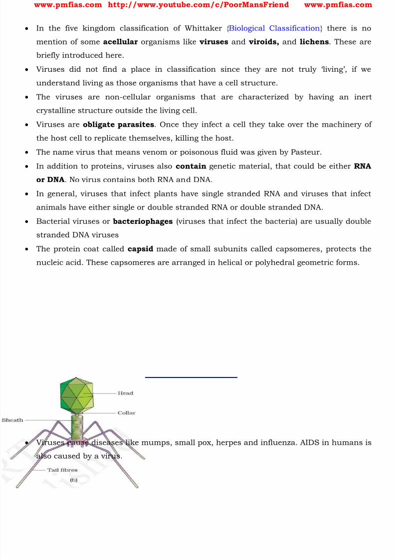
8/16/2019 Biology for Prelims Exam
http://slidepdf.com/reader/full/biology-for-prelims-exam 159/384
www.pmfias.com http://www.youtube.com/c/PoorMansFriend www.pmfias.com
In the five kingdom classification of Whittaker { Biological Classification } there is no
mention of some acellular organisms like viruses and viroids, and lichens . These are
briefly introduced here.
Viruses did not find a place in classificat ion since they are not truly ‘living’, if we
understand living as those organisms that have a cell structure.
The viruses are non-cellular organisms that are characterized by having an inertcrystalline structure outside the living cell.
Viruses are obligate parasites . Once they infect a cell they take over the machinery of
the host cell to replicate themselves, killing the host.
The name virus that means venom or poisonous fluid was given by Pasteur.
In addition to proteins, viruses also contain genetic material, that could be either RNA
or DNA . No virus contains both RNA and DNA.
In general, viruses that infect plants have single stranded RNA and viruses that infectanimals have either single or double stranded RNA or double stranded DNA.
Bacterial viruses or bacteriophages (viruses that infect the bacteria) are usually double
stranded DNA viruses
The protein coat called capsid made of small subunits called capsomeres, protects the
nucleic acid. These capsomeres are arranged in helical or polyhedral geometric forms.
Viruses cause diseases like mumps, small pox, herpes and influenza. AIDS in humans is
also caused by a virus.

8/16/2019 Biology for Prelims Exam
http://slidepdf.com/reader/full/biology-for-prelims-exam 160/384

8/16/2019 Biology for Prelims Exam
http://slidepdf.com/reader/full/biology-for-prelims-exam 161/384
www.pmfias.com http://www.youtube.com/c/PoorMansFriend www.pmfias.com
Plant Parts – Root, Stem, Leaf, Transpiration, Respiration in Plants, Flower, Androecium,
Gynoecium, Fruit, Transport Of Water And Minerals In Plants.
Plant Parts and Their Functions – Structural Organization in Plants
The Root
The main functions of the root system are absorption of water and minerals from the
soil, providing a proper anchorage to the plant parts, storing reserve food material and
synthesis of plant growth regulators .
In majority of the dicotyledonous plants, the direct elongation of the radicle leads to the
formation of primary root which grows inside the soil. It bears lateral roots of several
orders that are referred to as secondary, tertiary, etc. roots. The primary roots and its
branches constitute the tap root system , as seen in the mustard plant.
In monocotyledonous plants, the primary root is short lived and is replaced by a largenumber of roots. These roots originate from the base of the stem and constitute the
fibrous root system , as seen in the wheat plant.
In some plants, like grass, Monstera and the banyan tree, roots arise from parts of the
plant other than the radicle and are called adventitious roots .

8/16/2019 Biology for Prelims Exam
http://slidepdf.com/reader/full/biology-for-prelims-exam 162/384
www.pmfias.com http://www.youtube.com/c/PoorMansFriend www.pmfias.com
http://www.biosci.ohio-
state.edu/~plantbio/osu_pcmb/pcmb_lab_resources/images/pcmb101/stms_rts_lvs/root_
types.jpg
The root is covered at the apex by a thimble-like structure called the root cap . It
protects the tender apex of the root as it makes its way through the soil.
Tap roots of carrot, turnip and adventitious roots of sweet potato, get swollen and store
food.
Hanging structures that support a banyan tree are called prop roots . Similarly, the
stems of maize and sugarcane have supporting roots coming out of the lower nodes of
the stem. These are called stilt roots .
In some plants such as Rhizophora growing in swampy areas, many roots come out of
the ground and grow vertically upwards. Such roots, called pneumatophores , help to
get oxygen for respiration.
The Stem
8/16/2019 Biology for Prelims Exam
http://slidepdf.com/reader/full/biology-for-prelims-exam 163/384
www.pmfias.com http://www.youtube.com/c/PoorMansFriend www.pmfias.com
The region of the stem where leaves are born are called nodes while internodes are the
portions between two nodes.
Some stems perform the function of storage of food, support, protection and of vegetative
propagation.
Underground stems of potato, ginger, turmeric, zaminkand, colocasia are modified to
store food in them. Stem tendrils which develop from axillary buds , are slender and spirally coiled and
help plants to climb such as in gourds (cucumber, pumpkins, watermelon) and
grapevines.
Axillary buds of stems may also get modified into woody, straight and pointed thorns
Thorns are found in many plants such as Citrus, Bougainvillea. They protect plants
from browsing animals.
Some plants of arid regions modify their stems into flattened (Opuntia), or fleshycylindrical (Euphorbia) structures. They contain chlorophyll and carry out
photosynthesis.
Underground stems of some plants such as grass and strawberry, etc., spread to new
niches and when older parts die new plants are formed.
The Leaf
Leaves originate from shoot apical meristems . Leaf develops at the node and bears a
bud in its axil. The axillary bud later develops into a branch .
A typical leaf consists of three main parts: leaf base, petiole and lamina .
The petiole help hold the blade to light. Long thin flexible petioles allow leaf blades to
flutter in wind, thereby cooling the leaf and bringing fresh air to leaf surface.
The lamina or the leaf blade is the green expanded part of the leaf with veins and
veinlets. There is, usually, a middle prominent vein, which is known as the midrib.
Veins provide rigidity to the leaf blade and act as channels of transport for water,
minerals and food materials.
Leaf Venation
The arrangement of veins and the veinlets in the lamina of leaf is termed as venation.
When the veinlets form a network, the venation is termed as reticulate . When the veins
run parallel to each other within a lamina, the venation is termed as parallel.
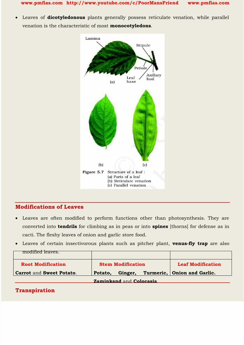
8/16/2019 Biology for Prelims Exam
http://slidepdf.com/reader/full/biology-for-prelims-exam 164/384
www.pmfias.com http://www.youtube.com/c/PoorMansFriend www.pmfias.com
Leaves of dicotyledonous plants generally possess reticulate venation, while parallel
venation is the characteristic of most monocotyledons .
Modifications of Leaves
Leaves are often modified to perform functions other than photosynthesis. They are
converted into tendrils for climbing as in peas or into spines [thorns] for defense as in
cacti. The fleshy leaves of onion and garlic store food.
Leaves of certain insectivorous plants such as pitcher plant, venus-fly trap are also
modified leaves.
Root Modification Stem Modification Leaf Modification
Carrot and Sweet Potato . Potato, Ginger, Turmeric,
Zaminkand and Colocasia .
Onion and Garlic.
Transpiration
8/16/2019 Biology for Prelims Exam
http://slidepdf.com/reader/full/biology-for-prelims-exam 165/384
www.pmfias.com http://www.youtube.com/c/PoorMansFriend www.pmfias.com
Plants absorb mineral nutrients and water from the soil. Not all the water absorbed is
utilised by the plant. The water evaporates through the stomata present on the surface
of the leaves by the process of transpiration.
The evaporation of water from leaves generates a suction pull (the same that you
produce when you suck water through a straw) which can pull water to great heights in
the tall trees. Transpiration also cools the plant.
Water absorption through roots can be increased by keeping the plants
a) in the shade
b) in dim light
c) under the fan
d) covered with a polythene bag
When we place a plant under the fan the speed of air flow is very high. Transpiration will
take place in presence of high air flow through the stomata. Rate of transpiration
increases during windy condition.
Increase in the rate of transpiration increases the water absorption also because when
transpiration occurs, it will create a transpiration pull and more water absorption will
take place.
Do Plants Also Respire?
In plants each part can independently take in oxygen from the air and give out carbon
dioxide. Even roots can respire. Can you guess what would happen if a potted plant is
overwatered?
Plants carry out photosynthesis only during the day and respiration both during the
day time as well as night .
Did you know? For us oxygen is essential, but for those organisms which do not use it,
oxygen is toxic. In fact, our white blood cells use oxygen to kill invading bacteria. Even
for humans, it may be dangerous to breathe pure oxygen for long.
The Flower
The flower is the reproductive unit in the angiosperms . It is meant for sexual
reproduction. Androecium and gynoecium are reproductive organs.
When a flower has both androecium and gynoecium, it is bisexual . A flower having
either only stamens or only carpels is unisexual .

8/16/2019 Biology for Prelims Exam
http://slidepdf.com/reader/full/biology-for-prelims-exam 166/384
www.pmfias.com http://www.youtube.com/c/PoorMansFriend www.pmfias.com
Aestivation: The mode of arrangement of sepals or petals in floral bud with respect to
the other members of the same whorl is known as aestivation.
Parts of a flower
Androecium
Androecium is composed of stamens . Each stamen which represents the male
reproductive organ consists of a stalk or a filament and an anther. Each anther is
usually bilobed and each lobe has two chambers, the pollen-sacs. The pollen grains are
produced in pollen-sacs. A sterile stamen is called staminode .
Gynoecium
Gynoecium is the female reproductive part of the flower and is made up of one or more
carpels. A carpel consists of three parts namely stigma, style and ovary.
After fertilization, the ovules develop into seeds and the ovary matures into a fruit.
Placentation: The arrangement of ovules within the ovary is known as placentation.
The Fruit
The fruit is a characteristic feature of the flowering plants.
It is a mature or ripened ovary , developed after fertilisation.
If a fruit is formed without fertilisation of the ovary, it is called a parthenocarpic fruit .
The Seed: The ovules after fertilisation, develop into seeds .

8/16/2019 Biology for Prelims Exam
http://slidepdf.com/reader/full/biology-for-prelims-exam 167/384
www.pmfias.com http://www.youtube.com/c/PoorMansFriend www.pmfias.com
Transport Of Water And Minerals In Plants
Plants absorb water and minerals by the roots. The roots have root hair. The root hair
increase the surface area of the root for the absorption of water and mineral nutrients
dissolved in water. The root hair is in contact with the water present between the soil
particles.
Can you guess how water moves from the root to the leaves? What kind of transport
system is present in plants?
Plants have pipe-like vessels to transport water and nutrients from the soil. The vessels
are made of special cells, forming the vascular tissue.
The vascular tissue for the transport of water and nutrients in the plant is called the
xylem . The xylem forms a continuous network of channels that connects roots to the
leaves through the stem and branches and thus transports water to the entire plant
leaves synthesise food. The food has to be transported to all parts of the plant. This is done by the vascular
tissue called the phloem . Thus, xylem and phloem transport substances in plants.
Summary
Flowering plants exhibit enormous variation in shape, size, structure, mode of nutrition,
life span, habit and habitat. They have well developed root and shoot systems.
Root system is either tap root or fibrous. Generally, dicotyledonous plants have tap rootswhile monocotyledonous plants have fibrous roots.
The roots in some plants get modified for storage of food, mechanical support and
respiration. The shoot system is differentiated into stem, leaves, flowers and fruits.
The morphological features of stems like the presence of nodes and internodes,
multicellular hair and positively phototropic nature help to differentiate the stems from
roots. Stems also get modified to perform diverse functions such as storage of food,
vegetative propagation and protection under different conditions. Leaf is a lateral outgrowth of stem developed exogeneously at the node. These are green
in colour to perform the function of photosynthesis. Leaves exhibit marked variations in
their shape, size, margin, apex and extent of incisions of leaf blade (lamina). Like other
parts of plants, the leaves also get modified into other structures such as tendrils,
spines for climbing and protection respectively.
8/16/2019 Biology for Prelims Exam
http://slidepdf.com/reader/full/biology-for-prelims-exam 168/384
www.pmfias.com http://www.youtube.com/c/PoorMansFriend www.pmfias.com
The flower is a modified shoot, meant for sexual reproduction. The flowers are arranged
in different types of inflorescences. They exhibit enormous variation in structure,
symmetry, position of ovary in relation to other parts, arrangement of petals, sepals,
ovules etc.
After fertilisation, the ovary is converted into fruits and ovules into seeds. Seeds either
may be monocotyledonous or dicotyledonous. They vary in shape, size and period ofviability. The floral characteristics form the basis of classification and identification of
flowering plants. This can be illustrated through semi-technical descriptions of families.
Hence, a flowering plant is described in a definite sequence by using scientific terms.
The floral features are represented in the summarised form as floral diagrams and floral
formula.
Water comes out of leaves in the form of vapour by a process called transpiration. Plants
release a lot of water into the air through this process.

8/16/2019 Biology for Prelims Exam
http://slidepdf.com/reader/full/biology-for-prelims-exam 169/384
www.pmfias.com http://www.youtube.com/c/PoorMansFriend www.pmfias.com
Plantae - Plant Kingdom, Algae – Thallophytes: Green Algae, Brown Algae, Red Algae, Uses
of algae, Bryophytes, Pteridophytes, Cryptogamae.
Plantae
Classification among plants depends on
(a) whether the plant body has well differentiated, distinct components,(b) whether the differentiated plant body has special tissues for the transport of water
and other substances within it,
(c) ability to bear seeds, and
(d) whether the seeds are enclosed within fruits.
Phylogenetic classification [evolutionary relationships], cytotaxonomy [cytological
information like chromosome number, structure, behavior] and chemotaxonomy
[chemical constituents of the plant], are used by taxonomists for classifying plants.
Plant Kingdom
Plants are multicellular eukaryotes with cell walls mainly made of cellulose { Plant
Cell vs. Animal Cell }.
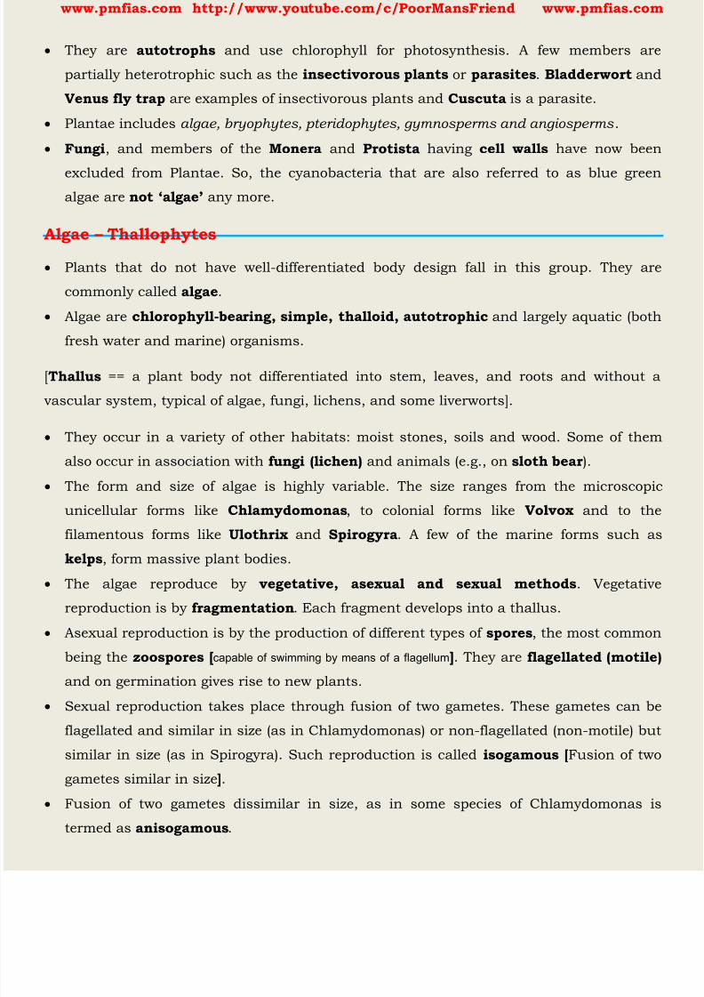
8/16/2019 Biology for Prelims Exam
http://slidepdf.com/reader/full/biology-for-prelims-exam 170/384
www.pmfias.com http://www.youtube.com/c/PoorMansFriend www.pmfias.com
They are autotrophs and use chlorophyll for photosynthesis. A few members are
partially heterotrophic such as the insectivorous plants or parasites . Bladderwort and
Venus fly trap are examples of insectivorous plants and Cuscuta is a parasite.
Plantae includes algae, bryophytes, pteridophytes, gymnosperms and angiosperms .
Fungi , and members of the Monera and Protista having cell walls have now been
excluded from Plantae. So, the cyanobacteria that are also referred to as blue greenalgae are not ‘algae’ any more.
Algae – Thallophytes
Plants that do not have well-differentiated body design fall in this group. They are
commonly called algae .
Algae are chlorophyll-bearing, simple, thalloid, autotrophic and largely aquatic (both
fresh water and marine) organisms.
[Thallus == a plant body not differentiated into stem, leaves, and roots and without a
vascular system, typical of algae, fungi, lichens, and some liverworts].
They occur in a variety of other habitats: moist stones, soils and wood. Some of them
also occur in association with fungi (lichen) and animals (e.g., on sloth bear ).
The form and size of algae is highly variable. The size ranges from the microscopic
unicellular forms like Chlamydomonas , to colonial forms like Volvox and to the
filamentous forms like Ulothrix and Spirogyra . A few of the marine forms such as
kelps , form massive plant bodies.
The algae reproduce by vegetative, asexual and sexual methods . Vegetative
reproduction is by fragmentation . Each fragment develops into a thallus.
Asexual reproduction is by the production of different types of spores , the most common
being the zoospores [ capable of swimming by means of a flagellum ]. They are flagellated (motile)
and on germination gives rise to new plants.
Sexual reproduction takes place through fusion of two gametes. These gametes can be
flagellated and similar in size (as in Chlamydomonas) or non-flagellated (non-motile) but
similar in size (as in Spirogyra). Such reproduction is called isogamous [ Fusion of two
gametes similar in size ].
Fusion of two gametes dissimilar in size, as in some species of Chlamydomonas is
termed as anisogamous .

8/16/2019 Biology for Prelims Exam
http://slidepdf.com/reader/full/biology-for-prelims-exam 171/384
www.pmfias.com http://www.youtube.com/c/PoorMansFriend www.pmfias.com
Fusion between one large, non-motile (static) female gamete and a smaller, motile male
gamete is termed oogamous , e.g., Volvox, Fucus. [Compare this with human sperm and
ovum]
Chlorophyceae – Green Algae The members of chlorophyceae are commonly called green algae .
The plant body may be unicellular, colonial or filamentous.
They are usually grass green due to the dominance of pigments chlorophyll a and b
The pigments are localised in definite chloroplasts.
Most of the members have one or more storage bodies called pyrenoids located in the
chloroplasts. Pyrenoids contain protein besides starch. Some algae may store food in the
form of oil droplets.
Green algae usually have a rigid cell wall made of an inner layer of cellulose and an
outer layer of pectose .
Vegetative reproduction usually takes place by fragmentation or by formation of different
types of spores.
Asexual reproduction is by flagellated zoospores produced in zoosporangia.
The sexual reproduction shows considerable variation in the type and formation of sex
cells and it may be isogamous, anisogamous or oogamous. Some commonly found green algae are: Chlamydomonas, Volvox, Ulothrix, Spirogyra
and Chara .
Phaeophyceae – Brown Algae
The members of phaeophyceae or brown algae are found primarily in marine habitats .
8/16/2019 Biology for Prelims Exam
http://slidepdf.com/reader/full/biology-for-prelims-exam 172/384
www.pmfias.com http://www.youtube.com/c/PoorMansFriend www.pmfias.com
They show great variation in size and form. They range from simple branched,
filamentous forms (Ectocarpus) to profusely branched forms as represented by kelps,
which may reach a height of 100 metres.
They possess chlorophyll a, c, carotenoids and xanthophylls . They vary in colour
from olive green to various shades of brown depending upon the amount of the
xanthophyll pigment, fucoxanthin present in them. The vegetative cells have a cellulosic wall usually covered on the outside by a gelatinous
coating of algin . The protoplast contains, in addition to plastids, a centrally located
vacuole and nucleus.
Vegetative reproduction takes place by fragmentation.
Asexual reproduction in most brown algae is by biflagellate zoospores that are pear-
shaped and have two unequal laterally attached flagella.
Sexual reproduction maybe isogamous, anisogamous or oogamous. Union of gametes may take place in water or within the oogonium (oogamous species).
The gametes are pyriform (pear-shaped) and bear two laterally attached flagella.
The common forms are Ectocarpus, Dictyota, Laminaria, Sargassum and Fucus .
Rhodophyceae – Red Algae
The members of rhodophyceae are commonly called red algae because of the
predominance of the red pigment, r-phycoerythrin in their body.
Majority of the red algae are marine with greater concentrations found in the warmer
areas.
They occur in both well-lighted regions close to the surface of water and also at great
depths in oceans where relatively little light penetrates.
The red thalli of most of the red algae are multicellular. Some of them have complex
body organisation.
The food is stored as floridean starch which is very similar to amylopectin and glycogen
in structure.
The red algae usually reproduce vegetatively by fragmentation.
They reproduce asexually by non-motile spores and sexually by non-motile gametes.
Sexual reproduction is oogamous.
The common members are: Polysiphonia, Porphyra, Gracilaria and Gelidium .
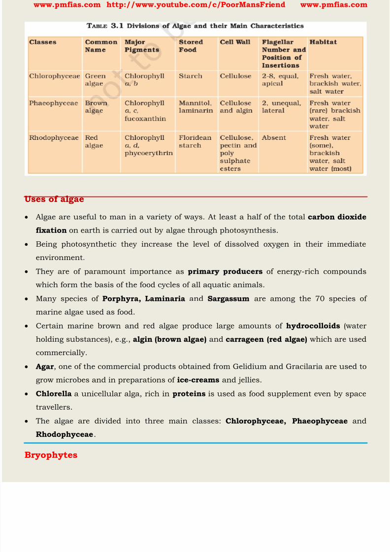
8/16/2019 Biology for Prelims Exam
http://slidepdf.com/reader/full/biology-for-prelims-exam 173/384
www.pmfias.com http://www.youtube.com/c/PoorMansFriend www.pmfias.com
Uses of algae
Algae are useful to man in a variety of ways. At least a half of the total carbon dioxide
fixation on earth is carried out by algae through photosynthesis.
Being photosynthetic they increase the level of dissolved oxygen in their immediate
environment.
They are of paramount importance as primary producers of energy-rich compounds
which form the basis of the food cycles of all aquatic animals.
Many species of Porphyra, Laminaria and Sargassum are among the 70 species of
marine algae used as food.
Certain marine brown and red algae produce large amounts of hydrocolloids (water
holding substances), e.g., algin (brown algae) and carrageen (red algae) which are used
commercially.
Agar , one of the commercial products obtained from Gelidium and Gracilaria are used to
grow microbes and in preparations of ice-creams and jellies.
Chlorella a unicellular alga, rich in proteins is used as food supplement even by space
travellers.
The algae are divided into three main classes: Chlorophyceae, Phaeophyceae and
Rhodophyceae .
Bryophytes
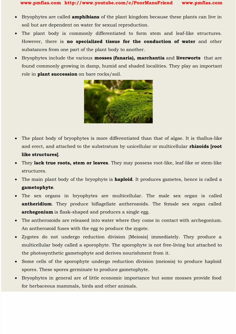
8/16/2019 Biology for Prelims Exam
http://slidepdf.com/reader/full/biology-for-prelims-exam 174/384
www.pmfias.com http://www.youtube.com/c/PoorMansFriend www.pmfias.com
Bryophytes are called amphibians of the plant kingdom because these plants can live in
soil but are dependent on water for sexual reproduction.
The plant body is commonly differentiated to form stem and leaf-like structures.
However, there is no specialized tissue for the conduction of water and other
substances from one part of the plant body to another.
Bryophytes include the various mosses (funaria), marchantia and liverworts that arefound commonly growing in damp, humid and shaded localities. They play an important
role in plant succession on bare rocks/soil.
The plant body of bryophytes is more differentiated than that of algae. It is thallus-like
and erect, and attached to the substratum by unicellular or multicellular rhizoids [root
like structures] .
They lack true roots, stem or leaves . They may possess root-like, leaf-like or stem-like
structures.
The main plant body of the bryophyte is haploid . It produces gametes, hence is called a
gametophyte .
The sex organs in bryophytes are multicellular. The male sex organ is called
antheridium . They produce biflagellate antherozoids. The female sex organ called
archegonium is flask-shaped and produces a single egg.
The antherozoids are released into water where they come in contact with archegonium.
An antherozoid fuses with the egg to produce the zygote.
Zygotes do not undergo reduction division [Meiosis] immediately. They produce a
multicellular body called a sporophyte. The sporophyte is not free-living but attached to
the photosynthetic gametophyte and derives nourishment from it.
Some cells of the sporophyte undergo reduction division (meiosis) to produce haploid
spores. These spores germinate to produce gametophyte.
Bryophytes in general are of little economic importance but some mosses provide food
for herbaceous mammals, birds and other animals.
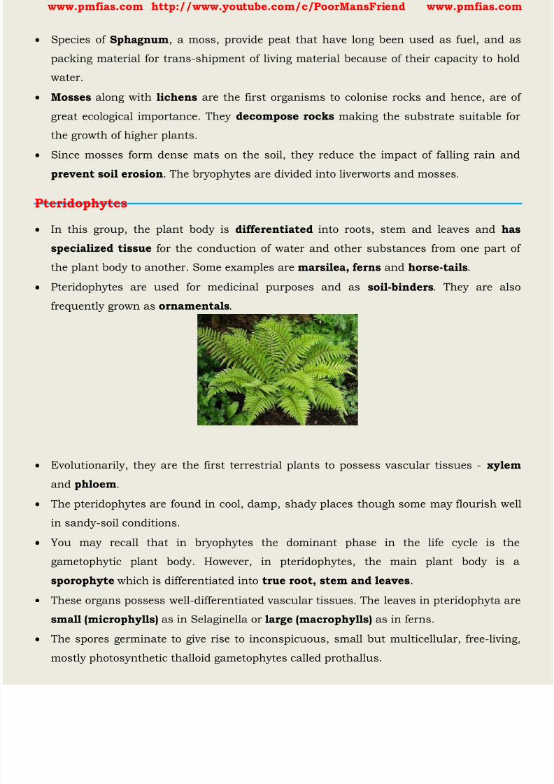
8/16/2019 Biology for Prelims Exam
http://slidepdf.com/reader/full/biology-for-prelims-exam 175/384
www.pmfias.com http://www.youtube.com/c/PoorMansFriend www.pmfias.com
Species of Sphagnum , a moss, provide peat that have long been used as fuel, and as
packing material for trans-shipment of living material because of their capacity to hold
water.
Mosses along with lichens are the first organisms to colonise rocks and hence, are of
great ecological importance. They decompose rocks making the substrate suitable for
the growth of higher plants. Since mosses form dense mats on the soil, they reduce the impact of falling rain and
prevent soil erosion . The bryophytes are divided into liverworts and mosses.
Pteridophytes
In this group, the plant body is differentiated into roots, stem and leaves and has
specialized tissue for the conduction of water and other substances from one part of
the plant body to another. Some examples are marsilea, ferns and horse-tails .
Pteridophytes are used for medicinal purposes and as soil-binders . They are also
frequently grown as ornamentals .
Evolutionarily, they are the first terrestrial plants to possess vascular tissues - xylem
and phloem .
The pteridophytes are found in cool, damp, shady places though some may flourish well
in sandy-soil conditions.
You may recall that in bryophytes the dominant phase in the life cycle is the
gametophytic plant body. However, in pteridophytes, the main plant body is a
sporophyte which is differentiated into true root, stem and leaves .
These organs possess well-differentiated vascular tissues. The leaves in pteridophyta are
small (microphylls) as in Selaginella or large (macrophylls) as in ferns.
The spores germinate to give rise to inconspicuous, small but multicellular, free-living,
mostly photosynthetic thalloid gametophytes called prothallus.
8/16/2019 Biology for Prelims Exam
http://slidepdf.com/reader/full/biology-for-prelims-exam 176/384
www.pmfias.com http://www.youtube.com/c/PoorMansFriend www.pmfias.com
These gametophytes require cool, damp, shady places to grow. Because of this specific
restricted requirement and the need for water for fertilisation, the spread of living
pteridophytes is limited and restricted to narrow geographical regions.
Cryptogamae
The thallophytes , the bryophytes and the pteridophytes have naked embryos thatare called spores .
The reproductive organs of plants in all these three groups are very inconspicuous, and
they are therefore called ‘ cryptogamae ’, or ‘those with hidden reproductive organs ’.
Kingdom Plantae – Phanerogams – Plants with Seeds: Gymnosperms, Angiosperms:
Monocots and Dicots or Dicotyledon and Monocotyledon.
Phanerogams – Plants with Seeds
Plants with well differentiated reproductive tissues that ultimately make seeds are called
phanerogams .
Seeds are the result of the reproductive process. They consist of the embryo along with
stored food, which serves for the initial growth of the embryo during germination.
This group is further classified, based on whether the seeds are naked or enclosed in
fruits, giving us two groups: gymnosperms and angiosperms .
Gymnosperms
This term is made from two greek words: gymno – means naked and sperma – means
seed.
The plants of this group bear naked seeds [ ovules are not enclosed by any ovary wall
and are usually perennial, evergreen and woody . The seeds that develop post-
fertilisation are naked too. Examples are pines , such as deodar.
Gymnosperms include medium-sized trees or tall trees and shrubs. One of the
gymnosperms, the giant redwood tree Seguoia is one of the tallest tree species.
The roots are generally tap roots {Plant Parts and Their Functions }. Roots in some
genera have fungal association in the form of mycorrhiza (Pinus) , while in some others
(Cgcas ) small specialised roots called coralloid roots are associated with N2-fixing
cyanobacteria.

8/16/2019 Biology for Prelims Exam
http://slidepdf.com/reader/full/biology-for-prelims-exam 177/384
www.pmfias.com http://www.youtube.com/c/PoorMansFriend www.pmfias.com
The leaves in gymnosperms are well-adapted to withstand extremes of temperature,
humidity and wind.
In conifers, the needle-like leaves reduce the surface area. Their thick cuticle and
sunken stomata also help to reduce water loss.
The gymnosperms are heterosporous ; they produce haploid microspores and
megaspores. The two kinds of spores are produced within sporangia that are borne on sporophylls
which are arranged spirally along an axis to form lax or compact strobili or cones .
The strobili bearing microsporophylls and microsporangia are called microsporangiate
or male strobili .
The microspores develop into a male gametophytic generation which is highly reduced
and is confined to only a limited number of cells. This reduced gametophyte is called a
pollen grain. The development of pollen grains take place within the microsporangia .
The cones bearing megasporophylls with ovules or megasporangia are called
macrosporangiate or female strobili .
The male or female cones or strobili may be borne on the same tree ( Pinus ). However, in
cycas male cones and megasporophylls are borne on different trees.
Unlike bryophytes and pteridophytes { Bryophytes – Pteridophytes }, in gymnosperms the
male and the female gametophytes do not have an independent free-living existence.
They remain within the sporangia retained on the sporophytes.
The pollen grain is released from the microsporangium. They are carried in air currents
and come in contact with the opening of the ovules borne on megasporophylls.
The pollen tube carrying the male gametes grows towards archegonia in the ovules and
discharge their contents near the mouth of the archegonia.
Following fertilisation, zygote develops into an embryo and the ovules into seeds. These
seeds are not covered .
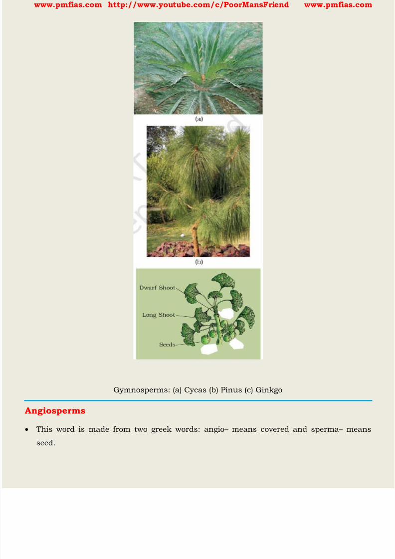
8/16/2019 Biology for Prelims Exam
http://slidepdf.com/reader/full/biology-for-prelims-exam 178/384
www.pmfias.com http://www.youtube.com/c/PoorMansFriend www.pmfias.com
Gymnosperms: (a) Cycas (b) Pinus (c) Ginkgo
Angiosperms
This word is made from two greek words: angio – means covered and sperma – means
seed.
8/16/2019 Biology for Prelims Exam
http://slidepdf.com/reader/full/biology-for-prelims-exam 179/384
www.pmfias.com http://www.youtube.com/c/PoorMansFriend www.pmfias.com
Unlike the gymnosperms where the ovules are naked, in the angiosperms or flowering
plants , the pollen grains and ovules are developed in specialised structures called
flowers .
The seeds develop inside an organ which is modified to become a fruit . These are also
called flowering plants .
The male sex organ in a flower is the stamen . Each stamen consists of a slenderfilament with an anther at the tip. The anthers, following Meiosis , produce pollen
grains .
The female sex organ in a flower is the pistil or the carpel . Pistil consists of an ovary
enclosing one to many ovules. Within ovules are present highly reduced female
gametophytes termed embryo-sacs. The embryo-sac formation is preceded by meiosis.
Hence, each of the cells of an embryo-sac is haploid .
Each embryo-sac has a three-celled egg apparatus - one egg cell and two synergids,three antipodal cells and two polar nuclei. The polar nuclei eventually fuse to produce a
diploid secondary nucleus.
Pollen grain, after dispersal from the anthers, are carried by wind or various other
agencies to the stigma of a pistil. This is termed as pollination .
The pollen grains germinate on the stigma and the resulting pollen tubes grow through
the tissues of stigma and style and reach the ovule.
The pollen tubes enter the embryo-sac where two male gametes are discharged. One of
the male gametes fuses with the egg cell to form a zygote (syngamy) .
The other male gamete fuses with the diploid secondary nucleus to produce the triploid
primary endosperm nucleus (PEN).
Because of the involvement of two fusions, this event is termed as double fertilisation
an event unique to angiosperms .

8/16/2019 Biology for Prelims Exam
http://slidepdf.com/reader/full/biology-for-prelims-exam 180/384
www.pmfias.com http://www.youtube.com/c/PoorMansFriend www.pmfias.com
Figure: Life cycle of an angiosperm
The zygote develops into an embryo (with one or two cotyledons) and the PEN develops
into endosperm which provides nourishment to the developing embryo.
The synergids and antipodals degenerate after fertilisation. During these events the
ovules develop into seeds and the ovaries develop into fruit.
Plant embryos in seeds have structures called cotyledons . Cotyledons are called ‘ seed
leaves ’ because in many instances they emerge and become green when the seed
germinates. Thus, cotyledons represent a bit of pre-designed plant in the seed.
Monocots and Dicots
The angiosperms are divided into two groups on the basis of the number of cotyledons
present in the seed.
Plants with seeds having a single cotyledon are called monocotyledonous or monocots
Plants with seeds having two cotyledons are called dicots.
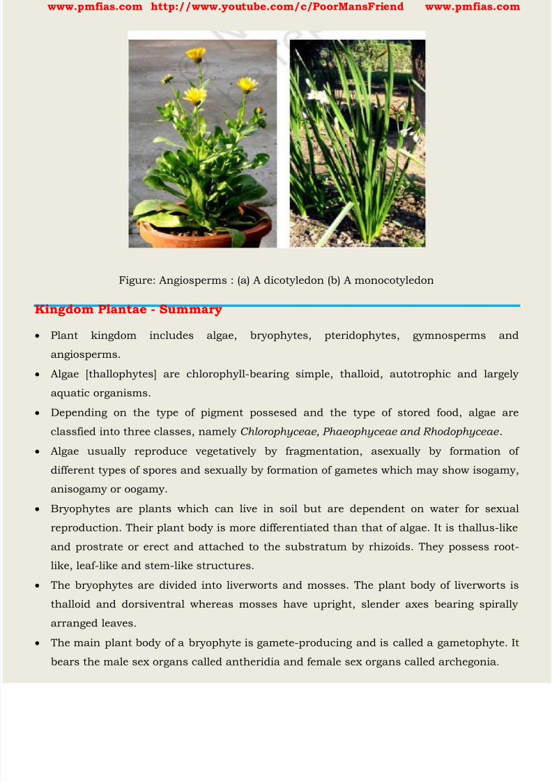
8/16/2019 Biology for Prelims Exam
http://slidepdf.com/reader/full/biology-for-prelims-exam 181/384
www.pmfias.com http://www.youtube.com/c/PoorMansFriend www.pmfias.com
Figure: Angiosperms : (a) A dicotyledon (b) A monocotyledon
Kingdom Plantae - Summary
Plant kingdom includes algae, bryophytes, pteridophytes, gymnosperms and
angiosperms.
Algae [thallophytes] are chlorophyll-bearing simple, thalloid, autotrophic and largely
aquatic organisms.
Depending on the type of pigment possesed and the type of stored food, algae areclassfied into three classes, namely Chlorophyceae, Phaeophyceae and Rhodophyceae .
Algae usually reproduce vegetatively by fragmentation, asexually by formation of
different types of spores and sexually by formation of gametes which may show isogamy,
anisogamy or oogamy.
Bryophytes are plants which can live in soil but are dependent on water for sexual
reproduction. Their plant body is more differentiated than that of algae. It is thallus-like
and prostrate or erect and attached to the substratum by rhizoids. They possess root-like, leaf-like and stem-like structures.
The bryophytes are divided into liverworts and mosses. The plant body of liverworts is
thalloid and dorsiventral whereas mosses have upright, slender axes bearing spirally
arranged leaves.
The main plant body of a bryophyte is gamete-producing and is called a gametophyte. It
bears the male sex organs called antheridia and female sex organs called archegonia.
8/16/2019 Biology for Prelims Exam
http://slidepdf.com/reader/full/biology-for-prelims-exam 182/384
www.pmfias.com http://www.youtube.com/c/PoorMansFriend www.pmfias.com
The male and female gametes produced fuse to form zygote which produces a
multicellular body called a sporophyte. It produces haploid spores. The spores germinate
to form gametophytes.
In pteridophytes the main plant is a sporophyte which is differentiated into true root,
stem and leaves. These organs possess well-differentiated vascular tissues. The
sporophytes bear sporangia which produce spores. The spores germinate to form gametophytes which require cool, damp places to grow.
The gametophytes bear male and female sex organs called antheridia and archegonia,
respectively. Water is required for transfer of male gametes to archegonium where zygote
is formed after fertilisation. The zygote produces a sporophyte.
The gymnosperms are the plants in which ovules are not enclosed by any ovary wall.
After fertilisation the seeds remain exposed and therefore these plants are called naked-
seeded plants. The gymnosperms produce microspores and megaspores which are produced in
microsporangia and megasporangia borne on the sporophylls. The sporophylls -
microsporophylls and megasporophylls - are arranged spirally on axis to form male and
female cones, respectively. The pollen grain germinates and pollen tube releases the
male gamete into the ovule, where it fuses with the egg cell in archegonia. Following
fertilisation, the zygote develops into embryo and the ovules into seeds.
In angiosperms, the male sex organs (stamen) and female sex organs (pistil) are borne in
a flower. Each stamen consists of a filament and an anther. The anther produces pollen
grains (male gametophyte) after meiosis. The pistil consists of an ovary enclosing one to
many ovules. Within the ovule is the female gametophyte or embryo sac which contains
the egg cell.
The pollen tube enters the embryo-sac where two male gametes are discharged. One
male gamete fuses with egg cell (syngamy) and other fuses with diploid secondary
nucleus (triple fusion). This phenomenon of two fusions is called double fertilisation and
is unique to angiosperms. The angiosperms are divided into two classes - the
dicotyledons and the monocotyledons.
During the life cycle of any sexually reproducing plant, there is alternation of
generations between gamete producing haploid gametophyte and spore producing
diploid sporophyte. However, different plant groups as well as individuals may show
different patterns of life cycles - haplontic, diplontic or intermediate.

8/16/2019 Biology for Prelims Exam
http://slidepdf.com/reader/full/biology-for-prelims-exam 183/384
www.pmfias.com http://www.youtube.com/c/PoorMansFriend www.pmfias.com
Match the following (column I with column II)
Column I Column II
Chlamydomonas Moss
Cycas Pteridophyte
Selaginella Algae
Sphagnum Gymnosperm

8/16/2019 Biology for Prelims Exam
http://slidepdf.com/reader/full/biology-for-prelims-exam 184/384
www.pmfias.com http://www.youtube.com/c/PoorMansFriend www.pmfias.com
Plant Tissue - Meristematic Tissue, Permanent Tissue - Simple Permanent Tissue:
Parenchyma, Chlorenchyma. Complex Permanent Tissue: Xylem, Phloem.
Plant Tissues
Plants are stationary or fixed – they don’t move. Most of the tissues they have are
supportive, which provides them with structural strength. Most of the plant tissues are dead, since dead cells can provide mechanical strength as
easily as live ones, and need less maintenance.
Animals on the other hand move around in search of food, mates and shelter. They
consume more energy as compared to plants. Most of the tissues they contain are living.
Another difference between animals and plants is in the pattern of growth. The growth
in plants is limited to certain regions, while this is not so in animals.
There are some tissues in plants that divide throughout their life. These tissues are
localised in certain regions.
Based on the dividing capacity of the tissues, various plant tissues can be classified as
growing or meristematic tissue and permanent tissue .
Cell growth in animals is more uniform. So, there is no such demarcation of dividing
and non-dividing regions in animals.
The structural organisation of organs and organ systems is far more specialised and
localised in complex animals than even in very complex plants. This fundamental
difference reflects the different modes of life pursued by these two major groups of
organisms, particularly in their different feeding methods.
Also, they are differently adapted for a sedentary existence on one hand (plants) and
active locomotion on the other (animals), contributing to this difference in organ system
design.
Meristematic Tissue
The growth of plants occurs only in certain specific regions. This is because the dividing
tissue , also known as meristematic tissue, is located only at these points.
Depending on the region where they are present, meristematic tissues are classified as
apical, lateral and intercalary .
New cells produced by meristem are initially like those of meristem itself, but as they
grow and mature, their characteristics slowly change and they become differentiated as
components of other tissues.

8/16/2019 Biology for Prelims Exam
http://slidepdf.com/reader/full/biology-for-prelims-exam 185/384
www.pmfias.com http://www.youtube.com/c/PoorMansFriend www.pmfias.com
1. Apical meristem is present at the growing tips of stems and roots and increases the
length of the stem and the root.
2. The girth of the stem or root increases due to lateral meristem (cambium) .
3. Intercalary meristem is the meristem at the base of the leaves or internodes (on
either side of the node) on twigs. As the cells of this tissue are very active, they have dense cytoplasm, thin cellulose
walls and prominent nuclei . They lack vacuoles .
Permanent Tissue
What happens to the cells formed by meristematic tissue? They take up a specific role
and lose the ability to divide. As a result, they form a permanent tissue.
This process of taking up a permanent shape, size, and a function is calleddifferentiation . Cells of meristematic tissue differentiate to form different types of
permanent tissue.
Simple Permanent Tissue
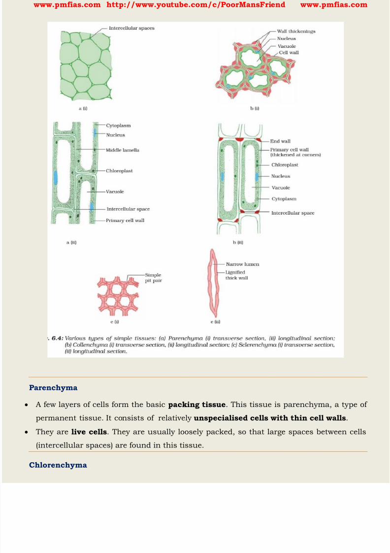
8/16/2019 Biology for Prelims Exam
http://slidepdf.com/reader/full/biology-for-prelims-exam 186/384
www.pmfias.com http://www.youtube.com/c/PoorMansFriend www.pmfias.com
Parenchyma
A few layers of cells form the basic packing tissue . This tissue is parenchyma, a type of
permanent tissue. It consists of relatively unspecialised cells with thin cell walls .
They are live cells . They are usually loosely packed, so that large spaces between cells
(intercellular spaces) are found in this tissue.
Chlorenchyma
8/16/2019 Biology for Prelims Exam
http://slidepdf.com/reader/full/biology-for-prelims-exam 187/384
www.pmfias.com http://www.youtube.com/c/PoorMansFriend www.pmfias.com
This tissue provides support to plants and also stores food . In some situations, it
contains chlorophyll and performs photosynthesis, and then it is called chlorenchyma.
Aerenchyma
In aquatic plants, large air cavities are present in parenchyma to give buoyancy to the
plants to help them float. Such a parenchyma type is called aerenchyma. The
parenchyma of stems and roots also stores nutrients and water.
Collenchyma
The flexibility in plants is due to another permanent tissue, collenchyma. It allows easy
bending in various parts of a plant (leaf, stem) without breaking. It also provides
mechanical support to plants. We can find this tissue in leaf stalks below the epidermis.
The cells of this tissue are living, elongated and irregularly thickened at the corners.
There is very little intercellular space .
Sclerenchyma
Yet another type of permanent tissue is sclerenchyma. It is the tissue which makes the
plant hard and stiff . We have seen the husk of a coconut. It is made of
sclerenchymatous tissue. The cells of this tissue are dead . They are long and narrow as
the walls are thickened due to lignin (a chemical substance which acts as cement and
hardens them). Often these walls are so thick that there is no internal space inside the
cell. This tissue is present in stems, around vascular bundles, in the veins of leaves and
in the hard covering of seeds and nuts. It provides strength to the plant parts.
Epidermis
What you observe is the outermost layer of cells, called epidermis. The epidermis is
usually made of a single layer of cells.
In some plants living in very dry habitats, the Epidermis may be thicker since protection
against water loss is critical. The entire surface of a plant has this outer covering of epidermis. It protects all the parts
of the plant.
Epidermal cells on the aerial parts of the plant often secrete a waxy, water-resistant
layer on their outer surface. This aids in protection against loss of water, mechanical
injury and invasion by parasitic fungi.
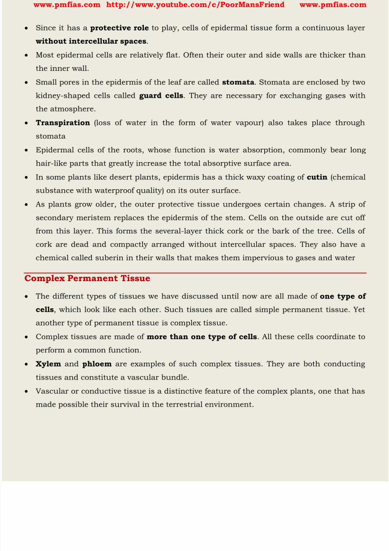
8/16/2019 Biology for Prelims Exam
http://slidepdf.com/reader/full/biology-for-prelims-exam 188/384
www.pmfias.com http://www.youtube.com/c/PoorMansFriend www.pmfias.com
Since it has a protective role to play, cells of epidermal tissue form a continuous layer
without intercellular spaces .
Most epidermal cells are relatively flat. Often their outer and side walls are thicker than
the inner wall.
Small pores in the epidermis of the leaf are called stomata . Stomata are enclosed by two
kidney-shaped cells called guard cells . They are necessary for exchanging gases withthe atmosphere.
Transpiration (loss of water in the form of water vapour) also takes place through
stomata
Epidermal cells of the roots, whose function is water absorption, commonly bear long
hair-like parts that greatly increase the total absorptive surface area.
In some plants like desert plants, epidermis has a thick waxy coating of cutin (chemical
substance with waterproof quality) on its outer surface. As plants grow older, the outer protective tissue undergoes certain changes. A strip of
secondary meristem replaces the epidermis of the stem. Cells on the outside are cut off
from this layer. This forms the several-layer thick cork or the bark of the tree. Cells of
cork are dead and compactly arranged without intercellular spaces. They also have a
chemical called suberin in their walls that makes them impervious to gases and water
Complex Permanent Tissue
The different types of tissues we have discussed until now are all made of one type of
cells , which look like each other. Such tissues are called simple permanent tissue. Yet
another type of permanent tissue is complex tissue.
Complex tissues are made of more than one type of cells . All these cells coordinate to
perform a common function.
Xylem and phloem are examples of such complex tissues. They are both conducting
tissues and constitute a vascular bundle.
Vascular or conductive tissue is a distinctive feature of the complex plants, one that has
made possible their survival in the terrestrial environment.
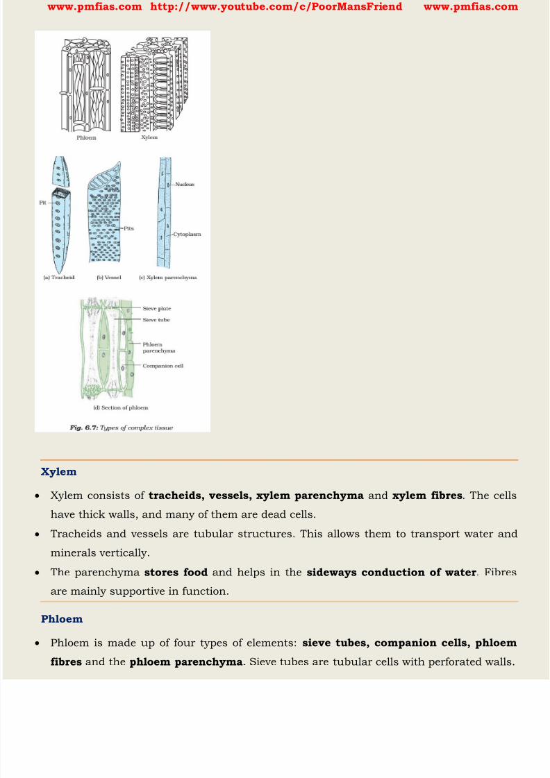
8/16/2019 Biology for Prelims Exam
http://slidepdf.com/reader/full/biology-for-prelims-exam 189/384
www.pmfias.com http://www.youtube.com/c/PoorMansFriend www.pmfias.com
Xylem
Xylem consists of tracheids, vessels, xylem parenchyma and xylem fibres . The cells
have thick walls, and many of them are dead cells.
Tracheids and vessels are tubular structures. This allows them to transport water and
minerals vertically.
The parenchyma stores food and helps in the sideways conduction of water . Fibres
are mainly supportive in function.
Phloem
Phloem is made up of four types of elements: sieve tubes, companion cells, phloem
fibres and the phloem parenchyma . Sieve tubes are tubular cells with perforated walls.
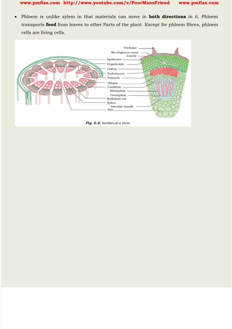
8/16/2019 Biology for Prelims Exam
http://slidepdf.com/reader/full/biology-for-prelims-exam 190/384
www.pmfias.com http://www.youtube.com/c/PoorMansFriend www.pmfias.com
Phloem is unlike xylem in that materials can move in both directions in it. Phloem
transports food from leaves to other Parts of the plant. Except for phloem fibres, phloem
cells are living cells.

8/16/2019 Biology for Prelims Exam
http://slidepdf.com/reader/full/biology-for-prelims-exam 191/384
www.pmfias.com http://www.youtube.com/c/PoorMansFriend www.pmfias.com
Nutrition in Plants - Photosynthesis, Saprotrophs, Symbiosis, Parasites, Macronutrients -
Micronutrients in Plants, Nitrogen Cycle, Nitrogen Fixation.
Source | Credits | Picture Credits: NCERT General Science
Nutrition in Plants
Nutrition is the mode of taking food by an organism and its utilization by the body. The mode of nutrition in which organisms make food themselves from simple
substances is called autotrophic (auto = self; trophos = nourishment) nutrition.
Therefore, plants are called autotrophs.
Animals and most other organisms take in readymade food prepared by the plants. They
are called heterotrophs (heteros = other) .
Insectivorous plants have both autotrophic and heterotrophic mode of nutrition.
Photosynthesis – Food Making Process In Plants
Carbon dioxide from air is taken in through the tiny pores present on the surface of the
leaves. These pores are surrounded by ‘guard cells’ . Such pores are called stomata .
Water and minerals are transported to the leaves by the vessels which run like pipes
throughout the root, the stem, the branches and the leaves. They form a continuous
path or passage for the nutrients to reach the leaf.
The leaves have a green pigment called chlorophyll . It helps leaves to capture theenergy of the sunlight. This energy is used to synthesise (prepare) food from carbon
dioxide and water . Since the synthesis of food occurs in the presence of sunlight, it is
called photosynthesis (Photo: light; synthesis: to combine) .
So we find that chlorophyll, sunlight, carbon dioxide and water are necessary to carry
out the process of photosynthesis. Thus, sun is the ultimate source of energy for all
living organisms.
Besides leaves, photosynthesis also takes place in other green parts of the plant — in
green stems and green branches .
The desert plants have scale or spine-like leaves to reduce loss of water by
transpiration. These plants have green stems which carry out photosynthesis.
During photosynthesis, chlorophyll containing cells of leaves, in the presence of
sunlight, use carbon dioxide and water to synthesise carbohydrates . The carbohydrates
ultimately get converted into starch .
8/16/2019 Biology for Prelims Exam
http://slidepdf.com/reader/full/biology-for-prelims-exam 192/384
www.pmfias.com http://www.youtube.com/c/PoorMansFriend www.pmfias.com
The leaves other than green also have chlorophyll. The large amount of red, brown and
other pigments mask the green colour. Photosynthesis takes place in these leaves also.
Chlorophyll Pigments
Within the leaves, the mesophyll cells have a large number of chloroplasts that are
responsible for CO2 fixation. A chromatographic separation of the leaf pigments shows that the colour that we see in
leaves is not due to a single pigment but due to four pigments: Chlorophyll a (bright or
blue green in the chromatogram), chlorophyll b (yellow green ), xanthophylls (yellow
and carotenoids (yellow to yellow-orange ).
Light Reactions and Dark Reactions
Within the chloroplast there is the membranous system consisting of grana, the stroma
lamellae, and the fluid stroma. There is a clear division of labour within the chloroplast.
The membrane system is responsible for trapping the light energy and also for the
synthesis of ATP and NADPH . [ light reactions ]
In the light reaction the light energy is absorbed by the pigments present in the antenna,
and funnelled to special chlorophyll a molecules called reaction centre chlorophylls.
In stroma, enzymatic reactions incorporate CO2 [chemosynthetic pathway- carbon fixing
reactions] into the plant leading to the synthesis of sugar , which in turn forms starch
[dark reactions ]
The former set of reactions, since they are directly light driven are called light
reactions . The latter are not directly light driven but are dependent on the products of
light reactions (ATP and NADPH). Hence, to distinguish the latter they are called, by
convention, as dark reactions . However, this should not be construed to mean that they
occur in darkness or that they are not lightdependent.

8/16/2019 Biology for Prelims Exam
http://slidepdf.com/reader/full/biology-for-prelims-exam 193/384
www.pmfias.com http://www.youtube.com/c/PoorMansFriend www.pmfias.com
Factors Affecting Photosynthesis
Law of Limiting Factors : If a chemical process is affected by more than one factor, then
its rate will be determined by the factor which is nearest to its minimal value : it is the
factor which directly affects the process if its quantity is changed.
For example, despite the presence of a green leaf and optimal light and CO2 conditions,
the plant may not photosynthesise if the temperature is very low. This leaf, if given the
optimal temperature, will start photosynthesising.
Carbon dioxide Concentration The concentration of CO 2 is very low in the atmosphere (between 0.03 and 0.04 per
cent). Increase in concentration upto 0.05 per cent can cause an increase in CO 2
fixation rates; beyond this the levels can become damaging over longer periods.
Temperature
The dark reactions being enzymatic are temperature controlled . Though the light
reactions are also temperature sensitive they are affected to a much lesser extent.
The temperature optimum for photosynthesis of different plants also depends on the
habitat that they are adapted to. Tropical plants have a higher temperature optimum
than the plants adapted to temperate climates.
Water
Water stress causes the stomata to close hence reducing the CO 2 availability.
8/16/2019 Biology for Prelims Exam
http://slidepdf.com/reader/full/biology-for-prelims-exam 194/384
www.pmfias.com http://www.youtube.com/c/PoorMansFriend www.pmfias.com
Besides, water stress also makes leaves wilt, thus, reducing the surface area ofthe leaves
and their metabolic activity as well.
Light
There is a linear relationship between incident light and CO2 fixation rates at low light
intensities.
At higher light intensities, gradually the rate does not show further increase as other
factors become limiting.
Saprotrophs – Fungi
Fungi have a different mode of nutrition. They secrete digestive juices on the dead and
decaying matter and convert it into a solution. Then they absorb the nutrients from it.
This mode of nutrition in which organisms take in nutrients in solution form from dead
and decaying matter is called saprotrophic nutrition.
Plants which use saprotrophic mode of nutrition are called saprotrophs. Mushrooms are
best example.
Symbiosis
Some organisms live together and share shelter and nutrients. This is called symbiotic
relationship. For example, certain fungi live in the roots of trees. The tree provides
nutrients to the fungus and, in return, receives help from it to take up water andnutrients from the soil.
Plants absorb mineral nutrients from the soil. So, their amounts in the soil keep on
declining. Fertilisers and manures contain plant nutrients such as nitrogen, potassium,
phosphorous, etc.
Usually crops require a lot of nitrogen to make proteins. After the harvest, the soil
becomes deficient in nitrogen.
Though nitrogen gas is available in plenty in the air, plants cannot use it in the mannerthey can use carbon dioxide. They need nitrogen in a soluble form.
The bacterium called Rhizobium can take atmospheric nitrogen and converts it into a
soluble form. But Rhizobium cannot make its own food. So it lives in the roots of gram,
peas, moon beans and other legumes and provides them with nitrogen (symbiosis).
Most of the pulses (dals) are obtained from leguminous plants . In return, the plants
provide food and shelter to the bacteria. They have a symbiotic relationship.
8/16/2019 Biology for Prelims Exam
http://slidepdf.com/reader/full/biology-for-prelims-exam 195/384
www.pmfias.com http://www.youtube.com/c/PoorMansFriend www.pmfias.com
Parasites
A parasite is an organism which lives in or on another organism and benefits by deriving
nutrients at the other's expense.
Plants like cuscuta are parasites. They take food from the host plant.
Amarbelis an example of: (i) autotroph (ii) parasite (iii) saprotroph (iv) host
Mineral Nutrition
The basic needs of all living organisms are essentially the same. They require
macromolecules, such as carbohydrates, proteins and fats, and water and minerals for
their growth and development.
Only a few elements have been found to be absolutely essential for plant growth and
metabolism. These elements are further divided into two broad categories based on their
quantitative requirements.
Macronutrients and Micronutrients in Plants
Macronutrients are generally present in plant tissues in large amounts. The
macronutrients include
1. Carbon
2. Hydrogen
3. Oxygen
4. Nitrogen
5. Phosphorous
6. Sulphur
7. Potassium
8. Calcium and
9. Magnesium .
Micronutrients or trace elements, are needed in very small amounts. These include1. Iron
2. Manganese
3. Copper
4. Molybdenum
5. Zinc
6. Boron
8/16/2019 Biology for Prelims Exam
http://slidepdf.com/reader/full/biology-for-prelims-exam 196/384
www.pmfias.com http://www.youtube.com/c/PoorMansFriend www.pmfias.com
7. Chlorine and
8. Nickel .
In addition to the 17 essential elements named above, there are some beneficial
elements such as
1. Sodium
2. Silicon3. Cobalt and
4. Selenium . They are required by higher plants.
Nitrogen
Nitrogen is required by all parts of a plant, particularly the meristematic tissues and
the metabolically active cells.
Nitrogen is one of the major constituents of proteins, nucleic acids, vitamins and
hormones.
Phosphorus
Phosphorus is a constituent of cell membranes , certain proteins, all nucleic acids and
nucleotides, and is required for all phosphorylation reactions.
Potassium
In plants, this is required in more abundant quantities in the meristematic tissues,
buds, leaves and root tips .
Potassium helps to maintain an anion-cation balance in cells and is involved in protein
synthesis, opening and closing of stomata, activation of enzymes and in the
maintenance of the turgidity of cells.
Calcium
Calcium is required by meristematic and differentiating tissues. During cell division it is
used in the synthesis of cell wall , particularly as calcium pectate in the middle lamella.
It accumulates in older leaves. It is involved in the normal functioning of the cell
membranes. It activates certain enzymes and plays an important role in regulating
metabolic activities.
Magnesium
8/16/2019 Biology for Prelims Exam
http://slidepdf.com/reader/full/biology-for-prelims-exam 197/384
www.pmfias.com http://www.youtube.com/c/PoorMansFriend www.pmfias.com
It activates the enzymes of respiration, photosynthesis and are involved in the synthesis
of DNA and RNA .
Nitrogen Cycle
Apart from carbon, hydrogen and oxygen, nitrogen is the most prevalent element in
living organisms. Nitrogen is a constituent of amino acids, proteins, hormones, chlorophylls and many of
the vitamins.
Plants compete with microbes for the limited nitrogen that is available in soil. Thus,
nitrogen is a limiting nutrient for both natural and agricultural eco-systems.
Nitrogen exists as two nitrogen atoms joined by a very strong triple covalent bond (N ≡
N). The process of conversion of nitrogen (N2) to ammonia is termed as nitrogen-
fixation.
In nature, lightning and ultraviolet radiation provide enough energy to convert
nitrogen to nitrogen oxides (NO, NO2, N2O).
Industrial combustions, forest fires, automobile exhausts and power-generating stations
are also sources of atmospheric nitrogen oxides.
Decomposition of organic nitrogen of dead plants and animals into ammonia is called
ammonification .
Some of this ammonia volatilises and re-enters the atmosphere but most of it is
converted into nitrate by soil bacteria.
Ammonia is first oxidised to nitrite by the bacteria Nitrosomonas and/or Nitrococcus .
The nitrite is further oxidised to nitrate with the help of the bacterium Nitrobacter
These steps are called nitrification . These nitrifying bacteria are chemoautotrophs .
The nitrate thus formed is absorbed by plants and is transported to the leaves. In leaves,
it is reduced to form ammonia that finally forms the amine group of amino acids .
Nitrate present in the soil is also reduced to nitrogen by the process of denitrification
Denitrification is carried by bacteria Pseudomonas and Thiobacillus .
The enzyme, nitrogenase which is capable of nitrogen reduction is present exclusively
in prokaryotes . Such microbes are called N2-fixers .
The nitrogen-fixing microbes could be free-living or symbiotic. Examples of free-living
nitrogen-fixing aerobic microbes are Azotobacter and Beijemickia while
Rhodospirillum is anaerobic and Bacillus free-living.

8/16/2019 Biology for Prelims Exam
http://slidepdf.com/reader/full/biology-for-prelims-exam 198/384
www.pmfias.com http://www.youtube.com/c/PoorMansFriend www.pmfias.com
In addition, a number of cyanobacteria such as Anabaena and Nostoc are also free-
living nitrogen-fixers.

8/16/2019 Biology for Prelims Exam
http://slidepdf.com/reader/full/biology-for-prelims-exam 199/384
www.pmfias.com http://www.youtube.com/c/PoorMansFriend www.pmfias.com

8/16/2019 Biology for Prelims Exam
http://slidepdf.com/reader/full/biology-for-prelims-exam 200/384
www.pmfias.com http://www.youtube.com/c/PoorMansFriend www.pmfias.com
Symbiotic Biological Nitrogen Fixation Several types of symbiotic biological nitrogen fixing associations are known. The most
prominent among them is the legume-bacteria relationship.
Species of rod-shaped Rhizobium has such relationship with the roots of several
legumes such as alfalfa, sweet clover, sweet pea, lentils, garden pea, broad bean,
clover beans , etc.
The most common association on roots is as nodules . These nodules are small
outgrowths on the roots. The microbe, Frankia, also produces nitrogen-fixing nodules on
the roots of nonleguminous plants (e.g., Alnus).
Both Rhizobium and Frankia . are free- living in soil, but as symbionts, can fix
atmospheric nitrogen.
Summary
Plants obtain their inorganic nutrients from air, water and soil.
8/16/2019 Biology for Prelims Exam
http://slidepdf.com/reader/full/biology-for-prelims-exam 201/384
www.pmfias.com http://www.youtube.com/c/PoorMansFriend www.pmfias.com
Plants absorb a wide variety of mineral elements.
Not all the mineral elements that they absorb are required by plants.
Out of the more than 105 elements discovered so far, less than 21 are essential and
beneficial for normal plant growth and development.
The elements required in large quantities are called macronutrients while those required
in less quantities or in trace are termed as micronutrients. These elements are either essential constituents of proteins, carbohydrates, fats, nucleic
acid etc., and/or take part in various metabolic processes.
Deficiency of each of these essential elements may lead to symptoms called deficiency
symptoms.
Chlorosis, necrosis, stunted growth, impaired cell division , etc., are some prominent
deficiency symptoms.
Plants absorb minerals through roots by either passive or active processes. They arecarried to all parts of the organism through xylem along with water transport.
Nitrogen is very essential for the sustenance of life. Plants cannot use atmospheric
nitrogen directly. But some of the plants in association with N2-fixing bacteria,
especially roots of legumes, can fix this atmospheric nitrogen into biologically usable
forms. Nitrogen fixation requires a strong reducing agent and energy in the form of ATP.
N2-fixation is accomplished with the help of nitrogenfixing microbes, mainly Rhizobium.
The enzyme nitrogenase which plays an important role in biological N2 fixation is verysensitive to oxygen . Most of the processes take place in anaerobic environment.
The energy, ATP, required is provided by the respiration of the host cells. Ammonia
produced following N2 fixation is incorporated into amino acids as the amino group.

8/16/2019 Biology for Prelims Exam
http://slidepdf.com/reader/full/biology-for-prelims-exam 202/384
www.pmfias.com http://www.youtube.com/c/PoorMansFriend www.pmfias.com
Sexual and Asexual Reproduction in Plants – Asexual Reproduction: Vegetative
Propagation, Budding, Spores – Sexual Reproduction: Unisexual, Bisexual.
Modes Of Reproduction in Plants
There are several ways by which plants produce their offspring. These are categorised
into two types: (i) asexual , and (ii) sexual reproduction. In asexual reproduction plants can give rise to new plants without seeds , whereas in
sexual reproduction, new plants are obtained from seeds.
Asexual Reproduction in Plants
Asexual reproduction occurs through:
1. Vegetative Propagation,
2. Budding,3. Fragmentation and
4. Spore formation.
Vegetative Propagation
Most plants have roots, stems and leaves. These are called the vegetative parts of a
plant.
Vegetative Propagation is a type of asexual reproduction in which new plants are
produced from roots, stems, leaves and buds. Since reproduction is through the
vegetative parts of the plant, it is known as vegetative propagation .
Bryophyllum (sprout leaf plant) has buds in the margins of leaves. If a leaf of this
plant falls on a moist soil, each bud can give rise to a new plant.
Bryophyllum (sprout leaf plant)

8/16/2019 Biology for Prelims Exam
http://slidepdf.com/reader/full/biology-for-prelims-exam 203/384
www.pmfias.com http://www.youtube.com/c/PoorMansFriend www.pmfias.com
The roots of some plants can also give rise to new plants. Sweet potato and dahlia are
examples.
Plants such as cacti produce new plants when their parts get detached from the main
plant body. Each detached part can grow into a new plant.
Plants produced by vegetative propagation take less time to grow and bear flowers and
fruits earlier than those produced from seeds.
The new plants are exact copies of the parent plant, as they are produced from a singleparent .
Budding
Yeast is a single-celled organism. The small bulb-like projection coming out from the
yeast cell is called a bud.
The bud gradually grows and gets detached from the parent cell and forms a new yeast
cell.
The new yeast cell grows, matures and produces more yeast cells. If this process
continues, a large number of yeast cells are produced in a short time.

8/16/2019 Biology for Prelims Exam
http://slidepdf.com/reader/full/biology-for-prelims-exam 204/384
www.pmfias.com http://www.youtube.com/c/PoorMansFriend www.pmfias.com
Fragmentation
When water and nutrients are available algae grow and multiply rapidly by
fragmentation. An alga breaks up into two or more fragments. These fragments or pieces grow into new
individuals. This process continues and they cover a large area in a short period of time.
Spore formation
Fungi on a bread piece grow from spores which are present in the air. When spores are
released they keep floating in the air.

8/16/2019 Biology for Prelims Exam
http://slidepdf.com/reader/full/biology-for-prelims-exam 205/384
www.pmfias.com http://www.youtube.com/c/PoorMansFriend www.pmfias.com
The spores are asexual reproductive bodies. As they are very light, they can cover long
distances.
Each spore is covered by a hard protective coat to withstand unfavorable conditions
such as high temperature and low humidity. So they can survive for a long time.
Under favourable conditions, a spore germinates and develops into a new individual.
Plants such as moss and ferns also reproduce by means of spores.
Sexual Reproduction in Plants – Unisexual and Bisexual
The flowers are the reproductive parts of a plant. The stamens are the male
reproductive part and the pistil is the female reproductive part.
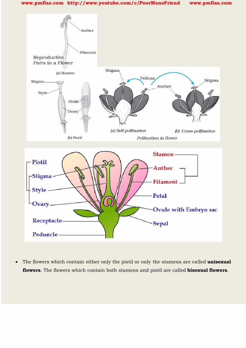
8/16/2019 Biology for Prelims Exam
http://slidepdf.com/reader/full/biology-for-prelims-exam 206/384
www.pmfias.com http://www.youtube.com/c/PoorMansFriend www.pmfias.com
The flowers which contain either only the pistil or only the stamens are called unisexual
flowers . The flowers which contain both stamens and pistil are called bisexual flowers .

8/16/2019 Biology for Prelims Exam
http://slidepdf.com/reader/full/biology-for-prelims-exam 207/384
www.pmfias.com http://www.youtube.com/c/PoorMansFriend www.pmfias.com
Corn, papaya and cucumber produce unisexual flowers, whereas mustard, rose and
petunia have bisexual flowers.
Both the male and the female unisexual flowers may be present in the same plant or in
different plants.
Anther contains pollen grains which produce male gametes . A pistil consists of stigma,
style and ovary. The ovary contains one or more ovules . The female gamete or the egg is
formed in an ovule. In sexual reproduction a male and a female gamete fuse to form a
zygote .
Pollination
Generally pollen grains have a tough protective coat which prevents them from drying
up. Since pollen grains are light, they can be carried by wind or water. Insects visit
flowers and carry away pollen on their bodies.
Some of the pollen lands on the stigma of a flower of the same kind. The transfer of
pollen from the anther to the stigma of a flower is called pollination . If the pollen lands on the stigma of the same flower it is called self-pollination . When
the pollen of a flower lands on the stigma of another flower of the same plant, or that of
a different plant of the same kind, it is called cross-pollination .
Fertilisation
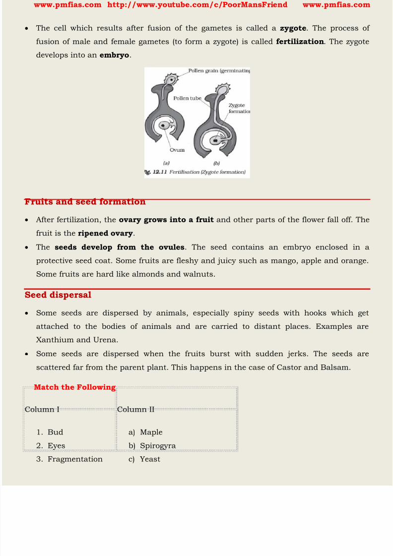
8/16/2019 Biology for Prelims Exam
http://slidepdf.com/reader/full/biology-for-prelims-exam 208/384
www.pmfias.com http://www.youtube.com/c/PoorMansFriend www.pmfias.com
The cell which results after fusion of the gametes is called a zygote . The process of
fusion of male and female gametes (to form a zygote) is called fertilization . The zygote
develops into an embryo .
Fruits and seed formation
After fertilization, the ovary grows into a fruit and other parts of the flower fall off. The
fruit is the ripened ovary .
The seeds develop from the ovules . The seed contains an embryo enclosed in a
protective seed coat. Some fruits are fleshy and juicy such as mango, apple and orange.
Some fruits are hard like almonds and walnuts.
Seed dispersal
Some seeds are dispersed by animals, especially spiny seeds with hooks which get
attached to the bodies of animals and are carried to distant places. Examples are
Xanthium and Urena.
Some seeds are dispersed when the fruits burst with sudden jerks. The seeds are
scattered far from the parent plant. This happens in the case of Castor and Balsam.
Match the Following
Column I Column II
1. Bud
2. Eyes
3. Fragmentation
a) Maple
b) Spirogyra
c) Yeast
8/16/2019 Biology for Prelims Exam
http://slidepdf.com/reader/full/biology-for-prelims-exam 209/384
www.pmfias.com http://www.youtube.com/c/PoorMansFriend www.pmfias.com
4. Wings
5. Spores
d) Bread mould
e) Potato
f) Rose
Asexual reproduction vs Sexual reproduction
Asexual reproduction Sexual reproduction
a) Only one parent plant is
involved.
a) Both male and female parents are
involved.
b) Occurs in unisexual plants. b) Occurs in bisexual plants.
c) Occurs in lower plants.c) Occurs in higher plants.
d) Reproductive organs are not
present.
d) Fully developed reproductive parts
are present
e) In most of the methods the
original parent disappears.
e) Original parents remain alive after
process of reproduction.
f)
Process like gamete formation orfertilization is not seen.
f) Fertilization of gametes give rises to
zygote.
g) Characteristics of only one
parent is inherited.
g) Characteristics of both parents are
inherited.
h) No need of seeds.h) Seeds are used to get new plants
from a flower.
Self-Pollination Cross-Pollination
1. Pollen grains are transferred to the
stigma of the same flower.
1. Pollen grains are carried to
stigma of another flower.

8/16/2019 Biology for Prelims Exam
http://slidepdf.com/reader/full/biology-for-prelims-exam 210/384
www.pmfias.com http://www.youtube.com/c/PoorMansFriend www.pmfias.com
2. Occurs in bisexual plants having
anther and stigma maturing at
same time.
3. It takes place in plants like wheat,
peas etc.
2. Occurs in bisexual flowers
having anther and stigma
maturing at different times.
3. It takes place in plants like
lady- finger, tomato, brinjal etc.
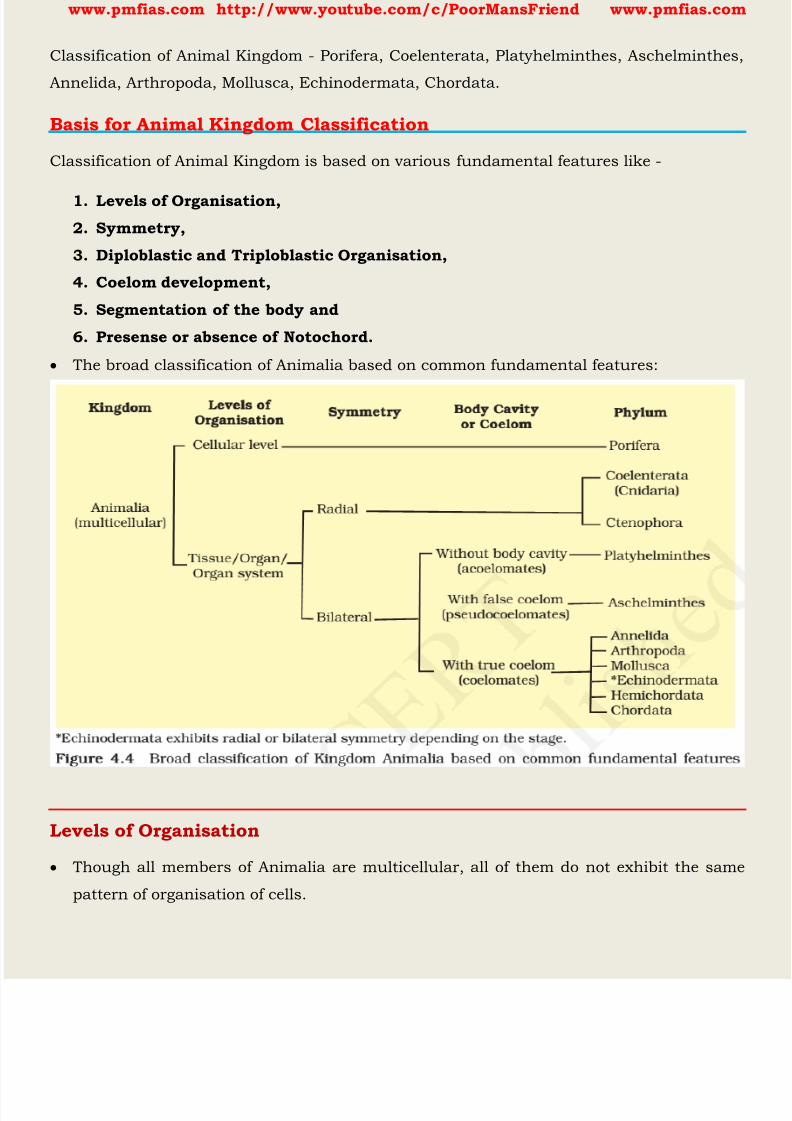
8/16/2019 Biology for Prelims Exam
http://slidepdf.com/reader/full/biology-for-prelims-exam 211/384
www.pmfias.com http://www.youtube.com/c/PoorMansFriend www.pmfias.com
Classification of Animal Kingdom - Porifera, Coelenterata, Platyhelminthes, Aschelminthes,
Annelida, Arthropoda, Mollusca, Echinodermata, Chordata.
Basis for Animal Kingdom Classification
Classification of Animal Kingdom is based on various fundamental features like -
1. Levels of Organisation,
2. Symmetry,
3. Diploblastic and Triploblastic Organisation,
4. Coelom development,
5. Segmentation of the body and
6. Presense or absence of Notochord.
The broad classification of Animalia based on common fundamental features:
Levels of Organisation
Though all members of Animalia are multicellular, all of them do not exhibit the same
pattern of organisation of cells.
8/16/2019 Biology for Prelims Exam
http://slidepdf.com/reader/full/biology-for-prelims-exam 212/384
www.pmfias.com http://www.youtube.com/c/PoorMansFriend www.pmfias.com
For example, in sponges, the cells are arranged as loose cell aggregates, i.e., they exhibit
cellular level of organisation . Some division of labour (activities) occur among the
cells.
In coelenterates, the arrangement of cells is more complex. Here the cells performing the
same function are arranged into tissues, hence is called tissue level of organisation .
A still higher level of organisation, i.e., organ level [ organ level of organisation ] isexhibited by members of Platyhelminthes and other higher phyla where tissues are
grouped together to form organs, each specialised for a particular function.
In animals like Annelids, Arthropods, Molluscs, Echinoderms and Chordates, organs
have associated to form functional systems, each system concerned with a specific
physiological function. This pattern is called organ system level of organisation .
Organ systems in different groups of animals exhibit various patterns of complexities.
For example, the digestive system in Platyhelminthes (incomplete digestive system)has only a single opening to the outside of the body that serves as both mouth and
anus, and is hence called incomplete. A complete digestive system has two openings,
mouth and anus.
Similarly, the circulatory system may be of two types: open type in which the blood is
pumped out of the heart and the cells and tissues are directly bathed in it and closed
type in which the blood is circulated through a series of vessels of varying diameters
(arteries, veins and capillaries).
Symmetry
Animals can be categorised on the basis of their symmetry.
Sponges are mostly asymmetrical, i.e., any plane that passes through the centre does
not divide them into equal halves.
When any plane passing through the central axis of the body divides the organism into
two identical halves, it is called radial symmetry . Coelenterates, Ctenophores and
Echinoderms have this kind of body plan.
Animals like Annelids, Arthropods, etc., where the body can be divided into identical left
and right halves in only one plane, exhibit bilateral symmetry .

8/16/2019 Biology for Prelims Exam
http://slidepdf.com/reader/full/biology-for-prelims-exam 213/384
www.pmfias.com http://www.youtube.com/c/PoorMansFriend www.pmfias.com
Diploblastic and Triploblastic Organisation
Animals in which the cells are arranged in two embryonic layers, an external ectoderm
and an internal endoderm , are called diploblastic animals , e.g., Coelenterates. An
undifferentiated layer, mesoglea , is present in between the ectoderm and the endoderm.
Those animals in which the developing embryo has a third germinal layer, mesoderm ,
in between the ectoderm and endoderm, are called triploblastic animals
(platyhelminthes to chordates).
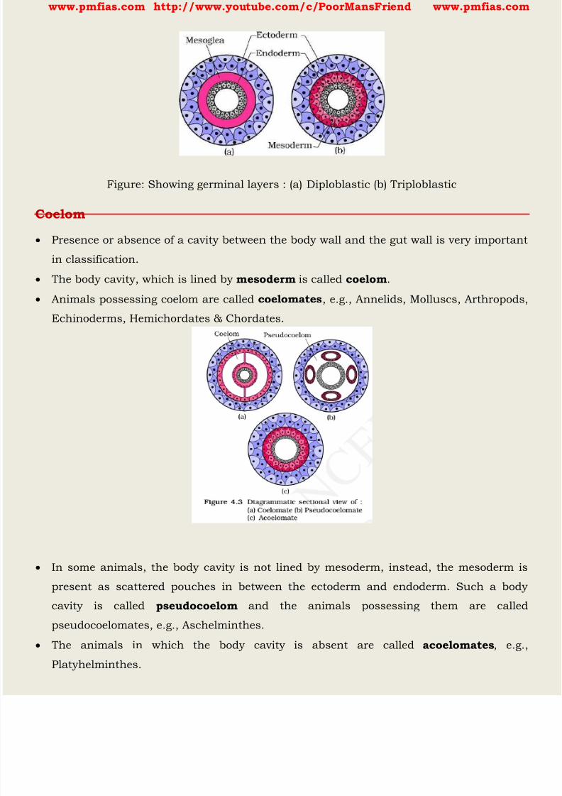
8/16/2019 Biology for Prelims Exam
http://slidepdf.com/reader/full/biology-for-prelims-exam 214/384
www.pmfias.com http://www.youtube.com/c/PoorMansFriend www.pmfias.com
Figure: Showing germinal layers : (a) Diploblastic (b) Triploblastic
Coelom
Presence or absence of a cavity between the body wall and the gut wall is very important
in classification.
The body cavity, which is lined by mesoderm is called coelom . Animals possessing coelom are called coelomates , e.g., Annelids, Molluscs, Arthropods,
Echinoderms, Hemichordates & Chordates.
In some animals, the body cavity is not lined by mesoderm, instead, the mesoderm is
present as scattered pouches in between the ectoderm and endoderm. Such a body
cavity is called pseudocoelom and the animals possessing them are called
pseudocoelomates, e.g., Aschelminthes.
The animals in which the body cavity is absent are called acoelomates , e.g.,
Platyhelminthes.
8/16/2019 Biology for Prelims Exam
http://slidepdf.com/reader/full/biology-for-prelims-exam 215/384
www.pmfias.com http://www.youtube.com/c/PoorMansFriend www.pmfias.com
Segmentation
In some animals, the body is externally and internally divided into segments with a
serial repetition of at least some organs.
For example, in earthworm, the body shows this pattern called metameric
segmentation and the phenomenon is known as metamerism .
Notochord
Notochord is a mesodermally [ the middle layer of cells or tissues of an embryo, or the parts derived from this
(e.g. cartilage, muscles, and bone) ] derived rod-like structure formed on the dorsal side
[posterior] during embryonic development in some animals.
Animals with notochord are called chordates and those animals which do not form this
structure are called non-chordates, e.g., Porifera to Echinoderms.
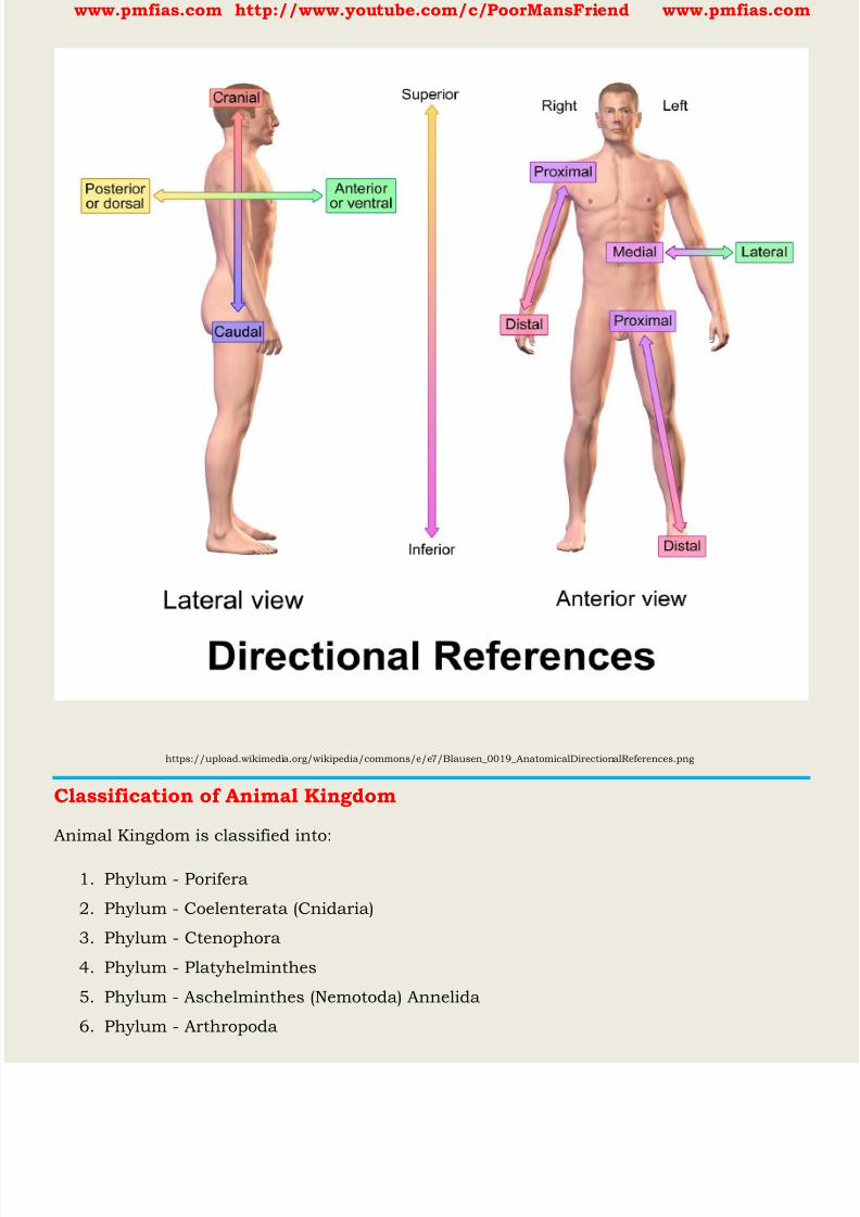
8/16/2019 Biology for Prelims Exam
http://slidepdf.com/reader/full/biology-for-prelims-exam 216/384
www.pmfias.com http://www.youtube.com/c/PoorMansFriend www.pmfias.com
https://upload.wikimedia.org/wikipedia/commons/e/e7/Blausen_0019_AnatomicalDirectionalReferences.png
Classification of Animal Kingdom
Animal Kingdom is classified into:
1. Phylum - Porifera
2. Phylum - Coelenterata (Cnidaria)
3. Phylum - Ctenophora
4. Phylum - Platyhelminthes
5. Phylum - Aschelminthes (Nemotoda) Annelida
6. Phylum - Arthropoda
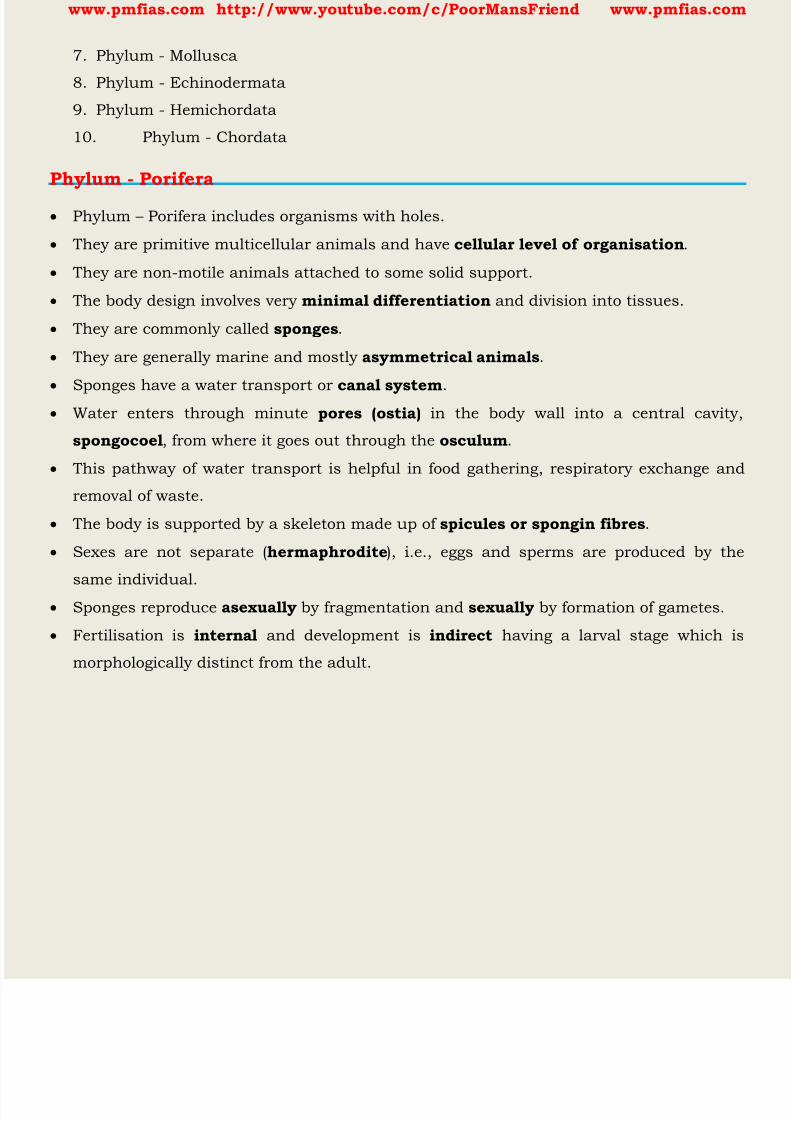
8/16/2019 Biology for Prelims Exam
http://slidepdf.com/reader/full/biology-for-prelims-exam 217/384
www.pmfias.com http://www.youtube.com/c/PoorMansFriend www.pmfias.com
7. Phylum - Mollusca
8. Phylum - Echinodermata
9. Phylum - Hemichordata
10. Phylum - Chordata
Phylum - Porifera
Phylum – Porifera includes organisms with holes.
They are primitive multicellular animals and have cellular level of organisation .
They are non-motile animals attached to some solid support.
The body design involves very minimal differentiation and division into tissues.
They are commonly called sponges .
They are generally marine and mostly asymmetrical animals .
Sponges have a water transport or canal system . Water enters through minute pores (ostia) in the body wall into a central cavity,
spongocoel , from where it goes out through the osculum .
This pathway of water transport is helpful in food gathering, respiratory exchange and
removal of waste.
The body is supported by a skeleton made up of spicules or spongin fibres .
Sexes are not separate ( hermaphrodite ), i.e., eggs and sperms are produced by the
same individual.
Sponges reproduce asexually by fragmentation and sexually by formation of gametes.
Fertilisation is internal and development is indirect having a larval stage which is
morphologically distinct from the adult.
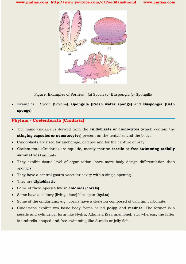
8/16/2019 Biology for Prelims Exam
http://slidepdf.com/reader/full/biology-for-prelims-exam 218/384
www.pmfias.com http://www.youtube.com/c/PoorMansFriend www.pmfias.com
Figure: Examples of Porifera : (a) Sycon (b) Euspongia (c) Spongilla
Examples: Sycon (Scypha), Spongilla (Fresh water sponge) and Euspongia (Bath
sponge) .
Phylum - Coelenterata (Cnidaria)
The name cnidaria is derived from the cnidoblasts or cnidocytes (which contain the
stinging capsules or nematocytes ) present on the tentacles and the body.
Cnidoblasts are used for anchorage, defense and for the capture of prey.
Coelenterata (Cnidaria) are aquatic, mostly marine sessile or free-swimming radially
symmetrical animals.
They exhibit tissue level of organization [have more body design differentiation than
sponges].
They have a central gastro-vascular cavity with a single opening.
They are diploblastic .
Some of these species live in colonies (corals) .
Some have a solitary [living alone] like – span ( hydra ).
Some of the cnidarians, e.g., corals have a skeleton composed of calcium carbonate.
Cnidarians exhibit two basic body forms called polyp and medusa . The former is a
sessile and cylindrical form like Hydra, Adamsia (Sea anemone), etc. whereas, the latter
is umbrella-shaped and free-swimming like Aurelia or jelly fish.

8/16/2019 Biology for Prelims Exam
http://slidepdf.com/reader/full/biology-for-prelims-exam 219/384
www.pmfias.com http://www.youtube.com/c/PoorMansFriend www.pmfias.com
Those cnidarians which exist in both forms exhibit alternation of generation
(Metagenesis), i.e., polyps produce medusae asexually and medusae form the polyps
sexually (e.g., Obelia).
Jellyfish and sea anemones are common examples.
Digestion is extracellular and intracellular.
Examples: Aurelia (jelly fish), Physalia (Portuguese man-of-war), Adamsia (Sea
anemone), Pennatula (Sea-pen), Gorgonia (Sea-fan) and Meandrina (Brain coral).
Phylum - Ctenophora
Ctenophora are commonly known as sea walnuts or comb jellies .
They exclusively marine, radially symmetrical, diploblastic animals.
They exhinit tissue level of organisation.
The body bears eight external rows of ciliated comb plates, which help in locomotion.
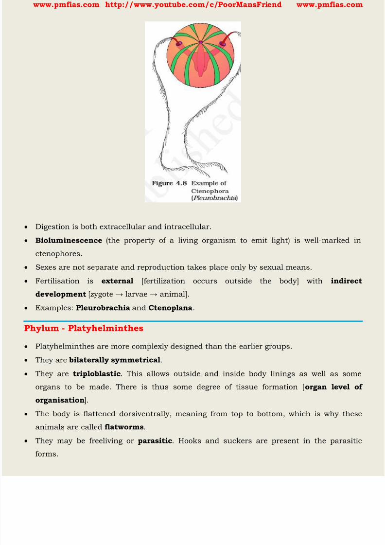
8/16/2019 Biology for Prelims Exam
http://slidepdf.com/reader/full/biology-for-prelims-exam 220/384
www.pmfias.com http://www.youtube.com/c/PoorMansFriend www.pmfias.com
Digestion is both extracellular and intracellular.
Bioluminescence (the property of a living organism to emit light) is well-marked in
ctenophores.
Sexes are not separate and reproduction takes place only by sexual means.
Fertilisation is external [fertilization occurs outside the body] with indirect
development [zygot e → larvae → animal].
Examples: Pleurobrachia and Ctenoplana .
Phylum - Platyhelminthes
Platyhelminthes are more complexly designed than the earlier groups.
They are bilaterally symmetrical .
They are triploblastic . This allows outside and inside body linings as well as some
organs to be made. There is thus some degree of tissue formation [ organ level of
organisation ].
The body is flattened dorsiventrally, meaning from top to bottom, which is why these
animals are called flatworms .
They may be freeliving or parasitic . Hooks and suckers are present in the parasitic
forms.

8/16/2019 Biology for Prelims Exam
http://slidepdf.com/reader/full/biology-for-prelims-exam 221/384

8/16/2019 Biology for Prelims Exam
http://slidepdf.com/reader/full/biology-for-prelims-exam 222/384
www.pmfias.com http://www.youtube.com/c/PoorMansFriend www.pmfias.com
Phylum - Annelida
Annelida are aquatic [marine and fresh water] or terrestrial; free-living, and sometimes
parasitic.
Their body surface is distinctly marked out into segments or metameres [metamerically
segmented] and, hence, the phylum name Annelida (Latin, annulus: little ring).
They exhibit organ-system level of body organization.
They are coelomate [true body cavity]. This allows true organs to be packaged in the
body structure.
They are bilateral symmetric and triploblastic .
They possess longitudinal and circular muscles which help in locomotion.
Aquatic annelids like Nereis possess lateral appendages, parapodia , which help in
swimming.
A closed circulatory system is present.
Nephridia (sing. nephridium) help in osmoregulation and excretion.
Neural system consists of paired ganglia (sing. ganglion) connected by lateral nerves to a
double ventral nerve cord.
Nereis , an aquatic form, is dioecious [Sexes are separate], but earthworms and leeches
are monoecious [having both the male and female reproductive organs in the same
individual].
Reproduction is sexual.

8/16/2019 Biology for Prelims Exam
http://slidepdf.com/reader/full/biology-for-prelims-exam 223/384
www.pmfias.com http://www.youtube.com/c/PoorMansFriend www.pmfias.com
Phylum - Arthropoda
Arthropoda is probably the largest group of animals. Over two-thirds of all named
species on earth are arthropods.
They have jointed legs (the word ‘arthropod’ means ‘jointed legs’).
Some familiar examples are prawns, butterflies, houseflies, spiders, scorpions and
crabs and some insects.
They exhibit organ-system level of organisation.
They are bilaterally symmetrical, triploblastic, segmented and coelomate animals. The
coelomic cavity is blood-filled.
The body of arthropods is covered by chitinous exoskeleton. The body consists of head,
thorax and abdomen.
There is an open circulatory system , and so the blood does not flow in well defined
blood vessels.
Respiratory organs are gills, book gills, book lungs or tracheal system.
Sensory organs like antennae, eyes (compound and simple), statocysts or balance
organs are present.
Excretion takes place through malpighian tubules .
They are mostly dioecious.
Fertilisation is usually internal.
They are mostly oviparous.
Development may be direct or indirect.
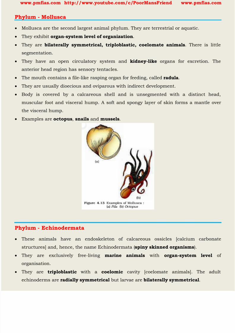
8/16/2019 Biology for Prelims Exam
http://slidepdf.com/reader/full/biology-for-prelims-exam 224/384
www.pmfias.com http://www.youtube.com/c/PoorMansFriend www.pmfias.com
Phylum - Mollusca
Mollusca are the second largest animal phylum. They are terrestrial or aquatic.
They exhibit organ-system level of organization .
They are bilaterally symmetrical, triploblastic, coelomate animals . There is little
segmentation.
They have an open circulatory system and kidney-like organs for excretion. The
anterior head region has sensory tentacles.
The mouth contains a file-like rasping organ for feeding, called radula .
They are usually dioecious and oviparous with indirect development.
Body is covered by a calcareous shell and is unsegmented with a distinct head,
muscular foot and visceral hump. A soft and spongy layer of skin forms a mantle over
the visceral hump.
Examples are octopus , snails and mussels .
Phylum - Echinodermata
These animals have an endoskeleton of calcareous ossicles [calcium carbonate
structures] and, hence, the name Echinodermata ( spiny skinned organisms ).
They are exclusively free-living marine animals with organ-system level of
organisation.
They are triploblastic with a coelomic cavity [coelomate animals]. The adult
echinoderms are radially symmetrical but larvae are bilaterally symmetrical .
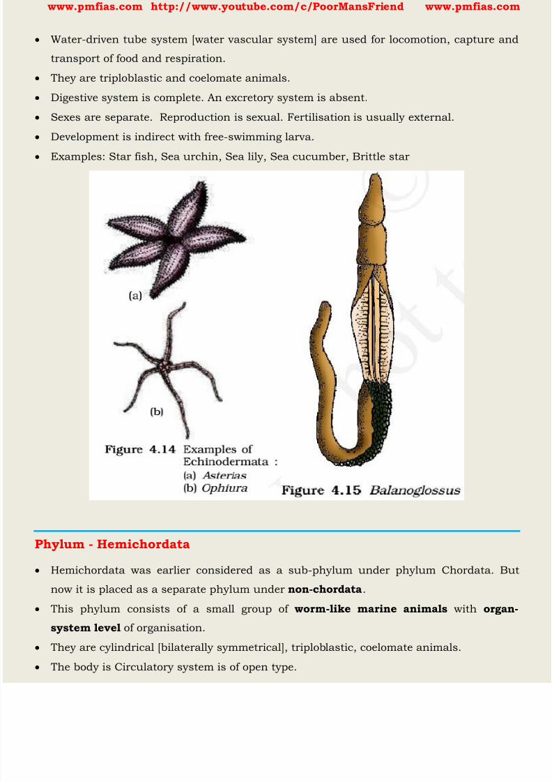
8/16/2019 Biology for Prelims Exam
http://slidepdf.com/reader/full/biology-for-prelims-exam 225/384
www.pmfias.com http://www.youtube.com/c/PoorMansFriend www.pmfias.com
Water-driven tube system [water vascular system] are used for locomotion, capture and
transport of food and respiration.
They are triploblastic and coelomate animals.
Digestive system is complete. An excretory system is absent.
Sexes are separate. Reproduction is sexual. Fertilisation is usually external.
Development is indirect with free-swimming larva. Examples: Star fish, Sea urchin, Sea lily, Sea cucumber, Brittle star
Phylum - Hemichordata Hemichordata was earlier considered as a sub-phylum under phylum Chordata. But
now it is placed as a separate phylum under non-chordata .
This phylum consists of a small group of worm-like marine animals with organ-
system level of organisation.
They are cylindrical [bilaterally symmetrical], triploblastic, coelomate animals.
The body is Circulatory system is of open type.

8/16/2019 Biology for Prelims Exam
http://slidepdf.com/reader/full/biology-for-prelims-exam 226/384
www.pmfias.com http://www.youtube.com/c/PoorMansFriend www.pmfias.com
Respiration takes place through gills.
Excretory organ is present.
Sexes are separate. Fertilisation is external. Development is indirect.
Examples: Balanoglossus and Saccoglossus.
Phylum - Chordata
Animals belonging to phylum Chordata are fundamentally characterised by the presence
of a notochord, a dorsal hollow nerve cord and paired pharyngeal gill slits.
They are bilaterally symmetrical, triploblastic, coelomate with organ-system level of
organisation.
They possess a post anal tail and a closed circulatory system.
Phylum Chordata is divided into three subphyla: Urochordata or Tunicata,
Cephalochordata and Vertebrata. Subphyla Urochordata and Cephalochordata are often referred to as protochordates and
are exclusively marine .
In Urochordata, notochord is present only in larval tail , while in Cephalochordata, it
extends from head to tail region and is persistent throughout their life.
Examples: Urochordata - Ascidia, Salpa, Doliolum; Cephalochordata - Amphioxus or
Lancelet.
All chordates possess the following features:
1. have a notochord
2. have a dorsal nerve cord
3. are triploblastic
4. have paired gill pouches
5. are coelomate.

8/16/2019 Biology for Prelims Exam
http://slidepdf.com/reader/full/biology-for-prelims-exam 227/384
www.pmfias.com http://www.youtube.com/c/PoorMansFriend www.pmfias.com
Vertebrata
These animals have a true vertebral column and internal skeleton, allowing a completely
different distribution of muscle attachment points to be used for movement.
The members of subphylum Vertebrata possess notochord during the embryonic period.
The notochord is replaced by a cartilaginous or bony vertebral column in the adult.
Thus all vertebrates are chordates but all chordates are not vertebrates.
Besides the basic chordate characters, vertebrates have a ventral muscular heart with
two, three or four chambers, kidneys for excretion and osmoregulation and pairedappendages which may be fins or limbs.
Vertibrates are bilaterally symmetrical, triploblastic, coelomic and segmented , with
complex differentiation of body tissues and organs.
Comparison of Chordates and Non-chordates
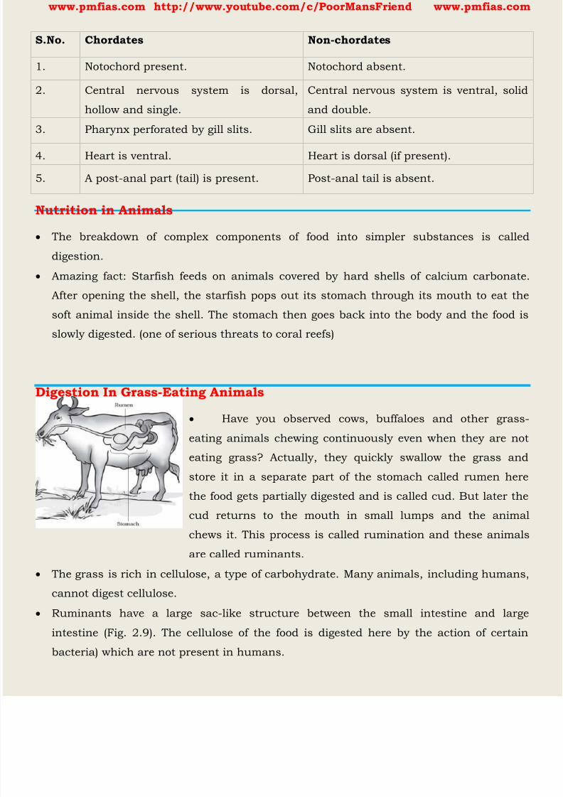
8/16/2019 Biology for Prelims Exam
http://slidepdf.com/reader/full/biology-for-prelims-exam 228/384
www.pmfias.com http://www.youtube.com/c/PoorMansFriend www.pmfias.com
Nutrition in Animals
The breakdown of complex components of food into simpler substances is called
digestion.
Amazing fact: Starfish feeds on animals covered by hard shells of calcium carbonate.After opening the shell, the starfish pops out its stomach through its mouth to eat the
soft animal inside the shell. The stomach then goes back into the body and the food is
slowly digested. (one of serious threats to coral reefs)
Digestion In Grass-Eating Animals
Have you observed cows, buffaloes and other grass-eating animals chewing continuously even when they are not
eating grass? Actually, they quickly swallow the grass and
store it in a separate part of the stomach called rumen here
the food gets partially digested and is called cud. But later the
cud returns to the mouth in small lumps and the animal
chews it. This process is called rumination and these animals
are called ruminants. The grass is rich in cellulose, a type of carbohydrate. Many animals, including humans,
cannot digest cellulose.
Ruminants have a large sac-like structure between the small intestine and large
intestine (Fig. 2.9). The cellulose of the food is digested here by the action of certain
bacteria) which are not present in humans.
S.No. Chordates Non-chordates
1. Notochord present. Notochord absent.
2. Central nervous system is dorsal,
hollow and single.
Central nervous system is ventral, solid
and double.
3. Pharynx perforated by gill slits. Gill slits are absent.
4. Heart is ventral. Heart is dorsal (if present).
5. A post-anal part (tail) is present. Post-anal tail is absent.
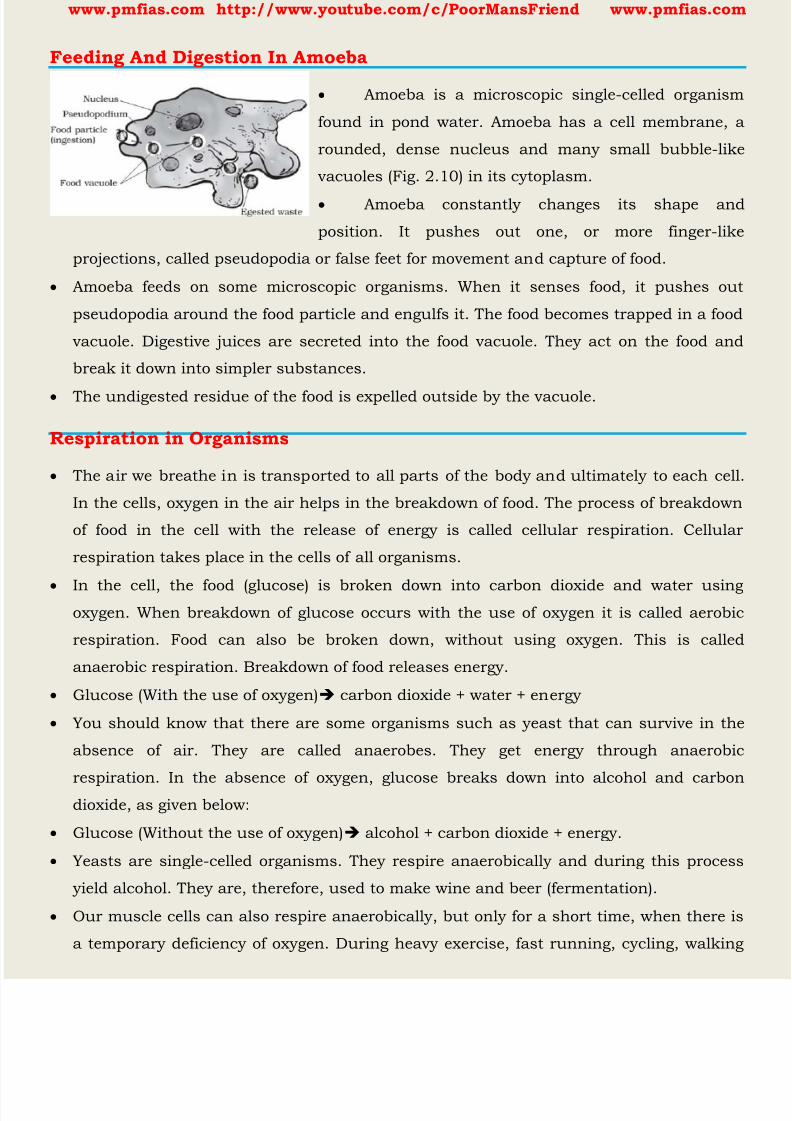
8/16/2019 Biology for Prelims Exam
http://slidepdf.com/reader/full/biology-for-prelims-exam 229/384
www.pmfias.com http://www.youtube.com/c/PoorMansFriend www.pmfias.com
Feeding And Digestion In Amoeba
Amoeba is a microscopic single-celled organism
found in pond water. Amoeba has a cell membrane, a
rounded, dense nucleus and many small bubble-like
vacuoles (Fig. 2.10) in its cytoplasm.
Amoeba constantly changes its shape and
position. It pushes out one, or more finger-like
projections, called pseudopodia or false feet for movement and capture of food.
Amoeba feeds on some microscopic organisms. When it senses food, it pushes out
pseudopodia around the food particle and engulfs it. The food becomes trapped in a food
vacuole. Digestive juices are secreted into the food vacuole. They act on the food and
break it down into simpler substances.
The undigested residue of the food is expelled outside by the vacuole.
Respiration in Organisms
The air we breathe in is transported to all parts of the body and ultimately to each cell.
In the cells, oxygen in the air helps in the breakdown of food. The process of breakdown
of food in the cell with the release of energy is called cellular respiration. Cellular
respiration takes place in the cells of all organisms.
In the cell, the food (glucose) is broken down into carbon dioxide and water usingoxygen. When breakdown of glucose occurs with the use of oxygen it is called aerobic
respiration. Food can also be broken down, without using oxygen. This is called
anaerobic respiration. Breakdown of food releases energy.
Glucose (With the use of oxygen) carbon dioxide + water + energy
You should know that there are some organisms such as yeast that can survive in the
absence of air. They are called anaerobes. They get energy through anaerobic
respiration. In the absence of oxygen, glucose breaks down into alcohol and carbondioxide, as given below:
Glucose (Without the use of oxygen) alcohol + carbon dioxide + energy.
Yeasts are single-celled organisms. They respire anaerobically and during this process
yield alcohol. They are, therefore, used to make wine and beer (fermentation).
Our muscle cells can also respire anaerobically, but only for a short time, when there is
a temporary deficiency of oxygen. During heavy exercise, fast running, cycling, walking
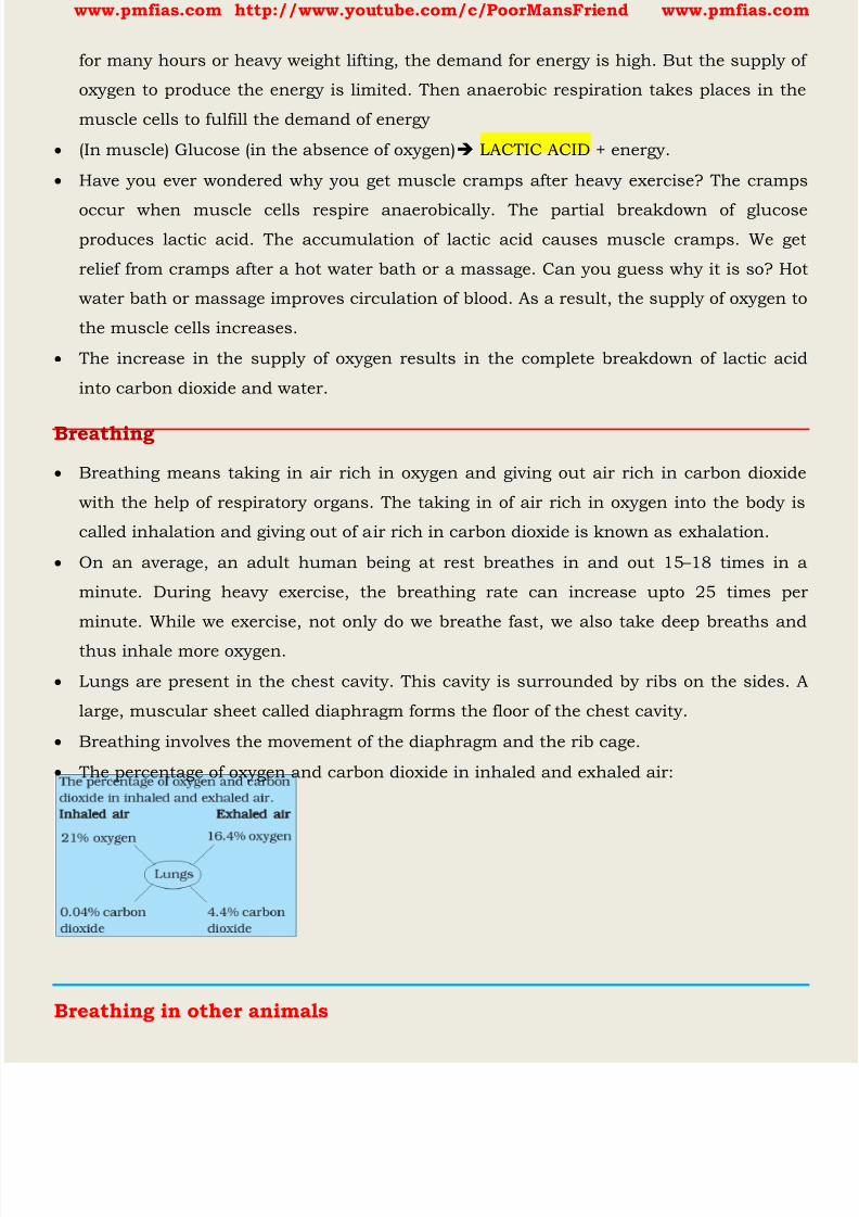
8/16/2019 Biology for Prelims Exam
http://slidepdf.com/reader/full/biology-for-prelims-exam 230/384
www.pmfias.com http://www.youtube.com/c/PoorMansFriend www.pmfias.com
for many hours or heavy weight lifting, the demand for energy is high. But the supply of
oxygen to produce the energy is limited. Then anaerobic respiration takes places in the
muscle cells to fulfill the demand of energy
(In muscle) Glucose (in the absence of oxygen) LACTIC ACID + energy.
Have you ever wondered why you get muscle cramps after heavy exercise? The cramps
occur when muscle cells respire anaerobically. The partial breakdown of glucoseproduces lactic acid. The accumulation of lactic acid causes muscle cramps. We get
relief from cramps after a hot water bath or a massage. Can you guess why it is so? Hot
water bath or massage improves circulation of blood. As a result, the supply of oxygen to
the muscle cells increases.
The increase in the supply of oxygen results in the complete breakdown of lactic acid
into carbon dioxide and water.
Breathing
Breathing means taking in air rich in oxygen and giving out air rich in carbon dioxide
with the help of respiratory organs. The taking in of air rich in oxygen into the body is
called inhalation and giving out of air rich in carbon dioxide is known as exhalation.
On an average, an adult human being at rest breathes in and out 15 – 18 times in a
minute. During heavy exercise, the breathing rate can increase upto 25 times per
minute. While we exercise, not only do we breathe fast, we also take deep breaths and
thus inhale more oxygen.
Lungs are present in the chest cavity. This cavity is surrounded by ribs on the sides. A
large, muscular sheet called diaphragm forms the floor of the chest cavity.
Breathing involves the movement of the diaphragm and the rib cage.
The percentage of oxygen and carbon dioxide in inhaled and exhaled air:
Breathing in other animals

8/16/2019 Biology for Prelims Exam
http://slidepdf.com/reader/full/biology-for-prelims-exam 231/384
www.pmfias.com http://www.youtube.com/c/PoorMansFriend www.pmfias.com
Cockroach: A cockroach has small openings on the sides of its body. Other insects also
have similar openings. These openings are called spiracles. Insects have a network of air
tubes called tracheae for gas exchange
Oxygen rich air rushes through spiracles into the tracheal tubes, diffuses into the body
tissue, and reaches every cell of the body. Similarly, carbon dioxide from the cells goes
into the tracheal tubes and moves out through spiracles. These air tubes or tracheae arefound only in insects and not in any other group of animals.
In fish Gills are well supplied with blood vessels for exchange of gases.
8/16/2019 Biology for Prelims Exam
http://slidepdf.com/reader/full/biology-for-prelims-exam 232/384
www.pmfias.com http://www.youtube.com/c/PoorMansFriend www.pmfias.com
Phylum - Chordata - Division In Vertebrata: Cyclostomata, Pisces, Chondrichthyes,
Osteichthyes, Amphibia, Amphibia, Reptilia, Aves, Mammalia.
Source | Credits | Picture Credits: NCERT General Science
Phylum - Chordata
Animals belonging to phylum Chordata are fundamentally characterised by the presence
of a notochord , a dorsal hollow nerve cord and paired pharyngeal [ relating to the pharynx
gill slits .
They are bilaterally symmetrical, triploblastic, coelomate with organ-system level of
organisation.
Phylum Chordata is divided into three subphyla: Urochordata or Tunicata,
Cephalochordata and Vertebrata .
Subphyla Urochordata and Cephalochordata are often referred to as protochordates andare exclusively marine .
In Urochordata, notochord is present only in larval tail , while in Cephalochordata, it
extends from head to tail region and is persistent throughout their life.
Examples: Urochordata - Ascidia, Salpa, Doliolum; Cephalochordata - Amphioxus or
Lancelet.
Vertebrata
These animals have a true vertebral column and internal skeleton , allowing a
completely different distribution of muscle attachment points to be used for movement.
Comparison of Chordates and Non-chordates
S.No. Chordates Non-chordates
1. Notochord present. Notochord absent.
2. Central nervous system is dorsal,
hollow and single.
Central nervous system is ventral, solid
and double.
3. Pharynx perforated by gill slits. Gill slits are absent.
4. Heart is ventral. Heart is dorsal (if present).
5. A post-anal part (tail) is present. Post-anal tail is absent.

8/16/2019 Biology for Prelims Exam
http://slidepdf.com/reader/full/biology-for-prelims-exam 233/384
www.pmfias.com http://www.youtube.com/c/PoorMansFriend www.pmfias.com
The members of subphylum Vertebrata possess notochord during the embryonic
period .
The notochord is replaced by a cartilaginous or bony vertebral column in the adult.
Thus all vertebrates are chordates but all chordates are not vertebrates.
Besides the basic chordate characters, vertebrates have a ventral muscular heart with
two, three or four chambers, kidneys for excretion and osmoregulation and pairedappendages which may be fins or limbs.
Vertibrates are bilaterally symmetrical, triploblastic, coelomic and segmented , with
complex differentiation of body tissues and organs.

8/16/2019 Biology for Prelims Exam
http://slidepdf.com/reader/full/biology-for-prelims-exam 234/384
www.pmfias.com http://www.youtube.com/c/PoorMansFriend www.pmfias.com
Division In Vertebrata
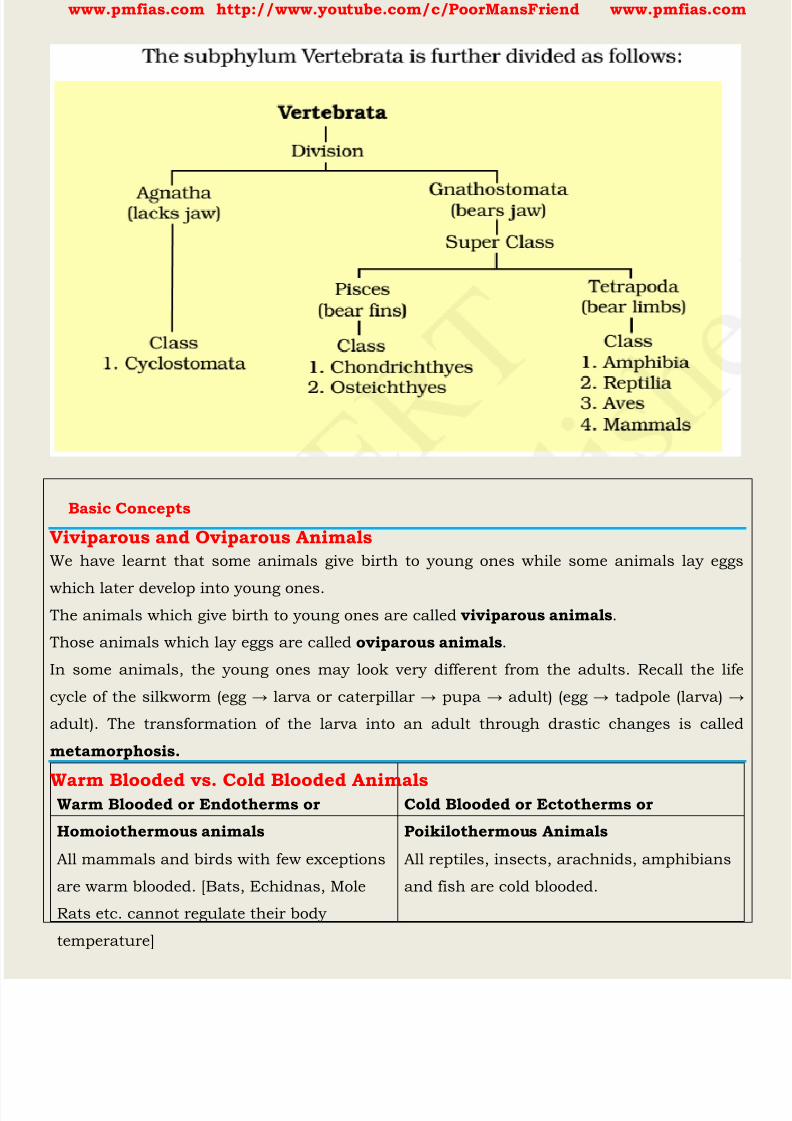
8/16/2019 Biology for Prelims Exam
http://slidepdf.com/reader/full/biology-for-prelims-exam 235/384
www.pmfias.com http://www.youtube.com/c/PoorMansFriend www.pmfias.com
Basic Concepts
Viviparous and Oviparous AnimalsWe have learnt that some animals give birth to young ones while some animals lay eggs
which later develop into young ones. The animals which give birth to young ones are called viviparous animals .
Those animals which lay eggs are called oviparous animals .
In some animals, the young ones may look very different from the adults. Recall the life
cycle of the silkworm (egg → larv a or caterpillar → pupa → adult) (egg → tadpole (larva) →
adult). The transformation of the larva into an adult through drastic changes is called
metamorphosis.
Warm Blooded vs. Cold Blooded AnimalsWarm Blooded or Endotherms or
Homoiothermous animals
Cold Blooded or Ectotherms or
Poikilothermous Animals
All mammals and birds with few exceptions
are warm blooded. [Bats, Echidnas, Mole
Rats etc. cannot regulate their body
temperature]
All reptiles, insects, arachnids, amphibians
and fish are cold blooded.
8/16/2019 Biology for Prelims Exam
http://slidepdf.com/reader/full/biology-for-prelims-exam 236/384
www.pmfias.com http://www.youtube.com/c/PoorMansFriend www.pmfias.com
They maintain a constant internal body
temperature irrespective of external
environment. [Can regulate their body
temperature by generating their own heat
when they are in a cooler environment, and
by cooling themselves when they are in ahotter environment]
Their body temperature changes according
to the external environment. [If a cold
blooded animal is taken to the equator its
body temperature increases and if taken to
the poles its body temperature decreases]
They can survive in a wide of environments
as they are able to regulate their body
temperature.
They cannot survive in a wide of
environments. [Tropical animals cannot
survive in the polar region and vice versa]
They require a lot of food for their survival.
Most of the food consumed is utilized to
maintain a constant body temperature.
Most of the food consumed is converted
into body mass. So they need less food
compared to warm blooded animals.
They are active in both warm and cold
environments.
They are active in warm environments and
are very sluggish in cold environments.
To stay cool, warm-blooded animals usually
sweat. Animals like elephants use their ears
to cool their body [large, thin ears which
loose heat quickly].
Some warm-blooded animals, especially
birds, migrate from colder to warmer
regions in the winter.
Mammals have hair, fur and birds have
feathers to help keep them warm.
Warm-blooded animals can also shiver to
generate more heat when they get too cold.
Cold-blooded animals often like to bask in
the sun to warm up and increase their
metabolism.
Some cold-blooded animals, such as bees
or dragonflies, shiver to stay warm when in
a cold environment.
Constant body temperature provide a nice
warm environment for viruses, bacteria and
parasites to live in.
Constantly changing body temperatures
make life more difficult for the parasites.
HibernationHibernation is a state of inactivity and metabolic depression in few endotherms [warm
blooded animals – bear, rodents] and ectotherms [many reptiles like snakes, turtles and
amphibians like frogs]. Snakes, lizards, toads, frogs, salamanders and most turtles will
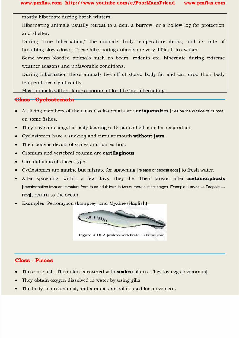
8/16/2019 Biology for Prelims Exam
http://slidepdf.com/reader/full/biology-for-prelims-exam 237/384
www.pmfias.com http://www.youtube.com/c/PoorMansFriend www.pmfias.com
mostly hibernate during harsh winters.
Hibernating animals usually retreat to a den, a burrow, or a hollow log for protection
and shelter.
During "true hibernation," the animal's body temperature drops, and its rate of
breathing slows down. These hibernating animals are very difficult to awaken.
Some warm-blooded animals such as bears, rodents etc. hibernate during extremeweather seasons and unfavorable conditions.
During hibernation these animals live off of stored body fat and can drop their body
temperatures significantly.
Most animals will eat large amounts of food before hibernating.
Class - Cyclostomata
All living members of the class Cyclostomata are ectoparasites [ives on the outside of its host
on some fishes.
They have an elongated body bearing 6-15 pairs of gill slits for respiration.
Cyclostomes have a sucking and circular mouth without jaws .
Their body is devoid of scales and paired fins.
Cranium and vertebral column are cartilaginous .
Circulation is of closed type.
Cyclostomes are marine but migrate for spawning [ release or deposit eggs ] to fresh water.
After spawning, within a few days, they die. Their larvae, after metamorphosis[transformation from an immature form to an adult form in two or more distinct stages. Example: Larvae → Tadpole →
Frog ], return to the ocean.
Examples: Petromyzon (Lamprey) and Myxine (Hagfish).
Class - Pisces
These are fish. Their skin is covered with scales /plates. They lay eggs [oviporous].
They obtain oxygen dissolved in water by using gills.
The body is streamlined, and a muscular tail is used for movement.
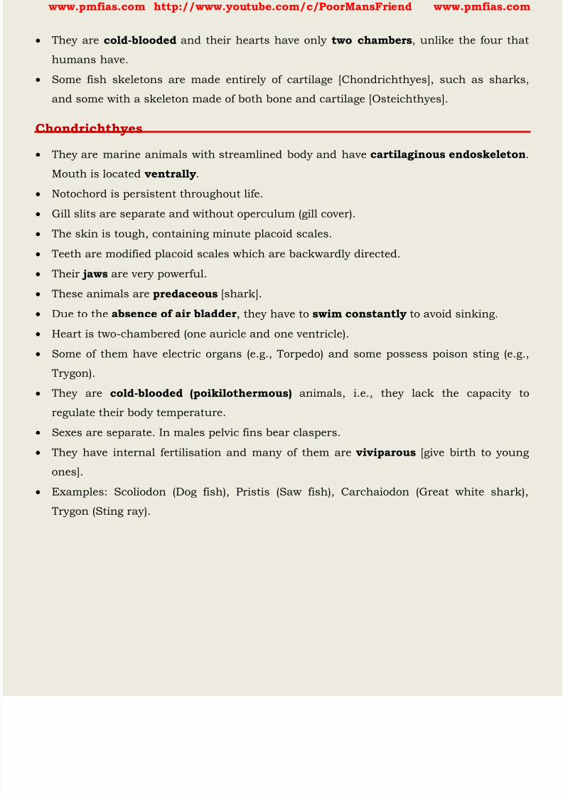
8/16/2019 Biology for Prelims Exam
http://slidepdf.com/reader/full/biology-for-prelims-exam 238/384
www.pmfias.com http://www.youtube.com/c/PoorMansFriend www.pmfias.com
They are cold-blooded and their hearts have only two chambers , unlike the four that
humans have.
Some fish skeletons are made entirely of cartilage [Chondrichthyes], such as sharks,
and some with a skeleton made of both bone and cartilage [Osteichthyes].
Chondrichthyes
They are marine animals with streamlined body and have cartilaginous endoskeleton
Mouth is located ventrally .
Notochord is persistent throughout life.
Gill slits are separate and without operculum (gill cover).
The skin is tough, containing minute placoid scales.
Teeth are modified placoid scales which are backwardly directed.
Their jaws are very powerful. These animals are predaceous [shark].
Due to the absence of air bladder , they have to swim constantly to avoid sinking.
Heart is two-chambered (one auricle and one ventricle).
Some of them have electric organs (e.g., Torpedo) and some possess poison sting (e.g.,
Trygon).
They are cold-blooded (poikilothermous) animals, i.e., they lack the capacity to
regulate their body temperature.
Sexes are separate. In males pelvic fins bear claspers.
They have internal fertilisation and many of them are viviparous [give birth to young
ones].
Examples: Scoliodon (Dog fish), Pristis (Saw fish), Carchaiodon (Great white shark),
Trygon (Sting ray).
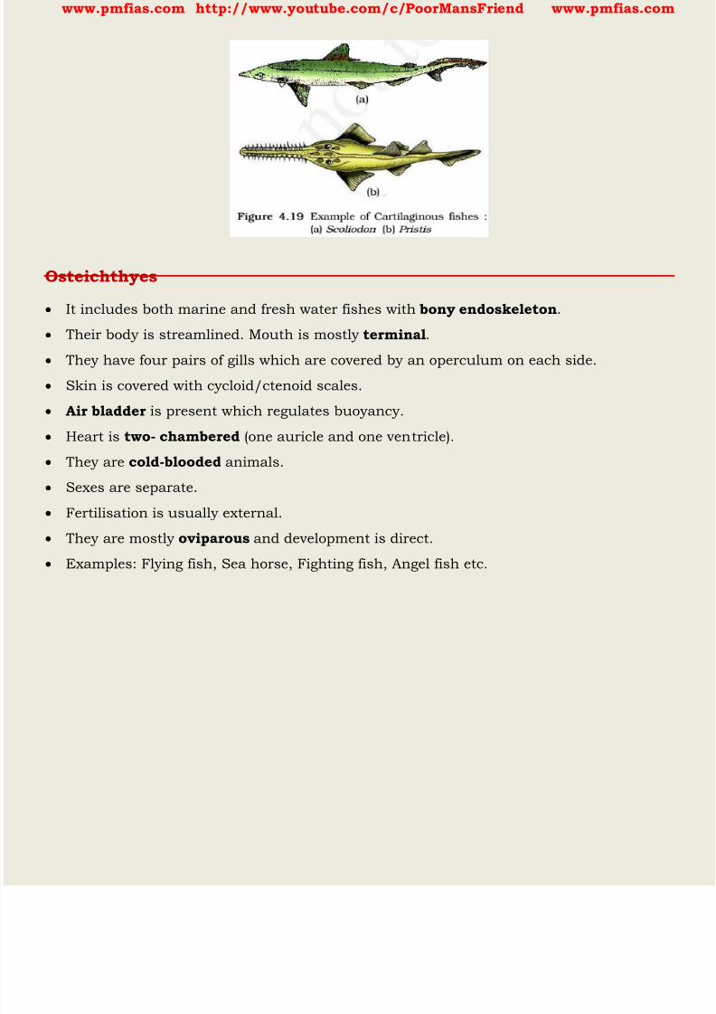
8/16/2019 Biology for Prelims Exam
http://slidepdf.com/reader/full/biology-for-prelims-exam 239/384
www.pmfias.com http://www.youtube.com/c/PoorMansFriend www.pmfias.com
Osteichthyes
It includes both marine and fresh water fishes with bony endoskeleton .
Their body is streamlined. Mouth is mostly terminal .
They have four pairs of gills which are covered by an operculum on each side.
Skin is covered with cycloid/ctenoid scales.
Air bladder is present which regulates buoyancy.
Heart is two- chambered (one auricle and one ventricle).
They are cold-blooded animals.
Sexes are separate.
Fertilisation is usually external.
They are mostly oviparous and development is direct.
Examples: Flying fish, Sea horse, Fighting fish, Angel fish etc.
8/16/2019 Biology for Prelims Exam
http://slidepdf.com/reader/full/biology-for-prelims-exam 240/384
www.pmfias.com http://www.youtube.com/c/PoorMansFriend www.pmfias.com
Class - Amphibia
As the name indicates (Gr., Amphi : dual, bios, life), amphibians can live in aquatic as
well as terrestrial habitats.
The amphibian skin is moist without scales [mucus glands in the skin]. The eyes have
eyelids. A tympanum represents the ear.
Alimentary canal, urinary and reproductive tracts open into a common chamber called
cloaca which opens to the exterior.
They have a three-chambered heart (two auricles and one ventricle). These are cold-
blooded animals.
Respiration is through gills, lungs and through skin.
Respiration is by gills, lungs and through skin.
Sexes are separate. Fertilisation is external.
They are oviparous and development is indirect.
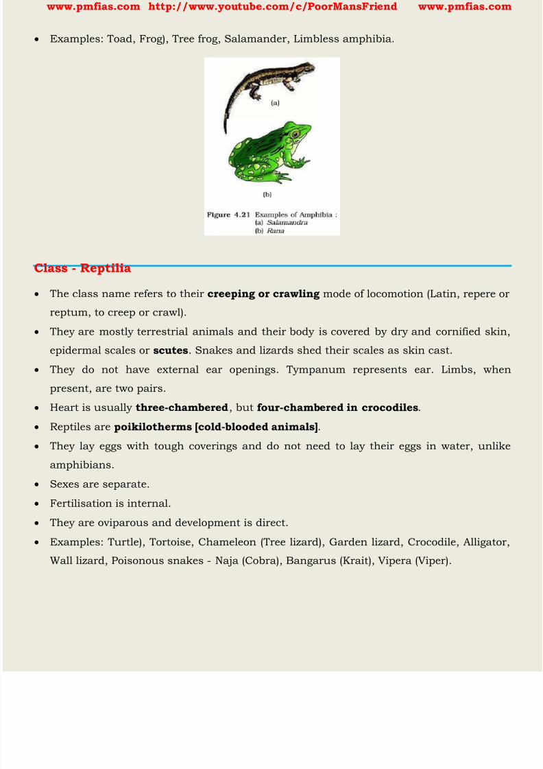
8/16/2019 Biology for Prelims Exam
http://slidepdf.com/reader/full/biology-for-prelims-exam 241/384
www.pmfias.com http://www.youtube.com/c/PoorMansFriend www.pmfias.com
Examples: Toad, Frog), Tree frog, Salamander, Limbless amphibia.
Class - Reptilia The class name refers to their creeping or crawling mode of locomotion (Latin, repere or
reptum, to creep or crawl).
They are mostly terrestrial animals and their body is covered by dry and cornified skin,
epidermal scales or scutes . Snakes and lizards shed their scales as skin cast.
They do not have external ear openings. Tympanum represents ear. Limbs, when
present, are two pairs.
Heart is usually three-chambered , but four-chambered in crocodiles .
Reptiles are poikilotherms [cold-blooded animals] .
They lay eggs with tough coverings and do not need to lay their eggs in water, unlike
amphibians.
Sexes are separate.
Fertilisation is internal.
They are oviparous and development is direct.
Examples: Turtle), Tortoise, Chameleon (Tree lizard), Garden lizard, Crocodile, Alligator,
Wall lizard, Poisonous snakes - Naja (Cobra), Bangarus (Krait), Vipera (Viper).
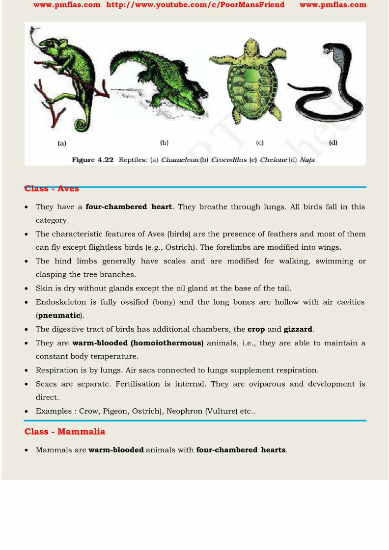
8/16/2019 Biology for Prelims Exam
http://slidepdf.com/reader/full/biology-for-prelims-exam 242/384
www.pmfias.com http://www.youtube.com/c/PoorMansFriend www.pmfias.com
Class - Aves They have a four-chambered heart . They breathe through lungs. All birds fall in this
category.
The characteristic features of Aves (birds) are the presence of feathers and most of them
can fly except flightless birds (e.g., Ostrich). The forelimbs are modified into wings.
The hind limbs generally have scales and are modified for walking, swimming or
clasping the tree branches.
Skin is dry without glands except the oil gland at the base of the tail. Endoskeleton is fully ossified (bony) and the long bones are hollow with air cavities
(pneumatic ).
The digestive tract of birds has additional chambers, the crop and gizzard .
They are warm-blooded (homoiothermous) animals, i.e., they are able to maintain a
constant body temperature.
Respiration is by lungs. Air sacs connected to lungs supplement respiration.
Sexes are separate. Fertilisation is internal. They are oviparous and development isdirect.
Examples : Crow, Pigeon, Ostrich), Neophron (Vulture) etc..
Class - Mammalia
Mammals are warm-blooded animals with four-chambered hearts .
8/16/2019 Biology for Prelims Exam
http://slidepdf.com/reader/full/biology-for-prelims-exam 243/384
www.pmfias.com http://www.youtube.com/c/PoorMansFriend www.pmfias.com
Most mammals familiar to us produce live young ones . However, a few of them, like the
Platypus and the Echidna lay eggs, and some, like kangaroos give birth to very poorly
developed young ones.
They are found in a variety of habitats - polar ice caps, deserts, mountains, forests,
grasslands and dark caves. Some of them have adapted to fly or live in water.
The most unique mammalian characteristic is the presence of milk producing glands(mammary glands) by which the young ones are nourished.
They have two pairs of limbs, adapted for walking, running, climbing, burrowing,
swimming or flying.
The skin of mammals is unique in possessing hair. External ears or pinnae are present.
Different types of teeth are present in the jaw.
Heart is four-chambered . They are homoiothermous [warm-blooded]. Respiration is by
lungs. Sexes are separate and fertilisation is internal.
They are viviparous with few exceptions and development is direct.
Examples: Oviparous – Platypus; Viviparous – Kangaroo, Flying fox), Delphinus
(Common dolphin), Balaenoptera (Blue whale), etc.
Animal Classification Summary
Porifera includes multicellular animals which exhibit cellular level of organisation and
have characteristic flagellated choanocytes.
The coelenterates have tentacles and bear cnidoblasts. They are mostly aquatic, sessile
or free-floating. The ctenophores are marine animals with comb plates.
The platyhelminths have flat body and exhibit bilateral symmetry. The parasitic forms
show distinct suckers and hooks.
Aschelminthes are pseudocoelomates and include parasitic as well as non-parasitic
round worms.
Annelids are metamerically segmented animals with a true coelom.
The arthropods are the most abundant group of animals characterised by the presence
of jointed appendages.
The molluscs have a soft body surrounded by an external calcareous shell. The body is
covered with external skeleton made of chitin.

8/16/2019 Biology for Prelims Exam
http://slidepdf.com/reader/full/biology-for-prelims-exam 244/384
www.pmfias.com http://www.youtube.com/c/PoorMansFriend www.pmfias.com
The echinoderms possess a spiny skin. Their most distinctive feature is the presence of
water vascular system.
The hemichordates are a small group of worm-like marine animals. They have a
cylindrical body with proboscis, collar and trunk.
Phylum Chordata includes animals which possess a notochord either throughout or
during early embryonic life. Other common features observed in the chordates are thedorsal, hollow nerve cord and paired pharyngeal gill slits.
Some of the vertebrates do not possess jaws (Agnatha) whereas most of them possess
jaws (Gnathostomata). Agnatha is represented by the class, Cyclostomata. They are the
most primitive chordates and are ectoparasites on fishes. Gnathostomata has two super
classes, Pisces and Tetrapoda.
Classes Chondrichthyes and Osteichthyes bear fins for locomotion and are grouped
under Pisces. The Chondrichthyes are fishes with cartilaginous endoskeleton and aremarine.
Classes, Amphibia, Reptilia, Aves and Mammalia have two pairs of limbs and are thus
grouped under Tetrapoda. The amphibians have adapted to live both on land and water.
Reptiles are characterised by the presence of dry and cornified skin. Limbs are absent in
snakes. Fishes, amphibians and reptiles are poikilothermous (coldblooded).
Aves are warm-blooded animals with feathers on their bodies and forelimbs modified
into wings for flying. Hind limbs are adapted for walking, swimming, perching or
clasping.
The unique features of mammals are the presence of mammary glands and hairs on the
skin. They commonly exhibit viviparity.
Match the following
1. Operculum
2. Parapodia
3. Scales4. Comb plates
5. Radula
6. Hairs
7. Choanocytes
8. Gill slits
a) Ctenophora
b) Mollusca
c) Poriferad) Reptilia
e) Annelida
f) Cyclostomata and Chondrichthyes
g) Mammalia
h) Osteichthyes
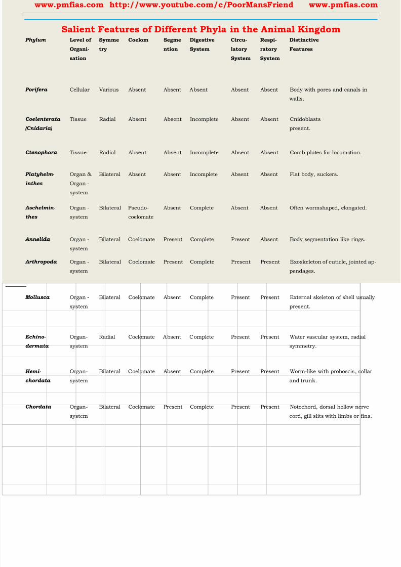
8/16/2019 Biology for Prelims Exam
http://slidepdf.com/reader/full/biology-for-prelims-exam 245/384
www.pmfias.com http://www.youtube.com/c/PoorMansFriend www.pmfias.com
Salient Features of Different Phyla in the Animal Kingdom
Phylum Level of
Organi-
sation
Symme
try
Coelom Segme
ntion
Digestive
System
Circu-
latory
System
Respi-
ratory
System
Distinctive
Features
Porifera Cellular Various Absent Absent Absent Absent Absent Body with pores and canals in
walls.
Coelenterata
(Cnidaria)
Tissue Radial Absent Absent Incomplete Absent Absent Cnidoblasts
present.
Ctenophora Tissue Radial Absent Absent Incomplete Absent Absent Comb plates for locomotion.
Platyhelm-
inthes
Organ &
Organ -
system
Bilateral Absent Absent Incomplete Absent Absent Flat body, suckers.
Aschelmin-
thes
Organ -
system
Bilateral Pseudo-
coelomate
Absent Complete Absent Absent Often wormshaped, elongated.
Annelida Organ -
system
Bilateral Coelomate Present Complete Present Absent Body segmentation like rings.
Arthropoda Organ -
system
Bilateral Coelomate Present Complete Present Present Exoskeleton of cuticle, jointed ap-
pendages.
Mollusca Organ -system
Bilateral Coelomate Absent Complete Present Present External skeleton of shell usuallypresent.
Echino-
dermata
Organ-
system
Radial Coelomate Absent Complete Present Present Water vascular system, radial
symmetry.
Hemi-
chordata
Organ-
system
Bilateral Coelomate Absent Complete Present Present Worm-like with proboscis, collar
and trunk.
Chordata Organ-
system
Bilateral Coelomate Present Complete Present Present Notochord, dorsal hollow nerve
cord, gill slits with limbs or fins.

8/16/2019 Biology for Prelims Exam
http://slidepdf.com/reader/full/biology-for-prelims-exam 246/384
www.pmfias.com http://www.youtube.com/c/PoorMansFriend www.pmfias.com
8/16/2019 Biology for Prelims Exam
http://slidepdf.com/reader/full/biology-for-prelims-exam 247/384
www.pmfias.com http://www.youtube.com/c/PoorMansFriend www.pmfias.com
Source | Credits | Picture Credits: NCERT General Science
Biotechnology, Genetic Engineering, Cloning, Recombinant DNA, Cloning Vectors,
Competent Host, Biotechnology And Its Applications in Medicine & Agriculture.
Biotechnology
Biotechnology deals with techniques of using live organisms or enzymes from organisms
to produce products and processes useful to humans.
Making curd, bread or wine, which are all microbe-mediated processes, could also be
thought as a form of biotechnology.
However, it is used in a restricted sense today, to refer to such of those processes which
use genetically modified organisms to achieve the same on a larger scale.
Modern biotechnology using genetically modified organisms was made possible only
when man learnt to alter the chemistry of DNA and construct recombinant DNA. This
key process is called recombinant DNA technology or genetic engineering .
This process involves the use of restriction endonucleases, DNA ligase, appropriate
plasmid or viral vectors to isolate and ferry the foreign DNA into host organisms,
expression of the foreign gene, purification of the gene product, i.e., the functional
protein and finally making a suitable formulation for marketing. Large scale production
involves use of bioreactors.
Genetic Engineering
Genetic engineering involves the techniques to alter the chemistry of genetic material
(DNA and RNA) and thus change the phenotype of the host organism.
Asexual reproduction preserves the genetic information, while sexual reproduction
permits variation.
Traditional hybridisation procedures used in plant and animal breeding, very often lead
to inclusion and multiplication of undesirable genes along with the desired genes.
The techniques of genetic engineering which include creation of recombinant DNA, use
of gene cloning and gene transfer , overcome this limitation and allows us to isolate
and introduce only one or a set of desirable genes without introducing undesirable genes
into the target organism.
There are three basic steps in genetically modifying an organism —
1. identification of DNA with desirable genes;
8/16/2019 Biology for Prelims Exam
http://slidepdf.com/reader/full/biology-for-prelims-exam 248/384
www.pmfias.com http://www.youtube.com/c/PoorMansFriend www.pmfias.com
2. introduction of the identified DNA into the host;
3. maintenance of introduced DNA in the host and transfer of the DNA to its progeny.
Cloning
DNA which is somehow transferred into an alien organism would not be able to multiply
itself in the progeny cells of the organism. But, when it gets integrated into the genome of the recipient, it may multiply and be
inherited along with the host DNA. This is because the alien piece of DNA has become
part of a chromosome, which has the ability to replicate.
In a chromosome there is a specific DNA sequence called the origin of replication
which is responsible for initiating replication.
Therefore, for the multiplication of any alien piece of DNA in an organism it needs to be
a part of a chromosome(s) which has a specific sequence known as ‘origin of replication’.
Thus, an alien DNA is linked with the origin of replication, so that, this alien piece of
DNA can replicate and multiply itself in the host organism. This can also be called as
cloning or making multiple identical copies of any template DNA .
Recombinant DNA (rDNA)
Recombinant DNA (rDNA) molecules are DNA molecules formed by laboratory methods
of genetic recombination (such as molecular cloning) to bring together genetic material
from multiple sources, creating sequences that would not otherwise be found in the
genome.
Recombinant DNA is possible because DNA molecules from all organisms share the
same chemical structure. They differ only in the nucleotide sequence within that
identical overall structure.
In most cases, organisms containing recombinant DNA have apparently normal
phenotypes. That is, their appearance, behavior and metabolism are usually unchanged.

8/16/2019 Biology for Prelims Exam
http://slidepdf.com/reader/full/biology-for-prelims-exam 249/384
www.pmfias.com http://www.youtube.com/c/PoorMansFriend www.pmfias.com
The cutting of DNA at specific locations became possible with the discovery of the so-called ‘molecular scissors’ - restriction enzymes .
Restriction enzymes belong to a larger class of enzymes called nucleases . These are of
two kinds; exonucleases and endonucleases.
Exonucleases remove nucleotides from the ends of the DNA whereas, endonucleases
make cuts at specific positions within the DNA.
The cut piece of DNA was then linked with the plasmid DNA. These plasmid DNA act as
vectors to transfer the piece of DNA attached to it. You probably know that mosquito acts as an insect vector to transfer the malarial
parasite Into human body.
In the same way, a plasmid can be used as vector to deliver an alien piece of DNA into
the host organism.
The linking of antibiotic resistance gene with the plasmid vector became possible with
the enzyme DNA ligase , which acts on cut DNA molecules and joins their ends. This
makes a new combination of circular autonomously replicating DNA created in vitro andis known as recombinant DNA .
When this DNA is transferred into Escherichia coli, a bacterium closely related to
Salmonella, it could replicate using the new host’s DNA polymerase enzyme and make
multiple copies. The ability to multiply copies of antibiotic resistance gene in E. coli was
called cloning of antibiotic resistance gene in E. coli.
Applications of Recombinant DNA Technology
8/16/2019 Biology for Prelims Exam
http://slidepdf.com/reader/full/biology-for-prelims-exam 250/384
www.pmfias.com http://www.youtube.com/c/PoorMansFriend www.pmfias.com
Recombinant DNA is widely used in biotechnology, medicine and research.
Recombinant DNA is used to identify, map and sequence genes, and to determine
their function.
Recombinant DNA is used to produce
Recombinant human insulin, Recombinant human growth hormone,
Recombinant blood clotting factor VIII,
Recombinant hepatitis B vaccine,
Insect-resistant crops etc.
Cloning Vectors
You may be surprised to know that we have learnt the lesson of transferring genes into
plants and animals from bacteria and viruses which have known this for ages - how to
deliver genes to transform eukaryotic cells and force them to do what the bacteria or
viruses want.
For example, Agrobacterioum tumifaciens, a pathogen of several dicot plants is able to
deliver a piece of DNA known as ‘T -DNA’ to transform normal plant cells into a tumor
and direct these tumor cells to produce the chemicals required by the pathogen.
Similarly, retroviruses in animals have the ability to transform normal cells into
cancerous cells.
A better understanding of the art of delivering genes by pathogens in their eukaryotic
hosts has generated knowledge to transform these tools of pathogens into useful vectors
for delivering genes of interest to humans.
The tumor inducing (Ti) plasmid of AgrobcLCterium tumifaciens has now been modified
into a cloning vector which is no more pathogenic to the plants but is still able to use
the mechanisms to deliver genes of our interest into a variety of plants.
Similarly, retroviruses have also been disarmed and are now used to deliver desirable
genes into animal cells.
So, once a gene or a DNA fragment has been ligated into a suitable vector it is
transferred into a bacterial, plant or animal host (where it multiplies).
Plasmids and bacteriophages [vectors] have the ability to replicate within bacterial cells
independent of the control of chromosomal DNA.
8/16/2019 Biology for Prelims Exam
http://slidepdf.com/reader/full/biology-for-prelims-exam 251/384
www.pmfias.com http://www.youtube.com/c/PoorMansFriend www.pmfias.com
Competent Host – Methods to Induce Alien DNA into Host Cells
Since DNA is a hydrophilic molecule , it cannot pass through cell membranes . In
order to force bacteria to take up the plasmid, the bacterial cells must first be made
‘competent’ to take up DNA. Recombinant DNA can then be forced into such cells by
incubating the cells with recombinant DNA on ice, followed by placing them briefly at
42 0C (heat shock), and then putting them back on ice. This enables the bacteria to take
up the recombinant DNA. This is not the only way to introduce alien DNA into host cells.
In a method known as micro-injection, recombinant DNA is directly injected into the
nucleus of an animal cell.
In another method, suitable for plants, cells are bombarded with high velocity micro-
particles of gold or tungsten coated with DNA in a method known as biolistics or gene
gun .
And the last method uses ‘disarmed pathogen’ vectors, which when allowed to infect the
cell, transfer the recombinant DNA into the host.
Biotechnology And Its Applications
Biotechnology essentially deals with industrial scale production of biopharmaceuticals
and biologicals using genetically modified microbes, fungi, plants and animals.
The applications of biotechnology include therapeutics, diagnostics, genetically modified
crops for agriculture, processed food, bioremediation, waste treatment, and energyproduction.
Cloning
Cloning is the production of an exact copy of a cell, any other living part, or a complete
organism.
Cloning of an animal was successfully performed for the first time by Ian Wilmut and his
colleagues at the Roslin Institute in Edinburgh, Scotland.
They cloned successfully a sheep named Dolly. Dolly was born in 1996 and was the first
mammal to be cloned.
During the process of cloning Dolly, a cell was collected from the mammary gland of a
female Finn Dorsett sheep. Simultaneously, an egg was obtained from a Scottish
blackface ewe. The nucleus was removed from the egg. Then, the nucleus of the
mammary gland cell from the Finn Dorsett sheep was inserted into the egg of the
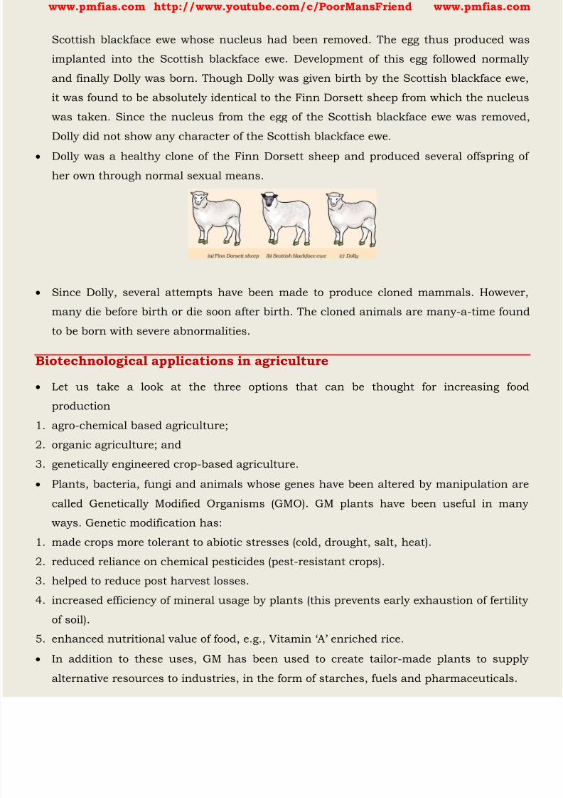
8/16/2019 Biology for Prelims Exam
http://slidepdf.com/reader/full/biology-for-prelims-exam 252/384
www.pmfias.com http://www.youtube.com/c/PoorMansFriend www.pmfias.com
Scottish blackface ewe whose nucleus had been removed. The egg thus produced was
implanted into the Scottish blackface ewe. Development of this egg followed normally
and finally Dolly was born. Though Dolly was given birth by the Scottish blackface ewe,
it was found to be absolutely identical to the Finn Dorsett sheep from which the nucleus
was taken. Since the nucleus from the egg of the Scottish blackface ewe was removed,
Dolly did not show any character of the Scottish blackface ewe. Dolly was a healthy clone of the Finn Dorsett sheep and produced several offspring of
her own through normal sexual means.
Since Dolly, several attempts have been made to produce cloned mammals. However,
many die before birth or die soon after birth. The cloned animals are many-a-time found
to be born with severe abnormalities.
Biotechnological applications in agriculture
Let us take a look at the three options that can be thought for increasing food
production
1. agro-chemical based agriculture;
2. organic agriculture; and
3. genetically engineered crop-based agriculture.
Plants, bacteria, fungi and animals whose genes have been altered by manipulation are
called Genetically Modified Organisms (GMO). GM plants have been useful in many
ways. Genetic modification has:
1. made crops more tolerant to abiotic stresses (cold, drought, salt, heat).
2. reduced reliance on chemical pesticides (pest-resistant crops).
3. helped to reduce post harvest losses.
4. increased efficiency of mineral usage by plants (this prevents early exhaustion of fertility
of soil).
5. enhanced nutritional value of food, e.g., Vitamin ‘A’ enriched rice.
In addition to these uses, GM has been used to create tailor-made plants to supply
alternative resources to industries, in the form of starches, fuels and pharmaceuticals.

8/16/2019 Biology for Prelims Exam
http://slidepdf.com/reader/full/biology-for-prelims-exam 253/384
www.pmfias.com http://www.youtube.com/c/PoorMansFriend www.pmfias.com
Bt toxin is produced by a bacterium called Bacillus thuringiensis (Bt for short).
Some strains of Bacillus thuringiensis produce proteins that kill certain insects such as
tobacco budworm, armyworm, beetles and dipterans flies, mosquitoes.
Why does this toxin not kill the Bacillus? Actually, the Bt toxin protein exist as inactive
protoxins but once an insect ingest the inactive toxin, it is converted into an active form
of toxin due to the alkaline pH of the gut which solubilise the crystals. Bt toxin gene has been cloned from the bacteria and been expressed in plants to provide
resistance to insects without the need for insecticides; in effect created a bio-pesticide.
Examples are Bt cotton, Bt corn, rice, tomato, potato and soyabean etc.
Biotechnological applications in medicine
The recombinant DNA technological processes have made immense impact in the area of
healthcare by enabling mass production of safe and more effective therapeutic
drugs .
Further, the recombinant therapeutics do not induce unwanted immunological
responses as is common in case of similar products isolated from non-human sources.
At present, about 30 recombinant therapeutics have been approved for human-use the
world over. In India, 12 of these are presently being marketed.
Genetically Engineered Insulin
Management of adult-onset diabetes is possible by taking insulin at regular time
intervals. What would a diabetic patient do if enough human-insulin was not available?
If you discuss this, you would soon realise that one would have to isolate and use
insulin from other animals. Would the insulin isolated from other animals be just as
effective as that secreted by the human body itself and would it not elicit an immune
response in the human body?
Now, imagine if bacterium were available that could make human insulin. Suddenly the
whole process becomes so simple. You can easily grow a large quantity of the bacteria
and make as much insulin as you need.
Think about whether insulin can be orally administered to diabetic people or not. Why?
Insulin used for diabetes was earlier extracted from pancreas of slaughtered cattle and
pigs.
Insulin from an animal source, though caused some patients to develop allergy or other
types of reactions to the foreign protein.
8/16/2019 Biology for Prelims Exam
http://slidepdf.com/reader/full/biology-for-prelims-exam 254/384
www.pmfias.com http://www.youtube.com/c/PoorMansFriend www.pmfias.com
Insulin consists of two short polypeptide chains: chain A and chain B, that are linked
together by disulphide bridges.
In mammals, including humans, insulin is synthesised as a pro-hormone (like a pro-
enzyme, the pro-hormone also needs to be processed before it becomes a fully mature
and functional hormone) which contains an extra stretch called the C peptide. This C
peptide is not present in the mature insulin and is removed during maturation intoinsulin.The main challenge for production of insulin using rDNA techniques was getting
insulin assembled into a mature form.
In 1983, Eli Lilly an American company prepared two DNA sequences corresponding to
A and B, chains of human insulin and introduced them in plasmids of E. coli to produce
insulin chains. Chains A and B were produced separately, extracted and combined by
creating disulfide bonds to form human insulin.
Gene Therapy
If a person is born with a hereditary disease, can a corrective therapy be taken for such
a disease? Gene therapy is an attempt to do this.
Gene therapy is a collection of methods that allows correction of a gene defect that has
been diagnosed in a child/embryo.
Here genes are inserted into a person’s cells and tissues to treat a disease. Correction of
a genetic defect involves delivery of a normal gene into the individual or embryo to take
over the function of and compensate for the non-functional gene.
The first clinical gene therapy was given in 1990 to a 4-year old girl with adenosine
deaminase (ADA) deficiency. This enzyme is crucial for the immune system to function.
The disorder is caused due to the deletion of the gene for adenosine deaminase.
In some children ADA deficiency can be cured by bone marrow transplantation; in
others it can be treated by enzyme replacement therapy, in which functional ADA is
given to the patient by injection.
But the problem with both of these approaches that they are not completely curative.
As a first step towards gene therapy, lymphocytes from the blood of the patient are
grown in a culture outside the body. A functional ADA cDNA (using a retroviral vector) is
then introduced into these lymphocytes, which are subsequently returned to the patient.
However, as these cells are not immortal, the patient requires periodic infusion of such
genetically engineered lymphocytes.

8/16/2019 Biology for Prelims Exam
http://slidepdf.com/reader/full/biology-for-prelims-exam 255/384
www.pmfias.com http://www.youtube.com/c/PoorMansFriend www.pmfias.com
However, if the gene isolate from marrow cells producing ADA is introduced into cells at
early embryonic stages, it could be a permanent cure.
Molecular Diagnosis
You know that for effective treatment of a disease, early diagnosis and understanding its
pathophysiology is very important. Using conventional methods of diagnosis (serum and urine analysis, etc.) early detection
is not possible.
Recombinant DNA technology , Polymerase Chain Reaction (PCR) and Enzyme
Linked Immuno-sorbent Assay (ELISA) are some of the techniques that serve the
purpose of early diagnosis.
Presence of a pathogen (bacteria, viruses, etc.) is normally suspected only when the
pathogen has produced a disease symptom. By this time the concentration of pathogen
is already very high in the body.
However, very low concentration of a bacteria or virus (at a time when the symptoms of
the disease are not yet visible) can be detected by amplification of their nucleic acid by
PCR.
PCR is now routinely used to detect HIV in suspected AIDS patients. It is being used to
detect mutations in genes in suspected cancer patients too. It is a powerful techqnique
to identify many other genetic disorders.
ELISA is based on the principle of antigen-antibody interaction . Infection by pathogen
can be detected by the presence of antigens (proteins, glycoproteins, etc.) or by detecting
the antibodies synthesised against the pathogen.
Transgenic animals
Animals that have had their DNA manipulated to possess and express an extra (foreign)
gene are known as transgenic animals.
Transgenic rats, rabbits, pigs, sheep, cows and fish have been produced, although over
95 per cent of all existing transgenic animals are mice.
Why are these animals being produced? How can man benefit from such modifications?
Let us try and explore some of the common reasons.
Normal physiology and development: Transgenic animals can be specifically designed
to allow the study of how genes are regulated, and how they affect the normal functions
of the body and its development, e.g., study of complex factors involved in growth such
8/16/2019 Biology for Prelims Exam
http://slidepdf.com/reader/full/biology-for-prelims-exam 256/384
www.pmfias.com http://www.youtube.com/c/PoorMansFriend www.pmfias.com
as insulin-like growth factor. By introducing genes from other species that alter the
formation of this factor and studying the biological effects that result, information is
obtained about the biological role of the factor in the body.
Study of disease: Many transgenic animals are designed to increase our understanding
of how genes contribute to the development of disease. These are specially made to serve
as models for human diseases so that investigation of new treatments for diseases ismade possible. Today transgenic models exist for many human diseases such as cancer,
cystic fibros is, rheumatoid arthritis and Alzheimer’s.
Biological products: Medicines required to treat certain human diseases can contain
biological products, but such products are often expensive to make. Transgenic animals
that produce useful biological products can be created by the introduction of the portion
of DNA (or genes) which codes for a particular product such as human protein. Similar
attempts are being made for treatment of phenylketonuria (PKU) and cystic fibrosis. In
1997, the first transgenic cow, Rosie, produced human protein-enriched milk (2.4 grams
per litre). The milk contained the human alpha-lactalbumin and was nutritionally a
more balanced product for human babies than natural cow-milk.
Vaccine safety: Transgenic mice are being developed for use in testing the safety of
vaccines before they are used on humans. Transgenic mice are being used to test the
safety of the polio vaccine. If successful and found to be reliable, they could replace the
use of monkeys to test the safety of batches of the vaccine.
Chemical safety testing: This is known as toxicity/safety testing. The procedure is the
same as that used for testing toxicity of drugs. Transgenic animals are made that carry
genes which make them more sensitive to toxic substances than non-transgenic
animals. They are then exposed to the toxic substances and the effects studied. Toxicity
testing in such animals will allow us to obtain results in less time.
Biotechnology: Ethical Issues
The manipulation of living organisms by the human race cannot go on any further,
without regulation. Some ethical standards are required to evaluate the morality of all
human activities that might help or harm living organisms.
Going beyond the morality of such issues, the biological significance of such things is
also important. Genetic modification of organisms can have unpredicatable results when
such organisms are introduced into the ecosystem.

8/16/2019 Biology for Prelims Exam
http://slidepdf.com/reader/full/biology-for-prelims-exam 257/384
www.pmfias.com http://www.youtube.com/c/PoorMansFriend www.pmfias.com
Therefore, the Indian Government has set up organisations such as GEAC (Genetic
Engineering Approval Committee) , which will make decisions regarding the validity of
GM research and the safety of introducing GM-organisms for public services.
The modification/usage of living organisms for public services (as food and medicine
sources, for example) has also created problems with patents granted for the same.
There is growing public anger that certain companies are being granted patents forproducts and technologies that make use of the genetic materials, plants and other
biological resources that have long been identified, developed and used by farmers and
indigenous people of a specific region/country.
Rice is an important food grain, the presence of which goes back thousands of years in
Asia’s agricultural history. There are an estimated 200,000 varieties of rice in India
alone. The diversity of rice in India is one of the richest in the world.
Basmati rice is distinct for its unique aroma and flavour and 27 documented varieties ofBasmati are grown in India. There is reference to Basmati in ancient texts, folklore and
poetry, as it has been grown for centuries.
In 1997, an American company got patent rights on Basmati rice through the US Patent
and T rademark Office. This allowed the company to sell a ‘new’ variety of Basmati, in the
US and abroad. This ‘new’ variety of Basmati had actually been derived from Indian
farmer’s varieties.
Indian Basmati was crossed with semi-dwarf varieties and claimed as an invention or a
novelty. The patent extends to functional equivalents, implying that other people selling
Basmati rice could be restricted by the patent.
Several attempts have also been made to patent uses, products and processes based on
Indian traditional herbal medicines, e.g., turmeric neem. If we are not vigilant and we do
not immediately counter these patent applications, other countries/individuals may
encash on our rich legacy and we may not be able to do anything about it.
Biopiracy is the term used to refer to the use of bio-resources by multinational
companies and other organisations without proper authorisation from the countries and
people concerned without compensatory payment.
Most of the industrialised nations are rich financially but poor in biodiversity and
traditional knowledge. In contrast the developing and the underdeveloped world is rich
in biodiversity and traditional knowledge related to bio-resources.
8/16/2019 Biology for Prelims Exam
http://slidepdf.com/reader/full/biology-for-prelims-exam 258/384
www.pmfias.com http://www.youtube.com/c/PoorMansFriend www.pmfias.com
Traditional knowledge related to bio-resources can be exploited to develop modern
applications and can also be used to save time, effort and expenditure during their
commercialisation.
There has been growing realisation of the injustice, inadequate compensation and
benefit sharing between developed and developing countries. Therefore, some nations
are developing laws to prevent such unauthorised exploitation of their bio-resources andtraditional knowledge.
The Indian Parliament has recently cleared the second amendment of the Indian Patents
Bill, that takes such issues into consideration, including patent terms emergency
provisions and research and development initiative.
Summary
Biotechnology has given to humans several useful products by using microbes, plant,
animals and their metabolic machinery. Recombinant DNA technology has made it possible to engineer microbes, plants and
animals such that they have novel capabilities.
Genetically Modified Organisms have been created by using methods other than natural
methods to transfer one or more genes from one organism to another, generally using
techniques such as recombinant DNA technology.
GM plants have been useful in increasing crop yields, reduce post-harvest losses and
make crops more tolerant of stresses.
There are several GM crop plants with improved nutritional value of foods and reduced
the reliance on chemical pesticides (pest-resistant crops).
Recombinant DNA technological processes have made immense impact in the area of
healthcare by enabling mass production of safe and more effective therapeutics.
Since the recombinant therapeutics are identical to human proteins, they do not induce
unwanted immunological responses and are free from risk of infection as was observed
in case of similar products isolated from non-human sources. Human insulin is made in
bacteria yet its structure is absolutely identical to that of the natural molecule.
Transgenic animals are also used to understand how genes contribute to the
development of a disease by serving as models for human diseases, such as cancer,
cystic fibrosis, rheumatoid arthritis and Alzheimer’s.

8/16/2019 Biology for Prelims Exam
http://slidepdf.com/reader/full/biology-for-prelims-exam 259/384
www.pmfias.com http://www.youtube.com/c/PoorMansFriend www.pmfias.com
Gene therapy is the insertion of genes into an individual’s cells and tissues to treat
diseases especially hereditary diseases. It does so by replacing a defective mutant allele
with a functional one or gene targeting which involves gene amplification.
Viruses that attack their hosts and introduce their genetic material into the host cell as
part of their replication cycle are used as vectors to transfer healthy genes or more
recently portions of genes.
8/16/2019 Biology for Prelims Exam
http://slidepdf.com/reader/full/biology-for-prelims-exam 260/384
www.pmfias.com http://www.youtube.com/c/PoorMansFriend www.pmfias.com
Human Digestive System - Digestive Glands, Enzyme Action in Stomach, Enzyme Action in
Small Intestine, Absorption of Digested Products, Disorders of Digestive System.
Human Digestive System
Biomacromolecules (carbohydrates, proteins etc.) in food cannot be utilized by our body
in their original form. They have to be broken down and converted into simple substances (glucose, amino
acids etc.) in the digestive system.
During the digestion process, Biomacromolecules like
1. carbohydrates get broken into simple sugars such as glucose,
2. fats into fatty acids and glycerol,
3. proteins into amino acids.
This process of conversion of complex food substances to simple absorbable forms is
called digestion .
Alimentary Canal
The food passes through a continuous canal called alimentary canal . The canal can be
divided into various compartments: (1) the buccal cavity, (2) foodpipe or oesophagus, (3)
stomach, (4) small intestine, (5) large intestine ending in the rectum and (6) the anus.
The activities of the gastro-intestinal tract [ alimentary canal ] are under neural and
hormonal control for proper coordination of different parts.
The sight, smell and/or the presence of food in the oral cavity can stimulate the
secretion of saliva.
Gastric and intestinal secretions are also, similarly, stimulated by neural signals.
The muscular activities of different parts of the alimentary canal can also be moderated
by neural mechanisms.

8/16/2019 Biology for Prelims Exam
http://slidepdf.com/reader/full/biology-for-prelims-exam 261/384
www.pmfias.com http://www.youtube.com/c/PoorMansFriend www.pmfias.com
Buccal Cavity or Oral Cavity – Teeth, Tongue, Saliva
The process of taking food into the body is called ingestion . Ingestion happens through
mouth. The mouth leads to the buccal cavity or oral cavity.
The oral cavity has a number of teeth and a muscular tongue. Each tooth is embedded
in a socket of jaw bone.
Majority of mammals including human being forms two sets of teeth during their life, a
set of temporary milk or deciduous teeth [milk teeth] replaced by a set of permanent
or adult teeth [permanent teeth] .
An adult human has 32 permanent teeth which are of four different types, namely,
incisors (I), canine (C), premolars (PM) & molars (M) .
Arrangement of teeth in each half of the upper and lower jaw in the order I, C, PM, M is
represented by a dental formula which in human is 2123/2123 [2-I,1-C,2-PM,3-M]

8/16/2019 Biology for Prelims Exam
http://slidepdf.com/reader/full/biology-for-prelims-exam 262/384
www.pmfias.com http://www.youtube.com/c/PoorMansFriend www.pmfias.com
The hard chewing surface of the teeth, made up of enamel (Enamel is the hardest
substance in the human body and contains the highest percentage of minerals ), helps
in the mastication (chewing) of food.
Our mouth has the salivary glands which secrete saliva. The saliva breaks down the
starch into sugars .
The saliva secreted into the oral cavity contains electrolytes (Na+, K+, Cl", HCOs) and
enzymes, SALIVARY AMYLASE and LYSOZYME .
The chemical process of digestion is initiated in the oral cavity by the hydrolytic action of
the carbohydrate splitting enzyme, the salivary amylase . About 30 per cent of starch is hydrolysed here by this enzyme (optimum pH 6.8) into a
disaccharide - maltose .
Lysozyme present in saliva acts as an antibacterial agent that prevents infections.
The tongue is a fleshy muscular organ attached at the back to the floor of the buccal
cavity. It mixes saliva with the food during chewing and helps in swallowing food.
8/16/2019 Biology for Prelims Exam
http://slidepdf.com/reader/full/biology-for-prelims-exam 263/384
8/16/2019 Biology for Prelims Exam
http://slidepdf.com/reader/full/biology-for-prelims-exam 264/384
www.pmfias.com http://www.youtube.com/c/PoorMansFriend www.pmfias.com
Small intestine
Small intestine is distinguishable into three regions, a ‘C’ shaped duodenum , a long
coiled middle portion jejunum and a highly coiled ileum .
The opening of the stomach into the duodenum is guarded by the pyloric sphincter
Ileum opens into the large intestine.
The small intestine is highly coiled and is about 7.5 meters long . It receives secretions
from the liver and the pancreas. Besides, its wall also secretes juices.
The digested food passes into the blood vessels in the wall of the intestine. This process
is called absorption .
The inner walls of the small intestine have thousands of finger-like outgrowths. These
are called villi (singular villus) . The villi increase the surface area for absorption of
the digested food.
Villi are supplied with a network of capillaries and a large lymph (a colourless fluid
containing white blood cells) vessel called the lacteal .
The absorbed substances are transported via the blood vessels to different organs of the
body where they are used to build complex substances such as the proteins required by
the body. This is called assimilation .
In the cells, glucose breaks down with the help of oxygen into carbon dioxide and
water , and energy is released.
The food that remains undigested and unabsorbed then enters into the large intestine.
Large intestine
The large intestine is wider and shorter than small intestine. It is about 1.5 metre in
length. Its function is to absorb water and some salts from the undigested food material.
The remaining waste passes into the rectum and remains there as semi-solid faeces. The
faecal matter is removed through the anus from time-to-time. This is called egestion .
Ingestion → Digestion → Absorption → Assimilation → Egestion
It consists of caecum , colon and rectum . Caecum is a small blind sac which hosts
some symbiotic micro-organisms .
A narrow finger -like tubular projection, the vermiform appendix which is a vestigial
organ [small remnant of something that was once more noticeable], arises from the
caecum.

8/16/2019 Biology for Prelims Exam
http://slidepdf.com/reader/full/biology-for-prelims-exam 265/384
www.pmfias.com http://www.youtube.com/c/PoorMansFriend www.pmfias.com
Appendix was helpful in digesting roughage (fibrous indigestible material in vegetable
foodstuffs which aids the passage of food and waste products through the gut). Thousands
of years ago, when man used to eat roots, leaves, etc., it was essential. But now it has lost
its significance.
The caecum opens into the colon. The colon is divided into three parts - an ascending, a
transverse and a descending part. The descending part opens into the rectum whichopens out through the anus.
No significant digestive activity occurs in the large intestine. The functions of large
intestine are: absorption of some water, minerals and certain drugs; secretion of mucus
which helps in adhering the waste (undigested) particles together and lubricating it for
an easy passage.
The undigested, unabsorbed substances called faeces enters into the caecum of the
large intestine through ileo-caecal valve, which prevents the back flow of the faecalmatter. It is temporarily stored in the rectum till defaecation.
Layers of Alimentary Canal
The wall of alimentary canal from oesophagus to rectum possesses four layers namely
serosa, muscularis, sub-mucosa and mucosa .
1. Serosa is the outermost layer and is made up of a thin mesothelium (epithelium of
visceral organs) with some connective tissues.
2. Muscularis is formed by smooth muscles .3. The submucosal layer is formed of loose connective tissues containing nerves, blood
and lymph vessels. In duodenum, glands are also present in sub-mucosa.
The innermost layer lining the lumen of the alimentary canal is the mucosa. This layer
forms irregular folds ( rugae ) in the stomach and small finger-like foldings called villi in
the small intestine. Mucosal epithelium has goblet cells which secrete mucus that
help in lubrication . Mucosa also forms glands in the stomach ( gastric glands ).
8/16/2019 Biology for Prelims Exam
http://slidepdf.com/reader/full/biology-for-prelims-exam 266/384
www.pmfias.com http://www.youtube.com/c/PoorMansFriend www.pmfias.com
Digestive Glands
The digestive glands associated with the alimentary canal include the salivary glands ,
the liver and the pancreas .
Salivary glands
Saliva is mainly produced by three pairs of salivary glands, the parotids (cheek), the
sub-maxillary (lower jaw) and the sub-linguals (below the tongue).
These glands situated just outside the buccal cavity secrete salivary juice into the buccal
cavity.
The saliva breaks down the starch into sugars .
Liver
The liver is a reddish brown gland situated in the upper part of the abdomen on theright side.
It is the largest gland in the body.
It secretes bile juice that is stored in a sac called the gall bladder .
The bile plays an important role in the digestion of fats .
It has two lobes. The hepatic lobules are the structural and functional units of liver
containing hepatic cells .
The bile secreted by the hepatic cells passes through the hepatic ducts and is stored andconcentrated in a thin muscular sac called the gall bladder.
The duct of gall bladder (cystic duct) along with the hepatic duct from the liver ,
forms the common bile duct .
The bile duct and the pancreatic duct open together into the duodenum as the common
hepato-pancreatic duct which is guarded by a sphincter called the sphincter of Oddi .

8/16/2019 Biology for Prelims Exam
http://slidepdf.com/reader/full/biology-for-prelims-exam 267/384
www.pmfias.com http://www.youtube.com/c/PoorMansFriend www.pmfias.com
Pancreas
The pancreas is a large cream coloured gland located just below the stomach.
The pancreatic juice acts on carbohydrates and proteins and changes them into
simpler forms.
The partly digested food now reaches the lower part of the small intestine where the
intestinal juice [succus entericus] completes the digestion of all components of the food.
The pancreas is a compound (both exocrine and endocrine ) elongated organ situated
between the limbs of the ‘U’ shaped duodenum.
The exocrine portion secretes an alkaline pancreatic juice containing enzymes and the
endocrine portion secretes hormones, insulin and glucagon .
Digestion – Enzyme Action in Stomach
The stomach stores the food for 4-5 hours. The food mixes thoroughly with the acidic
gastric juice of the stomach by the churning movements of its muscular wall and is
called the chyme .
The proenzyme [inactive precursor of an enzyme] pepsinogen, on exposure to
hydrochloric acid gets converted into the active enzyme PEPSIN , the proteolytic
(breakdown of proteins or peptides into amino acids) enzyme of the stomach.
Pepsin converts proteins into proteoses and peptones (peptides).
The mucus and bicarbonates present in the gastric juice play an important role in
lubrication and protection of the mucosal epithelium from excoriation by the highly
concentrated hydrochloric acid. HCl provides the acidic pH (pH 1.8) optimal for
pepsins.
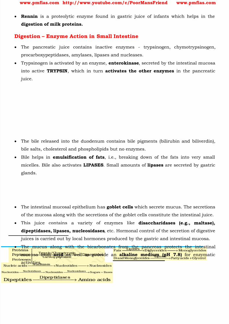
8/16/2019 Biology for Prelims Exam
http://slidepdf.com/reader/full/biology-for-prelims-exam 268/384
www.pmfias.com http://www.youtube.com/c/PoorMansFriend www.pmfias.com
Rennin is a proteolytic enzyme found in gastric juice of infants which helps in the
digestion of milk proteins.
Digestion – Enzyme Action in Small Intestine
The pancreatic juice contains inactive enzymes - trypsinogen, chymotrypsinogen,
procarboxypeptidases, amylases, lipases and nucleases. Trypsinogen is activated by an enzyme, enterokinase , secreted by the intestinal mucosa
into active TRYPSIN , which in turn activates the other enzymes in the pancreatic
juice.
The bile released into the duodenum contains bile pigments (bilirubin and biliverdin),
bile salts, cholesterol and phospholipids but no enzymes.
Bile helps in emulsification of fats , i.e., breaking down of the fats into very small
micelles. Bile also activates LIPASES . Small amounts of lipases are secreted by gastric
glands.
The intestinal mucosal epithelium has goblet cells which secrete mucus. The secretions
of the mucosa along with the secretions of the goblet cells constitute the intestinal juice.
This juice contains a variety of enzymes like disaccharidases (e.g., maltase),
dipeptidases, lipases, nucleosidases , etc. Hormonal control of the secretion of digestive
juices is carried out by local hormones produced by the gastric and intestinal mucosa.
The mucus along with the bicarbonates from the pancreas protects the intestinal
mucosa from acid as well as provide an alkaline medium (pH 7.8) for enzymatic
activities.
8/16/2019 Biology for Prelims Exam
http://slidepdf.com/reader/full/biology-for-prelims-exam 269/384
www.pmfias.com http://www.youtube.com/c/PoorMansFriend www.pmfias.com
The breakdown of biomacromolecules mentioned above occurs in the duodenum region
of the small intestine.
The simple substances thus formed are absorbed in the jejunum and ileum regions of
the small intestine.
The undigested and unabsorbed substances are passed on to the large intestine.
Absorption of Digested Products
Absorption is the process by which the end products of digestion pass through the
intestinal mucosa into the blood or lymph .
Small amounts of monosaccharides like glucose, amino acids and some electrolytes like
chloride ions are generally absorbed by simple diffusion . The passage of these
substances into the blood depends upon the concentration gradients.
However, sometimes substances like glucose and amino acids are absorbed with the
help of carrier proteins. This mechanism is called the facilitated transport .
Transport of water depends upon the osmotic gradient. Active transport occurs against
the concentration gradient and hence requires energy. Various nutrients like amino
acids, monosaccharides like glucose, electrolytes like Na+ are absorbed into the blood by
this mechanism.
Fatty acids and glycerol being insoluble , cannot be absorbed into the blood. They are
first incorporated into small droplets called micelles which move into the intestinal
mucosa. They are re-formed into very small protein coated fat globules called the
chylomicrons which are transported into the lymph vessels (lacteals) in the villi. These
lymph vessels ultimately release the absorbed substances into the blood stream.
Absorption of substances takes place in different parts of the alimentary canal, like
mouth, stomach, small intestine and large intestine. However, maximum absorption
occurs in the small intestine.
Summary of Absorption in Different Parts of Digestive System
Mouth Stomach Small Intestine LargeIntestine
Certain drugs coming incontact with the mucosaof mouth and lower sideof the tongue areabsorbed into the bloodcapillaries lining them.
Absorption ofwater, simplesugars, andalcohol etc.takes place.
Principal organ for absorption of nutrients. Thedigestion is completed here and the finalproducts of digestion such as glucose,fructose, fatty acids, glycerol and amino acidsare absorbed through the mucosa into theblood stream and lymph.
Absorption ofwater, someminerals anddrugs takesplace.
8/16/2019 Biology for Prelims Exam
http://slidepdf.com/reader/full/biology-for-prelims-exam 270/384
8/16/2019 Biology for Prelims Exam
http://slidepdf.com/reader/full/biology-for-prelims-exam 271/384
www.pmfias.com http://www.youtube.com/c/PoorMansFriend www.pmfias.com
Human Respiratory System - General Science NCERT - Respiration Breathing and Exchange of
Gases, Mechanism of Breathing, Mechanism of Breathing.
Respiration – Breathing and Exchange of Gases
Oxygen (O2) is utilized by the organisms to indirectly break down nutrient molecules
like glucose and to derive energy for performing various activities. Carbon dioxide (CO2)
which is harmful is also released during the above catabolic reactions . It is, therefore,
evident that O2 has to be continuously provided to the cells and CO2 produced by the
cells have to be released out. This process of exchange of O2 from the atmosphere with
CO2 produced by the cells is called breathing, commonly known as respiration.
Metabolic Pathways
Metabolic pathways that lead to a more complex structure from a simpler structure are
called biosynthetic pathways or anabolic pathways . Example: acetic acid becomescholesterol.
Metabolic pathways that lead to a simpler structure from a complex structure are called
catabolic pathways . Example: glucose becomes lactic acid in our skeletal muscle .
Anabolic pathways consume energy . Assembly of a protein from amino acids requires
energy input.
On the other hand, catabolic pathways lead to the release of energy. For example, when
glucose is degraded to lactic acid in our skeletal muscle, energy is liberated. Thismetabolic pathway from glucose to lactic acid which occurs in 10 metabolic steps is
called glycolysis .
Living organisms have learnt to trap this energy liberated during degradation and store
it in the form of chemical bonds.
As and when needed, this bond energy is utilized for biosynthetic, osmotic and
mechanical work that we perform.
The most important form of energy currency in living systems is the bond energy in achemical called adenosine triphosphate (ATP) .
Mechanisms of breathing vary among different groups of animals depending mainly on
their habitats and levels of organization.
Lower invertebrates like sponges, coelenterates, flatworms, etc., exchange O2 with CO2
by simple diffusion over their entire body surface.

8/16/2019 Biology for Prelims Exam
http://slidepdf.com/reader/full/biology-for-prelims-exam 272/384
www.pmfias.com http://www.youtube.com/c/PoorMansFriend www.pmfias.com
Earthworms use their moist cuticle and insects have a network of tubes ( tracheal
tubes ) to transport atmospheric air within the body.
Special vascularized structures called gills are used by most of the aquatic arthropods
and molluscs whereas vascularised bags called lungs are used by the terrestrial forms
for the exchange of gases.
Among vertebrates, fishes use gills whereas reptiles, birds and mammals respirethrough lungs. Amphibians like frogs can respire through their moist skin also.
Mammals usually have a well-developed respiratory system.
Human Respiratory System
We have a pair of external nostrils opening out above the upper lips. It leads to a nasal
chamber through the nasal passage. The nasal chamber opens into the pharynx , a
portion of which is the common passage for food and air.
The pharynx opens through the larynx region into the trachea. Larynx is a cartilaginous
box which helps in sound production and hence called the sound box .
During swallowing glottis can be covered by a thin elastic cartilaginous flap called
epiglottis to prevent the entry of food into the larynx.
Trachea is a straight tube which divides into a right and left primary bronchi . Each
bronchi undergoes repeated divisions to form the secondary and tertiary bronchi and
bronchioles ending up in very thin terminal bronchioles. The tracheae, primary,
secondary and tertiary bronchi are supported by incomplete cartilaginous rings .
Each terminal bronchiole gives rise to a number of very thin, irregular-walled and
vascularised bag-like structures called alveoli . The branching network of bronchi,
bronchioles and alveoli comprise the lungs.
We have two lungs which are covered by a double layered pleura , with pleural fluid
between them. It reduces friction on the lung-surface. The outer pleural membrane is
in close contact with the thoracic lining whereas the inner pleural membrane is in
contact with the lung surface.
The part starting with the external nostrils up to the terminal bronchioles constitute the
conducting part whereas the alveoli and their ducts form the respiratory or exchange
part of the respiratory system.
The conducting part transports the atmospheric air to the alveoli, clears it from foreign
particles, humidifies and also brings the air to body temperature . Exchange part is
the site of actual diffusion of O2 and CO2 between blood and atmospheric air.

8/16/2019 Biology for Prelims Exam
http://slidepdf.com/reader/full/biology-for-prelims-exam 273/384
www.pmfias.com http://www.youtube.com/c/PoorMansFriend www.pmfias.com
The lungs are situated in the thoracic chamber which is anatomically an air-tight
chamber. The thoracic chamber is formed dorsally by the vertebral column , ventrally
by the sternum [breastbone], laterally by the ribs and on the lower side by the dome-
shaped diaphragm .
The anatomical setup of lungs in thorax is such that any change in the volume of the
thoracic cavity will be reflected in the lung (pulmonary) cavity . Such an arrangement isessential for breathing, as we cannot directly alter the pulmonary volume.
Respiration involves the following steps:
1. Breathing or pulmonary ventilation by which atmospheric air is drawn in and CO2 rich
alveolar air is released out.
2. Diffusion of gases (O2 and CO2) across alveolar membrane.
3. Transport of gases by the blood.
4. Diffusion of O2 and CO2 between blood and tissues.
5. Utilisation of O2 by the cells for catabolic reactions and resultant release of CO2.
Mechanism of Breathing
Breathing involves two stages: inspiration during which atmospheric air is drawn in
and expiration by which the alveolar air is released out.
The movement of air into and out of the lungs is carried out by creating a pressure
gradient between the lungs and the atmosphere.
Inspiration can occur if the pressure within the lungs (intra-pulmonary pressure) is less
than the atmospheric pressure, i.e., there is a negative pressure in the lungs with
respect to atmospheric pressure. Similarly, expiration takes place when the intra-
pulmonary pressure is higher than the atmospheric pressure.
The diaphragm and a specialized set of muscles – external and internal intercostals
between the ribs, help in generation of such gradients.
Inspiration is initiated by the contraction of diaphragm which increases the volume of
thoracic chamber in the antero-posterior axis. The contraction of external inter-costal
muscles lifts up the ribs and the sternum causing an increase in the volume of the
thoracic chamber in the dorso-ventral axis. The overall increase in the thoracic
volume causes a similar increase in pulmonary volume .
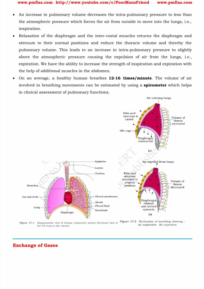
8/16/2019 Biology for Prelims Exam
http://slidepdf.com/reader/full/biology-for-prelims-exam 274/384
www.pmfias.com http://www.youtube.com/c/PoorMansFriend www.pmfias.com
An increase in pulmonary volume decreases the intra-pulmonary pressure to less than
the atmospheric pressure which forces the air from outside to move into the lungs, i.e.,
inspiration.
Relaxation of the diaphragm and the inter-costal muscles returns the diaphragm and
sternum to their normal positions and reduce the thoracic volume and thereby the
pulmonary volume. This leads to an increase in intra-pulmonary pressure to slightlyabove the atmospheric pressure causing the expulsion of air from the lungs, i.e.,
expiration. We have the ability to increase the strength of inspiration and expiration with
the help of additional muscles in the abdomen.
On an average, a healthy human breathes 12-16 times/minute . The volume of air
involved in breathing movements can be estimated by using a spirometer which helps
in clinical assessment of pulmonary functions.
Exchange of Gases

8/16/2019 Biology for Prelims Exam
http://slidepdf.com/reader/full/biology-for-prelims-exam 275/384
www.pmfias.com http://www.youtube.com/c/PoorMansFriend www.pmfias.com
Alveoli are the primary sites of exchange of gases. Exchange of gases also occur
between blood and tissues . O2 and CO2 are exchanged in these sites by simple
diffusion mainly based on pressure/concentration gradient.
Partial pressure of gasses, Solubility of the gases as well as the thickness of the
membranes involved in diffusion are some important factors that can affect the rate of
diffusion. Pressure contributed by an individual gas in a mixture of gases is called partial
pressure.
Transport of Gases
Blood is the medium of transport for O2 and CO2.
About 97 per cent of O2 is transported by RBCs in the blood. The remaining 3 per cent
of O2 is carried in a dissolved state through the plasma.
Nearly 20-25 per cent of CO2 is transported by RBCs whereas 70 per cent of it is carried
as bicarbonate . About 7 per cent of CO2 is carried in a dissolved state through
plasma.
Transport of Oxygen
Haemoglobin is a red coloured iron containing pigment present in the RBCs. O2 can
bind with haemoglobin in a reversible manner to form oxyhaemoglobin .
Each haemoglobin molecule can carry a maximum of four molecules of O2. Binding ofoxygen with haemoglobin is primarily related to partial pressure of O2.
Partial pressure of CO2, hydrogen ion concentration and temperature are the other
factors which can interfere with this binding.
Transport of Carbon dioxide

8/16/2019 Biology for Prelims Exam
http://slidepdf.com/reader/full/biology-for-prelims-exam 276/384
www.pmfias.com http://www.youtube.com/c/PoorMansFriend www.pmfias.com
CO2 is carried by haemoglobin as carbamino-haemoglobin (about 20-25 per cent). This
binding is related to the partial pressure of CO2. Partial pressure of O2 is a major factor
which could affect this binding. RBCs contain a very high concentration of the enzyme,
carbonic anhydrase and minute quantities of the same is present in the plasma too.
Nearly 70 per cent of carbon dioxide is transported as bicarbonate (HCO3) with the help
of the enzyme carbonic anhydrase. At the tissue site where partial pressure of CO2 is high due to catabolism [ the breakdown of
complex molecules in living organisms to form simpler ones, together with the release of energy ], CO2 diffuses
into blood (RBCs and plasma) and forms HCO3 and H+-.
At the alveolar site where pCO2 is low, the reaction proceeds in the opposite direction
leading to the formation of CO2 and H2O.
Thus, CO2 trapped as bicarbonate at the tissue level and transported to the alveoli is
released out as CO2. Every 100 ml of deoxygenated blood delivers approximately 4 ml ofCO2 to the alveoli.
Regulation of Respiration
Human beings have a significant ability to maintain and moderate the respiratory
rhythm to suit the demands of the body tissues. This is done by the neural system.
A specialised centre present in the medulla region of the brain called respiratory
rhythm centre is primarily responsible for this regulation.
Another centre present in the pons region of the brain called pneumotaxic centre can
moderate the functions of the respiratory rhythm centre. Neural signal from this centre
can reduce the duration of inspiration and thereby alter the respiratory rate.
A chemosensitive area is situated adjacent to the rhythm centre which is highly sensitive
to CO2 and hydrogen ions. Increase in these substances can activate this centre, which
in turn can signal the rhythm centre to make necessary adjustments in the respiratory
process by which these substances can be eliminated.
Disorders of Respiratory System
Asthma: Asthma is a difficulty in breathing causing wheezing due to inflammation of
bronchi and bronchioles.
Emphysema: Emphysema is a chronic disorder in which alveolar walls are damaged
due to which respiratory surface is decreased. One of the major causes of this is
cigarette smoking .

8/16/2019 Biology for Prelims Exam
http://slidepdf.com/reader/full/biology-for-prelims-exam 277/384
www.pmfias.com http://www.youtube.com/c/PoorMansFriend www.pmfias.com
Occupational Respiratory Disorders: In certain industries, especially those involving
grinding or stone-breaking, so much dust is produced that the defense mechanism of
the body cannot fully cope with the situation. Long exposure can give rise to
inflammation leading to fibrosis (proliferation of fibrous tissues) and thus causing
serious lung damage. Workers in such industries should wear protective masks.

8/16/2019 Biology for Prelims Exam
http://slidepdf.com/reader/full/biology-for-prelims-exam 278/384
www.pmfias.com http://www.youtube.com/c/PoorMansFriend www.pmfias.com
Human Excretory System – Kidneys, Urine Formation, Tubules: Proximal Convoluted
Tubule (PCT), Henle’s Loop, Distal Convoluted Tubule (DCT), Collecting Duct.
Excretory System
When our cells perform their functions, certain waste products are released in to the
blood stream. These are toxic and hence need to be removed from the body. The process of removal of wastes produced in the cells of the living organisms is called
excretion. The parts involved in excretion forms the excretory system.
Waste removal is done by the blood capillaries in the kidneys.
When the blood reaches the two kidneys, it contains both useful and harmful
substances. The useful substances are absorbed back into the blood. The wastes are
removed as urine.
From the kidneys, the urine goes into the urinary bladder through tube-like ureters . It
is stored in the bladder and is passed out through the urinary opening at the end of a
muscular tube called urethra . The kidneys, ureters, bladder and urethra form the
excretory system.
An adult human being normally passes about 1 – 1.8 L of urine in 24 hours, and the
urine consists of 95% water, 2.5 % urea and 2.5% other waste products .
Excretory Products and their Elimination
Animals accumulate ammonia, urea, uric acid, carbon dioxide, water and ions like
Na+, K+, Cl -, phosphate, sulphate, etc., either by metabolic activities or by other means
like excess ingestion. These substances have to be removed totally or partially.
Ammonia, urea and uric acid are the major forms of nitrogenous wastes excreted by
the animals.
The way in which waste chemicals are removed from the body of the animal depends on
the availability of water.
Ammonia is the most toxic form and requires large amount of water for its
elimination, whereas uric acid, being the least toxic , can be removed with a minimum
loss of water.
Aquatic animals like fishes, excrete cell waste in gaseous form (ammonia) which
directly dissolves in water.
Some land animals like birds, lizards, snakes excrete a semi-solid, white coloured
compound ( uric acid ).
8/16/2019 Biology for Prelims Exam
http://slidepdf.com/reader/full/biology-for-prelims-exam 279/384
www.pmfias.com http://www.youtube.com/c/PoorMansFriend www.pmfias.com
The major excretory product in humans is urea which is excreted through urine.
Sometimes a person’s kidneys may stop working due to infection or injury. As a result of
kidney failure, waste products start accumulating in the blood. Such persons cannot
survive unless their blood is filtered periodically through an artificial kidney. This
process is called dialysis .
The process of excreting ammonia is Ammonotelism. Many bony fishes, aquaticamphibians and aquatic insects are ammonotelic in nature.
Ammonia, as it is readily soluble, is generally excreted by diffusion across body surfaces
or through gill surfaces (in fish) as ammonium ions. Kidneys do not play any
significant role in its removal.
Terrestrial adaptation necessitated the production of lesser toxic nitrogenous wastes like
urea and uric acid for conservation of water.
Mammals, many terrestrial amphibians and marine fishes mainly excrete urea and arecalled ureotelic animals . Ammonia produced by metabolism is converted into urea in
the liver of these animals and released into the blood which is filtered and excreted out
by the kidneys.
Some amount of urea may be retained in the kidney matrix of some of these animals to
maintain a desired osmolarity [ the concentration of a solution expressed as the total number of solute
particles per litre ].
Reptiles, birds, land snails and insects excrete nitrogenous wastes as uric acid in the
form of pellet or paste with a minimum loss of water and are called uricotelic animals .
A survey of animal kingdom presents a variety of excretory structures. In most of the
invertebrates, these structures are simple tubular forms whereas vertebrates have
complex tubular organs called kidneys. Some of these structures are mentioned here.
Protonephridia or flame cells are the excretory structures in Platyhelminthes
(Flatworms, e.g., Planaria), rotifers, some annelids and the cephalochordate.
Protonephridia are primarily concerned with ionic and fluid volume regulation, i.e.,
osmoregulation. Nephridia are the tubular excretory structures of earthworms and
other annelids. Nephridia help to remove nitrogenous wastes and maintain a fluid and
ionic balance.
Malpighian tubules are the excretory structures of most of the insects including
cockroaches. Malpighian tubules help in the removal of nitrogenous wastes and
osmoregulation.
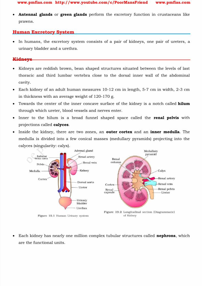
8/16/2019 Biology for Prelims Exam
http://slidepdf.com/reader/full/biology-for-prelims-exam 280/384
www.pmfias.com http://www.youtube.com/c/PoorMansFriend www.pmfias.com
Antennal glands or green glands perform the excretory function in crustaceans like
prawns.
Human Excretory System
In humans, the excretory system consists of a pair of kidneys, one pair of ureters, a
urinary bladder and a urethra.
Kidneys
Kidneys are reddish brown, bean shaped structures situated between the levels of last
thoracic and third lumbar vertebra close to the dorsal inner wall of the abdominal
cavity.
Each kidney of an adult human measures 10-12 cm in length, 5-7 cm in width, 2-3 cm
in thickness with an average weight of 120-170 g.
Towards the center of the inner concave surface of the kidney is a notch called hilum
through which ureter, blood vessels and nerves enter.
Inner to the hilum is a broad funnel shaped space called the renal pelvis with
projections called calyces .
Inside the kidney, there are two zones, an outer cortex and an inner medulla . The
medulla is divided into a few conical masses (medullary pyramids) projecting into the
calyces (singularity: calyx).
Each kidney has nearly one million complex tubular structures called nephrons , which
are the functional units.
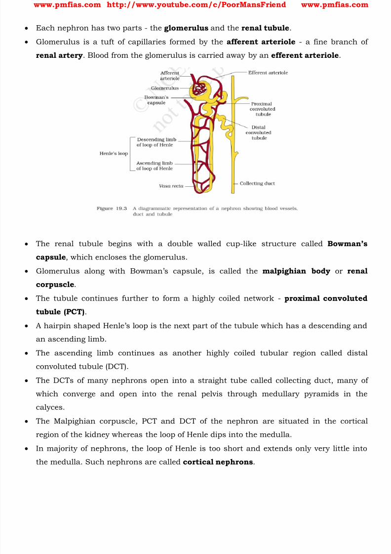
8/16/2019 Biology for Prelims Exam
http://slidepdf.com/reader/full/biology-for-prelims-exam 281/384
www.pmfias.com http://www.youtube.com/c/PoorMansFriend www.pmfias.com
Each nephron has two parts - the glomerulus and the renal tubule .
Glomerulus is a tuft of capillaries formed by the afferent arteriole - a fine branch of
renal artery . Blood from the glomerulus is carried away by an efferent arteriole .
The renal tubule begins with a double walled cup-like structure called Bowman’s
capsule , which encloses the glomerulus.
Glomerulus along with Bowman’s capsule, is called the malpighian body or renal
corpuscle .
The tubule continues further to form a highly coiled network - proximal convoluted
tubule (PCT) .
A hairpin shaped Henle’s l oop is the next part of the tubule which has a descending and
an ascending limb.
The ascending limb continues as another highly coiled tubular region called distal
convoluted tubule (DCT).
The DCTs of many nephrons open into a straight tube called collecting duct, many of
which converge and open into the renal pelvis through medullary pyramids in the
calyces.
The Malpighian corpuscle, PCT and DCT of the nephron are situated in the cortical
region of the kidney whereas the loop of Henle dips into the medulla.
In majority of nephrons, the loop of Henle is too short and extends only very little into
the medulla. Such nephrons are called cortical nephrons .

8/16/2019 Biology for Prelims Exam
http://slidepdf.com/reader/full/biology-for-prelims-exam 282/384
www.pmfias.com http://www.youtube.com/c/PoorMansFriend www.pmfias.com
In some of the nephrons, the loop of Henle is very long and runs deep into the medulla.
These nephrons are called juxta medullary nephrons .
The efferent arteriole emerging from the glomerulus forms a fine capillary network
around the renal tubule called the peritubular capillaries .
A minute vessel of this network runs parallel to the Henle’s loop forming a ‘U’ shaped
vasa recta. Vasa recta is absent or highly reduced in cortical nephrons.
Urine Formation
Urine formation involves three main processes namely, glomerular filtration,
reabsorption and secretion , that takes place in different parts of the nephron.
The first step in urine formation is the filtration of blood, which is carried out by the
glomerulus and is called glomerular filtration .
On an average, 1100-1200 ml of blood is filtered by the kidneys per minute.
The glomerular capillary blood pressure causes filtration of blood through 3 layers, i.e.,
the endothelium of glomerular blood vessels , the epithelium of Bowman’s capsule
and a basement membrane between these two layers.
The epithelial cells of Bowman’s capsule called podocytes are arranged in an intricate
manner so as to leave some minute spaces called filtration slits or slit pores. Blood is
filtered so finely through these membranes, that almost all the constituents of the
plasma except the proteins pass onto the lumen of the Bowman’s capsule. Therefore, it
is considered as a process of ultra-filtration .
The amount of the filtrate formed by the kidneys per minute is called glomerular
filtration rate (GFR) . GFR in a healthy individual is approximately 125 ml/minute, i.e.,
180 liters per day !

8/16/2019 Biology for Prelims Exam
http://slidepdf.com/reader/full/biology-for-prelims-exam 283/384
www.pmfias.com http://www.youtube.com/c/PoorMansFriend www.pmfias.com
The kidneys have built-in mechanisms for the regulation of glomerular filtration rate.
One such efficient mechanism is carried out by juxta glomerular apparatus (JGA) .
A comparison of the volume of the filtrate formed per day (180 liters per day) with that of
the urine released (1.5 litres), suggest that nearly 99 per cent of the filtrate has to be
reabsorbed by the renal tubules. This process is called reabsorption .
The tubular epithelial cells in different segments of nephron perform this either byactive or passive mechanisms. For example, substances like glucose, amino acids, Na+,
etc., in the filtrate are reabsorbed actively whereas the nitrogenous wastes are absorbed
by passive transport. Reabsorption of water also occurs passively in the initial segments
of the nephron.
During urine formation, the tubular cells secrete substances like H+, K+ and ammonia
into the filtrate. Tubular secretion is also an important step in urine formation as it
helps in the maintenance of ionic and acid base balance of body fluids.
Function of the Tubules
Proximal Convoluted Tubule (PCT)
PCT is lined by simple cuboidal epithelium which increases the surface area for
reabsorption. Nearly all of the essential nutrients, and 70-80 per cent of electrolytes and
water are reabsorbed by this segment.
PCT also helps to maintain the pH and ionic balance of the body fluids by selectivesecretion of hydrogen ions, ammonia and potassium ions into the filtrate and by
absorption of HCO3- from it.
Henle’s Loop
Reabsorption is minimum in its ascending limb. However, this region plays a significant
role in the maintenance of high osmolarity of medullary interstitial fluid.
The descending limb of loop of Henle is permeable to water but almost impermeable to
electrolytes. This concentrates the filtrate as it moves down.
The ascending limb is impermeable to water but allows transport of electrolytes actively
or passively. Therefore, as the concentrated filtrate pass upward, it gets diluted due to
the passage of electrolytes to the medullary fluid.
Distal Convoluted Tubule (DCT)
8/16/2019 Biology for Prelims Exam
http://slidepdf.com/reader/full/biology-for-prelims-exam 284/384
www.pmfias.com http://www.youtube.com/c/PoorMansFriend www.pmfias.com
Conditional reabsorption of Na+ and water takes place in this segment. DCT is also
capable of reabsorption of HCO3- and selective secretion of hydrogen and potassium
ions and NH3 to maintain the pH and sodium-potassium balance in blood.
Collecting Duct
This long duct extends from the cortex of the kidney to the inner parts of the medulla. Large amounts of water could be reabsorbed from this region to produce a concentrated
urine.
This segment allows passage of small amounts of urea into the medullary interstitium to
keep up the osmolarity.
It also plays a role in the maintenance of pH and ionic balance of blood by the selective
secretion of H+ and K+ ions.
Mechanism of Concentration of the Filtrate
Mammals have the ability to produce a concentrated urine. The Henle’s loop and vasa
recta play a significant role in this.
The flow of filtrate in the two limbs of Henle’s loop is in opposite directions and thus
forms a counter current.
The flow of blood through the two limbs of vasa recta is also in a counter current
pattern.
The proximity bet ween the Henle’s loop and vasa recta, as well as the counter current in
them help in maintaining an increasing osmolarity towards the inner medullary
interstitium. This gradient is mainly caused by NaCl and urea .
NaCl is transported by the ascending limb of Henle’s loop which is exchanged with the
descending limb of vasa recta. NaCl is returned to the interstitium by the ascending
portion of vasa recta.
Similarly, small amounts of urea enter the thin segment of the ascending limb of Henle’s
loop which is transported back to the interstitium by the collecting tubule.
The above described transport of substances facilitated by the special arrangement of
Henle’s loop and vasa recta is called the counter current mechanism . This mechanism
helps to maintain a concentration gradient in the medullary interstitium.
Presence of such interstitial gradient helps in an easy passage of water from the
collecting tubule thereby concentrating the filtrate (urine). Human kidneys can produce
urine nearly four times concentrated than the initial filtrate formed.
8/16/2019 Biology for Prelims Exam
http://slidepdf.com/reader/full/biology-for-prelims-exam 285/384
www.pmfias.com http://www.youtube.com/c/PoorMansFriend www.pmfias.com
Regulation of Kidney Function
The functioning of the kidneys is efficiently monitored and regulated by hormonal
feedback mechanisms involving the hypothalamus, JGA and to a certain extent, the
heart .
Osmoreceptors in the body are activated by changes in blood volume, body fluid volume
and ionic concentration. An excessive loss of fluid from the body can activate these
receptors which stimulate the hypothalamus to release antidiuretic hormone (ADH) or
vasopressin from the neurohypophysis .
ADH facilitates water reabsorption from latter parts of the tubule, thereby preventing
diuresis [ increased or excessive production of urine ].
An increase in body fluid volume can switch off the osmoreceptors and suppress the
ADH release to complete the feedback.
ADH can also affect the kidney function by its constrictory effects on blood vessels. This
causes an increase in blood pressure . An increase in blood pressure can increase the
glomerular blood flow and thereby the GFR.
The JGA plays a complex regulatory role. A fall in glomerular blood flow/glomerular
blood pressure/GFR can activate the JG cells to release renin which converts
angiotensinogen in blood to angiotensin I and further to angiotensin II.
Angiotensin II, being a powerful vasoconstrictor, increases the glomerular blood
pressure and thereby GFR. Angiotensin II also activates the adrenal cortex to release Aldosterone . Aldosterone
causes reabsorption of Na+ and water from the distal parts of the tubule. This also leads
to an increase in blood pressure and GFR. This complex mechanism is generally known
as the Renin-Angiotensin mechanism .
An increase in blood flow to the atria of the heart can cause the release of Atrial
Natriuretic Factor (ANF) . ANF can cause vasodilation (dilation of blood vessels) and
thereby decrease the blood pressure. ANF mechanism, therefore, acts as a check on therenin-angiotensin mechanism.
Micturition
Urine formed by the nephrons is ultimately carried to the urinary bladder where it is
stored till a voluntary signal is given by the central nervous system (CNS). This signal is
initiated by the stretching of the urinary bladder as it gets filled with urine. In response,
8/16/2019 Biology for Prelims Exam
http://slidepdf.com/reader/full/biology-for-prelims-exam 286/384
www.pmfias.com http://www.youtube.com/c/PoorMansFriend www.pmfias.com
the stretch receptors on the walls of the bladder send signals to the CNS. The CNS
passes on motor messages to initiate the contraction of smooth muscles of the bladder
and simultaneous relaxation of the urethral sphincter causing the release of urine. The
process of release of urine is called micturition and the neural mechanisms causing it
is called the micturition reflex .
An adult human excretes, on an average, 1 to 1.5 litres of urine per day. The urineformed is a light yellow coloured watery fluid which is slightly acidic (pH-6.0) and has a
characterestic odour.
On an average, 25-30 gm of urea is excreted out per day. Various conditions can affect
the characteristics of urine.
Analysis of urine helps in clinical diagnosis of many metabolic discorders as well as
malfunctioning of the kidney. For example, presence of glucose (Glycosuria) and
ketone bodies (Ketonuria) in urine are indicative of diabetes mellitus .
Role of other Organs in Excretion
Other than the kidneys, lungs, liver and skin also help in the elimination of excretory
wastes.
Our lungs remove large amounts of CO2 (approximately 200mL/ minute) and also
significant quantities of water every day.
Liver, the largest gland in our body, secretes bile-containing substances like bilirubin,
biliverdin, cholesterol, degraded steroid hormones, vitamins and drugs . Most of
these substances ultimately pass out alongwith digestive wastes.
The sweat and sebaceous glands in the skin can eliminate certain substances through
their secretions. Sweat produced by the sweat glands is a watery fluid containing NaCl
small amounts of urea, lactic acid , etc.
Though the primary function of sweat is to facilitate a cooling effect on the body surface,
it also helps in the removal of some of the wastes mentioned above.
Sebaceous glands eliminate certain substances like sterols, hydrocarbons and waxes
through sebum. This secretion provides a protective oily covering for the skin. Small
amounts of nitrogenous wastes could be eliminated through saliva too.
Disorders of the Excretory System

8/16/2019 Biology for Prelims Exam
http://slidepdf.com/reader/full/biology-for-prelims-exam 287/384
www.pmfias.com http://www.youtube.com/c/PoorMansFriend www.pmfias.com
Malfunctioning of kidneys can lead to accumulation of urea in blood, a condition called
uremia , which is highly harmful and may lead to kidney failure. In such patients, urea
can be removed by a process called hemodialysis .
Blood drained from a convenient artery is pumped into a dialyzing unit after adding an
anticoagulant like heparin . The unit contains a coiled cellophane tube surrounded by a
fluid (dialyzing fluid) having the same composition as that of plasma except thenitrogenous wastes.
The porous cellophane membrane of the tube allows the passage of molecules based on
concentration gradient. As nitrogenous wastes are absent in the dialyzing fluid, these
substances freely move out, thereby clearing the blood.
The cleared blood is pumped back to the body through a vein after adding anti-heparin
to it. This method is a boon for thousands of uremic patients all over the world.
Kidney transplantation is the ultimate method in the correction of acute renal failures(kidney failure). A functioning kidney is used in transplantation from a donor, preferably
a close relative, to minimise its chances of rejection by the immune system of the host.
Modern clinical procedures have increased the success rate of such a complicated
technique.
Renal calculi: Stone or insoluble mass of crystallised salts ( oxalates , etc.) formed
within the kidney.
Glomerulonephritis: Inflammation of glomeruli of kidney.
Summary
Many nitrogen containing substances, ions, CO2, water, etc., that accumulate in the
body have to be eliminated.
Nature of nitrogenous wastes formed and their excretion vary among animals, mainly
depending on the habitat (availability of water).
Ammonia, urea and uric acid are the major nitrogenous wastes excreted.
Protonephridia, nephridia, malpighian tubules, green glands and the kidneys are
the common excretory organs in animals. They not only eliminate nitrogenous wastes
but also help in the maintenance of ionic and acid-base balance of body fluids.
In humans, the excretory system consists of one pair of kidneys, a pair of ureters, a
urinary bladder and a urethra .
Each kidney has over a million tubular structures called nephrons . Nephron is the
functional unit of kidney and has two portions - glomerulus and renal tubule .

8/16/2019 Biology for Prelims Exam
http://slidepdf.com/reader/full/biology-for-prelims-exam 288/384
www.pmfias.com http://www.youtube.com/c/PoorMansFriend www.pmfias.com
Glomerulus is a tuft of capillaries formed from afferent arterioles, fine branches of renal
artery.
The renal tubule starts with a double walled Bowman’s capsule and is further
differentiated into a proximal convoluted tubule (PCT), Henle’s loop (HL) and distal
convoluted tubule (DCT) .
The DCTs of many nephrons join to a common collecting duct many of which ultimatelyopen into the renal pelvis through the medullary pyramids. The Bowman’s capsule
encloses the glomerulus to form Malpighian or renal corpuscle .
Urine formation involves three main processes, i.e., filtration, reabsorption and
secretion .
Filtration is a non-selective process performed by the glomerulus using the glomerular
capillary blood pressure. About 1200 ml of blood is filtered by the glomerulus per
minute to form 12 5 ml of filtrate in the Bowman’s capsule per minute (GFR). JGA, a specialised portion of the nephrons, plays a significant role in the regulation of
GFR.
Nearly 99 per cent reabsorption of the filtrate takes place through different parts of the
nephrons.
PCT is the major site of reabsorption and selective secretion. HL [Henle’s Loop] primarily
helps to maintain osmolar gradient within the kidney interstitium.
DCT and collecting duct allow extensive reabsorption of water and certain electrolytes,
which help in osmoregulation: H+, K+ and NH3 could be secreted into the filtrate by the
tubules to maintain the ionic balance and pH of body fluids.
A counter current mechanism operates between the two limbs of the loop of Henle and
those of vasa recta (capillary parallel to Henle’s loop). The filtrate gets concentrated as it
moves down the descending limb but is diluted by the ascending limb. Electrolytes and
urea are retained in the interstitium by this arrangement.
DCT and collecting duct concentrate the filtrate about four times, an excellent
mechanism of conservation of water.
Urine is stored in the urinary bladder till a voluntary signal from CNS carries out its
release through urethra, i.e., micturition. Skin, lungs and liver also assist in excretion.
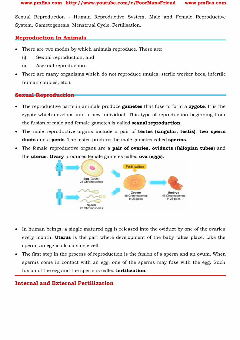
8/16/2019 Biology for Prelims Exam
http://slidepdf.com/reader/full/biology-for-prelims-exam 289/384
www.pmfias.com http://www.youtube.com/c/PoorMansFriend www.pmfias.com
Sexual Reproduction - Human Reproductive System, Male and Female Reproductive
System, Gametogenesis, Menstrual Cycle, Fertilisation.
Reproduction In Animals
There are two modes by which animals reproduce. These are:
(i)
Sexual reproduction, and(ii) Asexual reproduction.
There are many organisms which do not reproduce (mules, sterile worker bees, infertile
human couples, etc.).
Sexual Reproduction
The reproductive parts in animals produce gametes that fuse to form a zygote . It is the
zygote which develops into a new individual. This type of reproduction beginning from
the fusion of male and female gametes is called sexual reproduction .
The male reproductive organs include a pair of testes (singular, testis) , two sperm
ducts and a penis . The testes produce the male gametes called sperms .
The female reproductive organs are a pair of ovaries, oviducts (fallopian tubes) and
the uterus . Ovary produces female gametes called ova (eggs) .
In human beings, a single matured egg is released into the oviduct by one of the ovaries
every month. Uterus is the part where development of the baby takes place. Like thesperm, an egg is also a single cell.
The first step in the process of reproduction is the fusion of a sperm and an ovum. When
sperms come in contact with an egg, one of the sperms may fuse with the egg. Such
fusion of the egg and the sperm is called fertilization .
Internal and External Fertilization
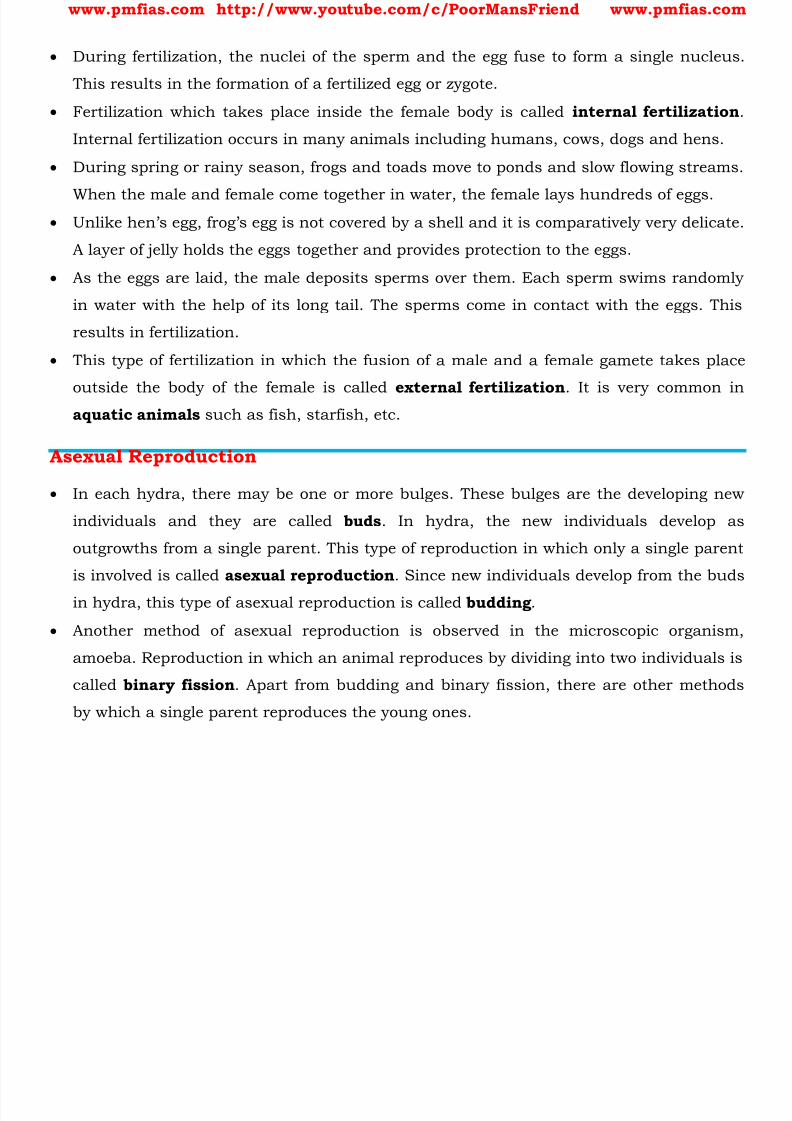
8/16/2019 Biology for Prelims Exam
http://slidepdf.com/reader/full/biology-for-prelims-exam 290/384
www.pmfias.com http://www.youtube.com/c/PoorMansFriend www.pmfias.com
During fertilization, the nuclei of the sperm and the egg fuse to form a single nucleus.
This results in the formation of a fertilized egg or zygote.
Fertilization which takes place inside the female body is called internal fertilization
Internal fertilization occurs in many animals including humans, cows, dogs and hens.
During spring or rainy season, frogs and toads move to ponds and slow flowing streams.
When the male and female come together in water, the female lays hundreds of eggs. Unlike hen’s egg, frog’s egg is not covered by a shell and it is comparatively very delicate.
A layer of jelly holds the eggs together and provides protection to the eggs.
As the eggs are laid, the male deposits sperms over them. Each sperm swims randomly
in water with the help of its long tail. The sperms come in contact with the eggs. This
results in fertilization.
This type of fertilization in which the fusion of a male and a female gamete takes place
outside the body of the female is called external fertilization . It is very common inaquatic animals such as fish, starfish, etc.
Asexual Reproduction
In each hydra, there may be one or more bulges. These bulges are the developing new
individuals and they are called buds . In hydra, the new individuals develop as
outgrowths from a single parent. This type of reproduction in which only a single parent
is involved is called asexual reproduction . Since new individuals develop from the buds
in hydra, this type of asexual reproduction is called budding .
Another method of asexual reproduction is observed in the microscopic organism,
amoeba. Reproduction in which an animal reproduces by dividing into two individuals is
called binary fission . Apart from budding and binary fission, there are other methods
by which a single parent reproduces the young ones.

8/16/2019 Biology for Prelims Exam
http://slidepdf.com/reader/full/biology-for-prelims-exam 291/384
www.pmfias.com http://www.youtube.com/c/PoorMansFriend www.pmfias.com
Human Reproductive System
The reproductive events in humans include
1. formation of gametes ( gametogenesis ), i.e., sperms in males and ovum in females,
2. transfer of sperms into the female genital tract ( insemination ),
3. fusion of male and female gametes ( fertilisation ) leading to formation of zygote .
4. formation and development of blastocyst and its attachment to the uterine wall
(implantation ),
5. embryonic development ( gestation ) and
6. delivery of the baby ( parturition ).
Male Reproductive System The male reproductive system is located in the pelvis region. It includes a pair of testes
along with accessory ducts, glands and the external genitalia .
The testes are situated outside the abdominal cavity within a pouch called scrotum . The
scrotum helps in maintaining the low temperature of the testes (2 – 2.5 0C lower than
the normal internal body temperature) necessary for spermatogenesis.
Each testis has about 250 testicular lobules. Each lobule contains one to three highly
coiled seminiferous tubules in which sperms are produced . Each seminiferous tubule is lined on its inside by two types of cells called male germ
cells (spermatogonia) and Sertoli cells .
The male germ cells undergo meiotic divisions finally leading to sperm formation, while
Sertoli cells provide nutrition to the germ cells.
The regions outside the seminiferous tubules called interstitial spaces, contain small
blood vessels and interstitial cells or Leydig cells . Leydig cells synthesise and secrete
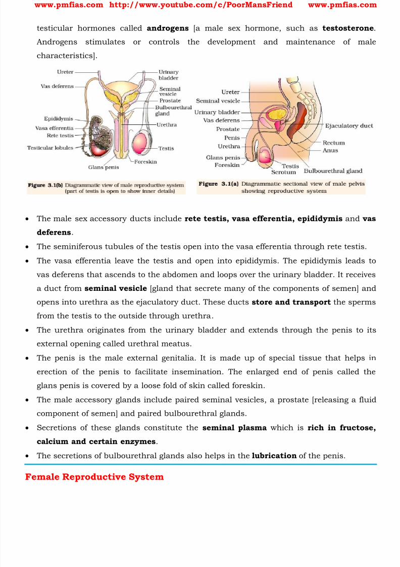
8/16/2019 Biology for Prelims Exam
http://slidepdf.com/reader/full/biology-for-prelims-exam 292/384
www.pmfias.com http://www.youtube.com/c/PoorMansFriend www.pmfias.com
testicular hormones called androgens [a male sex hormone, such as testosterone
Androgens stimulates or controls the development and maintenance of male
characteristics].
The male sex accessory ducts include rete testis, vasa efferentia, epididymis and vas
deferens .
The seminiferous tubules of the testis open into the vasa efferentia through rete testis.
The vasa efferentia leave the testis and open into epididymis. The epididymis leads to
vas deferens that ascends to the abdomen and loops over the urinary bladder. It receives
a duct from seminal vesicle [gland that secrete many of the components of semen] and
opens into urethra as the ejaculatory duct. These ducts store and transport the sperms
from the testis to the outside through urethra.
The urethra originates from the urinary bladder and extends through the penis to its
external opening called urethral meatus.
The penis is the male external genitalia. It is made up of special tissue that helps in
erection of the penis to facilitate insemination. The enlarged end of penis called the
glans penis is covered by a loose fold of skin called foreskin.
The male accessory glands include paired seminal vesicles, a prostate [releasing a fluid
component of semen] and paired bulbourethral glands.
Secretions of these glands constitute the seminal plasma which is rich in fructose,
calcium and certain enzymes .
The secretions of bulbourethral glands also helps in the lubrication of the penis.
Female Reproductive System

8/16/2019 Biology for Prelims Exam
http://slidepdf.com/reader/full/biology-for-prelims-exam 293/384
www.pmfias.com http://www.youtube.com/c/PoorMansFriend www.pmfias.com
The female reproductive system consists of a pair of ovaries along with a pair of
oviducts, uterus, cervix, vagina and the external genitalia located in pelvic region.
These parts of the system along with a pair of the mammary glands are integrated
structurally and functionally to support the processes of ovulation, fertilisation,
pregnancy, birth and child care.
Ovaries are the primary female sex organs [testis in males] that produce the femalegamete ( ovum ) [sperm in males] and several steroid hormones (ovarian hormones).
The ovaries are located one on each side of the lower abdomen. Each ovary is connected
to the pelvic wall and uterus by ligaments.
Each ovary is covered by a thin epithelium which encloses the ovarian stroma. The
stroma is divided into two zones – a peripheral cortex and an inner medulla.
The oviducts (fallopian tubes), uterus and vagina constitute the female accessory
ducts.
Each fallopian tube extends from the periphery of each ovary to the uterus, the part
closer to the ovary is the funnel-shaped infundibulum.
The edges of the infundibulum possess finger-like projections called fimbriae, which
help in collection of the ovum after ovulation . The infundibulum leads to a wider part
of the oviduct called ampulla.
The last part of the oviduct, isthmus has a narrow lumen and it joins the uterus. The
uterus is single and it is also called womb. The shape of the uterus is like an inverted
pear.
It is supported by ligaments attached to the pelvic wall. The uterus opens into vagina
through a narrow cervix. The cavity of the cervix is called cervical canal which along
with vagina forms the birth canal.
8/16/2019 Biology for Prelims Exam
http://slidepdf.com/reader/full/biology-for-prelims-exam 294/384
www.pmfias.com http://www.youtube.com/c/PoorMansFriend www.pmfias.com
The wall of the uterus has three layers of tissue. The external thin membranous
perimetrium, middle thick layer of smooth muscle, myometrium and inner glandular
layer called endometrium that lines the uterine cavity.
The endometrium undergoes cyclical changes during menstrual cycle while the
myometrium exhibits strong contraction during delivery of the baby.
The female external genitalia include mons pubis, labia majora, labia minora, hymenand clitoris.
Mons pubis is a cushion of fatty tissue covered by skin and pubic hair. The labia majora
are fleshy folds of tissue, which extend down from the mons pubis and surround the
vaginal opening.
Thelabia minora are paired folds of tissue under the labia majora. The opening of the
vagina is often covered partially by a membrane called hymen.
The clitoris is a tiny finger-like structure which lies at the upper junction of the two
labia minora above the urethral opening.
The hymen is often torn during the first coitus (intercourse) . However, it can also be
broken by a sudden fall or jolt, insertion of a vaginal tampon, active participation in
some sports like horseback riding, cycling, etc. In some women the hymen persists even after coitus. In fact, the presence or absence of
hymen is not a reliable indicator of virginity or sexual experience.
A functional mammary gland is characteristic of all female mammals. The mammary
glands are paired structures (breasts) that contain glandular tissue and variable
amount of fat.
8/16/2019 Biology for Prelims Exam
http://slidepdf.com/reader/full/biology-for-prelims-exam 295/384
www.pmfias.com http://www.youtube.com/c/PoorMansFriend www.pmfias.com
The glandular tissue of each breast is divided into 15-20 mammary lobes containing
clusters of cells called alveoli . The cells of alveoli secrete milk , which is stored in the
cavities (lumens) of alveoli. The alveoli open into mammary tubules.
The tubules of each lobe join to form a mammary duct. Several mammary ducts join to
form a wider mammary ampulla which is connected to lactiferous duct through which
milk is sucked out.
Gametogenesis
The primary sex organs – the testis in the males and the ovaries in the females
produce gametes, i.e, sperms and ovum , respectively, by the process called
gametogenesis .
In testis, the immature male germ cells (spermatogonia) produce sperms by
spermatogenesis that begins at puberty.
The spermatogonia (sing. spermatogonium) present on the inside wall of seminiferous
tubules multiply by mitotic division and increase in numbers. Each spermatogonium
is diploid and contains 46 chromosomes.
Some of the spermatogonia called primary spermatocytes periodically undergo meiosis .
A primary spermatocyte completes the first meiotic division (reduction division) leading
to formation of two equal, haploid cells called secondary spermatocytes, which have
only 23 chromosomes each.
The secondary spermatocytes undergo the second meiotic division to produce four
equal, haploid spermatids.

8/16/2019 Biology for Prelims Exam
http://slidepdf.com/reader/full/biology-for-prelims-exam 296/384
www.pmfias.com http://www.youtube.com/c/PoorMansFriend www.pmfias.com
What would be the number of chromosome in the spermatids? 23 chromosomes.
The spermatids are transformed into spermatozoa (sperms) by the process called
spermiogenesis. After spermiogenesis, sperm heads become embedded in the Sertoli
cells, and are finally released from the seminiferous tubules by the process called
spermiation.
Spermatogenesis starts at the age of puberty due to significant increase in the secretionof gonadotropin releasing hormone (GnRH) . This, if you recall, is a hypothalamic
hormone .
The increased levels of GnRH then acts at the anterior pituitary gland and stimulates
secretion of two gonadotropins – luteinising hormone (LH) and follicle stimulating
hormone (FSH) .
LH acts at the Leydig cells and stimulates synthesis and secretion of androgens
Androgens, in turn, stimulate the process of spermatogenesis. FSH acts on the Sertoli cells and stimulates secretion of some factors which help in the
process of spermiogenesis.
Sperm is a microscopic structure composed of a head, neck, a middle piece and a tail. A
plasma membrane envelops the whole body of sperm.

8/16/2019 Biology for Prelims Exam
http://slidepdf.com/reader/full/biology-for-prelims-exam 297/384
www.pmfias.com http://www.youtube.com/c/PoorMansFriend www.pmfias.com
The sperm head contains an elongated haploid nucleus, the anterior portion of which is
covered by a cap-like structure, acrosome. The acrosome is filled with enzymes that help
fertilization of the ovum.
The middle piece possesses numerous mitochondria , which produce energy for the
movement of tail that facilitate sperm motility essential for fertilization.
The human male ejaculates about 200 to 300 million sperms during a coitus of which,for normal fertility, at least 60 per cent sperms must have normal shape and size and at
least 40 per cent of them must show vigorous motility.
Sperms released from the seminiferous tubules, are transported by the accessory ducts.
Secretions of epididymis, vas deferens, seminal vesicle and prostate are essential for
maturation and motility of sperms .
The seminal plasma along with the sperms constitute the semen . The functions of male
sex accessory ducts and glands are maintained by the testicular hormones(androgens) .
The process of formation of a mature female gamete is called oogenesis which is
markedly different from spermatogenesis.
Oogenesis is initiated during the embryonic development stage when a couple of
million gamete mother cells (oogonia) are formed within each fetal ovary; no more
oogonia are formed and added after birth .
These cells start division and enter into prophase-I of the meiotic division and get
temporarily arrested at that stage, called primary oocytes .
Each primary oocyte then gets surrounded by a layer of granulosa cells and is called
the primary follicle .
A large number of these follicles degenerate during the phase from birth to puberty
Therefore, at puberty only 60,000-80,000 primary follicles are left in each ovary.
The primary follicles get surrounded by more layers of granulosa cells and a new theca
and are called secondary follicles. The secondary follicle soon transforms into a tertiary
follicle which is characterised by a fluid filled cavity called antrum.
At this stage the primary oocyte within the tertiary follicle grows in size and completes
its first meiotic division . It is an unequal division resulting in the formation of a large
haploid secondary oocyte and a tiny first polar body.
The secondary oocyte retains bulk of the nutrient rich cytoplasm of the primary oocyte.

8/16/2019 Biology for Prelims Exam
http://slidepdf.com/reader/full/biology-for-prelims-exam 298/384
www.pmfias.com http://www.youtube.com/c/PoorMansFriend www.pmfias.com
The tertiary follicle further changes into the mature follicle or Graafian follicle . The
secondary oocyte forms a new membrane called zona pellucida surrounding it.
The Graafian follicle now ruptures to release the secondary oocyte ( ovum ) from the ovary
by the process called ovulation .
Menstrual Cycle
The reproductive cycle in the female primates (e.g. monkeys, apes and human beings) is
called menstrual cycle. The first menstruation begins at puberty and is called
menarche .
In human females, menstruation is repeated at an average interval of about 28/29 days,
and the cycle of events starting from one menstruation till the next one is called the
menstrual cycle .
One ovum is released (ovulation) during the middle of each menstrual cycle. The cycle
starts with the menstrual phase, when menstrual flow occurs and it lasts for 3-5 days.

8/16/2019 Biology for Prelims Exam
http://slidepdf.com/reader/full/biology-for-prelims-exam 299/384
www.pmfias.com http://www.youtube.com/c/PoorMansFriend www.pmfias.com
The menstrual flow results due to breakdown of endometrial lining of the uterus and its
blood vessels which forms liquid that comes out through vagina. Menstruation only
occurs if the released ovum is not fertilized .
Lack of menstruation may be indicative of pregnancy . However, it may also be caused
due to some other underlying causes like stress, poor health etc.
The menstrual phase is followed by the follicular phase. During this phase, the primaryfollicles in the ovary grow to become a fully mature Graafian follicle and simultaneously
the endometrium of uterus regenerates through proliferation.
These changes in the ovary and the uterus are induced by changes in the levels of
pituitary and ovarian hormones.
The secretion of gonadotropins (LH and FSH) increases gradually during the follicular
phase, and stimulates follicular development as well as secretion of estrogens by the
growing follicles. Both LH and FSH attain a peak level in the middle of cycle (about 14 th day). Rapid
secretion of LH leading to its maximum level during the mid-cycle called LH surge
induces rupture of Graafian follicle and thereby the release of ovum (ovulation).
The ovulation (ovulatory phase) is followed by the luteal phase during which the
remaining parts of the Graafian follicle transform as the corpus luteum.
The corpus luteum secretes large amounts of progesterone which is essential for
maintenance of the endometrium. Such an endometrium is necessary for implantation
of the fertilised ovum and other events of pregnancy.
During pregnancy all events of the menstrual cycle stop and there is no menstruation.
In the absence of fertilisation, the corpus luteum degenerates. This causes
disintegration of the endometrium leading to menstruation, marking a new cycle.
In human beings, menstrual cycles ceases around 50 years of age; that is termed as
menopause .
Cyclic menstruation is an indicator of normal reproductive phase and extends between
menarche and menopause.
Fertilisation And Implantation
During copulation (coitus) semen is released by the penis into the vagina (insemination).
The motile sperms swim rapidly, pass through the cervix, enter into the uterus and
finally reach the ampullary region of the fallopian tube .

8/16/2019 Biology for Prelims Exam
http://slidepdf.com/reader/full/biology-for-prelims-exam 300/384
www.pmfias.com http://www.youtube.com/c/PoorMansFriend www.pmfias.com
The ovum released by the ovary is also transported to the ampullary region where
fertilisation takes place.
Fertilisation can only occur if the ovum and sperms are transported simultaneously to
the ampullary region . This is the reason why not all copulations lead to fertilisation and
pregnancy.
The process of fusion of a sperm with an ovum is called fertilisation. During fertilisation,
a sperm comes in contact with the zona pellucida layer of the ovum and induces
changes in the membrane that block the entry of additional sperms. Thus, it ensures
that only one sperm can fertilise an ovum.
The secretions of the acrosome help the sperm enter into the cytoplasm of the ovum
through the zona pellucida and the plasma.
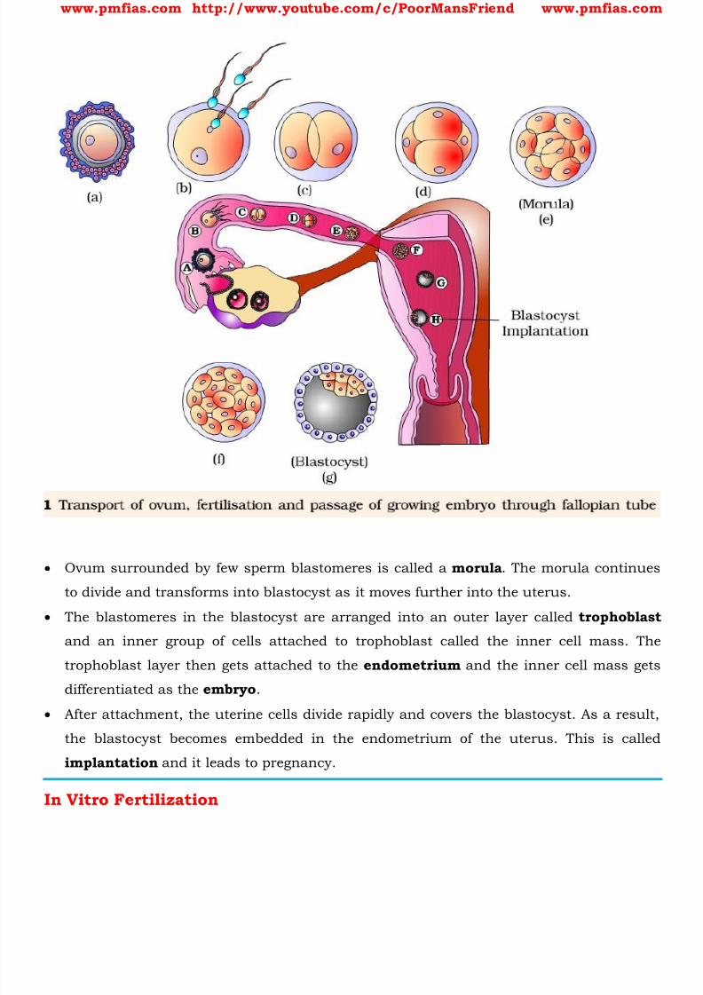
8/16/2019 Biology for Prelims Exam
http://slidepdf.com/reader/full/biology-for-prelims-exam 301/384
www.pmfias.com http://www.youtube.com/c/PoorMansFriend www.pmfias.com
Ovum surrounded by few sperm blastomeres is called a morula . The morula continues
to divide and transforms into blastocyst as it moves further into the uterus.
The blastomeres in the blastocyst are arranged into an outer layer called trophoblast
and an inner group of cells attached to trophoblast called the inner cell mass. The
trophoblast layer then gets attached to the endometrium and the inner cell mass gets
differentiated as the embryo .
After attachment, the uterine cells divide rapidly and covers the blastocyst. As a result,
the blastocyst becomes embedded in the endometrium of the uterus. This is called
implantation and it leads to pregnancy.
In Vitro Fertilization
8/16/2019 Biology for Prelims Exam
http://slidepdf.com/reader/full/biology-for-prelims-exam 302/384
www.pmfias.com http://www.youtube.com/c/PoorMansFriend www.pmfias.com
Have you heard of test tube babies? In some women oviducts are blocked . These
women are unable to bear babies because sperms cannot reach the egg for fertilization.
In such cases, doctors collect freshly released egg and sperms and keep them together
for a few hours for IVF or In Vitro Fertilization (fertilization outside the body ).
In case fertilization occurs, the zygote is allowed to develop for about a week and then it
is placed in the mother’s uterus. Complete development takes place in the uterus andthe baby is born like any other baby.
Babies born through this technique are called test-tube babies . This term is actually
misleading because babies cannot grow in test tubes.
Pregnancy And Embryonic Development
After implantation, finger-like projections appear on the trophoblast called chorionic villi
which are surrounded by the uterine tissue and maternal blood.
The chorionic villi and uterine tissue become interdigitated with each other and jointly
form a structural and functional unit between developing embryo (foetus) and maternal
body called placenta .
The placenta facilitate the supply of oxygen and nutrients to the embryo and also
removal of carbon dioxide and excretory/waste materials produced by the embryo.
The placenta is connected to the embryo through an umbilical cord which helps in the
transport of substances to and from the embryo.
Placenta also acts as an endocrine tissue and produces several hormones like human
chorionic gonadotropin (hCG), human placental lactogen (hPL), estrogens, progestogens,
etc.
In the later phase of pregnancy, a hormone called relaxin is also secreted by the ovary.
Let us remember that hCG, hPL and relaxin are produced in women only during
pregnancy .
In addition, during pregnancy the levels of other hormones like estrogens, progestogens,
cortisol, prolactin, thyroxine, etc., are increased several folds in the maternal blood.
Increased production of these hormones is essential for supporting the fetal growth,
metabolic changes in the mother and maintenance of pregnancy.
Immediately after implantation, the inner cell mass (embryo) differentiates into an outer
layer calledectoderm and an inner layer called endoderm. A mesoderm soon appears
between the ectoderm and the endoderm [ triploblastic ]. These three layers give rise to
all tissues (organs) in adults.
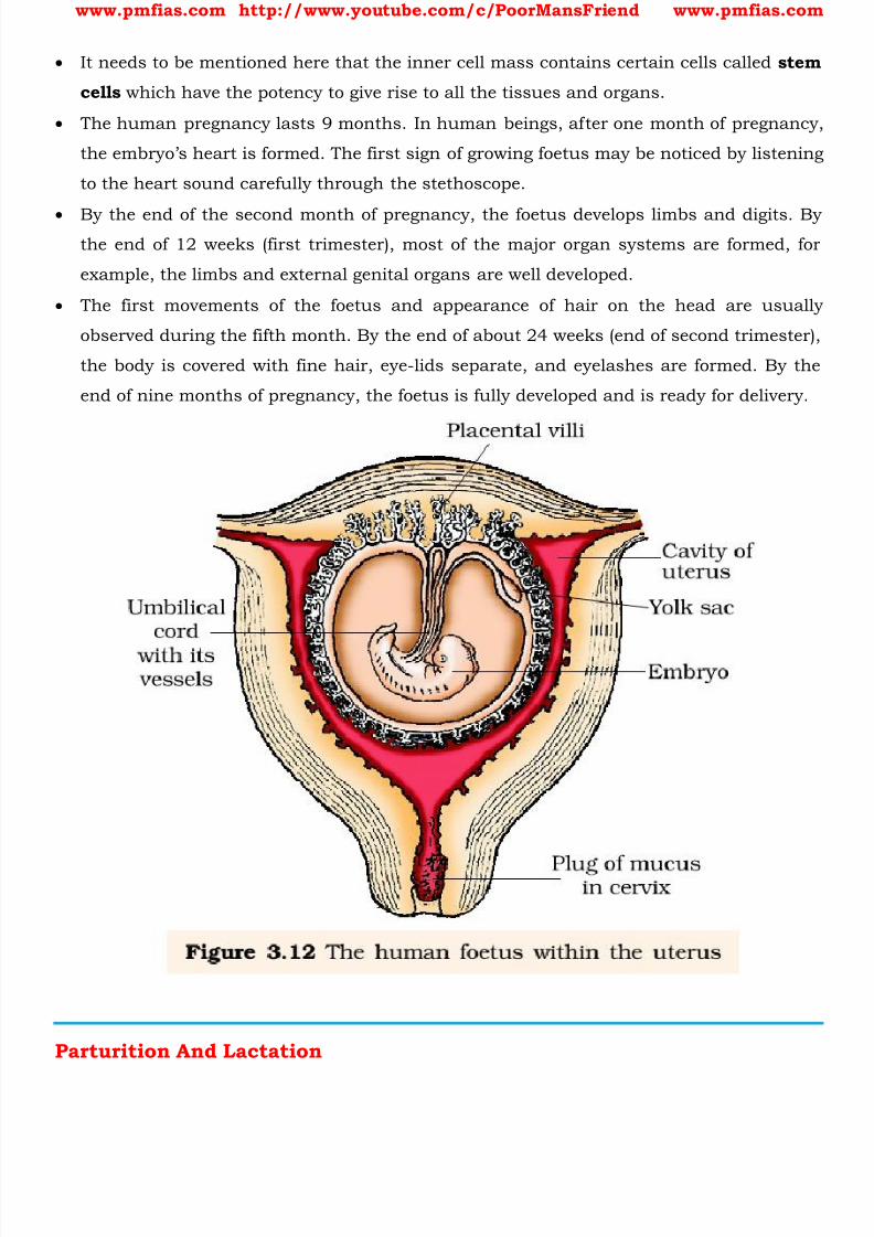
8/16/2019 Biology for Prelims Exam
http://slidepdf.com/reader/full/biology-for-prelims-exam 303/384
www.pmfias.com http://www.youtube.com/c/PoorMansFriend www.pmfias.com
It needs to be mentioned here that the inner cell mass contains certain cells called stem
cells which have the potency to give rise to all the tissues and organs.
The human pregnancy lasts 9 months. In human beings, after one month of pregnancy,
the embryo’s heart is formed. The first sign of growing foetus may be noticed by listening
to the heart sound carefully through the stethoscope.
By the end of the second month of pregnancy, the foetus develops limbs and digits. Bythe end of 12 weeks (first trimester), most of the major organ systems are formed, for
example, the limbs and external genital organs are well developed.
The first movements of the foetus and appearance of hair on the head are usually
observed during the fifth month. By the end of about 24 weeks (end of second trimester),
the body is covered with fine hair, eye-lids separate, and eyelashes are formed. By the
end of nine months of pregnancy, the foetus is fully developed and is ready for delivery.
Parturition And Lactation

8/16/2019 Biology for Prelims Exam
http://slidepdf.com/reader/full/biology-for-prelims-exam 304/384
www.pmfias.com http://www.youtube.com/c/PoorMansFriend www.pmfias.com
The average duration of human pregnancy is about 9 months which is called the
gestation period. Vigorous contraction of the uterus at the end of pregnancy causes
expulsion/delivery of the foetus. This process of delivery of the foetus (childbirth) is
called parturition .
Parturition is induced by a complex neuroendocrine mechanism. The signals for
parturition originate from the fully developed foetus and the placenta which induce milduterine contractions called foetal ejection reflex. This triggers release of oxytocin from
the maternal pituitary .
Oxytocin acts on the uterine muscle and causes stronger uterine contractions, which in
turn stimulates further secretion of oxytocin. The stimulatory reflex between the uterine
contraction and oxytocin secretion continues resulting in stronger and stronger
contractions. This leads to expulsion of the baby out of the uterus through the birth
canal – parturition. Soon after the infant is delivered, the placenta is also expelled out of the uterus. The
mammary glands of the female undergo differentiation during pregnancy and starts
producing milk towards the end of pregnancy by the process called lactation . This helps
the mother in feeding the newborn.
The milk produced during the initial few days of lactation is called colostrum which
contains several antibodies absolutely essential to develop resistance for the new-born
babies.
Breast-feeding during the initial period of infant growth is recommended by doctors for
bringing up a healthy baby.
Summary
Humans are sexually reproducing and viviparous.
The male reproductive system is composed of a pair of testes, the male sex accessory
ducts and the accessory glands and external genitalia.
Each testis has about 250 compartments called testicular lobules , and each lobule
contains one to three highly coiled seminiferous tubules .
Each seminiferous tubule is lined inside by spermatogonia and Sertoli cells.
The spermatogonia undergo meiotic divisions leading to sperm formation , while Sertoli
cells provide nutrition to the dividing germ cells.
The Leydig cells outside the seminiferous tubules, synthesise and secrete testicular
hormones called androgens .
8/16/2019 Biology for Prelims Exam
http://slidepdf.com/reader/full/biology-for-prelims-exam 305/384
www.pmfias.com http://www.youtube.com/c/PoorMansFriend www.pmfias.com
The male external genitalia is called penis.
The female reproductive system consists of a pair of ovaries, a pair of oviducts, a uterus,
a vagina, external genitalia, and a pair of mammary glands.
The ovaries produce the female gamete (ovum) and some steroid hormones (ovarian
hormones).
Ovarian follicles in different stages of development are embedded in the stroma. The oviducts, uterus and vagina are female accessory ducts.
The uterus has three layers namely perimetrium, myometrium and endometrium.
The female external genitalia includes mons pubis, labia majora, labia minora, hymen
and clitoris.
The mammary glands are one of the female secondary sexual characteristics.
Spermatogenesis results in the formation of sperms that are transported by the male sex
accessory ducts. A normal human sperm is composed of a head, neck, a middle piece and tail.
The process of formation of mature female gametes is called oogenesis.
The reproductive cycle of female primates is called menstrual cycle.
Menstrual cycle starts only after attaining sexual maturation (puberty).
During ovulation only one ovum is released per menstrual cycle.
The cyclical changes in the ovary and the uterus during menstrual cycle are induced by
changes in the levels of pituitary and ovarian hormones . After coitus, sperms are transported to the junction of the isthmus and ampulla, where
the sperm fertilizes the ovum leading to formation of a diploid zygote.
The presence of X or Y chromosome in the sperm determines the sex of the embryo.
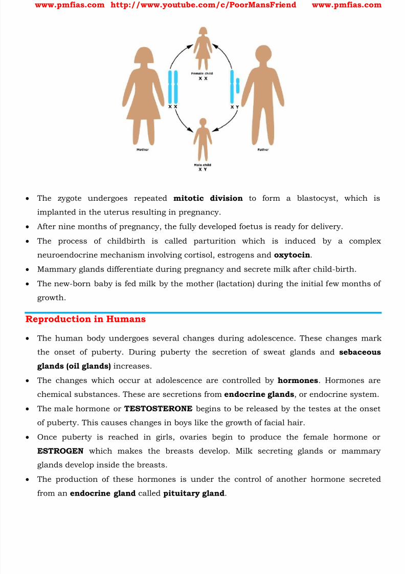
8/16/2019 Biology for Prelims Exam
http://slidepdf.com/reader/full/biology-for-prelims-exam 306/384
www.pmfias.com http://www.youtube.com/c/PoorMansFriend www.pmfias.com
The zygote undergoes repeated mitotic division to form a blastocyst, which isimplanted in the uterus resulting in pregnancy.
After nine months of pregnancy, the fully developed foetus is ready for delivery.
The process of childbirth is called parturition which is induced by a complex
neuroendocrine mechanism involving cortisol, estrogens and oxytocin .
Mammary glands differentiate during pregnancy and secrete milk after child-birth.
The new-born baby is fed milk by the mother (lactation) during the initial few months of
growth.
Reproduction in Humans
The human body undergoes several changes during adolescence. These changes mark
the onset of puberty. During puberty the secretion of sweat glands and sebaceous
glands (oil glands) increases.
The changes which occur at adolescence are controlled by hormones . Hormones are
chemical substances. These are secretions from endocrine glands , or endocrine system.
The male hormone or TESTOSTERONE begins to be released by the testes at the onset
of puberty. This causes changes in boys like the growth of facial hair.
Once puberty is reached in girls, ovaries begin to produce the female hormone or
ESTROGEN which makes the breasts develop. Milk secreting glands or mammary
glands develop inside the breasts.
The production of these hormones is under the control of another hormone secreted
from an endocrine gland called pituitary gland .
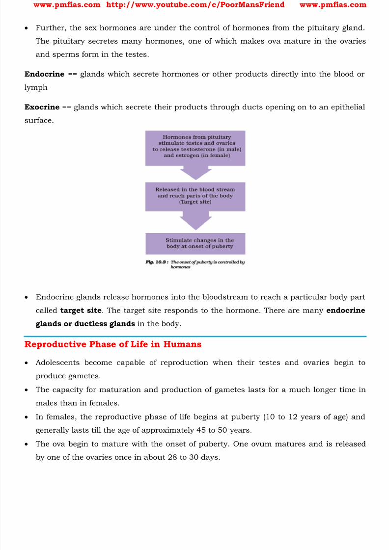
8/16/2019 Biology for Prelims Exam
http://slidepdf.com/reader/full/biology-for-prelims-exam 307/384
www.pmfias.com http://www.youtube.com/c/PoorMansFriend www.pmfias.com
Further, the sex hormones are under the control of hormones from the pituitary gland.
The pituitary secretes many hormones, one of which makes ova mature in the ovaries
and sperms form in the testes.
Endocrine == glands which secrete hormones or other products directly into the blood or
lymph
Exocrine == glands which secrete their products through ducts opening on to an epithelial
surface.
Endocrine glands release hormones into the bloodstream to reach a particular body part
called target site . The target site responds to the hormone. There are many endocrine
glands or ductless glands in the body.
Reproductive Phase of Life in Humans
Adolescents become capable of reproduction when their testes and ovaries begin to
produce gametes.
The capacity for maturation and production of gametes lasts for a much longer time in
males than in females.
In females, the reproductive phase of life begins at puberty (10 to 12 years of age) and
generally lasts till the age of approximately 45 to 50 years.
The ova begin to mature with the onset of puberty. One ovum matures and is released
by one of the ovaries once in about 28 to 30 days.
8/16/2019 Biology for Prelims Exam
http://slidepdf.com/reader/full/biology-for-prelims-exam 308/384
www.pmfias.com http://www.youtube.com/c/PoorMansFriend www.pmfias.com
During this period, the wall of the uterus becomes thick so as to receive the egg, in case
it is fertilized and begins to develop. This results in pregnancy.
If fertilization does not occur, the released egg, and the thickened lining of the uterus
along with its blood vessels are shed off. This causes bleeding in women which is called
menstruation .
Menstruation occurs once in about 28 to 30 days. The first menstrual flow begins atpuberty and is termed menarche .
At 45 to 50 years of age, the menstrual cycle stops. Stoppage of menstruation is termed
menopause . Initially, menstrual cycle may be irregular. It take some time to become
regular.
Menstrual cycle is controlled by hormones . The cycle includes the maturation of the
egg, its release, thickening of uterine wall and its breakdown if pregnancy does not
occur. In case the egg is fertilized it begins to divide and then gets embedded in the uterus for
further development.
Inside the fertilized egg or zygote is the instruction for determining the sex of the baby.
This instruction is present in the thread-like structures, called chromosomes in the
fertilized egg.
All human beings have 23 pairs of chromosomes in the nuclei of their cells. Two
chromosomes out of these are the sex chromosomes, named X and Y.
A female has two X chromosomes, while a male has one X and one Y chromosome. The
gametes (egg and sperm) have only one set of chromosomes. The unfertilized egg always
has one X chromosome. Sperms are of two kinds. One kind has an X chromosome, and
the other kind has a Y chromosome.
Reproductive Health and Birth Control Methods
India was amongst the first countries in the world to initiate action plans and
programmes at a national level to attain total reproductive health as a social goal.
These programmes called ‘ family planning ’ were initiated in 1951.
Statutory ban on amniocentesis (a foetal sex determination test based on the
chromosomal pattern in the amniotic fluid surrounding the developing embryo) for
sex-determination to legally check increasing female feticides.
‘Saheli’ -a new oral contraceptive for the females was developed by scientists at
Central Drug Research Institute (CDRI) in Lucknow, India.
8/16/2019 Biology for Prelims Exam
http://slidepdf.com/reader/full/biology-for-prelims-exam 309/384
8/16/2019 Biology for Prelims Exam
http://slidepdf.com/reader/full/biology-for-prelims-exam 310/384
www.pmfias.com http://www.youtube.com/c/PoorMansFriend www.pmfias.com
Another effective and popular method is the use of Intra Uterine Devices (IUDs). These
devices are inserted by doctors or expert nurses in the uterus through vagina. These
Intra Uterine Devices are presently available as Copper T (CuT), non-medicated IUDs
(e.g., Lippes loop), copper releasing IUDs (CuT, Cu7, Multiload 375) and the hormone
releasing IUDs (Progestasert, LNG-20).
IUDs Increase phagocytosis of sperms within the uterus and the Cu ions releasedsuppress sperm motility and the fertilising capacity of sperms. The hormone releasing
IUDs, in addition, make the uterus unsuitable for implantation and the cervix hostile to
the sperms. IUDs are ideal contraceptives for the females who want to delay pregnancy
and/or space children. It is one of most widely accepted methods of contraception in
India.
Pills
Oral administration of small doses of either progestogens or progestogen-estrogen
combinations is another contraceptive method used by the females. They are used in the
form of tablets and hence are popularly called the pills. Pills have to be taken daily for a
period of 21 days starting preferably within the first five days of menstrual cycle. After a
gap of 7 days (during which menstruation occurs) it has to be repeated in the same
pattern till the female desires to prevent conception. They inhibit ovulation and
implantation as well as alter the quality of cervical mucus to prevent/retard entry of
sperms . Pills are very effective with lesser side effects and are well accepted by the
females.
Progestogens alone or in combination with estrogen can also be used by females as
injections or implants under the skin. Their mode of action is similar to that of pills and
their effective periods are much longer. Administration of progestogens or progestogen-
estrogen combinations or IUDs within 72 hours of coitus have been found to be very
effective as emergency contraceptives as they could be used to avoid possible
pregnancy due to rape or casual unprotected intercourse.
Surgical Methods
Surgical methods, also called sterilization, are generally advised for the male/female
partner as a terminal method to prevent any more pregnancies. Surgical intervention
blocks gamete transport and thereby prevent conception.

8/16/2019 Biology for Prelims Exam
http://slidepdf.com/reader/full/biology-for-prelims-exam 311/384
www.pmfias.com http://www.youtube.com/c/PoorMansFriend www.pmfias.com
Sterilisation procedure in the male is called ‘vasectomy’ and that in the female,
‘tubectomy’ .
In vasectomy, a small part of the vas deferens is removed or tied up through a small
incision on the scrotum whereas in tubectomy, a small part of the fallopian tube is
removed or tied up through a small incision in the abdomen or through vagina. These
techniques are highly effective but their reversibility is very poor.
No doubt, the widespread use of these methods have a significant role in checking
uncontrolled growth of population. However, their possible ill-effects like nausea,
abdominal pain, breakthrough bleeding, irregular menstrual bleeding or even breast
cancer, though not very significant, should not be totally ignored.
Sexually Transmitted Diseases (STDs)
Diseases or infections which are transmitted through sexual intercourse are collectivelycalled sexually transmitted diseases (STD) or venereal diseases (VD) or reproductive
tract infections (RTI).
Gonorrhoea, syphilis, genital herpes, chlamydiasis, genital warts, trichomoniasis,
hepatitis-B and of course, the most discussed infection in the recent years, HIV leading
to AIDS are some of the common STDs.

8/16/2019 Biology for Prelims Exam
http://slidepdf.com/reader/full/biology-for-prelims-exam 312/384
www.pmfias.com http://www.youtube.com/c/PoorMansFriend www.pmfias.com
Some of these infections like hepatitis-B and HIV can also be transmitted by sharing of
injection needles, surgical instruments, etc., with infected persons, transfusion of blood,
or from an infected mother to the foetus too. Except for hepatitis-B, genital herpes and
HIV infections, other diseases are completely curable if detected early and treated
properly. Early symptoms of most of these are minor and include itching, fluid
discharge, slight pain, swellings, etc., in the genital region. Infected females may oftenbe asymptomatic and hence, may remain undetected for long. Absence or less significant
symptoms in the early stages of infection and the social stigma attached to the STDs,
deter the infected persons from going for timely detection and proper treatment. This
could lead to complications later, which include pelvic inflammatory diseases (PID),
abortions, still births, ectopic pregnancies, infertility or even cancer of the reproductive
tract. STDs are a major threat to a healthy society. Therefore, prevention or early
detection and cure of these diseases are given prime consideration under the
reproductive health-care programmes. Though all persons are vulnerable to these
infections, their incidences are reported to be very high among persons in the age group
of 15-24 years - the age group to which you also belong. Don’t panic. Prevention is in
your hands. You could be free of these infections if you follow the simple principles given
below:
Avoid sex with unknown partners/multiple partners.
Always use condoms during coitus.
In case of doubt, go to a qualified doctor for early detection and get complete treatment if
diagnosed with disease.
In vitro fertilisation (IVF-fertilisation outside the body in almost similar conditions as
that in the body) followed by embryo transfer (ET) is one of such methods. In this
method, popularly known as test tube baby programme, ova from the wife/donor
(female) and sperms from the husband/donor (male) are collected and are induced to
form zygote under simulated conditions in the laboratory. The zygote or early embryos
(with upto 8 blastomeres) could then be transferred into the fallopian tube (ZIFT-zygote
intra fallopian transfer) and embryos with more than 8 blastomeres, into the uterus (IUT
- intra uterine transfer), to complete its further development. Embryos formed by in-vivo
fertilisation (fusion of gametes within the female) also could be used for such transfer to
assist those females who cannot conceive.
Transfer of an ovum collected from a donor into the fallopian tube (GIFT - gamete intra
fallopian transfer) of another female who cannot produce one, but can provide suitable

8/16/2019 Biology for Prelims Exam
http://slidepdf.com/reader/full/biology-for-prelims-exam 313/384
www.pmfias.com http://www.youtube.com/c/PoorMansFriend www.pmfias.com
environment for fertilisation and further development is another method attempted.
Intra cytoplasmic sperm injection (ICSI) is another specialised procedure to form an
embryo in the laboratory in which a sperm is directly injected into the ovum. Infertility
cases either due to inability of the male partner to inseminate the female or due to very
low sperm counts in the ejaculates, could be corrected by artificial insemination (AI)
technique. In this technique, the semen collected either from the husband or a healthydonor is artificially introduced either into the vagina or into the uterus (IUI - intra-
uterine insemination) of the female.
Though options are many, all these techniques require extremely high precision
handling by specialized professionals and expensive instrumentation. Therefore, these
facilities are presently available only in very few centers in the country. Obviously their
benefits is affordable to only a limited number of people. Emotional, religious and social
factors are also deterrents in the adoption of these methods. Since the ultimate aim of
all these procedures is to have children, in India we have so many orphaned and
destitute children, who would probably not survive till maturity, unless taken care of.
Our laws permit legal adoption and it is as yet, one of the best methods for couples
looking for parenthood.
SUMMARY
Diseases or infections transmitted through sexual intercourse are called Sexually
Transmitted Diseases (STDs). Pelvic Inflammatory Diseases (PIDs), still birth, infertility
are some of the complications of them. Early detection facilitate better cure of these
diseases. Avoiding sexual intercourse with unknown/multiple partners, use of condoms
during coitus are some of the simple precautions to avoid contracting STDs.
Inability to conceive or produce children even after 2 years of unprotected sexual
cohabitation is called infertility. Various methods are now available to help such
couples. In Vitro fertilisation followed by transfer of embryo into the female genital tract
is one such method and is commonly known as the ‘Test Tube Baby’ Programme.
8/16/2019 Biology for Prelims Exam
http://slidepdf.com/reader/full/biology-for-prelims-exam 314/384
www.pmfias.com http://www.youtube.com/c/PoorMansFriend www.pmfias.com
Types of Movements. Muscular System – Muscle Types: Skeletal Muscles, Visceral Muscles,
Cardiac Muscles. Skeletal System: Major Joints.
Types of Movements
Voluntary movements are called locomotion. Walking, running, climbing, flying,
swimming are all some forms of locomotory movements. Cells of the human body exhibit three main types of movements, namely, amoeboid,
ciliary and muscular .
Some specialized cells in our body like macrophages and leucocytes in blood exhibit
amoeboid movement. It is effected by pseudopodia formed by the streaming of
protoplasm (as in Amoeba).
Ciliary movement occurs in most of our internal tubular organs which are lined by
ciliated epithelium .
The coordinated movements of cilia in the trachea help us in removing dust particles
and some of the foreign substances inhaled along with the atmospheric air.
Passage of ova through the female reproductive tract is also facilitated by the ciliary
movement.
Movement of our limbs, jaws, tongue, etc. require muscular movement. The contractile
property of muscles are effectively used for locomotion and other movements by human
beings and majority of multicellular organisms. Locomotion requires a perfect
coordinated activity of muscular, skeletal and neural systems.
Muscular System – Muscle Types
Muscle is a specialized tissue of mesodermal origin. [ Mesodermal == the middle layer of
cells or tissues of an embryo, or the parts derived from this (e.g. cartilage, muscles, and
bone)]
About 40-50 per cent of the body weight of a human adult is contributed by muscles.
They have special properties like excitability, contractility, extensibility and elasticity.
Muscles have been classified using different criteria, namely location, appearance and
nature of regulation of their activities. Based on their location, three types of muscles
are identified : (i) Skeletal (ii) Visceral [the internal organs in the main cavities of the
body] and (iii) Cardiac.
Skeletal Muscles
8/16/2019 Biology for Prelims Exam
http://slidepdf.com/reader/full/biology-for-prelims-exam 315/384
www.pmfias.com http://www.youtube.com/c/PoorMansFriend www.pmfias.com
Skeletal muscles are closely associated with the skeletal components of the body. They
have a striped appearance under the microscope and hence are called striated muscles .
As their activities are under the voluntary control of the nervous system, they are known
as voluntary muscles too. They are primarily involved in locomotory actions and
changes of body postures.
Each organized skeletal muscle in our body is made of a number of muscle bundles orfascicles held together by a common connective tissue layer called fascia .
Each muscle bundle contains a number of muscle fibres. Each muscle fibre is lined by
the plasma membrane called sarcolemma enclosing the sarcoplasm.
The endoplasmic reticulum, i.e., sarcoplasmic reticulum of the muscle fibres is the store
house of calcium ions .
A characteristic feature of the muscle fibre is the presence of a large number of parallelly
arranged filaments in the sarcoplasm called myofilaments or myofibrils . Each myofibril has alternate dark and light bands on it. The striated appearance is due
to the distribution pattern of two important proteins - Actin and Myosin .
Actin and myosin are polymerized proteins with contractility. A motor neuron carries
signal to the muscle fibre which generates an action potential in it. This causes the
release of Ca++ from sarcoplasmic reticulum.
Ca++ activates actin which binds to the myosin head to form a cross bridge. These cross
bridges pull the actin filaments causing them to slide over the myosin filaments and
thereby causing contraction. Ca++ are then returned to sarcoplasmic reticulum which
inactivate the actin. Cross bridges are broken and the muscles relax.
Muscles are classified as Red and White fibres based primarily on the amount of red
coloured myoglobin pigment in them.

8/16/2019 Biology for Prelims Exam
http://slidepdf.com/reader/full/biology-for-prelims-exam 316/384
www.pmfias.com http://www.youtube.com/c/PoorMansFriend www.pmfias.com
Visceral Muscles Visceral muscles are located in the inner walls of hollow visceral organs of the body like
the alimentary canal, reproductive tract, etc.
They do not exhibit any striation and are smooth in appearance. Hence, they are called
smooth muscles (nonstriated muscle).
Their activities are not under the voluntary control of the nervous system and are
therefore known as involuntary muscles .
They assist, for example, in the transportation of food through the digestive tract andgametes through the genital tract.
Cardiac Muscles
As the name suggests, Cardiac muscles are the muscles of heart. Many cardiac muscle
cells assemble in a branching pattern to form a cardiac muscle.
Based on appearance, cardiac muscles are striated . They are involuntary in nature as
the nervous system does not control their activities directly.
Skeletal System
Skeletal system consists of a framework of bones and a few cartilages. Bone and
cartilage are specialized connective tissues.
The former has a very hard matrix due to calcium salts in it and the latter has slightly
pliable matrix due to chondroitin salts .

8/16/2019 Biology for Prelims Exam
http://slidepdf.com/reader/full/biology-for-prelims-exam 317/384
www.pmfias.com http://www.youtube.com/c/PoorMansFriend www.pmfias.com
In human beings, this system is made up of 206 bones and a few cartilages. It is
grouped into two principal divisions - the axial and the appendicular skeleton.
Axial skeleton comprises 80 bones distributed along the main axis of the body. The
skull, vertebral column, sternum and ribs constitute axial skeleton.
Joints
Joints are essential for all types of movements involving the bony parts of the body.
Locomotory movements are no exception to this.
Joints are points of contact between bones, or between bones and cartilages.
Joints have been classified into three major structural forms, namely, fibrous,
cartilaginous and synovial.
Fibrous joints do not allow any movement . This type of joint is shown by the flat skull
bones which fuse end-to-end with the help of dense fibrous connective tissues in the
form of sutures, to form the cranium.
In cartilaginous joints , the bones involved are joined together with the help of
cartilages. The joint between the adjacent vertebrae in the vertebral column is of this
pattern and it permits limited movements .
Synovial joints are characterized by the presence of a fluid filled synovial cavity
between the articulating surfaces of the two bones. Such an arrangement allows
considerable movement . These joints help in locomotion and many other movements.
Ball and socket joint, hinge joint (knee joint), pivot joint, gliding joint and saddle joint
are some examples.
Major Joints
1. Ball and socket joints
2. Pivotal Joint: The joint where our neck joins the head is a pivotal joint.
3. Hinge joints
4. Fixed joints There are some bones in our head that are joined together at some joints. The bones
cannot move at these joints. Such joints are called fixed joints .
When you open your mouth wide, you can move your lower jaw away from your head,
isn’t it? Try to move your upper jaw, now. Are you able to move it? There is a joint
between the upper jaw and the rest of the head which is a fixed joint.
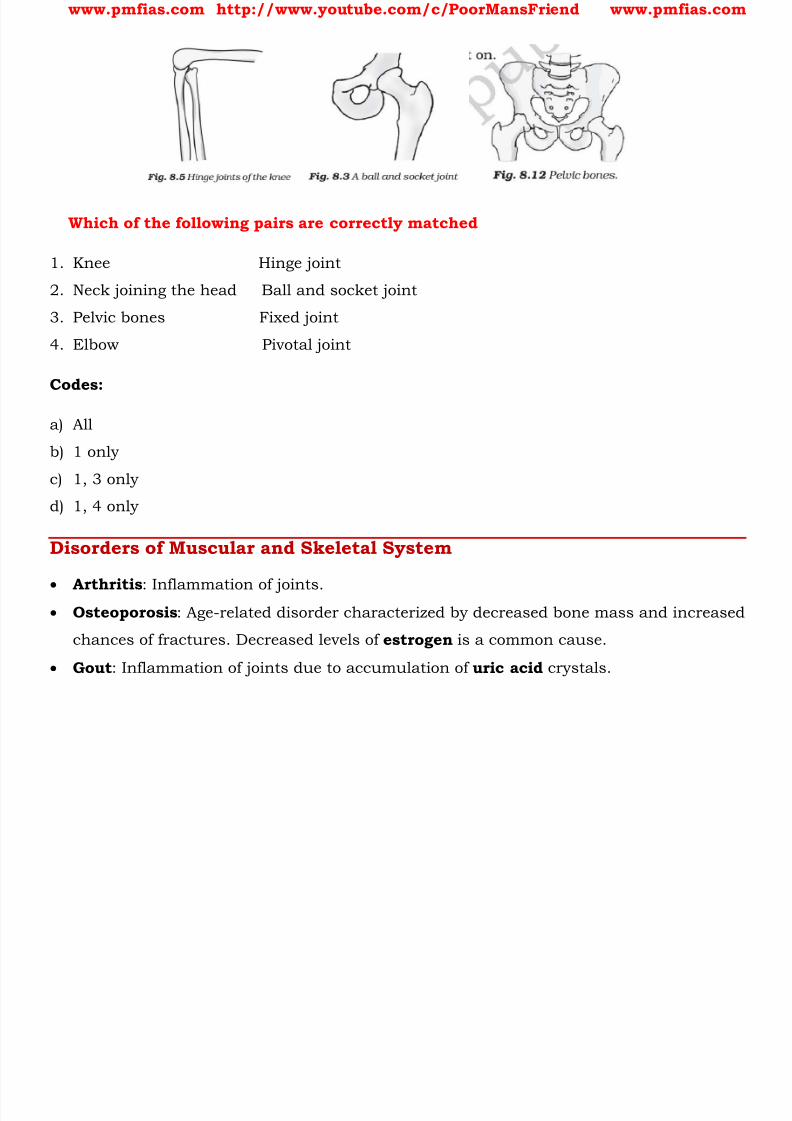
8/16/2019 Biology for Prelims Exam
http://slidepdf.com/reader/full/biology-for-prelims-exam 318/384
www.pmfias.com http://www.youtube.com/c/PoorMansFriend www.pmfias.com
Which of the following pairs are correctly matched
1. Knee Hinge joint
2. Neck joining the head Ball and socket joint
3. Pelvic bones Fixed joint
4. Elbow Pivotal joint
Codes:
a) All
b) 1 only
c) 1, 3 only
d) 1, 4 only
Disorders of Muscular and Skeletal System
Arthritis : Inflammation of joints. Osteoporosis : Age-related disorder characterized by decreased bone mass and increased
chances of fractures. Decreased levels of estrogen is a common cause.
Gout : Inflammation of joints due to accumulation of uric acid crystals.

8/16/2019 Biology for Prelims Exam
http://slidepdf.com/reader/full/biology-for-prelims-exam 319/384
www.pmfias.com http://www.youtube.com/c/PoorMansFriend www.pmfias.com
Neural Control and Coordination
As you know, the functions of the organs/organ systems in our body must be
coordinated to maintain homeostasis [ the maintenance of a stable equilibrium ].
Coordination is the process through which two or more organs interact and complement
the functions of one another.
The neural system provides an organized network of point-to-point connections for a
quick coordination.
The endocrine system provides chemical integration through hormones.
Human Neural System
The human neural system is divided into two parts :
1. the central neural system (CNS)
2. the peripheral neural system (PNS)
The CNS includes the brain and the spinal cord and is the site of information
processing and control.
The PNS comprises of all the nerves of the body associated with the CNS (brain and
spinal cord).
Peripheral Neural System (PNS)
The nerve fibres of the PNS are of two types:
1. afferent fibres → tissues/organs to brain .
2. efferent fibres → brain to tissues/organs .
The afferent nerve fibres transmit impulses from tissues/organs to the CNS and the
efferent fibres transmit regulatory impulses from the CNS to the concerned peripheral
tissues/organs.
The PNS is divided into two divisions called somatic neural system and autonomic
neural system .
The somatic neural system relays impulses from the CNS to skeletal muscles while the
autonomic neural system transmits impulses from the CNS to the involuntary organs
and smooth muscles of the body.
1. Somatic Neural System → Brain to Voluntary muscles .
2. Autonomic Neural System → Brain to Involuntary muscles .
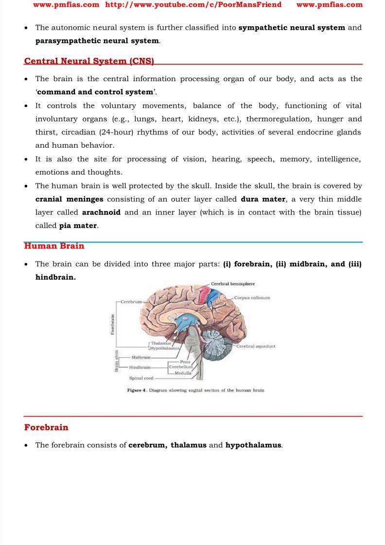
8/16/2019 Biology for Prelims Exam
http://slidepdf.com/reader/full/biology-for-prelims-exam 320/384
www.pmfias.com http://www.youtube.com/c/PoorMansFriend www.pmfias.com
The autonomic neural system is further classified into sympathetic neural system and
parasympathetic neural system .
Central Neural System (CNS)
The brain is the central information processing organ of our body, and acts as the
‘ command and control system
’.
It controls the voluntary movements, balance of the body, functioning of vital
involuntary organs (e.g., lungs, heart, kidneys, etc.), thermoregulation, hunger and
thirst, circadian (24-hour) rhythms of our body, activities of several endocrine glands
and human behavior.
It is also the site for processing of vision, hearing, speech, memory, intelligence,
emotions and thoughts.
The human brain is well protected by the skull. Inside the skull, the brain is covered by
cranial meninges consisting of an outer layer called dura mater , a very thin middle
layer called arachnoid and an inner layer (which is in contact with the brain tissue)
called pia mater .
Human Brain
The brain can be divided into three major parts: (i) forebrain, (ii) midbrain, and (iii)
hindbrain.
Forebrain
The forebrain consists of cerebrum, thalamus and hypothalamus .

8/16/2019 Biology for Prelims Exam
http://slidepdf.com/reader/full/biology-for-prelims-exam 321/384
www.pmfias.com http://www.youtube.com/c/PoorMansFriend www.pmfias.com
Cerebrum forms the major part of the human brain. A deep cleft divides the cerebrum
longitudinally into two halves, which are termed as the left and right cerebral
hemispheres.
The hemispheres are connected by a tract of nerve fibres called corpus callosum .
The layer of cells which covers the cerebral hemisphere is called cerebral cortex . The
cerebral cortex is referred to as the grey matter due to its greyish appearance. Theneuron cell bodies are concentrated here giving the colour.
The cerebral cortex contains motor areas, sensory areas and large regions that are
neither clearly sensory nor motor in function. These regions called as the association
areas are responsible for complex functions like intersensory associations, memory
and communication .
Fibres of the tracts are covered with the myelin sheath , which constitute the inner part
of cerebral hemisphere. They give an opaque white appearance to the layer and, hence,is called the white matter .
The cerebrum wraps around a structure called thalamus, which is a major coordinating
centre for sensory and motor signaling .
Another very important part of the brain called hypothalamus lies at the base of the
thalamus. The hypothalamus contains a number of centres which control body
temperature, urge for eating and drinking . It also contains several groups of
neurosecretory cells, which secrete hormones called hypothalamic hormones .
The inner parts of cerebral hemispheres and a group of associated deep structures like
amygdala, hippocampus, etc., form a complex structure called the limbic lobe or limbic
system . Along with the hypothalamus, it is involved in the regulation of sexual
behaviour, expression of emotional reactions (e.g., excitement, pleasure, rage and
fear), and motivation.
Midbrain
The midbrain is located between the thalamus/hypothalamus of the forebrain and pons
of the hindbrain. A canal called the cerebral aqueduct passess through the midbrain.
The dorsal portion of the midbrain consists mainly of four round swellings (lobes) called
corpora quadrigemina . Midbrain and hindbrain form the brain stem.
Hindbrain
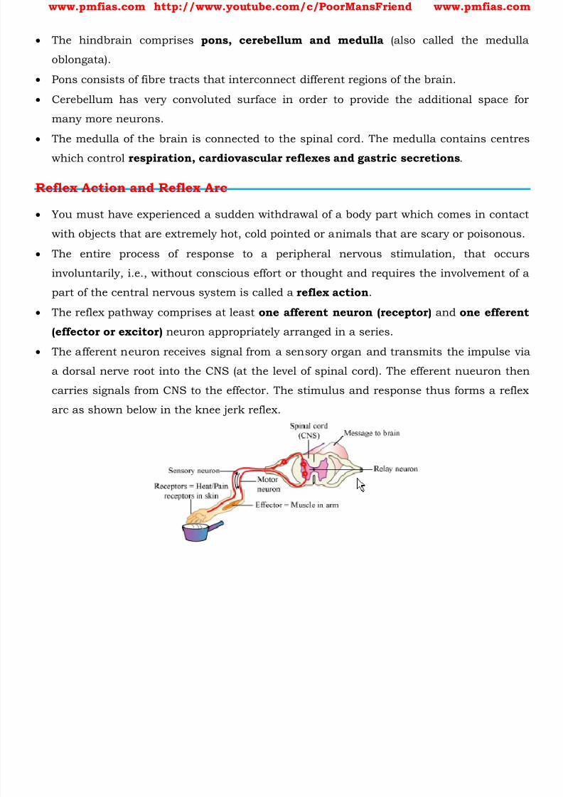
8/16/2019 Biology for Prelims Exam
http://slidepdf.com/reader/full/biology-for-prelims-exam 322/384
www.pmfias.com http://www.youtube.com/c/PoorMansFriend www.pmfias.com
The hindbrain comprises pons, cerebellum and medulla (also called the medulla
oblongata).
Pons consists of fibre tracts that interconnect different regions of the brain.
Cerebellum has very convoluted surface in order to provide the additional space for
many more neurons.
The medulla of the brain is connected to the spinal cord. The medulla contains centreswhich control respiration, cardiovascular reflexes and gastric secretions .
Reflex Action and Reflex Arc
You must have experienced a sudden withdrawal of a body part which comes in contact
with objects that are extremely hot, cold pointed or animals that are scary or poisonous.
The entire process of response to a peripheral nervous stimulation, that occurs
involuntarily, i.e., without conscious effort or thought and requires the involvement of a
part of the central nervous system is called a reflex action .
The reflex pathway comprises at least one afferent neuron (receptor) and one efferent
(effector or excitor) neuron appropriately arranged in a series.
The afferent neuron receives signal from a sensory organ and transmits the impulse via
a dorsal nerve root into the CNS (at the level of spinal cord). The efferent nueuron then
carries signals from CNS to the effector. The stimulus and response thus forms a reflex
arc as shown below in the knee jerk reflex.

8/16/2019 Biology for Prelims Exam
http://slidepdf.com/reader/full/biology-for-prelims-exam 323/384
www.pmfias.com http://www.youtube.com/c/PoorMansFriend www.pmfias.com
Eye
Our paired eyes are located in sockets of the skull called orbits.
The adult human eye ball is nearly a spherical structure. The wall of the eye ball is
composed of three layers.
The external layer is composed of a dense connective tissue and is called the sclera . The
anterior portion of this layer is called the cornea . The middle layer, choroid , contains
many blood vessels and looks bluish in colour.
The choroid layer is thin over the posterior two-thirds of the eye ball, but it becomes
thick in the anterior part to form the ciliary body.
The ciliary body itself continues forward to form a pigmented and opaque structure
called the iris which is the visible coloured portion of the eye.
The eye ball contains a transparent crystalline lens which is held in place by ligaments
attached to the ciliary body. In front of the lens, the aperture surrounded by the iris is
called the pupil . The diameter of the pupil is regulated by the muscle fibres of iris.
The inner layer is the retina and it contains three layers of neural cells - from inside to
outside - ganglion cells, bipolar cells and photoreceptor cells .
There are two types of photoreceptor cells, namely, rods and cones . These cells contain
the light-sensitive proteins called the photopigments .
The daylight (photopic) vision and colour vision are functions of cones and the twilight
(scotopic) vision is the function of the rods.
The rods contain a purplish-red protein called the rhodopsin or visual purple, which
contains a derivative of Vitamin A .
8/16/2019 Biology for Prelims Exam
http://slidepdf.com/reader/full/biology-for-prelims-exam 324/384
www.pmfias.com http://www.youtube.com/c/PoorMansFriend www.pmfias.com
In the human eye, there are three types of cones which possess their own characteristic
photopigments that respond to red, green and blue lights. The sensations of different
colours are produced by various combinations of these cones and their photopigments.
When these cones are stimulated equally, a sensation of white light is produced.
The optic nerves leave the eye and the retinal blood vessels enter it at a point medial to
and slightly above the posterior pole of the eye ball. Photoreceptor cells are not presentin that region and hence it is called the blind spot .
At the posterior pole of the eye lateral to the blind spot, there is a yellowish pigmented
spot called macula lutea with a central pit called the fovea . The fovea is a thinned-out
portion of the retina where only the cones are densely packed . It is the point where the
visual acuity (resolution) is the greatest.
The space between the cornea and the lens is called the aqueous chamber and contains
a thin watery fluid called aqueous humor . The space between the lens and the retina iscalled the vitreous chamber and is filled with a transparent gel called vitreous humor .
The Ear
The ears perform two sensory functions, hearing and maintenance of body balance.
Anatomically, the ear can be divided into three major sections called the outer ear, the
middle ear and the inner ear.
The outer ear consists of the pinna and external auditory meatus (canal).
The pinna collects the vibrations in the air which produce sound. The external auditory
meatus leads inwards and extends up to the tympanic membrane (the ear drum) .
There are very fine hairs and wax-secreting glands in the skin of the pinna and the
meatus.
The tympanic membrane is composed of connective tissues covered with skin outside
and with mucus membrane inside.
The middle ear contains three ossicles called malleus, incus and stapes which are
attached to one another in a chainlike fashion.
The malleus is attached to the tympanic membrane and the stapes is attached to the
oval window of the cochlea.
The ear ossicles increase the efficiency of transmission of sound waves to the inner ear.
An Eustachian tube connects the middle ear cavity with the pharynx. The Eustachian
tube helps in equalising the pressures on either sides of the ear drum.
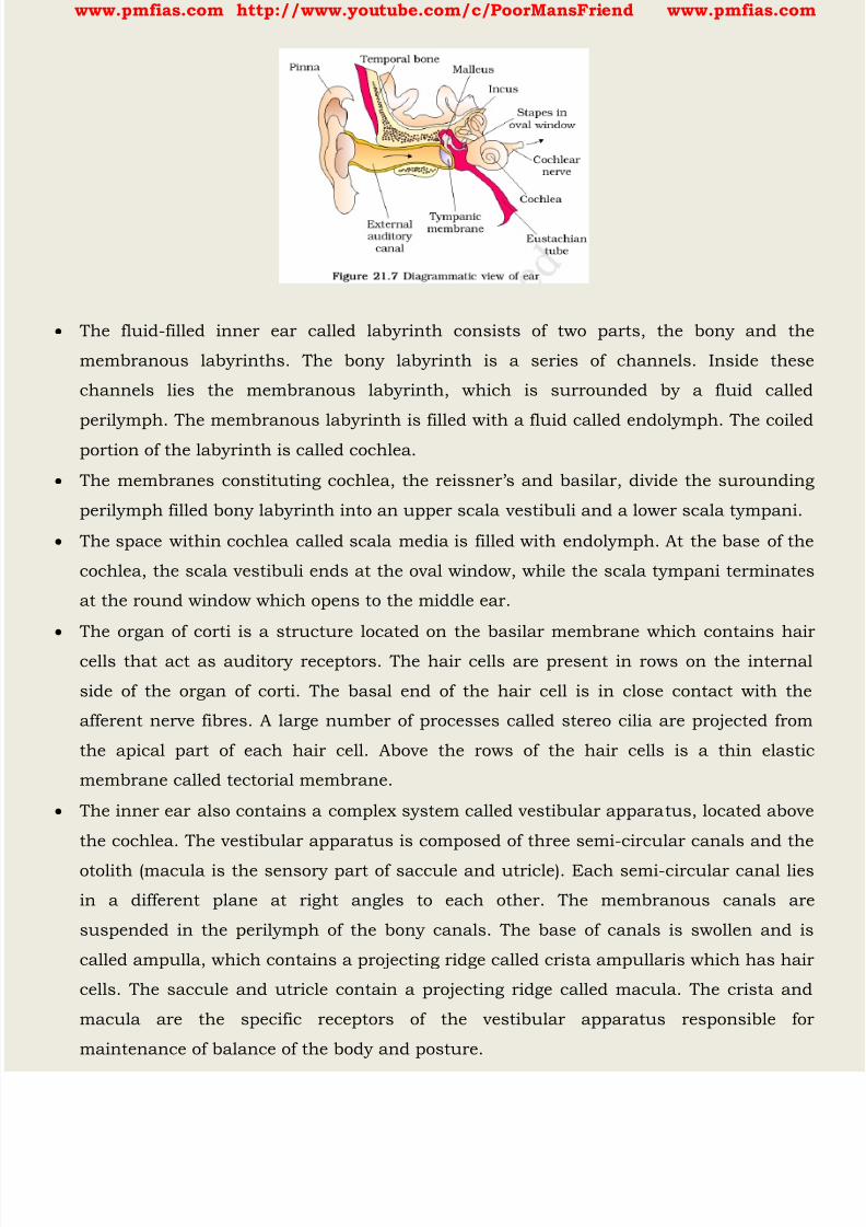
8/16/2019 Biology for Prelims Exam
http://slidepdf.com/reader/full/biology-for-prelims-exam 325/384
www.pmfias.com http://www.youtube.com/c/PoorMansFriend www.pmfias.com
The fluid-filled inner ear called labyrinth consists of two parts, the bony and the
membranous labyrinths. The bony labyrinth is a series of channels. Inside these
channels lies the membranous labyrinth, which is surrounded by a fluid called
perilymph. The membranous labyrinth is filled with a fluid called endolymph. The coiled
portion of the labyrinth is called cochlea.
The membranes cons tituting cochlea, the reissner’s and basilar, divide the surounding
perilymph filled bony labyrinth into an upper scala vestibuli and a lower scala tympani.
The space within cochlea called scala media is filled with endolymph. At the base of the
cochlea, the scala vestibuli ends at the oval window, while the scala tympani terminates
at the round window which opens to the middle ear.
The organ of corti is a structure located on the basilar membrane which contains hair
cells that act as auditory receptors. The hair cells are present in rows on the internal
side of the organ of corti. The basal end of the hair cell is in close contact with the
afferent nerve fibres. A large number of processes called stereo cilia are projected from
the apical part of each hair cell. Above the rows of the hair cells is a thin elastic
membrane called tectorial membrane.
The inner ear also contains a complex system called vestibular apparatus, located above
the cochlea. The vestibular apparatus is composed of three semi-circular canals and the
otolith (macula is the sensory part of saccule and utricle). Each semi-circular canal lies
in a different plane at right angles to each other. The membranous canals are
suspended in the perilymph of the bony canals. The base of canals is swollen and is
called ampulla, which contains a projecting ridge called crista ampullaris which has hair
cells. The saccule and utricle contain a projecting ridge called macula. The crista and
macula are the specific receptors of the vestibular apparatus responsible for
maintenance of balance of the body and posture.
8/16/2019 Biology for Prelims Exam
http://slidepdf.com/reader/full/biology-for-prelims-exam 326/384
www.pmfias.com http://www.youtube.com/c/PoorMansFriend www.pmfias.com
Neurons
The neural system coordinates and integrates functions as well as metabolic and
homeostatic activities of all the organs.
Neurons, the functional units of neural system are excitable cells due to a differential
concentration gradient of ions across the membrane. The electrical potential difference
across the resting neural membrane is called the ‘resting potential’.
The nerve impulse is conducted along the axon membrane in the form of a wave of
depolarisation and repolarisation.
A synapse is formed by the membranes of a pre-synaptic neuron and a post-synaptic
neuron which may or may not be separated by a gap called synaptic cleft. Chemicals
involved in the transmission of impulses at chemical synapses are called
neurotransmitters.
Human neural system consists of two parts : (i) central neural system (CNS) and (ii) the
peripheral neural system. The CNS consists of the brain and spiral cord. The brain can
be divided into three major parts : (i) forebrain, (ii) midbrain and (iii) hindbrain. The
forebrain consists of cerebrum, thalamus and hypothalamus. The cerebrum is
longitudinally divided into two halves that are connected by the corpus callosum. A very
important part of the forebrain called hypothalamus controls the body temperature,
eating and drinking. Inner parts of cerebral hemispheres and a group of associated deep
structures form a complex structure called limbic system which is concerned witholfaction, autonomic responses, regulation of sexual behaviour, expression of emotional
reactions, and motivation. The midbrain receives and integrates visual, tactile and
auditory inputs. The hindbrain comprises pons, cerebellum and medulla. The
cerebellum integrates information received from the semicircular canals of the ear and
the auditory system. The medulla contains centres, which control respiration,
cardiovascular reflexes, and gastric secretions. Pons consist of fibre tracts that
interconnect different regions of the brain. The entire process of involuntary response toa peripheral nervous stimulation is called reflex action. Information regarding changes
in the environment is received by the CNS through the sensory organs which are
processed and analysed. Signals are then sent for necessary adjustments. The wall of
the human eye ball is composed of three layers. The external layer is composed of
cornea and sclera. Inside sclera is the middle layer, which is called the choroid. Retina,
the innermost layer, contains two types of photoreceptor cells, namely rods and cones.
8/16/2019 Biology for Prelims Exam
http://slidepdf.com/reader/full/biology-for-prelims-exam 327/384
www.pmfias.com http://www.youtube.com/c/PoorMansFriend www.pmfias.com
The daylight (photopic) vision and colour vision are functions of cones and twilight
(scotopic) vision is the function of the rods. The light enters through cornea, the lens
and the images of objects are formed on the retina.
The ear can be divided into the outer ear, the middle ear and the inner ear. The middle
ear contains three ossicles called malleus, incus and stapes. The fluid filled inner ear is
called the labyrinth, and the coiled portion of the labyrinth is called cochlea. The organof corti is a structure which contains hair cells that act as auditory receptors and is
located on the basilar membrane. The vibrations produced in the ear drum are
transmitted through the ear ossicles and oval window to the fluid-filled inner ear. Nerve
impulses are generated and transmitted by the afferent fibres to the auditory cortex of
the brain. The inner ear also contains a complex system located above the cochlea called
vestibular apparatus. It is influenced by gravity and movements, and helps us in
maintaining balance of the body and posture.

8/16/2019 Biology for Prelims Exam
http://slidepdf.com/reader/full/biology-for-prelims-exam 328/384
www.pmfias.com http://www.youtube.com/c/PoorMansFriend www.pmfias.com
Endocrine Glands and Hormones
Endocrine glands lack ducts and are hence, called ductless glands . Their secretions are
called hormones .
Hormone is a chemical produced by endocrine glands and released into the blood and
transported to a distantly located target organ.
Hormones are non-nutrient chemicals which act as intercellular messengers and are
produced in trace amounts.
Invertebrates possess very simple endocrine systems with few hormones whereas a large
number of chemicals act as hormones and provide coordination in the vertebrates. The
human endocrine system is described here.
The endocrine glands and hormone producing diffused tissues/cells located in different
parts of our body constitute the endocrine system. Pituitary, pineal, thyroid, adrenal,pancreas, parathyroid, thymus and gonads (testis in males and ovary in females)
are the organized endocrine bodies in our body.
In addition to these, some other organs, e.g., gastrointestinal tract, liver, kidney,
heart also produce hormones.
A brief account of the structure and functions of all major endocrine glands and
hypothalamus of the human body is given in the following sections.
8/16/2019 Biology for Prelims Exam
http://slidepdf.com/reader/full/biology-for-prelims-exam 329/384
www.pmfias.com http://www.youtube.com/c/PoorMansFriend www.pmfias.com
Hypothalamus
Hypothalamus is the part of the forebrain and it regulates a wide spectrum of body
functions.
It contains several groups of neurosecretory cells called nuclei which produce hormones.
These hormones regulate the synthesis and secretion of pituitary hormones .
However, the hormones produced by hypothalamus are of two types, the releasing
hormones (which stimulate secretion of pituitary hormones) and the inhibiting
hormones (which inhibit secretions of pituitary hormones).
For example a hypothalamic hormone called Gonadotrophin releasing hormone (GnRH)
stimulates the pituitary synthesis and release of gonadotrophins.
On the other hand, somatostatin from the hypothalamus inhibits the release of growth
hormone from the pituitary.
These hormones originating in the hypothalamic neurons, pass through axons and are
released from their nerve endings. These hormones reach the pituitary gland through a
portal circulatory system and regulate the functions of the anterior pituitary. The
posterior pituitary is under the direct neural regulation of the hypothalamus.
Pituitary Gland
The pituitary gland is located in a bony cavity called sella tursica and is attached to
hypothalamus by a stalk. It is divided anatomically into an adenohypophysis and a neurohypophysis .
Adenohypophysis consists of two portions, pars distalis and pars intermedia .
The pars distalis region of pituitary, commonly called anterior pituitary , produces
1. Growth Hormone (GH),
2. Prolactin (PRL),
3. Thyroid Stimulating Hormone (TSH),
4.
Adrenocorticotrophic Hormone (ACTH),5. Luteinizing Hormone (LH) and
6. Follicle Stimulating Hormone (FSH).
Pars intermedia secretes only one hormone called Melanocyte Stimulating Hormone
(MSH).
However, in humans, the pars intermedia is almost merged with pars distalis.
8/16/2019 Biology for Prelims Exam
http://slidepdf.com/reader/full/biology-for-prelims-exam 330/384
www.pmfias.com http://www.youtube.com/c/PoorMansFriend www.pmfias.com
Neurohypophysis (pars nervosa) also known as posterior pituitary, stores and releases
two hormones called oxytocin and vasopressin , which are actually synthesised by the
hypothalamus and are transported axonally to neurohypophysis.
Over-secretion of GH stimulates abnormal growth of the body leading to gigantism and
low secretion of GH results in stunted growth resulting in pituitary dwarfism .
Prolactin regulates the growth of the mammary glands and formation of milk in them. TSH stimulates the synthesis and secretion of thyroid hormones from the thyroid
gland.
ACTH stimulates the synthesis and secretion of steroid hormones called
glucocorticoids from the adrenal cortex .
LH and FSH stimulate gonadal activity and hence are called gonadotrophins .
In males, LH stimulates the synthesis and secretion of hormones called androgens from
testis. In males, FSH and androgens regulate spermatogenesis . In females, LH induces ovulation of fully mature follicles ( graafian follicles ) and
maintains the corpus luteum, formed from the remnants of the graafian follicles after
ovulation. FSH stimulates growth and development of the ovarian follicles in females.
MSH acts on the melanocytes (melanin containing cells) and regulates pigmentation
of the skin.
Oxytocin acts on the smooth muscles of our body and stimulates their contraction. In
females, it stimulates a vigorous contraction of uterus at the time of child birth , andmilk ejection from the mammary gland.
Vasopressin acts mainly at the kidney and stimulates resorption of water and
electrolytes by the distal tubules and thereby reduces loss of water through urine
(diuresis ). Hence, it is also called as Anti-Diuretic Hormone (ADH).
Pineal Gland
The pineal gland is located on the dorsal side of forebrain.
Pineal secretes a hormone called melatonin .
Melatonin plays a very important role in the regulation of a 24-hour (diurnal) rhythm
of our body .
For example, it helps in maintaining the normal rhythms of sleep-wake cycle, body
temperature.
8/16/2019 Biology for Prelims Exam
http://slidepdf.com/reader/full/biology-for-prelims-exam 331/384
www.pmfias.com http://www.youtube.com/c/PoorMansFriend www.pmfias.com
In addition, melatonin also influences metabolism, pigmentation , the menstrual cycle
as well as our defense capability .
Thyroid Gland
The thyroid gland is composed of two lobes which are located on either side of the
trachea . Both the lobes are interconnected with a thin flap of connective tissue called isthmus .
The thyroid gland is composed of follicles and stromal tissues. Each thyroid follicle is
composed of follicular cells, enclosing a cavity. These follicular cells synthesize two
hormones, tetraiodothyronine or thyroxine (T4) and triiodothyronine (T3) .
Iodine is essential for the normal rate of hormone synthesis in the thyroid. Deficiency of
iodine in our diet results in hypothyroidism and enlargement of the thyroid gland,
commonly called goitre .
Hypothyroidism during pregnancy causes defective development and maturation of
the growing baby leading to stunted growth (cretinism), mental retardation, low
intelligence quotient, abnormal skin, deaf-mutism, etc.
In adult women, hypothyroidism may cause menstrual cycle to become irregular.
Due to cancer of the thyroid gland or due to development of nodules of the thyroid
glands, the rate of synthesis and secretion of the t hyroid hormones is increased to
abnormal high levels leading to a condition called hyperthyroidism which adversely
affects the body physiology.
Thyroid hormones play an important role in the regulation of the basal metabolic rate .
These hormones also support the process of red blood cell formation .
Thyroid hormones control the metabolism of carbohydrates, proteins and fats.
Maintenance of water and electrolyte balance is also influenced by thyroid hormones.
Thyroid gland also secretes a protein hormone called Thyrocalcitonin (TCT) which
regulates the blood calcium levels .
Parathyroid Gland
In humans, four parathyroid glands are present on the back side of the thyroid gland,
one pair each in the two lobes of the thyroid gland.
The parathyroid glands secrete a peptide hormone called Parathyroid Hormone (PTH)
The secretion of PTH is regulated by the circulating levels of calcium ions. Parathyroid
hormone (PTH) increases the Ca2+ levels in the blood .
8/16/2019 Biology for Prelims Exam
http://slidepdf.com/reader/full/biology-for-prelims-exam 332/384
www.pmfias.com http://www.youtube.com/c/PoorMansFriend www.pmfias.com
PTH acts on bones and stimulates the process of bone resorption (dissolution/
demineralisation).
PTH also stimulates reabsorption of Ca2+ by the renal tubules and increases Ca2+
absorption from the digested food.
It is, thus, clear that PTH is a Hypercalcemic Hormone , i.e., it increases the blood
Ca2+ levels. Along with TCT, it plays a significant role in calcium balance in the body.
Thymus
The thymus gland is a lobular structure located between lungs behind sternum on the
ventral side of aorta.
The thymus plays a major role in the development of the immune system .
This gland secretes the peptide hormones called Thymosins .
Thymosins play a major role in the differentiation of T-lymphocytes , which provide cell-
mediated immunity.
In addition, thymosins also promote production of antibodies to provide humoral
immunity.
Thymus is degenerated in old individuals resulting in a decreased production of
thymosins. As a result, the immune responses of old persons become weak.
Adrenal Gland
Our body has one pair of adrenal glands, one at the anterior part of each kidney. The
gland is composed of two types of tissues. The centrally located tissue is called the
adrenal medulla , and outside this lies the adrenal cortex .
The adrenal medulla secretes two hormones called adrenaline or epinephrine and
noradrenaline or norepinephrine . These are commonly called as catecholamines .
Adrenaline and noradrenaline are rapidly secreted in response to stress of any kind and
during emergency situations and are called emergency hormones or hormones of
Fight or Flight .
These hormones increase alertness, pupilary dilation, piloerection (raising of hairs),
sweating etc.
Both the hormones increase the heartbeat , the strength of heart contraction and the
rate of respiration.

8/16/2019 Biology for Prelims Exam
http://slidepdf.com/reader/full/biology-for-prelims-exam 333/384
www.pmfias.com http://www.youtube.com/c/PoorMansFriend www.pmfias.com
Catecholamines also stimulate the breakdown of glycogen resulting in an increased
concentration of glucose in blood .
In addition, they also stimulate the breakdown of lipids and proteins.
The adrenal cortex secretes many hormones, commonly called as corticoids . The
corticoids, which are involved in carbohydrate metabolism are called glucocorticoids. In
our body, cortisol is the main glucocorticoid. Corticoids, which regulate the balance of water and electrolytes in our body are called
mineralocorticoids. Aldosterone is the main mineralocorticoid in our body.
Glucocorticoids stimulate gluconeogenesis, lipolysis and proteolysis; and inhibit cellular
uptake and utilisation of amino acids.
Cortisol is also involved in maintaining the cardio-vascular system as well as the kidney
functions.
Glucocorticoids, particularly cortisol, produces anti-inflammatory reactions andsuppresses the immune response.
Cortisol stimulates the RBC production.
Aldosterone acts mainly at the renal tubules and stimulates the reabsorption of Na+ and
water and excretion of K+ and phosphate ions. Thus, aldosterone helps in the
maintenance of electrolytes, body fluid volume, osmotic pressure and blood pressure.
Small amounts of androgenic steroids are also secreted by the adrenal cortex which play
a role in the growth of axial hair, pubic hair and facial hair during puberty.
Pancreas
Pancreas is a composite gland which acts as both exocrine and endocrine gland .
The endocrine pancreas consists of ‘ Islets of Langerhans ’. There are about 1 to 2
million Islets of Langerhans in a normal human pancreas representing only 1 to 2 per
cent of the pancreatic tissue.
The two main types of cells in the Islet of Langerhans are called a-cells and p-cells. The
a-cells secrete a hormone called glucagon , while the p-cells secrete INSULIN .
Glucagon is a peptide hormone, and plays an important role in maintaining the normal
blood glucose levels . Glucagon acts mainly on the liver cells (hepatocytes) and
stimulates glycogenolysis resulting in an increased blood sugar (hyperglycemia) .
8/16/2019 Biology for Prelims Exam
http://slidepdf.com/reader/full/biology-for-prelims-exam 334/384
www.pmfias.com http://www.youtube.com/c/PoorMansFriend www.pmfias.com
In addition, this hormone stimulates the process of gluconeogenesis which also
contributes to hyperglycemia. Glucagon reduces the cellular glucose uptake and
utilisation. Thus, glucagon is a hyperglycemic hormone .
Insulin is a peptide hormone, which plays a major role in the regulation of glucose
homeostasis . Insulin acts mainly on hepatocytes and adipocytes (cells of adipose
tissue), and enhances cellular glucose uptake and utilisation . As a result, there is arapid movement of glucose from blood to hepatocytes and adipocytes resulting in
decreased blood glucose levels (hypoglycemia) .
Insulin also stimulates conversion of glucose to glycogen (glycogenesis) in the target
cells. The glucose homeostasis in blood is thus maintained jointly by the two - insulin
and glucagons.
Prolonged hyperglycemia leads to a complex disorder called diabetes mellitus which is
associated with loss of glucose through urine and formation of harmful compoundsknown as ketone bodies . Diabetic patients are successfully treated with insulin
therapy.
Testis
A pair of testis is present in the scrotal sac (outside abdomen) of male individuals. Testis
performs dual functions as a primary sex organ as well as an endocrine gland .
Testis is composed of seminiferous tubules and stromal or interstitial tissue. The Leydig
cells or interstitial cells, which are present in the intertubular spaces produce a group of
hormones called androgens mainly testosterone .
Androgens regulate the development, maturation and functions of the male accessory
sex organs like epididymis, vas deferens, seminal vesicles, prostate gland, urethra etc.
These hormones stimulate muscular growth, growth of facial and axillary hair,
aggressiveness, low pitch of voice etc.
Androgens play a major stimulatory role in the process of spermatogenesis (formation
of spermatozoa).
Androgens act on the central neural system and influence the male sexual behavior
(libido) .
These hormones produce anabolic (synthetic) effects on protein and carbohydrate
metabolism.
Ovary
8/16/2019 Biology for Prelims Exam
http://slidepdf.com/reader/full/biology-for-prelims-exam 335/384
www.pmfias.com http://www.youtube.com/c/PoorMansFriend www.pmfias.com
Females have a pair of ovaries located in the abdomen. Ovary is the primary female sex
organ which produces one ovum during each menstrual cycle. In addition, ovary also
produces two groups of steroid hormones called estrogen and progesterone.
Ovary is composed of ovarian follicles and stromal tissues. The estrogen is synthesized
and secreted mainly by the growing ovarian follicles. After ovulation, the ruptured follicle
is converted to a structure called corpus luteum, which secretes mainly progesterone . Estrogens produce wide ranging actions such as stimulation of growth and activities of
female secondary sex organs , development of growing ovarian follicles, appearance of
female secondary sex characters (e.g., high pitch of voice, etc.), mammary gland
development. Estrogens also regulate female sexual behavior .
Progesterone supports pregnancy. Progesterone also acts on the mammary glands and
stimulates the formation of alveoli (sac-like structures which store milk) and milk
secretion.
Hormones of Heart, Kidney and Gastrointestinal Tract
As mentioned earlier, hormones are also secreted by some tissues which are not
endocrine glands. For example, the atrial wall of our heart secretes a very important
peptide hormone called Atrial Natriuretic Factor (ANF) , which decreases blood
pressure . When blood pressure is increased, ANF is secreted which causes dilation of
the blood vessels. This reduces the blood pressure.
The juxtaglomerular cells of kidney produce a peptide hormone called erythropoietin
which stimulates Erythropoiesis (formation of RBC) .
Endocrine cells present in different parts of the gastro-intestinal tract secrete four major
peptide hormones, namely Gastrin, Secretin, Cholecystokinin (CCK) and Gastric
Inhibitory Peptide (GIP) .
Gastrin acts on the gastric glands and stimulates the secretion of hydrochloric acid
and pepsinogen .
Secretin acts on the exocrine pancreas and stimulates secretion of water and
bicarbonate ions.
CCK acts on both pancreas and gall bladder and stimulates the secretion of pancreatic
enzymes and bile juice , respectively.
GIP inhibits gastric secretion and motility.
Several other non-endocrine tissues secrete hormones called growth factors. These
factors are essential for the normal growth of tissues and their repairing/regeneration.
8/16/2019 Biology for Prelims Exam
http://slidepdf.com/reader/full/biology-for-prelims-exam 336/384
www.pmfias.com http://www.youtube.com/c/PoorMansFriend www.pmfias.com
Mechanism of Hormone Action
Hormones produce their effects on target tissues by binding to specific proteins called
hormone receptors located in the target tissues only.
Hormone receptors present on the cell membrane of the target cells are called
membrane-bound receptors and the receptors present inside the target cell are called
intracellular receptors, mostly nuclear receptors (present in the nucleus).
Binding of a hormone to its receptor leads to the formation of a hormone-receptor
complex. Each receptor is specific to one hormone only and hence receptors are specific.
Hormone-Receptor complex formation leads to certain biochemical changes in the target
tissue. Target tissue metabolism and hence physiological functions are regulated by
hormones.
Hormones which interact with membrane-bound receptors normally do not enter the
target cell, but generate second messengers which in turn regulate cellular metabolism. Hormones which interact with intracellular receptors (e.g., steroid hormones,
iodothyronines, etc.) mostly regulate gene expression or chromosome function by the
interaction of hormone-receptor complex with the genome. Cumulative biochemical
actions result in physiological and developmental effects.
On the basis of their chemical nature, hormones can be divided into groups:
1. peptide, polypeptide, protein hormones (e.g., insulin, glucagon, pituitary hormones,hypothalamic hormones, etc.)
2. steroids (e.g., cortisol, testosterone, estradiol and progesterone)
3. iodothyronines (thyroid hormones)
4. amino-acid derivatives (e.g., epinephrine).
Summary
There are special chemicals which act as hormones and provide chemical coordination,
integration and regulation in the human body.
These hormones regulate metabolism, growth and development of our organs, the
endocrine glands or certain cells.
The endocrine system is composed of hypothalamus, pituitary and pineal, thyroid,
adrenal, pancreas, parathyroid, thymus and gonads (testis and ovary). In addition to

8/16/2019 Biology for Prelims Exam
http://slidepdf.com/reader/full/biology-for-prelims-exam 337/384
www.pmfias.com http://www.youtube.com/c/PoorMansFriend www.pmfias.com
these, some other organs, e.g., gastrointestinal tract, kidney, heart etc., also produce
hormones.
The pituitary gland is divided into three major parts, which are called as pars distalis,
pars intermedia and pars nervosa.
Pars distalis produces six trophic hormones. Pars intermedia secretes only one
hormone, while pars nervosa (neurohypophysis) secretes two hormones. The pituitary hormones regulate the growth and development of somatic tissues and
activities of peripheral endocrine glands.
Pineal gland secretes melatonin, which plays a very important role in the regulation of
24-hour (diurnal) rhythms of our body (e.g., rhythms of sleep and state of being awake,
body temperature, etc.).
The thyroid gland hormones play an important role in the regulation of the basal
metabolic rate, development and maturation of the central neural system,erythropoiesis, metabolism of carbohydrates, proteins and fats, menstrual cycle.
Another thyroid hormone, i.e., thyrocalcitonin regulates calcium levels in our blood by
decreasing it.
The parathyroid glands secrete parathyroid hormone (PTH) which increases the blood
Ca2+ levels and plays a major role in calcium homeostasis.
Thyroid and adrenals secrete their hormones when they receive orders from the pituitary
through its hormones.
Metamorphosis in insects is controlled by insect hormones. In a frog, it is controlled by
thyroxine, the hormone produced by thyroid. Thyroxine production requires the
presence of iodine in water. If the water in which the tadpoles are growing does not
contain sufficient iodine, the tadpoles cannot become adults.
The thymus gland secretes thymosins which play a major role in the differentiation of T-
lymphocytes, which provide cell-mediated immunity. In addition, thymosins also
increase the production of antibodies to provide humoral immunity.
Adrenal glands secrete hormones which maintain the correct salt balance in the blood.
The adrenal gland is composed of the centrally located adrenal medulla and the outer
adrenal cortex. The adrenal medulla secretes epinephrine and norepinephrine. These
hormones increase alertness, pupilary dilation, piloerection, sweating, heart beat,
strength of heart contraction, rate of respiration, glycogenolysis, lipolysis, proteolysis.
8/16/2019 Biology for Prelims Exam
http://slidepdf.com/reader/full/biology-for-prelims-exam 338/384
www.pmfias.com http://www.youtube.com/c/PoorMansFriend www.pmfias.com
The adrenal cortex secretes glucocorticoids and mineralocorticoids. Glucocorticoids
stimulate gluconeogenesis, lipolysis, proteolysis, erythropoiesis, cardio-vascular system,
blood pressure, and glomerular filtration rate and inhibit inflammatory reactions by
suppressing the immune response.
Mineralocorticoids regulate water and electrolyte contents of the body. The endocrine
pancreas secretes glucagon and insulin. Glucagon stimulates glycogenolysis and gluconeogenesis resulting in hyperglycemia.
Insulin stimulates cellular glucose uptake and utilisation, and glycogenesis resulting in
hypoglycemia. Insulin deficiency and/or insulin resistance result in a disease called
diabetes mellitus.
The testis secretes androgens, which stimulate the development, maturation and
functions of the male accessory sex organs, appearance of the male secondary sex
characters, spermatogenesis, male sexual behaviour, anabolic pathways anderythropoiesis.
The ovary secretes estrogen and progesterone. Estrogen stimulates growth and
development of female accessory sex organs and secondary sex characters. Progesterone
plays a major role in the maintenance of pregnancy as well as in mammary gland
development and lactation.
The atrial wall of the heart produces atrial natriuretic factor which decreases the blood
pressure. Kidney produces erythropoietin which stimulates erythropoiesis.
The gastrointestinal tract secretes gastrin, secretin, cholecystokinin and gastric
inhibitory peptide. These hormones regulate the secretion of digestive juices and help in
digestion.

8/16/2019 Biology for Prelims Exam
http://slidepdf.com/reader/full/biology-for-prelims-exam 339/384
www.pmfias.com http://www.youtube.com/c/PoorMansFriend www.pmfias.com
Immunity – Innate Immunity - Acquired Immunity, Active - Passive Immunity. Vaccination
and Immunization, Allergies, Auto Immunity, Immune System in the Body.
Immunity is of two types: (i) Innate immunity and (ii) Acquired immunity.
Innate Immunity
Innate immunity is non-specific type of defense, that is present at the time of birth .
Innate immunity is accomplished by providing different types of barriers to the entry of
the foreign agents into our body.
Innate immunity consist of four types of barriers. These are —
Physical Barriers: Skin on our body is the main barrier which prevents entry of the
micro-organisms. Mucus coating of the epithelium lining the respiratory,
gastrointestinal and urogenital tracts also help in trapping microbes entering our body. Physiological Barriers: Acid in the stomach, saliva in the mouth, tears from eyes-all
prevent microbial growth.
Cellular Barriers: Certain types of leukocytes (WBC) of our body like polymorpho-
nuclear leukocytes (PMNL-neutrophils) and monocytes and natural killer (type of
lymphocytes ) in the blood as well as macrophages in tissues can phagocytose and
destroy microbes.
Cytokine Barriers: Virus-infected cells secrete proteins called interferons whichprotect non-infected cells from further viral infection.
Acquired Immunity
Acquired immunity is pathogen specific . It is characterized by memory . This means
that our body when it encounters a pathogen for the first time produces a response
called primary response which is of low intensity.
Subsequent encounter with the same pathogen elicits a highly intensified secondary or
anamnestic response . This is ascribed to the fact that our body appears to have
memory of the first encounter.
The primary and secondary immune responses are carried out with the help of two
special types of lymphocytes present in our blood, i.e., B-lymphocytes and T-
lymphocytes .

8/16/2019 Biology for Prelims Exam
http://slidepdf.com/reader/full/biology-for-prelims-exam 340/384
www.pmfias.com http://www.youtube.com/c/PoorMansFriend www.pmfias.com
The B-lymphocytes produce an army of proteins in response to pathogens into our
blood to fight with them. These proteins are called Antibodies [ a blood protein produced by the
body in response to and counteracting an antigen ].
The T-cells themselves do not secrete antibodies but help B cells produce them.
Each antibody molecule has four peptide chains , two small called light chains and two
longer called heavy chains. Hence, an antibody is represented as H2L2.
Different types of antibodies are produced in our body. IgA, IgM, IgE, IgG are some of
them.
Because these antibodies are found in the blood, the response is also called as humoral
immune response . This is one of the two types of our acquired immune response –
antibody mediated . The second type is called cell-mediated immune response or cell
mediated immunity (CMI). The T-lymphocytes mediate CMI .
Very often, when some human organs like heart, eye, liver, kidney fail to function
satisfactorily, transplantation is the only remedy to enable the patient to live a normal
life. Then a search begins - to find a suitable donor. Why is it that the organs cannot be
taken from just anybody? What is it that the doctors check?
Grafts from just any source - an animal, another primate, or any human beings cannot
be made since the grafts would be rejected sooner or later. Tissue matching, blood
group matching are essential before undertaking any graft/transplant and even after
this the patient has to take immuno-suppresants all his/her life. The body is able to
differentiate ‘self’ and ‘nonself’ and the cell-mediated immune response is responsible
for the graft rejection .
Active and Passive Immunity
When a host is exposed to antigens [ a substance which the body recognizes as alien and which induces
an immune response ], which may be in the form of living or dead microbes or other proteins,

8/16/2019 Biology for Prelims Exam
http://slidepdf.com/reader/full/biology-for-prelims-exam 341/384
www.pmfias.com http://www.youtube.com/c/PoorMansFriend www.pmfias.com
antibodies are produced in the host body. This type of immunity is called active
immunity .
Active immunity is slow and takes time to give its full effective response. Injecting the
microbes deliberately during immunization or infectious organisms gaining access into
body during natural infection induce active immunity.
When ready-made antibodies are directly given to protect the body against foreignagents, it is called passive immunity .
Do you know why mother’s milk is considered very essential for the newborn infant? The
yellowish fluid colostrum secreted by mother during the initial days of lactation has
abundant antibodies (IgA) to protect the infant.
The foetus also receives some antibodies from their mother, through the placenta during
pregnancy. These are some examples of passive immunity .
Vaccination and Immunization
The principle of immunization or vaccination is based on the property of ‘ memory ’ of the
immune system.
In vaccination, a preparation of antigenic proteins of pathogen or
inactivated/weakened pathogen (vaccine) are introduced into the body.
The antibodies produced in the body against these antigens would neutralize the
pathogenic agents during actual infection.
The vaccines also generate memory - B and T-cells that recognize the pathogen quickly
on subsequent exposure and overwhelm the invaders with a massive production of
antibodies.
If a person is infected with some deadly microbes to which quick immune response is
required as in tetanus , we need to directly inject the preformed antibodies, or antitoxin
(a preparation containing antibodies to the toxin).
Even in cases of snakebites, the injection which is given to the patients, contain
preformed antibodies against the snake venom. This type of immunization is called
passive immunization .
Recombinant DNA technology has allowed the production of antigenic polypeptides
of pathogen in bacteria or yeast . Vaccines produced using this approach allow large
scale production and hence greater availability for immunization, e.g., hepatitis B
vaccine produced from yeast.
8/16/2019 Biology for Prelims Exam
http://slidepdf.com/reader/full/biology-for-prelims-exam 342/384
www.pmfias.com http://www.youtube.com/c/PoorMansFriend www.pmfias.com
Allergies
Did this happen to you? When you have gone to a new place and suddenly you started
sneezing, wheezing for no explained reason, and when you came away, your symptoms
disappeared?
Some of us are sensitive to some particles in the environment. The above-mentioned
reaction could be because of allergy to pollen, mites, etc., which are different in different
places.
The exaggerated response of the immune system to certain antigens present in the
environment is called allergy. The substances to which such an immune response is
produced are called allergens . The antibodies produced to these are of IgE type .
Common examples of allergens are mites in dust, pollens, animal dander, etc.
Symptoms of allergic reactions include sneezing, watery eyes, running nose and
difficulty in breathing.
Allergy is due to the release of chemicals like histamine and serotonin from the mast
cells .
For determining the cause of allergy, the patient is exposed to or injected with very small
doses of possible allergens, and the reactions studied.
The use of drugs like anti-histamine, adrenalin and steroids quickly reduce the
symptoms of allergy.
Somehow, modern-day life style has resulted in lowering of immunity and moresensitivity to allergens - more and more children in metro cities of India suffer from
allergies and asthma due to sensitivity to the environment. This could be because of the
protected environment provided early in life.
Auto Immunity
Memory-based acquired immunity evolved in higher vertebrates based on the ability to
differentiate foreign organisms (e.g., pathogens) from self-cells. While we still do not understand the basis of this, two corollaries of this ability have to
be understood.
One, higher vertebrates can distinguish foreign molecules as well as foreign organisms.
Most of the experimental immunology deals with this aspect.
Two, sometimes, due to genetic and other unknown reasons, the body attacks self-
cells . This results in damage to the body and is called auto-immune disease .
8/16/2019 Biology for Prelims Exam
http://slidepdf.com/reader/full/biology-for-prelims-exam 343/384
www.pmfias.com http://www.youtube.com/c/PoorMansFriend www.pmfias.com
Rheumatoid arthritis which affects many people in our society is an auto-immune
disease.
Immune System in the Body
The human immune system consists of lymphoid organs , tissues, cells and soluble
molecules like antibodies. As you have read, immune system is unique in the sense thatit recognizes foreign antigens, responds to these and remembers them. The immune
system also plays an important role in allergic reactions, auto-immune diseases and
organ transplantation.
Lymphoid Organs: These are the organs where origin and/or maturation and
proliferation of Lymphocytes occur.
The primary lymphoid organs are bone marrow and thymus where immature
lymphocytes differentiate into antigen-sensitive lymphocytes.
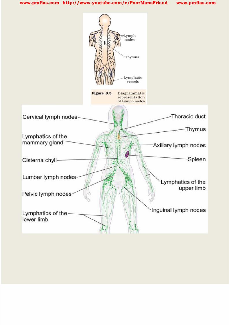
8/16/2019 Biology for Prelims Exam
http://slidepdf.com/reader/full/biology-for-prelims-exam 344/384
www.pmfias.com http://www.youtube.com/c/PoorMansFriend www.pmfias.com

8/16/2019 Biology for Prelims Exam
http://slidepdf.com/reader/full/biology-for-prelims-exam 345/384
www.pmfias.com http://www.youtube.com/c/PoorMansFriend www.pmfias.com
After maturation the lymphocytes migrate to secondary lymphoid organs like spleen,
lymph nodes, tonsils, Peyer’s patches of small intestine and appendix .
The secondary lymphoid organs provide the sites for interaction of lymphocytes with the
antigen, which then proliferate to become effector cells .
The bone marrow is the main lymphoid organ where all blood cells including
lymphocytes are produced.
The thymus is a lobed organ located near the heart and beneath the breastbone. The
thymus is quite large at the time of birth but keeps reducing in size with age and by the
time puberty is attained it reduces to a very small size.
Both bone-marrow and thymus provide micro-environments for the development and
maturation of T-lymphocytes .
The spleen is a large bean shaped organ. It mainly contains lymphocytes and
phagocytes . It acts as a filter of the blood by trapping blood-borne micro-organisms.
Spleen also has a large reservoir of erythrocytes .
8/16/2019 Biology for Prelims Exam
http://slidepdf.com/reader/full/biology-for-prelims-exam 346/384
www.pmfias.com http://www.youtube.com/c/PoorMansFriend www.pmfias.com
The lymph nodes are small solid structures located at different points along the
lymphatic system. Lymph nodes serve to trap the micro-organisms or other antigens,
which happen to get into the lymph and tissue fluid.
Antigens trapped in the lymph nodes are responsible for the activation of lymphocytes
present there and cause the immune response.
There is lymphoid tissue also located within the lining of the major tracts (respiratory,digestive and urogenital tracts) called mucosal-associated lymphoid tissue (MALT) . It
constitutes about 50 per cent of the lymphoid tissue in human body.
Summary
Health is not just the absence of disease. It is a state of complete physical, mental,
social and psychological well-being.
Diseases like typhoid, cholera, pneumonia, fungal infections of skin, malaria and many
others are a major cause of distress to human beings.
Vector-borne diseases like malaria especially one caused by Plasmodium falciparum , if
not treated, may prove fatal.
Our immune system plays the major role in preventing these diseases when we are
exposed to disease-causing agents.
The innate defenses of our body like skin, mucous membranes, antimicrobial
substances present in our tears, saliva and the phagocytic cells help to block the entry
of pathogens into our body.
If the pathogens succeed in gaining entry to our body, specific antibodies (humoral
immune response) and cells (cell mediated immune response) serve to kill these
pathogens.
Immune system has memory. On subsequent exposure to same pathogen, the immune
response is rapid and more intense. This forms the basis of protection afforded by
vaccination and immunization.
Source: NCERT Science Textbooks Class 6-12

8/16/2019 Biology for Prelims Exam
http://slidepdf.com/reader/full/biology-for-prelims-exam 347/384
www.pmfias.com http://www.youtube.com/c/PoorMansFriend www.pmfias.com
AIDS – Acquired Immuno Deficiency Syndrome – Causes of AIDS – Mechanism of HIV
Proliferation in Human Body – Prevention of AIDS.
AIDS – Acquired Immuno Deficiency Syndrome
The word AIDS stands for Acquired Immuno Deficiency Syndrome . This means
deficiency of immune system, acquired during the lifetime of an individual indicatingthat it is not a congenital disease [disease or abnormality present from birth].
‘Syndrome’ means a group of symptoms.
AIDS was first reported in 1981 and in the last twenty-five years or so, it has spread all
over the world.
Causes of AIDS
AIDS is caused by the Human Immuno Deficiency Virus (HIV) , a member of a group of
viruses called Retrovirus , which have an envelope enclosing the RNA genome .
Transmission of HIV-infection generally occurs by
a) sexual contact with infected person,
b) by transfusion of contaminated blood and blood products,
c) by sharing infected needles as in the case of intravenous drug abusers and
d) from infected mother to her child through placenta.
So, people who are at high risk of getting this infection includes
a) individuals who have multiple sexual partners,
b) drug addicts who take drugs intravenously,
c) individuals who require repeated blood transfusions and
d) children born to an HIV infected mother.
It is important to note that HIV/AIDS is not spread by mere touch or physical contact; it
spreads only through body fluids . It is, hence, imperative, for the physical and
psychological well-being, that the HIV/AIDS infected persons are not isolated from
family and society. There is always a time-lag between the infection and appearance of AIDS symptoms.
This period may vary from a few months to many years (usually 5-10 years).
Mechanism of HIV Proliferation in Human Body

8/16/2019 Biology for Prelims Exam
http://slidepdf.com/reader/full/biology-for-prelims-exam 348/384
www.pmfias.com http://www.youtube.com/c/PoorMansFriend www.pmfias.com
After getting Into the body of the person, the virus enters into macrophages where RNA
genome of the virus replicates to form viral DNA with the help of the enzyme Reverse
Transcriptase .
This viral DNA gets incorporated into host cell’s DNA and directs the infected cells to
produce virus particles. The macrophages continue to produce virus and in this way
acts like a HIV factory.
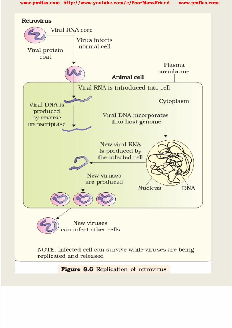
8/16/2019 Biology for Prelims Exam
http://slidepdf.com/reader/full/biology-for-prelims-exam 349/384
www.pmfias.com http://www.youtube.com/c/PoorMansFriend www.pmfias.com
8/16/2019 Biology for Prelims Exam
http://slidepdf.com/reader/full/biology-for-prelims-exam 350/384
www.pmfias.com http://www.youtube.com/c/PoorMansFriend www.pmfias.com
Simultaneously, HIV enters into helper T-lymphocytes (Th) , replicates and produce
progeny viruses. The progeny viruses released in the blood attack other helper T-
lymphocytes. This is repeated leading to a progressive decrease in the number of helper
T-lymphocytes in the body of the infected person. During this period, the person suffers
from bouts of fever, diarrhoea and weight loss .
Due to decrease in the number of helper T lymphocytes, the person starts suffering frominfections that could have been otherwise overcome such as those due to bacteria
especially Mycobacterium, viruses, fungi and even parasites like Toxoplasma. The
patient becomes so immuno-deficient that he/she is unable to protect himself/herself
against these infections.
Prevention of AIDS
A widely used diagnostic test for AIDS is Enzyme Linked Immuno-Sorbent Assay
(ELISA) .
Treatment of AIDS with anti-retroviral drugs is only partially effective. They can only
prolong the life of the patient but cannot prevent death, which is inevitable.
As AIDS has no cure, prevention is the best option. Moreover, HIV infection, more often,
spreads due to conscious behavior patterns and is not something that happens
inadvertently, like pneumonia or typhoid.
Of course, infection in blood transfusion patients, new-borns (from mother) etc., may
take place due to poor monitoring. The only excuse may be ignorance and it has been
rightly said - “don’t die of ignorance”.
In our country the National AIDS Control Organization (NACO) and other non-
governmental organizations (NGOs) are doing a lot to educate people about AIDS.
WHO has started a number of programmes to prevent the spreading of HIV infection.
Making blood (from blood banks) safe from HIV, ensuring the use of only disposable
needles and syringes in public and private hospitals and clinics, free distribution of
condoms, controlling drug abuse, advocating safe sex and promoting regular check-ups
for HIV in susceptible populations, are some such steps taken up.
Infection with HIV or having AIDS is something that should not be hidden - since then,
the infection may spread to many more people.
HIV/AIDS-infected people need help and sympathy instead of being shunned by society.
Unless society recognizes it as a problem to be dealt with in a collective manner - the
chances of wider spread of the disease increase manifold.
8/16/2019 Biology for Prelims Exam
http://slidepdf.com/reader/full/biology-for-prelims-exam 351/384
www.pmfias.com http://www.youtube.com/c/PoorMansFriend www.pmfias.com
It is a malady that can only be tackled, by the society and medical fraternity acting
together, to prevent the spread of the disease.
Cancer
Cancer is one of the most dreaded diseases of human beings and is a major cause of
death all over the globe. More than a million Indians suffer from cancer and a large number of them die from it
annually.
The mechanisms that underlie development of cancer or oncogenic transformation of
cells, its treatment and control have been some of the most intense areas of research in
biology and medicine.
In our body, cell growth and differentiation is highly controlled and regulated. In cancer
cells, there is breakdown of these regulatory mechanisms.
Normal cells show a property called contact inhibition by virtue of which contact with
other cells inhibits their uncontrolled growth.
Cancer cells appears to have lost this property of contact inhibition. As a result of this,
cancerous cells just continue to divide giving rise to masses of cells called tumors .
Types of Tumors
Tumors are of two types: benign and malignant .
Benign tumors normally remain confined to their original location and do not spread to
other parts of the body and cause little damage.
The malignant tumors, on the other hand are a mass of proliferating cells called
neoplastic or tumor cells . These cells grow very rapidly, invading and damaging the
surrounding normal tissues.
As these cells actively divide and grow they also starve the normal cells by competing for
vital nutrients.
Cells sloughed from such tumors reach distant sites through blood, and wherever they
get lodged in the body, they start a new tumor there. This property called Metastasis is
the most feared property of malignant tumors.
Causes of Cancer
Transformation of normal cells into cancerous neoplastic cells may be induced by
physical, chemical or biological agents. These agents are called carcinogens .

8/16/2019 Biology for Prelims Exam
http://slidepdf.com/reader/full/biology-for-prelims-exam 352/384
www.pmfias.com http://www.youtube.com/c/PoorMansFriend www.pmfias.com
Ionizing radiations like X-rays and gamma rays and non-ionizing radiations like UV
cause DNA damage leading to neoplastic transformation.
The chemical carcinogens present in tobacco smoke have been identified as a major
cause of lung cancer.
Cancer causing viruses called oncogenic viruses have genes called viral oncogenes .
Furthermore, several genes called cellular oncogenes (c-onc) or proto oncogenes havebeen identified in normal cells which, when activated under certain conditions, could
lead to oncogenic transformation of the cells.
Cancer Detection and Diagnosis
Early detection of cancers is essential as it allows the disease to be treated successfully
in many cases.
Cancer detection is based on biopsy and histopathological studies of the tissue and
blood and bone marrow tests for increased cell counts in the case of leukemias .
In biopsy, a piece of the suspected tissue cut into thin sections is stained and examined
under microscope (histopathological studies) by a pathologist.
Techniques like radiography (use of X-rays) , CT (computed tomography) and MRI
(magnetic resonance imaging) are very useful to detect cancers of the internal organs.
Computed tomography uses X-rays to generate a three-dimensional image of the
internals of an object.
MRI uses strong magnetic fields and non-ionising radiations to accurately detect
pathological and physiological changes in the living tissue.

8/16/2019 Biology for Prelims Exam
http://slidepdf.com/reader/full/biology-for-prelims-exam 353/384
www.pmfias.com http://www.youtube.com/c/PoorMansFriend www.pmfias.com
Antibodies against cancer-specific antigens are also used for detection of certain
cancers.
Techniques of molecular biology can be applied to detect genes in individuals with
inherited susceptibility to certain cancers. Identification of such genes, which predispose
an Individual to certain cancers, may be very helpful in prevention of cancers.
Such individuals may be advised to avoid exposure to particular carcinogens to whichthey are susceptible (e.g., tobacco smoke in case of lung cancer).
Treatment of cancer
The common approaches for treatment of cancer are surgery, radiation therapy and
immunotherapy .
In radiotherapy, tumor cells are Irradiated lethally, taking proper care of the normal
tissues surrounding the tumor mass.
Several chemotherapeutic drugs are used to kill cancerous cells. Some of these are
specific for particular tumors. Majority of drugs have side effects like hair loss, anemia,
etc.
Most cancers are treated by combination of surgery, radiotherapy and chemotherapy.
Tumor cells have been shown to avoid detection and destruction by immune system.
Therefore, the patients are given substances called biological response modifiers such
as a-interferon which activate their immune system and help in destroying the tumor.
Source: NCERT Science Textbooks Class 6-12
Cell Organelles | Plant Cell vs. Animal Cell Carbohydrates | Monosaccharides | Polysaccharides Proteins | Amino Acids | Enzymes Vitamins and Minerals – Deficiency Diseases Fats | Healthy Fats and Unhealthy Fats Animal Tissues – Epithelium, Connective Tissues Human Digestive System | Digestive Glands Respiratory System – NCERT General Science Endocrine Glands and Hormones Human Neural System | Human Brain Muscular and Skeletal System Nucleic acids – DNA and RNA | Recombinant DNA Mitosis | Cell Cycle | Cell Division Meiosis | Mitosis – Meiosis Comparison Inheritance – Mendel’s Laws of Inheritance Chromosomal Theory | Human Genome Project Sex Determination | Genetic Disorders
http://www.pmfias.com/sex-determination-genetic-disorders-mendelian-disorders-chromosomal-disorders/
http://www.pmfias.com/sex-determination-genetic-disorders-mendelian-disorders-chromosomal-disorders/

8/16/2019 Biology for Prelims Exam
http://slidepdf.com/reader/full/biology-for-prelims-exam 354/384
www.pmfias.com http://www.youtube.com/c/PoorMansFriend www.pmfias.com
Diseases Caused by Microorganisms Microbes In Human Welfare | Useful Microbes Immunity | Human Immune System
Drugs and Alcohol Abuse
The drugs, which are commonly abused are opioids, cannabinoids and coca alkaloids .
Majority of these are obtained from flowering plants . Some are obtained from fungi . Opioids are the drugs, which bind to specific opioid receptors present in our central
nervous system and gastrointestinal tract.
Heroin , commonly called smack is chemically diacetylmorphine which is a white,
odourless, bitter crystalline compound. This is obtained by acetylation of morphine ,
which is extracted from the latex of poppy plant Papaver somniferum . Generally taken
by snorting and injection, heroin is a depressant and slows down body functions .
Cannabinoids are a group of chemicals, which interact with cannabinoid receptorspresent principally in the brain.
Natural cannabinoids are obtained from the inflorescences of the plant Cannabis sativa
The flower tops, leaves and the resin of cannabis plant are used in various combinations
to produce marijuana, hashish, charas and ganja . Generally taken by inhalation and
oral ingestion, these are known for their effects on cardiovascular system of the body.
These days cannabinoids are also being abused by some sportspersons.
Coca alkaloid or cocaine is obtained from coca plant Erythroxylum coca , native to
South America. It interferes with the transport of the neuro-transmitter dopamine .
Cocaine, commonly called coke or crack is usually snorted. It has a potent stimulating
action on central nervous system, producing a sense of euphoria and increased energy.
Excessive dosage of cocaine causes hallucinations .
Other well-known plants with hallucinogenic properties are Atropa belladona and
Datura.
Drugs like barbiturates, amphetamines, benzodiazepines, lysergic acid diethyl amides
(LSD), and other similar drugs, that are normally used as medicines to help patients
cope with mental illnesses like depression and insomnia, are often abused.
Morphine is a very effective sedative and painkiller , and is very useful in patients who
have undergone surgery.
Several plants, fruits and seeds having hallucinogenic properties have been used for
hundreds of years in folk-medicine, religious ceremonies and rituals all over the globe.

8/16/2019 Biology for Prelims Exam
http://slidepdf.com/reader/full/biology-for-prelims-exam 355/384
www.pmfias.com http://www.youtube.com/c/PoorMansFriend www.pmfias.com
When these are taken for a purpose other than medicinal use or in amounts/frequency
that impairs one’s physical, physiological or psychological functions, it constitutes drug
abuse.
Smoking also paves the way to hard drugs. Tobacco has been used by human beings for
more than 400 years. It is smoked, chewed or used as a snuff. Tobacco contains a large
number of chemical substances including nicotine , an alkaloid. Nicotine stimulates adrenal gland to release adrenaline and nor-adrenaline into blood
circulation, both of which raise blood pressure and increase heart rate.
Smoking is associated with increased incidence of cancers of lung, urinary bladder and
throat, bronchitis, emphysema, coronary heart disease, gastric ulcer, etc.
Tobacco chewing is associated with increased risk of cancer of the oral cavity. Smoking
increases carbon monoxide (CO) content in blood and reduces the concentration of
haembound oxygen . This causes oxygen deficiency in the body. When one buys packets of cigarettes one cannot miss the statutory warning that is
present on the packing which warns against smoking and says how it is injurious to
health. Yet, smoking is very prevalent in society, both among young and old.
Knowing the dangers of smoking and chewing tobacco, and its addictive nature, the
youth and old need to avoid these habits. Any addict requires counselling and medical
help to get rid of the habit.
Effects of Drug/Alcohol Abuse
The immediate adverse effects of drugs and alcohol abuse are manifested in the form of
reckless behavior, vandalism and violence.
Excessive doses of drugs may lead to coma and death due to respiratory failure, heart
failure or cerebral hemorrhage.
A combination of drugs or their intake along with alcohol generally results in overdosing
and even deaths.
The most common warning signs of drug and alcohol abuse among youth include drop
in academic performance, unexplained absence from school/college, lack of interest in
personal hygiene, withdrawal, isolation, depression, fatigue, aggressive and rebellious
behaviour, deteriorating relationships with family and friends, loss of interest in
hobbies, change in sleeping and eating habits, fluctuations in weight, appetite, etc.
There may even be some far-reaching implications of drug/alcohol abuse. If an abuser is
unable to get money to buy drugs/alcohol he/she may turn to stealing.
8/16/2019 Biology for Prelims Exam
http://slidepdf.com/reader/full/biology-for-prelims-exam 356/384
www.pmfias.com http://www.youtube.com/c/PoorMansFriend www.pmfias.com
The adverse effects are just not restricted to the person who is using drugs or alcohol. At
times, a drug/alcohol addict becomes the cause of mental and financial distress to
his/her entire family and friends.
Those who take drugs intravenously (direct injection into the vein using a needle and
syringe), are much more likely to acquire serious infections like AIDS and hepatitis B .
The viruses, which are responsible for these diseases, are transferred from one person toanother by sharing of infected needles and syringes.
Both AIDS and Hepatitis B infections are chronic infections and ultimately fatal. AIDS
can be transmitted to one’s life partner through sexual contact while Hepatitis B is
transmitted through infected blood .
The use of alcohol during adolescence may also have long-term effects. It could lead to
heavy drinking in adulthood. The chronic use of drugs and alcohol damages nervous
system and liver (cirrhosis) . The use of drugs and alcohol during pregnancy is alsoknown to adversely affect the foetus.
Another misuse of drugs is what certain sportspersons do to enhance their performance.
They (mis)use narcotic analgesics, anabolic steroids, diuretics and certain
hormones in sports to increase muscle strength and bulk and to promote
aggressiveness and as a result increase athletic performance.
The side-effects of the use of anabolic steroids in females include masculinization
(features like males), increased aggressiveness, mood swings, depression, abnormal
menstrual cycles, excessive hair growth on the face and body, enlargement of clitoris,
deepening of voice.
In males it includes acne, increased aggressiveness, mood swings, depression, reduction
of size of the testicles, decreased sperm production, potential for kidney and liver
dysfunction, breast enlargement, premature baldness, enlargement of the prostate
gland. These effects may be permanent with prolonged use.
In the adolescent male or female, severe facial and body acne, and premature closure of
the growth centers of the long bones may result in stunted growth.
Prevention and Control
The age- old adage of ‘prevention is better than cure’ holds true here also. It is also true
that habits such as smoking, taking drug or alcohol are more likely to be taken up at a
young age, more during adolescence. Hence, it is best to identify the situations that may

8/16/2019 Biology for Prelims Exam
http://slidepdf.com/reader/full/biology-for-prelims-exam 357/384
www.pmfias.com http://www.youtube.com/c/PoorMansFriend www.pmfias.com
push an adolescent towards use of drugs or alcohol, and to take remedial measures well
in time. In this regard, the parents and the teachers have a special responsibility.
Parenting that combines with high levels of nurturance and consistent discipline, has
been associated with lowered risk of substance (alcohol/drugs/tobacco) abuse. Some of
the measures mentioned here would be particularly useful for prevention and control of
alcohol and drugs abuse among adolescents Avoid undue peer pressure - Every child has his/her own choice and personality, which
should be respected and nurtured. A child should not be pushed unduly to perform
beyond his/her threshold limits; be it studies, sports or other activities.
Education and counselling - Educating and counselling him/ her to face problems and
stresses, and to accept disappointments and failures as a part of life. It would also be
worthwhile to channelize the child’s energy into healthy pursuits like sports, reading,
music, yoga and other extracurricular activities. Seeking help from parents and peers - Help from parents and peers should be sought
immediately so that they can guide appropriately. Help may even be sought from close
and trusted friends. Besides getting proper advise to sort out their problems, this would
help young to vent their feelings of anxiety and guilt.
Looking for danger signs - Alert parents and teachers need to look for and identify the
danger signs discussed above. Even friends, if they find someone using drugs or alcohol,
should not hesitate to bring this to the notice of parents or teacher in the best interests
of the person concerned. Appropriate measures would then be required to diagnose the
malady and the underlying causes. This would help in initiating proper remedial steps
or treatment.
Seeking professional and medical help - A lot of help is available in the form of highly
qualified psychologists, psychiatrists, and de-addiction and rehabilitation programmes
to help individuals who have unfortunately got in the quagmire of drug/alcohol abuse.
With such help, the affected individual with sufficient efforts and will power, can get rid
of the problem completely and lead a perfectly normal and healthy life.
Summary
Health is not just the absence of disease. It is a state of complete physical, mental,
social and psychological well-being.
Diseases like typhoid, cholera, pneumonia, fungal infections of skin, malaria and many
others are a major cause of distress to human beings.

8/16/2019 Biology for Prelims Exam
http://slidepdf.com/reader/full/biology-for-prelims-exam 358/384
www.pmfias.com http://www.youtube.com/c/PoorMansFriend www.pmfias.com
Vector-borne diseases like malaria especially one caused by Plasmodium falciparum , if
not treated, may prove fatal.
Our immune system plays the major role in preventing these diseases when we are
exposed to disease-causing agents.
The innate defenses of our body like skin, mucous membranes, antimicrobial
substances present in our tears, saliva and the phagocytic cells help to block the entryof pathogens into our body.
If the pathogens succeed in gaining entry to our body, specific antibodies (humoral
immune response) and cells (cell mediated immune response) serve to kill these
pathogens.
Immune system has memory. On subsequent exposure to same pathogen, the immune
response is rapid and more intense. This forms the basis of protection afforded by
vaccination and immunization.
8/16/2019 Biology for Prelims Exam
http://slidepdf.com/reader/full/biology-for-prelims-exam 359/384
www.pmfias.com http://www.youtube.com/c/PoorMansFriend www.pmfias.com
Acute and Chronic Diseases, Communicable Diseases, Organ-Specific Diseases, Principles
of Treatment, Principles of Prevention, Childhood Diseases in Indian Children.
Some important concepts related to diseases are discussed in the below posts:
Vitamins and Minerals – Deficiency Diseases
Sex Determination | Genetic Disorders Diseases Caused by Microorganisms
Immunity | Human Immune System
AIDS | Acquired Immuno Deficiency Syndrome
Cancer | Causes of Cancer – NCERT
Acute and Chronic Diseases
Some diseases last for only very short periods of time, and these are called acute
diseases . We all know from experience that the common cold lasts only a few days.
Other ailments can last for a long time, even as much as a lifetime, and are called
chronic diseases . An example is the infection causing elephantiasis , which is very
common in some parts of India.
Communicable Diseases
Microbial diseases that can spread from an infected person to a healthy person through
air, water, food or physical contact are called communicable diseases .
Examples of such diseases include cholera, common cold, chicken pox and
tuberculosis .
Example of a carrier is the female Anopheles mosquito , which carries the parasite of
malaria . Female Aedes mosquito acts as carrier of dengue virus .
Robert Köch (1876) discovered the bacterium (Bacillus anthracis ) which causes
anthrax disease.
How do infectious diseases spread? Many microbial agents can commonly move from an
affected person to someone else in a variety of ways. In other words, they can be
‘communicated’, and so are also called communicable diseases .
Such disease-causing microbes can spread through the air. Examples of such diseases
spread through the air are the common cold, pneumonia and tuberculosis .
http://www.pmfias.com/sex-determination-genetic-disorders-mendelian-disorders-chromosomal-disorders/
http://www.pmfias.com/sex-determination-genetic-disorders-mendelian-disorders-chromosomal-disorders/
http://www.pmfias.com/cancer-types-tumors-causes-cancer-cancer-detection-diagnosis-treatment-cancer/
http://www.pmfias.com/cancer-types-tumors-causes-cancer-cancer-detection-diagnosis-treatment-cancer/
http://www.pmfias.com/cancer-types-tumors-causes-cancer-cancer-detection-diagnosis-treatment-cancer/
http://www.pmfias.com/cancer-types-tumors-causes-cancer-cancer-detection-diagnosis-treatment-cancer/
http://www.pmfias.com/cancer-types-tumors-causes-cancer-cancer-detection-diagnosis-treatment-cancer/
8/16/2019 Biology for Prelims Exam
http://slidepdf.com/reader/full/biology-for-prelims-exam 360/384

8/16/2019 Biology for Prelims Exam
http://slidepdf.com/reader/full/biology-for-prelims-exam 361/384
www.pmfias.com http://www.youtube.com/c/PoorMansFriend www.pmfias.com
breathlessness. If the liver is targeted, there will be jaundice. If the brain is the target,
we will observe headaches, vomiting, fits or unconsciousness.
In addition to these tissue-specific effects of infectious disease, there will be other
common effects too.
Most of these common effects depe nd on the fact that the body’s immune system is
activated in response to infection. An active immune system recruits many cells to the affected tissue to kill off the disease-
causing microbes. This recruitment process is called inflammation . As a part of this
process, there are local effects such as swelling and pain, and general effects such as
fever.
In some cases, the tissue-specificity of the infection leads to very general-seeming
effects. For example, in HIV infection, the virus goes to the immune system and
damages its function. Thus, many of the effects of HIV-aids are because the body can nolonger fight off the many minor infections that we face every day. Instead, every small
cold can become pneumonia . Similarly, a minor gut infection can produce major
diarrhoea with blood loss. Ultimately, it is these other infections that kill people
suffering from HIV-aids.
Principles of Treatment
There are two ways to treat an infectious disease. One would be to reduce the effects of
the disease and the other to kill the cause of the disease.
For the first, we can provide treatment that will reduce the symptoms. The symptoms
are usually because of inflammation. For example, we can take medicines that bring
down fever, reduce pain or loose motions. We can take bed rest so that we can conserve
our energy. This will enable us to have more of it available to focus on healing.
But this kind of symptom-directed treatment by itself will not make the infecting
microbe go away and the disease will not be cured. For that, we need to be able to kill off
the microbes.
How do we kill microbes? One way is to use medicines that kill microbes. We have seen
earlier that microbes can be classified into different categories. They are viruses,
bacteria, fungi or protozoa.
Each of these groups of organisms will have some essential biochemical life process
which is peculiar to that group and not shared with the other groups. These processes
8/16/2019 Biology for Prelims Exam
http://slidepdf.com/reader/full/biology-for-prelims-exam 362/384

8/16/2019 Biology for Prelims Exam
http://slidepdf.com/reader/full/biology-for-prelims-exam 363/384
www.pmfias.com http://www.youtube.com/c/PoorMansFriend www.pmfias.com
As an example, let us take antibiotics. They commonly block biochemical pathways
important for bacteria . Many bacteria, for example, make a cell-wall to protect
themselves. The antibiotic penicillin blocks the bacterial processes that build the cell
wall. As a result, the growing bacteria become unable to make cell-walls, and die easily.
Human cells don’t make a cell-wall anyway, so penicillin cannot have such an effect on
us. Penicillin will have this effect on any bacteria that use such processes for making
cell-walls. Similarly, many antibiotics work against many species of bacteria rather than
simply working against one group.
But viruses do not use these pathways at all, and that is the reason why antibiotics do
not work against viral infections. If we have a common cold, taking antibiotics does not
reduce the severity or the duration of the disease. However, if we also get a bacterial
infection along with the viral cold, taking antibiotics will help. Even then, the antibiotic
will work only against the bacterial part of the infection, not the viral infection.
Principles of Prevention
What are the specific ways of prevention? They relate to a peculiar property of the
immune system that usually fights off microbial infections.

8/16/2019 Biology for Prelims Exam
http://slidepdf.com/reader/full/biology-for-prelims-exam 364/384

8/16/2019 Biology for Prelims Exam
http://slidepdf.com/reader/full/biology-for-prelims-exam 365/384
www.pmfias.com http://www.youtube.com/c/PoorMansFriend www.pmfias.com
Famously, two centuries ago, an English physician named Edward Jenner , realized that
milkmaids who had had cowpox did not catch smallpox even during epidemics.
Cowpox is a very mild disease. Jenner tried deliberately giving cowpox to people, and
found that they were now resistant to smallpox. This was because the smallpox virus is
c losely related to the cowpox virus. ‘Cow’ is ‘Vacca’ in latin, and cowpox is ‘Vaccinia’.
Childhood Diseases in Indian Children
Gastroentitis
Gastroentitis is an infection in the digestive system and it is one of the most common
childhood illnesses.
Symptoms of gastroentitis include diarrhoea, nausea and vomiting, tummy cramps, and
fever.
One of the main risks with gastroentitis is that it causes dehydration in children.
Rickets
Rickets occurs due to Vitamin D deficiency .
Deficiency of Vitamin D occurs in a child because of lack of exposure to sunlight .
Lack of adequate calcium in the diet can also cause rickets.
Rickets is a disease which involves softening and weakening of bones in children.
Children between the ages of 6 to 24 months are at the highest risk of developing thedisease because that is the age when their bones are rapidly growing.
Conjunctivitis
Conjunctivitis is caused due to inflammation of the conjunctiva .
Conjunctiva is the outermost layer of the eye and the inner surface of the eyelids.
Conjunctivitis often starts in one eye at first and then spreads to the other eye.
For children suffering from conjunctivitis it is important to see a doctor to know what
kind of conjunctivitis it is.
Symptoms of conjunctivitis include redness of eyes, irritation in the eye, and eye
watering.
Scabies
Scabies is an infection of the skin.
Scabies is caused by tiny insects called mites .

8/16/2019 Biology for Prelims Exam
http://slidepdf.com/reader/full/biology-for-prelims-exam 366/384
www.pmfias.com http://www.youtube.com/c/PoorMansFriend www.pmfias.com
These scabies mites burrow into the skin and lay eggs which become adult mites very
soon.
Symptoms of this infection include superficial burrows, rash and severe itching.
Blisters on the palm and soles of the feet are characteristic symptoms of scabies in
infants.
Scabies is one of the highly contagious diseases and a child can develop it by cominginto contact with someone else who has been infected.
Children with scabies must not be sent to school or day care until it gets completely
cured.
Upper Respiratory Tract infection (URTI)
Upper Respiratory Tract Infections are extremely common due to air pollution and
vehicular emission.
Upper respiratory tract infections include common cold, influenza and sore throat.
Tonsillitis is also one of upper respiratory tract infections.
Tonsillitis is caused due to infection of the tonsils.
Tonsils are the areas of lymphoid tissue on either side of the throat.
Symptoms of tonsillitis include a severe sore throat, coughing, headache and difficulty
swallowing.
Tuberculosis
Tuberculosis also affects children and is known as Primary Complex or Childhood
Tuberculosis infection.
Children under the age of two years are more at risk of developing tuberculosis because
their immune system is under developed or still developing.
Tuberculosis is completely curable and early diagnosis can help in effective treatment.
Typhoid It is a water borne disease rampant in children due to poor sanitation.
Cases of typhoid are more common in countries like India and some other South Asian
countries and in other low developed nations and have been seen lesser in countries like
the USA.
Symptoms of typhoid in children are poor appetite, body ache, discomfort in abdomen,
lethargy and weakness, fever with rising and falling pattern.
8/16/2019 Biology for Prelims Exam
http://slidepdf.com/reader/full/biology-for-prelims-exam 367/384
www.pmfias.com http://www.youtube.com/c/PoorMansFriend www.pmfias.com
Some children may also experience headache, chest congestion, diarrhoea and vomiting
and rose spots on the abdomen.
Bronchitis and Asthma
Bronchitis and asthma are common in children.
Bronchitis and asthma are caused due to high exposure to air borne pollutants.
Bronchitis and asthma need to be treated with antibiotics and bronchodilators.
Some Other Diseases
Diseases Caused by Worms
Ascaris , the common round worm and Wuchereria , the filarial worm , are some of the
helminths which are known to be pathogenic to man. Ascaris, an intestinal parasite
causes ascariasis . Symptoms of these disease include internal bleeding, muscular pain, fever, anemia and
blockage of the intestinal passage. The eggs of the parasite are excreted along with the
faeces of infected persons which contaminate soil, water, plants, etc. A healthy person
acquires this infection through contaminated water, vegetables, fruits, etc.
Wuchereria (W. bancrofti and W. malayi) , the filarial worms cause a slowly developing
chronic inflammation of the organs in which they live for many years, usually the
lymphatic vessels of the lower limbs and the disease is called elephantiasis orfilariasis. The genital organs are also often affected, resulting in gross deformities. The
pathogens are transmitted to person through the bite by the female mosquite.
Old Age Diseases: Dementia
Dementia is “one of the major causes of disability and dependency among older people
worldwide”
Pollution related diseases: Silicosis Silicosis is a lung disorder caused by inhalation, retention and pulmonary reaction to
crystalline silica, as a result of exposure during mining, stone crushing and quarrying
activities.
Zoonotic Diseases

8/16/2019 Biology for Prelims Exam
http://slidepdf.com/reader/full/biology-for-prelims-exam 368/384
www.pmfias.com http://www.youtube.com/c/PoorMansFriend www.pmfias.com
zoonotic diseases — are spread between animals and humans, and are common in
societies where poverty is widespread
Chikungunya, dengue, Avian influenza, plague, SARS and acute encephalitis syndrome
(AES) are some of the zoonotic diseases.

8/16/2019 Biology for Prelims Exam
http://slidepdf.com/reader/full/biology-for-prelims-exam 369/384
www.pmfias.com http://www.youtube.com/c/PoorMansFriend www.pmfias.com
Blood – Blood Vessels, Body Fluids and Circulation: Plasma, Formed Elements: Red Blood
Cells (RBC), White Blood Cells (WBC), Platelets, Coagulation of Blood, Blood Groups.
Blood
It transports substances like digested food from the small intestine to the other parts of
the body. It carries oxygen from the lungs to the cells of the body. It also transportswaste for removal from the body.
Blood is a liquid, which has cells of various kinds suspended in it. The fluid part of the
blood is called plasma .
One type of cells are the red blood cells (RBC) which contain a red pigment called
haemoglobin .
Haemoglobin bind with oxygen and transports it to all the parts of the body and
ultimately to all the cells. The presence of haemoglobin makes blood appear red .
The blood also has white blood cells (WBC) which fight against germs that may enter
our body.
The clot is formed because of the presence of another type of cells in the blood, called
platelets .
Blood Vessels
They are two types of blood vessels namely arteries and veins .
Veins are the blood vessels that carry carbon dioxide-rich blood [impure blood] from
all parts of the body back to the heart. Pulmonary vein is an exception as it carries
oxygen-rich blood [pure blood] from lungs to heart. The veins have thin walls .
Arteries are the blood vessels that carry oxygen-rich blood from heart to all parts of the
body. Pulmonary artery is an exception as it carries carbon dioxide-rich blood from
heart to lungs. The arteries have thick walls as the pressure acting on then is high.
Blood FROM Heart → Artery
Blood TO Heart → Vein
Arteries divide into smaller vessels. On reaching the tissues, they divide further into
extremely thin tubes called capillaries . The capillaries join up to form veins which
empty into the heart.
Body Fluids and Circulation

8/16/2019 Biology for Prelims Exam
http://slidepdf.com/reader/full/biology-for-prelims-exam 370/384
www.pmfias.com http://www.youtube.com/c/PoorMansFriend www.pmfias.com
Blood is a special connective tissue consisting of a fluid matrix, plasma, and formed
elements.
Plasma
Plasma is a straw coloured, viscous fluid constituting nearly 55 per cent of the blood.
90-92 per cent of plasma is water and proteins contribute 6-8 per cent of it. Fibrinogen, globulins and albumins are the major proteins.
Fibrinogens are needed for clotting or coagulation of blood.
Globulins primarily are involved in defense mechanisms of the body
Albumins help in osmotic balance .
Plasma also contains small amounts of minerals like Na+, Ca++, Mg++, HCO3-, Cl-, etc.
Glucose, amino acids, lipids, etc., are also present in the plasma as they are always in
transit in the body. Factors for coagulation or clotting of blood are also present in the plasma in an inactive
form. Plasma without the clotting factors is called serum .
Formed Elements
Erythrocytes, leucocytes and platelets are collectively called formed elements and
they constitute nearly 45 per cent of the blood.
Red Blood Cells (RBC) Erythrocytes or red blood cells (RBC) are the most abundant of all the cells in blood.
A healthy adult man has, on an average, 5 million to 5.5 million of RBCs mm-3 of blood.
RBCs are formed in the red bone marrow in the adults. RBCs are devoid of nucleus in
most of the mammals and are biconcave in shape.
They have a red coloured, iron containing complex protein called haemoglobin, hence
the colour and name of these cells.
RBCs have an average life span of 120 days after which they are destroyed in the spleen
(graveyard of RBCs) .
White Blood Cells (WBC)
Leucocytes are also known as white blood cells (WBC) as they are colorless due to the
lack of haemoglobin. They are nucleated and are relatively lesser in number which
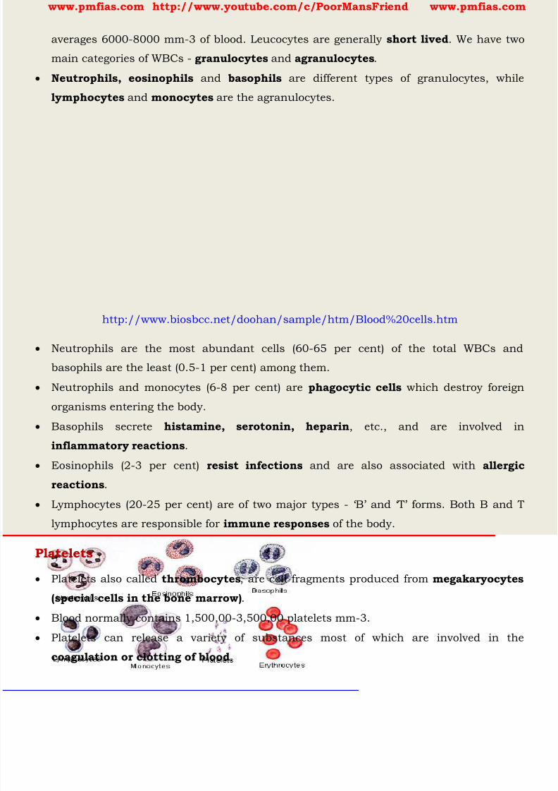
8/16/2019 Biology for Prelims Exam
http://slidepdf.com/reader/full/biology-for-prelims-exam 371/384
www.pmfias.com http://www.youtube.com/c/PoorMansFriend www.pmfias.com
averages 6000-8000 mm-3 of blood. Leucocytes are generally short lived . We have two
main categories of WBCs - granulocytes and agranulocytes .
Neutrophils, eosinophils and basophils are different types of granulocytes, while
lymphocytes and monocytes are the agranulocytes.
http://www.biosbcc.net/doohan/sample/htm/Blood%20cells.htm
Neutrophils are the most abundant cells (60-65 per cent) of the total WBCs and
basophils are the least (0.5-1 per cent) among them.
Neutrophils and monocytes (6-8 per cent) are phagocytic cells which destroy foreign
organisms entering the body.
Basophils secrete histamine, serotonin, heparin , etc., and are involved in
inflammatory reactions .
Eosinophils (2-3 per cent) resist infections and are also associated with allergic
reactions .
Lymphocytes (20-25 per cent) are of two major types - ‘B’ and ‘T’ forms. Both B and T
lymphocytes are responsible for immune responses of the body.
Platelets Platelets also called thrombocytes , are cell fragments produced from megakaryocytes
(special cells in the bone marrow) .
Blood normally contains 1,500,00-3,500,00 platelets mm-3.
Platelets can release a variety of substances most of which are involved in the
coagulation or clotting of blood .
8/16/2019 Biology for Prelims Exam
http://slidepdf.com/reader/full/biology-for-prelims-exam 372/384
www.pmfias.com http://www.youtube.com/c/PoorMansFriend www.pmfias.com
Coagulation of Blood
Blood exhibits coagulation or clotting in response to an injury or trauma. This is a
mechanism to prevent excessive loss of blood from the body.
Dark reddish brown scum is formed at the site of a cut or an injury over a period of
time. It is a clot or coagulam formed mainly of a network of threads called fibrins in
which dead and damaged formed elements of blood are trapped.
Fibrins are formed by the conversion of inactive fibrinogens in the plasma by the enzyme
thrombin .
Thrombins, in turn are formed from another inactive substance present in the plasma
called prothrombin . An enzyme complex, thrombokinase , is required for the above
reaction. This complex is formed by a series of linked enzymic reactions (cascade
process) involving a number of factors present in the plasma in an inactive state.
An injury or a trauma stimulates the platelets in the blood to release certain factors
which activate the mechanism of coagulation. Certain factors released by the tissues at
the site of injury also can initiate coagulation. Calcium ions play a very important role
in clotting.
Lymph (Tissue Fluid)
As the blood passes through the capillaries in tissues, some water along with many
small water soluble substances move out into the spaces between the cells of tissuesleaving the larger proteins and most of the formed elements in the blood vessels. This
fluid released out is called the interstitial fluid or tissue fluid .
Interstitial fluid or tissue fluid has the same mineral distribution as that in plasma.
Exchange of nutrients, gases, etc., between the blood and the cells always occur through
this fluid.
An elaborate network of vessels called the lymphatic system collects this fluid and
drains it back to the major veins. The fluid present in the lymphatic system is called thelymph .
Lymph is a colourless fluid containing specialized lymphocytes which are responsible
for the immune responses of the body. Lymph is also an important carrier for
nutrients, hormones, etc.
Fats are absorbed through lymph in the lacteals present in the intestinal villi.
Blood Groups

8/16/2019 Biology for Prelims Exam
http://slidepdf.com/reader/full/biology-for-prelims-exam 373/384
www.pmfias.com http://www.youtube.com/c/PoorMansFriend www.pmfias.com
As you know, blood of human beings differ in certain aspects though it appears to be
similar. Various types of grouping of blood has been done. Two such groupings - the
ABO and Rh - are widely used all over the world.
ABO grouping
ABO grouping is based on the presence or absence of two surface antigens (chemicalsthat can induce immune response) on the RBCs namely A and B.
Similarly, the plasma of different individuals contain two natural antibodies (proteins
produced in response to antigens).
The distribution of antigens and antibodies in the four groups of blood, A, B, AB and O
are given in Table below.
ABO blood groups are controlled by the gene I . The plasma membrane of the red blood
cells has sugar polymers that protrude from its surface and the kind of sugar is
controlled by the gene. The gene (I) has three alleles IA, IB and i .
The alleles IA and IB produce a slightly different form of the sugar while allele i does
not produce any sugar .
Because humans are diploid organisms, each person possesses any two of the three I
gene alleles.
IA and IB are completely dominant over i , in other words when IA and i are present
only IA expresses (because i does not produce any sugar), and when IB and i are present
IB expresses.
But when IA and IB are present together they both express their own types of sugars:
this is because of co-dominance . Hence red blood cells have both A and B types of
sugars.
Since there are three different alleles, there are six different combinations of these three
alleles that are possible, and therefore, a total of six different genotypes of the human
ABO blood types. How many phenotypes are possible?

8/16/2019 Biology for Prelims Exam
http://slidepdf.com/reader/full/biology-for-prelims-exam 374/384
www.pmfias.com http://www.youtube.com/c/PoorMansFriend www.pmfias.com
Here there are 6 Genotypes and 4 Phenotypes [A, B, AB and O].
You probably know that during blood transfusion, any blood cannot be used; the blood
of a donor has to be carefully matched with the blood of a recipient before any blood
transfusion to avoid severe problems of clumping (destruction of RBC) .
From the above mentioned table it is evident that group ‘O’ blood can be donated to
persons with any other blood group and hence ‘O’ group individuals are called
‘ universal donors’ .
Persons with ‘AB’ group can accept blood from per sons with AB as well as the other
groups of blood. Therefore, such persons are called ‘universal recipients’ .
Rh grouping
Another antigen , the Rh antigen similar to one present in Rhesus monkeys (hence
Rh), is also observed on the surface of RBCs of majority ( nearly 80 per cent ) of
humans. Such individuals are called Rh positive (Rh+ve) and those in whom this
antigen is absent are called Rh negative (Rh-ve) . An Rh-ve person, if exposed to Rh+ve blood, will form specific antibodies against the Rh
antigens. Therefore, Rh group should also be matched before transfusions.
A special case of Rh incompatibility (mismatching) has been observed between the Rh-ve
blood of a pregnant mother with Rh+ve blood of the foetus.
Rh antigens of the foetus do not get exposed to the Rh-ve blood of the mother in the first
pregnancy as the two bloods are well separated by the placenta.
8/16/2019 Biology for Prelims Exam
http://slidepdf.com/reader/full/biology-for-prelims-exam 375/384
www.pmfias.com http://www.youtube.com/c/PoorMansFriend www.pmfias.com
However, during the delivery of the first child, there is a possibility of exposure of the
maternal blood to small amounts of the Rh+ve blood from the foetus.
In such cases, the mother starts preparing antibodies against Rh antigen in her blood.
In case of her subsequent pregnancies, the Rh antibodies from the mother (Rh-ve) can
leak into the blood of the foetus (Rh+ve) and destroy the foetal RBCs.
This could be fatal to the foetus or could cause severe anaemia and jaundice to thebaby. This condition is called erythroblastosis foetalis .
This can be avoided by administering anti-Rh antibodies to the mother immediately
after the delivery of the first child.
Circulatory System – Human Circulatory System: Heart, Heartbeat, Cardiac Cycle,
Electrocardiograph (ECG), Double Circulation, Disorders of Circulatory System.
Circulatory System The English physician, William Harvey (A.D.1578 – 1657), discovered the circulation of
blood.
The circulatory patterns are of two types - open or closed .
Open circulatory system is present in arthropods and molluscs in which blood pumped
by the heart passes through large vessels into open spaces or body cavities called
sinuses .
Annelids and chordates have a closed circulatory system in which the blood pumpedby the heart is always circulated through a closed network of blood vessels. This pattern
is considered to be more advantageous as the flow of fluid can be more precisely
regulated.
All vertebrates possess a muscular chambered heart. Fishes have a 2-chambered heart
with an atrium and a ventricle. Amphibians and the reptiles (except crocodiles) have
a 3-chambered heart with two atria and a single ventricle, whereas crocodiles, birds
and mammals possess a 4-chambered heart with two atria and two ventricles. In fishes the heart pumps out deoxygenated blood which is oxygenated by the gills and
supplied to the body parts from where deoxygenated blood is returned to the heart
(single circulation) .
In amphibians and reptiles, the left atrium receives oxygenated blood from the
gills/lungs/skin and the right atrium gets the deoxygenated blood from other body
8/16/2019 Biology for Prelims Exam
http://slidepdf.com/reader/full/biology-for-prelims-exam 376/384
www.pmfias.com http://www.youtube.com/c/PoorMansFriend www.pmfias.com
parts. However, they get mixed up in the single ventricle which pumps out mixed blood
(incomplete double circulation) .
In birds and mammals, oxygenated and deoxygenated blood received by the left and
right atria respectively passes on to the ventricles of the same sides. The ventricles
pump it out without any mixing up, i.e., two separate circulatory pathways are present
in these organisms, hence, these animals have double circulation . Let us study thehuman circulatory system.
Does sponges and hydra also have blood? Animals such as sponges and Hydra do not
possess any circulatory system. The water in which they live brings food and oxygen as
it enters their bodies. The water carries away waste materials and carbon dioxide as it
moves out. Thus, these animals do not need a circulatory fluid like the blood.
Human Circulatory System
Heart
The heart has four chambers . The two upper chambers are called the atria (singular:
atrium) and the two lower chambers are called the ventricles .
The partition between the chambers helps to avoid mixing up of blood rich in oxygen
with the blood rich in carbon dioxide.
Heartbeat
The walls of the chambers of the heart are made up of muscles. These muscles contract
and relax rhythmically. This rhythmic contraction followed by its relaxation constitutes
a heartbeat.
Human circulatory system, also called the blood vascular system consists of a
muscular chambered heart, a network of closed branching blood vessels and blood, the
fluid which is circulated.
Heart, the mesodermally derived organ [ the middle layer of cells or tissues of an embryo, or the parts
derived from this (e.g. cartilage, muscles, and bone) ], is situated in the thoracic cavity, in between
the two lungs, slightly tilted to the left. It has the size of a clenched fist.
It is protected by a double walled membranous bag, pericardium , enclosing the
pericardial fluid .
Our heart has four chambers, two relatively small upper chambers called atria and two
larger lower chambers called ventricles .

8/16/2019 Biology for Prelims Exam
http://slidepdf.com/reader/full/biology-for-prelims-exam 377/384
www.pmfias.com http://www.youtube.com/c/PoorMansFriend www.pmfias.com
A thin, muscular wall called the interatrial septum separates the right and the left
atria, whereas a thick-walled, the inter-ventricular septum , separates the left and the
right ventricles.
The atrium and the ventricle of the same side are also separated by a thick fibrous
tissue called the atrio-ventricular septum . However, each of these septa are provided
with an opening through which the two chambers of the same side are connected. The opening between the right atrium and the right ventricle is guarded by a valve
formed of three muscular flaps or cusps, the tricuspid valve , whereas a bicuspid or
mitral valve guards the opening between the left atrium and the left ventricle.
The openings of the right and the left ventricles into the pulmonary artery and the
aorta respectively are provided with the semilunar valves .
The valves in the heart allows the flow of blood only in one direction, i.e., from the atria
to the ventricles and from the ventricles to the pulmonary artery or aorta. These valvesprevent any backward flow.
8/16/2019 Biology for Prelims Exam
http://slidepdf.com/reader/full/biology-for-prelims-exam 378/384
www.pmfias.com http://www.youtube.com/c/PoorMansFriend www.pmfias.com
The entire heart is made of cardiac muscles . The walls of ventricles are much thicker
than that of the atria.
A specialized cardiac musculature called the nodal tissue is also distributed in the
heart. A patch of this tissue is present in the right upper corner of the right atrium
called the sino-atrial node (SAN) .
Another mass of this tissue is seen in the lower left corner of the right atrium close tothe atrio-ventricular septum called the atrio-ventricular node (AVN) .
A bundle of nodal fibres, atrioventricular bundle (AV bundle) continues from the AVN
which passes through the atrio-ventricular septa to emerge on the top of the inter-
ventricular septum and immediately divides into a right and left bundle. These branches
give rise to minute fibres throughout the ventricular musculature of the respective sides
and are called purkinje fibres . These fibres along with right and left bundles are known
as bundle of His . The nodal musculature has the ability to generate action potentials without any
external stimuli, i.e., it is autoexcitable .
However, the number of action potentials that could be generated in a minute vary at
different parts of the nodal system.
The SAN can generate the maximum number of action potentials, i.e., 70-75/min, and is
responsible for initiating and maintaining the rhythmic contractile activity of the heart.
Therefore, it is called the pacemaker . Our heart normally beats 70-75 times in a minute
(average 72 beats/min).
Cardiac Cycle
Watch these videos for easy understanding
https://www.youtube.com/watch?v=gn6QmETEm8s ;
https://www.youtube.com/watch?v=yGlFBzaTuoI;

8/16/2019 Biology for Prelims Exam
http://slidepdf.com/reader/full/biology-for-prelims-exam 379/384
www.pmfias.com http://www.youtube.com/c/PoorMansFriend www.pmfias.com
To begin with, all the four chambers of heart are in a relaxed state , i.e., they are in
joint diastole .
As the tricuspid and bicuspid valves are open, blood from the pulmonary veins and vena
cava flows into the left and the right ventricle respectively through the left and right
atria. The semilunar valves are closed at this stage.
The SAN now generates an action potential which stimulates both the atria to undergo a
simultaneous contraction - the atrial systole . This increases the flow of blood into the
ventricles by about 30 per cent.
The action potential is conducted to the ventricular side by the AVN and AV bundle from
where the bundle of His transmits it through the entire ventricular musculature. This
causes the ventricular muscles to contract, ( ventricular systole ), the atria undergoes
relaxation (diastole), coinciding with the ventricular systole.
Ventricular systole increases the ventricular pressure causing the closure of tricuspid
and bicuspid valves due to attempted backflow of blood into the atria.
As the ventricular pressure increases further, the semilunar valves guarding the
pulmonary artery (right side) and the aorta (left side) are forced open, allowing the blood
in the ventricles to flow through these vessels into the circulatory pathways.
The ventricles now relax (ventricular diastole) and the ventricular pressure falls causing
the closure of semilunar valves which prevents the backflow of blood into the ventricles.
8/16/2019 Biology for Prelims Exam
http://slidepdf.com/reader/full/biology-for-prelims-exam 380/384
www.pmfias.com http://www.youtube.com/c/PoorMansFriend www.pmfias.com
As the ventricular pressure declines further, the tricuspid and bicuspid valves are
pushed open by the pressure in the atria exerted by the blood which was being emptied
into them by the veins. The blood now once again moves freely to the ventricles. The
ventricles and atria are now again in a relaxed ( joint diastole ) state, as earlier.
Soon the SAN generates a new action potential and the events described above are
repeated in that sequence and the process continues. This sequential event in the heart which is cyclically repeated is called the cardiac cycle
and it consists of systole and diastole of both the atria and ventricles.
As mentioned earlier, the heart beats 72 times per minute, i.e., that many cardiac cycles
are performed per minute. From this it could be deduced that the duration of a cardiac
cycle is 0.8 seconds.
During a cardiac cycle, each ventricle pumps out approximately 70 mL of blood which is
called the stroke volume. The stroke volume multiplied by the heart rate (no. of beatsper min.) gives the cardiac output.
Therefore, the cardiac output can be defined as the volume of blood pumped out by each
ventricle per minute and averages 5000 mL or 5 litres in a healthy individual.
The body has the ability to alter the stroke volume as well as the heart rate and thereby
the cardiac output. For example, the cardiac output of an athlete will be much higher
than that of an ordinary man.
During each cardiac cycle two prominent sounds are produced which can be easily
heard through a stethoscope. The first heart sound (lub) is associated with the closure
of the tricuspid and bicuspid valves whereas the second heart sound (dub) is
associated with the closure of the semilunar valves . These sounds are of clinical
diagnostic significance.
Electrocardiograph (ECG)
Electro-cardiograph is used to obtain an electrocardiogram (ECG) . ECG is a graphical
representation of the electrical activity of the heart during a cardiac cycle.
To obtain a standard ECG, a patient is connected to the machine with three electrical
leads (one to each wrist and to the left ankle) that continuously monitor the heart
activity.
For a detailed evaluation of the heart’s function, multiple leads are attached to the chest
region. Here, we will talk only about a standard ECG.
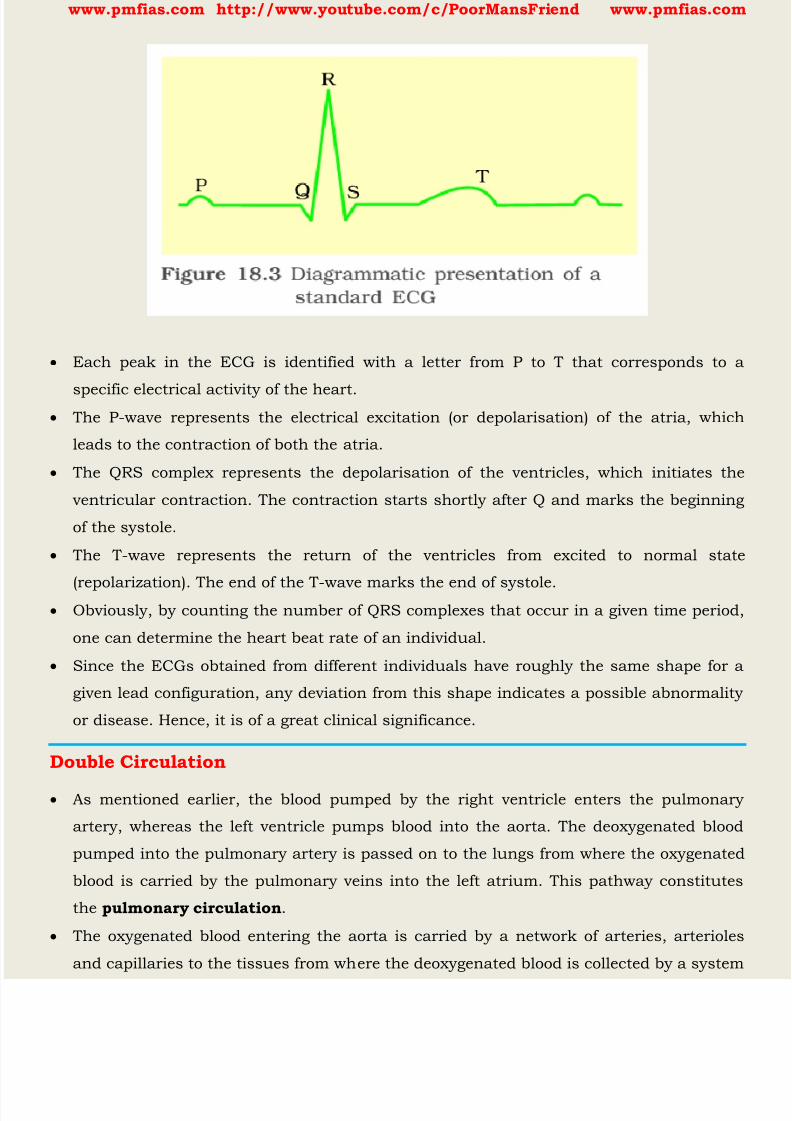
8/16/2019 Biology for Prelims Exam
http://slidepdf.com/reader/full/biology-for-prelims-exam 381/384
www.pmfias.com http://www.youtube.com/c/PoorMansFriend www.pmfias.com
Each peak in the ECG is identified with a letter from P to T that corresponds to a
specific electrical activity of the heart. The P-wave represents the electrical excitation (or depolarisation) of the atria, which
leads to the contraction of both the atria.
The QRS complex represents the depolarisation of the ventricles, which initiates the
ventricular contraction. The contraction starts shortly after Q and marks the beginning
of the systole.
The T-wave represents the return of the ventricles from excited to normal state
(repolarization). The end of the T-wave marks the end of systole. Obviously, by counting the number of QRS complexes that occur in a given time period,
one can determine the heart beat rate of an individual.
Since the ECGs obtained from different individuals have roughly the same shape for a
given lead configuration, any deviation from this shape indicates a possible abnormality
or disease. Hence, it is of a great clinical significance.
Double Circulation
As mentioned earlier, the blood pumped by the right ventricle enters the pulmonary
artery, whereas the left ventricle pumps blood into the aorta. The deoxygenated blood
pumped into the pulmonary artery is passed on to the lungs from where the oxygenated
blood is carried by the pulmonary veins into the left atrium. This pathway constitutes
the pulmonary circulation .
The oxygenated blood entering the aorta is carried by a network of arteries, arterioles
and capillaries to the tissues from where the deoxygenated blood is collected by a system
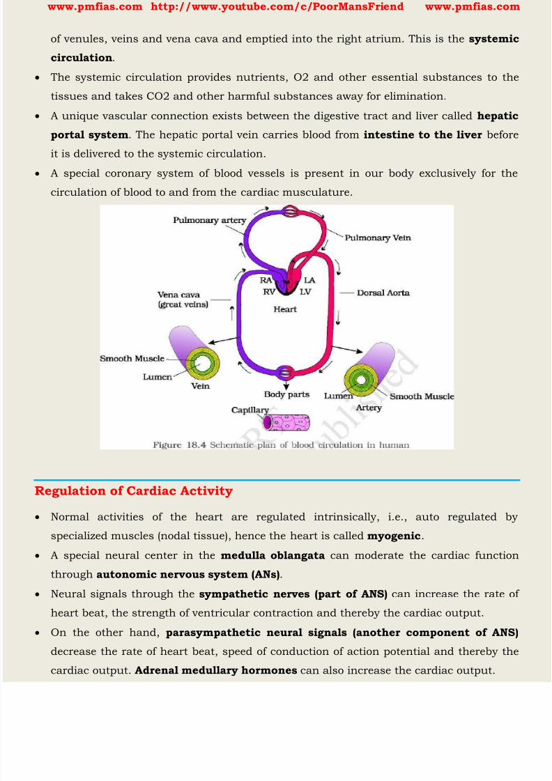
8/16/2019 Biology for Prelims Exam
http://slidepdf.com/reader/full/biology-for-prelims-exam 382/384
www.pmfias.com http://www.youtube.com/c/PoorMansFriend www.pmfias.com
of venules, veins and vena cava and emptied into the right atrium. This is the systemic
circulation .
The systemic circulation provides nutrients, O2 and other essential substances to the
tissues and takes CO2 and other harmful substances away for elimination.
A unique vascular connection exists between the digestive tract and liver called hepatic
portal system . The hepatic portal vein carries blood from intestine to the liver beforeit is delivered to the systemic circulation.
A special coronary system of blood vessels is present in our body exclusively for the
circulation of blood to and from the cardiac musculature.
Regulation of Cardiac Activity
Normal activities of the heart are regulated intrinsically, i.e., auto regulated by
specialized muscles (nodal tissue), hence the heart is called myogenic .
A special neural center in the medulla oblangata can moderate the cardiac function
through autonomic nervous system (ANs) .
Neural signals through the sympathetic nerves (part of ANS) can increase the rate of
heart beat, the strength of ventricular contraction and thereby the cardiac output.
On the other hand, parasympathetic neural signals (another component of ANS)
decrease the rate of heart beat, speed of conduction of action potential and thereby the
cardiac output. Adrenal medullary hormones can also increase the cardiac output.

8/16/2019 Biology for Prelims Exam
http://slidepdf.com/reader/full/biology-for-prelims-exam 383/384
www.pmfias.com http://www.youtube.com/c/PoorMansFriend www.pmfias.com
Disorders of Circulatory System
High Blood Pressure (Hypertension)
Hypertension is the term for blood pressure that is higher than normal (120/80).
In this measurement 120 mm Hg (millimetres of mercury pressure) is the systolic, or
pumping, pressure and 80 mm Hg is the diastolic, or resting, pressure.
If repeated checks of blood pressure of an individual is 140/90 (140 over 90) or higher,
it shows hypertension .
High blood pressure leads to heart diseases and also affects vital organs like brain and
kidney.
Coronary Artery Disease (CAD)
Coronary Artery Disease, often referred to as atherosclerosis , affects the vessels that
supply blood to the heart muscle. It is caused by deposits of calcium, fat, cholesterol
and fibrous tissues , which makes the lumen of arteries narrower.
Angina
It is also called ‘angina pectoris’ . A symptom of acute chest pain appears when no
enough oxygen is reaching the heart muscle.
Angina can occur in men and women of any age but it is more common among the
middle-aged and elderly. It occurs due to conditions that affect the blood flow.
Heart Failure
Heart failure means the state of heart when it is not pumping blood effectively enough to
meet the needs of the body. It is sometimes called congestive heart failure because
congestion of the lungs is one of the main symptoms of this disease.
Heart failure is not the same as cardiac arrest (when the heart stops beating) or a
heart attack (when the heart muscle is suddenly damaged by an inadequate bloodsupply).
Summary
Vertebrates circulate blood, a fluid connective tissue, in their body, to transport
essential substances to the cells and to carry waste substances from there. Another
fluid, lymph (tissue fluid) is also used for the transport of certain substances.
8/16/2019 Biology for Prelims Exam
http://slidepdf.com/reader/full/biology-for-prelims-exam 384/384
www.pmfias.com http://www.youtube.com/c/PoorMansFriend www.pmfias.com
Blood comprises of a fluid matrix, plasma and formed elements. Red blood cells (RBCs,
erythrocytes), white blood cells (WBCs, leucocytes) and platelets (thrombocytes)
constitute the formed elements. Blood of humans are grouped into A, B, AB and O
systems based on the presence or absence of two surface antigens, A, B on the RBCs.
Another blood grouping is also done based on the presence or absence of another
antigen called Rhesus factor (Rh) on the surface of RBCs. The spaces between cells inthe tissues contain a fluid derived from blood called tissue fluid. This fluid called lymph
is almost similar to blood except for the protein content and the formed elements.
All vertebrates and a few invertebrates have a closed circulatory system. Our circulatory
system consists of a muscular pumping organ, heart, a network of vessels and a fluid,
blood.
Heart has two atria and two ventricles. Cardiac musculature is auto-excitable. Sino-
atrial node (SAN) generates the maximum number of action protentials per minute (70-75/min) and therefore, it sets the pace of the activities of the heart. Hence it is called the
Pacemaker.
The action potential causes the atria and then the ventricles to undergo contraction
(systole) followed by their relaxation (diastole). The systole forces the blood to move from
the atria to the ventricles and to the pulmonary artery and the aorta. The cardiac cycle
is formed by sequential events in the heart which is cyclically repeated and is called the
cardiac cycle.
A healthy person shows 72 such cycles per minute. About 70 mL of blood is pumped out
by each ventricle during a cardiac cycle and it is called the stroke or beat volume.
Volume of blood pumped out by each ventricle of heart per minute is called the cardiac
output and it is equal to the product of stroke volume and heart rate (approx 5 litres).
The electrical activity of the heart can be recorded from the body surface by using
electrocardiograph and the recording is called electrocardiogram (ECG) which is of
clinical importance.
We have a complete double circulation, i.e., two circulatory pathways, namely,