BIOH111 SN11 Muscular System · PDF fileoIntegumentary system oSkeletal system ... Cardiac...
40
© Endeavour College of Natural Health endeavour.edu.au 1 BIOH111 o Cell Module o Tissue Module o Integumentary system o Skeletal system o Muscle system o Nervous system o Endocrine system
Transcript of BIOH111 SN11 Muscular System · PDF fileoIntegumentary system oSkeletal system ... Cardiac...
© Endeavour College of Natural Health endeavour.edu.au 1
BIOH111
oCell Module
oTissue Module
o Integumentary system
oSkeletal system
oMuscle system
oNervous system
oEndocrine system
© Endeavour College of Natural Health endeavour.edu.au 2
TEXTBOOK AND
REQUIRED/RECOMMENDED READINGS
o Principles of anatomy and physiology. Tortora et al; 14th
edition: Chapter 10
© Endeavour College of Natural Health endeavour.edu.au 3
BIOH111 – MUSCLE SYSTEM MODULE
o Session 11 (Lectures 17 and 18) – Muscle
physiology: Building of muscle organ – cells,
tissue, organ and muscle contraction process and
regulation
o Session 12 (Lectures 19 and 20) - Skeletal muscle
metabolism
o Session 13 (Lectures 21 and 22) – Major muscle
groups
BIOH111
Lectures 17 and 18
Muscle physiology: Building of muscle organ
and muscle contraction process and regulation
Department of Bioscience
endeavour.edu.au
© Endeavour College of Natural Health endeavour.edu.au 5
PREPARATION FOR THIS SESSION
o Review:
• plasma membrane and endoplasmic reticulum
structure and function
• difference between channels and receptors – think
about this: can they be both in one protein?
• regulated exocytosis
• tissue types
© Endeavour College of Natural Health endeavour.edu.au 6
OBJECTIVESLecture 17:
Muscle cells and tissue
Name and describe muscle cells and their function in building and function of
skeletal muscle tissue
Describe structure of connective tissue, nervous tissue and blood supply within
the structure and function of skeletal muscle tissue
Regulation of muscle contraction
Describe process of muscle contraction at the neuromuscular junction
Lecture 18:
Muscle contraction process
Describe the sliding filament theory of muscle contraction from both an
anatomical and physiological viewpoint
Discuss the contraction mechanism and the structures required for this occur
© Endeavour College of Natural Health endeavour.edu.au 7
FUNCTIONS OF MUSCULAR SYSTEM
1. Producing body movements - occurs at the cellular level
where chemical energy is changed into mechanical energy
2. Stabilizing body positions – skeletal muscle contractions
stabilize joints and maintain body positions (e.g.
siting/standing)
3. Regulating organ volumes - bands of smooth muscle
called sphincters
4. Movement of substances within the body - blood, lymph,
urine, air, food and fluids, sperm
5. Producing heat - involuntary contractions of skeletal
muscle (shivering)
© Endeavour College of Natural Health endeavour.edu.au 8
PROPERTIES OF MUSCLE TISSUE
1. Excitability
• respond to chemicals released from nerve cells
2. Conductivity
• ability to propagate electrical signals over membrane
3. Contractility
• ability to shorten and generate force
4. Extensibility
• ability to be stretched without damaging the tissue
5. Elasticity
• ability to return to original shape after being stretched
© Endeavour College of Natural Health endeavour.edu.au 9
MUSCLE TISSUEConsists of elongated cells called musle fibres or myocytes
that are modified for contraction using ATP as energy.
Muscle tissue provides motion, maintenance of posture and
heat.
o Classified into 3 types based on function and location:
1. Skeletal muscle tissue – striated, voluntary control
2. Cardiac muscle tissue - striated, involuntary control
3. Smooth (visceral) muscle tissue – non-striated,
involuntary control
© Endeavour College of Natural Health endeavour.edu.au 10
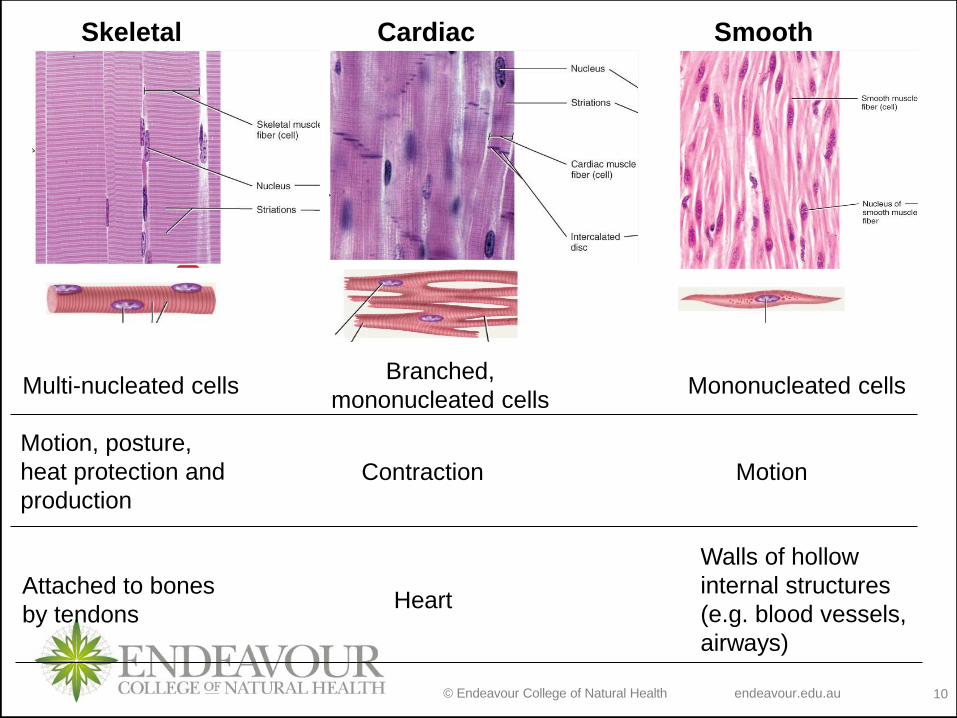
Skeletal Cardiac Smooth
Multi-nucleated cellsBranched,
mononucleated cellsMononucleated cells
Motion, posture,
heat protection and
production
Contraction Motion
Attached to bones
by tendonsHeart
Walls of hollow
internal structures
(e.g. blood vessels,
airways)

© Endeavour College of Natural Health endeavour.edu.au 11
SKELETAL MUSCLE TISSUE
o Skeletal muscle is made up of several tissues:
muscle cells (fibers), adipose tissue, connective tissue and
nervous tissue
o 1 skeletal muscle = 1 organ
Building a muscle: cells → tissue → organ
© Endeavour College of Natural Health endeavour.edu.au 12
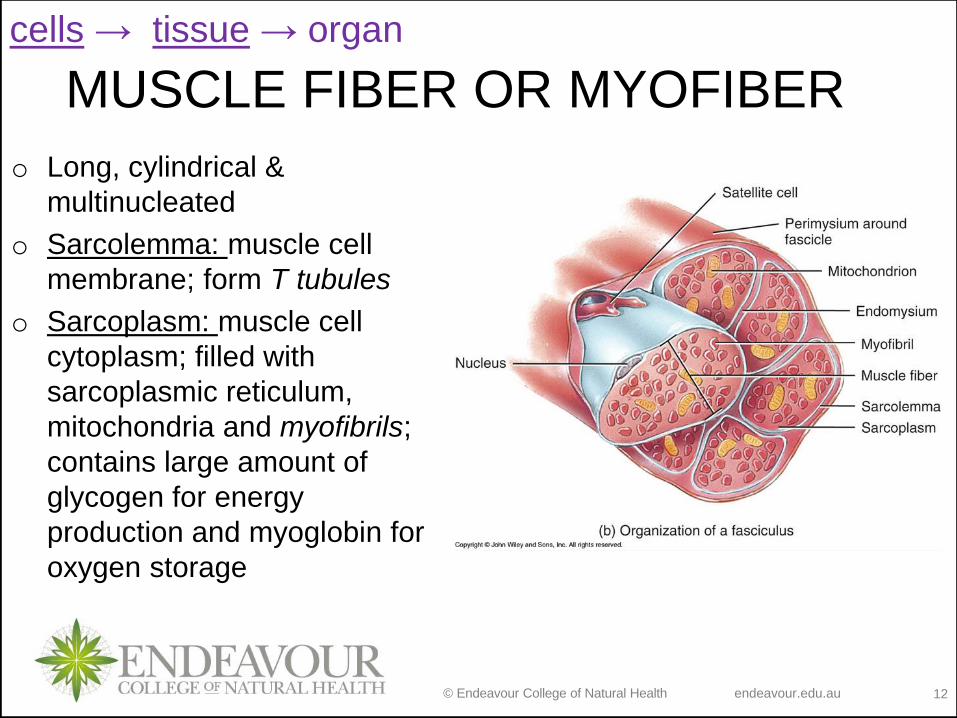
MUSCLE FIBER OR MYOFIBER
o Long, cylindrical &
multinucleated
o Sarcolemma: muscle cell
membrane; form T tubules
o Sarcoplasm: muscle cell
cytoplasm; filled with
sarcoplasmic reticulum,
mitochondria and myofibrils;
contains large amount of
glycogen for energy
production and myoglobin for
oxygen storage
cells → tissue → organ
© Endeavour College of Natural Health endeavour.edu.au 13
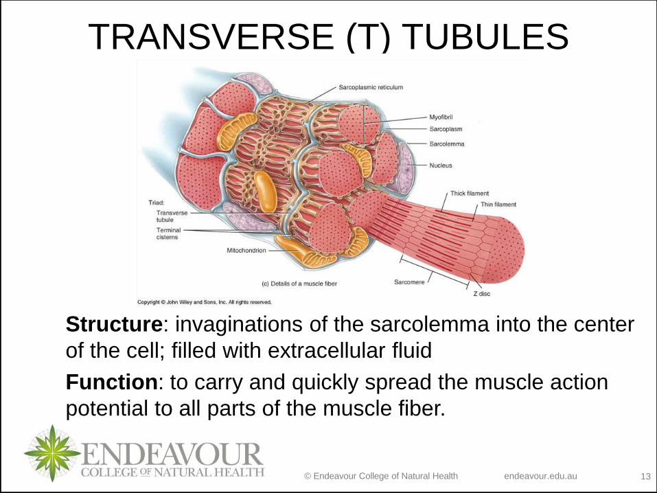
TRANSVERSE (T) TUBULES
Structure: invaginations of the sarcolemma into the center
of the cell; filled with extracellular fluid
Function: to carry and quickly spread the muscle action
potential to all parts of the muscle fiber.
© Endeavour College of Natural Health endeavour.edu.au 14
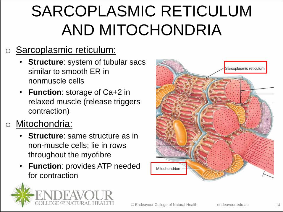
SARCOPLASMIC RETICULUM
AND MITOCHONDRIAo Sarcoplasmic reticulum:
• Structure: system of tubular sacs
similar to smooth ER in
nonmuscle cells
• Function: storage of Ca+2 in
relaxed muscle (release triggers
contraction)
o Mitochondria:
• Structure: same structure as in
non-muscle cells; lie in rows
throughout the myofibre
• Function: provides ATP needed
for contraction
© Endeavour College of Natural Health endeavour.edu.au 15
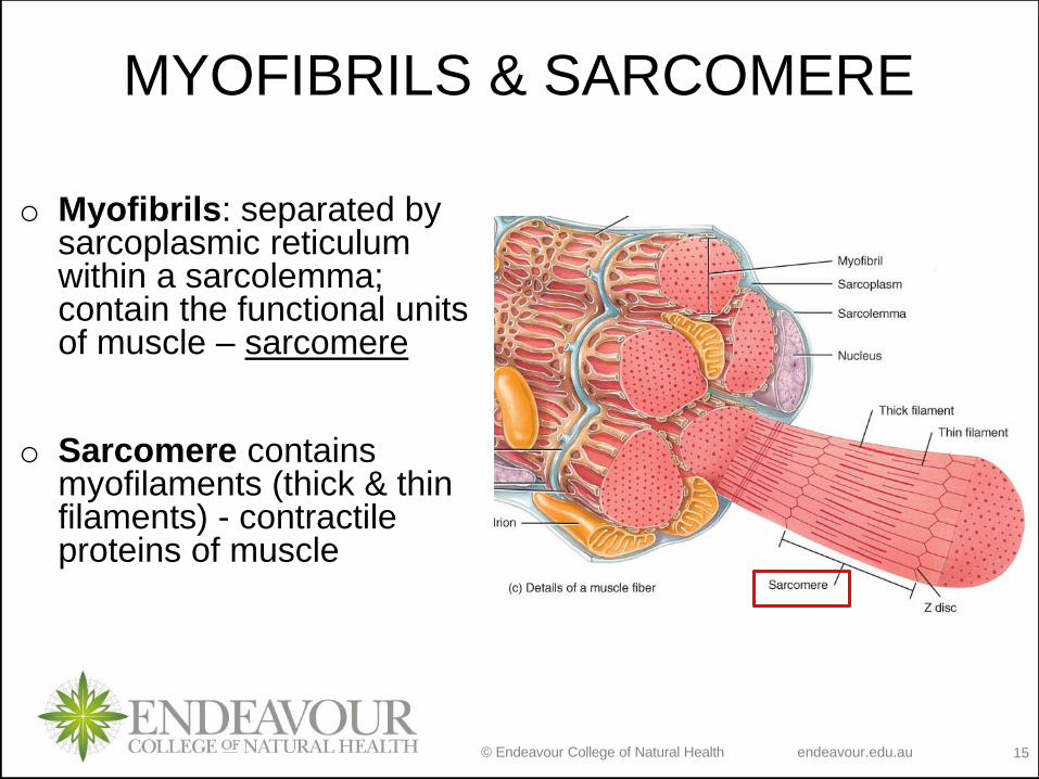
MYOFIBRILS & SARCOMERE
o Myofibrils: separated by sarcoplasmic reticulum within a sarcolemma; contain the functional units of muscle – sarcomere
o Sarcomere contains myofilaments (thick & thin filaments) - contractile proteins of muscle
© Endeavour College of Natural Health endeavour.edu.au 16
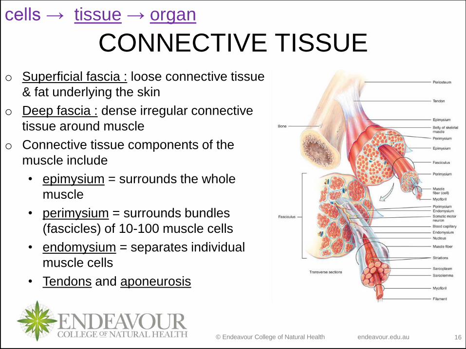
CONNECTIVE TISSUEo Superficial fascia : loose connective tissue
& fat underlying the skin
o Deep fascia : dense irregular connective
tissue around muscle
o Connective tissue components of the
muscle include
• epimysium = surrounds the whole
muscle
• perimysium = surrounds bundles
(fascicles) of 10-100 muscle cells
• endomysium = separates individual
muscle cells
• Tendons and aponeurosis
cells → tissue → organ
© Endeavour College of Natural Health endeavour.edu.au 17
CONNECTIVE TISSUE
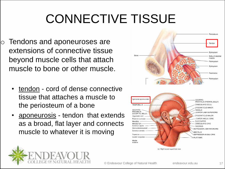
o Tendons and aponeuroses are
extensions of connective tissue
beyond muscle cells that attach
muscle to bone or other muscle.
• tendon - cord of dense connective
tissue that attaches a muscle to
the periosteum of a bone
• aponeurosis - tendon that extends
as a broad, flat layer and connects
muscle to whatever it is moving
© Endeavour College of Natural Health endeavour.edu.au 18
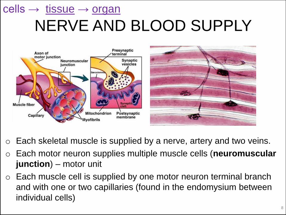
NERVE AND BLOOD SUPPLY
o Each skeletal muscle is supplied by a nerve, artery and two veins.
o Each motor neuron supplies multiple muscle cells (neuromuscular
junction) – motor unit
o Each muscle cell is supplied by one motor neuron terminal branch
and with one or two capillaries (found in the endomysium between
individual cells)
cells → tissue → organ
© Endeavour College of Natural Health endeavour.edu.au 19
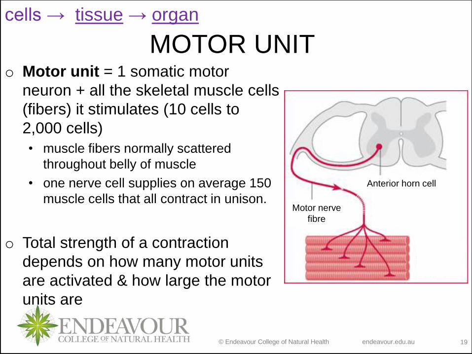
MOTOR UNITo Motor unit = 1 somatic motor
neuron + all the skeletal muscle cells
(fibers) it stimulates (10 cells to
2,000 cells)
• muscle fibers normally scattered
throughout belly of muscle
• one nerve cell supplies on average 150
muscle cells that all contract in unison.
o Total strength of a contraction
depends on how many motor units
are activated & how large the motor
units are
cells → tissue → organ
Anterior horn cell
Motor nerve
fibre
© Endeavour College of Natural Health endeavour.edu.au 20
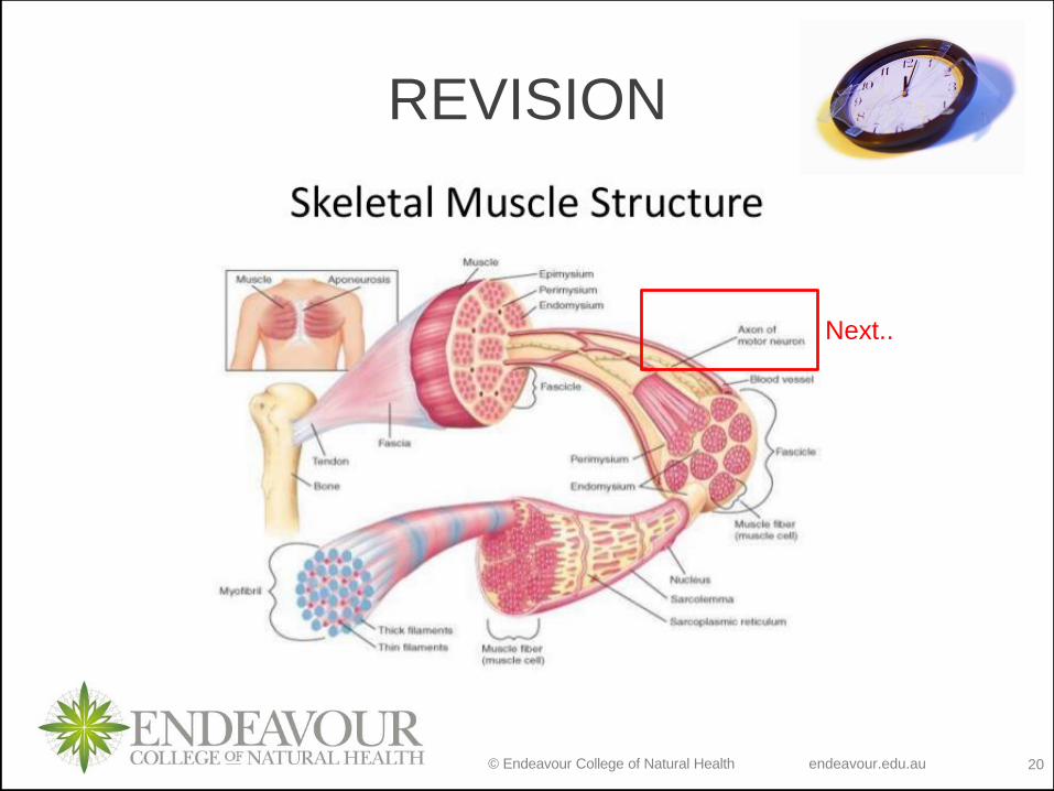
REVISION
Next..
© Endeavour College of Natural Health endeavour.edu.au 22
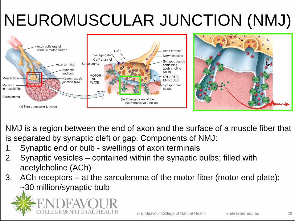
NEUROMUSCULAR JUNCTION (NMJ)
NMJ is a region between the end of axon and the surface of a muscle fiber that
is separated by synaptic cleft or gap. Components of NMJ:
1. Synaptic end or bulb - swellings of axon terminals
2. Synaptic vesicles – contained within the synaptic bulbs; filled with
acetylcholine (ACh)
3. ACh receptors – at the sarcolemma of the motor fiber (motor end plate);
~30 million/synaptic bulb
© Endeavour College of Natural Health endeavour.edu.au 23
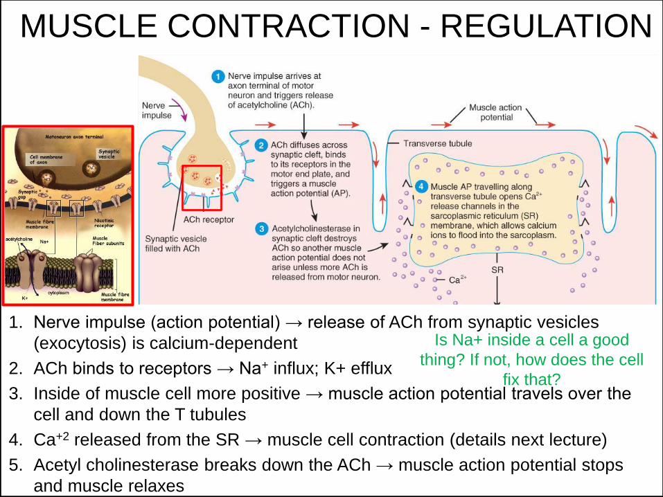
MUSCLE CONTRACTION - REGULATION
1. Nerve impulse (action potential) → release of ACh from synaptic vesicles
(exocytosis) is calcium-dependent
2. ACh binds to receptors → Na+ influx; K+ efflux
3. Inside of muscle cell more positive → muscle action potential travels over the
cell and down the T tubules
4. Ca+2 released from the SR → muscle cell contraction (details next lecture)
5. Acetyl cholinesterase breaks down the ACh → muscle action potential stops
and muscle relaxes
Is Na+ inside a cell a good
thing? If not, how does the cell
fix that?
© Endeavour College of Natural Health endeavour.edu.au 26
OBJECTIVESLecture 17:
Muscle cells and tissue
Name and describe muscle cells and their function in building and function of
skeletal muscle tissue
Describe structure of connective tissue, nervous tissue and blood supply within
the structure and function of skeletal muscle tissue
Regulation of muscle contraction
Describe process of muscle contraction at the neuromuscular junction
Lecture 18:
Muscle contraction process
Describe the sliding filament theory of muscle contraction from both an
anatomical and physiological viewpoint
Discuss the contraction mechanism and the structures required for this occur
© Endeavour College of Natural Health endeavour.edu.au 27
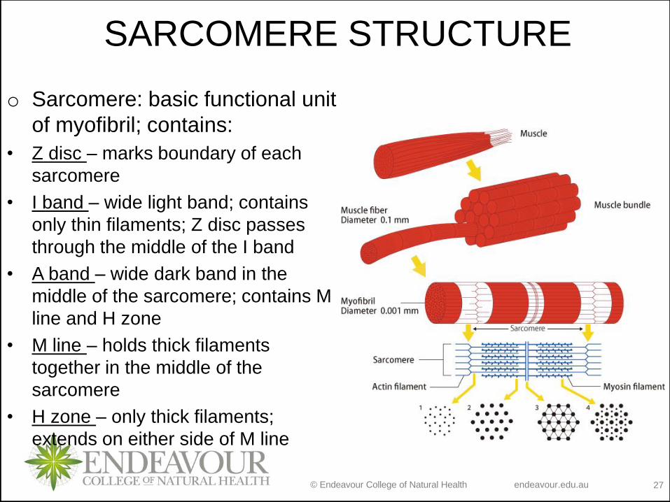
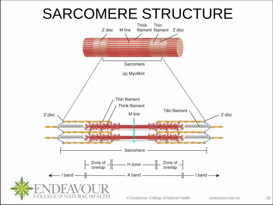
SARCOMERE STRUCTURE
o Sarcomere: basic functional unit
of myofibril; contains:
• Z disc – marks boundary of each
sarcomere
• I band – wide light band; contains
only thin filaments; Z disc passes
through the middle of the I band
• A band – wide dark band in the
middle of the sarcomere; contains M
line and H zone
• M line – holds thick filaments
together in the middle of the
sarcomere
• H zone – only thick filaments;
extends on either side of M line
© Endeavour College of Natural Health endeavour.edu.au 28
SARCOMERE STRUCTURE
© Endeavour College of Natural Health endeavour.edu.au 29
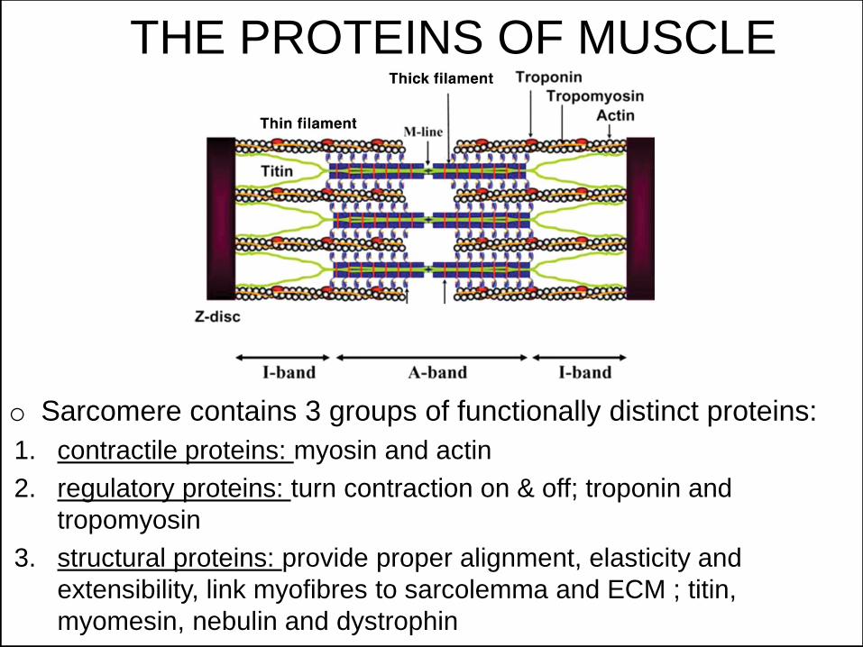
THE PROTEINS OF MUSCLE
o Sarcomere contains 3 groups of functionally distinct proteins:
1. contractile proteins: myosin and actin
2. regulatory proteins: turn contraction on & off; troponin and
tropomyosin
3. structural proteins: provide proper alignment, elasticity and
extensibility, link myofibres to sarcolemma and ECM ; titin,
myomesin, nebulin and dystrophin
© Endeavour College of Natural Health endeavour.edu.au 30
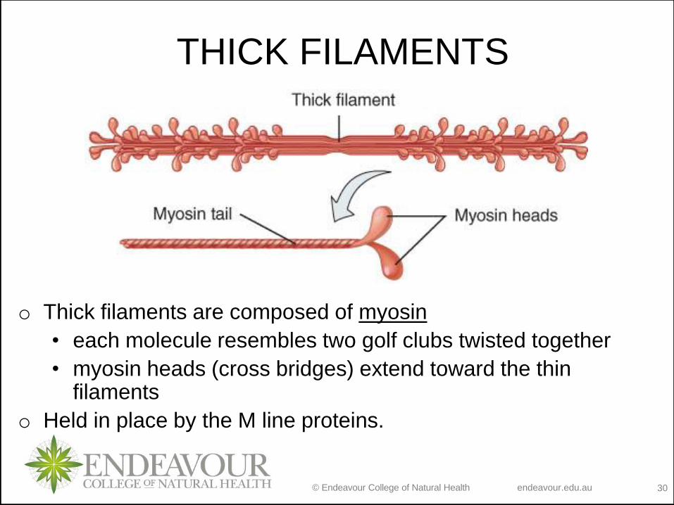
THICK FILAMENTS
o Thick filaments are composed of myosin
• each molecule resembles two golf clubs twisted together
• myosin heads (cross bridges) extend toward the thin filaments
o Held in place by the M line proteins.
© Endeavour College of Natural Health endeavour.edu.au 31
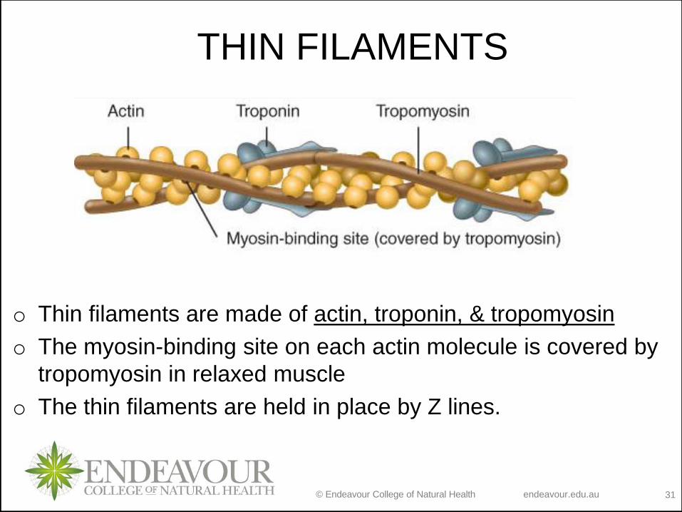
o Thin filaments are made of actin, troponin, & tropomyosin
o The myosin-binding site on each actin molecule is covered by
tropomyosin in relaxed muscle
o The thin filaments are held in place by Z lines.
THIN FILAMENTS
© Endeavour College of Natural Health endeavour.edu.au 32
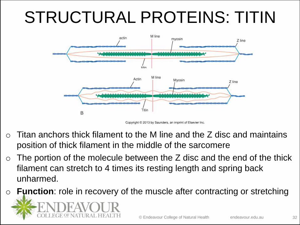
o Titan anchors thick filament to the M line and the Z disc and maintains
position of thick filament in the middle of the sarcomere
o The portion of the molecule between the Z disc and the end of the thick
filament can stretch to 4 times its resting length and spring back
unharmed.
o Function: role in recovery of the muscle after contracting or stretching
STRUCTURAL PROTEINS: TITIN
© Endeavour College of Natural Health endeavour.edu.au 33
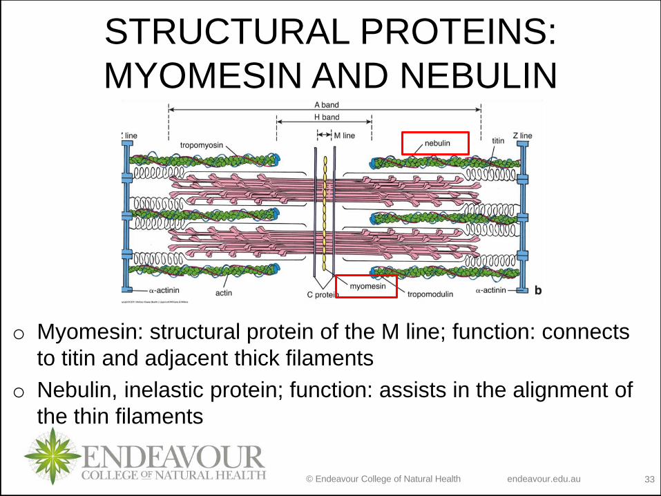
STRUCTURAL PROTEINS:
MYOMESIN AND NEBULIN
o Myomesin: structural protein of the M line; function: connects
to titin and adjacent thick filaments
o Nebulin, inelastic protein; function: assists in the alignment of
the thin filaments
© Endeavour College of Natural Health endeavour.edu.au 34
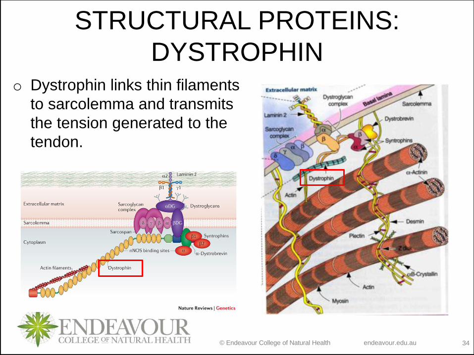
o Dystrophin links thin filaments
to sarcolemma and transmits
the tension generated to the
tendon.
STRUCTURAL PROTEINS:
DYSTROPHIN
© Endeavour College of Natural Health endeavour.edu.au 35
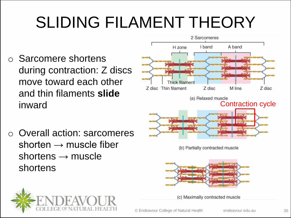
o Sarcomere shortens
during contraction: Z discs
move toward each other
and thin filaments slide
inward
o Overall action: sarcomeres
shorten → muscle fiber
shortens → muscle
shortens
SLIDING FILAMENT THEORY
Contraction cycle
© Endeavour College of Natural Health endeavour.edu.au 36
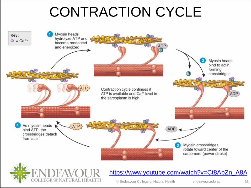
CONTRACTION CYCLE
https://www.youtube.com/watch?v=Ct8AbZn_A8A
© Endeavour College of Natural Health endeavour.edu.au 37
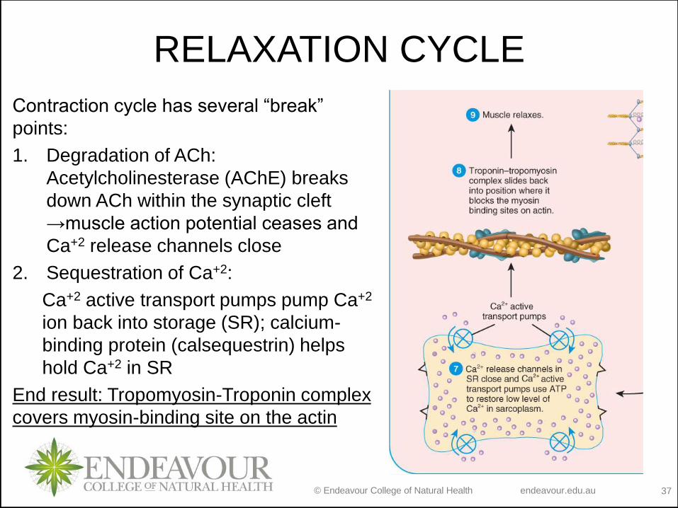
RELAXATION CYCLE
Contraction cycle has several “break”
points:
1. Degradation of ACh:
Acetylcholinesterase (AChE) breaks
down ACh within the synaptic cleft
→muscle action potential ceases and
Ca+2 release channels close
2. Sequestration of Ca+2:
Ca+2 active transport pumps pump Ca+2
ion back into storage (SR); calcium-
binding protein (calsequestrin) helps
hold Ca+2 in SR
End result: Tropomyosin-Troponin complex
covers myosin-binding site on the actin
© Endeavour College of Natural Health endeavour.edu.au 38
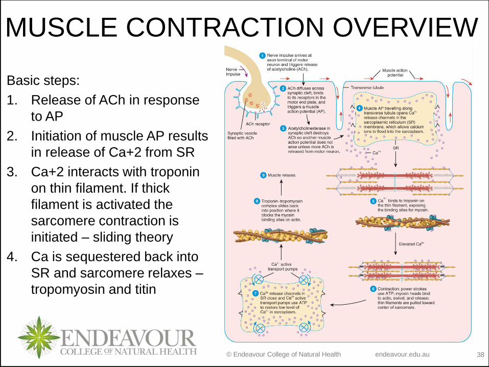
MUSCLE CONTRACTION OVERVIEW
Basic steps:
1. Release of ACh in response
to AP
2. Initiation of muscle AP results
in release of Ca+2 from SR
3. Ca+2 interacts with troponin
on thin filament. If thick
filament is activated the
sarcomere contraction is
initiated – sliding theory
4. Ca is sequestered back into
SR and sarcomere relaxes –
tropomyosin and titin

© Endeavour College of Natural Health endeavour.edu.au 39
2 molecules are critical for contraction:
Ca+2 and ATP.
We know where Ca+2 comes from, but
where does the ATP used in contraction
cycle come from? Next session!!

© Endeavour College of Natural Health endeavour.edu.au 41
WHY IS IT HARDER TO LIFT SOMETHING
WITH AN EXTENDED ARM?
o Length-Tension relationship
o Optimal overlap at the 100%
o Cell too stretched – myosin heads
are not close to actin so no binding
can occur → little force is produced
o Cell is too short: myosin heads
overlap with actin, thick filaments
crumpled by Z discs so little binding
can occur →little force is producedGraph of Force of contraction (Tension)
vs Length of sarcomere
© Endeavour College of Natural Health endeavour.edu.au 42
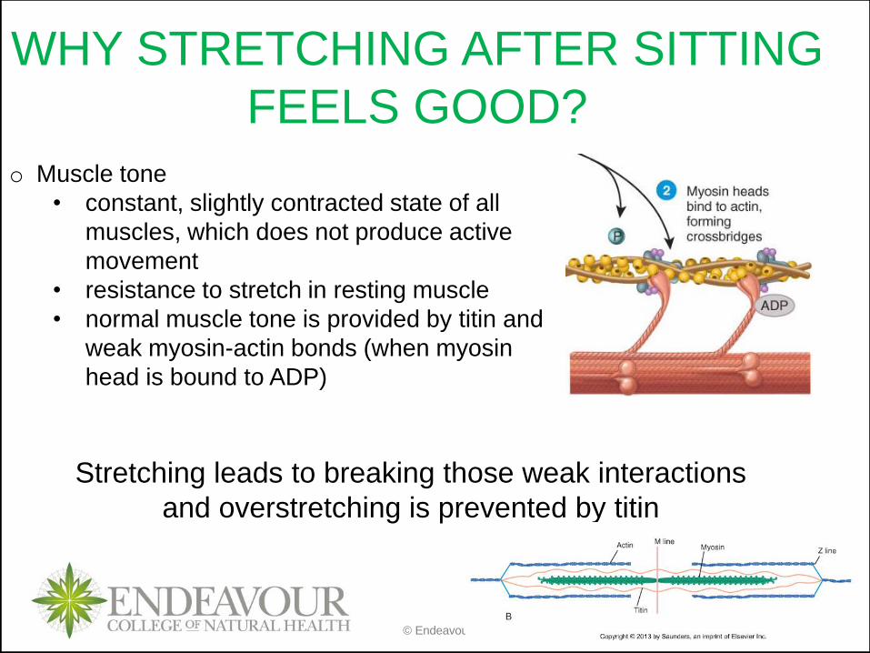
WHY STRETCHING AFTER SITTING
FEELS GOOD?
o Muscle tone
• constant, slightly contracted state of all
muscles, which does not produce active
movement
• resistance to stretch in resting muscle
• normal muscle tone is provided by titin and
weak myosin-actin bonds (when myosin
head is bound to ADP)
Stretching leads to breaking those weak interactions
and overstretching is prevented by titin
© Endeavour College of Natural Health endeavour.edu.au 43
Recap of Session 11
Muscle tissue is classified into 3 types: skeletal, smooth and cardiac
(we only cover skeletal in BIOH111)
Skeletal muscle cells (myofibers) contain a specialised component
called myofibril which contain a unit responsible for muscle contraction
– sarcomere
Sarcomere contraction is regulated by nervous system at the NMJ and
the outcome of the regulation is release of calcium from SER
Sarcomere is composed of 3 functional groups of protein: regulatory,
structural and contractile. All work together to enable sarcomere,
myofibril and myofiber to contract
© Endeavour College of Natural Health endeavour.edu.au 44
PREPARATION FOR NEXT SESSION
o Complete any missing concepts and linking words from
Session 11
o Review:
• mitochondria structure and function
• contraction cycle