Growth & Development of Skeletal Muscle. Skeletal, Striated, Voluntary Muscle.
Upload
claire-chandlerCategory
view
225download
3
Muscle StructureMuscle Structure
Animal Growth and Animal Growth and DevelopmentDevelopment
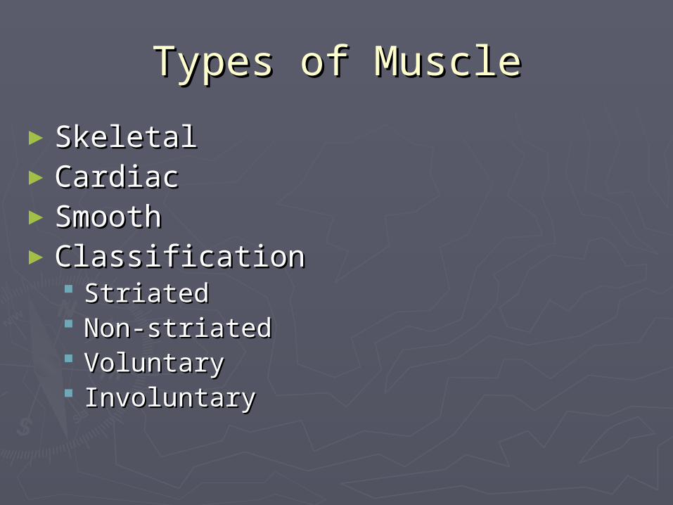
Types of MuscleTypes of Muscle
►SkeletalSkeletal►CardiacCardiac►SmoothSmooth►ClassificationClassification
StriatedStriated Non-striatedNon-striated VoluntaryVoluntary InvoluntaryInvoluntary
Skeletal structureSkeletal structure
► Based on size, shape and locationBased on size, shape and location►Origin- where muscles originate on one side Origin- where muscles originate on one side
of the joint of the joint ► Insertion- where muscles terminate on the Insertion- where muscles terminate on the
other side of the jointother side of the joint► Tendon – point of attachment at either Tendon – point of attachment at either
insertion or origininsertion or origin► Fascia- a thin sheet of connective tissue at Fascia- a thin sheet of connective tissue at
attachmentattachment
OrganizationOrganization
►EpimysiumEpimysium►PerimysiumPerimysium►EndomysiumEndomysium
SarcolemmaSarcolemma
Muscles are multinucleatedMuscles are multinucleated
Muscle CytoskeletonMuscle Cytoskeleton
►MicrofilamentsMicrofilaments► Intermediate filamentsIntermediate filaments►MicrotubulesMicrotubules
►Myofibrils are microfilamentous Myofibrils are microfilamentous organelles of muscle fibersorganelles of muscle fibers
►Sarcomere – smallest contractile unit Sarcomere – smallest contractile unit of muscleof muscle
MyofibrilsMyofibrils
►Made up of thick and thin filaments Made up of thick and thin filaments called myosin and actincalled myosin and actin
►Z-line – outer demarcation of the Z-line – outer demarcation of the sarcomeresarcomere Made up predominantly of alpha-actininMade up predominantly of alpha-actinin
►A- bandA- band►H – ZoneH – Zone► I - bandI - band
MyosinMyosin
►Occupies 80-87% of the total volume of the Occupies 80-87% of the total volume of the muscle fibermuscle fiber
► Contains both heavy and light chain Contains both heavy and light chain molecules (heavy and light meromyosin)molecules (heavy and light meromyosin)
► Positioned in the center of the sarcomerePositioned in the center of the sarcomere► Rachet effect – acts in contraction to make Rachet effect – acts in contraction to make
the sarcomere shorten because it pulls on the sarcomere shorten because it pulls on actin filaments, thus shortens one Z-line to actin filaments, thus shortens one Z-line to anotheranother
ActinActin
►Second most abundant in muscle fibers Second most abundant in muscle fibers and makes up almost 20% of the total and makes up almost 20% of the total protein protein
►Filamentous actin (F-actin) is predominantFilamentous actin (F-actin) is predominant►G-actin or globular is smaller yet is found G-actin or globular is smaller yet is found
to aid in the development of F-actinto aid in the development of F-actin►Numerous G-actins develop one F-actinNumerous G-actins develop one F-actin
Regulatory proteinsRegulatory proteins
►Tropomyosin – another filamentous Tropomyosin – another filamentous protein of the thin filamentprotein of the thin filament
►Troponin – also is a filamentous Troponin – also is a filamentous protein that surrounds actin and holds protein that surrounds actin and holds the actin strands togetherthe actin strands together
►Troponin is involved in calcium binding Troponin is involved in calcium binding upon release of or increased calcium upon release of or increased calcium levels in the muscle to initiate levels in the muscle to initiate contractioncontraction

Additional Components of Additional Components of Muscle FibersMuscle Fibers
►Sarcoplasmic reticulum – membranous Sarcoplasmic reticulum – membranous system of tubules that forms a network system of tubules that forms a network around each myofibrilaround each myofibril
►T – tubules – (transverse tubules) T – tubules – (transverse tubules) surround myofibrils at the A-I band jct. surround myofibrils at the A-I band jct.
►Figure 2.34Figure 2.34► In fact, the system between two T-In fact, the system between two T-
tubules is called the Sarcoplasmic tubules is called the Sarcoplasmic reticulumreticulum
Additional Components of Additional Components of Muscle FibersMuscle Fibers
► Sarcoplasmic reticulum consists of two Sarcoplasmic reticulum consists of two structuresstructures Terminal cisternaeTerminal cisternae Fenestrated collarFenestrated collar
►These act as Ca storage reservoirsThese act as Ca storage reservoirs
► The structure formed from the two terminal The structure formed from the two terminal cisternae and on each side of the T-tubule is cisternae and on each side of the T-tubule is called a triad. Note: there is no physical called a triad. Note: there is no physical contact between the T-tubule and the terminal contact between the T-tubule and the terminal cisternaecisternae
► Communication occurs via electrical messagesCommunication occurs via electrical messages
Additional Components of Additional Components of Muscle FibersMuscle Fibers
► The electrical systemThe electrical system Dihydropyridine and ryanodine recptors Dihydropyridine and ryanodine recptors When an electrical stimuli occurs at these receptors, When an electrical stimuli occurs at these receptors,
the SR channel changes conformation and allows Ca the SR channel changes conformation and allows Ca into the cytoplasminto the cytoplasm
Calsequestrin is the part of the fiber/cell (protein) Calsequestrin is the part of the fiber/cell (protein) that binds calcium in the SRthat binds calcium in the SR
► Nucleus – muscle fibers are multinucleatedNucleus – muscle fibers are multinucleated► Mitochondria- most of the energy is madeMitochondria- most of the energy is made► Sarcoplasm – cellular cytoplasm of the muscle Sarcoplasm – cellular cytoplasm of the muscle
cellcell
Cardiac MuscleCardiac Muscle
►Cardiocytes –cardiac muscle fibersCardiocytes –cardiac muscle fibers►Striated and involuntary (uniform Striated and involuntary (uniform
contractions)contractions)►Communication of these type Communication of these type
contractions occurs at connections contractions occurs at connections called intercalated discscalled intercalated discs
►Cardiac myofibrils are attached directly Cardiac myofibrils are attached directly to the intercalated discs and allow the to the intercalated discs and allow the heart to act in syncytium or as a groupheart to act in syncytium or as a group
Cardiac muscleCardiac muscle
► Cardiac thin filaments are more variable in Cardiac thin filaments are more variable in length as compared to skeletal muscle fiberslength as compared to skeletal muscle fibers
► Cardiac muscle must contract even under Cardiac muscle must contract even under extreme conditionsextreme conditions
► Because of the increased variability, this allows Because of the increased variability, this allows more overlap for contraction during stressed more overlap for contraction during stressed times for more uniform, consistent contractiontimes for more uniform, consistent contraction
► Cardiac muscle fibers are more branched and Cardiac muscle fibers are more branched and have more mitochondria than skeletal musclehave more mitochondria than skeletal muscle

Smooth muscleSmooth muscle
►Seen as sheet-like muscle massesSeen as sheet-like muscle masses► Is very elastic and pliableIs very elastic and pliable
►One example is around the uterus where it has to One example is around the uterus where it has to stretchstretch
►It thickens during pregnancy and becomes more It thickens during pregnancy and becomes more thin while the female is openthin while the female is open
►Smooth muscle is more triangular and Smooth muscle is more triangular and only has one nucleus per cellonly has one nucleus per cell
►Thick and thin filaments are not Thick and thin filaments are not symmetrically arranged like striated symmetrically arranged like striated musclemuscle
Smooth muscleSmooth muscle
►As compared to skeletal muscle, smooth As compared to skeletal muscle, smooth muscle has dense bodies that are muscle has dense bodies that are analogous to Z-linesanalogous to Z-lines Actin molecules are attached to dense Actin molecules are attached to dense
bodies bodies Thick filaments are likewise positioned Thick filaments are likewise positioned
throughout the cells that overlap the thin throughout the cells that overlap the thin filamentsfilaments
As a result, when contraction occurs, forces As a result, when contraction occurs, forces react in varying directionsreact in varying directions
MyogenesisMyogenesis
►Origin of muscle which begins at fertilizationOrigin of muscle which begins at fertilization►Develops from mesodermal somitesDevelops from mesodermal somites►DNA responsibilityDNA responsibility►Muscle is continually modified throughout Muscle is continually modified throughout
growth and developmentgrowth and development►Mitosis – increase in cell numbers or cell Mitosis – increase in cell numbers or cell
divisiondivision►During mitosis, the entire genome or DNA has During mitosis, the entire genome or DNA has
to be replicatedto be replicated► Figure 4.4Figure 4.4

Muscle cell cycleMuscle cell cycle
►Four distinct phasesFour distinct phases Gap 1 (G1) all cells enter the cell cycle Gap 1 (G1) all cells enter the cell cycle
from this phasefrom this phase►Most variable phase in lengthMost variable phase in length►Respond to cues from the environmentRespond to cues from the environment
Synthetic or S-phase is the time where the Synthetic or S-phase is the time where the cell dedicates to DNA replications or cell dedicates to DNA replications or synthesissynthesis
Third phase is called G2, and follows the S-Third phase is called G2, and follows the S-phasephase
Muscle cell cycleMuscle cell cycle
►G2 phase represents when the G2 phase represents when the intracellular architecture remodels itself intracellular architecture remodels itself to accommodate physical division of the to accommodate physical division of the cell mass in mitosis or M-phasecell mass in mitosis or M-phase
►M-phase is the shortest phaseM-phase is the shortest phase►After this, it re-enters the G1 phaseAfter this, it re-enters the G1 phase►Growth factors are often small proteins Growth factors are often small proteins
that influence the environment that that influence the environment that initiate cell growthinitiate cell growth
Muscle cell cycleMuscle cell cycle
► These small proteins bind to receptorsThese small proteins bind to receptors► Also, steroids can bind and create an Also, steroids can bind and create an
environment for growthenvironment for growth Ex. IGF-1, testosterone, etc.Ex. IGF-1, testosterone, etc.
► Another alternative for cells in the G1 phase is Another alternative for cells in the G1 phase is to exist in a protracted G1 phase or G0 phase. to exist in a protracted G1 phase or G0 phase.
► Cells are capable of remaining dormant, which Cells are capable of remaining dormant, which can re-enter the proliferative cycle without can re-enter the proliferative cycle without dividingdividing
► These cells in muscle are called satellite cellsThese cells in muscle are called satellite cells

Muscle cell determinationMuscle cell determination
►DeterminationDetermination Cells must migrate from the somites Cells must migrate from the somites
(matured mesodermal cells (Fig. 3.1 and (matured mesodermal cells (Fig. 3.1 and 3.2)3.2)
Cells that are determined to develop into Cells that are determined to develop into muscle are termed as myoblasts muscle are termed as myoblasts (precursors for muscle fibers) Fig. 4.4(precursors for muscle fibers) Fig. 4.4
Muscle regulatory factor gene (MRFs)Muscle regulatory factor gene (MRFs)►These trigger the expression of one or more These trigger the expression of one or more
genes for myogenesisgenes for myogenesis

Muscle cell determinationMuscle cell determination
►Transcription factors include MRFs and Transcription factors include MRFs and are responsible for turning on are responsible for turning on transcription of other genes located in transcription of other genes located in the nucleus and contain helix-loop-helix the nucleus and contain helix-loop-helix motif motif
►When two of these proteins combine they When two of these proteins combine they bind to the regulatory region and cause bind to the regulatory region and cause the gene to be expressed or repressedthe gene to be expressed or repressed
►Thus, MRFs are control point for Thus, MRFs are control point for myogenesismyogenesis
Muscle cell determinationMuscle cell determination
►Helix-loop-helix proteins include:Helix-loop-helix proteins include: MyogeninMyogenin MRF-4MRF-4 Myo-DMyo-D Myf-5Myf-5
►Fig. 4.5 MRFs actionFig. 4.5 MRFs action

Basic Genetics and Basic Genetics and CytogeneticsCytogenetics
Two Basic ConceptsTwo Basic ConceptsDNA- Polymer of Nucleotide Bases
Complementary Base Pairs
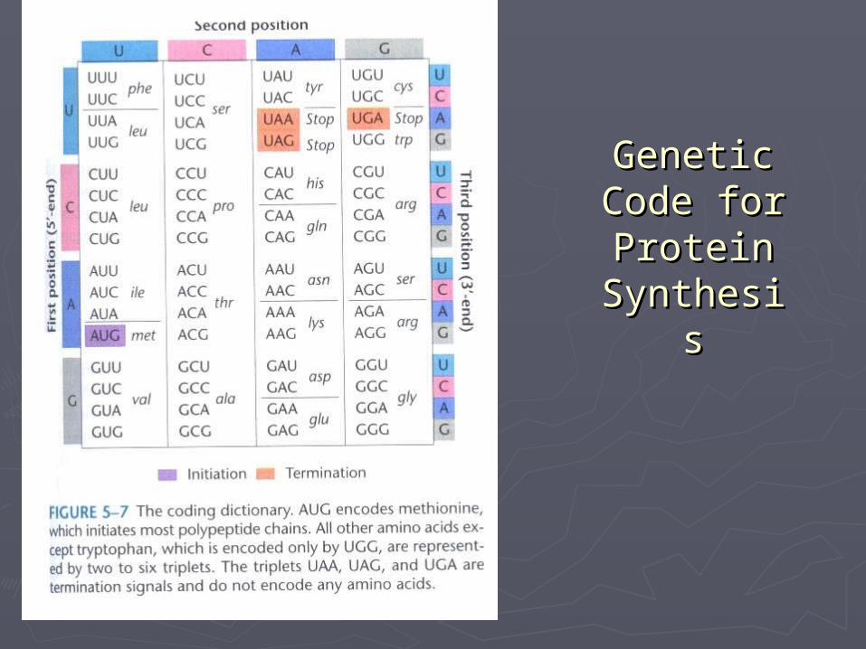
I. 1 Gene – 1 Protein (Gene determining the Phenotype) DNA is the Genetic Code for Protein Synthesis
II. Gene is the Unit of inheritance (Inherited from both Parents) Complementary Base Pares – DNA Duplication and Gamete production.
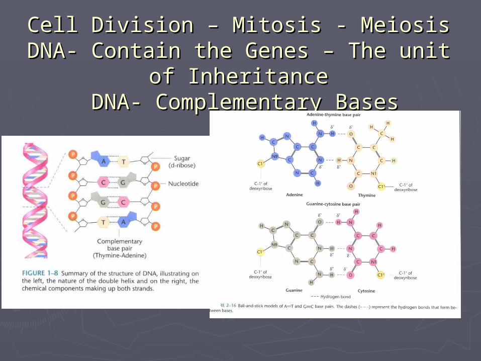
Cell Division – Mitosis - MeiosisCell Division – Mitosis - MeiosisDNA- Contain the Genes – The unit of DNA- Contain the Genes – The unit of
InheritanceInheritance DNA- Complementary Bases DNA- Complementary Bases

Chromosomes- Chromatin Chromosomes- Chromatin and DNAand DNA
DNA – Genetic Code for Protein DNA – Genetic Code for Protein SynthesisSynthesisDNA – Polymer of Nucleotide DNA – Polymer of Nucleotide Bases Bases Nucleotide Sequence –Amino acid Nucleotide Sequence –Amino acid Sequence of the Protein (Shape Sequence of the Protein (Shape and function)and function)
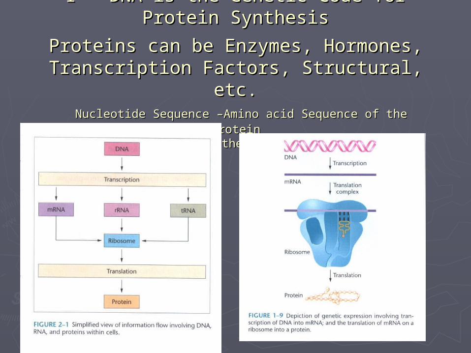
I - DNA is the Genetic Code for Protein I - DNA is the Genetic Code for Protein SynthesisSynthesis
Proteins can be Enzymes, Hormones, Proteins can be Enzymes, Hormones, Transcription Factors, Structural, etc.Transcription Factors, Structural, etc. Nucleotide Sequence –Amino acid Sequence of the ProteinNucleotide Sequence –Amino acid Sequence of the Protein
Shape Determine the function/effect)Shape Determine the function/effect)
Genetic Genetic Code for Code for Protein Protein
SynthesisSynthesis
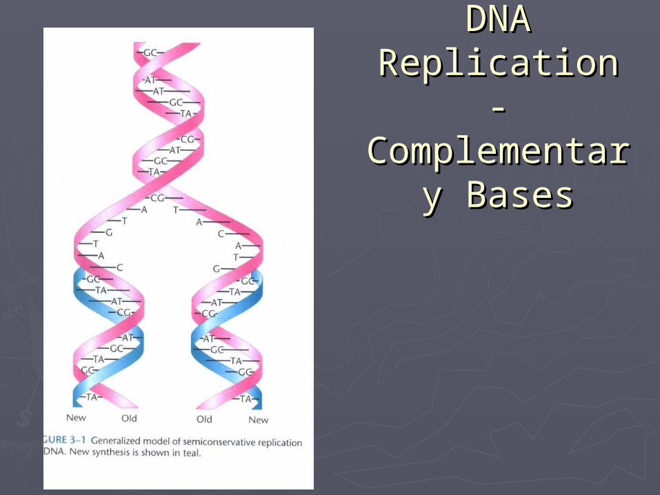
DNA DNA Replication - Replication -
ComplementarComplementary Basesy Bases
Differentiation and FusionDifferentiation and Fusion
►Replication competent – capable of Replication competent – capable of progressing the cell cycle and giving rise progressing the cell cycle and giving rise to additional myoblasts even though to additional myoblasts even though determined muscle cells & myoblasts determined muscle cells & myoblasts have express muscle regulatory factor have express muscle regulatory factor genesgenes
►Myoblasts do not contract because they Myoblasts do not contract because they do not contain contractile proteins yetdo not contain contractile proteins yet
►Before this, cells must receive signals Before this, cells must receive signals that induce them to differentiatethat induce them to differentiate
The “Differentiating” processThe “Differentiating” process
►Cells stop dividingCells stop dividing►Cells begin to align with one anotherCells begin to align with one another►Cell membranes begin to fuse together Cell membranes begin to fuse together
to form an immature muscle fiber to form an immature muscle fiber (myotube)(myotube)
►Muscle specific genes are regulated to Muscle specific genes are regulated to initiateinitiate Genes for muscle creatine kinase and Genes for muscle creatine kinase and
acetylcholine receptor subunit, myosin and acetylcholine receptor subunit, myosin and actin are triggeredactin are triggered
FusionFusion
► The exact process is still cloudyThe exact process is still cloudy► Yet, we know that small tubules or Yet, we know that small tubules or
attachments are apparent and dense attachments are apparent and dense structures beneath membranes are structures beneath membranes are generated to form tight junctions between generated to form tight junctions between myoblastsmyoblasts
► Finally, two bilipid membranes become one Finally, two bilipid membranes become one and dissolve in the cytoplasm of the newly and dissolve in the cytoplasm of the newly formed multinucleated cell. Note: Ca ions formed multinucleated cell. Note: Ca ions play a significant role in this processplay a significant role in this process

MaturationMaturation
►Once myoblasts differentiate and fuse Once myoblasts differentiate and fuse to form myotubes, resulting cells do to form myotubes, resulting cells do not continue to express a given set of not continue to express a given set of tenestenes
►The new muscle fibers change The new muscle fibers change following fusionfollowing fusion
►This process is called maturationThis process is called maturation
Morphological Aspects of Morphological Aspects of MyogenesisMyogenesis
►Once the somite has reached the two Once the somite has reached the two layered, dermomyotome stage cells layered, dermomyotome stage cells begin to accumulate ventrally to form begin to accumulate ventrally to form the myotome.the myotome.
►Myotome – a compartment of the somiteMyotome – a compartment of the somite►This is believed to involve migration of This is believed to involve migration of
cells from the dorsomedial ridge and cells from the dorsomedial ridge and ventrolateral part of the dermomyotome ventrolateral part of the dermomyotome to below it creating the myotometo below it creating the myotome
Morphological Aspects of Morphological Aspects of MyogenesisMyogenesis
►Recall that skeletal muscle originates Recall that skeletal muscle originates from somites which is developed from from somites which is developed from the mesodermthe mesoderm
►Myogenesis appears to involve Myogenesis appears to involve migration from the dorsomedial ridge migration from the dorsomedial ridge and ventrolateral spects of the and ventrolateral spects of the dermomyotome to immediately below dermomyotome to immediately below the dermomyotome, thus creating a the dermomyotome, thus creating a myotome Fig 4.8myotome Fig 4.8
Morphological Aspects of Morphological Aspects of MyogenesisMyogenesis
►Muscle does not just appear during Muscle does not just appear during prenatal developmentprenatal development
► It results from biphasic processesIt results from biphasic processes►Occurs from two populations of muscle Occurs from two populations of muscle
fiber myoblastsfiber myoblasts►During myoblast differentiation, During myoblast differentiation,
myoblasts cluster align in a vast myoblasts cluster align in a vast network of connective tissue and fuse to network of connective tissue and fuse to form an immature muscle fiber referred form an immature muscle fiber referred to as a myotubeto as a myotube

Morphological Aspects of Morphological Aspects of MyogenesisMyogenesis
►Primary myotubes are the first to developPrimary myotubes are the first to develop►These primary myotubes are the These primary myotubes are the
structure for which others attach and structure for which others attach and complete the final fiber structure - Figure complete the final fiber structure - Figure 4.114.11
►The myotube formation can be referred The myotube formation can be referred to as the second phase of myogenesis to as the second phase of myogenesis (fetal myoblasts) whereas the first phase (fetal myoblasts) whereas the first phase of primary myotubes are referred to as of primary myotubes are referred to as embryonic myoblastsembryonic myoblasts
Morphological Aspects of Morphological Aspects of MyogenesisMyogenesis
► Upon receiving signals to differentiate, the Upon receiving signals to differentiate, the secondary myotube use the primary myotubes secondary myotube use the primary myotubes as a template for alignment and organizationas a template for alignment and organization
► This alignment of fetal with embryonic myotubes This alignment of fetal with embryonic myotubes align closely and facilitates the fusion processalign closely and facilitates the fusion process
► After contraction the primary fibers remain After contraction the primary fibers remain intact and constant whereas the secondary intact and constant whereas the secondary muscle fibers or myotubes are splintered awaymuscle fibers or myotubes are splintered away
► This process of producing primary myotubes is This process of producing primary myotubes is self-governing in natureself-governing in nature
Morphological Aspects of Morphological Aspects of MyogenesisMyogenesis
►Depending on the species, muscle Depending on the species, muscle development occurs during the first 2/3 development occurs during the first 2/3 of prenatal developmentof prenatal development
►The total # of myofibers is considered The total # of myofibers is considered established by 90 days post-conception established by 90 days post-conception with pigs Fig. 4.12 - K State gestation with pigs Fig. 4.12 - K State gestation studystudy
►As muscle growth increases, muscle As muscle growth increases, muscle fiber diameter increases fiber diameter increases
MyofibrillogenesisMyofibrillogenesis
►The primary f(x)n of muscle is to contractThe primary f(x)n of muscle is to contract►This is done by the addition of myuofibrils This is done by the addition of myuofibrils
to each cell during developmentto each cell during development►Again, skeletal muscle is highly organized Again, skeletal muscle is highly organized
and contains many proteinsand contains many proteins►Stress fibers are those that are bundles of Stress fibers are those that are bundles of
myofilaments which have contractile myofilaments which have contractile possibilitiespossibilities
MyofibrillogenesisMyofibrillogenesis
► The stress cells are located at the periphery and The stress cells are located at the periphery and interact with membrane bound proteins called interact with membrane bound proteins called extracellular and cell-adhesion moleculesextracellular and cell-adhesion molecules
► Integrins (extracellular adhesion molecules) are Integrins (extracellular adhesion molecules) are proteins that interact with extracellular proteins proteins that interact with extracellular proteins of connective tissuesof connective tissues
► Two families that are responsible for holding cells Two families that are responsible for holding cells close in contact are: adherins and NCAM’sclose in contact are: adherins and NCAM’s
►Development of stress fibers to the cell Development of stress fibers to the cell membrane represents a template for the membrane represents a template for the development of nascent myofibrils (those initially development of nascent myofibrils (those initially responsible for developing muscle cells)responsible for developing muscle cells)
Muscle GrowthMuscle Growth
►Hypertrophy vs Hyperplasia Fig. 5.1Hypertrophy vs Hyperplasia Fig. 5.1►Muscle fibers are oriented in an oblique Muscle fibers are oriented in an oblique
fashion rather than parallel to the long fashion rather than parallel to the long axisaxis
►Muscle fiber # is best assessed when Muscle fiber # is best assessed when muscles contain more fibers that are muscles contain more fibers that are arranged in the long axis orientationarranged in the long axis orientation
► Individual muscle fibers do not necessarily Individual muscle fibers do not necessarily extend the entire length of the muscleextend the entire length of the muscle
Muscle GrowthMuscle Growth
►Mean packing density = # of fibers per unit Mean packing density = # of fibers per unit of a cross-sectional areaof a cross-sectional area
► Again, minimal increases in #’s of fibers Again, minimal increases in #’s of fibers occur during postnatal developmentoccur during postnatal development
► The occurrence when reported that #’s The occurrence when reported that #’s increased was due to a muscle fiber increased was due to a muscle fiber lengthening processlengthening process
► This may occur during muscle regeneration This may occur during muscle regeneration following an injury or some type of death to following an injury or some type of death to the myofiberthe myofiber
Muscle GrowthMuscle Growth
►Satellite cells of the muscle fiber that Satellite cells of the muscle fiber that are located between the sarcolemma are located between the sarcolemma and basal lamina are activated to to and basal lamina are activated to to proliferate and ultimately fuse with proliferate and ultimately fuse with other satellite cells to form new fibersother satellite cells to form new fibers
Factors affecting muscle fiber Factors affecting muscle fiber numbersnumbers
►Animal variationAnimal variation Varies from animal to animal and from Varies from animal to animal and from
muscle to musclemuscle to muscle
►MuscleMuscle The primary diff. in the size of muscle is the The primary diff. in the size of muscle is the
number of muscle fibers contained within number of muscle fibers contained within eacheach
This may range from a few thousand to This may range from a few thousand to billions depending on the muscle billions depending on the muscle ►What would be one of the largest muscles????What would be one of the largest muscles????

Factors affecting muscle fiber Factors affecting muscle fiber numbersnumbers
►SpeciesSpecies Excess muscle growth both #’s and size (both Excess muscle growth both #’s and size (both
length and diameter) contribute to the size of length and diameter) contribute to the size of muscles in various speciesmuscles in various species
Also, size of muscle fibers and thus muscles Also, size of muscle fibers and thus muscles contribute to the variation between speciescontribute to the variation between species► this would include mature cattle versus sheep, etc.this would include mature cattle versus sheep, etc.►Porcine semitendinosus has roughly 1/3 of the # of Porcine semitendinosus has roughly 1/3 of the # of
fibers as does the beef semitendinosus musclefibers as does the beef semitendinosus muscle
Factors affecting muscle fiber Factors affecting muscle fiber numbersnumbers
►NutritionNutrition Nutrition has a greater impact on muscle Nutrition has a greater impact on muscle
at specific stages of growth thus altering at specific stages of growth thus altering muscle development at various stages of muscle development at various stages of growth is imperativegrowth is imperative
Younger animals require more protein for Younger animals require more protein for muscle growth than older animalsmuscle growth than older animals
Also, in young animals, 600 lb. calves Also, in young animals, 600 lb. calves require more protein than a 900 lb. calf require more protein than a 900 lb. calf and those require more protein than those and those require more protein than those at 1000 lbs.at 1000 lbs.
Factors affecting muscle fiber Factors affecting muscle fiber numbersnumbers
►Swine – litter bearing species has to Swine – litter bearing species has to prioritize across the embryos and prioritize across the embryos and fetuses; thus, variations of muscle fetuses; thus, variations of muscle development among littermates occur development among littermates occur oftenoften Genetic differences may occur among Genetic differences may occur among
siblingssiblings Ex. Runts – those that weigh less than 2/3 Ex. Runts – those that weigh less than 2/3
of the mean wt. of a given litterof the mean wt. of a given litter
Factors affecting muscle fiber Factors affecting muscle fiber numbersnumbers
Runts ex.Runts ex.►If they survive, they will usually enter the If they survive, they will usually enter the
fattening phase earlier (earlier maturity pattern) fattening phase earlier (earlier maturity pattern) and not be as heavy muscled.and not be as heavy muscled.
►This may be due to undernourishment or simply This may be due to undernourishment or simply lesser development prenatallylesser development prenatally
Yet, if adequate nutrition was available Yet, if adequate nutrition was available during fetal muscle development, then during fetal muscle development, then numbers of muscle fibers are not affecting numbers of muscle fibers are not affecting during nutrition depravation during a during nutrition depravation during a postnatal state.postnatal state.
Factors affecting muscle fiber Factors affecting muscle fiber numbersnumbers
►Age – the # of muscle fibers that an Age – the # of muscle fibers that an animal develops is fixed at birth because animal develops is fixed at birth because hyperplasia occurs “in ovo” or “in utero”hyperplasia occurs “in ovo” or “in utero”
►Muscle fibers continue to die and Muscle fibers continue to die and regenerate with ageregenerate with age
►Yet, with age muscle fiber number does Yet, with age muscle fiber number does not change but when tissue mass is no not change but when tissue mass is no longer maintained then fibers are lostlonger maintained then fibers are lost
Factors affecting muscle fiber Factors affecting muscle fiber numbersnumbers
►Breed and genetic selectionBreed and genetic selection Faster growing breeds have more muscle Faster growing breeds have more muscle
fibers than slower growing breedsfibers than slower growing breeds This would also be true for lean vs fat type This would also be true for lean vs fat type
hogshogs Tables 5.1 and 5.2Tables 5.1 and 5.2 Selection for protein accretion is a result of Selection for protein accretion is a result of
selection for heavier muscled faster gaining selection for heavier muscled faster gaining animalsanimals
Therefore, domesticated animals will have Therefore, domesticated animals will have more muscle fibers than wild animals (ie. Pigs)more muscle fibers than wild animals (ie. Pigs)
Factors affecting muscle fiber Factors affecting muscle fiber numbersnumbers
► Sex – males tend to have greater numbers Sex – males tend to have greater numbers of muscle fibers than females at birthof muscle fibers than females at birth Species dependent – pigs are not really different Species dependent – pigs are not really different
yet bulls have more than heifersyet bulls have more than heifers This may be a result of greater androgenic This may be a result of greater androgenic
activity across the membrane for cattle greater activity across the membrane for cattle greater than for pigs. Ex. Freemartin in cattle vs litters than for pigs. Ex. Freemartin in cattle vs litters of pigs within the uterusof pigs within the uterus
When there are more myofibers developed When there are more myofibers developed prenatally, this gives rise to more hypertrophy prenatally, this gives rise to more hypertrophy postnatallypostnatally
Factors affecting muscle fiber Factors affecting muscle fiber numbersnumbers
►Genetic “conditions”Genetic “conditions” Double muscle (culard/doppellender) in cattleDouble muscle (culard/doppellender) in cattle
►Do not have double the # of muscles rather have Do not have double the # of muscles rather have muscles that have twice the number of fibersmuscles that have twice the number of fibers
►Muscle to bone ratios are closer to 6:1 instead of Muscle to bone ratios are closer to 6:1 instead of 5:1 in normal beef cattle5:1 in normal beef cattle
Callipyge condition in sheepCallipyge condition in sheep Association with adverse effects in Association with adverse effects in
reproduction, stress resistance, locomotion, reproduction, stress resistance, locomotion, etc.etc.
Muscle Fiber SizeMuscle Fiber Size
►Both longitudinal and radial growth Both longitudinal and radial growth effects the growth of muscle fiberseffects the growth of muscle fibers
►Mechanisms for increasing sizeMechanisms for increasing size Structural changes Structural changes
►Splitting of myofibrils accounts for the majority Splitting of myofibrils accounts for the majority of increased numbers during major stages of of increased numbers during major stages of muscle fiber hypertrophymuscle fiber hypertrophy
►Work or exercise stimulates increasing the Work or exercise stimulates increasing the radial growth of muscle fibersradial growth of muscle fibers
Muscle Fiber SizeMuscle Fiber Size
►Structural changes cont.Structural changes cont. Length is related to stretch by imposing Length is related to stretch by imposing
bone lengthening during skeletal growthbone lengthening during skeletal growth Length is added during growth by adding Length is added during growth by adding
sarcomeressarcomeres Sarcomere #’s can multiply 3x from birth Sarcomere #’s can multiply 3x from birth
to maturityto maturity Lengthening of sarcomeres also increases Lengthening of sarcomeres also increases
the length of muscle fibersthe length of muscle fibers

Muscle Fiber SizeMuscle Fiber Size
► Protein Synthesis and DegradationProtein Synthesis and Degradation The ability of the cell to synthesize or The ability of the cell to synthesize or
manufacture proteins affects the growth of the manufacture proteins affects the growth of the cell – DNAcell – DNA
DNA synthesisDNA synthesis►Transcribing DNA in to mRNATranscribing DNA in to mRNA►Movement of mRNA out of the nucleus into the Movement of mRNA out of the nucleus into the
cytoplasmcytoplasm►Translation of mRNA into proteinTranslation of mRNA into protein►Post-translational processing of the proteinPost-translational processing of the protein►Positioning of the protein to specific locationsPositioning of the protein to specific locations

Muscle Fiber SizeMuscle Fiber Size
►Protein degradation or proteolysisProtein degradation or proteolysis Use of enzymes within the cellUse of enzymes within the cell Net protein accretion or accumulation Net protein accretion or accumulation
determines muscle fiber hypertrophydetermines muscle fiber hypertrophy Three protein proteolysis systems in Three protein proteolysis systems in
musclemuscle►LysosomalLysosomal►CalpainCalpain►Ubiquitin-proteosome proteolytic pathwayUbiquitin-proteosome proteolytic pathway
Muscle Fiber SizeMuscle Fiber Size
►LysosomalLysosomal Cellular organelles packed with enzymes to Cellular organelles packed with enzymes to
degrade proteinsdegrade proteins One family of enzymes are called CathepsinsOne family of enzymes are called Cathepsins
►Non-specific proteases that work best at low pHNon-specific proteases that work best at low pH►Cystatin is responsible for deleterious degradationCystatin is responsible for deleterious degradation
►Calpain System Calpain System Consists of at least four major componentsConsists of at least four major components
Muscle Fiber SizeMuscle Fiber Size
►CalpainsCalpains Three of the four are specific intracellular Three of the four are specific intracellular
proteases: u-calpain (1), m-calpain (2), proteases: u-calpain (1), m-calpain (2), skeletal muscle calpain (3 or p94)skeletal muscle calpain (3 or p94)
Calpastatin – the fourth component- Calpastatin – the fourth component- inhibits calpainsinhibits calpains
The calpain system requires calciumThe calpain system requires calcium The calpain system is responsible for The calpain system is responsible for
protein turnover; ie. Myogenesis and protein turnover; ie. Myogenesis and myofibril assembly as well as proteolysismyofibril assembly as well as proteolysis
Muscle Fiber SizeMuscle Fiber Size
►Ubiquitin-proteosome proteolytic Ubiquitin-proteosome proteolytic pathway contains multiple enzymes, pathway contains multiple enzymes, ATP, and polypeptide co-factorsATP, and polypeptide co-factors Ubiquitin is covalently attached to Ubiquitin is covalently attached to
proteins intended to be broken downproteins intended to be broken down►Enzymes are: E1, E2, E3Enzymes are: E1, E2, E3
Muscle Fiber SizeMuscle Fiber Size
►Protein Accretion RatesProtein Accretion Rates Building vs degradationBuilding vs degradation
►When young, building will exceed When young, building will exceed degradations and eventually becomes zerodegradations and eventually becomes zero
►In an senescent state or with diseases, In an senescent state or with diseases, degradation may exceed building or accretion degradation may exceed building or accretion and muscle atrophy will occurand muscle atrophy will occur
►Factors such as nutrition, hormonal control, Factors such as nutrition, hormonal control, energy availability, etc. frequently make an energy availability, etc. frequently make an impact on accretionimpact on accretion
Muscle Fiber SizeMuscle Fiber Size
► Satellite cell recruitmentSatellite cell recruitment Increases in myofiber size is continually achieved Increases in myofiber size is continually achieved
through accretion of proteinsthrough accretion of proteins Are similar to myoblasts in that most are determined Are similar to myoblasts in that most are determined
and restricted to normal muscle accretion through and restricted to normal muscle accretion through protein accretionprotein accretion
Once activated cells proliferate and is capable of Once activated cells proliferate and is capable of differentiation and fusing with adjacent muscle fibersdifferentiation and fusing with adjacent muscle fibers
Satellite cells provide growing muscle fibers with Satellite cells provide growing muscle fibers with added DNA that increases the capacity of fibers to added DNA that increases the capacity of fibers to synthesize greater amounts of protein during cellular synthesize greater amounts of protein during cellular hypertrophyhypertrophy
Muscle Fiber SizeMuscle Fiber Size
►Satellite cellsSatellite cells The number of satellite cells are highest The number of satellite cells are highest
at birthat birth The capacity of muscle to synthesize The capacity of muscle to synthesize
protein is evaluated by using the protein is evaluated by using the RNA:DNA ratioRNA:DNA ratio
Fig 5.26 participation of adult satellite Fig 5.26 participation of adult satellite cells in regenerating muscle fiberscells in regenerating muscle fibers

Muscle Fiber SizeMuscle Fiber Size
►Factors affecting muscle fiber sizeFactors affecting muscle fiber size Sex – Table 5.5Sex – Table 5.5
►Regardless of breed, males have ~ 20% Regardless of breed, males have ~ 20% greater area of muscle fibers of bulls than greater area of muscle fibers of bulls than castratescastrates
►This is a consequence of androgens such as This is a consequence of androgens such as testosterone that enhances protein accretiontestosterone that enhances protein accretion
Muscle Fiber SizeMuscle Fiber Size
►NutritionNutrition Tables 5.6, 5.7, & 5.8 (Feed intake, Tables 5.6, 5.7, & 5.8 (Feed intake,
protein, and paylean effects)protein, and paylean effects) Pigs with ad libitum feeding have larger Pigs with ad libitum feeding have larger
fiber sizesfiber sizes Pigs with a higher plane of nutrition will Pigs with a higher plane of nutrition will
have larger fibershave larger fibers Overall, the effect of nutrient restriction Overall, the effect of nutrient restriction
on postnatal muscle mass is mediated on postnatal muscle mass is mediated through changes in muscle fiber sizethrough changes in muscle fiber size

Muscle Fiber SizeMuscle Fiber Size
►Age –Age – Muscle fiber size increases with age until Muscle fiber size increases with age until
maturitymaturity This does not occur or refer to normally This does not occur or refer to normally
matured slaughter animals at the end of a matured slaughter animals at the end of a finishing periodfinishing period
This refers to older more mature animals This refers to older more mature animals such as older cows, sows, ewes, etc.such as older cows, sows, ewes, etc.
Muscle Fiber SizeMuscle Fiber Size
►Growth promotantsGrowth promotants Again, Muscle growth is a result of Again, Muscle growth is a result of
increasing muscle fiber sizeincreasing muscle fiber size Many growth promotants act on the Many growth promotants act on the
endocrine/hormonal system to promote endocrine/hormonal system to promote muscle fiber size increase or protein muscle fiber size increase or protein accretionaccretion
►Genetic anomaliesGenetic anomalies Double muscling, callipyge examplesDouble muscling, callipyge examples