
Biofouling Vol. 31, No. 6, 481 - cheserver.ent.ohiou.edu - /cheserver.ent.ohiou.edu/Paper-gu/2015...
12
Laboratory investigation of the microbiologically influenced corrosion (MIC) resistance of a novel Cu-bearing 2205 duplex stainless steel in the presence of an aerobic marine Pseudomonas aeruginosa biofilm Jin Xia a,¶ , Chunguang Yang b,¶ , Dake Xu b *, Da Sun c , Li Nan b , Ziqing Sun b , Qi Li a , Tingyue Gu d and Ke Yang b a College of Chemistry, Liaoning University, Shenyang, China; b Institute of Metal Research, Chinese Academy of Sciences, Shenyang, China; c Key Laboratory of Biorheological Science and Technology, College of Bioengineering, Chongqing University, Chongqing, China; d Department of Chemical and Biomolecular Engineering, Institute for Corrosion and Multiphase Technology, Ohio University, Athens, OH, USA (Received 12 February 2015; accepted 8 June 2015) The microbiologically influenced corrosion (MIC) resistance of a novel Cu-bearing 2205 duplex stainless steel (2205 Cu-DSS) against an aerobic marine Pseudomonas aeruginosa biofilm was investigated. The electrochemical test results showed that R p increased and i corr decreased sharply after long-term immersion in the inoculation medium, suggesting that 2205 Cu-DSS possessed excellent MIC resistance to the P. aeruginosa biofilm. Fluorescence microscope images showed that 2205 Cu-DSS possessed a strong antibacterial ability, and its antibacterial efficiency after one and seven - days was 7.75% and 96.92%, respectively. The pit morphology comparison after 14 days between 2205 DSS and 2205 Cu-DSS demonstrated that the latter showed a considerably reduced maximum MIC pit depth compared with the former (1.44 μm vs 9.50 μm). The experimental results suggest that inhibition of the biofilm was caused by the copper ions released from the 2205 Cu-DSS, leading to its effective mitigation of MIC by P. aeruginosa. Keywords: 2205 Cu-DSS; Pseudomonas aeruginosa; MIC; biofilm; antimicrobial Introduction Duplex stainless steel (DSS) is constituted of approxi- mately equal volume fractions of ferrite (α) and austenite (γ), possessing the advantages of both austenitic and fer- ritic stainless steels (SSs). Therefore, it exhibits a desir- able combination of high strength and excellent corrosion resistance, and it is widely used in the marine environment (Jeon et al. 2013; Lee et al. 2014; Li, Ren et al. 2014). DSS contains high levels of chromium, molybdenum and nickel, resulting in excellent corrosion resistance and a high value of pitting resistance equiva- lent number (PREN). The PREN value is calculated from [wt% Cr + 3.3 (wt% Mo + 0.5 wt% W) + 16 wt% N] (Lee et al. 2014). Normally, the PREN of DSS exceeds 35. For a super duplex SS (SDSS), the value of PREN is up to 40–45 (Elhoud et al. 2010). When exposed to the marine environment, in spite of its high PREN value, DSS still suffers from severe localized corrosion, such as the pitting corrosion caused by microorganisms, known as microbiologically influenced corrosion (MIC) (Moradi et al. 2014). Liu (2014) found that the 2205 DSS was susceptible to MIC, and the 2205 DSS pipe on board a new yacht failed due to an extremely high corrosion rate (40 mm year -1 ). Moradi et al. (2014) found that a high concentration of chloride ion was present in the biofilm structure, leading to a loss of the amount of Cr in 2205 DSS underneath the biofilm and causing pitting corrosion. Pseudomonas aeruginosa is a Gram-negative motile rod-shaped bacterium, widely distributed in nature (Hamzah et al. 2013). It is reported that P. aeruginosa is frequently found in gasoline tanks, and it is considered a dominant bacterium in the marine environment (Manga et al. 2012). P. aeruginosa is said to be the pioneer colo- nizer in the process of biofilm formation in the aquatic environment (San et al. 2014) and it has been confirmed that it is an important corrosive bacterium in the marine environment, resulting in economic and environmental damage (Abdolahi et al. 2014). Researchers have investigated the MIC of some carbon steels and SSs and found these materials were severely corroded by P. aeruginosa (Yuan & Pehkonen 2007; Abdolahi et al. 2014; Hamzah et al. 2014). Copper exhibits its antibacterial property in several different forms, including free Cu 2+ , complexes of Cu 2+ and CuO nano particles (Sharifahmdian et al. 2015; Sun et al. 2015). Several antibacterial mechanisms for copper have been proposed (Sun et al. 2015): (1) elevated Cu 2+ levels inside a cell cause oxidative stress and the forma- tion of biocidal hydrogen peroxide; (2) excess copper *Corresponding author. Email: [email protected] ¶ These authors contributed equally to this work. © 2015 Taylor & Francis Biofouling, 2015 Vol. 31, No. 6, 481–492, http://dx.doi.org/10.1080/08927014.2015.1062089
-
Upload
trinhquynh -
Category
Documents
-
view
239 -
download
0
Transcript of Biofouling Vol. 31, No. 6, 481 - cheserver.ent.ohiou.edu - /cheserver.ent.ohiou.edu/Paper-gu/2015...

Laboratory investigation of the microbiologically influenced corrosion (MIC) resistance of anovel Cu-bearing 2205 duplex stainless steel in the presence of an aerobic marinePseudomonas aeruginosa biofilm
Jin Xiaa,¶, Chunguang Yangb,¶, Dake Xub*, Da Sunc, Li Nanb, Ziqing Sunb, Qi Lia, Tingyue Gud and Ke Yangb
aCollege of Chemistry, Liaoning University, Shenyang, China; bInstitute of Metal Research, Chinese Academy of Sciences, Shenyang,China; cKey Laboratory of Biorheological Science and Technology, College of Bioengineering, Chongqing University, Chongqing,China; dDepartment of Chemical and Biomolecular Engineering, Institute for Corrosion and Multiphase Technology, Ohio University,Athens, OH, USA
(Received 12 February 2015; accepted 8 June 2015)
The microbiologically influenced corrosion (MIC) resistance of a novel Cu-bearing 2205 duplex stainless steel (2205Cu-DSS) against an aerobic marine Pseudomonas aeruginosa biofilm was investigated. The electrochemical test resultsshowed that Rp increased and icorr decreased sharply after long-term immersion in the inoculation medium, suggestingthat 2205 Cu-DSS possessed excellent MIC resistance to the P. aeruginosa biofilm. Fluorescence microscope imagesshowed that 2205 Cu-DSS possessed a strong antibacterial ability, and its antibacterial efficiency after one and seven -days was 7.75% and 96.92%, respectively. The pit morphology comparison after 14 days between 2205 DSS and 2205Cu-DSS demonstrated that the latter showed a considerably reduced maximum MIC pit depth compared with the former(1.44 μm vs 9.50 μm). The experimental results suggest that inhibition of the biofilm was caused by the copper ionsreleased from the 2205 Cu-DSS, leading to its effective mitigation of MIC by P. aeruginosa.
Keywords: 2205 Cu-DSS; Pseudomonas aeruginosa; MIC; biofilm; antimicrobial
Introduction
Duplex stainless steel (DSS) is constituted of approxi-mately equal volume fractions of ferrite (α) and austenite(γ), possessing the advantages of both austenitic and fer-ritic stainless steels (SSs). Therefore, it exhibits a desir-able combination of high strength and excellentcorrosion resistance, and it is widely used in the marineenvironment (Jeon et al. 2013; Lee et al. 2014; Li, Renet al. 2014). DSS contains high levels of chromium,molybdenum and nickel, resulting in excellent corrosionresistance and a high value of pitting resistance equiva-lent number (PREN). The PREN value is calculated from[wt% Cr + 3.3 (wt% Mo + 0.5 wt% W) + 16 wt% N](Lee et al. 2014). Normally, the PREN of DSS exceeds35. For a super duplex SS (SDSS), the value of PREN isup to 40–45 (Elhoud et al. 2010). When exposed to themarine environment, in spite of its high PREN value,DSS still suffers from severe localized corrosion, such asthe pitting corrosion caused by microorganisms, knownas microbiologically influenced corrosion (MIC) (Moradiet al. 2014). Liu (2014) found that the 2205 DSS wassusceptible to MIC, and the 2205 DSS pipe on board anew yacht failed due to an extremely high corrosion rate(40 mm year−1). Moradi et al. (2014) found that a highconcentration of chloride ion was present in the biofilm
structure, leading to a loss of the amount of Cr in 2205DSS underneath the biofilm and causing pittingcorrosion.
Pseudomonas aeruginosa is a Gram-negative motilerod-shaped bacterium, widely distributed in nature(Hamzah et al. 2013). It is reported that P. aeruginosa isfrequently found in gasoline tanks, and it is considered adominant bacterium in the marine environment (Mangaet al. 2012). P. aeruginosa is said to be the pioneer colo-nizer in the process of biofilm formation in the aquaticenvironment (San et al. 2014) and it has been confirmedthat it is an important corrosive bacterium in the marineenvironment, resulting in economic and environmentaldamage (Abdolahi et al. 2014). Researchers haveinvestigated the MIC of some carbon steels and SSsand found these materials were severely corroded byP. aeruginosa (Yuan & Pehkonen 2007; Abdolahi et al.2014; Hamzah et al. 2014).
Copper exhibits its antibacterial property in severaldifferent forms, including free Cu2+, complexes of Cu2+
and CuO nano particles (Sharifahmdian et al. 2015; Sunet al. 2015). Several antibacterial mechanisms for copperhave been proposed (Sun et al. 2015): (1) elevated Cu2+
levels inside a cell cause oxidative stress and the forma-tion of biocidal hydrogen peroxide; (2) excess copper
*Corresponding author. Email: [email protected]¶These authors contributed equally to this work.
© 2015 Taylor & Francis
Biofouling, 2015Vol. 31, No. 6, 481–492, http://dx.doi.org/10.1080/08927014.2015.1062089
weakens cell membrane integrity, causing leakage ofspecific essential nutrients; and (3) copper binds withsome proteins, leading to their dysfunction and bioactiv-ity losses. Many antibacterial materials, such ascopper-coated metals and nano-copper materials, havebeen created (Sharifahmdian et al. 2015). Some studies(Ren et al. 2011; Xiang et al. 2012; Nan et al. 2015)found that Cu-bearing SSs could precipitate the copper-rich phase after the solution and aging treatment. Renet al. (2011) investigated the antibacterial efficacy of aCu-bearing SS against Escherichia coli and they attribu-ted its antibacterial property to the precipitation of theɛ-Cu phase. Hong and Koo (2005) studied the corrosionbehavior of a copper containing 304 SS, and suggestedthat the release of Cu2+ from the copper-rich phase couldkill sessile bacteria.
It is well known that MIC pitting corrosion is causedby microbial biofilms (Xu et al. 2013). Xu and Gu(2014) found that in order to obtain maintenance energyfor survival, sessile cells at the bottom of an electrogenicbiofilm on a carbon steel surface, when starved oforganic carbon (their normal electron donor), switched tooxidizing elemental iron to obtain electrons via direct orindirect electron transfer. Zhang et al. (2015) proved thatthe Desulfovibrioa vulgaris biofilm on a SS acceleratedpitting corrosion and weight loss when an electronmediator was added to promote electron transfer betweenthe metal and the sessile cells. Electrochemical evidencefor electron transfer between a corrosive biofilm and ametal surface in MIC was provided by Enning et al.(2012) and Venzlaff et al. (2013). MIC mitigation relieson the biofilm treatment. A recent study showed that304L-Cu SS was far more resistant to E. coli MIC com-pared with 304L SS (Nan et al. 2015). Copper has alsobeen added to ferritic and austenitic SSs to improve theirresistance to uniform corrosion in acid and chloride solu-tions (Hermas et al. 1995; Sourisseau et al. 2005; Pardoet al. 2006). Inspired by these studies, this work investi-gated a novel 2205 Cu-DSS for its efficacy against MICby marine P. aeruginosa biofilms. Electrochemical analy-sis, surface analytical methods and corrosion productanalysis were performed.
Materials and methods
Materials
The 2205 DSS was purchased from the Taiyuan Iron &Steel (Group) Co. Ltd, Taiyuan, Shanxi, China. The2205 Cu-DSS was made in a 25 kg vacuum inductionfurnace and hot forged to Φ 30 mm bars at the Instituteof Metal Research (IMR), Chinese Academy of Sciences,Shenyang, China. The chemical compositions (wt%) ofthe 2205 Cu-DSS and 2205 DSS specimens shown inTable 1 were determined by the Department of MaterialsAnalysis and Testing of IMR. The specimens were first
annealed at 1,050°C for 1 h and then quenched in water.The aim of the heat treatment was to allow copper todissolve to supersaturation in the matrix of each speci-men and to form a balanced microstructure of the α andγ phases. To precipitate the Cu-rich phase in the matrixof each specimen, the specimens were aged at 540°C for4 h after the solution treatment (Ren et al. 2012). Thetemperature and time of heat treatment were previouslyreported (Xiang et al. 2010, 2012). The specimens werecut into square coupons with dimensions of 10 mm ×10 mm × 5 mm. Prior to the experiments, coupons werepolished and cleaned, based on the procedure describedby Xu et al. (2012).
Culture medium and inoculum
P. aeruginosa MCCC 1A00099 was obtained from theMarine Culture Collection of China (MCCC), Xiamen,China. All the experiments were conducted in marine2216E liquid medium (Qingdao Hope Bio-technologyCo. Ltd, Qingdao, China) with the following composi-tion: 19.45 g l−1 NaCl, 5.98 g l−1 Mg2Cl, 3.24 g l−1
Na2SO4, 1.8 g l−1 CaCl2, 0.55 g l−1 KCl, 0.16 g l−1
Na2CO3, 0.08 g l−1 KBr, 0.034 g l−1 SrCl2, 0.08 g l−1
SrBr2, 0.022 g l−1 H3BO3, 0.004 g l−1 NaSiO3,0.0024 g l−1 NaF, 0.0016 g l−1 NH4NO3, 0.008 g l−1
NaH2PO4, 5.0 g l−1 peptone, 1.0 g l−1 yeast extract and0.1 g l−1 ferric citrate. The pH of the medium was 7.6 ±0.2. It was sterilized by autoclaving for 15 min at 121°C.The initial cell concentration immediately after inocula-tion was ~106 cells ml−1 measured by hemocytometerunder an optical microscope at 400× magnification.
Electrochemical studies
A conventional three-electrode glass cell with a liquidvolume of 500 ml was used for the electrochemical stud-ies (Beese et al. 2013). A platinum plate electrode(10 mm × 10 mm × 1 mm) was used as the counter elec-trode, and the saturated calomel electrode (SCE) wasused as the reference electrode. To create a workingelectrode sample, each vertically placed sample wasconnected with a copper wire at the back and thenmounted in epoxy resin that covered the entire back,leaving an exposed area of 1 cm2 at the front. The linearpolarization resistance (LPR) and cyclic potentialdynamic polarization were performed using an Autolabpotentiostat/galvanostat (Reference 600™, Gamry Instru-ments Inc., Warminster, PA, USA). The LPR measure-ments were recorded at a scan rate of 0.125 mV s−1 inthe range of –5 to 5 mV vs the open circuit potential(EOCP), and at a sampling frequency of 1 Hz. The cyclicpotential dynamic polarization curves were recorded at aforward scan rate of 0.1667 mV s−1 from –200 mV vsthe EOCP to the potential corresponding to the anodic
482 J. Xia et al.

current value of 100 μA cm−2 followed by reverse scanto Eocp, and a sampling frequency of 1 Hz (Antony et al.2008). The electrochemical data were analyzed usingEchem AnalystTM (http://www.gamry.com/products/potentiostats/reference-600/) software packages. To check thereproducibility, each experiment was conducted threetimes.
Surface analysis
The largest pit depth was measured under a Zeiss confo-cal laser scanning microscope (CLSM) (LSM 710, Zeiss,Jena, Germany) after the biofilms were removed, accord-ing to the Chinese National Standards (CNS) GB/T4334.4-2000 (Chinese National Standards 2000). Forthe CLSM surface analysis, the entire coupon surfacewas first scanned at a low resolution to confirm the loca-tion with the deepest pits. This region was then exam-ined at a higher resolution to produce a detailed pitdepth profile for reporting.
The corrosion products were tested using X-rayphotoelectron spectroscopy (XPS, ESCALAB250 surfaceanalysis system, Thermo VG, Waltham, MA, USA). XPSmeasurements were conducted by utilizing a monochro-matic X-ray source (a Al kα electrode at 15 kV and150 W). The pass energy of the spectra within the range of0–1,350 eV and the high resolution spectra were recordedusing 50 eV passing energy and 0.1 eV step.
Biofilm staining
Biofilms on 2205 Cu-DSS and 2205 DSS coupons werestained with a live/dead BacLight bacterial viability kit(Invitrogen, Eugene, OR, USA) according to the manu-facturer’s recommendations. The kits contained a mixtureof the green-fluorescent SYTO 9 stain and the red-fluo-rescent propidium iodide stain. When observed underCLSM, live cells showed a green fluorescence at anexcitation wavelength of 488 nm, while dead cellsappeared red, and partially damaged/dead cells yellow atan excitation wavelength of 559 nm. After the 2205 Cu-DSS and 2205 DSS coupons had been incubated in2216E medium inoculated with P. aeruginosa for one,seven, and 14 days, the coupons were taken out, gentlywashed with a phosphate buffer saline (PBS) solution toremove planktonic cells, and then dried at room tempera-ture. A Nikon CLSM (C2 Plus, Nikon, Tokyo, Japan)was used to observe the stained cells in the biofilm. The
biofilm thickness was measured in the 3-D scanningmode. For each exposure time, triplicate coupons weremeasured, and 10 sets of CLSM images were taken foreach coupon. The numbers of both live and dead sessileP. aeruginosa cells were quantified using the ImageJsoftware (National Institutes of Health, Bethesda, MD,USA).
Measurement of copper ion release
The coupons were immersed in 5 ml centrifuge tubescontaining 2 ml of sterilized 2216E medium. The Cu2+
concentration was measured after incubation for one,three, five, seven, nine, and 14 days by using atomicabsorption spectroscopy (AAS) (Z-2000, Hitachi, Tokyo,Japan). A graphite furnace was adopted for atomization.Air-C2H2 was used as the gas at a flow rate of1.8 l min−1. The flow rate of the oxidant gas was15.0 l min−1 with a pressure of 160 kPa. The lamp cur-rent was 280 mA, the wavelength was 324.8 nm and theslit width was 1.3 nm.
Results
Linear polarization resistance
In this work, the corrosion rates of 2205 DSS and 2205Cu-DSS were measured through icorr which was calcu-lated from the Stern–Geary equation (Stern & Geary1957). The Tafel slope B was assumed a typical value of26 mV, because icorr was relatively insensitive to B (Zouet al. 2011; Beese et al. 2013). Figure 1a shows thevariation in the polarization resistance (RP) vs exposuretime for 2205 DSS and 2205 Cu-DSS in medium inocu-lated with and without P. aeruginosa at 30°C. The initialRp values of 2205 DSS in the uninoculated medium,2205 DSS in the inoculated medium and 2205 Cu-DSSin the uninoculated medium were all equal to 200 kΩcm2. The Rp of 2205 DSS in the uninoculated mediumincreased sharply on the third day, and reached its maxi-mum value of 1,210 kΩ cm2 on the sixth day. The Rp of2205 Cu-DSS increased sharply on the ninth day, andreached 1,430 k Ω cm2 on the 14th day. This suggeststhat its passivity peaked three additional days later, but itreached a higher level compared with 2205 DSS in theuninoculated medium. In comparison with 2205 CuDSS, the Rp of 2205 DSS in the inoculated mediumfluctuated and increased slowly, but it never shot up,suggesting that the latter did not possess MIC resistance.
Table 1. Chemical composition of 2205 Cu-DSS and 2205 DSS (wt%).
Element Si Mn P S Ni Cr Mo Cu N Fe
Cu-DSS (wt%) 0.04 0.01 0.006 0.0034 6.03 23.63 2.90 3.02 0.23 BalanceDSS (wt%) 0.51 1.14 0.03 <0.001 5.89 23.22 3.10 – 0.17 Balance
Biofouling 483

The icorr value vs incubation time for 2205 DSS and2205 Cu-DSS with and without P. aeruginosa are shownin Figure 1b. The inhibition efficiency, ηp, was calculatedaccording to Equation 1 below (San et al. 2014):
gp ¼icorr uninhð Þ � icorr inhð Þ
icorr uninhð Þ�100% (1)
where icorr(uninh) and icorr(inh) are the uninhibited andinhibited corrosion current densities in the presence of P.aeruginosa, respectively. The ηp values calculated fromEquation 1 are shown in Table 2. In all cases the ηpshowed a general trend that shifted upward initially andthen started to decrease to a stable level on the 10th day.The ηp value was 65.7% on the 14th day for 2205Cu-DSS in the presence of P. aeruginosa compared withthat of 2205 DSS.
Tafel polarization measurement
Figure 2 shows the Tafel plots of 2205 DSS and 2205Cu-DSS with and without P. aeruginosa on the 14th dayof incubation. The electrochemical parameters (corrosion
potential, Ecorr; corrosion current density, icorr; cathodicTafel slope, βc; and anodic Tafel slope, βa) are listed inTable 3.
As shown in Figure 2, the Tafel plot curves of 2205DSS shifted markedly towards the positive direction inthe presence of P. aeruginosa compared with the abioticcurves. The curves of 2205 Cu-DSS in the presence ofP. aeruginosa shifted only slightly upward comparedwith the abiotic curves. However, there was also a con-siderable shift towards the direction of a lower icorr asindicated by Figure 2 and Table 3. When incubated withP. aeruginosa, the icorr of 2205 DSS was 0.20 μA cm−2,five times that for 2205 Cu-DSS (0.04 μA cm−2), sug-gesting that 2205 Cu-DSS exhibited better corrosionresistance to MIC by P. aeruginosa after long-termincubation. Its ηp value calculated from Equation 1 was80%, consistent with the data in Figure 1b and Table 2.In the presence of P. aeruginosa, the βc values of 2205DSS and 2205 Cu-DSS were both higher than their cor-responding values in the uninoculated medium, suggest-ing that cathodic oxygen reduction under the biofilm wasinhibited after a prolonged incubation period that proba-bly resulted in a much lower oxygen concentration underthe aerobic biofilm. The inserted diagram in Figure 2reveals that the pitting potentials of 2205 DSS and 2205Cu-DSS in the presence of P. aeruginosa were 1.050 V(vs SCE) and 0.977 V (vs SCE), respectively.
Surface and corrosion products analysis
When exposed to P. aeruginosa, the coupon surfaceswere all covered by a thick dark green layer containing
(a) (b)
Figure 1. The variations in (a) Rp, and (b) icorr with exposure time for 2205 DSS and 2205 Cu-DSS coupons in sterile medium withand without P. aeruginosa at 30°C.
Table 2. The inhibition efficiency of 2205 Cu-DSS in thepresence of P. aeruginosa.
Day 10 11 12 13 14
ηp% 33.4 55.1 64.1 62.6 65.7
ηp was calculated vs the icorr of 2205 DSS in the presence of P. aerugi-nosa; ηR was calculated vs the Rct of 2205 DSS in the presence of P.aeruginosa.
484 J. Xia et al.

the biofilm and corrosion products. The composition ofthe corrosion products layer was analyzed by XPS.Figure 3a shows the wide spectra of the layer. Its relativeconcentrations of elements (RACE) values are shown inTable 4. For both 2205 DSS and 2205 Cu-DSS in thepresence of P. aeruginosa, the RACE values of carbonand oxygen in the outer layer were higher than those onthe coupons in the uninoculated medium, which couldbe attributed to the formation of the P. aeruginosa bio-film. Based on the RACE results, the Cl values of 2205Cu-DSS were all much higher than those of 2205 DSSwith or without exposure to P. aeruginosa. The Cl 2pspectra obtained from the 2205 DSS and 2205 Cu-DSScoupons after exposure for 14 days to P. aeruginosa areshown in Figure 3b and c. Cu2(OH)3Cl exhibited a stron-ger peak at a binding energy of ~199.3 eV in Figure 3b,
indicating that copper ions were released from the 2205Cu-DSS matrix. Cu2(OH)3Cl was not detected in thecase of 2205 DSS, as shown in Figure 3c.
After coupon surface cleaning using nitric-hydrofluoric acid, the pit morphology was detected onthe surfaces of 2205 DSS and 2205 Cu-DSS that hadbeen exposed to P. aeruginosa for 14 days. The CLSM3-D images of the pit morphology on the bare couponsurfaces are presented in Figure 4. In Figure 4a and b,the 2205 DSS coupon exposed to P. aeruginosashowed the largest pit depth of 9.5 μm with a surfacediameter of 50 μm. In comparison, the largest pit depthon the 2205 Cu-DSS exposed to P. aeruginosa for14 days was only 1.4 μm, with a surface diameter of10 μm. The pit morphology results are consistent withthe electrochemical results in Figures 1 and 2.
1E-11 1E-10 1E-9 1E-8 1E-7 1E-6 1E-5 1E-4 1E-3 0.01 0.1-0.7
-0.6
-0.5
-0.4
-0.3
-0.2
-0.1
0.0
0.1
0.2
0.3
0.4
0.5
0.6E(
V vs
SC
E)
b
a
dc
1E-11 1E-10 1E-9 1E-8 1E-7 1E-6 1E-5 1E-4-0.8
-0.6
-0.4
-0.2
0.0
0.2
0.4
0.6
0.8
1.0
1.2
E(V vs
SC
E)
I(A cm—2)
b
d
Figure 2. Tafel plots of (a) 2205 DSS in the uninoculated medium; (b) 2205 DSS in the medium inoculated with P. aeruginosa; (c)2205 Cu-DSS in the uninoculated medium; and (d) 2205 Cu-DSS in the medium inoculated with P. aeruginosa after incubation for14 days. (The whole cyclic polarization curves of (b) and (d) are embedded in the top right corner.)
Table 3. The polarisation parameters of 2205 DSS and 2205 Cu-DSS in the sterile medium and in the presence of P. aeruginosa.
Sterile medium After inoculation
2205 DSS 2205 Cu-DSS 2205 DSS 2205 Cu-DSS
Ecorr / mV (vs SCE) −308.2 −478.5 −135.2 −437.1icorr / (μA cm-2) 0.01 0.13 0.20 0.04βa (V/dec) 0.18 0.99 0.56 0.62βc (V/dec) −0.09 −0.08 −0.58 −0.20
Biofouling 485

Live/dead cell staining of the biofilm
The biofilms on 2205 DSS and 2205 Cu-DSS couponsafter incubation with P. aeruginosa for one, seven, and14 days were characterized by CLSM. Figure 5a showsthat after incubation for one day, the P. aeruginosa bio-film established on the 2205 DSS surface was uniform;
and the dead (red) sessile cells were hardly detectable.However, the biofilm formed on the 2205 Cu-DSS sur-face was much more dispersed, and the dead sessile cellswere more numerous than those on the 2205 DSS sur-face after incubation for one day as shown in Figure 5b.After seven days, the 2205 Cu-DSS surface showed a
D
CB
2205 DSS in sterile medium 2205 DSS in the presence of 2205 DSS-Cu in sterile medium 2205 DSS-Cu in the presence of
Cl 2
p
C 1
s
Mg
KL
L
N 1
s
Mo
3sO
1s
Cr
2p
Fe 2
p
Na
1s
C K
LL
Inte
nsity
(Arb
. Uni
ts)
Binding energy (eV)
Cl L
MM
A
Inte
nsity
(Arb
. Uni
ts)
Binding energy (eV)
NaCl
2 3Cu (OH) Cl
Organic Cl
Peak sum
0 200 400 600 800 1000 1200 1400
196 197 198 199 200 201 202 203 204 196 197 198 199 200 201 202 203
Inte
nsit
y (A
rb. U
nits
)
Binding energy (eV)
NaCl
Peak sum
Organic Cl
(a)
(b) (c)
Figure 3. (a) The wide XPS spectra of the surfaces of the 2205 DSS and 2205 Cu-DSS in medium with and without P. aeruginosaafter incubation for 14 days; (b) the high resolution XPS spectra of Cl 2p for 2205 Cu-DSS after exposure to P. aeruginosa for14 days; and (c) the high resolution XPS spectra of Cl 2p for 2205 DSS after exposure to P. aeruginosa for 14 days.
486 J. Xia et al.
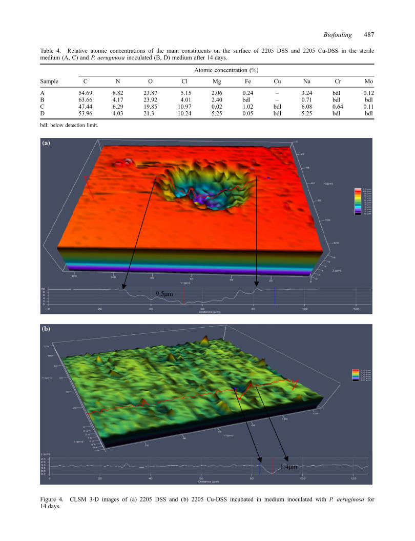
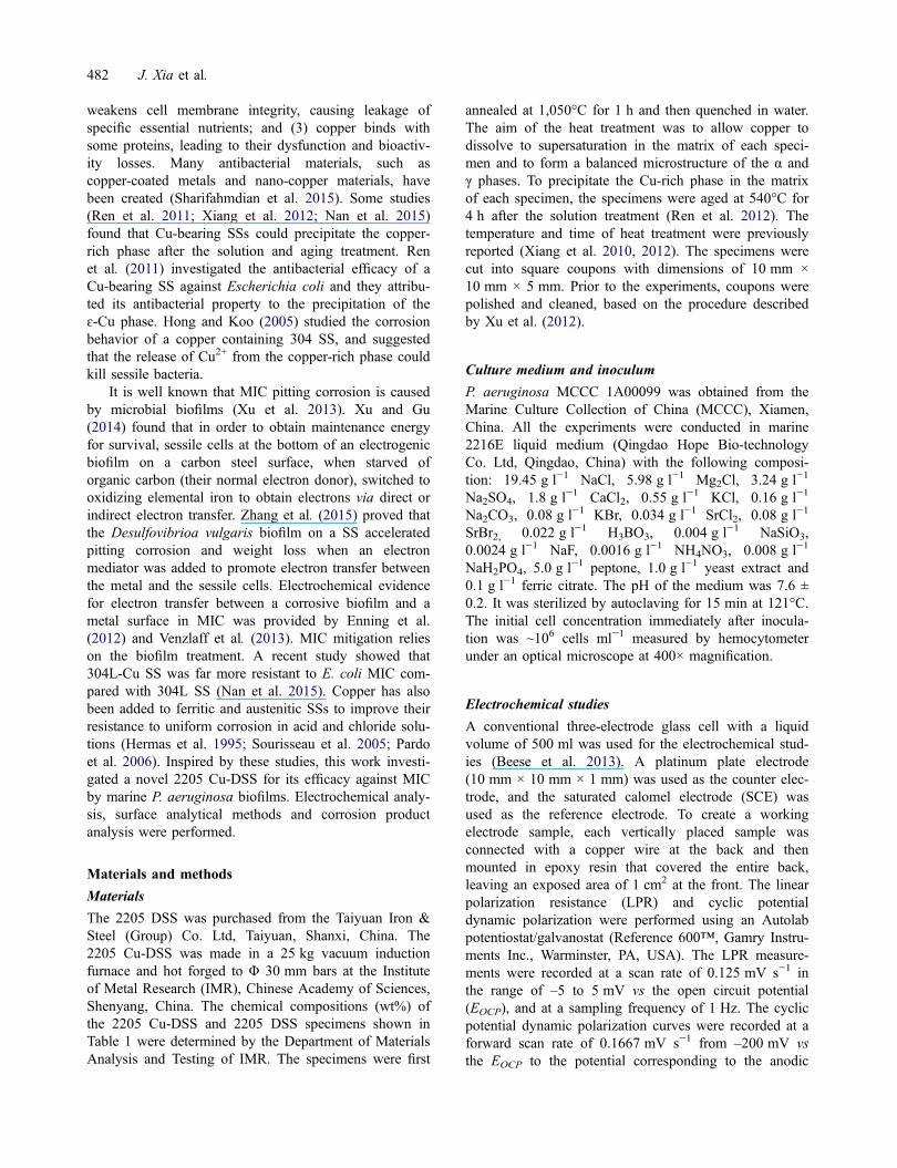
Table 4. Relative atomic concentrations of the main constituents on the surface of 2205 DSS and 2205 Cu-DSS in the sterilemedium (A, C) and P. aeruginosa inoculated (B, D) medium after 14 days.
Sample
Atomic concentration (%)
C N O Cl Mg Fe Cu Na Cr Mo
A 54.69 8.82 23.87 5.15 2.06 0.24 – 3.24 bdl 0.12B 63.66 4.17 23.92 4.01 2.40 bdl – 0.71 bdl bdlC 47.44 6.29 19.85 10.97 0.02 1.02 bdl 6.08 0.64 0.11D 53.96 4.03 21.3 10.24 5.25 0.05 bdl 5.25 bdl bdl
bdl: below detection limit.
9.5µm
1.4µm
(a)
(b)
Figure 4. CLSM 3-D images of (a) 2205 DSS and (b) 2205 Cu-DSS incubated in medium inoculated with P. aeruginosa for14 days.
Biofouling 487

(a) (b)
(c) (d)
0
2,000
4,000
6,000
8,000
10,000
12,000
14,000
Time (days)
Live cellDead cell
1 7
(f)
Cel
l num
ber
(cel
ls c
m-2
)
0.0
2.0x10³
4.0x10³
6.0x10³
8.0x10³
1.0x10⁴
1.2x10⁴
1.4x10⁴
1.6x10⁴
1.8x10⁴
2.0x10⁴
Live cellDead cell
Time (days)
Cel
l num
ber
(cel
ls c
m-2
)
1 7
(e)
0
10
20
30
40
50
60
70
80
71
Bio
film
thic
knes
s (m
m)
Time (days)
(g) 2205 DSS in the presence of2205 Cu-DSS in the presence of
Figure 5. Epifluorescence microscope images to show the growth of the P. aeruginosa biofilm on the surface of (a) 2205 DSS afterone day; (b) 2205 Cu-DSS after one day; (c) 2205 DSS after seven days; and (d) 2205 Cu-DSS after seven days. The calculated num-bers of live/dead cells are shown in (e) and (f). The biofilm thickness values for 2205 DSS and 2205 Cu-DSS coupons are presentedin (g).
488 J. Xia et al.
strong antibacterial ability compared with 2205 DSS asshown in Figure 5c and d. The biofilm on the 2205 DSSwas still intact, while the biofilm structure could nolonger be seen on the 2205 Cu-DSS surface. A relativelylarge quantity of dispersed dead sessile cells and a smallamount of live sessile cells were present on the 2205Cu-DSS surface. The antibacterial efficiency K of 2205Cu-DSS was calculated from Equation 2 (Li, Nan et al.2015):
K ¼ Nctrl � Nanti
Nctrl�100% (2)
where Nctrl represents the total number of sessile cells onthe 2205 DSS surface (control) and Nanti the total num-ber of sessile cells on the 2205 Cu-DSS surface. Thetotal number of sessile bacteria was the sum of the quan-tity of the live and dead bacteria. Figure 5e and f showsthe numbers of live and dead bacteria on the 2205 DSSand 2205 Cu-DSS surfaces after incubation for one andseven days, respectively. They indicate that the antibacte-rial efficiency of 2205 Cu-DSS after seven days(96.92%) was much higher than that after one day(7.75%).
The biofilm thickness was measured in a 3-D modeunder CLSM. Figure 5g shows that the biofilm thicknessvalues of 2205 DSS and 2205 Cu-DSS in the presenceof P. aeruginosa were 68.30 ± 6.16 μm and 60.70 ±7.80 μm, respectively. After exposure for seven days, thebiofilm thickness values of 2205 DSS and 2205 Cu-DSSwere 53.70 ± 2.91 μm and 47.81 ± 3.64 μm, respec-tively. In both cases, the biofilm on the 2205 Cu-DSSwas thinner than that on 2205 DSS. The Cu addition inthe matrix of 2205 Cu-DSS endowed it with antibacterialability, resulting in a thinner biofilm.
Discussion
The elements Fe, Cr, Mo and Cu were undetectable orlow in quantity in the biofilm when 2205 Cu-DSS wasincubated with the medium inoculated with P. aerugi-nosa after 14 days, probably because XPS did not pene-trate to the bottom of the relatively thick biofilm. Thesame observation (except Cu) was made for 2205 DSS.Yuan and Pehkonen (2007) observed the same resultswhen the biocorrosion behavior of the 304 SS in thepresence of P. aeruginosa was investigated.
Many researchers regard the cuprous dichloride anion(CuCl2
−) as the main form of soluble cuprous species inseawater or a NaCl solution (Kear et al. 2004; Badawyet al. 2005). Ma et al. (2015) proposed that Cu2(OH)3Clwas the oxidation product of Cu(I) oxide whichincreased the corrosion resistance of SS. This mayexplain why 2205 Cu-DSS showed a better MIC resis-tance performance than 2205 DSS.
In the presence of P. aeruginosa, the pH of the bulkculture medium was slightly changed from 7.8 initiallyto 8.2 finally on the 14th day. Although the pH under abiofilm can differ from that in the bulk fluid by up totwo units (Hidalgo et al. 2009), the relatively high pHsuggested that organic acid attack was not a factor in thisstudy. Figure 1b shows that the corrosion current densityof 2205 DSS in the presence of P. aeruginosa washigher than that of 2205 DSS in the uninoculatedmedium. This may be attributed to the catalysis ofP. aeruginosa on the reduction of oxygen (Cournet et al.2010), which accelerated the of corrosion process.Busalmen et al. (2002) also demonstrated that the catho-dic current density could be accelerated by the presenceof a P. aeruginosa biofilm.
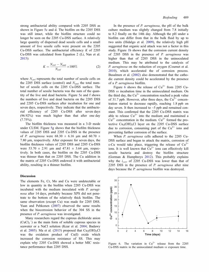
Figure 6 shows the release of Cu2+ from 2205 Cu-DSS vs incubation time in the uninoculated medium. Onthe third day, the Cu2+ concentration reached a peak valueof 31.7 ppb. However, after three days, the Cu2+ concen-tration started to decrease rapidly, reaching 1.8 ppb onday seven. It then increased to ~5 ppb and remained con-stant. This confirmed that the 2205 Cu-DSS matrix wasable to release Cu2+ into the medium and maintained aCu2+ concentration in the medium. Cu2+ formed the pro-tective Cu2(OH)3Cl layer on the 2205 Cu-DSS surfacedue to corrosion, consuming part of the Cu2+ ions andpreventing further corrosion of the surface.
When P. aeruginosa cells adhered to the 2205 Cu-DSS surface and began to attack the matrix, corrosion ofε-Cu would take place, triggering the release of Cu2+
ions. It is well known that Cu2+ ions can effectively killsessile bacteria and destroy the biofilm structure(Gorman & Humphreys 2012). This probably explainswhy the icorr of 2205 Cu-DSS was lower than that of2205 DSS in the presence of P. aeruginosa after ninedays because the P. aeruginosa biofilm was destroyed.
0 2 4 6 8 10 12 14 16
0
5
10
15
20
25
30
35
Cu2+
con
cent
ratio
n (p
pb)
Time (days)
Figure 6. The variation in Cu2+ release from the 2205Cu-DSS matrix in the uninoculated medium vs exposure time.
Biofouling 489

Biofilm treatment is vital in MIC mitigation. In thisstudy, the experimental data illustrate that with the addi-tion of Cu to 2205 DSS, the biofilm coverage, the sessileP. aeruginosa cell number and the largest MIC pit depthon 2205 Cu-DSS coupons were all significantly reduceddue to the antibacterial ability of Cu2+ released from themetal (Teitzel & Parsek 2003; Nan et al. 2008). The14-day data of live/dead biofilm staining were notobtained because after 14 days, most of the sessile cellson 2205 DSS and 2205 Cu-DSS surfaces were dead dueto the depletion of the nutrients in the 2216E medium.
Figure 7 illustrates how Cu2+ was released from theCu-rich phase from the 2205 Cu-DSS matrix and howP. aeruginosa MIC was mitigated. In the early stage,Cu2+ was easy to release because of the corrosion of thematrix by the P. aeruginosa biofilm. With the release ofCu2+ ions, the biofilm was gradually inhibited andeventually most of the sessile cells, especially those thatwere directly on the metal surface, were killed bythe Cu2+ ions as shown in Figure 7b after long-termimmersion.
It is well known in the literature that SS welds aremore prone to MIC attack. Post-weld heat treatment is aprerequisite for 2205-Cu DSS to exhibit its antibacterialability in an entire structure. This treatment may not bepractical in situ. Thus, special attention should be paid toMIC at the welds. This concern should be addressedbefore 2205-Cu DSS becomes a viable commercial suc-cess. Additional research regarding the weldability of2205-Cu is needed. As a newly emerged duplex SS, fur-ther study is needed. For example, its mechanical proper-ties such as toughness may be influenced by thepresence of the alpha prime. Because MIC pipeline orstorage tank leaks are often pinhole leaks, the largest pit
depth is a critical parameter in MIC. The results of thisstudy prove that the novel 2205 Cu-DSS exhibited betterMIC resistance than the commercial 2205 DSS.
Conclusions
The corrosion behavior of a novel 2205 Cu-DSS in thepresence of an aerobic marine P. aeruginosa biofilm wasinvestigated by analyzing electrochemical test data, ses-sile cell kills, corrosion pit sizes and corrosion products.The electrochemical tests indicated that 2205 Cu-DSShad better MIC resistance than the common 2205 DSSin the presence of P. aeruginosa after incubation fornine days. The XPS results revealed that a protectiveCu2(OH)3Cl layer was formed on the 2205 Cu-DSS sur-face. The pit morphology suggested that the pittingcorrosion resistance of 2205 Cu-DSS was significantlybetter than that of 2205 DSS in the presence ofP. aeruginosa. The antibacterial efficiency of 2205Cu-DSS after incubation for one and seven days withP. aeruginosa was found to be 7.75% and 96.92%, respec-tively, demonstrating excellent antibacterial efficacy afterincubation for seven days. The biocorrosion caused byP. aeruginosa biofilm was successfully mitigated by the2205 Cu-DSS after incubation for nine days. This couldbe explained by the fact that the release of Cu2+ from themetal triggered by the initial P. aeruginosa biofilm attackeffectively eradicated the biofilm subsequently.
The pitting corrosion caused by the corrosiveP. aeruginosa biofilm in marine environments has been aserious problem. This study showed that addition of theCu element in the currently used DSS created a newmetal with a superior antimicrobial ability against themarine P. aeruginosa biofilm. This will likely result in
(a) (b)
Cu2+
O2
Cu2+ Cu2+ Cu2+
Cl-, OH-
O2
2205 Cu-DSS matrix Passive filmε-Cu phase Live P. aeruginosa Dead P. aeruginosa
Cu2+ Cu2+
Cl-, OH-
Figure 7. Schematic illustration of the MIC resistance mechanism of 2205 Cu-DSS (a) at an early stage of incubation, and (b) aftera longer time.
490 J. Xia et al.
field applications, such as seawater cooling systems, asan environmentally friendly and cost-effective alternativeto biocide treatment of biofilms.
Conflict of interest disclosure statementNo potential conflict of interest was reported by the authors.
FundingThis study was financially supported by the Young MeritScholars program by the National Basic Research Program ofChina [973 Program No. 2014CB643300], the National Envi-ronmental Corrosion Platform (NECP) and by the Young MeritScholars program of the Institute of Metal Research, ChineseAcademy of Sciences.
ReferencesAbdolahi A, Hamzah E, Ibrahim Z, Hashim S. 2014. Micro-
bially influenced corrosion of steels by Pseudomonasaeruginosa. Corros Rev. 32:129–141.
Antony PJ, Raman RKS, Mohanram R, Kumar P, Raman R.2008. Influence of thermal aging on sulfate-reducing bacte-ria (SRB)-influenced corrosion behaviour of 2205 duplexstainless steel. Corros Sci. 50:1858–1864.
Badawy WA, Ismail KM, Fathi AM. 2005. Effect of Ni contenton the corrosion behaviour of Cu–Ni alloys in neutral chlo-ride solution. Electrochim Acta. 50:3603–3608.
Beese P, Venzlaff H, Srinivasan J, Garrelfs J, Stratmann M,Mayrhofer KJJ. 2013. Monitoring of anaerobic microbiallyinfluenced corrosion via electrochemical frequency modula-tion. Electrochim Acta. 105:239–247.
Busalmen JP, Vázquez M, Sánchez SRD. 2002. New evidenceon the catalase mechanism of microbial corrosion. Elec-trochim Acta. 47:1857–1865.
Chinese National Standards. 2000. Method of nitric-hydrofluoricacids test for stainless steel. GB/T4334:4–2000.
Cournet A, Bergé M, Roques C, Bergel A, Délia ML. 2010.Electrochemical reduction of oxygen catalyzed by Pseu-domonas aeruginosa. Electrochim Acta. 55:4902–4908.
Elhoud A, Ezuber H, Deans W. 2010. Influence of cold workand sigma phase on the pitting corrosion behavior of 25chromium super duplex stainless steel in 3.5% sodiumchloride solution. Mater Corros. 61:199–204.
Enning D, Venzlaff H, Garrelfs J, Dinh HT, Meyer V, MayrhoferK, Hassel AW, Stratmann M, Widdel F. 2012. Marinesulfate-reducing bacteria cause serious corrosion of ironunder electroconductive biogenic mineral crust: microbialiron corrosion under electroconductive crust. EnvironMicrobiol. 14:1772–1787.
Gorman JO, Humphreys H. 2012. Application of copper to pre-vent and control infection. Where are we now? J HospInfect. 81:1–7.
Hamzah E, Hussain MF, Ibrahim Z, Abdolahi A. 2013. Influ-ence of Pseudomonas aeruginosa bacteria on corrosionresistance of 304 stainless steel. Corros Eng Sci Technol.48:116–120.
Hamzah E, Hussain MF, Ibrahim Z, Abdolahi A. 2014. Corro-sion behaviour of carbon steel in sea water medium inpresence of Pseudomonas aeruginosa bacteria. Arab J SciEng. 39:6863–6870.
Hermas AA, Ogura K, Takagi S, Adachi T. 1995. Effects ofalloying additions on corrosion and passivation behaviorsof type 304 stainless steel. Corrosion. 51:3–10.
Hidalgo G, Burns A, Herz E, Hay AG, Houston PL, WiesnerU, Lion LW. 2009. Functional tomographic fluorescenceimaging of pH microenvironments in microbial biofilms byuse of silica nanoparticle sensors. Appl Environ Microbiol.75:7426–7435.
Hong IT, Koo CH. 2005. Antibacterial properties, corrosionresistance and mechanical properties of Cu-modified SUS304 stainless steel. Mater Sci Eng: A. 393:213–222.
Jeon S-H, Kim S-T, Lee I-S, Kim J-S, Kim K-T, Park Y-S.2013. Effects of Cu on the precipitation of intermetalliccompounds and the intergranular corrosion of hyper duplexstainless steels. Corros Sci. 66:217–224.
Kear G, Barker BD, Stokes K, Walsh FC. 2004. Electrochemi-cal corrosion behaviour of 90–10 Cu–Ni in chloride-basedelectrolytes. J Appl Electrochem. 34:659–669.
Lee J-S, Fushimi K, Nakanishi T, Hasegawa Y, Park Y-S.2014. Corrosion behaviour of ferrite and austenite phaseson super duplex stainless steel in a modified green-deathsolution. Corros Sci. 89:111–117.
Li M, Nan L, Xu D, Ren G, Yang K. 2015. Antibacterial per-formance of Cu-bearing stainless steel against microorgan-isms in tap water. J Mater Sci Technol. 31:243–251.
Li S, Ren X, Ji X, Gui Y. 2014. Effects of microstructurechanges on the superplasticity of 2205 duplex stainlesssteel. Mater Design. 55:146–151.
Liu W. 2014. Rapid MIC attack on 2205 duplex stainless steelpipe in a yacht. Eng Fail Anal. 42:109–120.
Ma AL, Jiang S, Zheng Y, Ke W. 2015. Corrosion product filmformed on the 90/10 copper–nickel tube in natural seawa-ter: composition/structure and formation mechanism.Corros Sci. 91:245–261.
Manga SS, Oyeleke SB, Librahim AD, Aliero AA, Bagudo AI.2012. Influence of bacteria associated with corrosion ofmetal. Continent J Microbiol. 6:19–25.
Moradi M, Song Z, Yang L, Jiang J, He J. 2014. Effect of mar-ine Pseudoalteromonas sp. on the microstructure and corro-sion behaviour of 2205 duplex stainless steel. Corros Sci.84:103–112.
Nan L, Liu Y, Lü M, Yang K. 2008. Study on antibacterialmechanism of copper-bearing austenitic antibacterial stain-less steel by atomic force microscopy. J Mater Sci-MaterM. 19:3057–3062.
Nan L, Xu D, Gu T, Song X, Yang K. 2015. Microbiologicalinfluenced corrosion resistance characteristics of a 304L-Custainless steel against Escherichia coli. Mater Sci Eng: C.48:228–234.
Pardo A, Merino MC, Carboneras M, Viejo F, Arrabal R,Muñoz J. 2006. Influence of Cu and Sn content in thecorrosion of AISI 304 and 316 stainless steels in H2SO4.Corros Sci. 48:1075–1092.
Ren L, Nan L, Yang K. 2011. Study of copper precipitationbehavior in a Cu-bearing austenitic antibacterial stainlesssteel. Mater Design. 32:2374–2379.
Ren L, Yang K, Guo L, Chai H. 2012. Preliminary study ofanti-infective function of copper-bearing stainless steel.Mater Sci Eng: C. 32:1204–1209.
San NO, Nazir H, Dönmez G. 2014. Microbially influenced corro-sion and inhibition of nickel–zinc and nickel–copper coatingsby Pseudomonas aeruginosa. Corros Sci. 79:177–183.
Sharifahmdian O, Salimijazi HR, Fathi MH, Mostaghimi J,Pershin L. 2015. Relationshiop between surface properties
Biofouling 491

and antibacterial behaviour of wire arc spray coppercoatings. Surf coat Technol. 233:74–79.
Sourisseau T, Chauveau E, Baroux B. 2005. Mechanism ofcopper action on pitting phenomena observed on stainlesssteels in chloride media. Corros Sci. 47:1097–1117.
Stern M, Geary AL. 1957. Electrochemical polarization. J Elec-trochem Soc. 104:56–63.
Sun D, Shahzad MB, Li M, Wang G, Xu D. 2015. Antimicro-bial materials with medical application. Mater Technol. 30:b90–b95.
Teitzel GM, Parsek MR. 2003. Heavy metal resistance of bio-film and planktonic Pseudomonas aeruginosa. Appl Envi-ron Microbiol. 69:2313–2320.
Venzlaff H, Enning D, Srinivasan J, Mayrhofer KJJ, HasselAW, Widdel F, Stratmann M. 2013. Accelerated cathodicreaction in microbial corrosion of iron due to directelectron uptake by sulfate-reducing bacteria. Corros Sci.66:88–96.
Xiang H, Fan J, Liu D, Guo P. 2012. Effects of antibacterialaging treatment on microstructure and properties of copper-containing duplex stainless steel. Acta Metall Sinica.48:1081–1088.
Xiang H, Huang W, Liu D, He F, Ruan F. 2010. Effects ofaging temperature on the microstructure and property of
2906 super duplex stainless steel. Trans Mater Heat Treat.31:86–90.
Xu D, Gu T. 2014. Carbon source starvation triggered moreaggressive corrosion against carbon steel by Desulfovibriovulgaris biofilm. Int Biodeter Biodegr. 91:74–81.
Xu D, Li Y, Song F, Gu T. 2013. Laboratory investigation ofmicrobiologically influenced corrosion of C1018 carbonsteel by nitrate reducing bacterium Bacillus licheniformis.Corros Sci. 77:385–390.
Xu D, Wen J, Gu T, Raad I. 2012. Biocide cocktail consistingof glutaraldehyde, ethylene diamine disuccinate (EDDS),and methanol for the mitigation of souring and biocorro-sion. Corrosion. 68:994–1002.
Yuan SJ, Pehkonen SO. 2007. Microbiologically influencedcorrosion of 304 stainless steel by aerobic PseudomonasNCIMB 2021 bacteria: AFM and XPS study. Colloid SurfB. 59:87–99.
Zhang P, Xu D, Li Y, Yang K, Gu T. 2015. Electron mediatorsaccelerate the microbiologically influenced corrosion of304 stainless steel by Desulfovibrio vulgaris biofilm. Bio-electrochemistry. 101:14–21.
Zou Y, Wang J, Zheng Y. 2011. Electrochemical techniques fordetermining corrosion rate of rusted steel in seawater.Corros Sci. 53:208–216.
492 J. Xia et al.


