Biochimica et Biophysica Acta - COnnecting REpositories · L. Sánchez-Felipe et al. / Biochimica...
10
Entry of Newcastle Disease Virus into the host cell: Role of acidic pH and endocytosis Lorena Sánchez-Felipe, Enrique Villar ⁎, Isabel Muñoz-Barroso ⁎ Departamento de Bioquímica y Biología Molecular, Universidad de Salamanca, Edificio Departamental Lab. 106/108, Plaza Doctores de la Reina s/n, 37007 Salamanca, Spain abstract article info Article history: Received 31 January 2013 Received in revised form 2 August 2013 Accepted 13 August 2013 Available online 28 August 2013 Keywords: Viral entry Endocytosis Low-pH NDV Paramyxovirus Most paramyxoviruses enter the cell by direct fusion of the viral envelope with the plasma membrane. Our pre- vious studies have shown the colocalization of Newcastle Disease Virus (NDV) with the early endosome marker EEA1 and the inhibition of NDV fusion by the caveolin-phosphorylating drug phorbol 12-myristate 13-acetate (PMA) prompted us to propose that NDV enters the cells via endocytosis. Here we show that the virus-cell fusion and cell-cell fusion promoted by NDV-F are increased by about 30% after brief exposure to low pH in HeLa and ELL-0 cells but not in NDV receptor- deficient cell lines such as GM95 or Lec1. After a brief low-pH exposure, the percentage of NDV fusion at 29 °C was similar to that at 37 °C without acid-pH stimulation, meaning that acid pH would decrease the energetic barrier to enhance fusion. Furthermore, preincubation of cells with the pro- tein kinase C inhibitor bisindolylmaleimide led to the inhibition of about 30% of NDV infectivity, suggesting that a population of virus enters cells through receptor-mediated endocytosis. Moreover, the involvement of the GTPase dynamin in NDV entry is shown as its specific inhibitor, dynasore, also impaired NDV fusion and infectiv- ity. Optimal infection of the host cells was significantly affected by drugs that inhibit endosomal acidification such as concanamycin A, monensin and chloroquine. These results support our hypothesis that entry of NDV into ELL- 0 and HeLa cells occurs through the plasma membrane as well as by dynamin- low pH- and receptor- dependent endocytosis. © 2013 Elsevier B.V. All rights reserved. 1. Introduction Newcastle Disease Virus (NDV), a prototype of paramyxovirus, is an avian enveloped RNA-negative strand virus that causes respiratory disease in domestic fowl, leading to huge economic losses in the poultry industry. The envelope of NDV contains two associated glycoproteins that mediate viral entry: the hemagglutinin-neuraminidase (HN) and fusion (F) proteins. HN is the receptor-binding protein that recognizes and binds to sialoglycoconjugates at the cell surface [1] and also has receptor-cleaving (sialidase) activity [2]. Based on crystallographic studies of the HN of NDV, two sialic acid-binding sites have been described [3,4], site II being located at the dimer interface and activated after engagement of site I to its receptor [4,5]. Site I exhibits both recep- tor-binding and sialidase activities while site II only has receptor binding activity [4]. Moreover, the triggering of F protein requires the presence of its homotypic attachment protein through its fusion promotion activity. Enveloped viruses enter the cell through two main mechanisms: by direct fusion of the viral envelope with the plasma membrane or by endocytosis. The use of the cellular endocytic machinery is commonly associated with low-pH-dependent activation of viral fusion proteins such as in influenza virus or in vesicular stomatitis virus [6]. Additionally, the fusion protein of Ebola virus is activated by acidic- pH-dependent proteases inside endosomes [7]. Moreover, recent stud- ies suggest that viruses with pH-independent fusion proteins may use endocytosis for entry (revised in [8]). Different families of viruses may use several different endocytic routes [9–11], the major pathway being the clathrin-mediated endocytosis used by viruses such as Semliki For- est and Sindbis alpha virus [12] or Hantaan virus [13]. Recent reports in- dicate that HIV also uses this route productively [14]. Caveola-mediated endocytosis is the second best characterized pathway used by SV40 and Ebola viruses to enter cells [15–18]. Although most viruses have been described to enter the cell through a unique mechanism, there are in- creasing reports showing that many viruses are able to use alternative uptake pathways to infect different target cells or even simultaneous entry mechanisms in the same cell type ([8,19] and references therein). With the exception of the fusion protein of the human metapneumovirus (HMPV), which depends on low-pH [20], para- myxovirus fusion proteins are activated at neutral pH, which sug- gests that viral entry occurs by direct fusion at the plasma membrane [2]. Nevertheless, recent reports point to the notion that the pH requirement for fusion does not necessarily imply the location of membrane fusion (revised in [21,22]). Some members of the paramyxoviruses such as Sendai, Nipah, human metapneumovirus and human respiratory syncytial virus (hRSV) [23–26] utilize the endocytic machinery for entry. Furthermore, as assessed by a R18 dequenching assay, our previous studies Biochimica et Biophysica Acta 1838 (2014) 300–309 Abbreviations: BIM, bisindolylmaleimide; moi, multiplicity of infection; R18, octadecylrhodamine B chloride; RBCs, red blood cells ⁎ Corresponding authors. E-mail addresses: [email protected] (E. Villar), [email protected] (I. Muñoz-Barroso). 0005-2736/$ – see front matter © 2013 Elsevier B.V. All rights reserved. http://dx.doi.org/10.1016/j.bbamem.2013.08.008 Contents lists available at ScienceDirect Biochimica et Biophysica Acta journal homepage: www.elsevier.com/locate/bbamem
Transcript of Biochimica et Biophysica Acta - COnnecting REpositories · L. Sánchez-Felipe et al. / Biochimica...

Biochimica et Biophysica Acta 1838 (2014) 300–309
Contents lists available at ScienceDirect
Biochimica et Biophysica Acta
j ourna l homepage: www.e lsev ie r .com/ locate /bbamem
Entry of Newcastle Disease Virus into the host cell: Role of acidic pHand endocytosis
Lorena Sánchez-Felipe, Enrique Villar ⁎, Isabel Muñoz-Barroso ⁎Departamento de Bioquímica y Biología Molecular, Universidad de Salamanca, Edificio Departamental Lab. 106/108, Plaza Doctores de la Reina s/n, 37007 Salamanca, Spain
Abbreviations: BIM, bisindolylmaleimide; moi, moctadecylrhodamine B chloride; RBCs, red blood cells⁎ Corresponding authors.
E-mail addresses: [email protected] (E. Villar), imunbar@
0005-2736/$ – see front matter © 2013 Elsevier B.V. All rhttp://dx.doi.org/10.1016/j.bbamem.2013.08.008
a b s t r a c t
a r t i c l e i n f oArticle history:Received 31 January 2013Received in revised form 2 August 2013Accepted 13 August 2013Available online 28 August 2013
Keywords:Viral entryEndocytosisLow-pHNDVParamyxovirus
Most paramyxoviruses enter the cell by direct fusion of the viral envelope with the plasma membrane. Our pre-vious studies have shown the colocalization of Newcastle Disease Virus (NDV) with the early endosome markerEEA1 and the inhibition of NDV fusion by the caveolin-phosphorylating drug phorbol 12-myristate 13-acetate(PMA) prompted us to propose that NDV enters the cells via endocytosis. Herewe show that the virus-cell fusionand cell-cell fusion promoted by NDV-F are increased by about 30% after brief exposure to low pH in HeLa andELL-0 cells but not in NDV receptor- deficient cell lines such as GM95 or Lec1. After a brief low-pH exposure,the percentage of NDV fusion at 29 °C was similar to that at 37 °C without acid-pH stimulation, meaning thatacid pHwould decrease the energetic barrier to enhance fusion. Furthermore, preincubation of cells with the pro-tein kinase C inhibitor bisindolylmaleimide led to the inhibition of about 30% of NDV infectivity, suggesting that apopulation of virus enters cells through receptor-mediated endocytosis. Moreover, the involvement of theGTPase dynamin in NDV entry is shown as its specific inhibitor, dynasore, also impaired NDV fusion and infectiv-ity. Optimal infection of the host cellswas significantly affected bydrugs that inhibit endosomal acidification suchas concanamycin A, monensin and chloroquine. These results support our hypothesis that entry of NDV into ELL-0 and HeLa cells occurs through the plasmamembrane aswell as by dynamin- low pH- and receptor- dependentendocytosis.
© 2013 Elsevier B.V. All rights reserved.
1. Introduction
Newcastle Disease Virus (NDV), a prototype of paramyxovirus, isan avian enveloped RNA-negative strand virus that causes respiratorydisease in domestic fowl, leading to huge economic losses in the poultryindustry. The envelope of NDV contains two associated glycoproteinsthat mediate viral entry: the hemagglutinin-neuraminidase (HN) andfusion (F) proteins. HN is the receptor-binding protein that recognizesand binds to sialoglycoconjugates at the cell surface [1] and also hasreceptor-cleaving (sialidase) activity [2]. Based on crystallographicstudies of the HN of NDV, two sialic acid-binding sites have beendescribed [3,4], site II being located at the dimer interface and activatedafter engagement of site I to its receptor [4,5]. Site I exhibits both recep-tor-binding and sialidase activities while site II only has receptor bindingactivity [4]. Moreover, the triggering of F protein requires the presence ofits homotypic attachment protein through its fusion promotion activity.
Enveloped viruses enter the cell through two main mechanisms: bydirect fusion of the viral envelope with the plasma membrane or byendocytosis. The use of the cellular endocytic machinery is commonlyassociated with low-pH-dependent activation of viral fusion proteinssuch as in influenza virus or in vesicular stomatitis virus [6].
ultiplicity of infection; R18,
usal.es (I. Muñoz-Barroso).
ights reserved.
Additionally, the fusion protein of Ebola virus is activated by acidic-pH-dependent proteases inside endosomes [7]. Moreover, recent stud-ies suggest that viruses with pH-independent fusion proteins may useendocytosis for entry (revised in [8]). Different families of viruses mayuse several different endocytic routes [9–11], the major pathway beingthe clathrin-mediated endocytosis used by viruses such as Semliki For-est and Sindbis alpha virus [12] or Hantaan virus [13]. Recent reports in-dicate that HIV also uses this route productively [14]. Caveola-mediatedendocytosis is the second best characterized pathway used by SV40 andEbola viruses to enter cells [15–18]. Although most viruses have beendescribed to enter the cell through a unique mechanism, there are in-creasing reports showing that many viruses are able to use alternativeuptake pathways to infect different target cells or even simultaneousentry mechanisms in the same cell type ([8,19] and references therein).
With the exception of the fusion protein of the humanmetapneumovirus (HMPV), which depends on low-pH [20], para-myxovirus fusion proteins are activated at neutral pH, which sug-gests that viral entry occurs by direct fusion at the plasmamembrane [2]. Nevertheless, recent reports point to the notionthat the pH requirement for fusion does not necessarily implythe location of membrane fusion (revised in [21,22]). Somemembers of the paramyxoviruses such as Sendai, Nipah, humanmetapneumovirus and human respiratory syncytial virus (hRSV)[23–26] utilize the endocytic machinery for entry. Furthermore,as assessed by a R18 dequenching assay, our previous studies

301L. Sánchez-Felipe et al. / Biochimica et Biophysica Acta 1838 (2014) 300–309
revealed the enhancement of NDV fusion with Cos-7 at acidic pH[27] and the colocalization of NDVwith the early endosomemarkerEEA1 [28]. These findings prompted us to suggest that the entryof NDV would occur through caveola-mediated endocytosis as anadditional mechanism to direct fusion with the plasma membrane[28]. To gain further insight into the molecular mechanisms of NDVentry, the present study focuses on the characterization of the NDVentry mechanism through the use of low-pH treatment of severalcell lines that differ in the expression of sialoglycoconjugates attheir surface and by analyzing the effect of different substancesthat either interfere with endocytosis or endosomal acidificationon NDV interaction with cells. NDV entry showed to be sensitiveto endosomal acid pH, and dynamin and protein kinase C inhibitorsblocked NDV fusion and infectivity. Our data support the idea thatNDV may penetrate HeLa and ELL-0 cells both by direct fusionat the cell surface and through receptor-mediated and dynamin-dependent endocytosis, at least in these cell lines.
2. Materials and methods
2.1. Cell lines and viruses
East Lansing Line (ELL-0) avian fibroblasts and HeLa cells wereobtained from the American Type Culture Collection (ATCC); MEB4and GM95 were obtained from Riken BRC Cell Bank (Tsukuba, Japan);ELL-0, HeLa, MEB4 andGM95 cells weremaintained in Dulbecco's mod-ified Eagle's medium (DMEM); Chinese hamster ovary (CHO) and mu-tant Lec1 were also purchased from ATCC and maintained in DMEM:F12 and AlphaMEM media respectively. All media were supplementedwith L-GlutaMax (580 mg l−1, Invitrogen), penicillin–streptomycin(100 U ml−1–100 μg ml−1) and 10% heat-inactivated fetal bovineserum (complete medium). The lentogenic “Clone 30” strain of NDVwas obtained from Intervet Laboratories (Salamanca, Spain). The viruswas grown and purified mainly as described previously [28]. For in-ectivity assays, a recombinant NDV derived from the Hitchner B1lentogenic strain (rNDV-F3aa-mRFP [29]), which expresses a mono-meric red-fluorescent protein [30] and kindly provided by Dr. AdolfoGarcía-Sastre, was used.
2.2. Reagents and antibodies
Bisindolylmaleimide I (BIM) was from Calbiochem; dynasore,concanamycin A, monensin, chloroquine, protease inhibitor cocktailand proteinase K were from Sigma Chemical Co; octadecylrhodamine Bchloride (R18), Hoechst 33258, calcein, lipofectamine and Alexa Fluor488 donkey anti-mouse antibody were from Molecular Probes. Mono-clonal anti-NP and polyclonal anti-NDV antibodies were generous giftsfromDr. García-Sastre (Emerging Pathogens Institute,Mount Sinai Schoolof Medicine, New York, USA); monoclonal anti-HN 14f and 1b antibodieswere kindly provided by Dr. M. Iorio (University of MassachusettsMedical School, Worcester, USA).
2.3. Virus titration
Virus titers were calculated in plaque-formation assays in Verocells, as described [31]. Plaque numbers were counted under a lightmicroscope and NDV infectivity was expressed as plaque-formingunits (pfu) ml−1.
2.4. Virus-binding assays
Monolayers of HeLa, ELL-0, MEB4, GM95, CHO and Lec1 cells on35 mm plates were previously treated with the different inhibitorsand then incubated with NDV at a moi of 1 for 1 h at 4 °C in the contin-uous presence of the inhibitor. Cells were washed with ice-cold PBS toremove unbound virus and then collected from the plates by incubation
with EDTA (520 μM) at 37 °C. Cells were pelleted by centrifugation andfixed with 2.5% paraformaldehyde for 30 min at 4 °C. After centrifuga-tion, cells were incubated with anti-HN 14f and 1b mAbs in blockingsolution (dilution 1:100) for 1 h at 4 °C. Then, the cellswere centrifugedand incubated with blocking solution containing anti-mouse AlexaFluor 488-conjugated IgG (5 μg ml−1) for 1 h at 4 °C. Cells were thenpelleted by centrifugation and resuspended in a suitable volume ofPBS for analysis in a FACScalibur flow cytometer (Becton Dickinson).At least 5 × 10⁴ cells were analyzed for each sample. Meanfluorescencevalues were normalized to the control values. Data were analyzed withWinMDI 2.9 software.
2.5. NDV–cell fusion assays
Purified NDVwas labeled with the fluorescent probe R18 essentiallyas described previously [32]. Cells plated in 24-well plates treatedor untreated with the inhibitors were incubated for 30 min at 4 °C onice with 2 μg of R18-NDV per plate. Then, cells were washed threetimes with cold PBS and incubated in complete medium containingHoechst 33258 (10 μg ml−1) for 1 h at 37 °C. They were fixed with 2%formaldehyde in PBS and the transfer of the rhodamine probe tocells was observed under an Olympus IX51 inverted fluorescencemicroscope. The percentage of fusion was calculated as the number ofpositive red-stained cells in 10 random fields with respect to the totalnumber of cells in these areas of the well.
2.6. NDV infectivity assays
Monolayers of cells were infected for 1 h at room temperature withdifferent dilutions of the recombinant NDV rNDV-F3aa-mRFP. After24 h at 37 °C, the cells were observed under an Olympus IX51 invertedfluorescence microscope with a 10× objective. The percentage of infec-tivity was calculated as the number of red-fluorescent cells out of thetotal number of cells in six random fields.
2.7. FACS infectivity assays
Monolayers of HeLa, ELL-0, MEB4, GM95, CHO and Lec1 cells on35 mm plates were treated with the inhibitors and then incubatedwith 1 moi of NDV for 1 h at 37 °C in the presence of inhibitor. Cellswere washed with ice-cold PBS to remove unbound virus andwere col-lected from the plates by incubation with EDTA (520 μM) at 37 °C. Cellswere pelleted by centrifugation and fixed with 2.5% paraformaldehydefor 30 min at 4 °C. Then, cells were blocked and permeabilized withPBS containing 1% BSA (blocking solution) and 0.1% TX100 for 20 minat 4 °C. After centrifugation, cells were incubated with anti-NP mAb inblocking solution (dilution 1:200) for 1 h at 4 °C. Then, they were cen-trifuged and incubated with blocking solution containing anti-mouseAlexa Fluor 488-conjugated IgG (5 μg ml−1) for 1 h at 4 °C. Cellswere then pelleted by centrifugation and resuspended in a suitablevolume of PBS for analysis with a FACScalibur flow cytometer (BectonDickinson). At least 5 × 10⁴ cells were analyzed for each sample.Mean fluorescence values and the % of cells with fluorescence higherthan the background were combined to quantify viral protein expres-sion and were normalized to the control values. Mean fluorescencevalues were normalized to control values. Data were analyzed withWinMDI 2.9 software.
2.8. Syncytium assays
Monolayers of cells were infectedwith 1moi of NDV for 1 h at 37 °C.At 7 h post-infection, viral F proteins were activated by digestion withacetyl trypsin [33]. Then, cells were incubated in complete mediumovernight and stained with Giemsa or crystal violet for syncytiumobservation. Representative fields were captured with an invertedmicroscope (Olympus IX51). Quantification of syncytiawas accomplished
0
20
40
60
80
HeLa ELL-0 MEB4 GM95 CHO Lec1
% F
usi
on
pH 7.4
pH 5.0
A
B
ELL
-0
HeL
a
******
pH 7.4 pH 5.0HoechstHoechst R18 R18
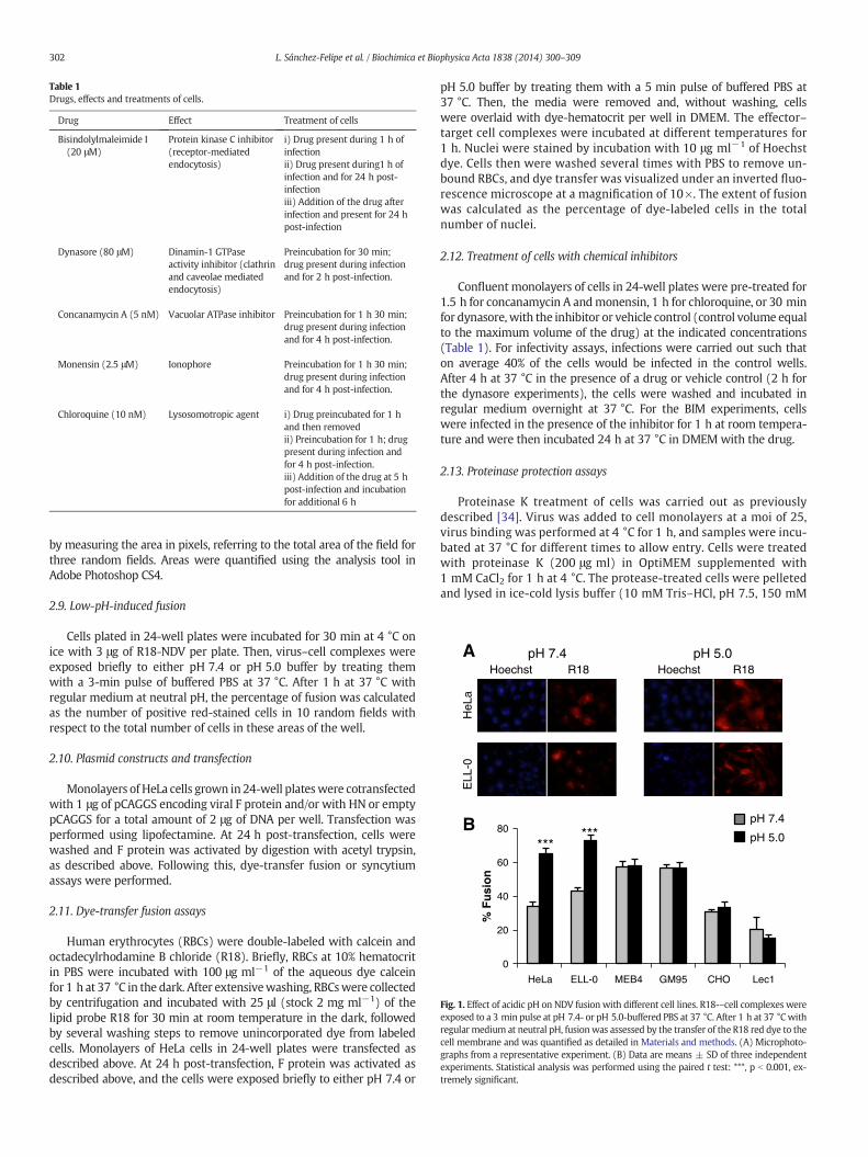
Fig. 1. Effect of acidic pH on NDV fusion with different cell lines. R18-–cell complexes wereexposed to a 3 min pulse at pH 7.4- or pH 5.0-buffered PBS at 37 °C. After 1 h at 37 °Cwithregular medium at neutral pH, fusionwas assessed by the transfer of the R18 red dye to thecell membrane and was quantified as detailed in Materials and methods. (A) Microphoto-graphs from a representative experiment. (B) Data are means ± SD of three independentexperiments. Statistical analysis was performed using the paired t test: ***, p b 0.001, ex-tremely significant.
Table 1Drugs, effects and treatments of cells.
Drug Effect Treatment of cells
Bisindolylmaleimide I(20 μM)
Protein kinase C inhibitor(receptor-mediatedendocytosis)
i) Drug present during 1 h ofinfectionii) Drug present during1 h ofinfection and for 24 h post-infectioniii) Addition of the drug afterinfection and present for 24 hpost-infection
Dynasore (80 μM) Dinamin-1 GTPaseactivity inhibitor (clathrinand caveolae mediatedendocytosis)
Preincubation for 30 min;drug present during infectionand for 2 h post-infection.
Concanamycin A (5 nM) Vacuolar ATPase inhibitor Preincubation for 1 h 30 min;drug present during infectionand for 4 h post-infection.
Monensin (2.5 μM) Ionophore Preincubation for 1 h 30 min;drug present during infectionand for 4 h post-infection.
Chloroquine (10 nM) Lysosomotropic agent i) Drug preincubated for 1 hand then removedii) Preincubation for 1 h; drugpresent during infection andfor 4 h post-infection.iii) Addition of the drug at 5 hpost-infection and incubationfor additional 6 h
302 L. Sánchez-Felipe et al. / Biochimica et Biophysica Acta 1838 (2014) 300–309
by measuring the area in pixels, referring to the total area of the field forthree random fields. Areas were quantified using the analysis tool inAdobe Photoshop CS4.
2.9. Low-pH-induced fusion
Cells plated in 24-well plates were incubated for 30 min at 4 °C onice with 3 μg of R18-NDV per plate. Then, virus–cell complexes wereexposed briefly to either pH 7.4 or pH 5.0 buffer by treating themwith a 3-min pulse of buffered PBS at 37 °C. After 1 h at 37 °C withregular medium at neutral pH, the percentage of fusion was calculatedas the number of positive red-stained cells in 10 random fields withrespect to the total number of cells in these areas of the well.
2.10. Plasmid constructs and transfection
Monolayers of HeLa cells grown in 24-well plateswere cotransfectedwith 1 μg of pCAGGS encoding viral F protein and/or with HN or emptypCAGGS for a total amount of 2 μg of DNA per well. Transfection wasperformed using lipofectamine. At 24 h post-transfection, cells werewashed and F protein was activated by digestion with acetyl trypsin,as described above. Following this, dye-transfer fusion or syncytiumassays were performed.
2.11. Dye-transfer fusion assays
Human erythrocytes (RBCs) were double-labeled with calcein andoctadecylrhodamine B chloride (R18). Briefly, RBCs at 10% hematocritin PBS were incubated with 100 μg ml−1 of the aqueous dye calceinfor 1 h at 37 °C in the dark. After extensivewashing, RBCswere collectedby centrifugation and incubated with 25 μl (stock 2 mg ml−1) of thelipid probe R18 for 30 min at room temperature in the dark, followedby several washing steps to remove unincorporated dye from labeledcells. Monolayers of HeLa cells in 24-well plates were transfected asdescribed above. At 24 h post-transfection, F protein was activated asdescribed above, and the cells were exposed briefly to either pH 7.4 or
pH 5.0 buffer by treating them with a 5 min pulse of buffered PBS at37 °C. Then, the media were removed and, without washing, cellswere overlaid with dye-hematocrit per well in DMEM. The effector–target cell complexes were incubated at different temperatures for1 h. Nuclei were stained by incubation with 10 μg ml−1 of Hoechstdye. Cells then were washed several times with PBS to remove un-bound RBCs, and dye transfer was visualized under an inverted fluo-rescence microscope at a magnification of 10×. The extent of fusionwas calculated as the percentage of dye-labeled cells in the totalnumber of nuclei.
2.12. Treatment of cells with chemical inhibitors
Confluent monolayers of cells in 24-well plates were pre-treated for1.5 h for concanamycin A andmonensin, 1 h for chloroquine, or 30 minfor dynasore, with the inhibitor or vehicle control (control volume equalto the maximum volume of the drug) at the indicated concentrations(Table 1). For infectivity assays, infections were carried out such thaton average 40% of the cells would be infected in the control wells.After 4 h at 37 °C in the presence of a drug or vehicle control (2 h forthe dynasore experiments), the cells were washed and incubated inregular medium overnight at 37 °C. For the BIM experiments, cellswere infected in the presence of the inhibitor for 1 h at room tempera-ture and were then incubated 24 h at 37 °C in DMEM with the drug.
2.13. Proteinase protection assays
Proteinase K treatment of cells was carried out as previouslydescribed [34]. Virus was added to cell monolayers at a moi of 25,virus binding was performed at 4 °C for 1 h, and samples were incu-bated at 37 °C for different times to allow entry. Cells were treatedwith proteinase K (200 μg ml) in OptiMEM supplemented with1 mM CaCl2 for 1 h at 4 °C. The protease-treated cells were pelletedand lysed in ice-cold lysis buffer (10 mM Tris–HCl, pH 7.5, 150 mM

303L. Sánchez-Felipe et al. / Biochimica et Biophysica Acta 1838 (2014) 300–309
NaCl, 1% NP-40) containing 1 mM phenylmethylsulfonyl fluoride(PMSF) and a cocktail of protease inhibitors. Viral proteins weredetected by Western blotting using primary rabbit polyclonal anti-NDV antibodies.
2.14. Cell viability assays
Cells were detached and a small quantity of them (200 μl) weremixed with 300 μl Trypan blue 0.4% (p/v) in PBS and with 300 μlof PBS. These products were mixed for 5–10 min and then 20 μl wastransferred to a counter chamber and the number of total cells andstained cells (dead cells) was counted. The percentage of viability wascalculated as the number of viable cells out of total number of cells.
0
5
10
15
20
25
30
35
40
45
50
pH 7.4
% a
rea
of
syn
citi
a
HeLaw/o virus pH 7.4 pH 5.0
A
BControl pH 7.4 pH 5.0 pH
% a
rea
of
syn
citi
a
0
5
10
15
20
25
pH 7.4 pH 5.0
HeLa
***
F + HN
Fig. 2. Enhancement of NDV F-promoted cell–cell fusion at acidic pH. (A)Monolayers of HeLa anthree pulses (3 min each) of pH 5.0- or pH 7.4-buffered PBS with 1 h interval between each pand at 24 h post-infection, syncytia were stained with Giemsa and the occupied areas in themethods. (B) HeLa cells were transfected with HN- and/or F-plasmid and the F0 precursor wpH 5.0- or pH 7.4-buffered PBS as in (A). At 48 h post-transfection, cells were fixed and staicells. Data are means ± SD of three independent experiments. **, p b 0.01, highly statistically
2.15. Statistics
The two-tailed unpaired Student's t-test was used to determine sta-tistical significance between two groups. Probability values of p b 0.001were considered extremely statistically significant, p b 0.01 extremelysignificant and p b 0.05 statistically significant. Statistical analyseswere performedwith the QuickCalcs program from GraphPad software.
3. Results and discussion
Most paramyxoviruses enter the host by fusion of their envelopewith the plasmamembrane at neutral pH. Nevertheless, we have previ-ously reported the enhancement of NDV fusionwith Cos-7 cells at acidic
pH 5.0
ELL-0w/o virus pH 7.4 pH 5.0
7.4 pH 5.0 pH 7.4 pH 5.0
**
F + HN
0
5
10
15
20
25
30
pH 7.4 pH 5.0
% a
rea
of
syn
citi
a
ELL-0
**
F HN
dELL-0 cellswere infectedwith 1moi of NDV for 1 h at 37 °C. Then, cellswere treatedwithulse. At 7 h post-infection, viral F proteins were activated by digestion with acetyl trypsinfield were quantified with the Adobe Photoshop program, as detailed in Materials andas activated with acetyl trypsin and immediately cells were treated with three pulses ofned with crystal violet for syncytium quantification as in (A). Control, mock-transfectedsignificant; ***, p b 0.001, extremely significant.

0
10
20
30
40
50
60
70
80
90
100
HeLa ELL-0 MEB4 GM95 CHO Lec1
% in
fect
ivit
y
***
** pH 7.4
pH 5.0
B
A
0
10
20
30
40
50
60
70
80
90
100
HeLa ELL-0 MEB4 GM95 CHO Lec1
% in
fect
ivit
y
***
** pH 7.4
pH 5.0
B
Fig. 3. Effect of acidic pH on NDV infectivity. Monolayers of the different cell lines wereinfected with rNDV-F3aa-mRFP at a moi of 10 (except on HeLa cells, at a moi of 1) for 1 hat room temperature. Then, cells were treated with three pulses (3 min each) of pH 5.0- orpH 7.4-buffered PBS with 1 h interval between each pulse. Infectivity was analyzed at 24 hpost-infection by calculating the percentage of red-fluorescent cells (infected cells) out ofthe total number of cells in three random fields. (A)Microphotographs from a representativeexperiment. (B)Data aremeans ± SDof three independent experiments. **, p b 0.01, highlystatistically significant; ***, p b 0.001, extremely significant.
304 L. Sánchez-Felipe et al. / Biochimica et Biophysica Acta 1838 (2014) 300–309
pH [27,28]. To further investigate the role of acidic pH in NDV entry, weanalyzed NDV fusionwith different cell lines after a short pulse of acidicpH, as detailed in Materials and methods. The different target cell linesvary in their surface glycoconjugate expression, i.e., the NDV receptor:HeLa, which are human epithelial cells commonly used in studies ofmany viruses includedNDV; ELL-0, a chicken fibroblast from the naturalhost of NDV; GM95, a glycosphingolipid-deficient cell line and its paren-tal cell line MEB4 [35]; CHO and their mutant Lec1 cells, deficient incomplex N-linked glycosylation but not in glycosphingolipids orO-glycoproteins [36]. To analyze virus–cell fusion, R18-NDV wasallowed to bind to cells for 30 min at 4 °C, after which virus–cellcomplexes were exposed briefly to either pH 7.4 or pH 5.0 bufferby treatment with a 3 min pulse of buffered PBS. After 1 h at37 °C with regular medium at neutral pH, the percentage of fusionwas calculated as the number of positive red-stained cells from thetotal number of cells.
The data summarized in Fig. 1 show that low pH pulse led to anincrease of about 30% of fusion in two of the cell lines, HeLa and ELL-0,suggesting that this enhancement might be cell type-specific, as wewill discuss later. Next, we wished to determine whether syncytiumformation was also stimulated by exposure to low pH in both virus-infected and HN/F transfected cells. HeLa and ELL-0 cells were infectedwith NDV at a moi of 1 for 1 h at 37 °C. Then, cells were treated withthree pulses (3 min each) of pH 5.0- or pH 7.4-buffered PBS, with 1 hinterval between each pulse. At 24 h post-infection syncytiawere quan-tified (Fig. 2A). As expected, syncytium formation was enhanced about2 times as compared with the control in both cell lines. These data cor-relate with the enhancement of viral infectivity observed after infectionwith the recombinant NDV rNDV-F3aa-mRFP (Fig. 3), as discussed later.Additionally, to determine whether HN protein was essential for fusionafter acidic pH treatment, HeLa cells were transfected with F and/or HNvectors; at 24 h post-transfection, cells were treated with three low-pHpulses, as above.When the cells were cotransfected with both F and HNplasmids, syncytium formation was increased 3.5 times after low-pHexposure (Fig. 2B) in comparisonwith the controls; acidic pH treatmentincreased both the number and the size of the syncytia (micrograph inFig. 2B). Nevertheless, NDV F protein did not promote the formation ofsyncytia in the absence of HN when treated with low pH (Fig. 2B).
To analyze the effect of acidic pH exposure on NDV infectivity, ELL-0,HeLa, MEB4, GM95, CHO and Lec1 were infected with the recombinantNDV rNDV-F3aa-mRFP for 1 h at room temperature at a moi of 10,except in HeLa cells, which were infected at a moi of 1 because HeLacells became fully infected at a moi of 10 (data not shown). Then, cellswere treated with three pulses (3 min each) of pH 5.0- or pH 7.4-buffered PBS with a 1 h interval. As detailed in Materials andmethods, infectivity was monitored at 24 h post-infection, calculat-ing the percentage of the red-fluorescent infected cells from thetotal number of cells. The results are summarized in Fig. 3 andSupplementary Fig. S1. Similar to fusion (Fig. 1), the low-pH treat-ment induced an enhanced infectivity exclusively in ELL-0 andHeLa cells, again supporting the idea that there are differences inthe entry mechanisms that depend on the cell line.
In the next series of experiments, we analyzed whether the acidicpH treatment was able to lower the temperature required for NDV Fprotein-induced fusion. In order to determine whether the possibleeffect was exerted differently in the fusion cascade, we used the dye-transfer-based fusion assay detailed in Materials and methods. HN-and F-transfected cells were allowed to fuse with RBCs doubly labeledwith the red probe R18 in their membrane and the green probe calceinin the cytoplasm. After proteolytic activation of F protein, cellswere treat-ed with a pulse of acidic or neutral pH (control) for 5 min at 37 °C. Then,fluorescence-labeled erythrocyteswere added and allowed to fuse for 1 hat 25 °C, 29 °C, 31 °C or 37 °C. As shown in Fig. 4, fusion did not occur at25 °C either at neutral pH or after low-pH treatment, although we didobserve an increase in RBCs binding to transfected cells at acidic pH atthis temperature (Fig. 4A, R18 panel, and Supplementary Fig. S2), which
might contribute to the enhancement of fusion at fusion-permissivetemperatures. Nevertheless, at the suboptimum temperatures of 29 °Cand 31 °C, the extent of fusion after treatment at pH 5.0 was similarto that of fusion at 37 °C at neutral pH. Finally, as seen in Fig. 2, afterlow-pH treatment fusion was increased about 2-fold at 37 °C. In allcases, transfer of the two probes – R18 and calcein – was similar,indicating that the enhancement of fusion due to the pulse ofacidic pH had a similar effect on hemifusion (transfer of R18 fromthe membrane of the erythrocytes to the membrane of the cell)and on complete fusion (transfer of calcein from the cytoplasm ofthe erythrocyte to the cytoplasm of the cell). As mentioned above,these data support the hypothesis that acid pH would decreasethe energetic barrier needed for protein activation prior to thetriggering of fusion, although this hypothetical decrease was notenough to allow fusion at 25 °C. Therefore, acidic pH might increasethe number of F proteins that undergo the conformational changesleading to membrane fusion, even though the presence of HN proteinwould still be required.

305L. Sánchez-Felipe et al. / Biochimica et Biophysica Acta 1838 (2014) 300–309
The enhancement of NDV fusion and infectivity by exposure to acidicpH reported above (Figs. 1–3) is in agreement with our previous results[27,28] and strongly supports a role for low pH in NDV entry and thenotion that a certain percentage of NDVmay enter cells by endocytosis,at least in certain cell lines such as HeLa, ELL-0 and Cos-7. Since inthe course of an infection a viruswould encounter low pH after endocy-tosis and trafficking through the endocytic pathway, we wanted to fur-ther explore the possible endocytic entry of NDV studying the effectof different substances that interfere either with endocytosis orendosomal acidification (Table 1). First, we incubated the targetcells in the presence of bisindolylmaleimide I (BIM), a highly selec-tive, cell-permeable, and reversible protein kinase C (PKC) inhibitor[37,38]. In most of the cells, cell viability as assayed by a Trypan blueexclusion method was higher than 90%, except in GM95, wherea strong cytopathic effect of the drug was observed (about 60%of cell death, data not shown). We analyzed the effect of BIMpreincubation on NDV infectivity under three different conditions:i) drug was present during infection and then withdrawn from theculture medium (Virus + BIM); ii) drug present for the durationof the assays, i.e., during infection and for 24 h post-infection(Virus + BIM/BIM); and iii) drug was added at 1 h post-infection
A
B
0
10
20
30
40
50
60
25°C 29°C
% F
usi
on
*
Tempe
Fig. 4. Effect of acidic pH on the temperature dependence of NDV F-promoted cell–cell fusion. Racetyl trypsin and cells were treated with pH 5.0- or pH 7.4-buffered PBS for 5 min at 37 °C. Th1 h. After washing unbound RBC, the extent of dye transfer was analyzed and quantified as theData concerning R18 transfer are shown and are similar to those of calcein transfer (not shown)three independent experiments. Statistical analyses were performed using the paired t test: * p
(Virus/BIM). The data are summarized in Fig. 5; since BIM per seis a compound that exhibits red fluorescence, the background(Fig. 5A, BIM) was subtracted from the red fluorescence due to themRFP expression occurring after rNDV-F3aa-mRFP infection. The in-hibitory effect of BIM was not observed when the drug was addedpost-infection, meaning that it must act during an early stage ofthe infection. The data concerning inhibition (approximately 26%of inhibition as compared with the control) correlated well withthe observed enhancement of viral fusion in both cell lines afterthe low-pH exposure shown in Fig. 3. The infectivity of NDV inGM95-treated cells was also reduced after BIM treatment, but theobserved reduction could have been be due to the negative effectof the drug on cell viability. According to our results, NDV couldpenetrate in HeLa and ELL-0 cells through receptor-mediated endo-cytosis, since the inhibition of PKC reduced NDV infectivity by 30% incomparison with the control (Fig. 5). Similarly, the activity of PKC isessential for the entry of different non-enveloped [39] or envelopedviruses, such as influenza virus [38], alphavirus, rhabdovirus, poxvi-rus and herpes virus [40], which has been related to the entrythrough receptor-mediated endocytosis. Furthermore, we have pre-viously suggested that caveolae/rafts could be a possible platform
31°C 37°C
*
**pH 7.4
pH 5.0
rature
BCs were double–NDV vectors. At 24 h post-transfection, F0 precursor was activated withen, cells were incubated with labeled RBC (0.2% hematocrit) at different temperatures forpercentage of red- (R18) or green- (calcein) stained cells out of the total number of cells.. (A) Microphotographs from a representative experiment. (B) Results are means ± SD ofb 0.05 statistically significant: ** p b 0.01 very significant.

306 L. Sánchez-Felipe et al. / Biochimica et Biophysica Acta 1838 (2014) 300–309
for NDV entry [28,41] and caveolae seem to be the major cell surfacelocations for PKC [42,43].
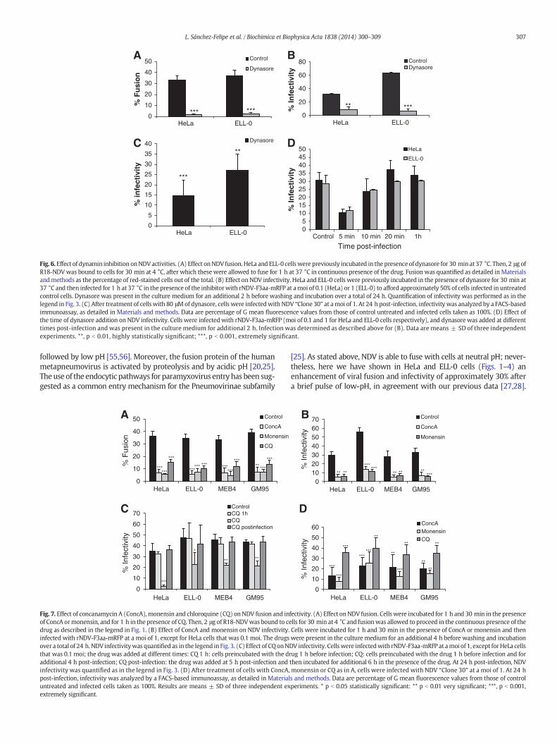
We also analyzed the effect of dynasore, a drug that inhibits theGTPase activity of dynamin 1, which has been implicated in clathrin-and caveola-mediated endocytosis [44]. Owing to the high toxicity ofthis drug in MEB4, GM95, CHO and Lec1 cells, these experiments wereonly performed in HeLa and ELL-0 cells, in which cell viability washigher than 95% after dynasore treatment (data not shown). The fusionof NDV with treated-HeLa and ELL-0 cells was almost completely abro-gated (Fig. 6A), as was infectivity, when assayed both by fluorescencemicroscopy after rNDV-F3aa-mRFP infection (Fig. 6B) and by FACS(Fig. 6C). Nevertheless, the binding of NDV to drug-treated cells wasnot modified (data not shown). However, the addition of dynasore at10 min post-infection resulted in only a minor decrease in infection,with no effect if added at 20 min post-infection (Fig. 6D), implying thatthe main role of dynamin was exerted during the early infection stepsand again supporting a role of endocytosis in NDV entry. The rate of fu-sion and infectivity inhibition exerted by dynasore (more than 80%)
0
20
40
60
80
100
120
Hela ELL-0 MEB4 GM95 CHO Lec1
% in
fect
ivit
y
Control
Virus +BIM
Virus +BIM/BIM
Virus/ BIM
B
A
***
**
Fig. 5. Effect of PKC inhibition on NDV infectivity. Monolayers of cells were treated withthe PKC-inhibitor BIM at different times of the infectivity experiments. Cells were infectedwith rNDV-F3aa-mRFP at a moi of 10 (1 moi for HeLa cells) for 1 h at room temperatureand infectivity was analyzed at 24 h post-infection by calculating the percentage of red-fluorescent cells (infected cells) out of the total number of cells in six random fields.Control, cells without the drug; Virus + BIM: cells were infected in the presence of thedrug and the drug was withdrawn after infection; Virus + BIM/BIM: cells were infectedin the presence of the drug that was also present for the duration of the assays; Virus/BIM: untreated cells were infected and the drug was added 1 h post-infection; BIM: non-infected cells in the presence of the drug. (A)Microphotographs froma representative exper-iment. (B) Data are means ± SD of three independent experiments. **, p b 0.01, highly sta-tistically significant; ***, p b 0.001, extremely significant.
was higher than that of BIM (Fig. 5) and than the observed enhancementof NDV fusion and infectivity after low-pH exposure (Figs. 1–4).Dynamin is essential not only for the fission of endocytic vesicles in theendocytic pathways but also for membrane fusion [45], being involvedin fusion pore expansion [46,47]. Additionally, dynamin has been impli-cated in the regulation of the actin cytoskeleton [48], which could ac-count for the stronger negative effect of dynasore preincubation ofcells on NDV activities. The drug might negatively affect not onlyendocytic entry but also the direct fusion of NDVwith the cell plasmamembrane. In sum, the data regarding the effect of dynasore on NDVfusion and infectivity discussed above indicate that, at least in HeLaand ELL-0 cells, NDV needs dynamin activity, as described for otherviruses such as HIV [14], HPV16 and BPV1 [49].
To prevent endosome acidification, cells were incubated with eitherconcanamycin A, an inhibitor of vacuolar H+-ATPase [50], monensin,an ionophore that disrupts the proton gradient across vesicular mem-branes [51], or chloroquine, a lysosomotropic agent that increases pHinside endocytic vesicles [52]. The concentration and drug treatmenttimes are specified in Table 1. Because of the high toxicity exerted bythe incubation of CHO and Lec1 cells with the three drugs, these exper-iments were performed in HeLa, ELL-0, MEB4 and GM95 cell lines,where treatment did not interfere with cell viability (cell viability wasabout 95% or higher, data not shown). The data concerning the effectof the three different compounds on NDV activities are summarizedin Fig. 7 and in Table 2. The treatment of cells with one of the threecompounds strongly inhibited virus–cell fusion, resulting in an approx-imately 80% reduction in virus–cell fusion after concanamycin A andmonensin treatments, whereas chloroquine preincubation reducedviral fusion by about 60% (Fig. 7A). Similarly, the three drugs stronglyreduced NDV infectivity (Fig. 7B, C, D). The negative effect of chloro-quine was not observed if the drug was withdrawn when the viruswas added (Fig. 7C, CQ 1 h) or was added at 5 h post-infection(Fig. 7C, CQ post-infection), meaning that the negative effect wouldbe exerted at the early steps of the viral cycle. Viral binding to the cellsurfacewas unaffected by any of the three compounds (Table 2). Never-theless, in ELL-0 andHeLa cells fusion and infectivity inhibition by inhib-itors of endosomal acidification (Fig. 7 and Table 2) were stronger thanthe enhancement observed after acid-pH treatment (Figs. 1 and 3). InHeLa and ELL-0 cells, inhibition of virus infectivity resulting frommonensin treatment was largely prevented by exposure to acidic pH(Supplementary Fig. S3). Moreover, concanamycin A, monensin andchloroquine would interfere with processes other than NDV fusion inendosomal compartments. In addition to raising the endosomal pH, ithas been reported that chloroquine has multiple effects on mammaliancells [53]. Chloroquine inhibition of SARS-CoV infection is proposed tobe exerted due to the interference with the terminal glycosylation ofthe viral receptor [54]. In addition, trafficking events other than NDVentry itself might be disturbed, since vesicular ATPase is involved inthe regulation of trafficking events in endosomal compartments.
As an alternative assay of endocytic internalization, we realizedproteinase K protection experiments as detailed in Materials andmethods. Uninternalized virions as well as cell surface glycoproteinsfrom virions that have fused directly with the plasma membranewould be digested by proteinase K, contrary to endocytosed virionsthat became protected from digestion. In ELL-0 cells a significant pro-portion of HN glycoprotein became protected after 6 min of incubationat 37 °C (Fig. 8A),which correlateswith use of an endocytic pathway forvirus entry. We also carried out the experiments with HeLa and MEB4cells (Fig. 8B) looking for HN protection at 10 min after the initiationof entry. Although total digestion of viral glycoproteins was notobserved at 4 °C (Fig. 8B, lanes 6 and 8), after incubation at 37 °C for10 min we detected an increase in the proportion of non-digestedviral HN (Fig. 8B, lanes 5 and 7), strongly supporting that a percentageof virus would also use endocytosis for entering both cell lines.
Class I viral fusion proteins can be activated by at least three differenttriggers: low pH, receptor binding at neutral pH, or receptor binding
0
10
20
30
40
50
HeLa ELL-0
% F
usi
on
Control
Dynasore
A
*** ***0
20
40
60
80
HeLa ELL-0
% In
fect
ivit
y
ControlDynasore
*****
B
D
05
101520253035404550
Control 5 min 10 min 20 min 1h
% In
fect
ivit
y
HeLa
ELL-0
0
5
10
15
20
25
30
35
40
HeLa ELL-0
% in
fect
ivit
y Dynasore
Time post-infection
C
***
**
Fig. 6.Effect of dynamin inhibition onNDVactivities. (A) Effect onNDV fusion.HeLa and ELL-0 cellswere previously incubated in the presenceof dynasore for 30 min at 37 °C. Then, 2 μg ofR18-NDVwas bound to cells for 30 min at 4 °C, after which these were allowed to fuse for 1 h at 37 °C in continuous presence of the drug. Fusion was quantified as detailed in Materialsandmethods as the percentage of red-stained cells out of the total. (B) Effect on NDV infectivity. HeLa and ELL-0 cells were previously incubated in the presence of dynasore for 30 min at37 °C and then infected for 1 h at 37 °C in the presence of the inhibitorwith rNDV-F3aa-mRFP at amoi of 0.1 (HeLa) or 1 (ELL-0) to afford approximately 50% of cells infected in untreatedcontrol cells. Dynasore was present in the culture medium for an additional 2 h before washing and incubation over a total of 24 h. Quantification of infectivity was performed as in thelegend in Fig. 3. (C) After treatment of cells with 80 μMof dynasore, cells were infectedwith NDV “Clone 30” at amoi of 1. At 24 h post-infection, infectivity was analyzed by a FACS-basedimmunoassay, as detailed in Materials and methods. Data are percentage of G mean fluorescence values from those of control untreated and infected cells taken as 100%. (D) Effect ofthe time of dynasore addition on NDV infectivity. Cells were infected with rNDV-F3aa-mRFP (moi of 0.1 and 1 for HeLa and ELL-0 cells respectively), and dynasore was added at differenttimes post-infection and was present in the culture medium for additional 2 h. Infection was determined as described above for (B). Data are means ± SD of three independentexperiments. **, p b 0.01, highly statistically significant; ***, p b 0.001, extremely significant.
307L. Sánchez-Felipe et al. / Biochimica et Biophysica Acta 1838 (2014) 300–309
followed by low pH [55,56]. Moreover, the fusion protein of the humanmetapneumovirus is activated by proteolysis and by acidic pH [20,25].The use of the endocytic pathways for paramyxovirus entry has been sug-gested as a common entry mechanism for the Pneumovirinae subfamily
0
10
20
30
40
50
60
70
HeLa ELL-0 MEB4 GM95
ControlCQ 1hCQCQ postinfection
0
10
20
30
40
50
HeLa ELL-0 MEB4 GM95
Control
ConcA
Monensin
CQ
******
***
****** *** ***
***
***** ***
***
***
*
****
A
C
% F
usio
n %
Infe
ctiv
ity
Fig. 7. Effect of concanamycin A (ConcA), monensin and chloroquine (CQ) on NDV fusion and inof ConcA ormonensin, and for 1 h in the presence of CQ. Then, 2 μg of R18-NDVwas bound to cdrug as described in the legend in Fig. 1. (B) Effect of ConcA and monensin on NDV infectivity.infected with rNDV-F3aa-mRFP at a moi of 1, except for HeLa cells that was 0.1 moi. The drugsover a total of 24 h. NDV infectivitywas quantified as in the legend in Fig. 3. (C) Effect of CQonNthat was 0.1 moi; the drug was added at different times: CQ 1 h: cells preincubated with the dadditional 4 h post-infection; CQ post-infection: the drug was added at 5 h post-infection andinfectivity was quantified as in the legend in Fig. 3. (D) After treatment of cells with ConcA,post-infection, infectivity was analyzed by a FACS-based immunoassay, as detailed in Materialuntreated and infected cells taken as 100%. Results are means ± SD of three independent exextremely significant.
[25]. As stated above, NDV is able to fuse with cells at neutral pH; never-theless, here we have shown in HeLa and ELL-0 cells (Figs. 1–4) anenhancement of viral fusion and infectivity of approximately 30% aftera brief pulse of low-pH, in agreement with our previous data [27,28].
010203040506070
HeLa ELL-0 MEB4 GM95
Control
ConcA
Monensin
** ** ** ** *****
******
0
10
20
30
40
50
60
HeLa ELL-0 MEB4 GM95
ConcAMonensinCQ
***
***
***
******
***
**
**
**
**
**
**
B
D
% In
fect
ivity
%
Infe
ctiv
ity
fectivity. (A) Effect onNDV fusion. Cells were incubated for 1 h and 30 min in the presenceells for 30 min at 4 °C and fusionwas allowed to proceed in the continuous presence of theCells were incubated for 1 h and 30 min in the presence of ConcA or monensin and thenwere present in the culture medium for an additional 4 h before washing and incubationDV infectivity. Cellswere infectedwith rNDV-F3aa-mRFP at amoi of 1, except for HeLa cellsrug 1 h before infection; CQ: cells preincubated with the drug 1 h before infection and forthen incubated for additional 6 h in the presence of the drug. At 24 h post-infection, NDVmonensin or CQ as in A, cells were infected with NDV “Clone 30” at a moi of 1. At 24 hs and methods. Data are percentage of G mean fluorescence values from those of controlperiments. * p b 0.05 statistically significant: ** p b 0.01 very significant; ***, p b 0.001,
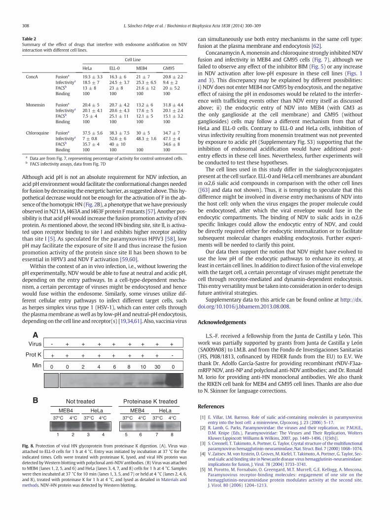
Table 2Summary of the effect of drugs that interfere with endosome acidification on NDVinteraction with different cell lines.
Cell Line
HeLa ELL-0 MEB4 GM95
ConcA Fusiona 19.3 ± 3.3 16.3 ± 6 21 ± 7 20.8 ± 2.2Infectivitya 18.5 ± 7 24.5 ± 3.7 25.3 ± 6.5 9.4 ± 2FACSb 13 ± 8 23 ± 8 21.6 ± 12 20 ± 5.2Binding 100 100 100 100
Monensin Fusiona 20.4 ± 5 20.7 ± 4.2 13.2 ± 6 31.8 ± 4.4Infectivitya 20.1 ± 4.1 20.6 ± 4.3 17.6 ± 5 20.1 ± 2.4FACSb 7.5 ± 4 25.1 ± 11 12.1 ± 5 15.1 ± 3.2Binding 100 100 100 100
Chloroquine Fusiona 37.5 ± 5.6 38.3 ± 7.5 30 ± 5 34.7 ± 7Infectivitya 7 ± 0.8 52.6 ± 6 48.3 ± 1.6 47.1 ± 4FACSb 35.7 ± 4 40 ± 10 34.6 ± 8Binding 100 100 100 100
a Data are from Fig. 7, representing percentage of activity for control untreated cells.b FACS infectivity assays, data from Fig. 7D
308 L. Sánchez-Felipe et al. / Biochimica et Biophysica Acta 1838 (2014) 300–309
Although acid pH is not an absolute requirement for NDV infection, anacid pH environmentwould facilitate the conformational changes neededfor fusionbydecreasing the energetic barrier, as suggested above. This hy-pothetical decrease would not be enough for the activation of F in the ab-sence of the homotypicHN (Fig. 2B), a phenotype thatwehave previouslyobserved inN211A, I463A and I463F protein Fmutants [57]. Another pos-sibility is that acid pHwould increase the fusion promotion activity of HNprotein. As mentioned above, the second HN binding site, site II, is activa-ted upon receptor binding to site I and exhibits higher receptor aviditythan site I [5]. As speculated for the paramyxovirus HPIV3 [58], lowpH may facilitate the exposure of site II and thus increase the fusionpromotion activity of the protein since site II has been shown to beessential in HPIV3 and NDV F activation [59,60].
Within the context of an in vivo infection, i.e., without lowering thepH experimentally, NDVwould be able to fuse at neutral and acidic pH,depending on the entry pathways. In a cell-type-dependent mecha-nism, a certain percentage of viruses might be endocytosed and hencewould fuse within the endosome. Similarly, some viruses utilize dif-ferent cellular entry pathways to infect different target cells, suchas herpes simplex virus type 1 (HSV-1), which can enter cells throughtheplasmamembrane aswell as by low-pH and neutral-pH endocytosis,depending on the cell line and receptor(s) [19,34,61]. Also, vaccinia virus
Not treated Proteinase K treated
MEB4MEB4 HeLaHeLa4°C4°C37°C 37°C
5 6 7 81 2 3 4
4°C4°C37°C 37°C
A
B
Min
Prot K
Virus - + + + + + + + +
+ + + + + + + + -
0 0 2 4 6 8 10 30 0
Fig. 8. Protection of viral HN glycoprotein from proteinase K digestion. (A). Virus wasattached to ELL-0 cells for 1 h at 4 °C. Entry was initiated by incubation at 37 °C for theindicated times. Cells were treated with proteinase K, lysed, and viral HN protein wasdetected byWestern blottingwith polyclonal anti-NDV antibodies. (B) Viruswas attachedto MEB4 (lanes 1, 2, 5, and 6) and HeLa (lanes 3, 4, 7, and 8) cells for 1 h at 4 °C. Sampleswere then incubated at 37 °C for 10 min (lanes 1, 3, 5, and 7) or held at 4 °C (lanes 2, 4, 6,and 8), treated with proteinase K for 1 h at 4 °C, and lysed as detailed in Materials andmethods. NDV-HN protein was detected by Western blotting.
can simultaneously use both entry mechanisms in the same cell type:fusion at the plasma membrane and endocytosis [62].
Concanamycin A,monensin and chloroquine strongly inhibited NDVfusion and infectivity in MEB4 and GM95 cells (Fig. 7), although wefailed to observe any effect of the inhibitor BIM (Fig. 5) or any increasein NDV activation after low-pH exposure in these cell lines (Figs. 1and 3). This discrepancy may be explained by different possibilities:i) NDV does not enterMEB4 nor GM95 by endocytosis, and the negativeeffect of raising the pH in endosomes would be related to the interfer-ence with trafficking events other than NDV entry itself as discussedabove; ii) the endocytic entry of NDV into MEB4 (with GM3 asthe only ganglioside at the cell membrane) and GM95 (withoutgangliosides) cells may follow a different mechanism from that ofHeLa and ELL-0 cells. Contrary to ELL-0 and HeLa cells, inhibition ofvirus infectivity resulting from monensin treatment was not preventedby exposure to acidic pH (Supplementary Fig. S3) supporting that theinhibition of endosomal acidification would have additional post-entry effects in these cell lines. Nevertheless, further experiments willbe conducted to test these hypotheses.
The cell lines used in this study differ in the sialoglycoconjugatespresent at the cell surface. ELL-0 andHeLa cellmembranes are abundantin α2,6 sialic acid compounds in comparison with the other cell lines([63] and data not shown). Thus, it is tempting to speculate that thisdifference might be involved in diverse entry mechanisms of NDV intothe host cell: only when the virus engages the proper molecule couldbe endocytosed, after which the viral envelope would fuse in theendocytic compartments. The binding of NDV to sialic acids in α2,6specific linkages could allow the endocytic entry of NDV, and couldbe directly required either for endocytic internalization or to facilitatesubsequent molecular changes enabling endocytosis. Further experi-ments will be needed to clarify this point.
Our data then support the notion that NDV might have evolved touse the low pH of the endocytic pathways to enhance its entry, atleast in certain cell lines. In addition to direct fusion of the viral envelopewith the target cell, a certain percentage of viruses might penetrate thecell through receptor-mediated and dynamin-dependent endocytosis.This entry versatilitymust be taken into consideration in order to designfuture antiviral strategies.
Supplementary data to this article can be found online at http://dx.doi.org/10.1016/j.bbamem.2013.08.008.
Acknowledgements
L.S.-F. received a fellowship from the Junta de Castilla y León. Thiswork was partially supported by grants from Junta de Castilla y León(SA009A08) to I.M.B. and from the Fondo de Investigaciones Sanitarias(FIS, PI08/1813, cofinanced by FEDER funds from the EU) to E.V. Wethank Dr. Adolfo García-Sastre for providing recombinant rNDV-F3aa-mRFP NDV, anti-NP and polyclonal anti-NDV antibodies; andDr. RonaldM. Iorio for providing anti-HN monoclonal antibodies. We also thankthe RIKEN cell bank for MEB4 and GM95 cell lines. Thanks are also dueto N. Skinner for language corrections.
References
[1] E. Villar, I.M. Barroso, Role of sialic acid-containing molecules in paramyxovirusentry into the host cell: a minireview, Glycoconj. J. 23 (2006) 5–17.
[2] R. Lamb, G. Parks, Paramyxoviridae: the viruses and their replication, in: P.M.H.E.,D.M. Knipe (Eds.), Paramyxoviridae: The Viruses and Their Replication, WoltersKluwer/Lippincott Williams & Wilkins, 2007, pp. 1449–1496, (1[5th]).
[3] S. Crennell, T. Takimoto, A. Portner, G. Taylor, Crystal structure of themultifunctionalparamyxovirus hemagglutinin-neuraminidase, Nat. Struct. Biol. 7 (2000) 1068–1074.
[4] V. Zaitsev, M. von Itzstein, D. Groves, M. Kiefel, T. Takimoto, A. Portner, G. Taylor, Sec-ond sialic acid binding site inNewcastle disease virus hemagglutinin-neuraminidase:implications for fusion, J. Virol. 78 (2004) 3733–3741.
[5] M. Porotto, M. Fornabaio, O. Greengard, M.T. Murrell, G.E. Kellogg, A. Moscona,Paramyxovirus receptor-binding molecules: engagement of one site on thehemagglutinin-neuraminidase protein modulates activity at the second site,J. Virol. 80 (2006) 1204–1213.

309L. Sánchez-Felipe et al. / Biochimica et Biophysica Acta 1838 (2014) 300–309
[6] L. Pelkmans, T. Burli, M. Zerial, A. Helenius, Caveolin-stabilized membrane domainsas multifunctional transport and sorting devices in endocytic membrane traffic, Cell118 (2004) 767–780.
[7] K. Chandran, N.J. Sullivan, U. Felbor, S.P. Whelan, J.M. Cunningham, Endosomalproteolysis of the Ebola virus glycoprotein is necessary for infection, Science 308(2005) 1643–1645.
[8] F.L. Cosset, D. Lavillette, Cell entry of enveloped viruses, Adv. Genet. 73 (2011) 121–183.[9] M. Marsh, A. Helenius, Virus entry: open sesame, Cell 124 (2006) 729–740.
[10] J. Mercer, A. Helenius, Virus entry by macropinocytosis, Nat. Cell Biol. 11 (2009)510–520.
[11] J. Mercer, M. Schelhaas, A. Helenius, Virus entry by endocytosis, Annu. Rev. Biochem.79 (2010) 803–833.
[12] A. Helenius, J. Kartenbeck, K. Simons, E. Fries, On the entry of Semliki forest virusinto BHK-21 cells, J. Cell Biol. 84 (1980) 404–420.
[13] M. Jin, J. Park, S. Lee, B. Park, J. Shin, K.J. Song, T.I. Ahn, S.Y. Hwang, B.Y. Ahn, K. Ahn,Hantaan virus enters cells by clathrin-dependent receptor-mediated endocytosis,Virology 294 (2002) 60–69.
[14] K. Miyauchi, Y. Kim, O. Latinovic, V. Morozov, G.B. Melikyan, HIV enters cells viaendocytosis and dynamin-dependent fusion with endosomes, Cell 137 (2009)433–444.
[15] H.A. Anderson, Y. Chen, L.C. Norkin, Bound simian virus 40 translocates tocaveolin-enriched membrane domains, and its entry is inhibited by drugs thatselectively disrupt caveolae, Mol. Biol. Cell 7 (1996) 1825–1834.
[16] L. Pelkmans, D. Puntener, A. Helenius, Local actin polymerization and dynaminrecruitment in SV40-induced internalization of caveolae, Science 296 (2002) 535–539.
[17] C.J. Empig, M.A. Goldsmith, Association of the caveola vesicular systemwith cellularentry by filoviruses, J. Virol. 76 (2002) 5266–5270.
[18] A. Sanchez, Analysis of filovirus entry into vero e6 cells, using inhibitors of endocy-tosis, endosomal acidification, structural integrity, and cathepsin (B and L) activity,J. Infect. Dis. 196 (Suppl. 2) (2007) S251–S258.
[19] E. Rahn, P. Petermann, M.J. Hsu, F.J. Rixon, D. Knebel-Morsdorf, Entry pathwaysof herpes simplex virus type 1 into human keratinocytes are dynamin- andcholesterol-dependent, PLoS One 6 (2011) e25464.
[20] R.M. Schowalter, S.E. Smith, R.E. Dutch, Characterization of human metapneumovirusF protein-promotedmembrane fusion: critical roles for proteolytic processing and lowpH, J. Virol. 80 (2006) 10931–10941.
[21] E.C. Smith, A. Popa, A. Chang, C. Masante, R.E. Dutch, Viral entry mechanisms: theincreasing diversity of paramyxovirus entry, FEBS J. 276 (2009) 7217–7227.
[22] A. Chang, R.E. Dutch, Paramyxovirus fusion and entry: multiple paths to a commonend, Viruses 4 (2012) 613–636.
[23] S. Diederich, L. Thiel, A.Maisner, Role of endocytosis and cathepsin-mediated activationin Nipah virus entry, Virology 375 (2008) 391–400.
[24] O. Pernet, C. Pohl, M. Ainouze, H. Kweder, R. Buckland, Nipah virus entry can occurby macropinocytosis, Virology 395 (2009) 298–311.
[25] R.M. Schowalter, A. Chang, J.G. Robach, U.J. Buchholz, R.E. Dutch, Low-pH triggeringof human metapneumovirus fusion: essential residues and importance in entry,J. Virol. 83 (2009) 1511–1522.
[26] A.A. Kolokoltsov, D. Deniger, E.H. Fleming, N.J. Roberts Jr., J.M. Karpilow, R.A. Davey,Small interfering RNA profiling reveals key role of clathrin-mediated endocytosisand early endosome formation for infection by respiratory syncytial virus, J. Virol.81 (2007) 7786–7800.
[27] K. San Roman, E. Villar, I. Munoz-Barroso, Acidic pH enhancement of the fusion ofNewcastle disease virus with cultured cells, Virology 260 (1999) 329–341.
[28] C. Cantin, J. Holguera, L. Ferreira, E. Villar, I. Munoz-Barroso, Newcastle disease virusmay enter cells by caveolae-mediated endocytosis, J. Gen. Virol. 88 (2007) 559–569.
[29] M.Mibayashi, L.Martinez-Sobrido, Y.M. Loo,W.B. Cardenas,M. Gale Jr., A. García-Sastre,Inhibition of retinoic acid-inducible gene I-mediated induction of beta interferon by theNS1 protein of influenza A virus, J. Virol. 81 (2007) 514–524.
[30] R.E. Campbell, O. Tour, A.E. Palmer, P.A. Steinbach, G.S. Baird, D.A. Zacharias, R.Y.Tsien, A monomeric red fluorescent protein, Proc. Natl. Acad. Sci. U. S. A. 99 (2002)7877–7882.
[31] K. San Roman, E. Villar, I. Munoz-Barroso, Mode of action of two inhibitory peptidesfrom heptad repeat domains of the fusion protein of Newcastle disease virus, Int.J. Biochem. Cell Biol. 34 (2002) 1207–1220.
[32] S.A. Connolly, R.A. Lamb, Paramyxovirus fusion: real-time measurement ofparainfluenza virus 5 virus–cell fusion, Virology 355 (2006) 203–212.
[33] J.K. Young, D. Li, M.C. Abramowitz, T.G. Morrison, Interaction of peptides withsequences from the Newcastle disease virus fusion protein heptad repeat regions,J. Virol. 73 (1999) 5945–5956.
[34] R.S. Milne, A.V. Nicola, J.C. Whitbeck, R.J. Eisenberg, G.H. Cohen, Glycoprotein Dreceptor-dependent, low-pH-independent endocytic entry of herpes simplex virustype 1, J. Virol. 79 (2005) 6655–6663.
[35] S. Ichikawa, N. Nakajo, H. Sakiyama, Y. Hirabayashi, A mouse B16 melanomamutantdeficient in glycolipids, Proc. Natl. Acad. Sci. U. S. A. 91 (1994) 2703–2707.
[36] V.C. Chu, G.R. Whittaker, Influenza virus entry and infection require host cellN-linked glycoprotein, Proc. Natl. Acad. Sci. U. S. A. 101 (2004) 18153–18158.
[37] D. Toullec, P. Pianetti, H. Coste, P. Bellevergue, T. Grand-Perret, M. Ajakane, V.Baudet, P. Boissin, E. Boursier, F. Loriolle, et al., The bisindolylmaleimide GF109203X is a potent and selective inhibitor of protein kinase C, J. Biol. Chem. 266(1991) 15771–15781.
[38] C.N. Root, E.G. Wills, L.L. McNair, G.R. Whittaker, Entry of influenza viruses into cellsis inhibited by a highly specific protein kinase C inhibitor, J. Gen. Virol. 81 (2000)2697–2705.
[39] P. Upla, V. Marjomaki, P. Kankaanpaa, J. Ivaska, T. Hyypia, F.G. Van Der Goot, J. Heino,Clustering induces a lateral redistribution of alpha 2 beta 1 integrin frommembranerafts to caveolae and subsequent protein kinase C-dependent internalization, Mol.Biol. Cell 15 (2004) 625–636.
[40] S.N. Constantinescu, C.D. Cernescu, L.M. Popescu, Effects of protein kinase C inhibitorson viral entry and infectivity, FEBS Lett. 292 (1991) 31–33.
[41] J.J. Martin, J. Holguera, L. Sanchez-Felipe, E. Villar, I. Munoz-Barroso, Cholesterol depen-dence of Newcastle Disease Virus entry, Biochim. Biophys. Acta 1818 (2012) 753–761.
[42] E.J. Smart, D.C. Foster, Y.S. Ying, B.A. Kamen, R.G. Anderson, Protein kinase C activa-tors inhibit receptor-mediated potocytosis by preventing internalization of caveolae,J. Cell Biol. 124 (1994) 307–313.
[43] C. Mineo, Y.S. Ying, C. Chapline, S. Jaken, R.G. Anderson, Targeting of protein kinase Calpha to caveolae, J. Cell Biol. 141 (1998) 601–610.
[44] E. Macia, M. Ehrlich, R. Massol, E. Boucrot, C. Brunner, T. Kirchhausen, Dynasore,a cell-permeable inhibitor of dynamin, Dev. Cell 10 (2006) 839–850.
[45] H.H. Low, J. Lowe, Dynamin architecture—from monomer to polymer, Curr. Opin.Struct. Biol. 20 (2010) 791–798.
[46] J.K. Jaiswal, V.M. Rivera, S.M. Simon, Exocytosis of post-Golgi vesicles is regulated bycomponents of the endocytic machinery, Cell 137 (2009) 1308–1319.
[47] A. Anantharam, M.A. Bittner, R.L. Aikman, E.L. Stuenkel, S.L. Schmid, D. Axelrod, R.W.Holz, A new role for the dynamin GTPase in the regulation of fusion pore expansion,Mol. Biol. Cell 22 (2011) 1907–1918.
[48] C. Gu, S. Yaddanapudi, A. Weins, T. Osborn, J. Reiser, M. Pollak, J. Hartwig, S. Sever,Direct dynamin–actin interactions regulate the actin cytoskeleton, EMBO J. 29(2010) 3593–3606.
[49] C.Y. Abban, N.A. Bradbury, P.I. Meneses, HPV16 and BPV1 infection can be blockedby the dynamin inhibitor dynasore, Am. J. Ther. 15 (2008) 304–311.
[50] S. Drose, K. Altendorf, Bafilomycins and concanamycins as inhibitors of V-ATPasesand P-ATPases, J. Exp. Biol. 200 (1997) 1–8.
[51] F.R. Maxfield, Weak bases and ionophores rapidly and reversibly raise the pH ofendocytic vesicles in cultured mouse fibroblasts, J. Cell Biol. 95 (1982) 676–681.
[52] C. de Duve, T. de Barsy, B. Poole, A. Trouet, P. Tulkens, F. Van Hoof, Commentary.Lysosomotropic agents, Biochem. Pharmacol. 23 (1974) 2495–2531.
[53] B. Thorens, P. Vassalli, Chloroquine and ammonium chloride prevent terminal glyco-sylation of immunoglobulins in plasma cells without affecting secretion, Nature 321(1986) 618–620.
[54] M.J. Vincent, E. Bergeron, S. Benjannet, B.R. Erickson, P.E. Rollin, T.G. Ksiazek, N.G.Seidah, S.T. Nichol, Chloroquine is a potent inhibitor of SARS coronavirus infectionand spread, Virol. J. 2 (2005) 69.
[55] L.J. Earp, S.E. Delos, H.E. Park, J.M. White, The many mechanisms of viral membranefusion proteins, Curr. Top. Microbiol. Immunol. 285 (2005) 25–66.
[56] J.M.White, S.E. Delos, M. Brecher, K. Schornberg, Structures andmechanisms of viralmembrane fusion proteins: multiple variations on a common theme, Crit. Rev.Biochem. Mol. Biol. 43 (2008) 189–219.
[57] J. Ayllon, E. Villar, I. Munoz-Barroso, Mutations in the ectodomain of Newcastledisease virus fusion protein confer a hemagglutinin-neuraminidase-independentphenotype, J. Virol. 84 (2010) 1066–1075.
[58] L.M. Palermo, M. Porotto, O. Greengard, A. Moscona, Fusion promotion by aparamyxovirus hemagglutinin-neuraminidase protein: pH modulation of receptoravidity of binding sites I and II, J. Virol. 81 (2007) 9152–9161.
[59] M. Porotto, S.G. Palmer, L.M. Palermo, A. Moscona, Mechanism of fusion triggeringby human parainfluenza virus type III: communication between viral glycoproteinsduring entry, J. Biol. Chem. 287 (2012) 778–793.
[60] M. Porotto, Z. Salah, I. DeVito, A. Talekar, S.G. Palmer, R. Xu, I.A. Wilson, A. Moscona,The second receptor binding site of the globular head of the Newcastle diseasevirus hemagglutinin-neuraminidase activates the stalk of multiple paramyxovirusreceptor binding proteins to trigger fusion, J. Virol. 86 (2012) 5730–5741.
[61] A.V. Nicola, A.M. McEvoy, S.E. Straus, Roles for endocytosis and low pH in herpessimplex virus entry into HeLa and Chinese hamster ovary cells, J. Virol. 77 (2003)5324–5332.
[62] A.C. Townsley, A.S. Weisberg, T.R. Wagenaar, B. Moss, Vaccinia virus entry into cellsvia a low-pH-dependent endosomal pathway, J. Virol. 80 (2006) 8899–8908.
[63] L. Sanchez-Felipe, E. Villar, I. Munoz-Barroso, Alpha2-3- and alpha2-6-N-linkedsialic acids allow efficient interaction of Newcastle Disease Virus with target cells,Glycoconj. J. 29 (2012) 539–549.

![Biochimica et Biophysica Acta - immed.org considerations/09.07.2017 updates/Membrane... · G.L. Nicolson, M.E. Ash / Biochimica et Biophysica Acta 1859 (2017) 1704–1724 1705 [8].](https://static.fdocuments.net/doc/165x107/5c684f1e09d3f2f5638b5509/biochimica-et-biophysica-acta-immed-considerations09072017-updatesmembrane.jpg)