Biochimica et Biophysica Acta - Stanford Universityweb.stanford.edu/group/heilshorn/Manuscript...
11
Review Protein-engineered biomaterials: Nanoscale mimics of the extracellular matrix ☆ Nicole H. Romano a , Debanti Sengupta b , Cindy Chung a , Sarah C. Heilshorn a, ⁎ a Department of Materials Science and Engineering, Stanford University, Stanford, CA 94305-4045, USA b Department of Chemistry, Stanford University, Stanford, CA 94305-4045, USA abstract article info Article history: Received 23 December 2009 Received in revised form 7 July 2010 Accepted 9 July 2010 Available online 18 July 2010 Keywords: Biomaterial Protein engineering Stem cell niche Extracellular matrix Tissue engineering Regenerative medicine Background: Traditional materials used as in vitro cell culture substrates are rigid and flat surfaces that lack the exquisite nano- and micro-scale features of the in vivo extracellular environment. While these surfaces can be coated with harvested extracellular matrix (ECM) proteins to partially recapitulate the bio-instructive nature of the ECM, these harvested proteins often exhibit large batch-to-batch variability and can be difficult to customize for specific biological studies. In contrast, recombinant protein technology can be utilized to synthesize families of 3 dimensional protein-engineered biomaterials that are cyto-compatible, reproducible, and fully customizable. Scope of Review: Here we describe a modular design strategy to synthesize protein-engineered biomaterials that fuse together multiple repeats of nanoscale peptide design motifs into full-length engineered ECM mimics. Major Conclusions: Due to the molecular-level precision of recombinant protein synthesis, these biomaterials can be tailored to include a variety of bio-instructional ligands at specified densities, to exhibit mechanical properties that match those of native tissue, and to include proteolytic target sites that enable cell-triggered scaffold remodeling. Furthermore, these biomaterials can be processed into forms that are injectable for minimally-invasive delivery or spatially patterned to enable the release of multiple drugs with distinct release kinetics. General significance: Given the reproducibility and flexibility of these protein-engineered biomaterials, they are ideal substrates for reductionist biological studies of cell–matrix interactions, for in vitro models of physiological processes, and for bio-instructive scaffolds in regenerative medicine therapies. This article is part of a Special Issue entitled Nanotechnologies - Emerging Applications in Biomedicine. © 2010 Elsevier B.V. All rights reserved. 1. Introduction: Moving cell culture into the third dimension The vast majority of in vitro mammalian cell culture studies are performed on flat, rigid substrates (most often tissue culture polystyrene (TCPS) or glass in the form of Petri dishes and microscope slides) that do little to mimic the exquisite three-dimensional (3D) nano- and micro-environments found in vivo. In the past, TCPS and glass have been used because they are inexpensive, highly reproduc- ible, cell-permissive for many cell types, and optically transparent. However, numerous recent studies have highlighted the importance of nano- and micro-scale structure [1], substrate mechanics [2,3], and 3D culture environments [4] in regulating cell adhesion, morphology, migration, signaling, and differentiation [5–7]. Therefore, in order to recreate physiologically relevant cell behavior in an artificial in vitro environment, it is imperative to design new 3D culture substrates that more accurately mimic the extracellular matrix (ECM). The ideal culture substrate would possess: (i) nano- and micro-scale repro- ducibility, (ii) flexibility of design, and (iii) the potential to be directly translated from lab-bench studies to clinical therapies. Here we describe recent efforts by our laboratory and others to address these goals through the molecular-level design of novel protein-based biomaterials, Table 1. These materials are completely constructed from engineered recombinant proteins that are designed to mimic many of the essential properties of natural ECM. Because these materials are synthesized by host organisms through precise translation of a genetic template, the resulting materials are highly reproducible at the molecular level. The genetic template is constructed in a modular fashion that enables easy customization of the engineered protein sequence and hence tailoring of matrix properties such as mechanical rigidity and cell adhesion [8–11]. Precise tuning of these matrix properties has been shown to influence cell behavior, including morphology, migration, gene regulation, intracellular signaling, and differentiation [12–14]. Therefore, these materials have wide poten- tial for use in reductionist in vitro studies of cell–matrix interactions, development of in vitro platforms that recapitulate complex in vivo Biochimica et Biophysica Acta 1810 (2011) 339–349 ☆ This article is part of a Special Issue entitled Nanotechnologies - Emerging Applications in Biomedicine. ⁎ Corresponding author. 476 Lomita Mall, McCullough 246, Stanford University, Stanford, CA 94305-4045, USA. Tel.: +1 650 723 3763: fax: +1 650 498 5596. E-mail address: [email protected] (S.C. Heilshorn). 0304-4165/$ – see front matter © 2010 Elsevier B.V. All rights reserved. doi:10.1016/j.bbagen.2010.07.005 Contents lists available at ScienceDirect Biochimica et Biophysica Acta journal homepage: www.elsevier.com/locate/bbagen
Transcript of Biochimica et Biophysica Acta - Stanford Universityweb.stanford.edu/group/heilshorn/Manuscript...
Biochimica et Biophysica Acta 1810 (2011) 339–349
Contents lists available at ScienceDirect
Biochimica et Biophysica Acta
j ourna l homepage: www.e lsev ie r.com/ locate /bbagen
Review
Protein-engineered biomaterials: Nanoscale mimics of the extracellular matrix☆
Nicole H. Romano a, Debanti Sengupta b, Cindy Chung a, Sarah C. Heilshorn a,⁎a Department of Materials Science and Engineering, Stanford University, Stanford, CA 94305-4045, USAb Department of Chemistry, Stanford University, Stanford, CA 94305-4045, USA
☆ This article is part of a Special Issue entitled NApplications in Biomedicine.⁎ Corresponding author. 476 Lomita Mall, McCullou
Stanford, CA 94305-4045, USA. Tel.: +1 650 723 3763:E-mail address: [email protected] (S.C. Heilsh
0304-4165/$ – see front matter © 2010 Elsevier B.V. Aldoi:10.1016/j.bbagen.2010.07.005
a b s t r a c t
a r t i c l e i n f oArticle history:
Received 23 December 2009Received in revised form 7 July 2010Accepted 9 July 2010Available online 18 July 2010Keywords:BiomaterialProtein engineeringStem cell nicheExtracellular matrixTissue engineeringRegenerative medicine
Background: Traditional materials used as in vitro cell culture substrates are rigid and flat surfaces that lackthe exquisite nano- and micro-scale features of the in vivo extracellular environment. While these surfacescan be coated with harvested extracellular matrix (ECM) proteins to partially recapitulate the bio-instructivenature of the ECM, these harvested proteins often exhibit large batch-to-batch variability and can be difficultto customize for specific biological studies. In contrast, recombinant protein technology can be utilized tosynthesize families of 3 dimensional protein-engineered biomaterials that are cyto-compatible, reproducible,and fully customizable.Scope of Review: Here we describe a modular design strategy to synthesize protein-engineered biomaterialsthat fuse together multiple repeats of nanoscale peptide design motifs into full-length engineered ECMmimics.Major Conclusions: Due to the molecular-level precision of recombinant protein synthesis, these biomaterialscan be tailored to include a variety of bio-instructional ligands at specified densities, to exhibit mechanical
properties that match those of native tissue, and to include proteolytic target sites that enable cell-triggeredscaffold remodeling. Furthermore, these biomaterials can be processed into forms that are injectable forminimally-invasive delivery or spatially patterned to enable the release of multiple drugs with distinctrelease kinetics.General significance: Given the reproducibility and flexibility of these protein-engineered biomaterials, theyare ideal substrates for reductionist biological studies of cell–matrix interactions, for in vitro models ofphysiological processes, and for bio-instructive scaffolds in regenerative medicine therapies.This article is part of a Special Issue entitled Nanotechnologies - Emerging Applications in Biomedicine.© 2010 Elsevier B.V. All rights reserved.
1. Introduction: Moving cell culture into the third dimension
The vast majority of in vitro mammalian cell culture studies areperformed on flat, rigid substrates (most often tissue culturepolystyrene (TCPS) or glass in the form of Petri dishes andmicroscopeslides) that do little to mimic the exquisite three-dimensional (3D)nano- and micro-environments found in vivo. In the past, TCPS andglass have been used because they are inexpensive, highly reproduc-ible, cell-permissive for many cell types, and optically transparent.However, numerous recent studies have highlighted the importanceof nano- and micro-scale structure [1], substrate mechanics [2,3], and3D culture environments [4] in regulating cell adhesion, morphology,migration, signaling, and differentiation [5–7]. Therefore, in order torecreate physiologically relevant cell behavior in an artificial in vitroenvironment, it is imperative to design new 3D culture substrates that
anotechnologies - Emerging
gh 246, Stanford University,fax: +1 650 498 5596.orn).
l rights reserved.
more accurately mimic the extracellular matrix (ECM). The idealculture substrate would possess: (i) nano- and micro-scale repro-ducibility, (ii) flexibility of design, and (iii) the potential to be directlytranslated from lab-bench studies to clinical therapies. Here wedescribe recent efforts by our laboratory and others to address thesegoals through the molecular-level design of novel protein-basedbiomaterials, Table 1.
These materials are completely constructed from engineeredrecombinant proteins that are designed to mimic many of theessential properties of natural ECM. Because these materials aresynthesized by host organisms through precise translation of a genetictemplate, the resulting materials are highly reproducible at themolecular level. The genetic template is constructed in a modularfashion that enables easy customization of the engineered proteinsequence and hence tailoring of matrix properties such as mechanicalrigidity and cell adhesion [8–11]. Precise tuning of these matrixproperties has been shown to influence cell behavior, includingmorphology, migration, gene regulation, intracellular signaling, anddifferentiation [12–14]. Therefore, these materials have wide poten-tial for use in reductionist in vitro studies of cell–matrix interactions,development of in vitro platforms that recapitulate complex in vivo

Table 1Comparison of various cell culture substrates and matrices.
Traditional surfaces Naturally derived gels Designed gels
Petri-dish/coverslip
Matrigel/collagen/fibrin
EngineeredECM
Dimensionality 2D 2D or 3D 2D or 3DReproducibility High Variable HighMechanicalproperties
Predetermined Low, poorreproducibility
Tunable
Adhesion ligands Requires coating Yes, predetermined Yes, tunableMatrix degradation No Yes, predetermined Yes, tunableFibrous network No Physiological Potentially
tunableCommercialavailability
Yes Yes No
Clinic translatability No Potential Potential
340 N.H. Romano et al. / Biochimica et Biophysica Acta 1810 (2011) 339–349
processes, and scaffolds for potential regenerative medicine therapies.In the following four sections, we will (i) describe the design conceptsused to synthesize these materials and then give specific examplesof how these materials can be used to further (ii) mechanisticunderstanding of cell–matrix interactions, (iii) development of invitro models of physiological processes, and (iv) advancement ofregenerative medicine therapies.
2. Designing protein-engineered mimics of ECM
Traditionally, when choosing a cell-culture substrate, the scientistmust decide between a natural or synthetic material, Table 1. Tissueculture polystyrene, often modified by coating with natural ECMproteins, tends to be the most popular culture substrate. But as cellstudies move toward 3D culture, naturally derived scaffolds are oftenchosen because they are commercially available in a convenientpowder form that can be reconstituted into a 3D hydrogel. Sincenatural materials such as Matrigel and collagen are derived directly
x
y
MIXING-INDUCED TWO-COMPONENT HYDROGEL
Component 1
Component 2
+
CHEMICALLY-CROSSLINKED ELASTIN-LIKE HYDROGEL
Elastin-like protein
Crosslinker
+
4
Fig. 1. Schematic of the modular design strategy used to create two families of protein-engrepeats of elastin-like modules, cell adhesion modules, and protease degradation modulesbonding between a crosslinker and multiple lysine amino acid residues on neighboring prorepeating peptide sequences that hetero-assemble [23]. The engineered proteins form a physPPxY domains on neighboring protein chains.
from mammalian sources, they offer physiologically relevant chem-istries and bio-functionalities. On the other hand, their biologicalorigins also impart an inflexibility of design; natural materials act as aone-size-fits-all system that cannot be easily customized. Further-more, the processing of commercially available natural materialsoften destroys higher order structures such as fibrils, can have largebatch-to-batch variations, andmay initiate immunogenic responses inin vivo studies [15]. It is for these reasons that synthetic polymerichydrogels, such as poly(ethylene glycol) (PEG) and poly(acrylamide)derivatives, were introduced as cell scaffolds [16–20]. While thesematerials are highly reproducible and customizable, they often lackthe nano- and micro-scale biological motifs that direct cell behaviorand must be carefully screened for potential cytotoxicity. Comparingnatural and synthetic materials, the former provides the advantage ofa highly biomimetic structure and chemistry, while the latter supportsreproducibility and customization.
In order to combine the advantages of natural and syntheticmaterials, engineered proteins can be designed for use as cell culturesubstrates that are biomimetic, reproducible, and customizable(Fig. 1). Through careful selection of the primary amino acid sequence,the resulting engineered ECM-mimetic proteins can be customized tohave the desired physical structures, biomechanical properties, andbiochemical properties for a particular application. In addition, sincethese proteins are based on the 20 canonical amino acids found inbiological systems, they are inherently cyto-compatible. The primaryamino acid sequence is designed by choosing shorter peptidemodulesthat are known to elicit a specific biological response and/or fold into aspecific physical structure. These peptide modules can be derivedfrom naturally occurring protein sequences [21], selected throughhigh-throughput screening of random sequences [8,22], or predictedthrough computational methods [23,24]. While engineered proteinsequences can also be fabricated using solid-phase synthetic chem-istry techniques, recombinant protein expression that utilizes thetranslational machinery of a host organism allows unparalleledmolecular-level control over the primary amino acid sequence [25].
WW Domain
PPxY Domain
RGDS + Spacer
Spacer
Bioactive Domain: Cell Adhesion
Bioactive Domain: Degradation
Chemical Crosslinker
Elastin-Like Domain
ineered biomaterials. Top: A chemically crosslinked hydrogel fabricated from multiple[8,53]. The engineered proteins form a chemical hydrogel network through covalenttein chains. Bottom: A mixing-induced two-component hydrogel fabricated from twoical hydrogel network through transient hydrogen bonding betweenWWdomains and

2D 3D
Expression
Purification
Transfection
Cloning
Design
Evaluation
E. coli
Fig. 2. Flow chart showing the sequence of experimental steps used to fabricate a protein-engineered biomaterial. Once a sequence of repetitive peptide modules is designed, theprimary amino acid sequence is encoded in a recombinant gene. Solid-state oligonucleotidesynthesis and molecular biology cloning are used to create a plasmid harboring therecombinant gene. The plasmid is transfected into the host of choice, often E. coli. Thebiosynthetic machinery of the host translates the genetic message into an expressedengineered protein. The target protein is purified away from the host contaminants; forexample, differential solubility induced by temperature cycling is often used to purifyelastin-like proteins [8]. The proteins are processed into a suitable scaffold throughchemicalorphysical crosslinking. The scaffolds canbeused for both2Dand3Dcell culture techniques.
341N.H. Romano et al. / Biochimica et Biophysica Acta 1810 (2011) 339–349
Through careful design of a flexible recombinant cloning strategy,each desired peptide module can be encoded in a specific oligonucle-otide that serves as amolecular building block (Fig. 2). Oligonucleotides
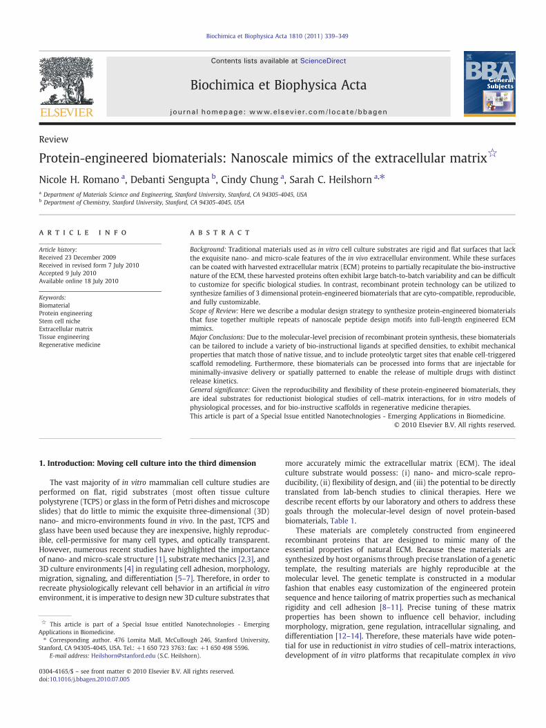
Fig. 3. Images of an elastin-like, protein-engineered biomaterial. (A) Photograph of a chmicrograph of human umbilical vein endothelial cells (HUVEC) growing within a 3D enviro
encoding the selected peptide modules are then spliced together tocreate a repetitive gene that encodes the engineered ECM-mimeticprotein. Several genetic building blocks can be mixed and matchedtogether to design multiple related recombinant genes that encode afamily of customized ECM-mimetic proteins with tailored scaffoldproperties. Once a recombinant gene encoding the desired primaryamino acid sequence is synthesized using traditional molecular biologyprotocols, the resulting plasmid is transformed into a carrier host.Escherichia coli bacteria are often chosen ashost organisms because theyare robust, divide rapidly, and require inexpensive fermentation media.The E. coli bacteria translate the genetic template into an exact primaryamino acid sequence. The newly synthesized engineered protein can bepurified using a variety of methods such as affinity chromatography[23], differential solubility [26], and size-exclusion chromatography,depending on the properties of the designed sequence. Thesetechniques can be optimized to yield significant amounts of purifiedengineered protein (e.g., up to 1.6 g/L) [27] with FDA-acceptable levelsof bacterial contaminants (e.g., as low as 0.065–0.115 endotoxin units/mg protein) [28], adequate for the formation of bulk cell culturesubstrates (Fig. 3).
Integrating concepts from biochemistry, molecular biology, andpolymer physics, our group has designed two ECM-mimetic proteinfamilies using this modular protein-engineering strategy (Fig. 1). Themodules chosen in our protein designs include sequences to initiatecell adhesion (e.g., integrin-binding peptides), sequences to conferresilience (e.g., elastin-like peptides), sequences to promote physicalcrosslinking (e.g., association peptides), and sequences to enablescaffold degradation (e.g., proteolytic target sites) [8,10,11,23,29,30].By utilizing these nanoscale design motifs in various combinations atdefined ratios, we can independently customize the properties of theresulting substrates, such as matrix stiffness, cell adhesivity, andproteolytic degradation. Within the field of engineered protein-basedbiomaterials, a large library of nanoscale peptide design motifs hasbeen explored [21,31–34]. These include motifs to induce minerali-zation [35] and signaling [36] as well as structural modules such assilk [37], collagen [38,39], and coiled-coil [40] peptides. Despite thislarge body of work, the amazing diversity of evolved proteinstructures represents an immense potential to design new protein-engineered biomaterials with novel functionalities.
In the following three sections, we will describe three potentialapplication areas where protein-engineered biomaterials are able toenhance biological andmedical research: fundamental studies of cell–matrix interactions, in vitro models of complex physiologicalphenomena, and development of scaffolds for regenerative medicine.For each section, we provide case studies using our own proteindesigns; however, the reader is also directed to additional examplescontained within the following recent reviews [41–46]. In addition tothe full-length protein-engineered materials described in this review,
emically crosslinked scaffold, 5 mm in diameter, 2 mm in height. (B) Phase contrastnment inside a chemically crosslinked scaffold.

342 N.H. Romano et al. / Biochimica et Biophysica Acta 1810 (2011) 339–349
much exciting work is also being performed in the field of small-molecular-weight, peptide-engineered materials. While this work isoutside the scope of the current review, the reader is directed to thefollowing excellent reviews of that field [47–49].
3. Protein-engineered biomaterials in reductionistcell–matrix studies
Fundamental studies of cell–matrix interactions are often difficultto interpret due to the complex crosstalk between biochemical andbiophysical factors. When performed in vitro, the ability to produceprecise experimental micro-environments with single-variable con-trol severely limits the possibility of confounding results. The use ofprotein-engineered biomaterials in place of traditional cell culture 2Dsubstrates and 3D matrices confers the ability to independentlymanipulate biochemical and biomechanical cues to perform reduc-tionist experiments with a series of definitive single-variable changes,thus parsing the complex crosstalk among multiple environmentalfactors. Furthermore, these biomaterials alleviate the potential batch-to-batch variability often exhibited by harvested, naturally occurringproteins.
Bio-instructive domain sequences incorporated into the recombi-nant protein system present specific biochemical cues to surroundingcells. As a first example, cell-adhesive peptide modules are commonlyincluded in protein-engineered biomaterials to initiate cell adhesionand subsequent cell signaling via specific ligand–receptor interactions(Fig. 4). In contrast, naturally occurring ECM proteins generally con-tain multiple bio-instructive ligands that may be present at varyingratios depending onmRNA splice variants, sources of harvested tissue,and methods of ECM protein purification. The types of adhesiveligands included in protein-engineered biomaterials include peptidemodules that are recognized by integrin cell surface receptors (e.g.,the RGDS and REDVmodules of fibronectin) [10,50,51], other ECM cellsurface receptors (e.g., the YIGSR module of laminin) [30], and cell–cell adhesion receptors (e.g., cadherin modules and neural-cell-adhesion-molecule modules) [30,52]. To verify that cell behavior isa direct consequence of a specific ligand–receptor interaction, anegative control protein-engineered biomaterial can be designed thatcontains a variant of the adhesive module with a scrambled aminoacid sequence to disrupt ligand–receptor binding [8]. Because boththe test biomaterial and the negative control biomaterial have nearlyidentical amino acid sequences, they generally have similar isoelectricpoints, hydrophobicities, mechanical properties, and structuralproperties. Therefore, a direct comparison of cell behavior on bio-materials containing the putative ligand and the sequence-scrambled
Fig. 4. Customizing the identity and density of bio-instructional ligands. Top: Encodingvarious nanoscale ligand modules into the primary amino acid sequence will yieldprotein-engineered biomaterials that elicit specific functionalities. Bottom: The densityof the ligand present in the scaffold can be tailored without altering the overall proteindensity; therefore, these are ideal scaffolds for reductionist single-variable studies.
ligand allows for elimination of other potentially confoundingexperimental variables (e.g., non-specific binding of function-block-ing antibodies or incomplete knockdown of receptor expression). Forexample, the adhesion of PC12 neuronal-like cells to elastin-mimeticbiomaterials was prevented by replacing the putative RGDS integrin-binding sequence with the scrambled RDGS control sequence,confirming the functionality of the cell-adhesive RGDS motif [8].
Beyond simply testing for the effects of the presence of a specificbio-instructive ligand, protein-engineered biomaterials also allowstraight-forward manipulation of the concentration of ligandspresented to each cell. For example, by simply mixing togetherprotein-engineered family members with the putative RGDS and thescrambled RDGS sequences at varying ratios, a family of scaffolds iscreated with specific ligand concentrations [8]. In this example, thetwo protein-engineered family members are pre-mixed and thenchemically crosslinked to form a single amorphous hydrogel;therefore, the RGDS ligands are assumed to be uniformly distributedthroughout the scaffold. Similar to the example presented above, herethe substrates have identical protein concentrations, isoelectricpoints, hydrophobicities, mechanical properties, and structuralproperties, enabling single-variable studies of ligand concentration.Neurite extension from differentiated PC12 cells was found to bedirectly related to the RGD ligand density between the concentrationrange of 0 to 1.82 RGD/nm2 [8]. Furthermore, these types ofengineered systems can often be designed to present much higherligand concentrations than would normally be found in vivo. Previouswork with an IKVAV sequence has demonstrated that high concen-trations may lead to enhanced neuronal (as opposed to glial)differentiation of neural stem cells [53]. Finally, by designing modularprotein-engineered scaffolds withmultiple bio-instructivemodules, itis possible to probe the synergistic effects of many different ligandcombinations [30]. This can be achieved simply by combinatoriallymixing together multiple protein-engineered family members thatcontain different nanoscale motifs at tailored concentrations. Alter-natively, the primary amino acid sequence of a single protein-engineered family member can be designed to include multiple bio-instructive modules at pre-determined ratios.
Although modular design of protein-engineered ECM mimics is apowerful tool for the simplification of complex experiments, theinsertion of a specific amino acid sequence into a recombinantprotein is not guaranteed to recapitulate the full activity of thatsequence when present in the naturally evolved protein [51,54]. Theidentity of flanking amino acids may impact the accessibility as wellas the secondary and tertiary structures of the target amino acidsequence, thereby altering the domain's functionality. For example,a single point mutagenesis in the primary amino acid sequence over40 residues away from a putative minimal binding peptide motifwas shown to alter the activity of the CS5 cell-binding domain in aprotein-engineered ECM mimic [55]. Therefore, the tertiary struc-ture and activity of all naturally derived sequences inserted intoprotein-engineered ECM mimics must be assayed to ensure that themodules are producing the intended effect. To help ensure thatpeptide bioactivity is retained when fused to adjacent peptidemodules, many groups include flexible spacer regions between thepeptide modules to encourage greater conformational flexibility[40]. Additionally, most peptide modules used to date in protein-engineered biomaterials have been based on relatively simpleamino acid sequences that are known to adopt a bioactiveconformation even when presented as short peptides (e.g., theRGD cell-binding domain). As the field of protein-engineeredbiomaterials continues to mature, new protein modules with morecomplicated tertiary structures and functional activities are begin-ning to be incorporated into modular synthetic proteins. Forexample, two enzymatic modules (an aldo-keto reductase domainand a polyphenol oxidase domain) have been successfully incorpo-rated into chimeric fusion proteins with self-assembling leucine

343N.H. Romano et al. / Biochimica et Biophysica Acta 1810 (2011) 339–349
zipper domains to form catalytically active protein hydrogels[56,57].
In addition to the influence of bio-instructional ligands on cellbehavior, the influence of matrix mechanical properties on celladhesion, morphology, migration, gene regulation, and stem celldifferentiation has been demonstrated for multiple cell types[2,3,20,58]. Many studies on cell–matrix mechanics interactionshave utilized naturally-derived ECM proteins such as collagen [59–61]. By altering the protein concentration (and hence density) of thematrix, the mechanical properties can be tuned to be stiffer or morecompliant. However, alterations in protein concentration simulta-neously change the density of bio-instructional ligands present in thematrix, making it difficult to parse apart the effects of these twovariables. In response, several groups have begun to use syntheticpolymeric matrices such as chemically crosslinked poly(acrylamide)gels for cell–matrix mechanics studies [19,20]. These materials aregenerally not inherently cell adhesive and must be subsequentlymodified with natural ECM proteins or minimal cell-binding peptidemodules to enhance cyto-compatibility [19]. Another alternative isthe use of protein-engineered ECM mimics, which enable direct andsimultaneous independent tuning of the ligand density and themechanical properties of the cell culture matrix (Fig. 5).
Engineered ECM mimics are generally amorphous hydrogels thatare composed of multiple protein chains crosslinked together throughchemical crosslinks (e.g., covalent bonding) [8], physical crosslinks(e.g., hydrogen bonding) [23,40], or even both [62]. Althoughmultiplefactorsmay be tailored to influence themechanical properties of theseECM mimics, the most accessible option is the modulation ofcrosslinking density. An increase in the density of crosslinks resultsin stiffer scaffolds (i.e., higher elastic moduli), while a decrease in thedensity of crosslinks results in more compliant scaffolds (i.e., lowerelastic moduli). Depending on the nature of the crosslinks in thedesigned ECMmimic, a variety of strategies can be utilized to alter thecrosslinking density while maintaining a constant ligand density. For
Fig. 5. Independent customization of ligand density and mechanical properties. Increasingdensity will not affect the mechanical properties of the scaffold. Increasing the crosslinkingscaffold rigidity without affecting the ligand density. Therefore, this strategy is used to crea
example, in chemically crosslinked systems, the amino acids lysine(K) and cysteine (C) are commonly used to induce site-specificcrosslinks through reaction with bi- or tri-functional crosslinkingmolecules [8,63]. By increasing the number of K or C residues in theprimary amino acid sequence of the ECM mimic, the density ofpotential crosslinks that can be formed is increased [64,65]. Similarly,simply increasing the efficiency of the crosslinking reaction (e.g., bymodulation of temperature, buffer conditions, or crosslinker concen-tration), the density of crosslinks can be easily tailored [8,64]. Forphysically crosslinked systems, which are held together by transientphysical bonds, increasing the number of physical crosslinking sitesper protein chain results in stiffer matrices [23]. Another approach isto increase the association energy between the physical crosslinkingsites, which also will increase the stiffness of the scaffold [23,40].Finally, for both chemically and physically crosslinked systems,designing longer engineered protein sequences can be used topromote protein chain entanglements, which act like pseudo-cross-links and stiffen the matrix [66]. For all of these strategies, carefuldesign of the primary amino acid sequence enables tailoring of thematrix mechanics while maintaining a constant ligand density.
Although modular design of protein-engineered materials impartscombinatorial flexibility, it also places additional responsibility on thedesigner. Careful attention must be paid to any changes in thematerial's properties as a result of redesigning the protein sequence.For example, a change in sequence to include more crosslinking sitesmay cause the protein to form secondary structures that were notpreviously present, thus altering the mechanical properties in anunintended way. As discussed above, alterations in primary aminoacid sequence may also affect ligand activity. Therefore, any tuning ofmechanical properties must be accompanied by an analysis of howthis customization may have impacted other variables.
One variable of particular importance to assess may be the meshsize of the crosslinked scaffold (sometimes also referred to as the“pore size”). Similar to the recently published results relating cellular
the bio-instructional ligand density (vertical axis) without altering the overall proteindensity (horizontal axis) without altering the overall protein density will increase thete a family of related ECM-mimetic biomaterials with customized properties.

Fig. 6. Schematic of physiological processes that can be modeled in vitro using protein-engineered biomaterials. Many physiological processes occur in 3D micro-environ-ments that are difficult to access in vivo and to recreate in vitro. Protein-engineeredbiomaterials are suitable scaffolds to be customized for ECM-mimetic in vitromodels ofcell–matrix remodeling, cell migration, collective cell–cell interactions, and stem/progenitor cell differentiation.
344 N.H. Romano et al. / Biochimica et Biophysica Acta 1810 (2011) 339–349
mechanotransduction behavior to matrix stiffness, the mesh size of acrosslinked scaffold is also known to be an important variable inregulating cell proliferation and migration in 3D matrices [67,68]. Ingeneral, stiffer scaffolds will also exhibit smaller mesh sizes. As thefrequency of crosslinking is increased, the mesh size available fordiffusing nutrients and soluble factors becomes smaller, resulting in amore rigid and diffusion-restrictivematrix [69]. In contrast, increasingmesh size influences the likelihood that cells seeded on a 2D scaffoldwill infiltrate into the matrix or that cells seeded within a 3D scaffoldwill have sufficient free volume to migrate [70,71]. Therefore, whileindependent customization of the scaffold elastic modulus and thescaffold ligand density can now be readily achieved, new strategiesare required to begin to dissect the intertwined effects of mechan-otransduction and scaffold mesh size.
4. In vitro models of physiological phenomena usingprotein-engineered materials
While a reductionist approach is required to parse the crosstalkamong several different signaling pathways during cell–matrix inter-actions, studies of physiologically relevant cellular phenomena oftenrequire complex signaling micro-environments. In vivo, a multitude of3D cues are presented to each cell in order to reinforce and refine aparticular directive; however, in vivo studies have several disadvantagessuch as limited experimenter access to the tissue site, limited ability toperform time-lapse imaging, limited ability to quantitatively customizethe micro-environment, and limited ability to perturb a specific cellphenotype without inadvertently disrupting other biological processes.As a consequence, the results of in vivo studies often can be difficult toquantitatively interpret because the effects of a single experimentalvariable are layered over the responses to a multitude of other cuespresent in the micro-environment. In addition, in vivo studies areexpensive to perform, require extensive technical training, and presentimportant ethical considerations. In response, in vitro 3D culturemodelsare being developed to enable quantitative analyses of physiologicallyrelevant phenomena such as matrix remodeling, cell migration,coordinated cell–cell interactions, and progenitor/stem cell differenti-ation (Fig. 6).
Each of these physiologically relevant phenomena is a dynamicprocess that occurs over a time frame of minutes to weeks. In vivo, themicro-environment is also dynamic, with alterations in bio-instruc-tional ligands, matrix structure, and matrix mechanics responding tothe changing needs of the local cells. In contrast, a static cell culturesubstrate, as discussed above, has pre-designed ligand density andscaffold mechanics to appropriately interact with cells at a singlespecific time point that may not be appropriate at later times.Therefore, temporal control over scaffold remodeling during the time-course of an experiment is a critical additional property that theexperimentalist must be able to control. One strategy to accomplishthis is to design synthetic substrates that are responsive to externaltriggers, such as light, to induce local changes in ligand density and/orscaffold mechanics [18,72]. For example, this technique has been usedto create in vitro microenvironments that regulate cell migration inresponse to dynamic changes in mechanics and regulate chondro-genic differentiation in response to dynamic changes in ligand density[18]. An alternative strategy to accomplish temporal control ofsubstrate properties is to mimic the proteolytic degradation thatoccurs during matrix remodeling in vivo [29,73–75].
For protein-engineered biomaterials, proteolytic target sites canbe designed directly into the primary amino acid sequence atspecified locations. During experimental studies, the researcher maychoose to trigger rapid degradation with the addition of exogenousproteases [29] or to allow cell-secreted proteases to control thetiming and extent of degradation [17,74]. Similarly, the extent ofdegradation and the size of the resulting degradation fragments canbe controlled by incorporating fewer or greater numbers of
proteolytic target sites into the scaffold [8]. In addition, the kineticsof the cleavage reaction can be customized by making pointmutations in the primary amino acid sequence [29]. For example,three elastin-like biomaterials were designed to degrade in responseto the protease urokinase plasminogen activator (uPA). By makingthree point mutations in the putative uPA target site, three scaffoldswith 97% sequence homology exhibited customized degradationrates that spanned two orders of magnitude [8]. Depending on thelocal concentration of protease, this range translated to degradationon the order of minutes to weeks.
The initiation of 3D cell migration in vitro is largely dependenton the scaffold's adhesivity and the ability to locally degrade [70].While light and laser-assisted photocleavage or scaffold ablationhave been used to trigger cell migration for in vitro studies [18],these strategies rely on experimenter-controlled parameters toinduce cell polarization into a leading, migrating edge. In contrast,the secretion of proteases by cells in vivo is hypothesized to be alocalized, directional process that precedes directed migration andthat is triggered by soluble cues such as gradients of growth factors[76]. For example, neurons are thought to selectively secrete theprotease uPA directly from the growth cone, i.e., the motile processlocated at the tip of an extending neurite, while the stationarysoma, i.e., cell body, is not believed to secrete this protease [77].

345N.H. Romano et al. / Biochimica et Biophysica Acta 1810 (2011) 339–349
Therefore, by matching the target proteolytic sites in the scaffold tothe relevant proteases involved in the cellular process of interest, itis possible to enable specific processes to occur (e.g., neuriteextension) while restricting other processes (e.g., soma migration).Using similar types of strategies, synthetic matrices that degrade inresponse to tissue plasminogen activator (tPA) [29], plasmin [17],and matrix metalloproteinase-2 (MMP-2) [73,78] have also beendesigned. Along with computational models of cell migration [79],these engineered scaffolds are beginning to elucidate the funda-mental mechanisms that govern 3D cell migration, which appear tobe quite different from previously studied 2D cell migrationmechanisms [4,80].
While the regulation of a single cell's migration is quiteintriguing, it is the coordinated migration of multiple cells thatultimately results in the development or regeneration of newtissues. The coordinated movement of multiple cells is regulatedthrough a complex interplay of cell–matrix and cell–cell interac-tions. Protein-engineered biomaterials can aid in the study of cell–cell interactions through two main strategies. First, peptide modulesthat initiate cell–cell signaling cascades, such as sequences derivedfrom cadherin [52] and neural cell adhesion molecule (NCAM) [30]cell–cell receptors, can be directly incorporated into the scaffold.These cell–cell adhesion mimics may be able to elicit responsesfrom a single cell that mimic cell–cell contact effects [52]. Second,ECM-mimetic scaffolds can be used as in vitro platforms for time-lapse study of coordinated cell motions. For example, 3D cultures ofendothelial cells within elastin-like ECM-mimetic scaffolds withappropriate elastic modulus, cell–ligand density, and biodegradabil-ity undergo coordinated cell migration to form network-likestructures (Fig. 3B). This in vitro network formation mimics acritical early step in in vivo angiogenesis, i.e., the formation of newblood vessels from existing conduits [81]. Building on these earlysuccesses, it may be possible to engineer scaffolds for in vitro co-culture studies of more complicated organogenesis processes. Forexample, the ECM-mimetic scaffold could be designed to permitlocal biodegradation and coordinated cell migration of a particularcell phenotype in response to protease secretion that occurs onlyduring a specific stage of development or during a specific diseaseprocess.
In addition to studies of matrix remodeling, cell migration, andcell–cell interactions, protein-engineered ECM mimics are also idealsubstrates for studies of progenitor and stem cell differentiation.Recent reports have highlighted the role that matrix elasticity[20,58], ligand–receptor interactions [18], and ligand density [82,83]can have on stem cell differentiation. These engineered stem cellniches enable systematic screening of multiple scaffold variables toelucidate the specific cell–microenvironment interactions that directspecific stages of differentiation [18,82,84]. To date, the majority ofthese studies have relied on synthetic polymeric scaffolds and 2Dculture environments; however, the expanded use of protein-engineered biomaterials will enable systematic scaffold perturbationin 3D micro-environments that more closely mimic physiologicallyrelevant stem cell niches. For example, adult murine neural stemcells were observed to self-renew and differentiate into glial (GFAP-positive) and neuronal (MAP2-positive) phenotypes in 3D physi-cally crosslinked, protein-engineered biomaterials [23]. Interesting-ly, even in these relatively compliant matrices with moduli in therange of 10–50 Pa (similar to the mechanical properties ofMatrigel), the neurites in these 3D cultures often extended overhundreds of microns [23]. In contrast, in published reports of neuralstem cell differentiation on engineered 2D substrates, scaffolds withmoduli near 10 Pa were unable to support self-renewal ordifferentiation [58]. This discrepancy suggests that similar to thebiological phenomenon of cell migration [4,79], the optimal micro-environmental cues to induce differentiation may be different in 2Dcompared to 3D.
5. Use of protein-engineered biomaterials in regenerativemedicine therapies
In the previous sections, we have discussed numerous approachesto studying cellular behavior, all of which involved the sequentiallayering of nanoscale engineered motifs in the protein-engineeredbiomaterial to gradually achieve higher levels of complexity. Thismodular design strategy also supports optimization of scaffolddesigns for potential clinical applications, although the scaffoldrequirements for translational therapies may be somewhat differentthan those required for in vitro biological studies. Design considera-tions for translational therapies can be grouped into three categories:biomimicry, therapeutic intervention, and clinical handling require-ments. The first and often most apparent approach is to specifyscaffold properties that mimic the ECM form and function of nativebiological tissue [6]. Alternatively, the therapeutic strategy might beto initiate tissue regeneration by performing drug delivery and/orredirecting the natural immune response to improve the rate ofregeneration [85]. Last, any translational therapy must take intoaccount the practical requirements that accompany its clinicalapplication. Each of these three design considerations is discussed inmore detail below.
While cell implantation studies conducted through matrix-free,direct cell injection into the site of injury have yielded encouragingresults, cell survival post-injection remains a major concern [86].Matrix-assisted cell implantation is thus viewed as a promisingalternative to direct cell injection. Although regenerative therapiesoften are focused on in vivo environments, many of these therapiesalso require some form of tissue expansion or manipulation in vitrobefore implantation. In particular, the worldwide donor shortagemakes in vitro cellular expansion a popular strategy to ensure theimplantation of a requisite number of cells. The choice of in vitromicro-environment for pre-implantation culture is critical, as cellswill respond to the biomechanical and biochemical cues present in theculture, which may alter subsequent cell behavior upon implantation.Therefore, the reproducibility and tunability of protein-engineeredbiomaterials makes them ideal scaffolds for cell expansion pre-implantation. An additional potential advantage of protein-engi-neered biomaterials is the possibility of directly expanding cellswithin a specific scaffold and then transplanting the entire construct(both cells and scaffold) into the host without requiring cell-harvestingprocedures.
In addition to matching of the biomechanical and biochemicalproperties of the scaffold to mimic the natural tissue, several researchgroups have also demonstrated thatmimicking structural aspects of thenatural tissue can also aid in promoting tissue regeneration [87–89]. Forexample, aligned fibers, channels, and pores have been utilized topromote longitudinal neurite guidance in scaffolds for spinal cord repair[78,90–92]. Similar patterning strategies can be employed usingprotein-engineeredbiomaterials. For example, nanoscalepeptidemotifscan be included in the protein design to promote the self-assembly ofaligned silk-like fibrils [93], photoactive chemical moieties can beincluded in the protein to enable photo-lithographic patterning [94],and sequential chemical crosslinking can be utilized to create compositestructures of multiple protein-engineered family members [29]. In thelatter example, internal pillars of a faster-degrading protein wereencapsulated within a matrix of a slower-degrading protein. Becausethe overall pore size of the scaffold was sufficient to allow the diffusionof proteases, the internal pillars were proteolytically degraded, leavingbehind an inverted replica in the form of internal channels with pre-determined dimensions within the surrounding scaffold [29]. Thesetypes of internal structures may be helpful in promoting longitudinalcellular alignment such as nerve fiber bundles or angiogenic sprouts.
While the aim of many regenerative medicine therapies is tomimic the in vivo tissue as closely as possible, in other scenarios thegoal is to provide a therapeutic intervention, such as delivery of a drug

Fig. 7. Schematic of drug delivery strategies for protein-engineered biomaterials. Therelease rate of an encapsulated drug can be customized through selection of anappropriate delivery strategy. The simplest strategy is to physically entrap the drugwithin the pores of the network and to allow delivery to occur through diffusion. Drugrelease can be retarded by designing interactions between the drug and network, suchas affinity binding, which decrease the effective diffusion rate. By covalently graftingthe drug to the protein chain, the drug release can be targeted to occur in response tonetwork degradation, such as protease-induced chain cleavage. Finally, if the drug is apeptide pharmaceutical, the drug can be designed directly into the primary amino acidsequence of the scaffold and released upon network degradation.
346 N.H. Romano et al. / Biochimica et Biophysica Acta 1810 (2011) 339–349
that will stimulate regeneration. These therapeutic interventions maynot correlate to physiological behavior, but they often take inspirationfrom natural pathways in wound healing and tissue regeneration.Several strategies can be utilized to deliver pharmacological agents(including small molecules, peptides, and protein pharmaceuticals)from protein-engineered biomaterials with customized release rates(Fig. 7). Often, drug delivery from a polymeric- or protein-basedscaffold can achieve more sustained release profiles compared to theburst profiles commonly associated with bolus injections [95]. Thesimplest strategy is to encapsulate the drug within a scaffold with atailored mesh size that restricts the diffusion-mediated release of thedrug [96]. To further slow the release, the drug can be non-covalentlytethered to the protein-engineered scaffold through transientphysical bonds [97]. The drug can also be directly covalently boundto the protein-engineered scaffold, either through the use of side-chain grafting [29] or direct incorporation into the primary amino acidsequence if the drug is a peptide pharmaceutical.
Covalent attachment of the drug within a protein-engineeredbiomaterial enables the triggered release in response to cell-secretedproteases (Fig. 8). Several diseases and injury states are characterizedby alterations in protease secretion [98–100]; therefore, biomaterialsthat encode protease target sites may be suitable for the design ofdisease-responsive treatment strategies that harness the degradativeaction of disease-regulated proteases to release pharmaceuticals ondemand. Further customization of the kinetics of proteolytic degra-dation can be combined with scaffold patterning to enable the releaseof multiple drugs with distinct spatial and temporal releaseprofiles [29]. For example, two fluorescently labeled model drugswere covalently attached to elastin-like proteins that are cleaved bythe protease uPA at two distinct rates. Each drug/engineered-proteincombination was patterned to form a disc-shaped “drug depot” thatwas encapsulated by a third engineered-protein gel that wasenzymatically inert.Upon exposure to the enzyme uPA, the fluor-ophore in the faster-degrading matrix depot was triggered to releasewith a burst-like profile while the fluorophore in the slower-degrading matrix depot was triggered to release with a sustainedprofile [29]. By combining multiple drug delivery strategies and fine-tuning the kinetics of release, future scaffold designs should enablethe triggered release of multiple drugs in response to specific cellbehaviors.
Finally, for all materials development for regenerative medicinetherapies, the surgical administration of the therapy in a clinicalsetting must be carefully considered. Regardless of the scientificcomplexity of the underlying regenerative mechanism, clinicaladministration of the cell/scaffold construct must be as straightfor-ward and reproducible as possible. For example, this could involvedesigning a scaffold that initially has a more rigid structure to enableeasy handling and surgical manipulation; however, after implanta-tion, the scaffold can undergo designed proteolytic degradation eitherto achieve a more compliant structure that mimics the mechanics ofnatural tissue or to reveal 3D voids that may guide cell behavior.
For many clinical applications, minimizing the use of invasivesurgical procedures through the direct injection of cells, drugs, and/orscaffolds is a key goal. However, direct injection of cells duringregenerative medicine therapies often results in extremely lowtransplanted cell viability [86]. Pre-encapsulation of transplantedcells within hydrogel scaffolds is being explored by several groups as ameans to increase transplanted cell viability, provide the transplantedcells with a hospitable micro-environment post-implantation, andultimately enhance the effectiveness of these promising therapies.While several injectable physical hydrogels including collagen,Matrigel, fibrin, and alginate are being investigated for cell encapsu-lation therapies, all of these materials require that the transplantedcells be briefly exposed to non-physiological conditions during theencapsulation process such as large shifts in temperature, pH, or ionicconcentration [40,101–104]. These encapsulation strategies mayirreversibly damage the encapsulated cells and accompanyingproteins and can be difficult to reproducibly control in a clinicalsetting. In response, a protein-engineered biomaterial was recentlydesigned to enable cell encapsulation at constant physiologicalconditions [23]. The two components of the hydrogel are freely-flowing liquids when kept separately; however, upon mixing, the twocomponents form physical crosslinks between hetero-associatingpeptide modules, resulting in the formation of a hydrogel. By simplypre-suspending cells, proteins, or drugs in either of the componentsprior to mixing, enables uniform encapsulation throughout thehydrogel. Because the hydrogel is held together through transientphysical crosslinks, thematerial is shear-thinning. Upon application ofshear force, such as that experienced by hand-injection through asyringe needle, the hydrogen bonds dissociate and allow the materialto flow as a liquid. Upon removal of shear force, the hydrogen bondsre-form and the material self-heals to form a hydrogel with identicalmechanical properties as before injection [23].

Fig. 8. Customization of drug release profiles from protein-engineered biomaterials. (A) Phase contrast micrograph of a 3D patterned elastin-like biomaterial designed to release twomodel drugs with two distinct spatial and temporal delivery profiles [57]. Scale bar=1 mm. Two fluorescently labeled model drugs were covalently grafted to two differentengineered proteins, patterned into two disc-shaped drug depots, and encapsulatedwithin a third engineered protein hydrogel by chemical crosslinking. The upper drug depot, whichis fabricated from a protein designed to slowly degrade in response to the protease uPA, provides a sustained release of themodel drug. The lower drug depot, which is fabricated froma protein designed to quickly degrade in response to uPA, provides a burst-like triggered release of the model drug. After several days, the upper drug depot is still present while thelower drug depot has completely disappeared. (B) Schematic of three potential drug release profiles that can be designed into protein-engineered biomaterials: burst release, timedburst release (which can be designed to occur in response to a specific biochemical trigger like the protease uPA from the example in panel A), and sustained release.
347N.H. Romano et al. / Biochimica et Biophysica Acta 1810 (2011) 339–349
Another promising application for injectable protein materials isthe delivery of therapeutic drugs for anti-cancer therapy. The precisemodular design of these materials allows an environmentallyresponsive targeting mechanism to be built directly into the drug[21]. In one example, an inhibitory peptide that blocks cancer cellproliferation was tethered to a thermo-responsive elastin-likepolypeptide carrier [34]. This thermo-responsive engineered proteinenables thermal targeting of cancerous cells while protecting thestability of the inhibitory peptide. Using the design techniquesdescribed in this review, there are numerous opportunities to useprotein engineering to deliver customized carrier systems for targetedtherapeutics.
While recombinantly engineered protein materials provide arobust and versatile platform for cell-based studies in vitro, their usein vivomust be accompanied by stringent purification steps as well asclinical trials. The most formidable opponent to the clinical applica-tion of protein-based materials is the possibility of immunogenicity.Regardless of the intended biomimicry that a protein material mayoffer, there are numerous opportunities for the material to elicit anunintended biological effect in the patient. In fact, many commonlyimplanted synthetic biomaterials previously thought to be inert arenow known to affect the immune system [105]. Due to the bacterialorigin of many recombinant proteins, high levels of endotoxin, whichcan trigger an innate immune response, may be present in thesamples. Endotoxin removal is commonly performed on commercial-ized recombinant protein therapeutics and can be achieved by severalmethods such as affinity column chromatography. Previousworkwithelastin-like biomaterials for potential use as small-diameter vasculargrafts has demonstrated the ability to remove endotoxin below U.S.Food and Drug Administration requirements for gram-scale implan-tation into adult humans [55]. This work also demonstrated that theresidual amounts of endotoxin present (0.065–0.115 endotoxin units/mg) were unable to elicit a response from primary human umbilicalvein endothelial cells in in vitro culture [55]. As with all biomaterialsincluding synthetic or naturally harvested materials, all post-purification handling must also minimize exposure to potentialendotoxin contamination. In addition to potential innate immuneresponses, the non-self amino acid sequences potentially present inprotein-engineered biomaterials may serve as epitopes for antibodyproduction by the adaptive immune system. Taking inspiration fromthe development process for pharmaceutical vaccines, biomaterialdesigners may be able to screen multiple amino acid sequences inorder to find protein materials that are less immunogenic [106]. As
with the development of all new customized biomaterials, potentialtranslation to a clinical setting will require thorough and independenttesting of each new material candidate.
Despite the obstacles involved in preparing protein materials fortherapeutic applications, protein-based biomaterial therapies arebeginning to enter clinical testing. For example, a silk-elastin proteinmaterial (NuCore from Spine Wave, Inc., originally designed byProtein Polymer Technologies, Inc.) was approved to enroll patients ina pilot clinical trial to assess product safety for potential use in spinaldisc arthroplasty [107]. In addition, many FDA-approved recombinantprotein therapies have been developed and marketed by thepharmaceutical industry. The widespread use of these recombinantlyderived drugs suggests a promising future for protein-based materialsin clinical therapies.
6. Conclusion
In summary, protein-engineered biomaterials are ideal scaffoldsfor specific biological and medical research endeavors that requirehighly reproducible, cyto-compatible, customizable materials. Be-cause protein-engineered biomaterials are synthesized using recom-binant protein techniques, they enable exact control over the primaryamino acid sequence. A modular design strategy is used to mix andmatch multiple nanoscale peptide motifs into a single full-lengthprotein that mimics many of the essential properties of natural ECM.Through careful design, these materials can be engineered to elicitspecific cellular behaviors through customization of cell–ligandinteractions, tailoring of scaffold mechanical properties, and temporalmodulation of scaffold bio-degradation.While these biomaterials maybe useful for a wide range of biological andmedical research activities,they are particularly well suited to enable single-variable reductioniststudies of cell–matrix interactions, to develop in vitro models ofcomplex 3D physiological phenomena, and to facilitate direct transla-tion between lab-bench studies and clinical studies of regenerativemedicine scaffolds.
Acknowledgments
N.R. acknowledges support from a Stanford Graduate Fellowshipand a National Science Foundation Graduate Fellowship. The authorsacknowledge funding from NIH 1DP2 OD006477-01, NSF EFRI-CBE-0735551, NSF DMR-0846363, and a Stanford Cardiovascular InstituteSeed Grant.

348 N.H. Romano et al. / Biochimica et Biophysica Acta 1810 (2011) 339–349
References
[1] H. Park, C. Cannizzaro, G. Vunjak-Novakovic, R. Langer, C.A. Vacanti, O.C.Farokhzad, Nanofabrication and microfabrication of functional materials fortissue engineering, Tissue Eng. 13 (2007) 1867–1877.
[2] D.E. Discher, P. Janmey, Y.L. Wang, Tissue cells feel and respond to the stiffness oftheir substrate, Science 310 (2005) 1139–1143.
[3] C.S. Chen, Mechanotransduction - a field pulling together? J. Cell Sci. 121 (2008)3285–3292.
[4] L.G. Griffith, M.A. Swartz, Capturing complex 3D tissue physiology in vitro, Nat.Rev. Mol. Cell Biol. 7 (2006) 211–224.
[5] B.S. Kim, D.J. Mooney, Development of biocompatible synthetic extracellularmatrices for tissue engineering, Trends Biotechnol. 16 (1998) 224–230.
[6] K. Saha, J.F. Pollock, D.V. Schaffer, K.E. Healy, Designing synthetic materials tocontrol stem cell phenotype, Curr. Opin. Chem. Biol. 11 (2007) 381–387.
[7] M.P. Lutolf, P.M. Gilbert, H.M. Blau, Designing materials to direct stem-cell fate,Nature 462 (2009) 433–441.
[8] K.S. Straley, S.C. Heilshorn, Independent tuning of multiple biomaterialproperties using protein engineering, Soft Matter 5 (2009) 114–124.
[9] T. Asakura, C. Tanaka, M.Y. Yang, J.M. Yao, M. Kurokawa, Production andcharacterization of a silk-like hybrid protein, based on the polyalanine region ofSamia cynthia ricini silk fibroin and a cell adhesive region derived fromfibronectin, Biomaterials 25 (2004) 617–624.
[10] A. Nicol, D.C. Gowda, D.W. Urry, Cell adhesion and growth on syntheticelastomeric matrices containing Arg-Gly-Asp-Ser-3, J. Biomed. Mater. Res. 26(1992) 393–413.
[11] A. Panitch, T. Yamaoka, M.J. Fournier, T.L. Mason, D.A. Tirrell, Design andbiosynthesis of elastin-like artificial extracellular matrix proteins containingperiodically spaced fibronectin CS5 domains, Macromolecules 32 (1999)1701–1703.
[12] D. Mooney, L. Hansen, J. Vacanti, R. Langer, S. Farmer, D. Ingber, Switching fromdifferentiation to growth in hepatocytes: Control by extracellular matrix, J. Cell.Physiol. 151 (1992) 497–505.
[13] S.P. Palecek, J.C. Loftus, M.H. Ginsberg, D.A. Lauffenburger, A.F. Horwitz, Integrin-ligand binding properties govern cell migration speed through cell-substratumadhesiveness, Nature 385 (1997) 537–540.
[14] A.S. Gobin, J.L. West, Cell migration through defined, synthetic extracellularmatrix analogues, FASEB J. 16 (2002) 751–753.
[15] L.R. Ellingsworth, F. Delustro, J.E. Brennan, S. Sawamura, J. McPherson, Thehuman response to reconstituted bovine collagen, J. Immunol. 136 (1986)877–882.
[16] J. Elisseeff, W. McIntosh, K. Anseth, S. Riley, P. Ragan, R. Langer, Photoencapsula-tion of chondrocytes in poly(ethylene oxide)-based semi-interpenetratingnetworks, J. Biomed. Mater. Res. Part A 51 (2000) 164–171.
[17] S. Halstenberg, A. Panitch, S. Rizzi, H. Hall, J.A. Hubbell, Biologically engineeredprotein-graft-poly(ethylene glycol) hydrogels: A cell adhesive and plasmin-degradable biosynthetic material for tissue repair, Biomacromolecules 3 (2002)710–723.
[18] A.M. Kloxin, A.M. Kasko, C.N. Salinas, K.S. Anseth, Photodegradable hydrogels fordynamic tuning of physical and chemical properties, Science 324 (2009) 59–63.
[19] C.A. Reinhart-King, M. Dembo, D.A. Hammer, Endothelial cell traction forces onRGD-derivatized polyacrylamide substrata, Langmuir 19 (2003) 1573–1579.
[20] A.J. Engler, S. Sen, H.L. Sweeney, D.E. Discher, Matrix elasticity directs stem celllineage specification, Cell 126 (2006) 677–689.
[21] Z. Megeed, J. Cappello, H. Ghandehari, Genetically engineered silk-elastinlikeprotein polymers for controlled drug delivery, Adv. Drug Deliv. Rev. 54 (2002)1075–1091.
[22] J.L. Harris, B.J. Backes, F. Leonetti, S. Mahrus, J.A. Ellman, C.S. Craik, Rapid andgeneral profiling of protease specificity by using combinatorial fluorogenicsubstrate libraries, Proc. Natl Acad. Sci. USA 97 (2000) 7754–7759.
[23] C. Wong Po Foo, J.S. Lee, W. Mulyasasmita, A. Parisi-Amon, S.C. Heilshorn, Two-component protein-engineered physical hydrogels for cell encapsulation, Proc.Natl Acad. Sci. USA 106 (2009) 22067–22072.
[24] W.P. Russ, D.M. Lowery, P. Mishra, M.B. Yaffe, R. Ranganathan, Natural-likefunction in artificial WW domains, Nature 437 (2005) 579–583.
[25] D.E. Meyer, A. Chilkoti, Genetically encoded synthesis of protein-based polymerswith precisely specified molecular weight and sequence by recursive directionalligation: Examples from the elastin-like polypeptide system, Biomacromolecules3 (2002) 357–367.
[26] D.E. Meyer, A. Chilkoti, Purification of recombinant proteins by fusion withthermally-responsive polypeptides, Nat. Biotechnol. 17 (1999) 1112–1115.
[27] D.C. Chow,M.R. Dreher, K. Trabbic-Carlson, A. Chilkoti, Ultra-high expression of athermally responsive recombinant fusion protein in E-coli, Biotechnol. Prog. 22(2006) 638–646.
[28] S.C. Heilshorn, K.A. DiZio, E.R. Welsh, D.A. Tirrell, Endothelial cell adhesion to thefibronectin CS5 domain in artificial extracellular matrix proteins, Biomaterials 24(2003) 4245–4252.
[29] K.S. Straley, S.C. Heilshorn, Dynamic, three-dimensional pattern formationwithin within enzyme-responsive hydrogels, Adv. Materials 21 (2009) 1–5.
[30] K.S. Straley, S.C. Heilshorn, Design and adsorption of modular engineeredproteins to prepare customized, neuron-compatible coatings, Front Neuroeng. 2(2009).
[31] S. Topp, V. Prasad, G.C. Cianci, E.R. Weeks, J.P. Gallivan, A genetic toolbox forcreating reversible Ca2+sensitive materials, J. Am. Chem. Soc. 128 (2006)13994–13995.
[32] M.K. McHale, L.A. Setton, A. Chilkoti, Synthesis and in vitro evaluation ofenzymatically cross-linked elastin-like polypeptide gels for cartilaginous tissuerepair, Tissue Eng. 11 (2005) 1768–1779.
[33] C.M. Elvin, A.G. Carr, M.G. Huson, J.M. Maxwell, R.D. Pearson, T. Vuocolo, N.E.Liyou, D.C. Wong, D.J. Merritt, N.E. Dixon, Synthesis and properties of crosslinkedrecombinant pro-resilin, Nature 437 (2005) 999–1002.
[34] G.L. Bidwell, A.A. Whittom, E. Thomas, D. Lyons, M.D. Hebert, D. Raucher, Athermally targeted peptide inhibitor of symmetrical dimethylation inhibitscancer-cell proliferation, Peptides 31 (2010) 834–841.
[35] C.Wong Po Foo, S.V. Patwardhan, D.J. Belton, B. Kitchel, D. Anastasiades, J. Huang,R.R. Naik, C.C. Perry, D.L. Kaplan, Novel nanocomposites from spider silk–silicafusion (chimeric) proteins, Proc. Natl Acad. Sci. 103 (2006) 9428–9433.
[36] C.Y. Liu, M.L. Apuzzo, D.A. Tirrell, Engineering of the extracellular matrix:Working toward neural stem cell programming and neurorestoration- conceptand progress report, Neurosurgery 52 (2003) 1165–1167.
[37] Y. Wang, H.J. Kim, G. Vunjak-Novakovic, D.L. Kaplan, Stem cell-based tissueengineering with silk biomaterials, Biomaterials 27 (2006) 6064–6082.
[38] H.J. Lee, J.S. Lee, T. Chansakul, C. Yu, J.H. Elisseeff, S.M.Yu,Collagenmimetic peptide-conjugatedphotopolymerizablePEGhydrogel, Biomaterials 27 (2006)5268–5276.
[39] V. Gauba, J.D. Hartgerink, Self-assembled heterotrimeric collagen triple helicesdirected through electrostatic interactions, J. Am. Chem. Soc. 129 (2007)2683–2690.
[40] W.A. Petka, J.L. Harden, K.P. McGrath, D. Wirtz, D.A. Tirrell, Reversible hydrogelsfrom self-assembling artificial proteins, Science 281 (1998) 389–392.
[41] C. Wong Po Foo, S.C. Heilshorn, Protein Engineered Biomaterials, in: S.J. Park, J.Cochran (Eds.), Protein Engineering and Design, Taylor and Francis Group, BocaRaton, 2010.
[42] D. Sengupta, S.C. Heilshorn, Protein-engineered biomaterials: highly tunabletissue engineering scaffolds, Tissue Eng. 16 (2010) 285–293.
[43] S.A. Maskarinec, D.A. Tirrell, Protein engineering approaches to biomaterialsdesign, Curr. Opin. Biotechnol. 16 (2005) 422–426.
[44] G.H. Altman, F. Diz, C. Jakuba, T. Calabro, R.L. Horan, J. Chen, H. Lu, J. Richmond,D.L. Kaplan, Silk-based biomaterials, Biomaterials 24 (2003) 401–416.
[45] K.L. Kiick, Biosynthetic methods for the production of advanced protein-basedmaterials, Polymer Rev. 47 (2007) 1–7.
[46] D. Chow, M.L. Nunalee, D.W. Lim, A.J. Simnick, A. Chilkoti, Peptide-basedbiopolymers in biomedicine and biotechnology, Mater. Sci. Eng. R-Reports 62(2008) 125–155.
[47] T.C. Holmes, Novel peptide-based biomaterial scaffolds for tissue engineering,Trends Biotechnol. 20 (2002) 16–21.
[48] D.N. Woolfson, M.G. Ryadnov, Peptide-based fibrous biomaterials: some thingsold, new and borrowed, Curr. Opin. Chem. Biol. 10 (2006) 559–567.
[49] S.G. Zhang, D.M. Marini, W. Hwang, S. Santoso, Design of nanostructuredbiological materials through self-assembly of peptides and proteins, Curr. Opin.Chem. Biol. 6 (2002) 865–871.
[50] A. Girotti, J. Reguera, J.C. Rodriguez-Cabello, F.J. Arias, M. Alonso, A.M. Testera,Design and bioproduction of a recombinant multi(bio)functional elastin-likeprotein polymer containing cell adhesion sequences for tissue engineeringpurposes, J. Mater. Sci. Mater. Med. 15 (2004) 479–484.
[51] J.C. Liu, S.C. Heilshorn, D.A. Tirrell, Comparative cell response to artificialextracellular matrix proteins containing the RGD and CS5 cell-binding domains,Biomacromolecules 5 (2004) 497–504.
[52] M. Nagaoka, H. Ise, T. Akaike, Immobilized E-cadherin model can enhance cellattachment and differentiation of primary hepatocytes but not proliferation,Biotechnol. Lett. 24 (2002) 1857–1862.
[53] G.A. Silva, C. Czeisler, K.L. Niece, E. Beniash, D.A. Harrington, J.A. Kessler, S.I.Stupp, Selective differentiation of neural progenitor cells by high-epitopedensity nanofibers, Science 303 (2004) 1352–1355.
[54] S.C. Heilshorn, J.C. Liu, D.A. Tirrell, Cell-binding domain context affects cellbehavior on engineered proteins, Biomacromolecules 6 (2005) 318–323.
[55] S.C. Heilshorn, K.A. DiZio, E.R. Welsh, D.A. Tirrell, Endothelial cell adhesion to thefibronectin CS5 domain in artificial extracellular matrix proteins, Biomaterials 24(2003) 4245–4252.
[56] I.R. Wheeldon, E. Campbell, S. Banta, A chimeric fusion protein engineered withdisparate functionalities-enzymatic activity and self-assembly, J. Mol. Biol. 392(2009) 129–142.
[57] I.R. Wheeldon, J.W. Gallaway, S.C. Barton, S. Banta, Bioelectrocatalytic hydrogelsfrom electron-conducting metallopolypeptides coassembled with bifunctionalenzymatic building blocks, Proc. Natl Acad. Sci. USA 105 (2008) 15275–15280.
[58] K. Saha, A.J. Keung, E.F. Irwin, Y. Li, L. Little, D.V. Schaffer, K.E. Healy, SubstrateModulus Directs Neural Stem Cell Behavior, Biophys. J. 95 (2008) 4426–4438.
[59] E. Lee,W.H. Lee, C.S. Kaetzel, G. Parry, M.J. Bissell, Interaction ofmousemammaryepithelial cells with collagen substrata: regulation of casein gene expression andsecretion, Proc. Natl Acad. Sci. USA 82 (1985) 1419–1423.
[60] K.R. Levental, H.M. Yu, L. Kass, J.N. Lakins, M. Egeblad, J.T. Erler, S.F. Fong, K.Csiszar, A. Giaccia, W. Weninger, M. Yamauchi, D.L. Gasser, V.M. Weaver, MatrixCrosslinking Forces Tumor Progression by Enhancing Integrin Signaling, Cell 139(2009) 891–906.
[61] T.A. Ulrich, A. Jain, K. Tanner, J.L. MacKay, S. Kumar, Probing cellularmechanobiology in three-dimensional culture with collagen-agarose matrices,Biomaterials 31 (2010) 1875–1884.
[62] R.E. Sallach, W. Cui, J. Wen, A. Martinez, V.P. Conticello, E.L. Chaikof, Elastin-mimetic protein polymers capable of physical and chemical crosslinking,Biomaterials 30 (2009) 409–422.
[63] D.W. Lim, D.L. Nettles, L.A. Setton, A. Chilkoti, Rapid Crosslinking of Elastin-likePolypeptides with Hydroxymethylphosphines in Aqueous Solution, Biomacro-molecules 8 (2007) 1463–1470.

349N.H. Romano et al. / Biochimica et Biophysica Acta 1810 (2011) 339–349
[64] P.J. Nowatzki, D.A. Tirrell, Physical properties of artificial extracellularmatrix proteinfilms prepared by isocyanate crosslinking, Biomaterials 25 (2003) 1261–1267.
[65] E.R. Welsh, D.A. Tirrell, Engineering the extracellular matrix: A novel approach topolymeric biomaterials. I. Control of the physical properties of artificial proteinmatrices designed to support adhesion of vascular endothelial cells, Biomacro-molecules 1 (2000) 23–30.
[66] W. Shen, K.C. Zhang, J.A. Kornfield, D.A. Tirrell, Tuning the erosion rate of artificialprotein hydrogels through control of network topology, Nat. Mater. 5 (2006)153–158.
[67] K. Trabbic-Carlson, L.A. Setton, A. Chilkoti, Swelling and mechanical behaviors ofchemically cross-linkedhydrogels of elastin-likepolypeptides, Biomacromolecules4 (2003) 572–580.
[68] D.J. Munoz-Pinto, A.S. Bulick, M.S. Hahn, Uncoupled investigation of scaffoldmodulus andmesh size on smoothmuscle cell behavior, J. Biomed. Mat. Res. A 90(2009) 303–316.
[69] N.A. Peppas, J.J. Sahlin, Hydrogels as mucoadhesive and bioadhesive materials: Areview, Biomaterials 17 (1996) 1553–1561.
[70] M.H. Zaman, L.M. Trapani, A. Siemeski, D. MacKellar, H. Gong, R.D. Kamm, A.Wells, D.A. Lauffenburger, P. Matsudaira, Migration of tumor cells in 3D matricesis governed by matrix stiffness along with cell-matrix adhesion and proteolysis,Proc. Natl Acad. Sci. USA 103 (2006) 10889–10894.
[71] G.P. Raeber, M.P. Lutolf, J.A. Hubbell, Molecularly engineeredPEG , hydrogels: Anovel model system for proteolytically mediated cell migration, Biophys. J. 89(2005) 1374–1388.
[72] Y. Luo, M.S. Shoichet, A photolabile hydrogel for guided three-dimensional cellgrowth and migration, Nat. Mater. 3 (2004) 249–253.
[73] J.L. West, J.A. Hubbell, Polymeric biomaterials with degradation sites forproteases involved in cell migration, Macromolecules 32 (1999) 241–244.
[74] D.Z. Seliktar, A.H. Lutolf, M.P. Wrana, J.L. Hubbell, MMP-2 sensitive, VEGF-bearing bioactive hydrogels for promotion of vascular healing, J. Biomed. Mat.Res. A 68 (2004) 704–716.
[75] Y. Chau, Y. Luo, A.C. Cheung, Y. Nagai, S. Zhang, J.B. Kobler, S.M. Zeitels, R. Langer,Incorporation of a matrix metalloproteinase-sensitive substrate into self-assembling peptides - a model for biofunctional scaffolds, Biomaterials 29(2008) 1713–1719.
[76] D. Moscatelli, M. Presta, D.B. Rifkin, Purification of a factor from human-placentathat stimulates capillary endothelial-cell protease production, DNA-synthesis,and migration, Proc. Natl Acad. Sci. USA 83 (1986) 2091–2095.
[77] N.W. Seeds, L.B. Siconolfi, S.P.Haffke,Neuronal extracellular proteases facilitate cellmigration, axonal growth, and pathfinding, Cell Tissue Res. 290 (1997) 367–370.
[78] O. Sarig-Nadir, N. Livnat, R. Zajdman, S. Shoham, D. Seliktar, Laser photoablationof guidance microchannels into hydrogels directs cell growth in threedimensions, Biophys. J. 96 (2009) 4743–4752.
[79] M.H. Zaman, R.D. Kamm, P. Matsudaira, D.A. Lauffenburger, Computationalmodel for cell migration in three-dimensional matrices, Biophys. J. 89 (2005)1389–1397.
[80] E. Cukierman, R. Pankov, D.R. Stevens, K.M. Yamada, Taking cell-matrixadhesions to the third dimension, Science 294 (2001) 1708–1712.
[81] W. Risau, Mechanisms of angiogenesis, Nature 386 (1997) 671–674.[82] G.M. Harbers, K.E. Healy, The effect of ligand type and density on osteoblast
adhesion, proliferation, and matrix mineralization, J. Biomed. Mater. Res. Part A75 (2005) 855–869.
[83] J.A. Rowley, D.J. Mooney, Alginate type and RGD density control myoblastphenotype, J. Biomed. Mater. Res. 60 (2002) 217–223.
[84] D.G. Anderson, D. Putnam, E.B. Lavik, T.A. Mahmood, R. Langer, Biomaterialmicroarrays: rapid, microscale screening of polymer-cell interaction, Biomaterials26 (2005) 4892–4897.
[85] O.A. Ali, N. Huebsch, L. Cao, G. Dranoff, D.J. Mooney, Infection-mimickingmaterials to program dendritic cells in situ, Nat. Mater. 8 (2009) 151–158.
[86] M. Zhang, D. Methot, V. Poppa, Y. Fujio, K. Walsh, C.E. Murry, Cardiomyocytegrafting for cardiac repair: Graft cell death and anti-death strategies, J. Mol. Cell.Cardiol. 33 (2001) 907–921.
[87] A.G. Mikos, G. Sarakinos, M.D. Lyman, D.E. Ingber, J.P. Vacanti, R. Langer,Prevascularization of porous biodegradable polymers, Biotechnol. Bioeng. 42(1993) 716–723.
[88] J. Malda, T.B.F. Woodfield, F. van der Vloodt, C. Wilson, D.E. Martens, J. Tramper,C.A. van Blitterswijk, J. Riesle, The effect of PEGT/PBT scaffold architecture on thecomposition of tissue engineered cartilage, Biomaterials 26 (2005) 63–72.
[89] J. Zeltinger, J.K. Sherwood, D.A. Graham, R. Mueller, L.G. Griffith, Effect of poresize and void fraction on cellular adhesion, proliferation, and matrix deposition,Tissue Eng. 7 (2001) 557–572.
[90] C. Miller, S. Jeftinija, S. Mallapragada, Synergistic effects of physical and chemicalguidance cues on neurite alignment and outgrowth on biodegradable polymersubstrates, Tissue Eng. 8 (2002) 367–378.
[91] I.P. Clements, Y.-T. Kim, A.W. English, X. Lu, A. Chung, R.V. Bellamkonda, Thin-film enhanced nerve guidance channels for peripheral nerve repair, Biomaterials30 (2009) 3834–3846.
[92] M.J. Webber, J.A. Kessler, S.I. Stupp, Emerging peptide nanomedicine toregenerate tissues and organs, J. Intern. Med. 267 (2010) 71–88.
[93] M. Stark, S. Grip, A. Rising, M. Hedhammar, W. Engstrom, G. Hjalm, J. Johansson,Macroscopic fibers self-assembled from recombinant miniature spider silkproteins, Biomacromolecules 8 (2007) 1695–1701.
[94] I.S. Carrico, S.A. Maskarinec, S.C. Heilshorn, M.L. Mock, J.C. Liu, P.J. Nowatzki, C.Franck, G. Ravichandran, D.A. Tirrell, Lithographic Patterning of PhotoreactiveCell-Adhesive Proteins, J. Am. Chem. Soc. 129 (2007) 4874–4875.
[95] T.P. Richardson, M.C. Peters, A.B. Ennett, D.J. Mooney, Polymeric system for dualgrowth factor delivery, Nat. Biotechnol. 19 (2001) 1029–1034.
[96] N.A. Peppas, K.B. Keys, M. Torres-Lugo, A.M. Lowman, Poly(ethylene glycol)-containing hydrogels in drug delivery, J. Control Release 62 (1999) 81–87.
[97] N. Yamaguchi, L. Zhang, B.S. Chae, C.S. Palla, E.M. Furst, K.L. Kiick, Growth factormediated assembly of cell receptor-responsive hydrogels, J. Am. Chem. Soc. 129(2007) 3040–3041.
[98] S.K. Sreenan, Y.P. Zhou, K. Otani, P.A. Hansen, K.P.M. Currie, C.Y. Pan, J.P. Lee, D.M.Ostrega, W. Pugh, Y. Horikawa, N.J. Cox, C.L. Hanis, C.F. Burant, A.P. Fox, G.I. Bell,K.S. Polonsky, Calpains play a role in insulin secretion and action, Diabetes 50(2001) 2013–2020.
[99] B. De Strooper, Proteases and proteolysis in alzheimer disease: a multifactorialview on the disease process, Physiol. Rev. 90 (2010) 465–494.
[100] N. Jessani, Y.S. Liu, M. Humphrey, B.F. Cravatt, Enzyme activity profiles of thesecreted andmembrane proteome that depict cancer cell invasiveness, Proc. NatlAcad. Sci. USA 99 (2002) 10335–10340.
[101] B.M. Gillette, J.A. Jensen, B.X. Tang, G.J. Yang, A. Bazargan-Lari, M. Zhong, S.K. Sia,In situ collagen assembly for integrating microfabricated three-dimensional cell-seeded matrices, Nat. Mater. 7 (2008) 636–640.
[102] S. Wang, D. Nagrath, P.C. Chen, F. Berthiaume, M.L. Yarmush, Three-dimensionalprimary hepatocyte culture in synthetic self-assembling peptide hydrogel,Tissue Eng. Part A 14 (2008) 227–236.
[103] M.A. Laflamme, K.Y. Chen, A.V. Naumova, V. Muskheli, J.A. Fugate, S.K. Dupras, H.Reinecke, C.H. Xu, M. Hassanipour, S. Police, C. O'Sullivan, L. Collins, Y.H. Chen, E.Minami, E.A. Gill, S. Ueno, C. Yuan, J. Gold, C.E. Murry, Cardiomyocytes derivedfrom human embryonic stem cells in pro-survival factors enhance function ofinfarcted rat hearts, Nat. Biotechnol. 25 (2007) 1015–1024.
[104] F. Cao, A.H. Sadrzadeh Rafie, O.J. Abilez, H.C. Wang, J.T. Blundo, B. Pruitt, C. Zarins,J.C. Wu, In vivo imaging and evaluation of different biomatrices for improvementof stem cell survival, J. Tissue Eng. Regen. Med. 1 (2007) 465–468.
[105] A.G. Mikos, L.V. McIntire, J.M. Anderson, J.E. Babensee, Host response to tissueengineered devices, Adv. Drug Deliv. Rev. 33 (1998) 111–139.
[106] Y. Lee, G. Ferrari, S.C. Lee, Estimating design space available for polyepitopesthrough consideration of major histocompatibility complex binding motifs,Biomed. Microdevices 12 (2010) 207–222.
[107] Q.-B. Bao, M. Songer, L. Pimenta, D. Werner, A. Reyes-Sanchez, M. Balsano, U.Agrillo, D. Coric, K. Davenport, H. Yuani, Nubac disc arthroplasty: preclinicalstudies and preliminary safety and efficacy evaluations, SAS J. 1 (2007)36–45.