Binding Mechanism and Structural Insights into the ...
21
doi.org/10.26434/chemrxiv.12463946.v1 Binding Mechanism and Structural Insights into the Identified Protein Target of Covid-19 with In-Vitro Effective Drug Ivermectin Parth Sarthi Sen Gupta, Satyaranjan Biswal, Saroj Kumar Panda, Abhik Kumar Ray, Malay Kumar Rana Submitted date: 11/06/2020 • Posted date: 12/06/2020 Licence: CC BY-NC-ND 4.0 Citation information: Sen Gupta, Parth Sarthi; Biswal, Satyaranjan; Panda, Saroj Kumar; Ray, Abhik Kumar; Rana, Malay Kumar (2020): Binding Mechanism and Structural Insights into the Identified Protein Target of Covid-19 with In-Vitro Effective Drug Ivermectin. ChemRxiv. Preprint. https://doi.org/10.26434/chemrxiv.12463946.v1 While an FDA approved drug Ivermectin was reported to dramatically reduce the cell line of SARS-CoV-2 by ~5000 folds within 48 hours, the precise mechanism of action and the COVID-19 molecular target involved in interaction with this in-vitro effective drug are unknown yet. Among 12 different COVID-19 targets studied here, the RNA dependent RNA polymerase (RdRp) with RNA and Helicase NCB site show the strongest affinity to Ivermectin amounting -10.4 kcal/mol and -9.6 kcal/mol, respectively. Molecular dynamics of corresponding protein-drug complexes reveals that the drug bound state of RdRp with RNA has better structural stability than the Helicase NCB site, with MM/PBSA free energy of -135.2 kJ/mol, almost twice that of Helicase (-76.6 kJ/mol). The selectivity of Ivermectin to RdRp is triggered by a cooperative interaction of RNA-RdRp by ternary complex formation. Identification of the target and its interaction profile with Ivermectin can lead to more powerful drug designs for COVID-19 and experimental exploration. File list (2) download file view on ChemRxiv Ivermectin_Manuscript_v1.pdf (3.03 MiB) download file view on ChemRxiv Supporting_Information.pdf (1.10 MiB)
Transcript of Binding Mechanism and Structural Insights into the ...
doi.org/10.26434/chemrxiv.12463946.v1
Binding Mechanism and Structural Insights into the Identified ProteinTarget of Covid-19 with In-Vitro Effective Drug IvermectinParth Sarthi Sen Gupta, Satyaranjan Biswal, Saroj Kumar Panda, Abhik Kumar Ray, Malay Kumar Rana
Submitted date: 11/06/2020 • Posted date: 12/06/2020Licence: CC BY-NC-ND 4.0Citation information: Sen Gupta, Parth Sarthi; Biswal, Satyaranjan; Panda, Saroj Kumar; Ray, Abhik Kumar;Rana, Malay Kumar (2020): Binding Mechanism and Structural Insights into the Identified Protein Target ofCovid-19 with In-Vitro Effective Drug Ivermectin. ChemRxiv. Preprint.https://doi.org/10.26434/chemrxiv.12463946.v1
While an FDA approved drug Ivermectin was reported to dramatically reduce the cell line of SARS-CoV-2 by~5000 folds within 48 hours, the precise mechanism of action and the COVID-19 molecular target involved ininteraction with this in-vitro effective drug are unknown yet. Among 12 different COVID-19 targets studiedhere, the RNA dependent RNA polymerase (RdRp) with RNA and Helicase NCB site show the strongestaffinity to Ivermectin amounting -10.4 kcal/mol and -9.6 kcal/mol, respectively. Molecular dynamics ofcorresponding protein-drug complexes reveals that the drug bound state of RdRp with RNA has betterstructural stability than the Helicase NCB site, with MM/PBSA free energy of -135.2 kJ/mol, almost twice thatof Helicase (-76.6 kJ/mol). The selectivity of Ivermectin to RdRp is triggered by a cooperative interaction ofRNA-RdRp by ternary complex formation. Identification of the target and its interaction profile with Ivermectincan lead to more powerful drug designs for COVID-19 and experimental exploration.
File list (2)
download fileview on ChemRxivIvermectin_Manuscript_v1.pdf (3.03 MiB)
download fileview on ChemRxivSupporting_Information.pdf (1.10 MiB)

1
Binding Mechanism and Structural Insights into the Identified Protein Target
of Covid-19 with In-Vitro Effective Drug Ivermectin
Parth Sarthi Sen Gupta1, Satyaranjan Biswal1, Saroj Kumar Panda1, Abhik Kumar Ray1,
and Malay Kumar Rana1*
1Department of Chemical Sciences, Indian Institute of Science Education and Research (IISER)
Berhampur, Odisha-760010, India.
Email: [email protected]
2
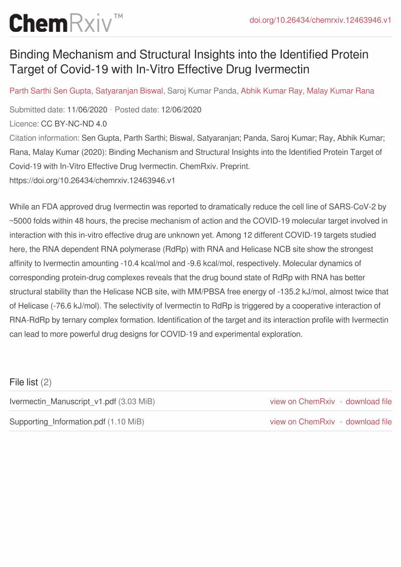
Abstract
While an FDA approved drug Ivermectin was reported to dramatically reduce the cell line of
SARS-CoV-2 by ~5000 folds within 48 hours, the precise mechanism of action and the COVID-
19 molecular target involved in interaction with this in-vitro effective drug are unknown yet.
Among 12 different COVID-19 targets studied here, the RNA dependent RNA polymerase (RdRp)
with RNA and Helicase NCB site show the strongest affinity to Ivermectin amounting -10.4
kcal/mol and -9.6 kcal/mol, respectively. Molecular dynamics of corresponding protein-drug
complexes reveals that the drug bound state of RdRp with RNA has better structural stability than
the Helicase NCB site, with MM/PBSA free energy of -135.2 kJ/mol, almost twice that of Helicase
(-76.6 kJ/mol). The selectivity of Ivermectin to RdRp is triggered by a cooperative interaction of
RNA-RdRp by ternary complex formation. Identification of the target and its interaction profile
with Ivermectin can lead to more powerful drug designs for COVID-19 and experimental
exploration.
Graphical TOC Entry
Keywords: Molecular docking, Molecular Dynamics, Ivermectin, COVID-19, Antiviral Drug

3
Covid-19, declared pandemic by WHO1, is a respiratory disease caused by a novel virus, SARS-
CoV-2, which is an enveloped, positive-sense, single-stranded RNA beta-coronavirus. COVID-19
is one of the seven pathogenic members of the coronaviridae family that includes several mild
common cold viruses, e.g., hCoV-OC43, HKU, and 229E2. Looking backward in the recent
decade, highly pathogenic human severe acute respiratory syndrome (SARS) coronavirus (SARS-
CoV) in 2002, and the Middle East respiratory syndrome (MERS) coronavirus (MERS-CoV) in
2012, with a fatality rate of 10% and 36%, respectively, have emerged3-5. Compared to MERS or
SARS, SARS-CoV-2 appears to spread more rapidly, making it difficult to contain that has been
a serious concern to the scientific community worldwide now6-8. An increasing number of cases
and death from the novel coronavirus worldwide certainly ushers an adverse global impact on
health and economics9. Considering the fatality and the epidemic nature of this disease, there is a
solemn need to find out preventive therapeutics as quickly as possible to curb this virus. However,
thus far, no clinically effective drug is approved for the treatment of this virus infection.
SARS-COV-2 has several conserved non-structural and structural patterns8, 10-11, which can be the
potential targets for the novel or repurposed drug discovery. Nonstructural protein 12, a conserved
protein in coronavirus, is an RNA-dependent RNA polymerase (RdRp) and responsible for
coronavirus replication/transcription. Given previous SARS-CoV and MERS-CoV inhibitors’
search considers RdRp as a significant drug target, in the case of SARS-COV-2, Nsp12-RdRp can
be most crucial for drug design on which no specific inhibitors have been reported until now.12
Together, being conserved and a necessary component for the replication of coronavirus, a multi-
functional protein, Nsp13-helicase, is another vital SARS-COV-2 target13, which can be
considered further for anti-viral drug discovery provided a very small number of Nsp13 inhibitors
reported to date14 .
Based on previous evidence of the effectiveness against the earlier SARS, MERS, etc., some FDA
approved antiviral drugs remdesivir, lopinavir, ritonavir, and oseltamivir are initially under
investigation against COVID-195 (The Scientist dated February 3, 2020. https://www.the-
scientist.com/news-opinion/flu-and-anti-hiv-drugs-show-efficacy-againstcoronavirus-67052).
Remdesivir, an inhibitor of RNA-dependent RNA polymerase, developed by Gilead to treat Ebola
virus infections, is currently in clinical trials for treating COVID-1914. Recently, a sudden

4
breakthrough in research at Monash University, Australia reporting Ivermectin, an anti-parasitic
drug, killing SARS-CoV-2 within 48 hours has gained a considerable attention worldwide8.
Ivermectin is an FDA-approved drug and has shown in vitro antiviral activity previously against a
broad range of viruses, including HIV, Dengue, influenza, and Zika virus5. With a lack of further
details as to how Ivermectin inhibiting these viruses, the exact mechanism and the target in which
Ivermectin interacts with SARS-CoV-2 is yet to be identified8. Such information can proliferate
the identification and design of more potent drugs than Ivermectin against SARS-CoV-2 in the
near future.
Therefore, the present work explores the interaction between all possible targets of SARS-CoV-2
with Ivermectin to identify the one which is specifically inhibited by the drug providing molecular
insights. Molecular docking of Ivermectin with twelve SARS-COV-2’s targets was carried out,
followed by binding mechanism exploration and structural stability analysis using molecular
dynamics (MD) simulation through the root-mean-square deviation (RMSD), root-mean-square
fluctuation (RMSF), radius of gyration (Rg), and binding free energy of the complexes of
Ivermectin with the best targets. The identification of potential target and revelation of the binding
mechanism for Ivermectin provide useful information for further exploration in the potential
therapeutic discovery against the SARS-CoV-2 pandemic.
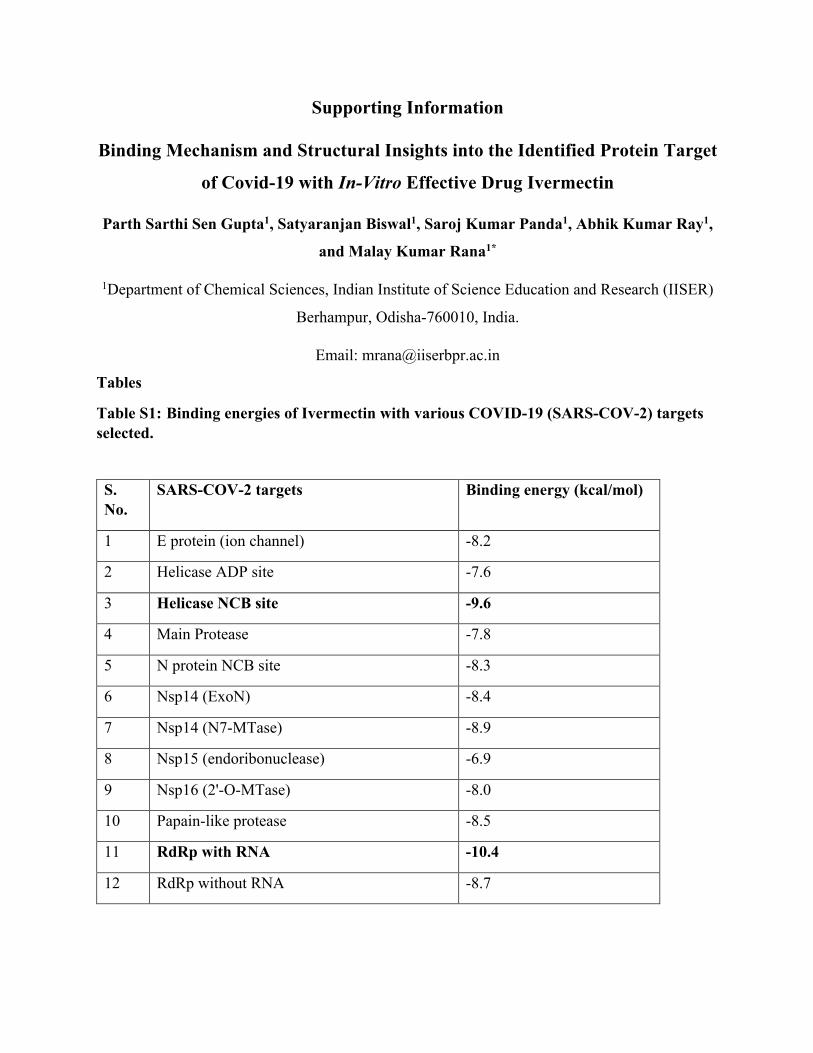
Based on docking scores, the interaction of Ivermectin with the twelve targets (as mentioned
above; Figure 1) is ranked as RNA dependent RNA polymerase (RdRp) with RNA (ΔG = -10.4)
> Helicase NCB site (ΔG = -9.6) > Nsp14 (N7-MTase) (ΔG = -8.9) > RdRp without RNA (ΔG =
-8.7) > Papain-like protease (ΔG = -8.5) > Nsp14(ExoN) (ΔG = -8.4) > N protein NCB site (ΔG
= -8.3) > E protein (ion channel) (ΔG = -8.2) > Nsp16 (2'-O-MTase) (ΔG = -8.0) > Main Protease
(ΔG = -7.8) > Helicase ADP site (ΔG = -7.6) > Nsp15(endoribonuclease) (ΔG = -6.9) (Table S1),
all ΔGs are in kcal/mol. Among all targets, RdRp with its cofactors Nsp7_Nsp8 and RNA has the
best docking score with Ivermectin, -10.4 kcal/mol, followed by Helicase in NCB site (-9.6
kcal/mol) (Table S1). ΔG below -9.0 kcal/mol demonstrates a significant interaction based on
which two targets, namely RdRp with RNA and Helicase in NCB site are shortlisted for further
investigation. The amino acid residues of these targets interacting with Ivermectin are provided in
Table S2.

5
Figure 1: The 3D diagrams of Ivermectin binding with twelve different protein targets of
SARS-COV-2 considered in this study.
The residues of the best scoring targets revealed in Table S2 involve in hydrogen bond (h-bond)
formation and π interactions. In the case of RdRp, ARG553, ARG555, and CYS622 participate in
h-bond formation, whereas HIS439 and ALA550 take part in π interactions. Besides, according to
Figure 2 for the RdRp-Ivermectin complex, RNA molecule interacts with Ivermectin via the
formation of a ternary complex, providing more stability. The energy contribution of RNA to the
RdRp-Ivermectin complex is -1.7 kcal/mol. In Figure 2, the residues predominantly interacting via
van der Walls interaction are LYS545, THR556, SER549, LYS551, ASP618, ASP623, SER682,
THR680, TYR619, ASN691, ASP760, THR687, and ARG836.

6
Figure 2: The 2D diagram depicting residues contributed majorly to the interaction between Ivermectin and the SARS-CoV-2 target RNA dependent RNA polymerase (RdRp).
On the other hand, residues involved in h-bond formation in the Helicase-Ivermectin complex are
LYS139, GLU142, ARG178, and ARG339 (Figure 3; Table S2). Π-interacting residues are
LEU138, VAL181, HIS230, and MET233. Similar to the RdRp-Ivermectin complex, residues
majorly involved in van der Waals interaction in the Helicase-Ivermectin complex are TYR180,
THR231, ASN179, ASN361, THR410, TYR382, LYS146, HIS311, SER310, CYS309, PRO408,
ALA407, THR380, and ARG409. Thus, from the interaction point of view, it is found to have four
and three h-bonds in the Helicase-Ivermectin and RdRp-Ivermectin complexes, respectively. In
addition to a greater number of h-bonds, the lower binding energy or more stability of RdRp-
Ivermectin compared to Helicase-Ivermectin stems from a cooperative interaction of -1.7 kcal/mol
due to a ternary complex formation of RNA with RdRp-Ivermectin.

7
Figure 3: The 2D diagram depicting residues contributed majorly to the interaction between
Ivermectin and the SARS-CoV-2 target Helicase in the NCB site.
Molecular Dynamics (MD) simulations have great significance to analyze the internal motions,
conformational changes, stability, etc. of protein-ligand complexes15-16. Utilizing the MD
trajectories generated, RMSD, RMSF, and Rg were computed and the results are discussed below.
Apart from allowing to assess the equilibration, quality of the run and convergence of MD
trajectories, RMSD is useful to investigate the stability of a protein in complex15-17. A larger
RMSD value is indicative of the lower stability of a protein complex. Here, the RMSD of the
Helicase-Ivermectin and RdRp-Ivermectin complexes with respect to the Ca atom was calculated
against the MD simulation time and is shown in Figures 4A and 4B, respectively. In the case of
the Helicase-Ivermectin complex, the average value of RMSD is around 0.4 nm, with fluctuations
at around 10-15 ns and 18 ns suggesting a loss of stability in these regions (Figure 4A). But, in
the case of the RdRp-Ivermectin complex, the average value of RMSD is around 3.8 nm and almost

8
no fluctuation has been seen during 50 ns simulation period, suggesting greater stability of the
complex throughout the whole dynamics (Figure 4B).
Figure 4: Plots of RMSD as a function of simulation time for (A) Helicase NCB site-
Ivermectin and (B) RdRp-RNA-Ivermectin.
RMSF is a dynamic parameter that measures the average main chain flexibility at each residue
position. RMSF measured with respect to backbone atoms of each amino acid residue for both the
complexes is presented in Figures 5A and 5B. In the case of the Helicase-Ivermectin complex, the
average value of RMSF is around 2.7 nm and higher spikes are noticed at the residues 50 to 90,
190, and 490 (Figure 5A). But, in the case of the RdRp-Ivermectin complex, smaller spikes rather
can be seen (Figure 5B) which corroborates with the RMSD analysis, indicating the more stability
of the complex.
The radius of gyration (Rg) describes the level of compaction of protein. It is defined as the mass-
weighted root-mean-square distance for a collection of atoms from their common center of mass.
Hence, the trajectory analysis for Rg provides the overall dimension of protein. In the case of the
Helicase-Ivermectin complex, the average value of Rg is 2.65 nm and there is a large fall at around
4.9 ns (Figure S1, A). In the case of the RdRp-Ivermectin complex, the average value of Rg is
around 2.8 nm and a small downward jump can be seen at around 19 ns before getting stabilized
(Figure S1, B). The larger average value of Rg for the RdRp-Ivermectin complex may be attributed
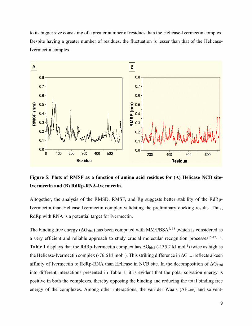
9
to its bigger size consisting of a greater number of residues than the Helicase-Ivermectin complex.
Despite having a greater number of residues, the fluctuation is lesser than that of the Helicase-
Ivermectin complex.
Figure 5: Plots of RMSF as a function of amino acid residues for (A) Helicase NCB site-
Ivermectin and (B) RdRp-RNA-Ivermectin.
Altogether, the analysis of the RMSD, RMSF, and Rg suggests better stability of the RdRp-
Ivermectin than Helicase-Ivermectin complex validating the preliminary docking results. Thus,
RdRp with RNA is a potential target for Ivermectin.
The binding free energy (ΔGbind) has been computed with MM/PBSA7, 18 ,which is considered as
a very efficient and reliable approach to study crucial molecular recognition processes15-17, 19.
Table 1 displays that the RdRp-Ivermectin complex has ΔGbind (-135.2 kJ mol-1) twice as high as
the Helicase-Ivermectin complex (-76.6 kJ mol-1). This striking difference in ΔGbind reflects a keen
affinity of Ivermectin to RdRp-RNA than Helicase in NCB site. In the decomposition of ΔGbind
into different interactions presented in Table 1, it is evident that the polar solvation energy is
positive in both the complexes, thereby opposing the binding and reducing the total binding free
energy of the complexes. Among other interactions, the van der Waals (ΔEvdW) and solvent-
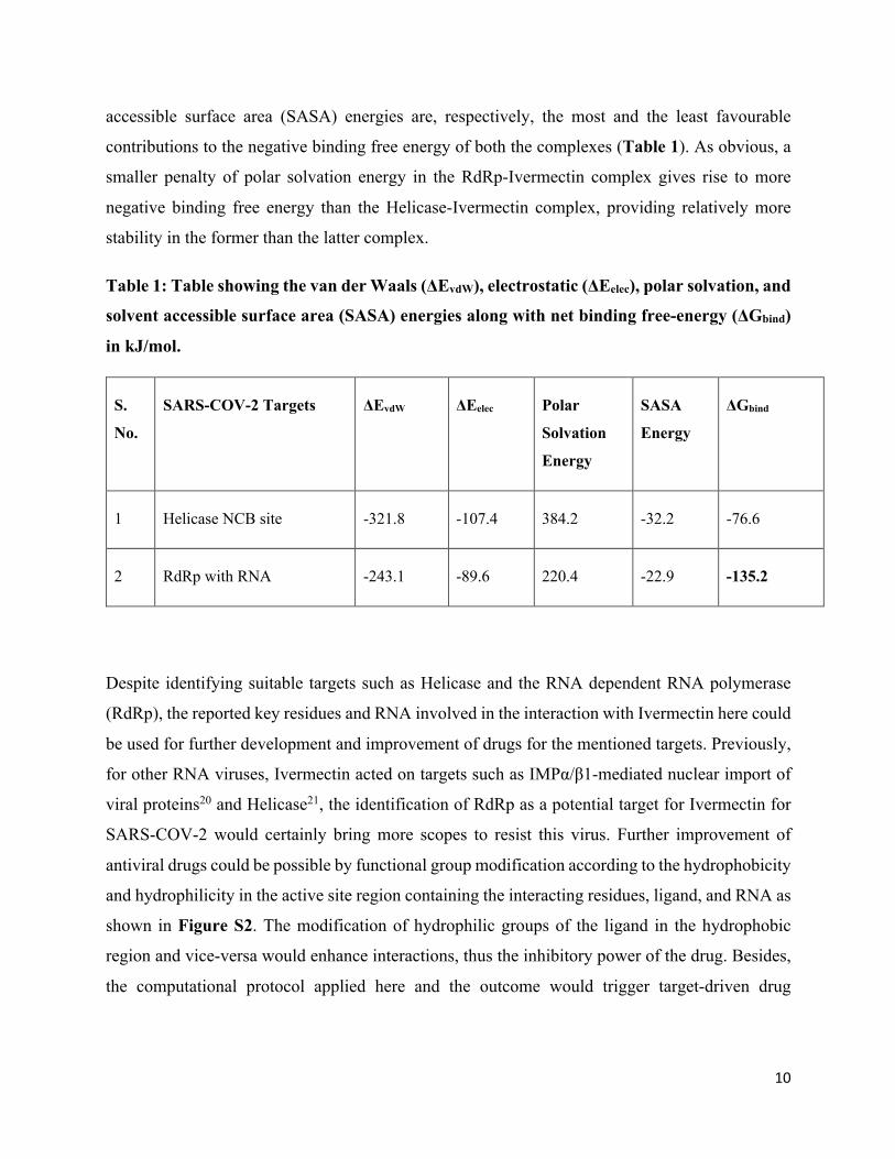
10
accessible surface area (SASA) energies are, respectively, the most and the least favourable
contributions to the negative binding free energy of both the complexes (Table 1). As obvious, a
smaller penalty of polar solvation energy in the RdRp-Ivermectin complex gives rise to more
negative binding free energy than the Helicase-Ivermectin complex, providing relatively more
stability in the former than the latter complex.
Table 1: Table showing the van der Waals (ΔEvdW), electrostatic (ΔEelec), polar solvation, and
solvent accessible surface area (SASA) energies along with net binding free-energy (ΔGbind)
in kJ/mol.
S.
No.
SARS-COV-2 Targets ΔEvdW ΔEelec Polar
Solvation
Energy
SASA
Energy
ΔGbind
1 Helicase NCB site -321.8 -107.4 384.2 -32.2 -76.6
2 RdRp with RNA -243.1 -89.6 220.4 -22.9 -135.2
Despite identifying suitable targets such as Helicase and the RNA dependent RNA polymerase
(RdRp), the reported key residues and RNA involved in the interaction with Ivermectin here could
be used for further development and improvement of drugs for the mentioned targets. Previously,
for other RNA viruses, Ivermectin acted on targets such as IMPα/β1-mediated nuclear import of
viral proteins20 and Helicase21, the identification of RdRp as a potential target for Ivermectin for
SARS-COV-2 would certainly bring more scopes to resist this virus. Further improvement of
antiviral drugs could be possible by functional group modification according to the hydrophobicity
and hydrophilicity in the active site region containing the interacting residues, ligand, and RNA as
shown in Figure S2. The modification of hydrophilic groups of the ligand in the hydrophobic
region and vice-versa would enhance interactions, thus the inhibitory power of the drug. Besides,
the computational protocol applied here and the outcome would trigger target-driven drug

11
discovery for SARS-COV-2 which seems to be the need of the hour and should promote studies
for other pathogens.
Ivermectin, an FDA-approved drug, has shown in vitro antiviral activity against SARS-COV-2.
But the exact mechanism and the specific target which Ivermectin inhibits were not known. In this
study, analyses of molecular docking study revealed that out of twelve molecular targets of SARS-
COV-2, two targets (RdRp with RNA and Helicase in NCP site) have better binding energies with
Ivermectin, lesser than -9.60 kcal/mol. Comparing between the two complexes of Ivermectin with
the two shortlisted targets, the RMSD, RMSF, Rg, and the binding free energy from MD suggest
greater stability of the RdRp-Ivermectin complex than that of Helicase. The presence of RNA
augments the interaction between RdRp-Ivermectin. Thus, RdRp is identified as the most probable
target for the in-vitro effective FDA approved drug Ivermectin, which can guide the experiment
as well as proliferate the design of more potential therapeutics against SARS-CoV-2.
Methodology
The twelve targets of SARS-COV-2 used in the study are Main Protease, Papain-like protease,
RdRp with RNA, RdRp without RNA, Helicase ADP site, Helicase NCB site, Nsp14(ExoN),
Nsp14(N7-MTase), Nsp15(endoribonuclease), Nsp16(2'-O-MTase), N protein NCB site, and E
protein(ion channel)11. If available, the structures of the targets were retrieved from the RCSB
database, otherwise, were modelled using Modeller11, 22 (Figure 1), followed by validation and
energy minimization with Gromacs23 of the generated structures. Downloaded from Drug Bank24,
the SMILES of Ivermectin was converted to the 3D coordinates using Open Babel25 and next
geometry optimization using Gaussian 1626 at the level DFT/B3LYP/6-31+G(d, p).
Utilizing COVID-19 docking server11, the initial docking of Ivermectin with twelve targets was
performed, the docking energies and poses of which were later verified manually by AutoDock
Vina27. The protein targets were prepared by removing water, adding missing hydrogens and the
Kollman charges to atoms. Addition of missing hydrogens if any, assignment of Gasteiger charges
and rotatable bonds were performed to Ivermectin. For sequential docking, the Autogrid size was
set to particular binding regions of each target with the default grid spacing. Lamarckian Genetic
Algorithm (GA 4.2) was used for the docking, which gives the top 10 estimated free energies of
binding. The best docking score and pose of complexes were taken, analyzed and rendered through
12
the Discovery Studio visualizer. The best docked complexes were taken for further molecular
dynamics study.
MD simulations were carried out for the RdRp with RNA-Ivermectin and Helicase-Ivermectin
complexes for a period of 50 ns using GROMACS (GROningenMAchine for Chemical
Simulations) v5.1 molecular dynamics package23. The unit cell defined as a cubical box, with a
minimal distance of 10 Å from the protein surface to the edges of the box, was solvated using the
Simple Point Charge (SPC) water model; the topologies of these selected targets and Ivermectin
were created by the GROMOS96 53a6 force field28. Counter-ions were added to make every
system electrically neutral at a salt concentration of 0.15 mol/L. Before the MD run, each system
was subjected to energy minimization by employing the steepest descent integrator for 5000 steps
with force convergence of <1000 kcal mol-1 nm-1.
Thereafter, each protein-ligand complex was equilibrated for 5 ns using canonical (NVT) and
isothermal-isobaric (NPT) ensembles. During equilibration, each system was coupled with the
Berendsen temperature and Parrinello-Rahman pressure controllers, respectively, to maintain
temperature 300 K and pressure 1 bar. The Particle Mesh Ewald (PME) algorithm29 was employed
to deal with the long-range Coulomb interactions with a Fourier grid spacing of 0.12 nm. The
short-range van der Waals interactions were given by the Lennard-Jones potential with a cut-off
distance of 1 nm. All bond lengths were constrained by the linear constraint solver (LINCS)
method30 .
Subsequently, 50 ns production run was performed. In principle, the same protocol was applied to
both systems. A time step of 2 fs was used and the coordinates were saved at every 10 ps during
the production run. For the structural analyses of every system, the resultant MD trajectories were
analyzed using the built-in modules of GROMACS and visual molecular dynamics (VMD 1.9.1)31.
The 2D plots depicting the intrinsic dynamical stabilities captured by the root-mean-square
deviation (RMSD), root-mean-square fluctuation (RMSF), and the radius of gyration (Rg) of the
complexes were generated by the Grace 5.1.23 program.
For binding free energy (ΔGbind) from MD trajectory, widely used Molecular Mechanics/Poisson-
Boltzmann Surface Area (MM/PBSA) was adapted16, 18, 32. ΔGbind in a solvent medium was
calculated as follows:
ΔGbind = Gcomplex - (Gprotein + Gligand), where G comprises the potential energy (EMM) in vacuum and
solvation free energy (Gsolv) for each. EMM consists of bonded and non-bonded interactions,

13
whereas Gsolv is the sum of electrostatic and non-polar solvation free energies. The ΔGbind of two
shortlisted complexes were calculated over 5000 frames taken at an equal interval during 50 ns
production run. Per-residue energy contribution to ΔGbind was also evaluated.
Acknowledgements The authors acknowledge IISER Berhampur for computational support. P.S.S.G also sincerely
acknowledges IISER Berhampur for providing him the Institute Postdoc Fellowship to carry out
this work.
Conflict of interest The authors report no conflicts of interest.
References 1. Cucinotta, D.; Vanelli, M. WHO declares COVID-19 a pandemic. Acta bio-medica: Atenei Parmensis 2020, 91 (1), 157-160. 2. Prajapat, M.; Sarma, P.; Shekhar, N.; Avti, P.; Sinha, S.; Kaur, H.; Kumar, S.; Bhattacharyya, A.; Kumar, H.; Bansal, S. Drug targets for corona virus: A systematic review. Indian journal of pharmacology 2020, 52 (1), 56. 3. Zhu, N.; Zhang, D.; Wang, W.; Li, X.; Yang, B.; Song, J.; Zhao, X.; Huang, B.; Shi, W.; Lu, R. A novel coronavirus from patients with pneumonia in China, 2019. New England Journal of Medicine 2020. 4. Sarma, P.; Sekhar, N.; Prajapat, M.; Avti, P.; Kaur, H.; Kumar, S.; Singh, S.; Kumar, H.; Prakash, A.; Dhibar, D. P. In-silico homology assisted identification of inhibitor of RNA binding against 2019-nCoV N-protein (N terminal domain). Journal of Biomolecular Structure and Dynamics 2020, (just-accepted), 1-11. 5. Chang, C.-k.; Sue, S.-C.; Yu, T.-h.; Hsieh, C.-M.; Tsai, C.-K.; Chiang, Y.-C.; Lee, S.-j.; Hsiao, H.-h.; Wu, W.-J.; Chang, W.-L. Modular organization of SARS coronavirus nucleocapsid protein. Journal of biomedical science 2006, 13 (1), 59-72. 6. Muralidharan, N.; Sakthivel, R.; Velmurugan, D.; Gromiha, M. M. Computational studies of drug repurposing and synergism of lopinavir, oseltamivir and ritonavir binding with SARS-CoV-2 Protease against COVID-19. Journal of Biomolecular Structure and Dynamics 2020, (just-accepted), 1-7. 7. Gupta, M. K.; Vemula, S.; Donde, R.; Gouda, G.; Behera, L.; Vadde, R. In-silico approaches to detect inhibitors of the human severe acute respiratory syndrome coronavirus envelope protein ion channel. Journal of Biomolecular Structure and Dynamics 2020, (just-accepted), 1-17. 8. Caly, L.; Druce, J. D.; Catton, M. G.; Jans, D. A.; Wagstaff, K. M. The FDA-approved Drug Ivermectin inhibits the replication of SARS-CoV-2 in vitro. Antiviral research 2020, 104787.
14
9. Petropoulos, F.; Makridakis, S. Forecasting the novel coronavirus COVID-19. PloS one 2020, 15 (3), e0231236. 10. Xu, X.; Zhai, Y.; Sun, F.; Lou, Z.; Su, D.; Xu, Y.; Zhang, R.; Joachimiak, A.; Zhang, X. C.; Bartlam, M. New antiviral target revealed by the hexameric structure of mouse hepatitis virus nonstructural protein nsp15. Journal of virology 2006, 80 (16), 7909-7917. 11. Kong, R.; Yang, G.; Xue, R.; Liu, M.; Wang, F.; Hu, J.; Guo, X.; Chang, S. COVID-19 Docking Server: An interactive server for docking small molecules, peptides and antibodies against potential targets of COVID-19. arXiv preprint arXiv:2003.00163 2020. 12. Gao, Y.; Yan, L.; Huang, Y.; Liu, F.; Zhao, Y.; Cao, L.; Wang, T.; Sun, Q.; Ming, Z.; Zhang, L. Structure of RNA-dependent RNA polymerase from 2019-nCoV, a major antiviral drug target. BioRxiv 2020. 13. Jia, Z.; Yan, L.; Ren, Z.; Wu, L.; Wang, J.; Guo, J.; Zheng, L.; Ming, Z.; Zhang, L.; Lou, Z. Delicate structural coordination of the Severe Acute Respiratory Syndrome coronavirus Nsp13 upon ATP hydrolysis. Nucleic acids research 2019, 47 (12), 6538-6550. 14. Cao, Y.-c.; Deng, Q.-x.; Dai, S.-x. Remdesivir for severe acute respiratory syndrome coronavirus 2 causing COVID-19: An evaluation of the evidence. Travel Medicine and Infectious Disease 2020, 101647. 15. Singh, V. K.; Srivastava, R.; Sen Gupta, P. S.; Naaz, F.; Chaurasia, H.; Mishra, R.; Rana, M. K.; Singh, R. K. Anti-HIV potential of diarylpyrimidine derivatives as non-nucleoside reverse transcriptase inhibitors: Design, synthesis, docking, TOPKAT analysis and molecular dynamics simulations. Journal of Biomolecular Structure and Dynamics 2020, (just-accepted), 1-18. 16. Sen Gupta, P. S.; Islam, R. N. U.; Banerjee, S.; Nayek, A.; Rana, M. K.; Bandyopadhyay, A. K. Screening and molecular characterization of lethal mutations of human homogentisate 1, 2 dioxigenase. Journal of Biomolecular Structure and Dynamics 2020, 1-11. 17. Sjulstok, E.; Solov'yov, I. A. Structural Explanations of FAD Binding in Drosophila Melanogaster Cryptochrome. The Journal of Physical Chemistry Letters 2020. 18. Hou, T.; Wang, J.; Li, Y.; Wang, W. Assessing the performance of the MM/PBSA and MM/GBSA methods. 1. The accuracy of binding free energy calculations based on molecular dynamics simulations. Journal of chemical information and modeling 2011, 51 (1), 69-82. 19. Tamura, K.; Sugita, Y. Free Energy Analysis of a Conformational Change of Heme ABC Transporter BhuUV-T. The Journal of Physical Chemistry Letters 2020. 20. Yang, S. N.; Atkinson, S. C.; Wang, C.; Lee, A.; Bogoyevitch, M. A.; Borg, N. A.; Jans, D. A. The broad spectrum antiviral ivermectin targets the host nuclear transport importin α/β1 heterodimer. Antiviral Research 2020, 104760. 21. Mastrangelo, E.; Pezzullo, M.; De Burghgraeve, T.; Kaptein, S.; Pastorino, B.; Dallmeier, K.; de Lamballerie, X.; Neyts, J.; Hanson, A. M.; Frick, D. N. Ivermectin is a potent inhibitor of flavivirus replication specifically targeting NS3 helicase activity: new prospects for an old drug. Journal of Antimicrobial Chemotherapy 2012, 67 (8), 1884-1894. 22. Liu, D.; Chen, X.; Long, D. NMR-Derived Conformational Ensemble of State 1 of Activated Ras Reveals Insights into a Druggable Pocket. The Journal of Physical Chemistry Letters 2020. 23. Pronk, S.; Páll, S.; Schulz, R.; Larsson, P.; Bjelkmar, P.; Apostolov, R.; Shirts, M. R.; Smith, J. C.; Kasson, P. M.; van der Spoel, D. GROMACS 4.5: a high-throughput and highly parallel open source molecular simulation toolkit. Bioinformatics 2013, 29 (7), 845-854.

15
24. Wishart, D. S.; Feunang, Y. D.; Guo, A. C.; Lo, E. J.; Marcu, A.; Grant, J. R.; Sajed, T.; Johnson, D.; Li, C.; Sayeeda, Z. DrugBank 5.0: a major update to the DrugBank database for 2018. Nucleic acids research 2018, 46 (D1), D1074-D1082. 25. O’Boyle, N.; Banck, M.; James, C.; Morley, C.; Vandermeersch, T.; Hutchison, G. Open babel: an open chemical toolbox. J Cheminf 3 (1): 33. 2011. 26. Frisch, M.; Trucks, G.; Schlegel, H.; Scuseria, G.; Robb, M.; Cheeseman, J.; Scalmani, G.; Barone, V.; Petersson, G.; Nakatsuji, H. Gaussian 16. Gaussian, Inc. Wallingford, CT: 2016. 27. Trott, O.; Olson, A. J. AutoDock Vina: improving the speed and accuracy of docking with a new scoring function, efficient optimization, and multithreading. Journal of computational chemistry 2010, 31 (2), 455-461. 28. Oostenbrink, C.; Villa, A.; Mark, A. E.; Van Gunsteren, W. F. A biomolecular force field based on the free enthalpy of hydration and solvation: the GROMOS force‐field parameter sets 53A5 and 53A6. Journal of computational chemistry 2004, 25 (13), 1656-1676. 29. Essman, U.; Perera, L.; Berkowitz, M.; Darden, T.; Lee, H.; Pedersen, L. A smooth particle mesh ewald potential. J. Chem. Phys 1995, 103, 8577-8592. 30. Hess, B.; Bekker, H.; Berendsen, H. J.; Fraaije, J. G. LINCS: a linear constraint solver for molecular simulations. Journal of computational chemistry 1997, 18 (12), 1463-1472. 31. Humphrey, W.; Dalke, A.; Schulten, K. VMD: visual molecular dynamics. Journal of molecular graphics 1996, 14 (1), 33-38. 32. Kumari, R.; Kumar, R.; Consortium, O. S. D. D.; Lynn, A. g_mmpbsa� A GROMACS tool for high-throughput MM-PBSA calculations. Journal of chemical information and modeling 2014, 54 (7), 1951-1962.

download fileview on ChemRxivIvermectin_Manuscript_v1.pdf (3.03 MiB)
Supporting Information
Binding Mechanism and Structural Insights into the Identified Protein Target
of Covid-19 with In-Vitro Effective Drug Ivermectin
Parth Sarthi Sen Gupta1, Satyaranjan Biswal1, Saroj Kumar Panda1, Abhik Kumar Ray1,
and Malay Kumar Rana1*
1Department of Chemical Sciences, Indian Institute of Science Education and Research (IISER)
Berhampur, Odisha-760010, India.
Email: [email protected]
Tables
Table S1: Binding energies of Ivermectin with various COVID-19 (SARS-COV-2) targets selected.
S. No.
SARS-COV-2 targets Binding energy (kcal/mol)
1 E protein (ion channel) -8.2
2 Helicase ADP site -7.6
3 Helicase NCB site -9.6
4 Main Protease -7.8
5 N protein NCB site -8.3
6 Nsp14 (ExoN) -8.4
7 Nsp14 (N7-MTase) -8.9
8 Nsp15 (endoribonuclease) -6.9
9 Nsp16 (2'-O-MTase) -8.0
10 Papain-like protease -8.5
11 RdRp with RNA -10.4
12 RdRp without RNA -8.7
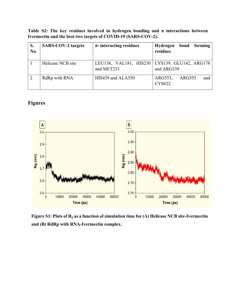
Table S2: The key residues involved in hydrogen bonding and π interactions between Ivermectin and the best two targets of COVID-19 (SARS-COV-2).
S. No.
SARS-COV-2 targets π- interacting residues Hydrogen bond forming residues
1 Helicase NCB site LEU138, VAL181, HIS230 and MET233
LYS139, GLU142, ARG178 and ARG339
2 RdRp with RNA HIS439 and ALA550 ARG553, ARG555 and CYS622
Figures
Figure S1: Plots of Rg as a function of simulation time for (A) Helicase NCB site-Ivermectin
and (B) RdRp with RNA-Ivermectin complex.
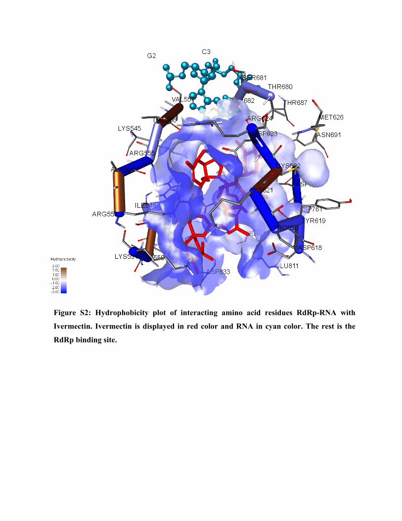
Figure S2: Hydrophobicity plot of interacting amino acid residues RdRp-RNA with
Ivermectin. Ivermectin is displayed in red color and RNA in cyan color. The rest is the
RdRp binding site.

download fileview on ChemRxivSupporting_Information.pdf (1.10 MiB)