Bilateral hydronephrosis and hydroureter after ...vri.cz/docs/vetmed/60-1-52.pdf1 Bilateral...
7
52 Case Report Veterinarni Medicina, 60, 2015 (1): 52–56 doi: 10.17221/7926-VETMED Bilateral hydronephrosis and hydroureter after ovariohysterectomy using nylon cable tie: a case report L.R. Mesquita 1 , S.C. Rahal 1 , L.M. Matsubara 1 , M.J. Mamprim 1 , C.R. Foschini 2 , L.G. Faria 3 , W.T. Kano 1 1 School of Veterinary Medicine and Animal Science, University Estadual Paulista (UNESP), Botucatu, Sao Paulo, Brazil 2 School of Engineer of Bauru (Foschini), University Estadual Paulista (UNESP), Bauru, Sao Paulo, Brazil 3 School of Agricultural and Veterinary Sciences, University Estadual Paulista (UNESP), Jaboticabal, Sao Paulo, Brazil ABSTRACT: A spayed crossbred female dog was presented due to progressive weight loss, emesis and anorexia over the preceding month. A complete blood count, urinalysis, serum biochemical panel, and ultrasound were initially performed. Computed tomography urography was performed as a complementary exam. Based on ultra- sound and CT findings an exploratory celiotomy was performed to remove hyperdense structures that could be the cause of the hydronephrosis and hydroureter in both kidneys. An extensive granulomatous reaction was found near the caudal pole to the left kidney. A nylon cable tie adhering firmly to this tissue was removed during surgical excision. On the dorsal surface of the bladder an extensive granulomatous reaction that had entrapped the right ureter was also noted. Another nylon cable tie was removed and the ureter was released. Eight months postopera- tively, the dog was in good general physical condition, showing appetite and vigour. Abdominal ultrasonography showed improvement of the hydronephrosis in both kidneys. The urea ratio was normal, but the creatinine level was slightly elevated, suggesting a guarded prognosis. Thus, bilateral hydronephrosis as observed in the present study should be considered as a major complication after elective ovariohysterectomy. Keywords: nylon cable tie; renal injury; surgery, computed tomography excretory; dog Complications after elective ovariohysterecto- mies may occur during surgery or at short or long intervals after the surgical procedure (Furneaux et al. 1973; Werner et al. 1992; Burrow et al. 2005; Goethem et al. 2006; Howe 2006; Adin 2011; Kennedy et al. 2011). Depending on the degree and type, the complication may be life-threaten- ing (Werner et al. 1992; de Gopegui et al. 1999; Goethem et al. 2006; Kennedy et al. 2011). Complications associated with the suture material or ligature include haemorrhage, abdominal wall de- hiscence, surgical wound infection, stump granuloma, fistulous draining tracts, inadvertent ureteral ligation, and chewed-out sutures (Kyles et al. 1996; Pollari and Bonnett 1996; Burrow et al. 2005; Goethem et al. 2006; Howe 2006; Adin 2011; DeTora and McCarthy 2011; Kennedy et al. 2011). Nylon cable ties have been used as ligating material during the spaying of cats and dogs since they are lower in cost and less time- consuming, especially in free campaigns that utilise minimally invasive celiotomy (Hammond et al. 1998; Barros et al. 2009; Lima et al. 2010). Complications reported in the use of nylon cable tie including loos- ening, fistulous tract, unilateral hydroureter and hy- dronephrosis (Werner et al. 1992; Murphy et al. 1998; Macedo et al. 2012). e purpose of this report was to describe another potential life-threatening com- plication encountered using this material - bilateral hydronephrosis and hydroureter – and the course of recovery after treatment.
Transcript of Bilateral hydronephrosis and hydroureter after ...vri.cz/docs/vetmed/60-1-52.pdf1 Bilateral...

52
Case Report Veterinarni Medicina, 60, 2015 (1): 52–56
doi: 10.17221/7926-VETMED
Bilateral hydronephrosis and hydroureter after ovariohysterectomy using nylon cable tie: a case report
L.R. Mesquita1, S.C. Rahal1, L.M. Matsubara1, M.J. Mamprim1, C.R. Foschini2, L.G. Faria3, W.T. Kano1
1School of Veterinary Medicine and Animal Science, University Estadual Paulista (UNESP), Botucatu, Sao Paulo, Brazil
2School of Engineer of Bauru (Foschini), University Estadual Paulista (UNESP), Bauru, Sao Paulo, Brazil
3School of Agricultural and Veterinary Sciences, University Estadual Paulista (UNESP), Jaboticabal, Sao Paulo, Brazil
ABSTRACT: A spayed crossbred female dog was presented due to progressive weight loss, emesis and anorexia over the preceding month. A complete blood count, urinalysis, serum biochemical panel, and ultrasound were initially performed. Computed tomography urography was performed as a complementary exam. Based on ultra-sound and CT findings an exploratory celiotomy was performed to remove hyperdense structures that could be the cause of the hydronephrosis and hydroureter in both kidneys. An extensive granulomatous reaction was found near the caudal pole to the left kidney. A nylon cable tie adhering firmly to this tissue was removed during surgical excision. On the dorsal surface of the bladder an extensive granulomatous reaction that had entrapped the right ureter was also noted. Another nylon cable tie was removed and the ureter was released. Eight months postopera-tively, the dog was in good general physical condition, showing appetite and vigour. Abdominal ultrasonography showed improvement of the hydronephrosis in both kidneys. The urea ratio was normal, but the creatinine level was slightly elevated, suggesting a guarded prognosis. Thus, bilateral hydronephrosis as observed in the present study should be considered as a major complication after elective ovariohysterectomy.
Keywords: nylon cable tie; renal injury; surgery, computed tomography excretory; dog
Complications after elective ovariohysterecto-mies may occur during surgery or at short or long intervals after the surgical procedure (Furneaux et al. 1973; Werner et al. 1992; Burrow et al. 2005; Goethem et al. 2006; Howe 2006; Adin 2011; Kennedy et al. 2011). Depending on the degree and type, the complication may be life-threaten-ing (Werner et al. 1992; de Gopegui et al. 1999; Goethem et al. 2006; Kennedy et al. 2011).
Complications associated with the suture material or ligature include haemorrhage, abdominal wall de-hiscence, surgical wound infection, stump granuloma, fistulous draining tracts, inadvertent ureteral ligation, and chewed-out sutures (Kyles et al. 1996; Pollari and Bonnett 1996; Burrow et al. 2005; Goethem et al.
2006; Howe 2006; Adin 2011; DeTora and McCarthy 2011; Kennedy et al. 2011). Nylon cable ties have been used as ligating material during the spaying of cats and dogs since they are lower in cost and less time-consuming, especially in free campaigns that utilise minimally invasive celiotomy (Hammond et al. 1998; Barros et al. 2009; Lima et al. 2010). Complications reported in the use of nylon cable tie including loos-ening, fistulous tract, unilateral hydroureter and hy-dronephrosis (Werner et al. 1992; Murphy et al. 1998; Macedo et al. 2012). The purpose of this report was to describe another potential life-threatening com-plication encountered using this material - bilateral hydronephrosis and hydroureter – and the course of recovery after treatment.
53
Veterinarni Medicina, 60, 2015 (1): 52–56 Case Report
doi: 10.17221/7926-VETMED
Case description
A six-year-old spayed crossbred female dog, weigh-ing 10 kg was presented to the Veterinary Hospital due to progressive weight loss, emesis and anorexia over the preceding month. The symptoms appeared after use of an anti-inflammatory drug. According to the owner, the female dog had been spayed in a free campaign approximately four years previously. Physical examination revealed signs of cachexia and lethargy, and pale mucous membrane. Pain signs in the mid-abdomen, enlarged kidneys and enlarged bladder were detected on palpation. A complete blood count (CBC), urinalysis, serum biochemi-cal panel, and ultrasound were initially performed. Computed tomography (CT) urography was per-formed as a complementary exam.
Hypochromic microcytic anaemia, elevated urea (122 mg/dl; reference range 21.4 to 59.92 mg/dl), el-evated creatinine (3.6 mg/dl; reference range 0.5 to 1.5 mg/dl), and hypokalaemia (2.9 mEq/l; reference range 4.37 to 5.65 mEq/l) were the most relevant ab-normalities. Abdominal ultrasonography (B-mode scan) (MyLab Alpha, Esaote, Genoa, Italy) showed bilateral renal enlargement with renal cortical atro-phy, loss of corticomedullary differentiation, dilated pelvis and dilatation of both ureters, suggesting hy-dronephrosis and hydroureter. Potassium chloride in lactated Ringer’s solution was administered (80 ml/kg/day). Under general anaesthesia, CT urography was performed with a helical scanner (Shimadzu SCT-7800CT, Shimadzu Corp., Kyoto, Japan) with the dog positioned in dorsal recumbency. A bolus injection of non-ionic iodinated contrast agent (2 ml of iopromide/kg, i.v.) was used after plain CT. The scanning parameters were as follows: voltage peak, 120 V; current, 150 mA; pitch, 1:0; and rotation time, 1 s/rotation. CT findings included bilateral hydrone-phrosis and thickening of the renal wall, the presence of an abnormal hyperdense structure in the caudal pole of the left kidney and another located on the dorsal surface of the bladder neck (Figure 1).
Based on these findings, an exploratory celiot-omy was performed to remove these hyperdense structures that could be the cause of the hydro-nephrosis and hydroureter in both kidneys. The dog was premedicated with morphine (0.5 mg/kg, i.m.). General anaesthesia was induced with propo-fol and maintained with isoflurane in oxygen. Transoperative analgesia included continuous infu-sion of 0.9% saline solution (500 ml) with morphine
(1 ml), lidocaine 2% (7.5 ml) and ketamine 10% (0.3 ml). A midline abdominal incision was extended from the xiphoid process to the pubis. Both kidneys were found to be enlarged, but the right kidney had a fluctuant consistency while the caudal pole of the left kidney had firm consistency on palpation. An extensive granulomatous reaction was found near the caudal pole of the left kidney. A nylon cable tie adhering firmly to this tissue was removed during surgical excision. A cable tie was not found on the right ovarian pedicle. In addition, the left ureter that had become entrapped in the surrounding tissue was released using sharp and blunt dissection. On the dor-sal surface of the bladder an extensive granulomatous reaction that had entrapped the right ureter was also noted. Another nylon cable tie was removed and the ureter was released. Omentum was placed over the freed sites of the ureters. Closure of the abdomen was routine, following irrigation of the abdominal cavity. The nylon cable ties were examined using a ste-reoscope (Carl Zeiss Citoval 2, Jenoptik Jena GmbH, Berlin, DDR). Defects and signs of degradation were observed at 25× and 50× magnifications (Figure 2).
The dog received ceftriaxone (20 mg/kg, q 12 h) immediately before surgery and for seven days after surgery, followed by omeprazole (1 mg/kg, q 24 h, p.o. for seven days) and furosemide (4 mg/kg,
Figure 1. Representative CT urography images before sur-gery. (a) Right kidney (*) with hydronephrosis; (b) left kidney (*) with hydronephrosis and thickening of renal wall; (c) bladder (●) and hyperdense structure (arrow) in the caudal pole of the left kidney; (d) hyperdense structure (arrow) located on the dorsal surface of the bladder neck (●)
54
Case Report Veterinarni Medicina, 60, 2015 (1): 52–56
doi: 10.17221/7926-VETMED
p.o. for 30 days). Tramadol chlorhydrate (4 mg/kg, s.c., q 12 h) was administrated for five days. The dog received intravenous fluid therapy for four days in a private practice due to the owner’s re-quest. Skin sutures were removed 10 days after surgery, and the dog showed better general physi-cal condition. The vomiting had stopped and the dog started to eat. New biochemistry analysis re-vealed improvement of creatinine (2.2 mg/dl) and potassium (4.5 mEq/l), but not of urea (125 mg/dl). On day 30 after surgery, the owner reported cloudy
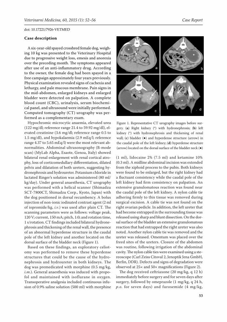
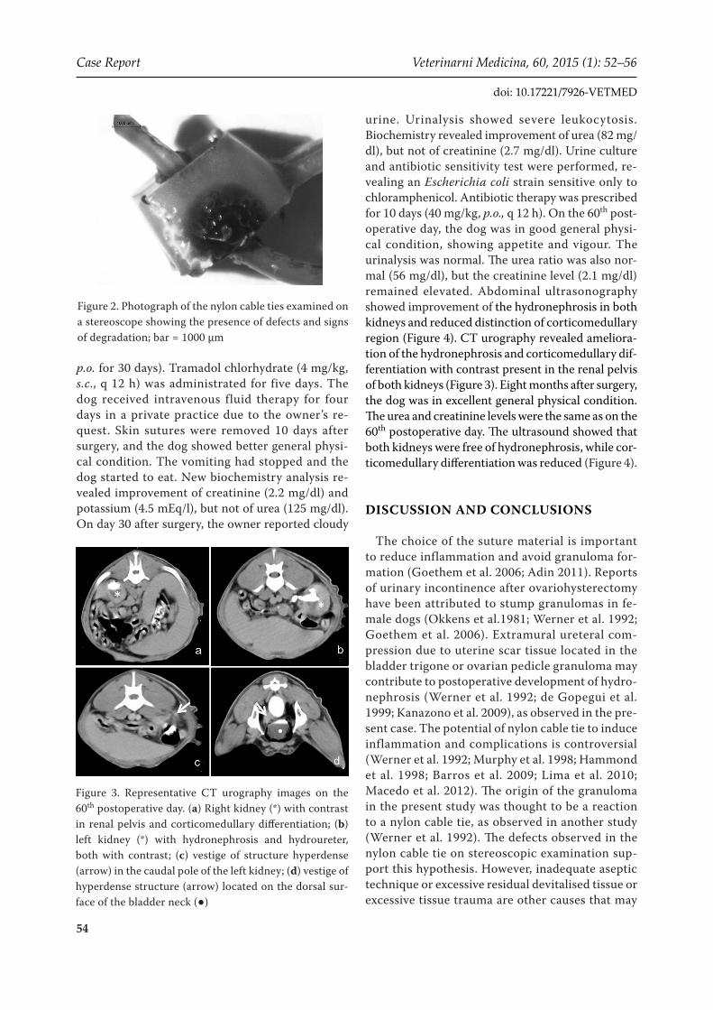
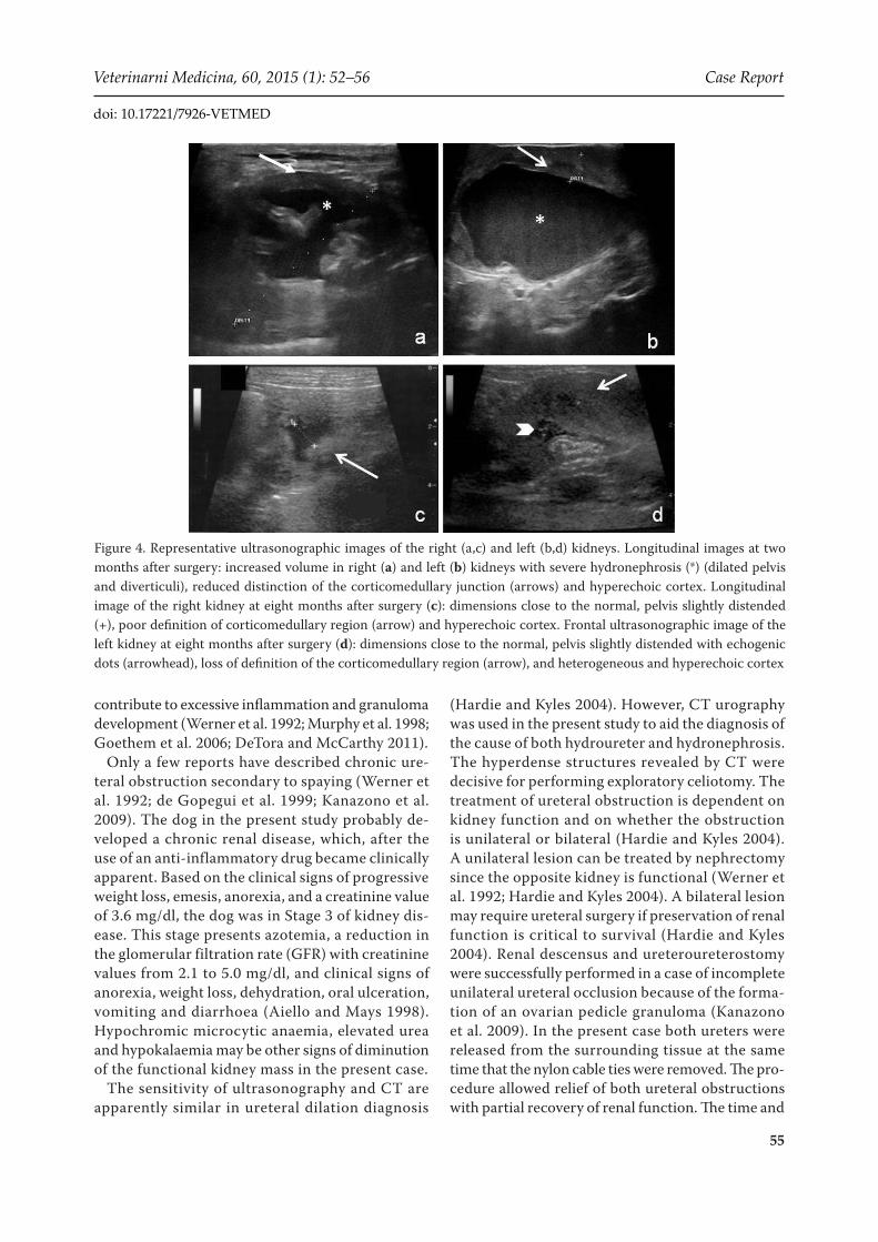
urine. Urinalysis showed severe leukocytosis. Biochemistry revealed improvement of urea (82 mg/dl), but not of creatinine (2.7 mg/dl). Urine culture and antibiotic sensitivity test were performed, re-vealing an Escherichia coli strain sensitive only to chloramphenicol. Antibiotic therapy was prescribed for 10 days (40 mg/kg, p.o., q 12 h). On the 60th post- operative day, the dog was in good general physi-cal condition, showing appetite and vigour. The urinalysis was normal. The urea ratio was also nor-mal (56 mg/dl), but the creatinine level (2.1 mg/dl) remained elevated. Abdominal ultrasonography showed improvement of the hydronephrosis in both kidneys and reduced distinction of corticomedullary region (Figure 4). CT urography revealed ameliora-tion of the hydronephrosis and corticomedullary dif-ferentiation with contrast present in the renal pelvis of both kidneys (Figure 3). Eight months after surgery, the dog was in excellent general physical condition. The urea and creatinine levels were the same as on the 60th postoperative day. The ultrasound showed that both kidneys were free of hydronephrosis, while cor-ticomedullary differentiation was reduced (Figure 4).
DISCUSSION AND CONCLUSIONS
The choice of the suture material is important to reduce inflammation and avoid granuloma for-mation (Goethem et al. 2006; Adin 2011). Reports of urinary incontinence after ovariohysterectomy have been attributed to stump granulomas in fe-male dogs (Okkens et al.1981; Werner et al. 1992; Goethem et al. 2006). Extramural ureteral com-pression due to uterine scar tissue located in the bladder trigone or ovarian pedicle granuloma may contribute to postoperative development of hydro-nephrosis (Werner et al. 1992; de Gopegui et al. 1999; Kanazono et al. 2009), as observed in the pre-sent case. The potential of nylon cable tie to induce inflammation and complications is controversial (Werner et al. 1992; Murphy et al. 1998; Hammond et al. 1998; Barros et al. 2009; Lima et al. 2010; Macedo et al. 2012). The origin of the granuloma in the present study was thought to be a reaction to a nylon cable tie, as observed in another study (Werner et al. 1992). The defects observed in the nylon cable tie on stereoscopic examination sup-port this hypothesis. However, inadequate aseptic technique or excessive residual devitalised tissue or excessive tissue trauma are other causes that may
Figure 2. Photograph of the nylon cable ties examined on a stereoscope showing the presence of defects and signs of degradation; bar = 1000 µm
Figure 3. Representative CT urography images on the 60th postoperative day. (a) Right kidney (*) with contrast in renal pelvis and corticomedullary differentiation; (b) left kidney (*) with hydronephrosis and hydroureter, both with contrast; (c) vestige of structure hyperdense (arrow) in the caudal pole of the left kidney; (d) vestige of hyperdense structure (arrow) located on the dorsal sur-face of the bladder neck (●)
55
Veterinarni Medicina, 60, 2015 (1): 52–56 Case Report
doi: 10.17221/7926-VETMED
contribute to excessive inflammation and granuloma development (Werner et al. 1992; Murphy et al. 1998; Goethem et al. 2006; DeTora and McCarthy 2011).
Only a few reports have described chronic ure-teral obstruction secondary to spaying (Werner et al. 1992; de Gopegui et al. 1999; Kanazono et al. 2009). The dog in the present study probably de-veloped a chronic renal disease, which, after the use of an anti-inflammatory drug became clinically apparent. Based on the clinical signs of progressive weight loss, emesis, anorexia, and a creatinine value of 3.6 mg/dl, the dog was in Stage 3 of kidney dis-ease. This stage presents azotemia, a reduction in the glomerular filtration rate (GFR) with creatinine values from 2.1 to 5.0 mg/dl, and clinical signs of anorexia, weight loss, dehydration, oral ulceration, vomiting and diarrhoea (Aiello and Mays 1998). Hypochromic microcytic anaemia, elevated urea and hypokalaemia may be other signs of diminution of the functional kidney mass in the present case.
The sensitivity of ultrasonography and CT are apparently similar in ureteral dilation diagnosis
(Hardie and Kyles 2004). However, CT urography was used in the present study to aid the diagnosis of the cause of both hydroureter and hydronephrosis. The hyperdense structures revealed by CT were decisive for performing exploratory celiotomy. The treatment of ureteral obstruction is dependent on kidney function and on whether the obstruction is unilateral or bilateral (Hardie and Kyles 2004). A unilateral lesion can be treated by nephrectomy since the opposite kidney is functional (Werner et al. 1992; Hardie and Kyles 2004). A bilateral lesion may require ureteral surgery if preservation of renal function is critical to survival (Hardie and Kyles 2004). Renal descensus and ureteroureterostomy were successfully performed in a case of incomplete unilateral ureteral occlusion because of the forma-tion of an ovarian pedicle granuloma (Kanazono et al. 2009). In the present case both ureters were released from the surrounding tissue at the same time that the nylon cable ties were removed. The pro-cedure allowed relief of both ureteral obstructions with partial recovery of renal function. The time and
Figure 4. Representative ultrasonographic images of the right (a,c) and left (b,d) kidneys. Longitudinal images at two months after surgery: increased volume in right (a) and left (b) kidneys with severe hydronephrosis (*) (dilated pelvis and diverticuli), reduced distinction of the corticomedullary junction (arrows) and hyperechoic cortex. Longitudinal image of the right kidney at eight months after surgery (c): dimensions close to the normal, pelvis slightly distended (+), poor definition of corticomedullary region (arrow) and hyperechoic cortex. Frontal ultrasonographic image of the left kidney at eight months after surgery (d): dimensions close to the normal, pelvis slightly distended with echogenic dots (arrowhead), loss of definition of the corticomedullary region (arrow), and heterogeneous and hyperechoic cortex
56
Case Report Veterinarni Medicina, 60, 2015 (1): 52–56
doi: 10.17221/7926-VETMED
degree of obstruction are important prognostic fac-tors for renal function recovery (Hardie and Kyles 2004). Apparently, dogs are able to survive with a small portion of functional renal tissue (Aiello and Mays 1998). The last postoperative laboratory ex-ams indicated that despite clinical improvement of the dog in the present case, the creatinine level was higher than normal, suggesting a guarded prognosis. In conclusion, bilateral hydronephrosis as observed in the present study should be considered as a major complication after elective ovariohysterectomy.
REFERENCES
Adin CA (2011): Complications of ovariohysterectomy and orchiectomy in companion animals. Veterinary Clinics of North America: Small Animal Practice 41, 1023–1039.
Aiello SE, Mays A (1998): The Merck Veterinary Manual. Wiley, New Jersey. 2305 pp.
Barros BJ, Sanches AWD, Pachaly JR (2009): The efficiency of nylon 6.6 (polyamide) cable ties as a method for mas-sive ligatures of ovarian pedicles and uterine stubs in ovariohysterectomy of bitches (Canis familiaris). Arquivo de Ciencias Veterinarias e Zoologia 12, 47–60.
Burrow R, Batchelor D, Cripps P (2005): Complications observed during and after ovariohysterectomy of 142 bitches at a veterinary teaching hospital. Veterinary Re-cord 157, 829–833.
de Gopegui RR, Espada Y, Majo N (1999): Bilateral hydroureter and hydronephrosis in a nine-year-old female German shep-herd dog. Journal of Small Animal Practice 40, 224–226.
DeTora M, McCarthy RJ (2011): Ovariohysterectomy versus ovariectomy for elective sterilization of female dogs and cats: is removal of the uterus necessary? Journal of the American Veterinary Medical Association 239, 1409–1412.
Furneaux RW, Boysen BG, Mero KN (1973): Complications of ovariohysterectomies. Canadian Veterinary Journal 14, 98–99.
Goethem BV, Schaefers-Okkens A, Kirpensteijn J (2006): Making a rational choice between ovariectomy and ovari-ohysterectomy in the dog: a discussion of the benefits of either technique. Veterinary Surgery 35, 136–143.
Hammond KJ, Sand R, Gold K, Herko M, MacDonald E, Giv-iden B, Keaton L, Riddlebaugh K, Castrodale L, Hebner J,
Zelinski K (1998): Are nylon cable ties safe? Journal of the American Veterinary Medical Association 212, 797–798.
Hardie EM, Kyles AE (2004): Management of ureteral ob-struction. Veterinary Clinics of North America: Small Animal Practice 34, 989–1010.
Howe LM (2006): Surgical methods of contraception and sterilization. Theriogenology 66, 500–509.
Kanazono S, Aikawa T, Yoshigae Y (2009): Unilateral hy-dronephrosis and partial ureteral obstruction by entrap-ment in a granuloma in a spayed dog. Journal of the American Animal Hospital Association 45, 301–304.
Kennedy KC, Tamburello KR, Hardie RJ (2011): Peri-operative morbidity associated with ovariohysterectomy performed as part of a third-year veterinary surgical-training program. Journal of Veterinary Medical Education 38, 408–413.
Kyles AE, Douglass JP, Rottman JB (1996): Pyelonephritis following inadvertent excision of the ureter during ovari-ohysterectomy in a bitch. Veterinary Record 139, 471–472.
Lima AFM, Luna SPL, Rodrigues MMP, Quitzan JG (2010): Histologic and videolaparoscopic evaluation of nylon tide tie and mononylon ovarian pedicle ligature in bitches submitted to minimal invasive ovariosalpingohysterec-tomy. Ars Veterinaria 26, 66–70.
Macedo AS, Dal-Bo IS, Quadros AM, Brambatti G, Reis KDHL, Brun MV, Alievi MM, Beck CAC (2012): Com-plications associated with ovariohysterectomy using ny-lon tie-rap as an hemostatic method. Acta Scientiae Veterinariae 40, 1–5.
Murphy ST, Newell SM, Burrows CF (1998): What is your diagnosis? Journal of the American Veterinary Medical Association 212, 193–196.
Okkens AC, Van de Gaag I, Biewenga WJ, Rothuizen J, Voorhout G (1981): Urological complications following ovariohysterectomy in dogs. Tijdschrift voor Dierge-neeskunde 106, 1189–1198.
Pollari FL, Bonnett BN (1996): Evaluation of postoperative complications following elective surgeries of dogs and cats at private practices using computer records. Cana-dian Veterinary Journal 37, 672–678.
Werner RE, Straughan AJ, Vezin D (1992): Nylon cable band reactions in ovariohysterectomized bitches. Journal of the American Veterinary Medical Association 200, 64–66.
Received: 2014–09–01Acepted after corrections: 2014–12–10
Corresponding Author:Sheila Canevese Rahal, University Estadual Paulista (UNESP), School of Veterinary Medicine and Animal Science, Department of Veterinary Surgery and Anesthesiology, Botucatu, Sao Paulo, Rubiao Junior s/n, 18618970, Brazil E-mail: [email protected]
SUBMITTED ON LINE © VETERINARY RESEARCH INSTITUTE, BRNO, CZECH REPUBLIC
(Hruska and Zalmanek, 2010: http://vetmed.vri.cz)
1
Mesquita LR, Rahal SC, Matsubara LM, Mamprim MJ, Foschini CR, Faria LG, Kano WT (2015) Bilateral hydronephrosis and hydroureter after ovariohysterectomy using nylon cable tie: a case report Veterinarni Medicina 60, 52-56 Additional material
References (available DOI included):
Adin Christopher A. (2011): Complications of Ovariohysterectomy and Orchiectomy in Companion
Animals. Veterinary Clinics of North America: Small Animal Practice, 41, 1023-1039. doi:10.1016/j.cvsm.2011.05.004
Aiello SE, Mays A (1998): The Merck Veterinary Manual. Wiley, New Jersey. 2305 pp. Barros BJ, Sanches AWD, Pachaly JR (2009): The efficiency of nylon 6.6 (polyamide) cable ties as a
method for massive ligatures of ovarian pedicles and uterine stubs in ovariohysterectomy of bitches (Canis familiaris). Arquivo de Ciencias Veterinarias e Zoologia 12, 47–60.
Burrow R., Batchelor D., Cripps P. (2005): Complications observed during and after
ovariohysterectomy of 142 bitches at a veterinary teaching hospital. Veterinary Record, 157, 829-833. doi:10.1136/vr.157.26.829
Gopegui R. Ruiz de, Espada Y., Majó N. (1999): Bilateral hydroureter and hydronephrosis in a nine-
year-old female German shepherd dog. Journal of Small Animal Practice, 40, 224-226 <doi:10.1111/j.1748-5827.1999.tb03066.x
DeTora M, McCarthy RJ (2011): Ovariohysterectomy versus ovariectomy for elective sterilization of
female dogs and cats: is removal of the uterus necessary? Journal of the American Veterinary Medical Association 239, 1409–1412.
Furneaux RW, Boysen BG, Mero KN (1973): Complications of ovariohysterectomies. Canadian
Veterinary Journal 14, 98–99. Goethem Bart, Schaefers-Okkens Auke, Kirpensteijn Jolle (2006): Making a Rational Choice Between
Ovariectomy and Ovariohysterectomy in the Dog: A Discussion of the Benefits of Either Technique. Veterinary Surgery, 35, 136-143. doi:10.1111/j.1532-950X.2006.00124.x
Hammond KJ, Sand R, Gold K, Herko M, MacDonald E, Gividen B, Keaton L, Riddlebaugh K,
Castrodale L, Hebner J, Zelinski K (1998): Are nylon cable ties safe? Journal of the American Veterinary Medical Association 212, 797–798.
Hardie Elizabeth M, Kyles Andrew E (2004): Management of ureteral obstruction. Veterinary Clinics of
North America: Small Animal Practice, 34, 989-1010. doi:10.1016/j.cvsm.2004.03.008
Howe Lisa M. (2006): Surgical methods of contraception and sterilization. Theriogenology, 66, 500-
509. doi:10.1016/j.theriogenology.2006.04.005
Kanazono Shinichi, Aikawa Takeshi, Yoshigae Yuki (2009): Unilateral Hydronephrosis and Partial
Ureteral Obstruction by Entrapment in a Granuloma in a Spayed Dog. Journal of the American
SUBMITTED ON LINE © VETERINARY RESEARCH INSTITUTE, BRNO, CZECH REPUBLIC
(Hruska and Zalmanek, 2010: http://vetmed.vri.cz)
2
Animal Hospital Association, 45, 301-304. doi:10.5326/0450301
Kennedy Katie C., Tamburello Kathereen R., Hardie Robert J. (2011): Peri-operative Morbidity
Associated with Ovariohysterectomy Performed as Part of a Third-Year Veterinary Surgical-Training Program. Journal of Veterinary Medical Education, 38, 408-413. doi:10.3138/jvme.38.4.408
Kyles A. E., Douglass J. P., Rottman J. B. (1996): Pyelonephritis following inadvertent excision of the
ureter during ovariohysterectomy in a bitch. Veterinary Record, 139, 471-472. doi:10.1136/vr.139.19.471
Lima AFM, Luna SPL, Rodrigues MMP, Quitzan JG (2010): Histologic and videolaparoscopic
evaluation of nylon tide tie and mononylon ovarian pedicle ligature in bitches submitted to minimal invasive ovariosalpingohysterectomy. Ars Veterinaria 26, 66–70.
Macedo AS, Dal-Bo IS, Quadros AM, Brambatti G, Reis KDHL, Brun MV, Alievi MM, Beck CAC
(2012): Complications associated with ovariohysterectomy using nylon tie-rap as an hemostatic method. Acta Scientiae Veterinariae 40, 1–5.
Murphy ST, Newell SM, Burrows CF (1998): What is your diagnosis? Journal of the American
Veterinary Medical Association 212, 193–196. Okkens AC, Van de Gaag I, Biewenga WJ, Rothuizen J, Voorhout G (1981): Urological complications
following ovariohysterectomy in dogs. Tijdschrift voor Diergeneeskunde 106, 1189–1198. Pollari FL, Bonnett BN (1996): Evaluation of postoperative complications following elective surgeries of
dogs and cats at private practices using computer records. Canadian Veterinary Journal 37, 672–678.
Werner RE, Straughan AJ, Vezin D (1992): Nylon cable band reactions in ovariohysterectomized
bitches. Journal of the American Veterinary Medical Association 200, 64–66.