Bidirectional Transfer of RNAi between Honey Bee and...
9
Bidirectional Transfer of RNAi between Honey Bee and Varroa destructor: Varroa Gene Silencing Reduces Varroa Population Yael Garbian . , Eyal Maori .¤ , Haim Kalev, Sharoni Shafir, Ilan Sela* The Hebrew University of Jerusalem, The Robert H. Smith Faculty of Agriculture, Food and Environment, Rehovot, Israel Abstract The mite Varroa destructor is an obligatory ectoparasite of the honey bee (Apis mellifera) and is one of the major threats to apiculture worldwide. We previously reported that honey bees fed on double-stranded RNA (dsRNA) with a sequence homologous to that of the Israeli acute paralysis virus are protected from the viral disease. Here we show that dsRNA ingested by bees is transferred to the Varroa mite and from mite on to a parasitized bee. This cross-species, reciprocal exchange of dsRNA between bee and Varroa engendered targeted gene silencing in the latter, and resulted in an over 60% decrease in the mite population. Thus, transfer of gene-silencing-triggering molecules between this invertebrate host and its ectoparasite could lead to a conceptually novel approach to Varroa control. Citation: Garbian Y, Maori E, Kalev H, Shafir S, Sela I (2012) Bidirectional Transfer of RNAi between Honey Bee and Varroa destructor: Varroa Gene Silencing Reduces Varroa Population. PLoS Pathog 8(12): e1003035. doi:10.1371/journal.ppat.1003035 Editor: David S. Schneider, Stanford University, United States of America Received February 21, 2012; Accepted September 9, 2012; Published December 20, 2012 Copyright: ß 2012 Garbian et al. This is an open-access article distributed under the terms of the Creative Commons Attribution License, which permits unrestricted use, distribution, and reproduction in any medium, provided the original author and source are credited. Funding: This work was supported in part by in-house funds of the B. Triwaks Bee Research Center, by Beeologics LLC, by USAID-MERC (grant no. TAMOU-08- M29-076) and by the Clore Israel Foundation. The funders had no role in study design, data collection and analysis, decision to publish, or preparation of the manuscript. Competing Interests: Patent pending: The authors assigned their rights to Yissum research development company of the Hebrew University of Jerusalem LTD, which together with Beeologics LLC submitted a patent application entitled ‘‘Compositions for controlling Varroa mites in bees’’ (Serial No. 61/251,339) on October 14, 2009. IS was the Chief Scientist of Beeologics. EM was an employee and consultant of Beeologics. This does not alter our adherence to all PLoS Pathogens policies on sharing data and materials. * E-mail: [email protected] ¤ Current address: Department of Veterinary Medicine, University of Cambridge, Cambridge, United Kingdom. . These authors contributed equally to this work. Introduction The European honey bee, Apis mellifera, plays a key role in pollination. One third of the world’s food crops as well as many wild plants depend on honey bees for pollination [1]. In recent years, honey bee colonies suffer from severe losses worldwide. Pests and pathogens are mainly involved in the decrease of honey bees vitality and colony losses including: mites, viruses, fungi, bacteria, and other insects [2]. However, the mite Varroa destructor is considered one of the greatest threats to apiculture, not only because of its direct deleterious effect, but also by being a vector of several important bee viruses [3– 8]. Varroa destructor is an obligatory parasite that feeds on the hemolymph of developing and mature honey bees. Varroa mites invade cells of bee larvae just before they are sealed, feed on the hemolymph of the developing bee and proliferate there. When the adult bee emerges, the attached female mites emerge with it. They may then transfer to another bee or another bee larval cell [3,9]. Without proper treatment, honeybee colonies infested with Varroa destructor typically collapse within 2 to 3 years [3,10]. Beekeepers use chemicals such as the organophosphate coumaphos, tau-fluvalinate and the formamidine amitraz to control Varroa, but the mites evolve resistance to such chemicals [11–14]. Therefore, alternative measures for Varroa control are sought, such as breeding bees for tolerance [3]. Here we report that Varroa gene expression can be modulated by RNA interference (RNAi) mediated by the bees, which may lead to a potential new conceptual approach to Varroa control. RNAi is an RNA-mediated sequence specific gene-silencing mechanism [15]. RNAi has been demonstrated to moderate gene expression in a wide variety of organisms including plants, mammals, insects and ticks [16,17]. The silencing pathway is initiated by the presence of endogenous or exogenous double- stranded RNAs (dsRNAs) that is then cleaved by RNase III-like enzymes resulting in small (21–26 bp) interfering RNAs (siRNA). SiRNAs guide protein complexes to RNAs carrying homologous sequences and target the RNA for degradation, or RNA-directed DNA methylation or chromatin remodeling [16]. Recently, transfer of RNAi from plants to insects and nematodes has been reported [18–21] as well as vertical transgenerational transfer of silencing signals [22]. RNAi affecting a germline has also been reported very recently [23]. DsRNA-mediated gene knockdown has also been demonstrated in Varroa by soaking the mite with dsRNA-containing solution [24]. In honey bees, ingestion of dsRNA has been successfully used to investigate gene function as well as for direct application against viral infection and the endoparasite Nosema ceranae [25–31]. Our previous indications of the effectiveness of ingested dsRNA- mediated silencing in bees [32] suggested that dsRNA is spread systemically in treated bees. We therefore hypothesized that ingested and systemically spread dsRNA might be horizontally transferred from treated bee to Varroa, in which case the bees could serve as RNAi vectors. Here we demonstrate reciprocal horizontal transfer of dsRNA ingested by honey bees to Varroa mites and then PLOS Pathogens | www.plospathogens.org 1 December 2012 | Volume 8 | Issue 12 | e1003035
Transcript of Bidirectional Transfer of RNAi between Honey Bee and...

Bidirectional Transfer of RNAi between Honey Bee andVarroa destructor: Varroa Gene Silencing Reduces VarroaPopulationYael Garbian., Eyal Maori.¤, Haim Kalev, Sharoni Shafir, Ilan Sela*
The Hebrew University of Jerusalem, The Robert H. Smith Faculty of Agriculture, Food and Environment, Rehovot, Israel
Abstract
The mite Varroa destructor is an obligatory ectoparasite of the honey bee (Apis mellifera) and is one of the major threats toapiculture worldwide. We previously reported that honey bees fed on double-stranded RNA (dsRNA) with a sequencehomologous to that of the Israeli acute paralysis virus are protected from the viral disease. Here we show that dsRNAingested by bees is transferred to the Varroa mite and from mite on to a parasitized bee. This cross-species, reciprocalexchange of dsRNA between bee and Varroa engendered targeted gene silencing in the latter, and resulted in an over 60%decrease in the mite population. Thus, transfer of gene-silencing-triggering molecules between this invertebrate host andits ectoparasite could lead to a conceptually novel approach to Varroa control.
Citation: Garbian Y, Maori E, Kalev H, Shafir S, Sela I (2012) Bidirectional Transfer of RNAi between Honey Bee and Varroa destructor: Varroa Gene SilencingReduces Varroa Population. PLoS Pathog 8(12): e1003035. doi:10.1371/journal.ppat.1003035
Editor: David S. Schneider, Stanford University, United States of America
Received February 21, 2012; Accepted September 9, 2012; Published December 20, 2012
Copyright: � 2012 Garbian et al. This is an open-access article distributed under the terms of the Creative Commons Attribution License, which permitsunrestricted use, distribution, and reproduction in any medium, provided the original author and source are credited.
Funding: This work was supported in part by in-house funds of the B. Triwaks Bee Research Center, by Beeologics LLC, by USAID-MERC (grant no. TAMOU-08-M29-076) and by the Clore Israel Foundation. The funders had no role in study design, data collection and analysis, decision to publish, or preparation of themanuscript.
Competing Interests: Patent pending: The authors assigned their rights to Yissum research development company of the Hebrew University of Jerusalem LTD,which together with Beeologics LLC submitted a patent application entitled ‘‘Compositions for controlling Varroa mites in bees’’ (Serial No. 61/251,339) onOctober 14, 2009. IS was the Chief Scientist of Beeologics. EM was an employee and consultant of Beeologics. This does not alter our adherence to all PLoSPathogens policies on sharing data and materials.
* E-mail: [email protected]
¤ Current address: Department of Veterinary Medicine, University of Cambridge, Cambridge, United Kingdom.
. These authors contributed equally to this work.
Introduction
The European honey bee, Apis mellifera, plays a key role in
pollination. One third of the world’s food crops as well as many wild
plants depend on honey bees for pollination [1]. In recent years,
honey bee colonies suffer from severe losses worldwide. Pests and
pathogens are mainly involved in the decrease of honey bees vitality
and colony losses including: mites, viruses, fungi, bacteria, and other
insects [2]. However, the mite Varroa destructor is considered one of the
greatest threats to apiculture, not only because of its direct deleterious
effect, but also by being a vector of several important bee viruses [3–
8]. Varroa destructor is an obligatory parasite that feeds on the
hemolymph of developing and mature honey bees. Varroa mites
invade cells of bee larvae just before they are sealed, feed on the
hemolymph of the developing bee and proliferate there. When the
adult bee emerges, the attached female mites emerge with it. They
may then transfer to another bee or another bee larval cell [3,9].
Without proper treatment, honeybee colonies infested with Varroa
destructor typically collapse within 2 to 3 years [3,10]. Beekeepers use
chemicals such as the organophosphate coumaphos, tau-fluvalinate
and the formamidine amitraz to control Varroa, but the mites evolve
resistance to such chemicals [11–14]. Therefore, alternative measures
for Varroa control are sought, such as breeding bees for tolerance [3].
Here we report that Varroa gene expression can be modulated by
RNA interference (RNAi) mediated by the bees, which may lead to a
potential new conceptual approach to Varroa control.
RNAi is an RNA-mediated sequence specific gene-silencing
mechanism [15]. RNAi has been demonstrated to moderate gene
expression in a wide variety of organisms including plants,
mammals, insects and ticks [16,17]. The silencing pathway is
initiated by the presence of endogenous or exogenous double-
stranded RNAs (dsRNAs) that is then cleaved by RNase III-like
enzymes resulting in small (21–26 bp) interfering RNAs (siRNA).
SiRNAs guide protein complexes to RNAs carrying homologous
sequences and target the RNA for degradation, or RNA-directed
DNA methylation or chromatin remodeling [16]. Recently,
transfer of RNAi from plants to insects and nematodes has been
reported [18–21] as well as vertical transgenerational transfer of
silencing signals [22]. RNAi affecting a germline has also been
reported very recently [23]. DsRNA-mediated gene knockdown
has also been demonstrated in Varroa by soaking the mite with
dsRNA-containing solution [24]. In honey bees, ingestion of
dsRNA has been successfully used to investigate gene function as
well as for direct application against viral infection and the
endoparasite Nosema ceranae [25–31].
Our previous indications of the effectiveness of ingested dsRNA-
mediated silencing in bees [32] suggested that dsRNA is spread
systemically in treated bees. We therefore hypothesized that
ingested and systemically spread dsRNA might be horizontally
transferred from treated bee to Varroa, in which case the bees could
serve as RNAi vectors. Here we demonstrate reciprocal horizontal
transfer of dsRNA ingested by honey bees to Varroa mites and then
PLOS Pathogens | www.plospathogens.org 1 December 2012 | Volume 8 | Issue 12 | e1003035
on to Varroa-parasitized bees. A significant phenotype, a measure
of Varroa control, was achieved by RNAi that was vectored
through bees to silence Varroa-specific genes.
Results
Direct and indirect horizontal transfer of dsRNA betweenbees and Varroa mites
To determine whether dsRNA is transferred from honey bees to
Varroa, initial studies were carried out with biologically irrelevant
dsRNA marker carrying a segment of the gene for green
fluorescent protein (GFP). The use of dsRNA-GFP (Table S1)
potentially reduced endogenous sequence ‘‘noise’’ and minimized
possible silencing of bee or Varroa gene expression (the list of
dsRNA sequences used throughout this study is shown in Table 1
and the actual sequences are depicted in Table S1). Since Varroa
mites suck substantial amounts of hemolymph from both adult and
developing bees in sealed brood cells, we tested whether dsRNA is
horizontally transferred in both cases. Adult bees were fed a
sucrose solution containing dsRNA, resulting in direct transfer of
dsRNA from adult bees to phoretic mites feeding on their
hemolymph. Adult nurse bees produce jelly that they feed to
developing larvae in brood cells, prior to sealing the cells: here, the
transfer of dsRNA from a developing bee in a sealed cell to mites
feeding on their hemolymph is indirect.
To test for direct horizontal transfer, we placed 30 worker bees
in plastic containers, and fed them with dsRNA-GFP in a 50%
sucrose solution for 8 days. Varroa mites were introduced to the
containers on the fifth day of feeding. After 3 days, Varroa that
were attached to the bees were removed. Transfer of dsRNA from
bees to mites was determined by RT-PCR of templates extracted
from the mites and the presence GFP sequence in the mite
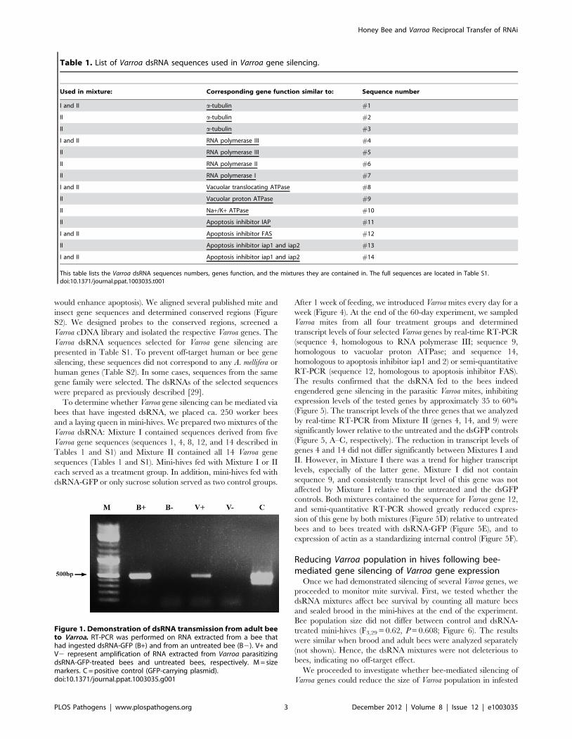
indicated bee-to-mite transfer (Figure 1).
Mites can absorb dsRNAs and siRNAs by physical contact [24].
To determine any biological function, it was necessary to establish
that RNAi triggers were delivered by food ingestion and not by
accidental contamination, and that the acquired dsRNA was
present in the bee hemolymph (the food source of Varroa). To this
end we strapped individual bees to a hollow plastic tube, and fed
them directly to their proboscis with dsRNA-GFP. This prevented
potential contamination of other bee parts by self or mutual
grooming with other bees. On the following day, we released each
bee to a separate plastic box, and added two female Varroa mites to
each of the above boxes. The Varroa were collected from a remote
hive that had not been exposed to dsRNA. On the third day, we
collected Varroa attached to the bees, and extracted hemolymph
from the bees. DsRNA-GFP was detected in RNA extracted from
hemolymph of dsRNA-GFP-treated bees. DsRNA-GFP was also
found in Varroa parasitizing the treated bees (Figure 2), indicating
that dsRNA transfer was via ingestion and that treated bees do
carry dsRNA in their hemolymph.
Pursuant to establishing a hemolymph-mediated RNAi transfer
we studied the possibility of indirect horizontal RNAi transfer. We
placed ca. 250 worker bees and a laying queen in mini-hives and
fed them dsRNA-GFP for 8 days. Varroa mites were introduced
into the mini-hives on the fifth day of feeding, and were later
collected from sealed larval/pupal cells. The presence of dsRNA-
GFP in Varroa mites collected from sealed cells on various days of
larval/pupal development indicated indirect transfer of dsRNA
from bee larvae (which were fed by nurse bees that had themselves
fed on dsRNA-containing sugar solution) to Varroa (Figure 3A).
Bidirectional horizontal transfer of dsRNA between beesand Varroa mites
Having shown direct and indirect transfer of dsRNA from bees
to Varroa, we wanted to test whether a mite that has acquired
dsRNA from a bee can then transfer it to another bee that it
parasitizes. In the direct transfer experiment, bees were fed
dsRNA-GFP in 50% sucrose solution for 8 days, and Varroa mites
were added on the fifth day of feeding. To test for bidirectional
horizontal transfer, on the eighth day the mites were removed
from the dsRNA-carrying bees and introduced into a container
with untreated bees for 4 days. DsRNA-GFP was detected in RNA
extracts of bees that had not consumed dsRNA, but were
parasitized by Varroa mites that previously parasitized dsRNA-
carrying bees (Figure 3B). The presence of dsRNA-GFP in the
parasitized bees indicated direct reciprocal transfer of dsRNA
from bee to Varroa and on to another bee.
DsRNA stability in hives. We tested the stability of dsRNA
under honey bee hive conditions in order to determine the
potential of dsRNA for Varroa control. We placed a cage with 10
bees in an empty second floor of a hive, separated by screen from
the populated bottom floor, so that the caged bees experienced an
environment similar to that of the hive. The caged bees had access
to a feeder containing dsRNA-GFP in 10 ml 50% sucrose solution,
identical to the dsRNA-GFP concentration in the other exper-
iments. Under these conditions, dsRNA-GFP slowly degraded
along the first three days. By day six, dsRNA-GFP was almost fully
degraded (Figure S1).
Silencing of Varroa gene expression mediated by beesingesting dsRNA
Once we had established horizontal transfer of physiologically
inert dsRNA between bees and Varroa, we explored the possibility
that RNAi that originated in the bee might affect gene expression
in the mite.
When this study was initiated, the Varroa genome had not been
elucidated (recently, partial Varroa genome information has been
released [33]). Therefore, we designed a number of genes whose
silencing was expected to harm the Varroa mite. We chose
fundamental housekeeping genes involved in cytoskeleton assem-
bly, energy transfer and transcription. In addition, we chose genes
involved in apoptosis inhibition (assuming that their silencing
Author Summary
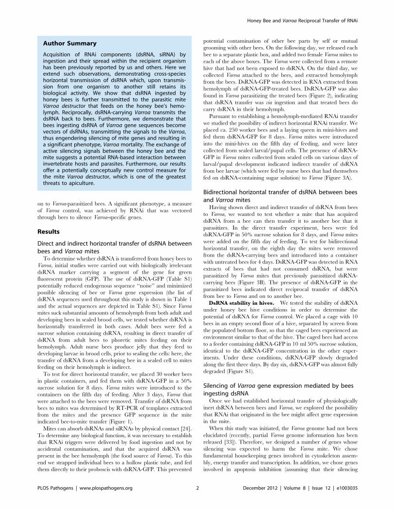
Acquisition of RNAi components (dsRNA, siRNA) byingestion and their spread within the recipient organismhas been previously reported by us and others. Here weextend such observations, demonstrating cross-specieshorizontal transmission of dsRNA which, upon transmis-sion from one organism to another still retains itsbiological activity. We show that dsRNA ingested byhoney bees is further transmitted to the parasitic miteVarroa destructor that feeds on the honey bee’s hemo-lymph. Reciprocally, dsRNA-carrying Varroa transmits thedsRNA back to bees. Furthermore, we demonstrate thatbees ingesting dsRNA of Varroa gene sequences becomevectors of dsRNAs, transmitting the signals to the Varroa,thus engendering silencing of mite genes and resulting ina significant phenotype, Varroa mortality. The exchange ofactive silencing signals between the honey bee and themite suggests a potential RNA-based interaction betweeninvertebrate hosts and parasites. Furthermore, our resultsoffer a potentially conceptually new control measure forthe mite Varroa destructor, which is one of the greatestthreats to apiculture.
Honey Bee and Varroa Reciprocal Transfer of RNAi
PLOS Pathogens | www.plospathogens.org 2 December 2012 | Volume 8 | Issue 12 | e1003035
would enhance apoptosis). We aligned several published mite and
insect gene sequences and determined conserved regions (Figure
S2). We designed probes to the conserved regions, screened a
Varroa cDNA library and isolated the respective Varroa genes. The
Varroa dsRNA sequences selected for Varroa gene silencing are
presented in Table S1. To prevent off-target human or bee gene
silencing, these sequences did not correspond to any A. mellifera or
human genes (Table S2). In some cases, sequences from the same
gene family were selected. The dsRNAs of the selected sequences
were prepared as previously described [29].
To determine whether Varroa gene silencing can be mediated via
bees that have ingested dsRNA, we placed ca. 250 worker bees
and a laying queen in mini-hives. We prepared two mixtures of the
Varroa dsRNA: Mixture I contained sequences derived from five
Varroa gene sequences (sequences 1, 4, 8, 12, and 14 described in
Tables 1 and S1) and Mixture II contained all 14 Varroa gene
sequences (Tables 1 and S1). Mini-hives fed with Mixture I or II
each served as a treatment group. In addition, mini-hives fed with
dsRNA-GFP or only sucrose solution served as two control groups.
After 1 week of feeding, we introduced Varroa mites every day for a
week (Figure 4). At the end of the 60-day experiment, we sampled
Varroa mites from all four treatment groups and determined
transcript levels of four selected Varroa genes by real-time RT-PCR
(sequence 4, homologous to RNA polymerase III; sequence 9,
homologous to vacuolar proton ATPase; and sequence 14,
homologous to apoptosis inhibitor iap1 and 2) or semi-quantitative
RT-PCR (sequence 12, homologous to apoptosis inhibitor FAS).
The results confirmed that the dsRNA fed to the bees indeed
engendered gene silencing in the parasitic Varroa mites, inhibiting
expression levels of the tested genes by approximately 35 to 60%
(Figure 5). The transcript levels of the three genes that we analyzed
by real-time RT-PCR from Mixture II (genes 4, 14, and 9) were
significantly lower relative to the untreated and the dsGFP controls
(Figure 5, A–C, respectively). The reduction in transcript levels of
genes 4 and 14 did not differ significantly between Mixtures I and
II. However, in Mixture I there was a trend for higher transcript
levels, especially of the latter gene. Mixture I did not contain
sequence 9, and consistently transcript level of this gene was not
affected by Mixture I relative to the untreated and the dsGFP
controls. Both mixtures contained the sequence for Varroa gene 12,
and semi-quantitative RT-PCR showed greatly reduced expres-
sion of this gene by both mixtures (Figure 5D) relative to untreated
bees and to bees treated with dsRNA-GFP (Figure 5E), and to
expression of actin as a standardizing internal control (Figure 5F).
Reducing Varroa population in hives following bee-mediated gene silencing of Varroa gene expression
Once we had demonstrated silencing of several Varroa genes, we
proceeded to monitor mite survival. First, we tested whether the
dsRNA mixtures affect bee survival by counting all mature bees
and sealed brood in the mini-hives at the end of the experiment.
Bee population size did not differ between control and dsRNA-
treated mini-hives (F3,29 = 0.62, P = 0.608; Figure 6). The results
were similar when brood and adult bees were analyzed separately
(not shown). Hence, the dsRNA mixtures were not deleterious to
bees, indicating no off-target effect.
We proceeded to investigate whether bee-mediated silencing of
Varroa genes could reduce the size of Varroa population in infested
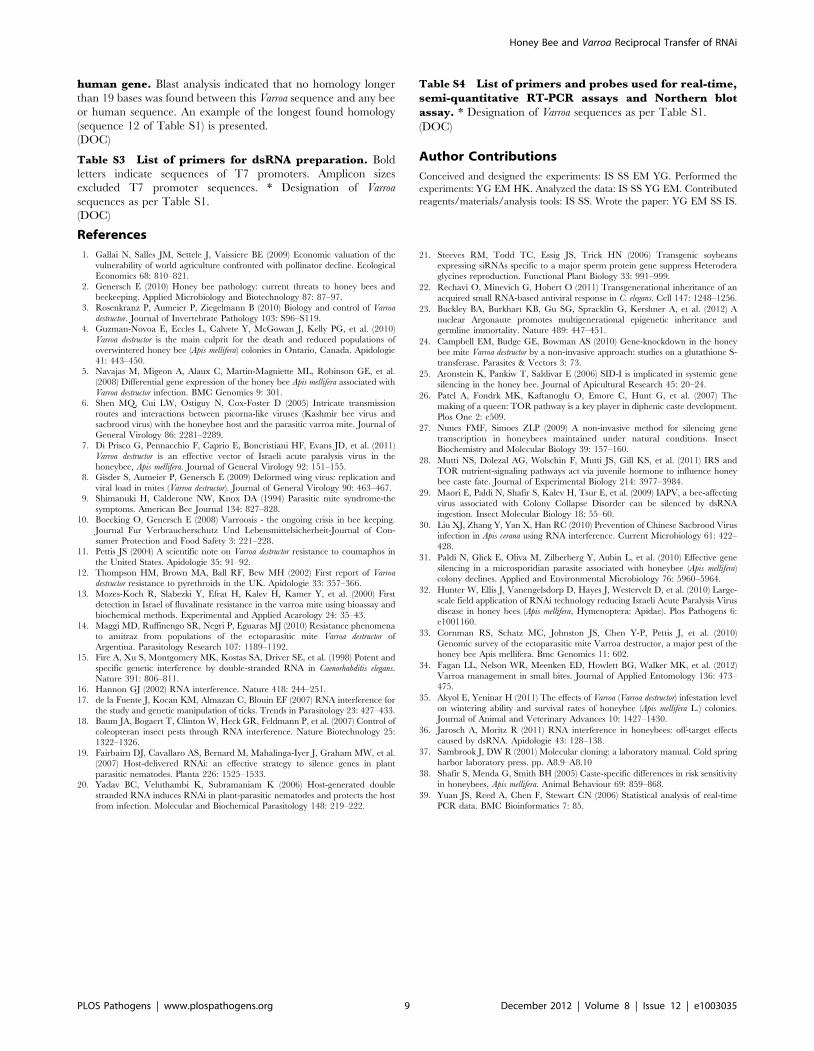
Table 1. List of Varroa dsRNA sequences used in Varroa gene silencing.
Used in mixture: Corresponding gene function similar to: Sequence number
I and II a-tubulin #1
II a-tubulin #2
II a-tubulin #3
I and II RNA polymerase III #4
II RNA polymerase III #5
II RNA polymerase II #6
II RNA polymerase I #7
I and II Vacuolar translocating ATPase #8
II Vacuolar proton ATPase #9
II Na+/K+ ATPase #10
II Apoptosis inhibitor IAP #11
I and II Apoptosis inhibitor FAS #12
II Apoptosis inhibitor iap1 and iap2 #13
I and II Apoptosis inhibitor iap1 and iap2 #14
This table lists the Varroa dsRNA sequences numbers, genes function, and the mixtures they are contained in. The full sequences are located in Table S1.doi:10.1371/journal.ppat.1003035.t001
Figure 1. Demonstration of dsRNA transmission from adult beeto Varroa. RT-PCR was performed on RNA extracted from a bee thathad ingested dsRNA-GFP (B+) and from an untreated bee (B2). V+ andV2 represent amplification of RNA extracted from Varroa parasitizingdsRNA-GFP-treated bees and untreated bees, respectively. M = sizemarkers. C = positive control (GFP-carrying plasmid).doi:10.1371/journal.ppat.1003035.g001
Honey Bee and Varroa Reciprocal Transfer of RNAi
PLOS Pathogens | www.plospathogens.org 3 December 2012 | Volume 8 | Issue 12 | e1003035

hives. We determined the number of Varroa individuals per bee by
examining the mite population on mature bees and in sealed
brood cells at the end of the experiment.
Varroa infestation was reduced in mini-hives treated with Varroa
dsRNA compared to the controls (F3,29 = 5.65, P = 0.0035;
Figure 7). The effect was greater with Mixture II, which targeted
more genes than Mixture I, reducing Varroa populations by an
average 53% compared to the dsRNA-GFP control, and by 61%
compared to the untreated control.
Discussion
Gene silencing following ingestion of dsRNA by honey bees has
been previously reported [25–32]. Here, we show that the ingested
dsRNA can be delivered across species to a bee-hemolymph-
dependent parasite. This RNAi transfer may cause silencing of
Varroa gene expression and reduce mite populations in hives.
Quantitative and semi-quantitative RT-PCR indicated that the
dsRNAs affected the targeted Varroa genes. From an RNA biology
point of view, the possibility that closely-associated organisms may
interact via RNAi pathways should be further explored.
The Varroa mite has been demonstrated to be a vector for bee
viruses [2,6–8]. Similarly, we show that the mite can also vector
RNAi-triggers, which were acquired from hemolymph of bees that
had consumed dsRNA. Additionally, we demonstrated that bees
vector biologically active dsRNA to the Varroa mite. The
occurrence of such reciprocal interactions raises the hypothesis
that bees may be potential vectors for Varroa-affecting viruses.
Although the main emphasis of this study is the cross-species
transfer and effect of RNAi, it also reflects on a promising concept
of Varroa control. With Varroa mites evolving resistance to the
chemicals used for their control [11–14], our study provides a
novel potential approach for relieving the most serious economic
burden on apiculture. Consistent with our findings of the stability
of dsRNA in the honey bee colony, it is stable enough to be
efficient when administered to the colony in sugar solution [29,32],
yet it eventually degrades in an environment favorable to bacterial
growth. Varroa mainly devastate colonies when Varroa population
grows unchecked. Effective control measures do not necessarily
need to completely eradicate the Varroa population [34,35].
Further studies would need to monitor the effect of dsRNA
treatment on Varroa population dynamics and long-term effect on
honey bee colonies under field conditions. Furthermore, the
dsRNA formula may be optimized by finding more vital target
genes that will lead to a greater reduction of the mite population in
hives with less amount of dsRNA fed to infested hive.
We prepared two dsRNA formulations: Mixture I targeted 5
Varroa gene sequences and Mixture II targeted 14 Varroa gene
sequences. Mixture II tended to reduce Varroa populations more
effectively (Figure 7). This suggests the existence of overlapping
metabolic pathways or that some of the gene products are stable
and remain active even though their respective gene’s expression
had been silenced.
We selected dsRNA sequences that are not homologous to
honey bee (or human) sequences. As in other reports [31,32],
silencing (in this case of the Varroa genes) did not affect the vigor of
the bees (Figure 6). Notably, we did not notice the off-target effects
reported by Jarosch and Moritz [36]. Although Varroa infestation
was greater in control vs. treatment mini-hives (Figure 7), this did
not affect the strength of the hives at the end of our experiment
Figure 2. Demonstration of dsRNA transmission from adult beeto Varroa via the bee hemolymph. Northern blot assay wasperformed on RNA extracted from a bee that had ingested dsRNA-GFP (B+) and from an untreated bee (B2), pooled RNA extracted fromhemolymph collected from bees that had ingested dsRNA-GFP (H+) andfrom untreated bees (H2), and pooled RNA extracted from Varroa mitesparasitizing dsRNA-GFP-treated bees (V+) and untreated bees (V2).C = positive control (GFP-carrying plasmid).doi:10.1371/journal.ppat.1003035.g002 Figure 3. Demonstration of dsRNA transfer from bee to Varroa
and from Varroa to bee. (A) Indirect dsRNA transmission from bee toVarroa parasitizing bee brood. RT-PCR of Varroa-extracted RNA.Numbers represent days from the beginning of dsRNA feeding. M = sizemarkers. C = positive control (GFP-carrying plasmid). + indicates samplescollected from dsRNA-GFP-treated hives. – indicates samples collectedfrom untreated, control hives. (B) DsRNA transmission from Varroa tobee. RT-PCR was performed on RNA extracted from a dsRNA-GFP-carrying Varroa (V+) and from Varroa devoid of dsRNA-GFP (V2). B+represents amplification of RNA from bees infested with dsRNA-GFP-carrying Varroa and B– represents amplification from bees infested withdsRNA-GFP-devoid Varroa. M and C: as in legend for A.doi:10.1371/journal.ppat.1003035.g003
Honey Bee and Varroa Reciprocal Transfer of RNAi
PLOS Pathogens | www.plospathogens.org 4 December 2012 | Volume 8 | Issue 12 | e1003035

(Figure 6). This is not surprising, since Varroa were present in our
hives for only about 7 weeks. Varroa damage is cumulative and is
minimal in newly infested hives (hives collapse after 2–3 years
[3,10]).
Materials and Methods
DsRNA preparationDsRNA was prepared according to Maori et al. [29]. Sequences
were amplified by PCR using specific primers including the 59 tail
of the T7 promoter (Table S1). PCR products were TA cloned
into the plasmid pDRIVE and sequenced. Amplicons were used as
template for in-vitro transcription.
RNA extraction and analysisTotal RNA for dsRNA-GFP detection experiments was
isolated from a single honey bee or from 10 Varroa mites, using
phenol-chloroform extraction (peqGOLD Trifast, Peqlab). Total
RNA for Varroa dsRNA experiments or for dsRNA-GFP
detection was isolated from 5 Varroa mites or from 50 mites,
respectively, with the ZR Tissue & Insect RNA MicroPrep (Zymo
Research) according to the manufacturer’s instructions. Eluted
RNA was treated with TURBO DNA-free kit (Ambion, Austin,
TX, USA) and tested for DNA contamination. Varroa RNA was
then co-precipitated with glycogen and 3 M sodium acetate in
70% ethanol and resuspended in 20 ml of RNAse-free water. The
amount and quality of the RNA samples were determined by
spectrophotometer (NanoDrop Technologies, Wilmington, DE,
USA). RNA from hemolymph was extracted using the phenol:
chloroform: Isoamyl alcohol method according to published
protocol [37].
DsRNA-GFP detection by RT-PCRDsRNA-GFP was detected by RT-PCR using Verso 1-Step
RT-PCR (Thermo Scientific) with specific GFP primers according
to the manufacturer’s protocol. Total RNA samples extracted
from 10 Varroa or 1 honey bee were used as templates.
DsRNA-GFP detection by northern blot analysisNorthern blot assay for detecting dsRNA-GFP was performed
as follows: Samples of RNA (500 ng) were electrophoresed on
1.2% agarose gel. The gel was washed with 0.25 M HCl,
denaturation solution, neutralization solution and 10XSSC before
the RNA was transferred to a positively charged nylon membrane
(Roche Diagnostics). The membrane was treated according to the
manufacturer’s protocol, with DIG-labeled probe (Roche Diag-
nostics) of GFP sequence corresponding to the sequence used as
template for dsRNA-GFP synthesis.
cDNA library constructionA Varroa cDNA library was prepared using a Smart cDNA
construction kit (Clontech) according to the manufacturer’s
instructions. Genes involved in four activity categories were
designated. Database-recorded mite and insect genes belonging to
those groups were aligned (Figure S2). Conserved sequences were
determined for each group and served as probes for selecting the
homologous genes from a Varroa cDNA library. The actual Varroa
genes were sequenced. Segments of Varroa genes, 200 to 450 bp in
length, which did not correspond in sequence to any bee or human
genes (identity of less than 21 consecutive bases; Table S2), were
selected. The selected Varroa sequences and GFP partial sequence
are presented in Table S3.
Gene expression: Real-time RT-PCR and semi-quantitativeRT-PCR
RNA (400 ng) was subjected to reverse transcription with
random hexamers using the Verso cDNA synthesis kit (Thermo
Scientific). Each sample of the obtained cDNA was diluted 1:50
before amplification. Real-time quantitative PCR was performed
by LightCycler 480 (Roche) and was analyzed with the instru-
ment’s software. The employed primers and probes are listed in
Table S4. The real-time PCR program was as follows: 95uC for
10 min, followed by 45 cycles of 95uC for 10 s and 60uC for 30 s.
At the end, samples were subjected to 40uC for 30 s. 18S rRNA
was used as an internal control for the standardization of RNA
levels.
The semi-quantitative PCR program was as follows: 95uC for
10 min, followed by 40 cycles, each consisting of 95uC for 10 s
and 65uC and 55uC for 30 s for the apoptosis inhibitor (FAS) and
its internal standardization control (actin), respectively, followed by
72uC for 30 s. Every three cycles starting from cycle 31 for FAS
and 29 for actin, a tube was taken out, incubated for 5 min at
72uC and stored at 220uC. Samples were analyzed on a 1.2%
agarose gel. Each qPCR experiment was repeated three times.
Figure 4. Schematic representation of the honey bee feeding regimen. The experiment lasted for 60 days. Top: schematic representation ofa honey bee’s life cycle. Bottom: experimental procedures on a timescale.doi:10.1371/journal.ppat.1003035.g004
Honey Bee and Varroa Reciprocal Transfer of RNAi
PLOS Pathogens | www.plospathogens.org 5 December 2012 | Volume 8 | Issue 12 | e1003035

Figure 5. Silencing of Varroa gene expression following horizontal transfer of dsRNA from bee to Varroa. (A–C) Results (mean 6 SE) ofreal-time RT-PCR of Varroa genes 4, 14 and 9, respectively (Table S1). RNA from Varroa infesting untreated bees, which did not ingest dsRNA, or thosethat ingested the physiologically inert dsRNA-GFP served as controls. Note that treatment I is devoid of sequence 9. Different letters above columnsindicate significant differences between treatments (P,0.05). Details of the RT-PCR assays and statistical analysis are described in the Materials andMethods. (D–E) Semi-quantitative RT-PCR for expression of the apoptosis inhibitor FAS gene (sequence 12, Table S1). RNA was extracted from Varroa
Honey Bee and Varroa Reciprocal Transfer of RNAi
PLOS Pathogens | www.plospathogens.org 6 December 2012 | Volume 8 | Issue 12 | e1003035

Regimen of dsRNA-GFP feedingTo test for direct transfer of dsRNA-GFP from adult bee to
mite, 1-day-old bees that emerged in an incubator were placed in
four plastic containers (30 bees per container). Two containers
were fed with 30 mg dsRNA-GFP in 200 ml of 50% sucrose
solution for 8 days, and the other two containers were controls, fed
only 50% sucrose solution. Adult female Varroa (n = 30) were
introduced into each container on day 5. After 3 days, Varroa that
were attached to bees were removed and collected and their RNA
was isolated for dsRNA-GFP analysis. A possible caveat of this
experiment was that dsRNA-GFP would be transferred to Varroa
through direct contamination with dsRNA-GFP in the containers,
or from contact with contaminated bee body parts.
We therefore performed an additional experiment to test for
dsRNA transfer to Varroa mites via the bee hemolymph. In the
morning of the experiment, 150 bees were collected from the area
of combs containing open larvae; these tend to be nurse bees. Each
bee was strapped into a hollow plastic tube in a manner that
ensured their ability to extend their proboscis, and minimized
injuries of the bees upon release [38]. The bees were divided into
two groups: Individuals in the first group were fed with 2.5 mg
dsRNA-GFP in a 5 ml 50% sucrose solution. Bees in the second
group served as the control group, and were fed with 5 ml 50%
sucrose solution only. In order to avoid contamination of dsRNA-
GFP on the bee body, the sucrose solution was given directly to the
proboscis. In addition, in order to prevent starvation of the bees
overnight, both groups (treated and control) were fed 5 ml 50%
sucrose solution in the evening. The following day, each bee was
released gently from the hollow plastic tube, placed in a clean cage
and supplied with candy (67% sugar powder and 33% honey).
Two female adult Varroa mites were added to each of the above
bees. Varroa were collected from a mite-infested hive that has never
been exposed to dsRNA. On the third day, each bee was
anaesthetized on ice, and 1–10 ml of hemolymph was collected.
The collection of hemolymph was performed by pricking a hole in
the inter-segmental membrane between the 2nd and 3rd
abdominal segment, and inserting a capillary tube. Prior to
hemolymph collection, Varroa mites, which were attached to the
bee’s body, were collected. All samples (Varroa mites, hemolymph
and bees) were placed directly on ice, and then stored at 280uCfor molecular analysis.
To test for bidirectional transfer of dsRNA-GFP from bee to
mite and on to another bee, some of the Varroa that had been
detached from bees were transferred to containers with newly
emerged, untreated bees for 4 days and their RNA was isolated for
dsRNA-GFP analysis. Every day, bees in all containers were given
an additional 1 ml sucrose solution after finishing their treatment.
In addition, bees had free access to a pollen patty consisting of
70% pollen mixed with sugar powder.
To test for indirect transfer of dsRNA-GFP from adult bee to
bee larva and on to mite feeding on the hemolymph of the
developing bee in a sealed cell, a cup of bees (ca. 250) and a laying
queen were introduced into each mini-hive (two replicates in each
of two enclosures). DsRNA-GFP (200 mg per hive) was provided
daily in 5 ml 50% sucrose solution for 8 days. Thirty Varroa mites
were introduced to the hives on the fifth day. Adult female Varroa
were collected from sealed cells from day 11 till day 30 and their
RNA was isolated for dsRNA-GFP analysis.
DsRNA-GFP stability in 50% sucrose solutionTo test the dsRNA stability in the hive, we placed 10 bees in a
cage, and exposed them to 10 ml of 50% sucrose solution that
contained 200 mg dsRNA-GFP. Hence, dsRNA-GFP final con-
centration was 20 ng/ml, identical to the dsRNA-GFP concentra-
tion that was used in the other experiments. The cage was placed
in a vacant second floor of a hive, separated by a screen from the
fed on bees that had ingested Mixture I or II (D). RNA was extracted from Varroa fed on dsRNA-untreated bees, and on bees that had ingested dsRNA-GFP (E). (F) Amplification of actin as a standardizing internal control. Reactions devoid of reverse transcriptase (RT) served as controls for the absenceof DNA contamination. Number of PCR cycles is indicated at the top of each electropherogram. SM = size markers.doi:10.1371/journal.ppat.1003035.g005
Figure 6. Mean (+ SE) total number of bees (capped brood and adults) in four treatments. Treatments did not differ significantly.doi:10.1371/journal.ppat.1003035.g006
Honey Bee and Varroa Reciprocal Transfer of RNAi
PLOS Pathogens | www.plospathogens.org 7 December 2012 | Volume 8 | Issue 12 | e1003035

populated bottom floor. Thus the caged bees were exposed to the
hive’s environment. Samples from the sugar solution were taken
on days 1, 2, 3 and 6, and placed on ice. The caged bees died on
day 2, possibly due to a heat wave. Sucrose concentration in the
samples was determined with a refractometer, and if needed,
equilibration with water was done. Samples were then stored at
280uC until analysis. Samples were analyzed on 1.2% agarose gel,
loaded with 5 ml from each sample (100 ng of dsRNA-GFP at time
zero).
Regimen of honey bee feeding with Varroa dsRNAThe experiment with Varroa dsRNA was conducted in mini-
hives, 12 mini-hives per replicate, and was repeated three times. In
each replicate, a cup of bees and a laying queen were placed in
each mini-hive. Three mini-hives were randomly assigned to one
of four netted enclosures, each representing a different feeding
treatment. Bees were fed 5 ml of 50% sucrose solution in troughs
placed in each mini-hive. The four treatments were: 1) sucrose
solution only (untreated control), 2) Mixture I (200 mg each of five
dsRNAs added to the sugar solution), 3) Mixture II (200 mg each
of 14 dsRNAs added to the sugar solution), and 4) dsRNA-GFP
(200 mg dsRNA) serving as an inert dsRNA control. Mini-hives
that fully consumed the treatment solutions were supplemented
with candy (67% sugar powder and 33% honey). In addition, the
bees were routinely fed pollen patties (70% pollen and 30% sugar
powder). Each replicate of the experiment lasted for 60 days
(Figure 4). Bees in each treatment were fed the respective solution
daily for the first 10 days and for the last 14 days, and twice a week
in the interim. Varroa mites were introduced into each mini-hive
from day 7 till day 14. In the first replicate, 30 mites were
introduced into each mini-hive; in the latter two replicates, 100
mites were introduced into each mini-hive. On day 60, all mature
bees were collected, counted and shaken with 70% ethanol
overnight in order to collect and count Varroa mites that fell off the
bees. All capped brood cells were opened to collect and count
Varroa mites. We calculated mites per bee (mature and developing).
Varroa mites, adult bees, emerging bees and pupae were stored for
molecular analyses.
Statistical analysisStatistical analyses were conducted with JMP statistical
software version 9 (SAS Institute, Cary, NC, USA). Statistical
significance was set at P,0.05. To test for significant differences
in relative expression, one-way ANOVA was conducted on ddCt
values [39]. Treatment was the main factor. To test for
differences in Varroa mite population, two-way ANOVA was
conducted on numbers of Varroa per bee in a block design with
treatment as main effect and experimental replicate as block. To
test for differences in total bee population, a similar two-way
ANOVA was conducted on the total number of bees (capped
brood and adults). Significant differences between treatments
were tested by the Tukey-Kramer (HSD) test.
Supporting Information
Figure S1 DsRNA stability in the hive. RT-PCR analysis of
dsRNA-GFP stability in 50% sucrose solution under hive
conditions. The solution was introduced to bees. Numbers
represent days from the time of dsRNA-GFP introduction.
SM = size markers.
(TIF)
Figure S2 An example of probe determination for pre-selected genes. A. Example sequences of mRNA for a tubulin of
mites and insects were aligned, a conservative region was selected
and a probe for selecting the homologous Varroa gene was
determined. B. The determined sequence of the selected a tubulin
probe based on the alignment of the known insect and mite
sequences (shown in A). Alternating bases (in parentheses) were
inserted to accommodate all possible sequence permutations.
(TIF)
Table S1 DsRNA-GFP sequence used as reporter se-quence and Varroa dsRNA sequences for Varroa genesilencing.
(DOC)
Table S2 An example of determining whether a selectedVarroa sequence may potentially off-target bee or
Figure 7. Mean (+ SE) number of Varroa mites per bee in four treatments. Different letters above columns indicate significant differencesbetween treatments (P,0.05).doi:10.1371/journal.ppat.1003035.g007
Honey Bee and Varroa Reciprocal Transfer of RNAi
PLOS Pathogens | www.plospathogens.org 8 December 2012 | Volume 8 | Issue 12 | e1003035
human gene. Blast analysis indicated that no homology longer
than 19 bases was found between this Varroa sequence and any bee
or human sequence. An example of the longest found homology
(sequence 12 of Table S1) is presented.
(DOC)
Table S3 List of primers for dsRNA preparation. Bold
letters indicate sequences of T7 promoters. Amplicon sizes
excluded T7 promoter sequences. * Designation of Varroa
sequences as per Table S1.
(DOC)
Table S4 List of primers and probes used for real-time,semi-quantitative RT-PCR assays and Northern blotassay. * Designation of Varroa sequences as per Table S1.
(DOC)
Author Contributions
Conceived and designed the experiments: IS SS EM YG. Performed the
experiments: YG EM HK. Analyzed the data: IS SS YG EM. Contributed
reagents/materials/analysis tools: IS SS. Wrote the paper: YG EM SS IS.
References
1. Gallai N, Salles JM, Settele J, Vaissiere BE (2009) Economic valuation of the
vulnerability of world agriculture confronted with pollinator decline. EcologicalEconomics 68: 810–821.
2. Genersch E (2010) Honey bee pathology: current threats to honey bees andbeekeeping. Applied Microbiology and Biotechnology 87: 87–97.
3. Rosenkranz P, Aumeier P, Ziegelmann B (2010) Biology and control of Varroa
destructor. Journal of Invertebrate Pathology 103: S96–S119.4. Guzman-Novoa E, Eccles L, Calvete Y, McGowan J, Kelly PG, et al. (2010)
Varroa destructor is the main culprit for the death and reduced populations ofoverwintered honey bee (Apis mellifera) colonies in Ontario, Canada. Apidologie
41: 443–450.5. Navajas M, Migeon A, Alaux C, Martin-Magniette ML, Robinson GE, et al.
(2008) Differential gene expression of the honey bee Apis mellifera associated with
Varroa destructor infection. BMC Genomics 9: 301.6. Shen MQ, Cui LW, Ostiguy N, Cox-Foster D (2005) Intricate transmission
routes and interactions between picorna-like viruses (Kashmir bee virus andsacbrood virus) with the honeybee host and the parasitic varroa mite. Journal of
General Virology 86: 2281–2289.
7. Di Prisco G, Pennacchio F, Caprio E, Boncristiani HF, Evans JD, et al. (2011)Varroa destructor is an effective vector of Israeli acute paralysis virus in the
honeybee, Apis mellifera. Journal of General Virology 92: 151–155.8. Gisder S, Aumeier P, Genersch E (2009) Deformed wing virus: replication and
viral load in mites (Varroa destructor). Journal of General Virology 90: 463–467.9. Shimanuki H, Calderone NW, Knox DA (1994) Parasitic mite syndrome-the
symptoms. American Bee Journal 134: 827–828.
10. Boecking O, Genersch E (2008) Varroosis - the ongoing crisis in bee keeping.Journal Fur Verbraucherschutz Und Lebensmittelsicherheit-Journal of Con-
sumer Protection and Food Safety 3: 221–228.11. Pettis JS (2004) A scientific note on Varroa destructor resistance to coumaphos in
the United States. Apidologie 35: 91–92.
12. Thompson HM, Brown MA, Ball RF, Bew MH (2002) First report of Varroa
destructor resistance to pyrethroids in the UK. Apidologie 33: 357–366.
13. Mozes-Koch R, Slabezki Y, Efrat H, Kalev H, Kamer Y, et al. (2000) Firstdetection in Israel of fluvalinate resistance in the varroa mite using bioassay and
biochemical methods. Experimental and Applied Acarology 24: 35–43.
14. Maggi MD, Ruffinengo SR, Negri P, Eguaras MJ (2010) Resistance phenomenato amitraz from populations of the ectoparasitic mite Varroa destructor of
Argentina. Parasitology Research 107: 1189–1192.15. Fire A, Xu S, Montgomery MK, Kostas SA, Driver SE, et al. (1998) Potent and
specific genetic interference by double-stranded RNA in Caenorhabditis elegans.Nature 391: 806–811.
16. Hannon GJ (2002) RNA interference. Nature 418: 244–251.
17. de la Fuente J, Kocan KM, Almazan C, Blouin EF (2007) RNA interference forthe study and genetic manipulation of ticks. Trends in Parasitology 23: 427–433.
18. Baum JA, Bogaert T, Clinton W, Heck GR, Feldmann P, et al. (2007) Control ofcoleopteran insect pests through RNA interference. Nature Biotechnology 25:
1322–1326.
19. Fairbairn DJ, Cavallaro AS, Bernard M, Mahalinga-Iyer J, Graham MW, et al.(2007) Host-delivered RNAi: an effective strategy to silence genes in plant
parasitic nematodes. Planta 226: 1525–1533.20. Yadav BC, Veluthambi K, Subramaniam K (2006) Host-generated double
stranded RNA induces RNAi in plant-parasitic nematodes and protects the hostfrom infection. Molecular and Biochemical Parasitology 148: 219–222.
21. Steeves RM, Todd TC, Essig JS, Trick HN (2006) Transgenic soybeansexpressing siRNAs specific to a major sperm protein gene suppress Heterodera
glycines reproduction. Functional Plant Biology 33: 991–999.
22. Rechavi O, Minevich G, Hobert O (2011) Transgenerational inheritance of an
acquired small RNA-based antiviral response in C. elegans. Cell 147: 1248–1256.
23. Buckley BA, Burkhart KB, Gu SG, Spracklin G, Kershner A, et al. (2012) A
nuclear Argonaute promotes multigenerational epigenetic inheritance and
germline immortality. Nature 489: 447–451.
24. Campbell EM, Budge GE, Bowman AS (2010) Gene-knockdown in the honey
bee mite Varroa destructor by a non-invasive approach: studies on a glutathione S-
transferase. Parasites & Vectors 3: 73.
25. Aronstein K, Pankiw T, Saldivar E (2006) SID-I is implicated in systemic gene
silencing in the honey bee. Journal of Apicultural Research 45: 20–24.
26. Patel A, Fondrk MK, Kaftanoglu O, Emore C, Hunt G, et al. (2007) The
making of a queen: TOR pathway is a key player in diphenic caste development.
Plos One 2: e509.
27. Nunes FMF, Simoes ZLP (2009) A non-invasive method for silencing gene
transcription in honeybees maintained under natural conditions. Insect
Biochemistry and Molecular Biology 39: 157–160.
28. Mutti NS, Dolezal AG, Wolschin F, Mutti JS, Gill KS, et al. (2011) IRS and
TOR nutrient-signaling pathways act via juvenile hormone to influence honeybee caste fate. Journal of Experimental Biology 214: 3977–3984.
29. Maori E, Paldi N, Shafir S, Kalev H, Tsur E, et al. (2009) IAPV, a bee-affecting
virus associated with Colony Collapse Disorder can be silenced by dsRNA
ingestion. Insect Molecular Biology 18: 55–60.
30. Liu XJ, Zhang Y, Yan X, Han RC (2010) Prevention of Chinese Sacbrood Virus
infection in Apis cerana using RNA interference. Current Microbiology 61: 422–
428.
31. Paldi N, Glick E, Oliva M, Zilberberg Y, Aubin L, et al. (2010) Effective gene
silencing in a microsporidian parasite associated with honeybee (Apis mellifera)
colony declines. Applied and Environmental Microbiology 76: 5960–5964.
32. Hunter W, Ellis J, Vanengelsdorp D, Hayes J, Westervelt D, et al. (2010) Large-
scale field application of RNAi technology reducing Israeli Acute Paralysis Virus
disease in honey bees (Apis mellifera, Hymenoptera: Apidae). Plos Pathogens 6:
e1001160.
33. Cornman RS, Schatz MC, Johnston JS, Chen Y-P, Pettis J, et al. (2010)
Genomic survey of the ectoparasitic mite Varroa destructor, a major pest of thehoney bee Apis mellifera. Bmc Genomics 11: 602.
34. Fagan LL, Nelson WR, Meenken ED, Howlett BG, Walker MK, et al. (2012)
Varroa management in small bites. Journal of Applied Entomology 136: 473–
475.
35. Akyol E, Yeninar H (2011) The effects of Varroa (Varroa destructor) infestation level
on wintering ability and survival rates of honeybee (Apis mellifera L.) colonies.
Journal of Animal and Veterinary Advances 10: 1427–1430.
36. Jarosch A, Moritz R (2011) RNA interference in honeybees: off-target effects
caused by dsRNA. Apidologie 43: 128–138.
37. Sambrook J, DW R (2001) Molecular cloning: a laboratory manual. Cold spring
harbor laboratory press. pp. A8.9–A8.10
38. Shafir S, Menda G, Smith BH (2005) Caste-specific differences in risk sensitivity
in honeybees, Apis mellifera. Animal Behaviour 69: 859–868.
39. Yuan JS, Reed A, Chen F, Stewart CN (2006) Statistical analysis of real-time
PCR data. BMC Bioinformatics 7: 85.
Honey Bee and Varroa Reciprocal Transfer of RNAi
PLOS Pathogens | www.plospathogens.org 9 December 2012 | Volume 8 | Issue 12 | e1003035