Benign childhood focal epilepsies: assessment of - Brain
23
REVIEW ARTICLE Benign childhood focal epilepsies: assessment of established and newly recognized syndromes Chrysostomos P. Panayiotopoulos, Michael Michael, Sue Sanders, Thalia Valeta and Michael Koutroumanidis Department of Clinical Neurophysiology and Epilepsies, St Thomas’ Hospital, Guy’s and St Thomas NHS Foundation Trust, London, UK Correspondence to: Michael Koutroumanidis, MD, Department of Clinical Neurophysiology and Epilepsies, St Thomas’ Hospital, London SE1 7EH, UK E-mail: [email protected] A big advance in epileptology has been the recognition of syndromes with distinct aetiology, clinical and EEG features, treatment and prognosis. A prime and common example of this is rolandic epilepsy that is well known by the general paediatricians for over 50 years, thus allowing a precise diagnosis that predicts an excel- lent prognosis. However, rolandic is not the only benign childhood epileptic syndrome. Converging evidence from multiple and independent clinical, EEG and magnetoencephalographic studies has documented Panayiotopoulos syndrome (PS) as a model of childhood autonomic epilepsy, which is also common and benign. Despite high prevalence, lengthy and dramatic features, PS as well as autonomic status epilepticus had eluded recognition because emetic and other ictal autonomic manifestations were dismissed as non-epileptic events of other dis- eases. Furthermore, PS because of frequent EEG occipital spikes has been erroneously considered as occipital epilepsy and thus confused with the idiopathic childhood occipital epilepsy of Gastaut (ICOE-G), which is another age-related but rarer and of unpredictable prognosis syndrome. Encephalitis is a common misdiagnosis for PS and migraine with visual aura for ICOE-G. Pathophysiologically, the symptomatogenic zone appears to correspond to the epileptogenic zone in rolandic epilepsy (sensory-motor symptomatology of the rolandic cortex) and the ICOE-G (occipital lobe symptomatology), while the autonomic clinical manifestations of PS are likely to be generated by variable and widely spread epileptogenic foci acting upon a temporarily hyperexci- table central autonomic network. Rolandic epilepsy, PS, ICOE-G and other possible clinical phenotypes of benign childhood focal seizures are likely to be linked together by a genetically determined, functional derangement of the systemic brain maturation that is age related (benign childhood seizure susceptibility syndrome). This is usually mild but exceptionally it may diverge to serious epileptic disorders such as epileptic encephalopathy with continuous spike and wave during sleep. Links with other benign and age-related seizures in early life such as febrile seizures, benign focal neonatal and infantile seizures is possible. Overlap with idiopathic generalized epilepsies is limited and of uncertain genetic significance. Taking all these into account, benign childhood focal seizures and related epileptic syndromes would need proper multi-disciplinary re-assessment in an evidence- based manner. Keywords: rolandic epilepsy; Panayiotopoulos syndrome; occipital epilepsy of Gastaut; autonomic seizures; autonomic status epilepticus; ictus emeticus; visual hallucinations; childhood seizure susceptibility; migraine, cyclic vomiting syndrome Abbreviations: AED = anti-epileptic drugs; BCSSS = benign childhood seizure susceptibility syndrome; CTS = centro-temporal spikes; CSWS = continuous spike and wave during sleep; FOS = fixation off sensitivity; GSES = giant somatosensory evoked spikes; GTCS = generalized tonic ^ clonic seizures; ICOE-G = idiopathic childhood occipital epilepsy of Gastaut; ILAE = International League against Epilepsy; IPS = intermittent photic stimulation; OPLS = Oro-pharyngo-laryngeal symptoms; PS = Panayiotopoulos syndrome Received April 14, 2008. Revised June 30, 2008. Accepted July 1, 2008 doi:10.1093/brain/awn162 Brain (2008), 131 , 2264^2286 ß The Author (2008). Published by Oxford University Press on behalf of the Guarantors of Brain. All rights reserved. For Permissions, please email: [email protected] Downloaded from https://academic.oup.com/brain/article/131/9/2264/283334 by guest on 01 January 2022
Transcript of Benign childhood focal epilepsies: assessment of - Brain

REVIEW ARTICLE
Benign childhood focal epilepsies: assessment ofestablished and newly recognized syndromesChrysostomos P. Panayiotopoulos, Michael Michael, Sue Sanders, Thalia Valeta andMichael Koutroumanidis
Department of Clinical Neurophysiology and Epilepsies, St Thomas’ Hospital, Guy’s and St Thomas NHS FoundationTrust,London, UK
Correspondence to: Michael Koutroumanidis, MD, Department of Clinical Neurophysiology and Epilepsies, St Thomas’Hospital, London SE1 7EH, UKE-mail: [email protected]
A big advance in epileptology has been the recognition of syndromes with distinct aetiology, clinical and EEGfeatures, treatment and prognosis. A prime and common example of this is rolandic epilepsy that is wellknown by the general paediatricians for over 50 years, thus allowing a precise diagnosis that predicts an excel-lent prognosis.However, rolandic is not the only benign childhood epileptic syndrome.Converging evidence frommultiple and independent clinical, EEG andmagnetoencephalographic studies has documented Panayiotopoulossyndrome (PS) as a model of childhood autonomic epilepsy, which is also common and benign. Despite highprevalence, lengthy and dramatic features, PS as well as autonomic status epilepticus had eluded recognitionbecause emetic and other ictal autonomic manifestations were dismissed as non-epileptic events of other dis-eases. Furthermore, PS because of frequent EEG occipital spikes has been erroneously considered as occipitalepilepsy and thus confused with the idiopathic childhood occipital epilepsy of Gastaut (ICOE-G), which isanother age-related but rarer and of unpredictable prognosis syndrome. Encephalitis is a commonmisdiagnosisfor PS and migraine with visual aura for ICOE-G. Pathophysiologically, the symptomatogenic zone appears tocorrespond to the epileptogenic zone in rolandic epilepsy (sensory-motor symptomatology of the rolandiccortex) and the ICOE-G (occipital lobe symptomatology), while the autonomic clinical manifestations of PSare likely to be generated by variable and widely spread epileptogenic foci acting upon a temporarily hyperexci-table central autonomic network. Rolandic epilepsy, PS, ICOE-G and other possible clinical phenotypes of benignchildhood focal seizures are likely to be linked together by a genetically determined, functional derangement ofthe systemic brain maturation that is age related (benign childhood seizure susceptibility syndrome). This isusually mild but exceptionally it may diverge to serious epileptic disorders such as epileptic encephalopathywith continuous spike and wave during sleep. Links with other benign and age-related seizures in early life suchas febrile seizures, benign focal neonatal and infantile seizures is possible. Overlap with idiopathic generalizedepilepsies is limited and of uncertain genetic significance.Taking all these into account, benign childhood focalseizures and related epileptic syndromes would need proper multi-disciplinary re-assessment in an evidence-based manner.
Keywords: rolandic epilepsy; Panayiotopoulos syndrome; occipital epilepsy of Gastaut; autonomic seizures; autonomicstatus epilepticus; ictus emeticus; visual hallucinations; childhood seizure susceptibility; migraine, cyclic vomiting syndrome
Abbreviations: AED=anti-epileptic drugs; BCSSS = benign childhood seizure susceptibility syndrome;CTS = centro-temporal spikes; CSWS = continuous spike and wave during sleep; FOS = fixation off sensitivity;GSES = giant somatosensory evoked spikes; GTCS = generalized tonic^ clonic seizures; ICOE-G = idiopathic childhoodoccipital epilepsy of Gastaut; ILAE = International League against Epilepsy; IPS = intermittent photic stimulation;OPLS = Oro-pharyngo-laryngeal symptoms; PS = Panayiotopoulos syndrome
Received April 14, 2008. Revised June 30, 2008. Accepted July 1, 2008
doi:10.1093/brain/awn162 Brain (2008), 131, 2264^2286
� The Author (2008). Published by Oxford University Press on behalf of the Guarantors of Brain. All rights reserved. For Permissions, please email: [email protected]
Dow
nloaded from https://academ
ic.oup.com/brain/article/131/9/2264/283334 by guest on 01 January 2022

Benign childhood focal seizures and related idiopathic epilep-tic syndromes affect approximately 22% of children withnon-febrile seizures and constitute a significant part of theeveryday practice of paediatricians, neurologists and electro-encephalographers. They comprise three identifiable electro-clinical syndromes recognized by the International Leagueagainst Epilepsy (ILAE) (Engel, 2006): rolandic epilepsywhich is well known, Panayiotopoulos syndrome (PS), acommon autonomic epilepsy, which is currently more readilydiagnosed and the idiopathic childhood occipital epilepsy ofGastaut (ICOE-G) including the idiopathic photosensitiveoccipital lobe epilepsy, a less common form with uncertainprognosis. There are also reports of children with benign focalseizures of predominantly affective symptoms, and claimshave been made for other clinical phenotypes associated withspecific inter-ictal EEG foci, such as frontal, midline orparietal, with or without giant somatosensory evoked spikes(GSES). Neurological and mental states and brain imaging arenormal, though because of their high prevalence any type ofbenign childhood focal seizures may incidentally occur inchildren with neurocognitive deficits or abnormal brainscans. The most useful diagnostic test is the EEG. In clinicalpractice, the combination of a normal child with infrequentseizures and an EEG showing disproportionately severe spikeactivity is highly suggestive of these benign childhoodsyndromes (Panayiotopoulos, 1999a).
All these conditions may be linked together in a broad,age-related and age-limited, benign childhood seizuresusceptibility syndrome (BCSSS), which may be geneticallydetermined (Panayiotopoulos, 1993).
This appraisal is based on over 30 years clinical research,extensive review of English and other journals includingrelevant book chapters and personal communications withexperts. Details of original studies, numerous case historiesand published reports not cited here can be found in ourprevious reviews (Panayiotopoulos, 1999a, 2002, 2007;Koutroumanidis, 2007).
Rolandic epilepsy (benign childhood epilepsywith centro-temporal spikes)Rolandic epilepsy is the best known and most commonbenign childhood focal epilepsy ((Beaussart and Faou, 1978;Loiseau et al., 1988; Bouma et al., 1997; Beaussart et al., 1999;Panayiotopoulos, 1999a; Dalla Bernardina et al., 2005; Wirrellet al., 2006; Fejerman, 2008). The age of onset ranges from 1to 14 years with 75% starting between 7 and 10 years. There isa 1.5 male predominance, prevalence is around 15% in child-ren aged 1–15 years with non-febrile seizures and incidence is10–20/100 000 children aged 0–15 years (Heijbel et al., 1975;Cavazzuti, 1980; Sidenvall et al., 1996; Astradsson et al., 1998;Berg et al., 1999; Larsson and Eeg-Olofsson, 2006).
Clinical manifestationsThe cardinal features of rolandic epilepsy are focal seizuresconsisting of unilateral facial sensory-motor symptoms (30%
of patients), oro-pharyngo-laryngeal symptoms (OPLS)(53%), speech arrest (40%) and hypersalivation (30%)(Beaussart, 1972; Loiseau and Beaussart, 1973; Lerman andKivity, 1975; Bouma et al., 1997; Panayiotopoulos, 1999a;Dalla Bernardina et al., 2005; Wirrell et al., 2006; Fejerman,2008). Ictal manifestations indicative of temporal lobeinvolvement do not occur in rolandic epilepsy, and theterm ‘centro-temporal’ refers only to the spike topographywhere it is partly a misnomer (see EEG section below).
Hemifacial sensory-motor seizures are mainly localized inthe lower lip and may spread to the ipsilateral hand. Motormanifestations are clonic contractions sometimes concurrentwith ipsilateral tonic deviation of the mouth, and sensorysymptoms consist of numbness in the corner of the mouth.OPLSs are unilateral sensory-motor symptoms of numbnessor paraesthesias (tingling, prickling or freezing) inside themouth, associated with strange sounds, such as death rattle,gargling, grunting and guttural sounds.
Hypersalivation, a prominent autonomic manifestation, isoften associated with hemifacial seizures, OPLSs and speecharrest. In speech arrest, the child is actually anarthric, unableto utter a single intelligible word and attempts to commu-nicate with gestures.
Consciousness and recollection are fully retained in morethan half (58%) of rolandic seizures. In the remainder,consciousness becomes impaired during the ictal progressand in one-third there is no recollection of ictal events.Progression to hemiconvulsions or generalized tonic–clonicseizures (GTCS) occurs in around half of children andhemiconvulsions may be followed by postictal Todd’shemiparesis (Panayiotopoulos, 1999a).
Three-quarters of rolandic seizures occur during non-REM (rapid eye movement) sleep, mainly at sleep onset orjust before awakening.
Rolandic seizures are usually brief lasting for 1–3 min.Focal motor, hemiconvulsive and generalized convulsivestatus epilepticus are rare at around 5% (Deonna et al., 1986;Wirrell et al., 1995; Panayiotopoulos, 1999a). Opercularstatus epilepticus usually occurs in children with atypicalevolution (Colamaria et al., 1991; Deonna et al., 1993;Fejerman et al., 2000) or may be induced by carbamazepineor lamotrigine (Caraballo et al., 1989; Parmeggiani et al.,2004). This state lasts for hours to months and consists ofongoing unilateral or bilateral contractions of the mouth,tongue or eyelids, positive or negative subtle perioral orother myoclonus, dysarthria, speech arrest, difficulties inswallowing, buccofacial apraxia and hypersalivation.
Other seizure typesDespite prominent hypersalivation, focal seizures withprimarily autonomic manifestations (autonomic seizures)are not considered part of the core clinical syndrome ofrolandic epilepsy. However, some children may presentwith independent autonomic seizures or seizures withmixed rolandic-autonomic manifestations including
Benign childhood focal epilepsies Brain (2008), 131, 2264^2286 2265
Dow
nloaded from https://academ
ic.oup.com/brain/article/131/9/2264/283334 by guest on 01 January 2022

emesis (see below in the relations between rolandic epilepsyand PS).
Primarily GTCS are considered part of rolandic epilepsyby the ILAE (Engel, 2006) and their occurrence can not beexcluded. However, from the published ictal recordings(Watanabe, 1996; Panayiotopoulos, 1999a; Wirrell et al.,2006) and the electroclinically unequivocal focal nature ofrolandic epilepsy, it can be inferred that at least the majorityof the GTCS follow rolandic activation, and are thereforesecondarily GTCS. Short-lived initial focal symptoms maypass unnoticed in daytime GTCS and are bound to be missedin nocturnal GTCS.
Typical absence seizures are considered rare (Beydounet al., 1992; Gelisse et al., 1999; Panayiotopoulos, 1999a)though a high incidence of them has also been reported(Beaumanoir et al., 1974).
ElectroencephalographyBy definition, centro-temporal spikes (CTS) are the hallmarkof benign childhood epilepsy with CTS (Fig. 1). However,although called centro-temporal, these spikes are mainlylocalized in the C3 and C4 (high central) or C5 and C6 (lowcentral) supra-sylvian and not temporal electrodes (Legardaet al., 1994; Panayiotopoulos, 1999a). CTS are often bilateraland typically activated by drowsiness and slow (non-REM)sleep, but not by overbreathing (Smith and Kellaway, 1964;Blom and Brorson, 1966; Clemens and Majoros, 1987). Inserial EEGs of the same child, CTS may occur right or left,infrequently or frequently, and appear small or giant, aloneor with spikes in other locations. Rarely, children withrolandic epilepsy may have normal EEG or CTS may appearonly during non-REM sleep (3–35%) (Panayiotopoulos,1999a). The incidence of extra-rolandic spikes in rolandicepilepsy is not precisely known but may be significant whenthese are sought (Drury and Beydoun, 1991).
Dipole EEG (Gregory and Wong, 1992; Tsai and Hung,1998; Jung et al., 2003), magnetoencephalography (MEG)(Minami et al., 1996; Huiskamp et al., 2004) and functionalMRI (Boor et al., 2007) studies have demonstrated that themain negative spike component of CTS is usually modelledby a single and stable tangential dipole source with thenegative pole maximum in the central region and thepositive pole maximum in the frontal region.
Brief 1–3 s generalized bursts of 3–5 Hz slow waves withintermixed small spikes without associated overt clinicalsymptoms may occur in about 4% of patients with rolandicepilepsy (Gelisse et al., 1999; Panayiotopoulos, 1999a).Typical 3 Hz spike–wave discharges are probably rare (seeabsence seizures above).
CTS are diagnostic markers of benign rolandic epilepsyonly in a suggestive clinical presentation. Their frequency,location and persistence do not determine the clinicalmanifestations, severity and frequency of seizures or theprognosis. It is well established that CTS are not specific to
rolandic epilepsy (Kellaway, 1980; Panayiotopoulos, 1999a)as they:
� occur in 2–3% of normal school-aged children, of whom510% develop rolandic seizures (Gibbs and Gibbs, 1967;Petersen and Eeg-Olofsson, 1971; Cavazzuti et al., 1980;Okubo et al., 1994).
� are common among relatives of children with rolandicepilepsy (Bray and Wiser, 1965; Bali et al., 2007)
� may occur in a variety of organic brain diseases with orwithout seizures, such as cerebral tumours, Rettsyndrome, fragile X syndrome and focal corticaldysplasia (Kellaway, 1980; Panayiotopoulos, 1999a)
� may incidentally be found in non-epileptic children withvarious symptoms, such as headache, speech and beha-vioural and learning difficulties (Gibbs and Gibbs, 1967).
Somatosensory stimulation is common form of activation ofCTS (10–20%) (De Marco and Tassinari, 1981;Panayiotopoulos, 1999a; Fonseca and Tedrus, 2000; Kubotaet al., 2000; Langill and Wong, 2003) and evokes GSES(Fig. 1), which correspond to mid- or long-latencysomatosensory evoked potentials (Manganotti et al., 1998).GSES, like spontaneous CTS, occur in children with orwithout seizures and disappear with age.
There have been around 20 reported ictal EEGs of rolandicseizures showing an initial paucity of spontaneous CTSbefore the onset of the ictal discharge, which appears in thecontralateral to the clinical manifestations rolandic regionsand consists of slow waves intermixed with spikes(Panayiotopoulos, 1999a; Wirrell et al., 2006; Fejermanet al., 2007) (Fig. 2). GTCS, when occurred, were precededby focal clinical and EEG features. (Watanabe, 1996;Panayiotopoulos, 1999a; Wirrell et al., 2006).
AetiologyRolandic epilepsy is genetically determined although con-ventional genetic influences may be less important than othermechanisms (Vadlamudi et al., 2004, 2006). There isevidence of linkage with chromosome 15q14 (Neubaueret al., 1998). Autosomal dominant inheritance with age-dependent penetrance refers to the EEG CTS and not to theclinical syndrome of rolandic epilepsy (Bray and Wiser, 1965;Bali et al., 2007).
Siblings or parents of patients with rolandic epilepsy mayrarely have the same type of seizures or other phenotypes ofBCSSS, such as PS. Reported occurrence of febrile seizuresranges from 10% to 20% (probably around 18%) of patients(Kajitani et al., 1992).
PathophysiologyAs indicated by the distribution of CTS, the epileptogeniczone in rolandic epilepsy involves neuronal networks withinthe rolandic cortex surrounding the central fissure bilaterally.This is congruent with the seizure symptomatogy
2266 Brain (2008), 131, 2264^2286 C. P. Panayiotopoulos et al.
Dow
nloaded from https://academ
ic.oup.com/brain/article/131/9/2264/283334 by guest on 01 January 2022

Fig. 1 Interictal EEG in rolandic epilepsy (top), PS (middle) and idiopathic childhood occipital epilepsy of Gastaut (bottom). (A) InterictalEEGwith CTS of two children with rolandic epilepsy separated by the vertical line. Left: spontaneous and GSES elicited by the patientsimultaneously tapping the tip of the fingers of both hands (arrows). Right: the same EEG sample at two different montages (extremeright is laplacian montage) to show that what appears as CTS are not temporal which on this occasion are localized in the lowercentral electrode (C5) of the laplacian montage. (B) Interictal EEG in five patients with PS separated by vertical lines. Despite similarclinical features, spikes are localized in the occipital, centro-temporal and frontal regions or they are frequently multifocal and may appearas clone-like, repetitive, spike^wave complexes. Brief generalized discharges of slow wave with small spikes (extreme right) are sometimesan interictal EEG feature. (C) Interictal EEG in two patients with ICOE-G separated by a vertical line. Left: Classical occipital paroxysmsdemonstrating FOS. Right: spontaneous scattered occipital spikes and occipital photosensitivity.
Benign childhood focal epilepsies Brain (2008), 131, 2264^2286 2267
Dow
nloaded from https://academ
ic.oup.com/brain/article/131/9/2264/283334 by guest on 01 January 2022
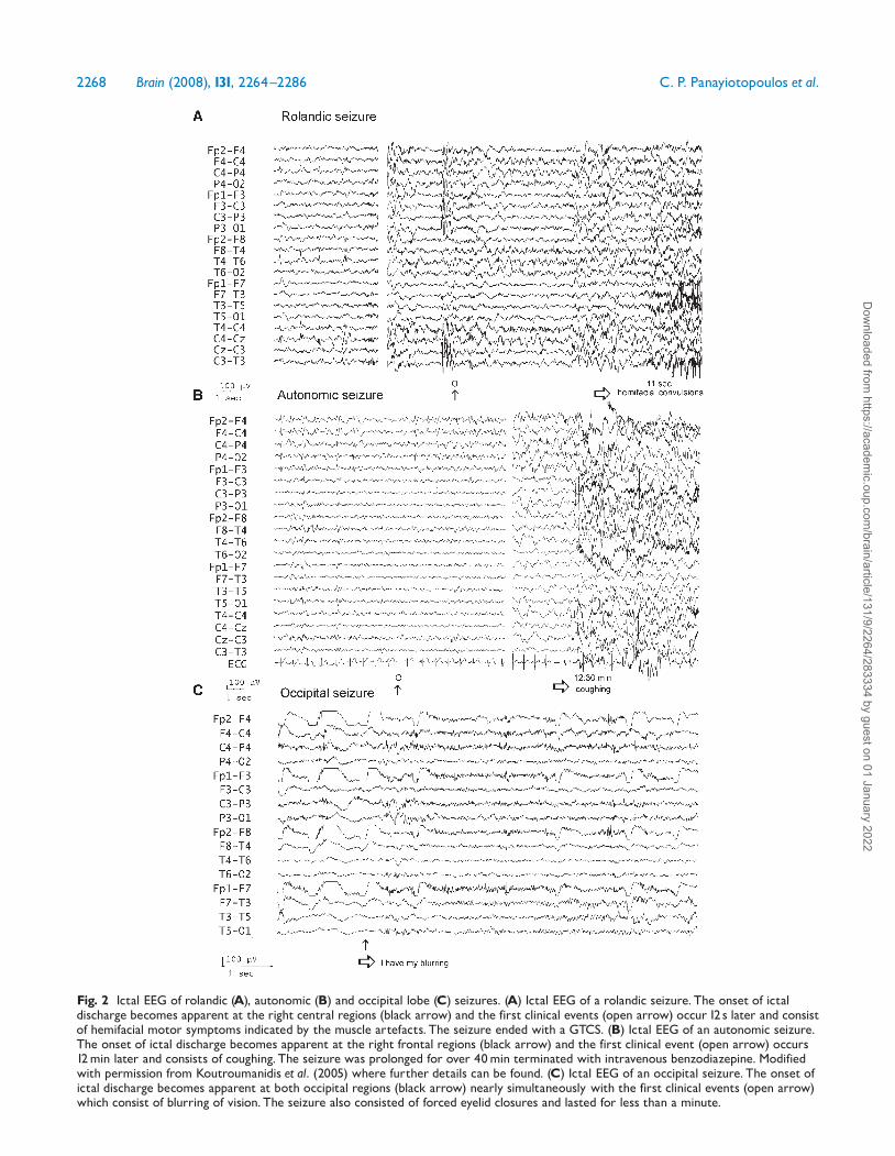
Fig. 2 Ictal EEG of rolandic (A), autonomic (B) and occipital lobe (C) seizures. (A) Ictal EEG of a rolandic seizure. The onset of ictaldischarge becomes apparent at the right central regions (black arrow) and the first clinical events (open arrow) occur 12 s later and consistof hemifacial motor symptoms indicated by the muscle artefacts. The seizure ended with a GTCS. (B) Ictal EEG of an autonomic seizure.The onset of ictal discharge becomes apparent at the right frontal regions (black arrow) and the first clinical event (open arrow) occurs12min later and consists of coughing. The seizure was prolonged for over 40min terminated with intravenous benzodiazepine. Modifiedwith permission from Koutroumanidis et al. (2005) where further details can be found. (C) Ictal EEG of an occipital seizure. The onset ofictal discharge becomes apparent at both occipital regions (black arrow) nearly simultaneously with the first clinical events (open arrow)which consist of blurring of vision. The seizure also consisted of forced eyelid closures and lasted for less than a minute.
2268 Brain (2008), 131, 2264^2286 C. P. Panayiotopoulos et al.
Dow
nloaded from https://academ
ic.oup.com/brain/article/131/9/2264/283334 by guest on 01 January 2022

(symptomatogenic zone) and in agreement with thosedescribed by Penfield and Rasmussen (1957) during electricalstimulation of the lower part of the pre-central and post-central gyrus in man.
The speech arrest is due to anarthia attributed to loss ofthe power and coordination of the musculature responsiblefor the articulation of words. There is no impairment of thecortical language networks. Hypersalivation most probablyrelates to the involvement of the superior bank of the sylvianfissure (Luders et al., 1987), but defining ictal symptomato-genesis by plotting the simple topographic coordinates of anictal discharge can hardly explain the high prevalence ofhypersalivation in benign rolandic epilepsy compared to itsexceptional only occurrence in adults with symptomatic fociof similar topography. Nor can it explain the opercularstatus epilepticus, with the lengthy for hours speech arrest,drooling and bilateral regional twitching that is associatedwith diffuse or bilateral rolandic spike wave activity, but doesnot propagate in a conventional way and does not involveother systems like for instance the motor strip or the lan-guage function. Therefore, at variance with the symptomaticadult focal epilepsies of comparable but more discretelylocalized topography, rolandic epilepsy reflects an age-relatedmaturational instability of the lower rolandic (somatosen-sory) cortex that represents the face and the oropharynxbilaterally (Koutroumanidis, 2007).
Evolution and prognosisThe prognosis for rolandic seizures is almost invariablyexcellent, with probably 52% risk of developing absenceseizures and less often GTCS in adult life (Beaumanoir et al.,1974; Lerman and Kivity, 1975; Blom and Heijbel, 1982;Loiseau et al., 1983; Panayiotopoulos, 1999a; Datta andSinclair, 2007; Koutroumanidis et al., 2008; Caraballo et al.,2008b). Remission occurs within 2–4 years from onset andbefore the age of 16 years. The total number of seizures islow, the majority of patients having fewer than 10 seizures;10–20% have just a single seizure. About 10–20% may havefrequent seizures, but these also remit with age.
Children with rolandic seizures may develop usually mildand reversible linguistic, cognitive and behavioural abnorm-alities during the active phase of the disease (Giordani et al.,2006; Nicolai et al., 2006; Riva et al., 2007; Kossoff et al.,2007; Kavros et al., 2008; Perkins et al., 2008). These may beworse in children with onset of seizures before 8 years ofage, high rate of occurrence and multifocal EEG spikes(Bulgheroni et al., 2008; Piccinelli et al., 2008; Boatman et al.,2008). The effect of anti-epileptic drugs (AED), the impact ofstigmatizing because of epilepsy, bias in selection of the mostserious cases and other factors have not been excluded inmost of these studies. The development, social adaptationand occupations of adults with a previous history of rolandicseizures was found normal (Blom and Heijbel, 1982; Loiseauet al., 1983).
Rarely (51%), rolandic epilepsy may evolve to more severesyndromes with linguistic, behavioural and neuropsycholo-gical deficits, such as Landau–Kleffner syndrome, atypicalfocal epilepsy of childhood or epilepsy with continuous spikeand wave during sleep (CSWS) (Fejerman et al., 2000) asexplained in the relevant section of this assessment.
Panayiotopoulos syndromePS is a common, childhood-related, susceptibility to auto-nomic seizures confirmed in long term studies of over 800children worldwide (Panayiotopoulos, 1981, 1988, 2002;Ferrie et al., 1997; Oguni et al., 1999; Kivity et al., 2000; Ladaet al., 2003; Ohtsu et al., 2003, 2008; Covanis et al., 2003;Caraballo et al., 2007; Dura-Trave et al., 2008).
The appraisal of PS here is based on in-depth analysis ofour own patients and review of the related literature up to2002 (Panayiotopoulos, 2002). Subsequent reports of around60 publications on this and related aspects have also beenthoroughly studied.
PS is defined as ‘benign age-related focal seizure disorderoccurring in early and mid-childhood. It is characterized byseizures, often prolonged, with predominantly autonomicsymptoms, and by an EEG that shows shifting and/ormultiple foci, often with occipital predominance’ (Ferrieet al., 2006). ‘Early onset benign childhood occipital epilepsy’often used as synonymous with PS (Taylor et al., 2003, 2008;Engel, 2006) does not represent the wide clinical, EEG andpathophysiological spectrum of PS, which is far beyond theoccipital neocortex (Martinovic, 2007).
Onset is from age 1 to 14 years with 76% starting between3 and 6 years. Both sexes are probably equally affected,though a female preponderance was found in some studies(Lada et al., 2003; Dura-Trave et al., 2008). Prevalence of PSmay be high though this is practically absent in designedcontrolled epidemiological studies (Berg et al., 1999; Jallonet al., 2001; Shinnar and Pellock, 2002; Cowan, 2002;Forsgren et al., 2005), which is understandable as thissyndrome was only recently formally recognized, its featuresimitate many other conditions and often manifests with asingle seizure only. In the original cohort of Panayiotopoulos(1988), prevalence was around 13% in children aged 3–6years with one or more non-febrile seizures, and 6% in theage group 1–15 years. These figures may be higher if childrenwho are currently considered to have atypicalclinical presentation are included in the syndrome(Panayiotopoulos, 2002; Covanis, 2006). PS is the mostcommon specific cause of non-febrile, non-convulsive statusepilepticus in childhood (Okanishi et al., 2008).
Clinical manifestationsThe hallmark of PS is ictal autonomic aberrations that mayinvolve any function of the autonomic system and mainlyemesis (70–80% of seizures). The following description ofclinical manifestations of PS are based on a synthetic analysisof available clinical historical data as perceived by patients
Benign childhood focal epilepsies Brain (2008), 131, 2264^2286 2269
Dow
nloaded from https://academ
ic.oup.com/brain/article/131/9/2264/283334 by guest on 01 January 2022
and witnessed by observers from our records and those pro-vided in the literature (Panayiotopoulos, 2002). Therefore,they may not accurately represent their true prevalence andsequence in PS.
Ictal autonomic symptomsSeizures commonly commence with autonomic manifesta-tions (80–90%), while consciousness and speech, as a rule,are preserved. Ictus emeticus (nausea and retching) culmi-nates in vomiting in 74–82% of seizures; in others, onlynausea or retching occurs and, in a quarter, emesis may notbe apparent. Emesis is usually the first apparent ictalsymptom, but it may also occur long after the onset ofother manifestations. Other autonomic manifestationsinclude pallor (28%), incontinence of urine (19%) andfaeces (3%), hypersalivation (10%), cyanosis (12%), mydria-sis (7%) and less often miosis (2%), coughing andabnormalities of intestinal motility (3%). Breathing (7%)and cardiac irregularities may be more common thanreported. Tachycardia is usually found, sometimes at theonset, in ictal EEG (Beaumanoir, 1993b; Oguni et al., 1999;Koutroumanidis et al., 2005; Parisi et al., 2005).Cardiorespiratory arrest is rare probably occurring in 1 per200 individuals; four cases out of around 1000 patients withPS have been reported but they all had complete recovery(Panayiotopoulos, 2002; Verrotti et al., 2005; Ferrie et al.,2006). Raised temperature has been documented in a fewcases (2%) after seizure onset. Cephalic auras of discomfortand odd sensations or headache commonly are describedwith other autonomic symptoms at seizure onset.
Syncopal-like manifestations occur in at least one-fifth ofseizures (Panayiotopoulos, 2002; Ferrie et al., 2006, 2007;Covanis, 2006; Caraballo et al., 2007). The child becomes‘completely unresponsive and flaccid like a rag doll’, whichmay precede, be concurrent with other seizure symptoms orbe the sole manifestation of a seizure (Oguni et al., 1999;Panayiotopoulos, 2002). They may occur while the patient isstanding, sitting, lying down or asleep and last from 1–2 minto half an hour.
Ictal behavioural changesRestlessness, agitation, terror or quietness, may occur at theonset of seizures, often in combination with other autonomicmanifestations.
Ictal non-autonomic symptomsPure autonomic seizures and pure autonomic status epil-epticus appear to occur in 10% of patients. They commenceand terminate solely with autonomic symptoms. However, inthe majority of seizures, autonomic manifestations arefollowed by conventional seizure symptoms. Nearly always,the child gradually or suddenly becomes confused or unres-ponsive. Other non-autonomic manifestations include inorder of prevalence unilateral deviation of the eyes oreyes opening (60–83%), speech arrest (8–13%), hemifacial
convulsions (6–13%), visual hallucinations (6–10%),OPLS (3%), unilateral drooping of the mouth (3%) andrarely (1%) eyelid or limb jerks, nystagmus or automatisms.The seizures may end with hemiconvulsions often withJacksonian marching (19–30%), or generalized convulsions(21–36%).
Autonomic manifestations may not be apparent atseizure onset even in witnessed diurnal seizures (510%).They may be absent, mild or missed in clinical observation.In these cases, eye deviation is the more common symptom.Visual symptoms are rare (1%) and not present inrecurrent seizures (Ferrie et al., 1997; Yalcin et al., 1997;Panayiotopoulos, 2002; Caraballo et al., 2007).
Duration of seizures and precipitating factorsThe seizures are usually lengthy of over 6 min and almosthalf of them last for 430 min to many hours, thus cons-tituting autonomic status epilepticus (Panayiotopoulos,2002; Ferrie et al., 2007). Lengthy seizures are equallycommon in sleep and wakefulness. Even after the most severeseizures and status, the patient is normal after a few hours’sleep. There is no record of residual neurological abnorm-alities. Hemiconvulsive or convulsive status epilepticus israre (4%).
Two-thirds of seizures start in sleep. Many seizures havebeen witnessed while travelling in a car, boat or aeroplane.The reason for this may be because in these circumstanceschildren easily fall asleep, seizures are more likely to bewitnessed and because travelling also precipitates motionsickness, to which children are particularly susceptible.
Intra-individual seizure variabilityThe same child may have brief and lengthy seizures,diurnal and nocturnal, with marked, inconspicuous, oreven without any autonomic changes (Ferrie et al., 1997;Oguni et al., 1999; Kivity et al., 2000; Panayiotopoulos, 2002;Lada et al., 2003; Ohtsu et al., 2003, 2008; Covanis et al.,2003; Caraballo et al., 2007). Even cardinal symptoms (suchas vomiting or eye deviation) may be present in one butabsent in another seizure. Seizures without autonomicmanifestations are rare (7%) and occur in patients whoalso have additional autonomic seizures (Panayiotopoulos,2002). Ictal video EEG recordings have documented thatautonomic symptoms and signs may vary between seizures ofthe same child (Koutroumanidis et al., 2005). There is nocorrelation between ictal semiology and topography ofinterictal spikes.
AetiologyPS, like rolandic epilepsy, is probably genetically determinedthough conventional genetic influences may be less impor-tant than other mechanisms (Taylor et al., 2008). Usually,there is no family history of similar seizures, althoughsiblings with PS or PS and rolandic epilepsy have beenreported (Ferrie et al., 1997; Lada et al., 2003; Covanis et al.,
2270 Brain (2008), 131, 2264^2286 C. P. Panayiotopoulos et al.
Dow
nloaded from https://academ
ic.oup.com/brain/article/131/9/2264/283334 by guest on 01 January 2022

2003; Caraballo et al., 2007; Livingston et al., 2008, Tayloret al., 2008). There is a high prevalence of febrile seizures(about 17%) (Panayiotopoulos, 2002).
SCN1A mutations have been recently reported in a child(Grosso et al., 2007) and two siblings (Livingston et al., 2008)with relatively early onset of seizures, prolonged time overwhich many seizures have occurred and strong associationwith febrile precipitants even after the age of 5 years. Thisis an area that needs further attention but may indicate thatSCN1A mutations contribute to a more severe phenotypeof PS.
PathophysiologyAutonomic symptoms of any type are often encountered inseizures, whether focal or generalized, in adults or children(Freeman, 2006; Ferrie et al., 2007; Goodman et al., 2008).They are generated by activation or inhibition of parts of thecentral autonomic network that involves the insular cortex,medial prefrontal cortex, amygdala, hypothalamus andventrolateral medulla (Goodman et al., 2008). The resultantautonomic disturbances depend on the brain areas involvedin seizure onset or propagation, and appear as single ormultiple symptoms some of which may be of localizing value(Elger, 2000).
In PS, the neuroanatomical and neurophysiologicalunderpinnings of autonomic manifestations are unknown.Any hypothesis of the pathophysiology of PS should explainsignificant pieces of evidence that converge from clinical,EEG and magnetoencephalographic studies.
First, autonomic seizures and autonomic status epilep-ticus with the symptomatology and sequence as in PSappear to be specific for childhood (Panayiotopoulos, 2004;Ferrie et al., 2007). For example, in adults, ictal vomitingoccurs rarely, and as a rule when consciousness is impairedfollowing other focal mainly temporal lobe symptoms,and is attributed to non-dominant mesial temporal lobeinvolvement (Kramer et al., 1988; Schauble et al., 2002;Koutroumanidis, 2003) In contrast, ictal vomiting inchildren is common, usually occurs when consciousness isintact without preceding focal cortical symptoms, andprobably has no localizing or lateralizing value (see ictalEEG). A possible explanation for this discrepancy may relateto the fact that children are constitutionally more vulnerableto emetic disturbances as exemplified by the ‘cyclic vomitingsyndrome’, a non-seizure disorder of unknown aetiology thatis also specific to childhood (Li et al., 1999) and associatedwith autonomic dysfunction (Chelimsky and Chelimsky,2007). Thus, the preferential involvement of emetic andother autonomic manifestations in PS may be attributed to amaturation-related susceptibility of the central autonomicnetwork (Panayiotopoulos, 2002, 2004).
Second, the epileptogenic zone in PS is wide and bilateralwith multifocal pockets in cortical areas surrounding majorfissures such as central, sylvian and mainly calcarine(Kanazawa et al., 2005; Yoshinaga et al., 2006; Saitoh et al.,2007, 2008).
Third, ictal autonomic symptomatology appears to pertainto any epileptogenic cortical onset zone, be this occipital,frontotemporal or frontal (Beaumanoir, 1993b; Oguni et al.,1999; Demirbilek and Dervent, 2004; Koutroumanidis et al.,2005; Parisi et al., 2005) (Fig. 2) and usually precede otherfocal cortical semiology. It is likely, that central autonomicnetworks have a lower threshold to epileptogenic activationthan those producing focal cortical semiology (occipital,frontal, central, parietal and less often temporal). Irrespectiveof the localization of their onset, ictal discharges may activatethe lower threshold autonomic centres (and therefore pro-duce autonomic manifestations) commonly before othercortical regions of relatively higher threshold that generatefocal cortical symptoms (sensory, motor, visual or other).Seizures remain purely autonomic if ictal neuronal activationof non-autonomic cortical areas fails to reach symptomato-genic threshold; otherwise they consist of autonomic andlocalization-related cortical symptoms and signs that mayonly rarely occur from onset. This hypothesis may explainwhy similar autonomic manifestations may appear fromanterior or posterior, right or left brain onsets. As seizuresprimarily involve a particular system (the autonomic), PSmay be considered as an electroclinical example of ‘systemepilepsy’ (Koutroumanidis, 2007).
To explain the paradoxical discrepancy between theprolonged and ample-looking ictal discharges that haveinvariably featured in all published ictal recordings(Beaumanoir, 1993b; Oguni et al., 1999; Vigevano et al.,2000; Demirbilek and Dervent, 2004; Koutroumanidis et al.,2005; Parisi et al., 2005) and the also consistent lack ofconspicuous cortical (motor or sensory) manifestations forseveral minutes into the seizure, one may also hypothesize asuboptimal ‘strength’ of the ictal electrical activity. Despitetheir scalp EEG phenomenology discharges presumably failto transform into dynamic cortico–cortical propagation andgenerate conventional cortical symptoms according to theirdistribution over the cerebrum (being though still capable ofactivating a more hyperexcitable autonomic network). Afterall, the magnitude (amplitude) of ictal discharges as theyappear in scalp recordings is hardly a reliable indicator of the‘vigour’ of the clinical manifestations: for example, drama-tically appearing hypermotor frontal lobe seizures may haveno scalp EEG correlates and high voltage generalized ordiffuse spike wave activity (such as in atypical absenceseizures of epileptic encephalopathies) may have only fewand mild clinical correlates (Koutroumanidis, 2007).
Syncopal-like attacks are difficult to explain. They may bea distinct seizure-type symptom similar to atonic seizures,but on some occasions they may be due to cardiac asystole(ictal syncope) generated by the seizure discharge.
ElectroencephalographyInter-ictal EEG findings show great variability (Fig. 1)(Panayiotopoulos, 1988, 2002; Oguni et al., 1999; Ladaet al., 2003; Ohtsu et al., 2003, 2008; Covanis et al., 2003;
Benign childhood focal epilepsies Brain (2008), 131, 2264^2286 2271
Dow
nloaded from https://academ
ic.oup.com/brain/article/131/9/2264/283334 by guest on 01 January 2022
Sanders et al., 2004; Ferrie et al., 2006; Caraballo et al., 2007).In about 90% of cases, the EEG reveals mainly multifocal,high amplitude, sharp slow wave complexes that may appearin any area, often shifting from one region to another in thesame or the contralateral hemisphere in sequential EEGs ofthe same child. Occipital spikes predominate but they do notoccur in a third of patients. Occipital paroxysms in theirclassical form with fixation off sensitivity (FOS) are evenrarer. Clone-like multifocal spikes are intriguing featureswhen they occur (19%). In routine EEG, these spikes appearto occur simultaneously at various locations in one or bothhemispheres but usually they are driven (secondarilyactivated) by a primary spike generator which is predomi-nantly posterior, leads the other spikes by a few ms and mayalso be the smallest of all or inconspicuous (Panayiotopoulos2002; Leal et al., 2007, 2008). Brief generalized discharges ofslow waves, intermixed with small spikes, may occur eitheralone (4%) or more often with focal spikes (15%). A singleroutine EEG may be normal in 10% of patients, and afew children have consistently normal wake EEGs beforea diagnostic sleep recording. Sleep typically accentuatesthe spike abnormalities, and photosensitivity is practicallyabsent.
As in benign rolandic epilepsy, the frequency, location andpersistence of spikes do not determine the clinical manifesta-tions, the duration, the severity and frequency of seizures ortheir prognosis. For instance, spikes may persist for manyyears after clinical remission or appear only once despitemultiple EEGs.
The multifocal potential for epileptogenesis in PS has alsobeen documented by EEG dipole analysis (Yoshinaga et al.,2006) and magnetoencephalography, which have implicatedareas along the parieto-occipital, calcarine and central sulcior in the frontal lobes (Kanazawa et al., 2005; Saitoh et al.,2007; Saito et al., 2008).
In the few reported ictal EEGs, the discharges consistmainly of unilateral rhythmic slow activity, usually inter-mixed with fast rhythms and small spikes. They start in widermore often in posterior than anterior regions, quicklybecome diffuse and last for many minutes (Beaumanoir,1993b; Oguni et al., 1999; Demirbilek and Dervent, 2004;Koutroumanidis et al., 2005; Parisi et al., 2005) (Fig. 2). Thefirst ictal clinical symptoms become apparent usually longafter the onset of the electrical discharge and present astachycardia, breathing irregularities, coughing or emesis,which would be unlikely to consider as seizure eventswithout an EEG.
Differential diagnosisPS is easy to diagnose because of the characteristic clusteringof clinical seizure semiology, which is often supported byinter-ictal EEG findings. The main problem is to recognizeemetic and other autonomic manifestations as seizure events,and not to dismiss them or erroneously consider them asunrelated to the ictus and as a feature of encephalitis,
migraine, syncope or gastroenteritis, which is the reason ofthe belated recognition of this common syndrome(Panayiotopoulos, 1988, 2002; Kivity and Lerman, 1992;Covanis, 2006).
A most difficult situation that demands experiencedevaluation is when a child is seen at the acute stage of aseizure when symptoms may dramatically accumulate insuccession and the diagnosis of true encephalitis is possible.A history of a previous similar seizure or full recovery after afew hours of sleep is reassuring and may help to avoidunnecessary investigations and promote withdrawal of anymedication that may have been initiated (Kivity and Lerman,1992; Sanders et al., 2004).
Approximately 10–20% of autonomic seizures andautonomic status epilepticus in children are due toheterogeneous cerebral pathology (Panayiotopoulos, 1988,2002). These symptomatic cases are betrayed by abnormalneurological or mental state, abnormal brain imaging andbackground EEG abnormalities.
PS is significantly different from the rolandic epilepsy andthe ICOE-G despite some overlapping clinical and/or EEGfeatures. These are detailed in the relevant section of thisarticle.
PrognosisPS is remarkably benign in terms of its evolution(Panayiotopoulos, 1988, 2002; Ferrie et al., 1997; Oguniet al., 1999; Kivity et al., 2000; Lada et al., 2003; Ohtsu et al.,2003; Covanis et al., 2003; Caraballo et al., 2007) butautonomic seizures are of concern in the rare context ofcardiorespiratory arrest though all four reported casesrecovered completely (Panayiotopoulos, 2002; Verrottiet al., 2005; Ferrie et al., 2006, 2007). The majority ofpatients have a single or less than five seizures untilremission. Only one quarter have multiple and sometimesvery frequent and prolonged seizures that may be resistant totreatment. Remission often occurs within 1–2 years of onsetbut probably 10% may have more protracted active seizureperiods. A fifth of patients develop rolandic and less oftenoccipital or other seizures but these are also age-related andremit (Panayiotopoulos, 2002). Atypical evolution of PSsimilar to those described in rolandic epilepsy is rare,probably 53% (Caraballo et al., 2001, 2007; Ferrie et al.,2002; Kikumoto et al., 2006).
The risk of epilepsy in adult life appears to be no higherthan in the general population (Panayiotopoulos, 2002;Ferrie et al., 2006; Caraballo et al., 2007).
Subtle neuropsychological deficits in some children duringthe active phase (Germano et al., 2005) may be syndrome-related but may also reflect effects of AED (most of thechildren were on AEDs including phenobarbital andvigabatrin) and/or other contributing factors. Prognosis ofcognitive function is good even for patients with atypicalevolutions (Caraballo et al., 2007).
2272 Brain (2008), 131, 2264^2286 C. P. Panayiotopoulos et al.
Dow
nloaded from https://academ
ic.oup.com/brain/article/131/9/2264/283334 by guest on 01 January 2022
Idiopathic childhood occipital epilepsyof GastautThe ICOE-G is a relatively rare form of pure occipitalepilepsy accounting for about 2–7% of benign childhoodfocal seizures (Gastaut, 1981, 1982a, b; Panayiotopoulos,1981, 1999a, b; Beaumanoir, 1983; Gastaut and Zifkin, 1987;Gastaut et al., 1992; Ferrie et al., 1997; Kivity et al., 2000;Covanis et al., 2005; Gobbi et al., 2008; Caraballo et al.,2008a). Age at onset ranges from 3 to 15 years, but most startbetween 8 and 11 years. Both sexes are equally affected.
Clinical manifestationsSeizures are occipital and primarily manifest with elemen-tary visual hallucinations, blindness or both (Gastaut,1982a; Gastaut and Zifkin, 1987; Gastaut et al., 1992;Panayiotopoulos, 1999a, b; Gobbi et al., 2008; Caraballoet al., 2008a). They are usually frequent, brief and diurnal.
Visual ictal symptomsElementary visual hallucinations are the most common andcharacteristic ictal symptom of ICOE-G. They are frequentlythe first and often the only seizure symptom. They developrapidly within seconds and consist mainly of small multi-coloured circular patterns that often appear in the peripheryof a visual field, becoming larger and multiplying during thecourse of the seizure, frequently moving towards the otherside.
Ictal blindness is probably the second most commonsymptom after visual hallucinations. It is sudden, usuallytotal and it is frequently the first and often the only seizuresymptom in patients who may also have other visual seizureswithout blindness. Impairment of visual awareness isconsistently reported by some patients before the appearanceof visual hallucinations.
Complex visual hallucinations such as faces and figures,and visual illusions such as micropsia, palinopsia and meta-morphopsia occur in510% of patients and mainly after theappearance of elementary visual hallucinations (Gastaut andZifkin, 1987).
Non-visual ictal occipital lobe symptoms and signsNon-visual occipital symptoms usually appear after theelementary visual hallucinations and these in order ofprevalence are deviation of the eyes, eyelid fluttering orrepetitive eye closures, pain and sensory hallucinations ofocular movements (Gastaut, 1982a; Gastaut and Zifkin,1987; Panayiotopoulos, 1999a, b; Gobbi et al., 2008;Caraballo et al., 2008a).
Deviation of the eyes, often associated with ipsilateralturning of the head, is the most common (in about 70% ofcases) non-visual ictal symptom. It usually starts after thecommencement of visual hallucinations and may be mild,but more often it is forceful tonic and may progress tohemiconvulsions and GTCS. Some children may have
seizures of eye deviation from the start without visualhallucinations and it is likely that these cases have a betterprognosis (Beaumanoir, 1983; Ferrie et al., 1997). Otherocular manifestations may include unidirectional ocularclonic seizures (oculoclonic seizures) that are rare, and eyelidfluttering or repetitive eye closures that occur in about 10%of patients, usually at a later stage when consciousness isimpaired. They signal an impending secondary GTCS.
Ictal headache, or mainly orbital pain, is a common ictalsymptom, and in a small number of patients it may startbefore the first visual or other ictal occipital symptoms.
ConsciousnessConsciousness is intact during the visual symptoms (simplefocal seizures), but may be disturbed or lost in the course ofthe seizure, usually before or at the time of eye deviation orconvulsions. Syncopal-like attacks are rare (Panayiotopoulos,2002).
Extra-occipital seizure progressionElementary visual hallucinations or other ictal symptoms mayprogress to complex focal seizures (14%), hemiconvulsions(43%) or GTCS (13%) (Gastaut and Zifkin, 1987). Complexfocal seizures of temporal lobe symptomatology are extremelyrare and may indicate a symptomatic cause (Panayiotopoulos,1999b). Ictal vomiting may occur with progression to thenon-dominant temporal lobe (Guerrini et al., 1995).
Post-ictal headachePost-ictal headache, mainly diffuse, but also severe, uni-lateral, pulsating and indistinguishable from migraine head-ache, occurs in half the patients, in 10% of whom it may beassociated with nausea and vomiting (Gastaut and Zifkin,1987; Panayiotopoulos, 1999a, b; Caraballo et al., 2008a).This occurs immediately, or 5–10 min after the end of thevisual hallucinations. The duration and severity of theheadache appears to be proportional to the duration andseverity of the preceding seizure although it may also occurafter brief simple visual seizures.
Seizure stereotypeFor any one patient, in every seizure, the elementary visualhallucinations have a fingerprint with a stereotypic appear-ance regarding morphology, colours, location, movement andother characteristics. Most of patients also know at what stageof their ictal manifestations a secondary GTCS may occur.
Duration and circadian distributionVisual seizures are usually brief, lasting from a few seconds to1–3 min if they occur alone without other occipital or extra-occipital spreading (Gastaut, 1982a; Gastaut and Zifkin,1987; Panayiotopoulos, 1999a, b; Covanis et al., 2005; Gobbiet al., 2008; Caraballo et al., 2008a). However, a few patientswith brief visual seizures may later develop lengthy visualseizures lasting for 10–20 min.
Benign childhood focal epilepsies Brain (2008), 131, 2264^2286 2273
Dow
nloaded from https://academ
ic.oup.com/brain/article/131/9/2264/283334 by guest on 01 January 2022

Visual seizures are predominantly diurnal and occur atany time of the day but some patients may also haveinfrequent seizures in sleep or on awakening.
Frequency of seizuresIf untreated, the majority of patients experience frequent briefvisual seizures ranging from several every day to one per weekor month. However, propagation to other seizure manifesta-tions, such as focal or generalized convulsions, is much lessfrequent occurring once per month, year or even rarer.
Precipitating factors and idiopathicphotosensitive occipital epilepsyThis is a matter of inclusion criteria. Gastaut consideredphotosensitivity as part of ICOE-G (Gastaut, 1982a; Gastautand Zifkin, 1987), while the ILAE Task Force recognizes‘idiopathic photosensitive occipital lobe epilepsy’ as asyndrome of reflex epilepsy with age-related onset (Engel,2001, 2006). Reflex occipital seizures induced by television,video games and intermittent photic stimulation (IPS)manifest with similar semiology as the spontaneous visualseizures (Aso et al., 1987; Michelucci and Tassinari, 1993;Guerrini et al., 1995, 1998; Yalcin et al., 2000;Panayiotopoulos, 2007). Deviation of the eyes, epigastricdiscomfort and vomiting, headache and generalized convul-sions may follow. Prognosis is uncertain. Some children mayhave only 1 or 2 seizures, but others may not remit. InterictalEEG shows spontaneous and photically induced occipitalspikes (Fig. 1). CTS may coexist. Ictal EEG documented theoccipital origin and the spreading of the discharges to thetemporal regions (Guerrini et al., 1995, 1998). There remainno other significant precipitating factors in ICOE-G ifphotosensitive patients are excluded. Despite FOS in EEG,only a few patients report seizure precipitation by goingfrom bright light to darkness or by darkness itself(Beaumanoir et al., 1989).
AetiologyThere is an increased family history of epilepsies (21–37%) ormigraine (9–16%) (Gastaut and Zifkin, 1987; Caraballo et al.,2008a) but familial ICOE-G appears to be rare (Nagendran etal., 1990; Grosso et al., 2008; Taylor et al., 2008).
PathophysiologyThe seizures are purely of occipital lobe origin. The epil-eptogenic zone involves networks within the occipital lobesand this localization is congruent with the symptomatogeniczone. Elementary visual hallucinations originate from theprimary visual cortex, complex visual hallucinations fromthe junction of the occipital with the parietal and temporallobes, formed visual illusions from the lateral occipital–posterior temporal junction and tonic deviation of the eyesfrom the medial occipital cortex, above or below thecalcarine sulcus. Ictal blindness may reflect bi-occipitalseizure spreading but this may not explain its suddenonset, without any other preceding manifestations. From the
EEG standpoint, the occipital paroxysms are usually bilateraland synchronous because they are activated in both occipitalregions by the elimination of fixation and central vision(FOS) (Panayiotopoulos, 1981) and not by thalamocorticalactivation proposed by Gastaut and Zifkin (1987).
The mechanisms for post-ictal headache are unknown. Itis likely that the occipital seizure discharge triggers a genuinemigraine headache through trigeminovascular or brain-stemmechanisms (Panayiotopoulos, 1999b, c).
Diagnostic proceduresBy definition, all tests other than the EEG are normal.However, high-resolution MRI is mandatory, becausesymptomatic occipital epilepsy present with the same clinicalEEG manifestations.
ElectroencephalographyThe inter-ictal EEG shows occipital paroxysms (Gastaut,1982a; Gastaut and Zifkin, 1987), often demonstrating FOS(Panayiotopoulos, 1981) (Fig. 1). Because terminology isoften unclear and FOS is not always tested, the prevalenceof classical occipital paroxysms with FOS is uncertain andranges from 100% (Gastaut and Zifkin, 1987), 88%(Caraballo et al., 2008a) to 19% (Panayiotopoulos, 1999a).Some patients may have only random occipital spikes,whereas others may have occipital spikes only in sleepEEG and some may have a consistently normal EEG(Panayiotopoulos, 1999b). Centro-temporal, frontal andGSES occur together with occipital spikes in around 20%of patients (Herranz Tanarro et al., 1984; Gastaut and Zifkin,1987). IPS consistently elicits occipital spikes and/or general-ized discharges in photosensitive patients.
As it happens with the rolandic spikes, occipital spikes arenot pathognomonic of any particular syndrome, becausethey also occur in a variety of organic brain diseases with orwithout seizures, in children with congenital or early onsetvisual and ocular deficits, and even in 0.5–1.2% of normalpre-school age children (Gibbs and Gibbs, 1952, 1967;Kellaway, 1980). They are common in young children with apeak age at first discovery of 4–5 years, and ‘tend todisappear in adult life, and the subsidence of the EEGabnormality is usually accompanied by a cessation ofseizures’ (Gibbs and Gibbs, 1952, 1967).
There are many reported ictal EEGs (Gastaut, 1982a; Asoet al., 1987; Gastaut and Zifkin, 1987; De Romanis et al.,1988, 1991; Beaumanoir, 1993a, b; Thomas et al., 2003).Seizure onset is preceded by regression of occipitalparoxysms, and is characterized by the sudden appearanceof an occipital discharge that consists of fast rhythms, fastspikes or both and is of much lower amplitude than theoccipital paroxysms. Elementary visual hallucinations relateto the initially fast spike activity and complex visualhallucinations may occur when the ictal discharge isslower. In oculoclonic seizures, spikes and spike–wavecomplexes are slower, and a localized ictal fast spikerhythm may occur before deviation of the eyes. Ictal EEG
2274 Brain (2008), 131, 2264^2286 C. P. Panayiotopoulos et al.
Dow
nloaded from https://academ
ic.oup.com/brain/article/131/9/2264/283334 by guest on 01 January 2022

during blindness is characterized by pseudo-periodic slowwaves and spikes, which differ from those seen in ictal visualhallucinations. There are usually no post-ictal abnormalities.
Differential diagnosisThe differential diagnosis of ICOE-G is mainly fromsymptomatic occipital epilepsy, migraine with aura, acephal-gic and basilar migraine where misdiagnosis is very high(Panayiotopoulos, 1999a, b).
Patients with symptomatic occipital epilepsy may oftenhave identical symptoms as ICOE-G with normal neuro-ophthalmological examination and routine brain imaging.Thus, high-resolution MRI is required to detect subtle lesions(Kuzniecky et al., 1997). Occipital seizures of mitochondrialdisorders, Lafora disease and coeliac disease should beconsidered (Panayiotopoulos, 1999a; Taylor et al., 2003).
The differential diagnosis of ICOE-G from migraine isusually easy if all clinical elements are properly assessed andsynthesized (Table 1). Contrary to visual seizures, visual auraof migraine develops slowly within minutes, lasts long for10–20 min and consists of mainly achromatic and linearpatterns (Panayiotopoulos, 1994; Russell and Olesen, 1996;Schott, 2007). Illustration of the visual symptoms of theattacks by the patient is a powerful tool in differentialdiagnosis and objective analysis. Orbital pain in the ictalphase of visual hallucinations is typical of occipital seizuresand does not occur in migraine. However, post-attackheadache is common and similar for both occipital epilepsyand migraine. Basilar migraine attacks also develop slowlywithin minutes, last for 30–60 min and consist of mainlybilateral impairment of vision associated with, or followedby, neurological symptoms such as vertigo, tinnitus, ataxia,bilateral weakness and dysaesthesiae, which do not occur inoccipital lobe epilepsy (Panayiotopoulos, 1999c).
Mistaking visual seizures as migraine attacks may becommon in publications referring to controversial diagnosticterms such as ‘migralepsy’ and ‘basilar migraine withoccipital paroxysms’. A critical review of such reportedcases indicates that these are likely to be genuine occipitalseizures imitating migraine (Panayiotopoulos, 1999c).
Despite some overlapping features, ICOE-G is distinctivefrom PS (Table 2) and the differences have been statistically
validated (Panayiotopoulos, 1999a). As a rule of thumbseizure onset is primarily with visual symptoms in ICOE-Gand with autonomic manifestations in PS. On the rareoccasion of ICOE-G with autonomic manifestations, thesealways occur after occipital lobe symptomatology.
PrognosisThe prognosis of ICOE-G is unclear, although available dataindicate that remission occurs in 50–60% of patients within2–4 years of onset (Gastaut and Zifkin, 1987;Panayiotopoulos, 1999b; Caraballo et al., 2008a). Seizuresshow a dramatically good response to carbamazepine in490% of patients. However, 40–50% of patients maycontinue having visual seizures and infrequent secondarilyGTCS. Rarely, atypical evolutions to epilepsy with CSWS andcognitive deterioration have been reported (Tenembaumet al., 1997). Also, rarely children with ICOE-G may manifestwith typical absence seizures, which usually appear after theonset of occipital seizures (Caraballo et al., 2005).
The performance scores for attention, memory andintellectual functioning were lower in patients with ICOE-G than control subjects though basic neurophysiologicalfunctions did not differ significantly (Gulgonen et al., 2000).
Other phenotypes of BCSSSThere are reports of children suffering from benign child-hood focal seizures with clinical EEG manifestations thatcannot be classified as rolandic epilepsy, PS or ICOE-G. Theymay represent rare, atypical or overlapping presentations ofBCSSS.
Benign childhood seizures with affectivesymptomsBenign childhood epilepsy with affective symptoms, reportedin less than 40 patients, is a clinical phenotype of BCSSS withfeatures common in both PS (behavioural and autonomicsymptoms) and rolandic epilepsy (speech arrest andhypersalivation) (Dalla Bernardina et al., 1992, 2007).Onset is between 2 and 9 years of age and both sexes areequally affected.
Table 1 Key differences between occipital seizures and migraine
Elements of visual hallucinations Occipital seizures Migraine
Speed of development fromonset to full image
Fast in seconds Slow in minutes
Speed and directionof movement
Fast in seconds and usually towardsthe centre of the visual fieldand contalateral to the side of onset
Slow in minutes and usually towards the peripheryof the visual field and ipsilateral to the side of onset
Quality Usually with bright colours andcircular shapes
Usually achromatic or black and white linear zigzag patterns
Duration Usually 1^3 min Usually over 15minProgression to transientneurological symptoms
Eye deviation, eyelid closures andsometimes convulsions
Scotoma, hemianopia, hemi-anaesthesia or hemi-paresis and forbasilar migraine, vertigo, ataxia, bilateral weaknessand dysaesthesiae
Benign childhood focal epilepsies Brain (2008), 131, 2264^2286 2275
Dow
nloaded from https://academ
ic.oup.com/brain/article/131/9/2264/283334 by guest on 01 January 2022

Table 2 Main features of rolandic epilepsy, Panayiotopoulos syndromes and idiopathic childhood epilepsy of Gastaut
Rolandic epilepsy Panayiotopoulos syndrome Idiopathic childhood occipitalepilepsy of Gastaut
Prevalence amongst children aged 1^15 yearswith non-febrile seizures (%)
15 6 0.5^1
Peak age at onset (years) 7^10 3^6 8^11Male to female ratio 1.5 1 1Seizure characteristicsTypical onset with Hemifacial sensory-motor or
oropharyngolaryngeal symptomsAutonomic symptoms mainlywith emesis
Visual symptoms mainly withelementary visual hallucinations
Hemifacial sensory-motor symptoms Common and often from onset Rare and not from onset Rare and not from onsetOropharyngolaryngeal symptoms Common and often from onset Rare and not from onset Have not been reportedSpeech arrest Common and often at onset Rare and not from onset Has not been reportedHypersalivation Common and often at onset Rare and not from onset Has not been reportedIctus emeticus Rare and not from onset Common and often at onset Rare and not from onsetAutonomic disturbances other thanvomiting and hypersalivation
Exceptional and not from onset Common and often at onset Exceptional and not from onset
Visual symptoms Have not been reported 7% but exceptional at onset Common and often at onsetDeviation of the eyes Frequent during sensory-motor symptoms Common but rarely at onset Common but rarely at onsetIctal behavioural changes Exceptional and not from onset Common and often at onset Have not been reportedDuration for 1^3min As a rule Rare As a ruleDuration of more than 5min Rare Common RarePartial status epilepticus (430min) Exceptional 40% ExceptionalTotal number of seizures 1^15 As a rule As a rule RareSingle seizures only (%) 10^20 30 ExceptionalFrequent seizures (%) 10 10 90Nocturnal (sleep only) (%) 70 64 ExceptionalFebrile convulsions 18 17 10Prognosis Excellent Excellent UncertainRemission within 1-2 years from first seizure Common Common Exceptional or rareSeizures after the age of 13 years Rare Exceptional CommonInterictal EEGCentrotemporal spikes alone As a rule and characteristic Rare Have not been reportedOccipital spikes Have not been reported 65% Probably 90%Spikes in other locations Probably uncommon Frequent ExceptionalBrief generalised discharged of 3^5Hz slowwaves with small spikes (%)
5 10 Exceptional
Somatosensory evoked spikes Common Rare Have not been reportedFixation-off sensitivity Has not been reported Rare May be less common than reportedPhotosensitivity Has not been reported Exceptional Probably 20^30%Normal EEG or focal slow after first seizure (%) �10 �10 �10Ictal EEG Slow activity with spikes Slow activity with spikes Fast spikes and fast rhythmsIctal onset Rolandic regions Anterior or posterior regions Occipital regions
2276Brain
(2008),131,2264^2286
C.P.Panayiotopoulos
etal.
Dow
nloaded from https://academ
ic.oup.com/brain/article/131/9/2264/283334 by guest on 01 January 2022
Seizures manifest with terror and screaming, auto-nomic disturbances (pallor, sweating, abdominal pain andhypersalivation), chewing and other automatisms, speecharrest and mild impairment of consciousness. These areusually brief, lasting 1–2 min, and frequent, occurring severaltimes a day in wakefulness or sleep. A fifth of patients havefebrile seizures and some may also have infrequent rolandicseizures. Generalized seizures have not been reported.
The inter-ictal EEG shows high-amplitude frontotemporaland parietotemporal spikes that are exaggerated by sleep.Ictal EEG discharges are mainly localized in the frontotem-poral, centro-temporal or parietal regions and are stereo-typical for each patient.
The response to treatment is excellent and remissionoccurs within 1–2 years from onset. Behavioural problemsmay be prominent during the active stage of the disease, butsubside later with seizure remittance.
Benign childhood epilepsy with parietal spikesand frequent extreme somatosensory-evokedspikesBenign childhood epilepsy with parietal spikes and frequentGSES (De Marco and Tassinari, 1981; Tassinari and DeMarco, 1992; Fonseca and Tedrus, 2000) has been proposedas another phenotype of BCSSS. The defining features areEEG spikes in the parietal regions, which are often elicited bytactile stimulation (Fig. 1). However, GSES are not specificfor any syndrome because they also occur in 10–20% ofchildren with rolandic seizures (Fonseca and Tedrus, 2000),in a few patients with PS (Panayiotopoulos, 1999a, 2002) andin children with no seizures (Negrin and De Marco, 1977).
Versive seizures of the head and body, often withoutimpairment of consciousness, are mainly diurnal andinfrequent. Frequent seizures and focal status epilepticusare exceptional.
Remission usually occurs within 1 year from seizure onset,but EEG abnormalities may persist for longer.
Benign childhood focal seizures associatedwith frontal or midline spikesBenign childhood focal seizures associated with frontal(Beaumanoir and Nahory, 1983; Martin-Santidrian et al.,1998; Panayiotopoulos, 1999a) or midline spikes (Bagdorfand Lee, 1993; Panayiotopoulos, 1999a) have been describedand long follow-up reports have confirmed a benign course,although no systematic studies have been published. How-ever, EEG spike foci specificity is questionable, as spikefoci of various locations (including frontal and midline)are also seen in rolandic epilepsy and more frequently inPS, and midline spikes are more common in childrenthan in adults (Kutluay et al., 2001; Sanders et al., 2002).
Recently, ‘benign infantile focal epilepsy with midlinespikes during sleep’ has been described as a new syndrome ofBCSSS (Bureau et al., 2002; Capovilla et al., 2006). Age
at onset is in the first 3 years of life and both sexes areequally affected. Seizures consist mainly of staring, motionarrest, cyanosis, loss of consciousness and stiffening of thearms. Clonic convulsions and automatisms are rare. Seizuresare brief from 1 to 5 min, mainly diurnal and are generallyinfrequent from one to three per year. There is a strongfamily history of undefined types of epileptic seizures withbenign epilepsies prevailing.
Inter-ictal EEG abnormalities are seen only in non-REMsleep and consist of small, mostly singular, midline spikes.The prognosis is excellent, with remission of seizures, normaldevelopment and normalization of the EEG before the age of4 years.
Differential diagnosis between seizures andsyndromes of BCSSSThe differential diagnosis between the main phenotypes ofBCSSS is easy in their typical presentations (Table 2).Problems may arise in children with clinical symptoms thatfall into two (or more) phenotypes or from overemphasizingon EEG localization. As in any other medical condition, asingle symptom is of limited syndromic significance. Thedifferential diagnosis requires that symptoms are mean-ingfully synthesized in regard to quality and quantity,chronological sequence, consistency, relation to other seizuremanifestations, the circumstances of their appearance andthe overall clustering of clinical EEG manifestations.
Rolandic epilepsy versus PSTheir differential diagnosis is usually easy (Table 2).However, there are some cases with overlapping features:
(a) One-tenth of children with PS often have typical andlengthy autonomic seizures with concurrent rolandicfeatures such as speech arrest, hemifacial convulsions,hypersalivation and OPS but these appear after theonset of autonomic symptoms and emesis (Ferrieet al., 1997; Oguni et al., 1999; Kivity et al., 2000;Panayiotopoulos, 2002; Lada et al., 2003; Caraballoet al., 2007). Conversely, these ictal symptoms occur atonset and usually without autonomic symptoms inrolandic epilepsy.
(b) One-tenth of children with PS develop pure rolandicseizures, either in parallel with autonomic seizures, orat a later age prior to final remission (Oguni et al.,1999; Panayiotopoulos, 2002; Lada et al., 2003;Caraballo et al., 2007).
(c) The topography of interictal spikes may overlap.Covanis et al. (2003) studied 24 otherwise normalchildren with focal non-febrile seizures who hademetic manifestations in at least one seizure andCTS in at least one EEG; 21 (83%) had ictal semiologytypical of PS but five also had concurrent rolandicsymptoms and four later developed pure rolandicseizures. The other four children (17%) had typical
Benign childhood focal epilepsies Brain (2008), 131, 2264^2286 2277
Dow
nloaded from https://academ
ic.oup.com/brain/article/131/9/2264/283334 by guest on 01 January 2022

rolandic seizures with concurrent ictus emeticus.Ohtsu et al. (2007) found that in early-onset rolandicepilepsy vomiting usually happened in the middle ofthe ictus, seizures, neurocognitive and behaviouralabnormalities were more frequent, while focal statusepilepticus and prolonged seizures were less commonthan in PS.
(d) Of siblings one may have rolandic seizures andanother PS, and there is a high prevalence of febrileseizures in both (Ferrie et al., 1997; Oguni et al., 1999;Panayiotopoulos, 2002; Caraballo et al., 2007).
Idiopathic childhood occipital epilepsy ofGastaut versus PSThe differentiation here is straightforward (Table 2) andstatistically validated (Panayiotopoulos, 1999a). The seizuresof ICOE-G are purely occipital and as such start and oftenend only with occipital lobe symptomatology. Further,seizures are mainly brief, frequent and diurnal. Rarely,seizures may be longer and also occur in sleep but these arealso fundamentally different to the rolandic epilepsy or theautonomic seizures and autonomic status epilepticus of PS.
Exceptionally, ictal vomiting may occur in ICOE-G butthis follows the appearance of visual symptomatology as ithappens with reflex photosensitive occipital seizures(Guerrini et al., 1995, 1998) and the same patient usuallyhas frequent brief occipital seizures. Conversely, visualsymptoms in PS, when present, are not the sole manifesta-tion of a seizure or stereotypical; only exceptionally (1%) arethey reported at seizure onset (Ferrie et al., 1997; Yalcin et al.,1997; Panayiotopoulos, 2002; Caraballo et al., 2007). Fromthe EEG standpoint, occipital spikes which characterizeICOE-G are also common in PS but these often occur withextra-occipital spikes and with shifting locations in sequen-tial EEG (Fig. 1). Further, ictal EEG is markedly differentbetween these syndromes (Fig. 2).
Reported difficulties in differentiating ICOE-G from PS(Genizi et al., 2007; Taylor et al., 2008) may arise whenemphasis is unduly placed on individual symptoms that mayoverlap rather than a comprehensive synthetic analysis oftheir quality, chronological sequence and other clusteringfeatures in the respective electro-clinical phenotypes, whichis the basis for precise differential diagnosis in clinicalpractice. If any other diagnostic approach is followed, theneven non-epileptic disorders such as migraine with auracould be deemed as overlapping with ICOE-G (visualhallucinations and headache), PS (lengthy duration andvomiting) or both (age, family history of epilepsies) (Table1). It may be because of these limitations and theretrospective character of their study that Taylor et al.(2008) found that only one of their 16 patients was typical inall respects of PS and that ICOE-G was as frequent as PS,which contrasts all previous prospective studies detailed inthis assessment. Such a discrepancy may indicate that PS is
still unrecognized and that the study does not represent thevast majority of typical PS.
Further, the commonly quoted argument that PS is notessentially different from ICOE-G considering that ‘theyounger the children are, the less likely they are to describevisual symptoms’ (Andermann and Zifkin, 1998) is nottenable: (i) more than two-thirds of children with PS areolder than 4 years and therefore able to describe their visualexperiences and (ii) there is no difference in seizurepresentation between younger and older children with PS.
A few patients with either PS or rolandic epilepsy maylater develop purely occipital seizures as of ICOE-G(Guerrini et al., 1997; Parmeggiani and Guerrini, 1999;Panayiotopoulos, 2002). These cases are easy to diagnose andindicate the intimate links of these disorders within theframework of BCSSS.
Benign childhood seizure susceptibilitysyndrome: a unified concept of benignchildhood focal seizuresRolandic epilepsy, PS, ICOE-G and other possible clinicalphenotypes of benign childhood focal seizures are likely to belinked together by a genetically determined, functionalderangement of the systemic brain maturation that is mildand age related (benign childhood seizure susceptibilitysyndrome) (Panayiotopoulos, 1993, 2002). They havedistinctive characteristics but they also share commonclinical and EEG features: seizures are infrequent, usuallynocturnal and remit within a few years from onset. Brief orprolonged seizures, even focal status epilepticus, may occuronly once in the patient’s lifetime. Despite the distinctivenessof their core clinical and EEG features, the natural historiesof these syndromes may show significant reciprocity: somechildren with rolandic epilepsy may present autonomicseizures referable to PS (and vice versa) before remittance,while other may have alternate autonomic and rolandicseizures. Some seizures may be of mixed character, andcertainly ictal autonomic manifestations, such as hypersali-vation, emesis, headache and syncopal-like attacks that areunusual in other epileptic syndromes in children or adults,are frequent in BCSSS, and may predominate. Affectedsiblings may have the same or another type of benignchildhood focal seizures, and febrile seizures are common.EEG spikes are regional (bilateral and multifocal) thanfocal—and as a rule disproportionately abundant to thefrequency of seizures—and there is a significant overlap ofinterictal topographies.
There is no reason to suggest that these syndromes differmerely because an ‘epileptogenic’ focus is slightly anterior orposterior, lateral or medial to the central regions. Therelevant ictal semeiologies and EEG findings suggest thateach one of these forms reflects constitutional hyperexcit-ability of a particular functional brain area or system: thelower rolandic (somatosensory) cortex that represents the
2278 Brain (2008), 131, 2264^2286 C. P. Panayiotopoulos et al.
Dow
nloaded from https://academ
ic.oup.com/brain/article/131/9/2264/283334 by guest on 01 January 2022

face and the oropharynx bilaterally in benign rolandicepilepsy, the occipital areas (cortical visual system) inICOE-G and the central autonomic network bilaterally anddiffusely in PS (Koutroumanidis, 2007). All these conditionsappear to be linked together by a genetically determined,functional derangement of the systemic brain maturationthat is mild and age related (Panayiotopoulos, 1993, 1999a).This derangement is often clinically silent and presents in490% of the susceptible children only with—also agerelated—EEG sharp and slow waves; the remaining one-tenth of these children have infrequent focal seizures. A smallnumber of susceptible children, with or without seizures,may also have minor and fully reversible neuropsychologicalsymptoms that are rarely clinically overt and can be detectedonly by formal neuropsychological testing. Finally, in avery small number of patients (probably 51%) thisdisturbance of brain maturation may further evolve into amore aggressive clinical state with seizures, neuropsycholo-gical manifestations and EEG abnormalities of variouscombinations and severity, such as atypical benign focalepilepsy of childhood, Landau–Kleffner syndrome andepilepsy with CSWS.
This concept of BCSSS is in agreement with previouslyexpressed views of ‘functional epilepsies of maturation’(Sorel and Rucquoy-Ponsar, 1969), ‘multifactorial pathogen-esis of epilepsies with benign focal epileptiform sharp waves’(Doose et al., 1997, 2002), ‘selective rates of maturation ofthe different cortical areas (Luders et al., 1987)’ and morerecently of possible ‘neurobiological spectrum’ betweenBCSSS and IGE (Taylor et al., 2003, 2008).
BCSSS, febrile and other idiopathic focalseizures in neonates and infantsOne of the most interesting aspects of benign childhoodseizures is their striking age-related sequence that appears toreflect enhanced epileptogenicity of the developing brain, asa whole and also of its functional systems, in different stagesof maturation. Benign neonatal and infantile seizures,rolandic epilepsy, PS, ICOE-G and other clinical phenotypesof BCSSS are specific to early life and do not occur in adults.This is also the case with most febrile seizures whose differentgenetic influences may explain their high prevalence amongstpatients with BCSSS and other more severe types of epilepsy,including the febrile plus phenotypes and genotypes(Scheffer et al., 2007; Harkin et al., 2007). It appears thatthere are three main periods of age-related childhoodsusceptibility to benign seizures: febrile, mainly generalized,convulsions first appear in early childhood at a peak age ofaround 18–22 months. Rolandic epilepsy and ICOE-Gmanifest with purely focal seizures and occur in latechildhood age. PS presents with mainly autonomic seizuresand covers the intermediate early childhood period withpeak at 4 or 5 years. The neonatal and early infantile periodsare not immune to focal seizure susceptibility either, asindicated by the benign neonatal seizures of the first few days
of life, (Plouin and Anderson, 2005) and the benign infantilefocal seizures of Watanabe and Vigevano (Vigevano et al.,2008). This point is exemplified by reports of children withneonatal seizures who later developed rolandic epilepsy(Maihara et al., 1999) or PS (Lada et al., 2003;Panayiotopoulos, 2007).
BCSSS, Landau^Kleffner syndrome, epilepsywith CSWS and atypical benign partialepilepsy of childhoodLandau–Kleffner syndrome and epilepsy with CSWS(Beaumanoir et al., 1995) are now considered by the ILAEas an entity named ‘epileptic encephalopathy of CSWSincluding Landau–Kleffner syndrome’ with a commonpathophysiological mechanism (Engel, 2006). Epilepticencephalopathy of CSWS including Landau–Kleffner syn-drome are functional disorders occurring at an age wherecortical synaptogenesis with abundant axonal sprouting andelemental functional network is being established in thebrain. Aggressive epileptogenic activity at this active periodof brain organization is detrimental for the establishment ofappropriate neuronal connections, normal brain develop-ment and functioning (Smith and Hoeppner, 2003). Thesedisorders may constitute a rare and extreme derailment ofBCSSS. EEG manifests with abundant and often continuoushigh amplitude sharp waves morphologically similar to theCTS and occipital paroxysms. Seizures are predominantlynocturnal and often resemble rolandic seizures. Otherwisetypical rolandic epilepsy (Fejerman et al., 2000), PS(Caraballo et al., 2001, 2005; Ferrie et al., 2002) and ICOE-G (Tenembaum et al., 1997) evolve to clinical and EEGfeatures of epileptic encephalopathy of CSWS includingLandau–Kleffner syndrome and atypical partial epilepsy ofchildhood (Saltik et al., 2005). Atypical benign partialepilepsy of childhood (Aicardi and Chevrie, 1982) isconsidered either a mild form of epileptic encephalopathyof CSWS (Arzimanoglou et al., 2004) or a more severeform of rolandic epilepsy (Doose et al., 2001; Hahn et al.,2001). The reason for this derailment of BCSSS is unknown,but may be related to location, epileptogenic thresholdand other intrinsic genetic and external superimposedfactors. Intense epileptic activity in the dominant temporalregion would affect linguistic capabilities as in Landau–Kleffner syndrome (Morrell et al., 1995). Conversely,the mainly frontal localization of CSWS primarily affectshigher cognitive and executive functioning (Galanopoulouet al., 2000; Smith and Hoeppner, 2003; Tassinariet al., 2005).
BCSSS and idiopathic generalized epilepsiesThe majority of BCSSS if properly diagnosed do not have anyclinical or EEG resemblance to idiopathic generalizedepilepsies, though others may disagree (Taylor et al., 2003,2008). Overlap of BCSSS with IGE is limited (Kivity et al.,
Benign childhood focal epilepsies Brain (2008), 131, 2264^2286 2279
Dow
nloaded from https://academ
ic.oup.com/brain/article/131/9/2264/283334 by guest on 01 January 2022

2000). However, a possible link, the type and extent of whichshould be explored further with clinical and genetic studiesmay be suggested by
(a) the occurrence of EEG generalized discharges inBCSSS (though these are often markedly differentfrom the classical generalized spike or polyspikedischarges of IGE);
(b) an undetermined but probably small proportion ofpatients with any type of BCSSS that may also suffertypical generalized convulsive or absence seizureseither during the active phase of BCSSS or moreoften at a later stage and
(c) an undetermined but probably small proportion ofpatients with syndromes of IGE including childhoodabsence epilepsy that may also have EEG focal spikesonly or together with any type of seizures of BCSSS(Beaumanoir et al., 1974; Taylor et al., 2003, 2008;Caraballo et al., 2008b).
Management of benign childhood focalseizuresShort- and long-term treatment strategies of benign child-hood focal seizures are largely empirical.
In the acute stage, control of the seizure is of paramountimportance. Benzodiazepines are used to terminate long-lasting seizures (410 min) and status epilepticus. Autonomicstatus epilepticus needs thorough evaluation for properdiagnosis and assessment of the neurological/autonomic stateof the child. Aggressive treatment should be avoided because ofthe risk of iatrogenic complications (Ferrie et al., 2006).
Continuous anti-epileptic medication is not usuallyrecommended. Although there may be effective AED thatmay prevent the occurrence of additional seizures, poten-tial adverse effects may not commensurate with the benefit.Decisions on management must take into account thefollowing (Bourgeois, 2000; Wirrell et al., 2006;Panayiotopoulos, 2007; Fejerman, 2008):
(a) Most children have excellent prognosis: 10–30% mayhave only a single seizure and 60–70% may have lessthan 10 in total. However, 10–20% of children mayhave frequent seizures, which are sometimes resistantto treatment.
(b) Remission of benign childhood focal seizures isexpected in all patients by the age of 15–16 years atthe latest.
(c) There is no evidence that the long-term prognosis isworse in untreated children (Ambrosetto andTassinari, 1990), although they may not be protectedagainst seizure recurrences and particularly GTCS(Peters et al., 2001).
(d) Some children become frightened, even by simplefocal seizures, and some parents are unable to cope
with the possibility of another fit despite firmreassurances.
(e) Persistence and frequency of EEG functional spikes donot predict clinical severity, frequency or degree ofliability to seizures
(f) In contrast to the other phenotypes of the BCSSS,patients with ICOE-G often suffer from frequentseizures and therefore prophylactic AED treatmentmay be mandatory. Secondarily, GTCS are probablyunavoidable without medication.
Continuous prophylaxis consists of daily monotherapy usingany AED that has proven efficacy in focal seizures andminimal adverse effects in children. The 2006 ILAEtreatment guidelines found that ‘no AED had level A orlevel B efficacy and effectiveness evidence as initial mono-therapy’ in rolandic epilepsy (Glauser et al., 2006). Of olderAED most authorities prefer carbamazepine in USA(Wheless et al., 2005) and valproic acid in Europe(Wheless et al., 2007), though these may have equivalentefficacy with all others (phenobarbital, phenytoin andclonazepam) (Ferrie et al., 1997; Bouma et al., 2002);carbamazepine may exaggerate seizures in a minority ofchildren with BCSSS including PS (Kikumoto et al., 2006)and valproate is associated with significant adverse effects.Recently, sulthiame (available only in a few countries) hasbeen revived as an excellent drug for the treatment ofrolandic epilepsy with EEG normalization (Bast et al., 2003;Ben Zeev et al., 2004) but this may be associated withcognitive abnormalities (Wirrell et al., 2008). Recommendednewer AED include levetiracetam (Verrotti et al., 2007;Kossoff et al., 2007), gabapentin (Bourgeois, 2000) andlamotrigine (Wheless et al., 2005). Lamotrigine on rareoccasions may cause seizure exacerbation and cognitivedeterioration (Cerminara et al., 2004).
Opinions of when to withdraw medication differ amongexperts, although all agree that there is no need to continuewith AED after the age 14 when most benign childhood focalseizures remit or 16 when they are practically non-existent; acommon recommendation is to start slow tapering the AEDafter a 1–3 years seizure free period (Bourgeois, 2000).
Parental needs and supportDespite their excellent prognosis, benign childhood focalseizures usually have a dramatic impact to parents. The mostdominant points of parental anxiety and concerns refer totheir uncertainty of the nature, the cause and the impact ofthe events on their child’s development as well as the lack ofappropriate information (Valeta, 2005). There is a need forsupportive family management, education and specificinstructions about emergency procedures for possiblesubsequent seizures. Education about BCSSS is the corner-stone of the optimal management. Parents should be givengeneral information about benign childhood focal seizuresand, in particular PS, in which seizures may have dramatic
2280 Brain (2008), 131, 2264^2286 C. P. Panayiotopoulos et al.
Dow
nloaded from https://academ
ic.oup.com/brain/article/131/9/2264/283334 by guest on 01 January 2022

features and last for many hours; the situation is frequentlycompounded by physicians’ uncertainty over diagnosis,management and prognosis. Parents who have watchedtheir child during a seizure may need specific psychologicalsupport to overcome anxiety and panic that may result inoverprotection and interfere in turn with parent–childseparation and independence (Valeta, 2007).
Conclusions and recommendationsIt is apparent from this assessment that BCSSS is a form-idable challenge in childhood epileptology. Despite signifi-cant progress made, we may have presented only the tip ofthe iceberg and it is likely that significant pieces are stillmissing or we may have misplaced them in the jigsaw puzzle.There are many practical, diagnostic, genetic, aetiol-ogical, electrophysiological, pathophysiological, theoretical,epidemiological, social and management issues still to beresolved. This will require a prospective multidisciplinaryand evidence-based approach. Significant evidence of theprocesses underpinning these diseases may emerge and ourold notions discarded. Furthermore, revisions should beproposed in the best spirit of unbiased work with an open-minded and dispassionate scientific decorum. Meanwhile,from the clinical point of view their phenotypic recognitionand their differentiation from other epileptic and non-epileptic disorders have significant prognostic and manage-ment implications.
ReferencesAicardi J, Chevrie JJ. Atypical benign partial epilepsy of childhood. Dev
Med Child Neurol 1982; 24: 281–92.
Ambrosetto G, Tassinari CA. Antiepileptic drug treatment of benign
childhood epilepsy with rolandic spikes: is it necessary? Epilepsia 1990;
31: 802–5.
Andermann F, Zifkin B. The benign occipital epilepsies of childhood: an
overview of the idiopathic syndromes and of the relationship to
migraine. Epilepsia 1998; 39 (Suppl 4): S9–23.
Arzimanoglou A, Guerrini R, Aicardi J, editors. Aicardi’s epilepsy in
children. Philadelphia: Lippincott Williams & Wilkins; 2004.
Aso K, Watanabe K, Negoro T, Takaesu E, Furune A, Takahashi I, et al.
Visual seizures in children. Epilepsy Res 1987; 1: 246–53.
Astradsson A, Olafsson E, Ludvigsson P, Bjorgvinsson H, Hauser WA.
Rolandic epilepsy: an incidence study in Iceland. Epilepsia 1998; 39:
884–6.
Bagdorf R, Lee SI. Midline spikes: is it another benign EEG pattern of
childhood? Epilepsia 1993; 34: 271–4.
Bali B, Kull LL, Strug LJ, Clarke T, Murphy PL, Akman CI, et al.
Autosomal dominant inheritance of centrotemporal sharp waves in
rolandic epilepsy families. Epilepsia 2007; 48: 2266–72.
Bast T, Volp A, Wolf C, Rating D. The influence of sulthiame on EEG in
children with benign childhood epilepsy with centrotemporal spikes
(BECTS). Epilepsia 2003; 44: 215–20.
Beaumanoir A. Infantile epilepsy with occipital focus and good prognosis.
Eur Neurol 1983; 22: 43–52.
Beaumanoir A. An EEG contribution to the study of migraine and of the
association between migraine and epilepsy in childhood. In:
Andermann F, Beaumanoir A, Mira L, Roger J, Tassinari CA, editors.
Occipital seizures and epilepsy in children. London: John Libbey &
company Ltd; 1993. p. 101–10.
Beaumanoir A. Semiology of occipital seizures in infants and children. In:
Andermann F, Beaumanoir A, Mira L, Roger J, Tassinari CA, editors.
Occipital seizures and epilepsies in children. London: John Libbey and
Company Ltd; 1993. p. 71–86.
Beaumanoir A, Ballis T, Varfis G, Ansari K. Benign epilepsy of childhood
with Rolandic spikes. A clinical, electroencephalographic, and telence-
phalographic study. Epilepsia 1974; 15: 301–15.
Beaumanoir A, Bureau M, Deonna T, Mira L, Tassinari CA. Continuous
spike and waves during slow sleep. Electrical status epilepticus during
slow sleep. London: John Libbey & company; 1993.
Beaumanoir A, Capizzi G, Nahory A, Yousfi Y. Scotogenic seizures. In:
Beaumanoir A, Gastaut H, Roger J, editors. Reflex seizures and reflex
epilepsies. Geneve: Medecine and Hygiene; 1989. p. 219–23.
Beaumanoir A, Nahory A. Benign partial epilepsies: 11 cases of frontal
partial epilepsy with favorable prognosis. [French]. Revue d
Electroencephalographie et de Neurophysiologie Clinique 1983; 13: 207–11.
Beaussart M. Benign epilepsy of children with Rolandic (centro-temporal)
paroxysmal foci. A clinical entity. Study of 221 cases. Epilepsia 1972; 13:
795–911.
Beaussart M, Faou R. Evolution of epilepsy with rolandic paroxysmal foci:
a study of 324 cases. Epilepsia 1978; 19: 337–42.
Beaussart M, Loiseau P, Roger H. The discovery of ‘benign rolandic
epilepsy’. In: Berkovic SF, Genton P, Hirsch E, Picard F, editors. Genetics
of focal epilepsies. London: John Libbey & Company; 1999. p. 3–6.
Ben Zeev B, Watemberg N, Lerman P, Barash I, Brand N, Lerman-Sagie T.
Sulthiame in childhood epilepsy. Pediatr Int 2004; 46: 521–4.
Berg AT, Shinnar S, Levy SR, Testa FM. Newly diagnosed epilepsy in
children: presentation at diagnosis. Epilepsia 1999; 40: 445–52.
Beydoun A, Garofalo EA, Drury I. Generalized spike-waves, multiple
loci, and clinical course in children with EEG features of benign
epilepsy of childhood with centrotemporal spikes. Epilepsia 1992; 33:
1091–6.
Blom S, Brorson LO. Central spikes or sharp waves (Rolandic spikes) in
children’s EEG and their clinical significance. Acta Paediatrica
Scandinavica 1966; 55: 385–93.
Blom S, Heijbel J. Benign epilepsy of children with centrotemporal EEG
foci: a follow-up study in adulthood of patients initially studied as
children. Epilepsia 1982; 23: 629–32.
Boatman DF, Trescher WH, Smith C, Ewen J, Los J, Wied HM, et al.
Cortical auditory dysfunction in benign rolandic epilepsy. Epilepsia
2008; 49: 1018–26.
Boor R, Jacobs J, Hinzmann A, Bauermann T, Scherg M, Boor S, et al.
Combined spike-related functional MRI and multiple source analysis in
the non-invasive spike localization of benign rolandic epilepsy. Clin
Neurophysiol 2007; 118: 901–9.
Bouma PA, Bovenkerk AC, Westendorp RG, Brouwer OF. The course of
benign partial epilepsy of childhood with centrotemporal spikes: a meta-
analysis. Neurology 1997; 48: 430–7.
Bourgeois BF. Drug treatment of benign focal epilepsies of childhood.
Epilepsia 2000; 41: 1057–8.
Bray PF, Wiser WC. Hereditary characteristics of familial temporal-central
focal epilepsy. Pediatric 1965; 36: 207–21.
Bulgheroni S, Franceschetti S, Vago C, Usilla A, Pantaleoni C, D’Arrigo S,
et al. Verbal dichotic listening performance and its relationship with
EEG features in benign childhood epilepsy with centrotemporal spikes.
Epilepsy Res 2008; 79: 31–8.
Bureau M, Cokar O, Maton B, Genton P, Dravet C. Sleep-related, low
voltage Rolandic and vertex spikes: an EEG marker of benignity in
infancy-onset focal epilepsies. Epileptic Disord 2002; 4: 15–22.
Capovilla G, Beccaria F, Montagnini A. ‘Benign focal epilepsy in infancy
with vertex spikes and waves during sleep’. Delineation of the syndrome
and recalling as ‘benign infantile focal epilepsy with midline spikes and
waves during sleep’ (BIMSE). Brain Dev 2006; 28: 85–91.
Caraballo R, Cersosimo R, Fejerman N. Panayiotopoulos syndrome: a
prospective study of 192 patients. Epilepsia 2007; 48: 1054–61.
Caraballo R, Fontana E, Michelizza B, Zullini B, Sgro V, Pajno-Ferrara F,
et al. Carbamazepine inducing atypical absences, atonic seizures and
continuous spike and waves during slow wave sleep. Boll Lega It Epil
1989; 66/67: 379–81.
Benign childhood focal epilepsies Brain (2008), 131, 2264^2286 2281
Dow
nloaded from https://academ
ic.oup.com/brain/article/131/9/2264/283334 by guest on 01 January 2022
Caraballo RH, Astorino F, Cersosimo R, Soprano AM, Fejerman N.
Atypical evolution in childhood epilepsy with occipital paroxysms
(Panayiotopoulos type). Epileptic Disord 2001; 3: 157–62.
Caraballo RH, Cersosimo RO, Fejerman N. Late-onset. ‘Gastaut type’,
childhood occipital epilepsy: an unusual evolution. Epileptic Disord
2005; 7: 341–6.
Caraballo RH, Cersosimo RO, Fejerman N. Childhood occipital epilepsy of
Gastaut: a study of 33 patients. Epilepsia 2008; 49: 288–97.
Caraballo RH, Fontana E, Darra F, Bongiorni L, Fiorini E, Cersosimo R,
et al. Childhood absence epilepsy and electroencephalographic focal
abnormalities with or without clinical manifestations. Seizure 2008.
Advance Access published on June 3, 2008, doi:10.1016/
j.seizure.2008.03.009.
Cavazzuti GB. Epidemiology of different types of epilepsy in school age
children of Modena, Italy. Epilepsia 1980; 21: 57–62.
Cavazzuti GB, Cappella L, Nalin A. Longitudinal study of epileptiform
EEG patterns in normal children. Epilepsia 1980; 21: 43–55.
Cerminara C, Montanaro ML, Curatolo P, Seri S. Lamotrigine-induced
seizure aggravation and negative myoclonus in idiopathic rolandic
epilepsy. Neurology 2004; 63: 373–5.
Chelimsky TC, Chelimsky GG. Autonomic abnormalities in cyclic
vomiting syndrome. J Pediatr Gastroenterol Nutr 2007; 44: 326–30.
Clemens B, Majoros E. Sleep studies in benign epilepsy of childhood with
rolandic spikes. II. Analysis of discharge frequency and its relation to
sleep dynamics. Epilepsia 1987; 28: 24–7.
Colamaria V, Sgro V, Caraballo R, Simeone M, Zullini E, Fontana E, et al.
Status epilepticus in benign rolandic epilepsy manifesting as anterior
operculum syndrome. Epilepsia 1991; 32: 329–34.
Covanis A. Panayiotopoulos syndrome: a benign childhood autonomic
epilepsy frequently imitating encephalitis, syncope, migraine, sleep
disorder, or gastroenteritis. Pediatrics 2006; 118: e1237–43.
Covanis A, Ferrie CD, Koutroumanidis M, Oguni H, Panayiotopoulos CP.
Panayiotopoulos syndrome and Gastaut type idiopathic childhood
occipital epilepsy. In: Roger J, Bureau M, Dravet C, Genton P,
Tassinari CA, Wolf P, editors. Epileptic syndromes in infancy, childhood
and adolescence. (fourth edition with video). Montrouge, France: John
Libbey Eurotext; 2005. p. 227–53.
Covanis A, Lada C, Skiadas K. Children with rolandic spikes and ictus
emeticus: rolandic epilepsy or Panayiotopoulos syndrome? Epileptic
Disord 2003; 5: 139–43.
Cowan LD. The epidemiology of the epilepsies in children. Ment Retard
Dev Disabil Res Rev 2002; 8: 171–81.
Dalla Bernardina B, Colamaria V, Chiamenti C, Capovilla G, Trevisan E,
Tassinari CA. Benign partial epilepsy with affective symptoms (‘benign
psychomotor epilepsy’). In: Roger J, Bureau M, Dravet C, Dreifuss FE,
Perret A, Wolf P, editors. Epileptic syndromes in infancy, childhood and
adolescence. 2nd edn. London: John Libbey & Company Ltd; 1992.
p. 219–23.
Dalla Bernardina B, Fontana E, Darra F. Are there other types of benign
focal epilepsies in childhood? In: Fejerman N, Caraballo RH, editors.
Benign focal epilepsies in infancy, childhood and adolescence.
Montrouge, France: John Libbey Eurotext; 2007. p. 169–78.
Dalla Bernardina B, Sgro M, Fejerman N. Epilepsy with centro-temporal
spikes and related syndromes. In: Roger J, Bureau M, Dravet C,
Genton P, Tassinari CA, Wolf P, editors. Epileptic syndromes in infancy,
childhood and adolescence. 4th edn. Montrouge, France: John Libbey
Eurotext; 2005. p. 203–25.
Datta A, Sinclair DB. Benign epilepsy of childhood with rolandic spikes:
typical and atypical variants. Pediatr Neurol 2007; 36: 141–5.
De Marco P, Tassinari CA. Extreme somatosensory evoked potential
(ESEP): an EEG sign forecasting the possible occurrence of seizures in
children. Epilepsia 1981; 22: 569–75.
De Romanis F, Buzzi MG, Cerbo R, Feliciani M, Assenza S, Agnoli A.
Migraine and epilepsy with infantile onset and electroencephalo-
graphic findings of occipital spike-wave complexes. Headache 1991;
31: 378–83.
De Romanis F, Feliciani M, Cerbo R. Migraine and other clinical
syndromes in children affected by EEG occipital spike-wave complexes.
Funct Neurol 1988; 3: 187–203.
Demirbilek V, Dervent A. Panayiotopoulos syndrome: video-EEG illustra-
tion of a typical seizure. Epileptic Disord 2004; 6: 121–4.
Deonna T, Ziegler AL, Despland PA, van Melle G. Partial epilepsy in
neurologically normal children: clinical syndromes and prognosis.
Epilepsia 1986; 27: 241–7.
Deonna TW, Roulet E, Fontan D, Marcoz JP. Speech and oromotor
deficits of epileptic origin in benign partial epilepsy of childhood with
rolandic spikes (BPERS). Relationship to the acquired aphasia-epilepsy
syndrome. Neuropediatrics 1993; 24: 83–7.
Doose H, Brigger-Heuer B, Neubauer B. Children with focal sharp waves:
clinical and genetic aspects. Epilepsia 1997; 38: 788–96.
Doose H, Hahn A, Neubauer BA, Pistohl J, Stephani U. Atypical ‘benign’
partial epilepsy of childhood or pseudo-lennox syndrome. Part II: family
study. Neuropediatrics 2001; 32: 9–13.
Doose H, Petersen B, Neubauer BA. Occipital sharp waves in idiopathic
partial epilepsies-clinical and genetic aspects. Epilepsy Res 2002; 48:
121–30.
Drury I, Beydoun A. Benign partial epilepsy of childhood with
monomorphic sharp waves in centrotemporal and other locations.
Epilepsia 1991; 32: 662–7.
Dura-Trave T, Yoldi-Petri ME, Gallinas-Victoriano F. Panayiotopoulos
syndrome: epidemiological and clinical characteristics and outcome. Eur
J Neurol 2008; 15: 336–41.
Elger CE. Semeiology of temporal lobe seizures. In: Oxbury JM, Polkey CE,
Duchowny M, editors. Intractable focal epilepsy. London: W.B.
Saunders; 2000. p. 63–8.
Engel J Jr. A proposed diagnostic scheme for people with epileptic seizures
and with epilepsy: report of the ILAE task force on classification and
terminology. Epilepsia 2001; 42: 796–803.
Engel J Jr. Report of the ILAE classification core group. Epilepsia 2006; 47:
1558–68.
Fejerman N. Benign childhood epilepsy with centrotemporal spikes. In:
Engel J Jr, Pedley TA, editors. Philadelphia: Lippincott Williams &
Wilkins, A Wolters Kluwer Business; 2008. p. 2369–77.
Fejerman N, Caraballo R, Tenembaum SN. Atypical evolutions of benign
localization-related epilepsies in children: are they predictable? Epilepsia
2000; 41: 380–90.
Fejerman N, Caraballo RH, editors. Benign focal epilepsies in infancy,
childhood and adolescence. Montrouge, France: John Libbey Eurotext;
2007.
Ferrie C, Caraballo R, Covanis A, Demirbilek V, Dervent A, Kivity S, et al.
Panayiotopoulos syndrome: a consensus view. Dev Med Child Neurol
2006; 48: 236–40.
Ferrie CD, Beaumanoir A, Guerrini R, Kivity S, Vigevano F, Takaishi Y,
et al. Early-onset benign occipital seizure susceptibility syndrome.
Epilepsia 1997; 38: 285–93.
Ferrie CD, Caraballo R, Covanis A, Demirbilek V, Dervent A, Fejerman N,
et al. Autonomic status epilepticus in Panayiotopoulos syndrome and
other childhood and adult epilepsies: a consensus view. Epilepsia 2007;
48: 1165–72.
Ferrie CD, Koutroumanidis M, Rowlinson S, Sanders S,
Panayiotopoulos CP. Atypical evolution of Panayiotopoulos syndrome:
a case report [published with video- sequences]. Epileptic Disord 2002;
4: 35–42.
Fonseca LC, Tedrus GM. Somatosensory evoked spikes and epileptic
seizures: a study of 385 cases. Clin Electroencephalogr 2000; 31: 71–5.
Forsgren L, Beghi E, Oun A, Sillanpaa M. The epidemiology of epilepsy in
Europe - a systematic review. Eur J Neurol 2005; 12: 245–53.
Freeman R. Cardiovascular manifestations of autonomic epilepsy. Clin
Auton Res 2006; 16: 12–7.
Galanopoulou AS, Bojko A, Lado F, Moshe SL. The spectrum of
neuropsychiatric abnormalities associated with electrical status epilepti-
cus in sleep. Brain Dev 2000; 22: 279–95.
2282 Brain (2008), 131, 2264^2286 C. P. Panayiotopoulos et al.
Dow
nloaded from https://academ
ic.oup.com/brain/article/131/9/2264/283334 by guest on 01 January 2022

Gastaut H. Benign epilepsy with occipital spike waves in children.
[French]. Bulletin et Memoires de l Academie Royale de Medecine de
Belgique 1981; 136: 540–55.
Gastaut H. A new type of epilepsy: benign partial epilepsy of childhood
with occipital spike-waves. Clin Electroencephalogr 1982; 13: 13–22.
Gastaut H. The benigne epilepsy in childhood with occipital
spike wave complexes. [German]. EEG-EMG Zeitschrift fur Elektroen-
zephalographie Elektromyographie und Verwandte Gebiete 1982; 13:
3–8.
Gastaut H, Roger J, Bureau M. Benign epilepsy of childhood with occipital
paroxysms. Up-date. In: Roger J, Bureau M, Dravet C, Dreifuss FE, Perret A,
Wolf P, editors. Epileptic syndromes in infancy, childhood and adolescence.
2nd edn. London: John Libbey & Company Ltd; 1992. p. 201–17.
Gastaut H, Zifkin BG. Benign epilepsy of childhood with occipital spike
and wave complexes. In: Andermann F, Lugaresi E, editors. Migraine
and epilepsy. Boston: Butterworths; 1987. p. 47–81.
Gelisse P, Genton P, Bureau M, Dravet C, Guerrini R, Viallat D, et al. Are
there generalised spike waves and typical absences in benign rolandic
epilepsy? Brain Dev 1999; 21: 390–6.
Genizi J, Zelnik N, Ravid S, Shahar E. Childhood epilepsy with occipital
paroxysms: difficulties in distinct segregation into either the early-onset
or late-onset epilepsy subtypes. J Child Neurol 2007; 22: 588–92.
Germano E, Gagliano A, Magazu A, Sferro C, Calarese T, Mannarino E,
et al. Benign childhood epilepsy with occipital paroxysms: neuropsy-
chological findings. Epilepsy Res 2005; 64: 137–50.
Gibbs FA, Gibbs EL. Atlas of electroencephalography. Vol. 2. Epilepsy.
Reading, MA.: Addison-Wesley; 1952.
Gibbs FA, Gibbs EL. Medical electroencephalography. Reading, MA:
Addison-Wesley Publishing Company; 1967.
Giordani B, Caveney AF, Laughrin D, Huffman JL, Berent S, Sharma U,
et al. Cognition and behavior in children with benign epilepsy with
centrotemporal spikes (BECTS). Epilepsy Res 2006; 70: 89–94.
Glauser T, Ben-Menachem E, Bourgeois B, Cnaan A, Chadwick D,
Guerreiro C, et al. ILAE treatment guidelines: evidence-based analysis of
antiepileptic drug efficacy and effectiveness as initial monotherapy for
epileptic seizures and syndromes. Epilepsia 2006; 47: 1094–120.
Gobbi G, Guerrini R, Grosso S. Late onset childhood occipital epilepsy
(Gastaut type). In: Engel J Jr, Pedley TA, editors. Philadelphia: Lippincott
Williams & Wilkins, A Wolters Kluwer Business; 2008. p. 2387–95.
Goodman JH, Stewart M, Drislane FW. Autonomic disturbances. In:
Engel J Jr, Pedley TA, editors. Philadelphia: Lippincott Williams &
Wilkins, A Wolters Kluwer Business; 2008. p. 1999–2005.
Gregory DL, Wong PK. Clinical relevance of a dipole field in rolandic
spikes. Epilepsia 1992; 33: 36–44.
Grosso S, Orrico A, Galli L, Di BR, Sorrentino V, Balestri P. SCN1A
mutation associated with atypical Panayiotopoulos syndrome.
Neurology 2007; 69: 609–11.
Grosso S, Vivarelli R, Gobbi G, Bartolo RD, Berardi R, Balestri P. Late-
onset childhood occipital epilepsy (Gastaut type): A family study. Eur J
Paediatr Neurol 2008. Advance Access published on January 30, 2008,
doi:10.1016/j.ejpn.2007.11.007.
Guerrini R, Bonanni P, Parmeggiani L, Belmonte A. Adolescent onset of
idiopathic photosensitive occipital epilepsy after remission of benign
rolandic epilepsy. Epilepsia 1997; 38: 777–81.
Guerrini R, Bonanni P, Parmeggiani L, Thomas P, Mattia D, Harvey AS,
et al. Induction of partial seizures by visual stimulation. Clinical and
electroencephalographic features and evoked potential studies. Adv
Neurol 1998; 75: 159–78.
Guerrini R, Dravet C, Genton P, Bureau M, Bonanni P, Ferrari AR, et al.
Idiopathic photosensitive occipital lobe epilepsy. Epilepsia 1995; 36:
883–91.
Gulgonen S, Demirbilek V, Korkmaz B, Dervent A, Townes BD.
Neuropsychological functions in idiopathic occipital lobe epilepsy.
Epilepsia 2000; 41: 405–11.
Hahn A, Pistohl J, Neubauer BA, Stephani U. Atypical ‘benign’ partial
epilepsy or pseudo-Lennox syndrome. Part I: symptomatology and long-
term prognosis. Neuropediatrics 2001; 32: 1–8.
Harkin LA, McMahon JM, Iona X, Dibbens L, Pelekanos JT, Zuberi SM,
et al. The spectrum of SCN1A-related infantile epileptic encephalopa-
thies. Brain 2007; 130: 843–52.
Heijbel J, Blom S, Bergfors PG. Benign epilepsy of children with
centrotemporal EEG foci. A study of incidence rate in outpatient care.
Epilepsia 1975; 16: 657–64.
Herranz Tanarro FJ, Saenz Lope E, Cristobal Sassot S. Occipital spike-wave
with or without benign childhood epilepsy. [French]. Revue d
Electroencephalographie et de Neurophysiologie Clinique 1984; 14: 1–7.
Huiskamp G, Van der MW, Van Huffelen A, Van Nieuwenhuizen O. High
resolution spatio-temporal EEG-MEG analysis of rolandic spikes. J Clin
Neurophysiol 2004; 21: 84–95.
Jallon P, Loiseau P, Loiseau J. Newly diagnosed unprovoked epileptic
seizures: presentation at diagnosis in CAROLE study. Coordination
Active du Reseau Observatoire Longitudinal de l’ Epilepsie. Epilepsia
2001; 42: 464–75.
Jung KY, Kim JM, Kim DW. Patterns of interictal spike propagation across
the central sulcus in benign rolandic epilepsy. Clin Electroencephalogr
2003; 34: 153–7.
Kajitani T, Kimura T, Sumita M, Kaneko M. Relationship between benign
epilepsy of children with centro-temporal EEG foci and febrile
convulsions. Brain Dev 1992; 14: 230–4.
Kanazawa O, Tohyama J, Akasaka N, Kamimura T. A magnetoencephalo-
graphic study of patients with Panayiotopoulos syndrome. Epilepsia
2005; 46: 1106–13.
Kavros PM, Clarke T, Strug LJ, Halperin JM, Dorta NJ, Pal DK.
Attention impairment in rolandic epilepsy: Systematic review. Epilepsia
2008. Advance Access published on April 11, 2008, doi:10.1111/j.1528-
1167.2008.01610.x.
Kellaway P. The incidence, significance and natural history of spike foci in
children. In: Henry CE, editor. Current clinical neurophysiology. update
on EEG and evoked potentials. New York: Elsevier/North Holland; 1980.
p. 151–75.
Kikumoto K, Yoshinaga H, Oka M, Ito M, Endoh F, Akiyama T, et al.
EEG and seizure exacerbation induced by carbamazepine in
Panayiotopoulos syndrome. Epileptic Disord 2006; 8: 53–6.
Kivity S, Ephraim T, Weitz R, Tamir A. Childhood epilepsy with occipital
paroxysms: clinical variants in 134 patients. Epilepsia 2000; 41: 1522–33.
Kivity S, Lerman P. Stormy onset with prolonged loss of consciousness in
benign childhood epilepsy with occipital paroxysms. J Neurol Neurosurg
Psychiatr 1992; 55: 45–8.
Kossoff EH, Los JG, Boatman DF. A pilot study transitioning children
onto levetiracetam monotherapy to improve language dysfunction
associated with benign rolandic epilepsy. Epilepsy Behav 2007; 11:
514–7.
Koutroumanidis M. Ictal vomiting in association with left temporal lobe
seizures in a left hemisphere language-dominant patient. Epilepsia 2003;
44: 1259.
Koutroumanidis M. Panayiotopoulos syndrome: an important electro-
clinical example of benign childhood system epilepsy. Epilepsia 2007; 48:
1044–53.
Koutroumanidis M, Aggelakis K, Panayiotopoulos CP. idiopathic epilepsy
with generalized tonic clonic seizures only vs. idiopathic epilepsy with
phantom absences and generalized tonic clonic seizures: one or two
syndromes? Epilepsia 2008. Advance Access published on July 4, 2008,
doi:10.1111/j.1528-1167.2008.01702.x.
Koutroumanidis M, Rowlinson S, Sanders S. Recurrent autonomic status
epilepticus in Panayiotopoulos syndrome: video/EEG studies. Epilepsy
Behav 2005; 7: 543–7.
Kramer RE, Luders H, Goldstick LP, Dinner DS, Morris HH, Lesser RP, et al.
Ictus emeticus: an electroclinical analysis. Neurology 1988; 38: 1048–52.
Kubota M, Takeshita K, Sakakihara Y, Yanagisawa M.
Magnetoencephalographic study of giant somatosensory evoked responses
in patients with rolandic epilepsy. J Child Neurol 2000; 15: 370–9.
Kutluay E, Passaro EA, Gomez-Hassan D, Beydoun A. Seizure semiology
and neuroimaging findings in patients with midline spikes. Epilepsia
2001; 42: 1563–8.
Benign childhood focal epilepsies Brain (2008), 131, 2264^2286 2283
Dow
nloaded from https://academ
ic.oup.com/brain/article/131/9/2264/283334 by guest on 01 January 2022
Kuzniecky R, Gilliam F, Morawetz R, Faught E, Palmer C, Black L.
Occipital lobe developmental malformations and epilepsy: clinical
spectrum, treatment, and outcome. Epilepsia 1997; 38: 175–81.
Lada C, Skiadas K, Theodorou V, Covanis A. A study of 43 patients with
Panayiotopoulos syndrome: a common and benign childhood seizure
suceptibility. Epilepsia 2003; 44: 81–8.
Langill L, Wong PK. Tactile-evoked rolandic discharges: a benign finding?
Epilepsia 2003; 44: 221–7.
Larson K, Eeg-Olofsson O. A population based study of epilepsy
in children from a Swedish county. Eur J Paediatr Neurol 2006; 10:
107–13.
Leal AJ, Ferreira JC, Dias AI, Calado E. Origin of frontal lobe spikes in
the early onset benign occipital lobe epilepsy (Panayiotopoulos
syndrome). Clin Neurophysiol 2008. Advance Access published on
July 11, 2008, doi:10.1016/j.clinph.2008.04.299.
Leal AJ, Nunes S, Dias AI, Vieira JP, Moreira A, Calado E. Analysis of the
generators of epileptic activity in early-onset childhood benign occipital
lobe epilepsy. Clin Neurophysiol 2007; 118: 1341–7.
Legarda S, Jayakar P, Duchowny M, Alvarez L, Resnick T. Benign rolandic
epilepsy: high central and low central subgroups. Epilepsia 1994; 35:
1125–9.
Lerman P, Kivity S. Benign focal epilepsy of childhood. A follow-up study
of 100 recovered patients. Arch Neurol 1975; 32: 261–4.
Li BU, Issenman RM, Sarna SK. Consensus statement—2nd International
Scientific Symposium on CVS. The faculty of the 2nd International
Scientific Symposium on Cyclic Vomiting Syndrome. Dig Dis Sci 1999;
44 (8 Suppl): 9S–11S.
Livingston J, Cross H, Birch R, Zuberi S. Inherited SCN1A mutation in
siblings with Panayiotopoulos syndrome. Dev Med Child Neurol 2008;
50 (Suppl 112): 3.
Loiseau P, Duche B, Cordova S, Dartigues JF, Cohadon S. Prognosis of
benign childhood epilepsy with centrotemporal spikes: a follow-up study
of 168 patients. Epilepsia 1988; 29: 229–35.
Loiseau P, Pestre M, Dartigues JF, Commenges D, Barberger-Gateau C,
Cohadon S. Long-term prognosis in two forms of childhood epilepsy:
typical absence seizures and epilepsy with rolandic (centrotemporal)
EEG foci. Ann Neurol 1983; 13: 642–8.
Luders HO, Lesser RP, Dinner DS, Morris III HH. Benign focal epilepsy of
childhood. In: Luders H, Lesser RP, editors. Epilepsy. Electroclinical
syndromes. Berlin, Heidelberg: Springer; 1987. p. 303–46.
Maihara T, Tsuji M, Higuchi Y, Hattori H. Benign familial neonatal
convulsions followed by benign epilepsy with centrotemporal spikes in
two siblings. Epilepsia 1999; 40: 110–3.
Manganotti P, Miniussi C, Santorum E, Tinazzi M, Bonato C, Marzi CA,
et al. Influence of somatosensory input on paroxysmal activity in benign
rolandic epilepsy with ‘extreme somatosensory evoked potentials’. Brain
1998; 121: 647–58.
Martin-Santidrian MA, Garaizar C, Prats-Vinas JM. [Frontal lobe epilepsy
in infancy: is there a benign partial frontal lobe epilepsy?]. Rev Neurol
1998; 26: 919–23.
Martinovic Z. The new ILAE report on classification and evidence-based
commentary on Panayiotopoulos syndrome and autonomic status
epilepticus. Epilepsia 2007; 48: 1215–6.
Michelucci R, Tassinari CA. Television-induced occipital seizures. In:
Andermann F, Beaumanoir A, Mira L, Roger J, Tassinari CA, editors.
Occipital seizures and epilepsies in children. London: John Libbey &
Company Ltd; 1993. p. 141–4.
Minami T, Gondo K, Yamamoto T, Yanai S, Tasaki K, Ueda K.
Magnetoencephalographic analysis of rolandic discharges in benign
childhood epilepsy. Ann Neurol 1996; 39: 326–34.
Morrell F, Whisler WW, Smith MC, Hoeppner TJ, de Toledo-Morrell L,
Pierre-Louis SJ, et al. Landau-Kleffner syndrome. Treatment with
subpial intracortical transection. Brain 1995; 118: 1529–46.
Nagendran K, Prior PF, Rossiter MA. Benign occipital epilepsy of
childhood: a family study. J R Soc Med 1990; 83: 804–5.
Negrin P, De Marco P. Parietal focal spikes evoked by tactile somatotopic
stimulation in sixty non-epileptic children: the nocturnal sleep and
clinical and EEG evolution. Electroencephalogr Clin Neurophysiol 1977;
43: 312–6.
Neubauer BA, Fiedler B, Himmelein B, Kampfer F, Lassker U, Schwabe G,
et al. Centrotemporal spikes in families with rolandic epilepsy: linkage to
chromosome 15q14. Neurology 1998; 51: 1608–12.
Nicolai J, Aldenkamp AP, Arends J, Weber JW, Vles JS. Cognitive and
behavioral effects of nocturnal epileptiform discharges in children with
benign childhood epilepsy with centrotemporal spikes. Epilepsy Behav
2006; 8: 56–70.
Oguni H, Hayashi K, Imai K, Hirano Y, Mutoh A, Osawa M. Study on the
early-onset variant of benign childhood epilepsy with occipital
paroxysms otherwise described as early-onset benign occipital seizure
susceptibility syndrome. Epilepsia 1999; 40: 1020–30.
Ohtsu M, Oguni H, Hayashi K, Funatsuka M, Imai K, Osawa M. EEG in
children with early-onset benign occipital seizure susceptibility syn-
drome: Panayiotopoulos syndrome. Epilepsia 2003; 44: 435–42.
Ohtsu M, Oguni H, Imai K, Funatsuka M, Osawa M. Early-onset form of
benign childhood epilepsy with centro-temporal EEG foci – a different
nosological perspective from panayiotopoulos syndrome.
Neuropediatrics 2008; 39: 14–9.
Okanishi T, Maegaki Y, Ohno K, Togari H. Underlying neurologic
disorders and recurrence rates of status epilepticus in childhood. Brain
Dev 2008. Advance Access published on March 24, 2008, doi:10.1016/
j.braindev.2008.02.003.
Okubo Y, Matsuura M, Asai T, Asai K, Kato M, Kojima T, et al. Epileptiform
EEG discharges in healthy children: prevalence, emotional and behavioral
correlates, and genetic influences. Epilepsia 1994; 35: 832–41.
Panayiotopoulos CP. Inhibitory effect of central vision on occipital lobe
seizures. Neurology 1981; 31: 1330–3.
Panayiotopoulos CP. Vomiting as an ictal manifestation of epileptic
seizures and syndromes. J Neurol Neurosurg Psychiatr 1988; 51:
1448–51.
Panayiotopoulos CP. Benign childhood partial epilepsies: benign childhood
seizure susceptibility syndromes [editorial]. J Neurol Neurosurg
Psychiatr 1993; 56: 2–5.
Panayiotopoulos CP. Elementary visual hallucinations in migraine and
epilepsy. J Neurol Neurosurg Psychiatr 1994; 57: 1371–4.
Panayiotopoulos CP. Benign childhood partial seizures and related
epileptic syndromes. London: John Libbey & Company Ltd; 1999.
Panayiotopoulos CP. Elementary visual hallucinations, blindness, and
headache in idiopathic occipital epilepsy: differentiation from migraine.
J Neurol Neurosurg Psychiatry 1999; 66: 536–40.
Panayiotopoulos CP. Visual phenomena and headache in occipital
epilepsy: a review, a systematic study and differentiation from migraine.
Epileptic Disord 1999; 1: 205–16.
Panayiotopoulos CP. Panayiotopoulos syndrome: a common and benign
childhood epileptic syndrome. London: John Libbey & Company; 2002.
Panayiotopoulos CP. Autonomic seizures and autonomic status epilepticus
peculiar to childhood: diagnosis and management. Epilepsy Behav 2004;
5: 286–95.
Panayiotopoulos CP. A clinical guide to epileptic syndromes and their
treatment. 2nd edn. London: Springer; 2007.
Parisi P, Ferri R, Pagani J, Cecili M, Montemitro E, Villa MP. Ictal video-
polysomnography and EEG spectral analysis in a child with severe
Panayiotopoulos syndrome. Epileptic Disord 2005; 7: 333–9.
Parmeggiani L, Guerrini R. Idiopathic partial epilepsy: electroclinical
demonstration of a prolonged seizure with sequential rolandic and
occipital involvement. Seizure spread due to regional susceptibility?
Epileptic Disord 1999; 1: 35–40.
Parmeggiani L, Seri S, Bonanni P, Guerrini R. Electrophysiological
characterization of spontaneous and carbamazepine-induced epileptic
negative myoclonus in benign childhood epilepsy with centro-temporal
spikes. Clin Neurophysiol 2004; 115: 50–8.
2284 Brain (2008), 131, 2264^2286 C. P. Panayiotopoulos et al.
Dow
nloaded from https://academ
ic.oup.com/brain/article/131/9/2264/283334 by guest on 01 January 2022

Penfield W, Rasmussen T. The cerebral cortex of man:A clinical study of
localisation of function. 4th edn. New York: The Macmillan Company;
1957.
Perkins FF Jr, Breier J, McManis MH, Castillo E, Wheless J, McGregor AL,
et al. Benign rolandic epilepsy—perhaps not so benign: use of magnetic
source imaging as a predictor of outcome. J Child Neurol 2008; 23:
389–93.
Peters JM, Camfield CS, Camfield PR. Population study of benign rolandic
epilepsy: is treatment needed? Neurology 2001; 57: 537–9.
Petersen I, Eeg-Olofsson O. The development of the electroencephalogram
in normal children from the age of 1 through 15 years. Non-paroxysmal
activity. Neuropadiatrie 1971; 2: 247–304.
Piccinelli P, Borgatti R, Aldini A, Bindelli D, Ferri M, Perna S, et al.
Academic performance in children with rolandic epilepsy. Dev Med
Child Neurol 2008; 50: 353–6.
Plouin P, Anderson VE. Benign familial and non-familial neonatal
seizures. In: Roger J, Bureau M, Dravet C, Genton P, Tassinari CA,
Wolf P, editors. Epileptic syndromes in infancy, childhood and
adolescence. 4th edn. Montrouge, France: John Libbey Eurotext Ltd;
2005. p. 3–15.
Riva D, Vago C, Franceschetti S, Pantaleoni C, D’Arrigo S, Granata T,
et al. Intellectual and language findings and their relationship to EEG
characteristics in benign childhood epilepsy with centrotemporal spikes.
Epilepsy Behav 2007; 10: 278–85.
Russell MB, Olesen J. A nosographic analysis of the migraine aura in a
general population. Brain 1996; 119: 355–61.
Saito N, Kanazawa O, Tohyama J, Akasaka N, Kamimura T, Toyabe SI,
et al. Brain maturation-related spike localization in Panayiotopoulos
syndrome: magnetoencephalographic study. Pediatr Neurol 2008; 38:
104–10.
Saitoh N, Kanazawa O, Toyama J, Akasaka N, Kamimura T.
Magnetoencephalographic findings of Panayiotopoulos syndrome with
frontal epileptic discharges. Pediatric Neurol 2007; 36: 190–4.
Saltik S, Uluduz D, Cokar O, Demirbilek V, Dervent A. A clinical and EEG
study on idiopathic partial epilepsies with evolution into ESES spectrum
disorders. Epilepsia 2005; 46: 524–33.
Sanders S, Rowlinson S, Koutroumanidis M, Ferrie CD,
Panayiotopoulos CP. Midline spikes in children and clinical correlations.
Epilepsia 2002; 43: 1436–9.
Sanders S, Rowlinson S, Manidakis I, Ferrie CD, Koutroumanidis M. The
contribution of the EEG technologists in the diagnosis of
Panayiotopoulos syndrome (susceptibility to early onset benign child-
hood autonomic seizures). Seizure 2004; 13: 565–73.
Schauble B, Britton JW, Mullan BP, Watson J, Sharbrough FW,
Marsh WR. Ictal vomiting in association with left temporal lobe
seizures in a left hemisphere language-dominant patient. Epilepsia 2002;
43: 1432–5.
Scheffer IE, Harkin LA, Grinton BE, Dibbens LM, Turner SJ, Zielinski MA,
et al. Temporal lobe epilepsy and GEFS + phenotypes associated with
SCN1B mutations. Brain 2007; 130: 100–9.
Schott GD. Exploring the visual hallucinations of migraine aura: the tacit
contribution of illustration. Brain 2007; 130: 1690–703.
Shinnar S, Pellock JM. Update on the epidemiology and prognosis of
pediatric epilepsy. J Child Neurol 2002; 17 (Suppl 1): S4–17.
Sidenvall R, Forsgren L, Heijbel J. Prevalence and characteristics of
epilepsy in children in northern Sweden. Seizure 1996; 5: 139–46.
Smith JMB, Kellaway P. Central (Rolandic) foci in children: an analysis of
200 cases. Electroencephalogr Clin Neurophysiol 1964; 17: 460–1.
Smith MC, Hoeppner TJ. Epileptic encephalopathy of late childhood:
Landau-Kleffner syndrome and the syndrome of continuous spikes
and waves during slow-wave sleep. J Clin Neurophysiol 2003; 20:
462–72.
Sorel L, Rucquoy-Ponsar M. Functional epilepsy of maturation.
Contribution of the EEG transverse montages in the diagnosis of this
form of epilepsy. [French] Revue Neurologique 1969; 121: 289–97.
Tassinari CA, Rubboli G, Volpi L, Billard C, Bureau M. Electrical status
epilepticus during slow sleep (ESES or CSWS) including acquired
epileptic aphasia (Landau-Kleffner syndrome). In: Roger J, Bureau M,
Dravet C, Genton P, Tassinari CA, Wolf P, editors. Epileptic syndromes
in infancy, childhood and adolescence. 4th edn. London: John Libbey &
Co Ltd; 2005. p. 295–314.
Tassinari CA, De Marco P. Benign partial epilepsy with extreme somato-
sensory evoked potentials. In: Roger J, Bureau M, Dravet C, Dreifuss FE,
Wolf P, Perret A, editors. Epileptic syndromes in infancy, childhood and
adolescense. 2nd edn. London: John Libbey; 1992. p. 225–9.
Taylor I, Berkovic SF, Kivity S, Scheffer IE. Benign occipital epilepsies of
childhood: clinical features and genetics. Brain 2008. Advance Access
published on July 9, 2008; doi:10.1093/brain/awn138.
Taylor I, Scheffer IE, Berkovic SF. Occipital epilepsies: identification of
specific and newly recognized syndromes. Brain 2003; 126: 753–69.
Tenembaum S, Deonna T, Fejerman N, Medina C, Ingvar-Maeder M,
Gubser-Mercati D. Continuous spike-waves and dementia in childhood
epilepsy with occipital paroxysms. J Epilepsy 1997; 10: 139–45.
Thomas P, Arzimanoglou A, Aicardi J. Benign idiopathic occipital epilepsy:
report of a case of the late (Gastaut) type. Epileptic Disord 2003; 5:
57–9.
Tsai ML, Hung KL. Topographic mapping and clinical analysis of benign
childhood epilepsy with centrotemporal spikes. Brain Dev 1998; 20:
27–32.
Vadlamudi L, Harvey AS, Connellan MM, Milne RL, Hopper JL,
Scheffer IE, et al. Is benign rolandic epilepsy genetically determined?
Ann Neurol 2004; 56: 129–32.
Vadlamudi L, Kjeldsen MJ, Corey LA, Solaas MH, Friis ML, Pellock JM,
et al. Analyzing the etiology of benign rolandic epilepsy: a multicenter
twin collaboration. Epilepsia 2006; 47: 550–5.
Valeta T. Parental attitude, reaction and education in benign childhood
focal seizures. In: Panayiotopoulos CP, editor. The epilepsies: seizures,
syndromes and management. Oxford: Bladon Medical Publishing; 2005.
p. 258–61.
Valeta T. Impact of newly identified epileptic seizures in patients and
family. In: Panayiotopoulos CP, editor. Vol. 3. Newly identified epileptic
seizures: diagnosis, procedures and management. Oxford: Medicinae;
2007. p. 138–44.
Verrotti A, Coppola G, Manco R, Ciambra G, Iannetti P, Grosso S, et al.
Levetiracetam monotherapy for children and adolescents with benign
rolandic seizures. Seizure 2007; 16: 271–5.
Verrotti A, Salladini C, Trotta D, di Corcia G, Chiarelli F. Ictal
cardiorespiratory arrest in Panayiotopoulos syndrome. Neurology
2005; 64: 1816–7.
Vigevano F, Lispi ML, Ricci S. Early onset benign occipital susceptibility
syndrome: video-EEG documentation of an illustrative case. Clin
Neurophysiol 2000; 111 (Suppl 2): S81–6.
Vigevano F, Specchio N, Caraballo R, Watanabe K. Benign familial and
nonfamilial seizures. In: Engel J Jr, Pedley TA, editors. Philadelphia:
Lippincott Williams & Wilkins, A Wolters Kluwer Business; 2008.
p. 2313–21.
Watanabe K. Benign partial epilepsies. In: Wallace S, editor. Epilepsy in
children. London: Chapman & Hall; 1996. p. 293–13.
Wheless JW, Clarke DF, Carpenter D. Treatment of pediatric
epilepsy: expert opinion, 2005. J Child Neurol 2005; 20 (Suppl 1):
S1–56.
Wheless JW, Clarke DF, Arzimanoglou A, Carpenter D. Treatment of
pediatric epilepsy: European expert opinion, 2007. Epileptic Disord
2007; 9: 353–412.
Wirrell E, Camfield CS, Camfield PR. Idiopathic and bening partial
epilepsies of childhood. In: Wyllie E, Gupta A, Lachhwani D, editors.
The treatment of epilepsy. 4th edn. Philadelphia: Lippincott Williams &
Wilkins; 2006. p. 373–89.
Wirrell E, Sherman EM, Vanmastrigt R, Hamiwka L. Deterioration in
cognitive function in children with benign epilepsy of childhood with
Benign childhood focal epilepsies Brain (2008), 131, 2264^2286 2285
Dow
nloaded from https://academ
ic.oup.com/brain/article/131/9/2264/283334 by guest on 01 January 2022

central temporal spikes treated with sulthiame. J Child Neurol 2008; 23:
14–21.
Wirrell EC, Camfield PR, Gordon KE, Dooley JM, Camfield CS. Benign
rolandic epilepsy: atypical features are very common. J Child Neurol
1995; 10: 455–8.
Yalcin AD, Kaymaz A, Forta H. Childhood occipital epilepsy: seizure
manifestations and electroencephalographic features. Brain Dev 1997;
19: 408–13.
Yalcin AD, Kaymaz A, Forta H. Reflex occipital lobe epilepsy. Seizure
2000; 9: 436–41.
Yoshinaga H, Koutroumanidis M, Kobayashi K, Shirasawa A, Kikumoto K,
Inoue T, et al. EEG dipole characteristics in Panayiotopoulos syndrome.
Epilepsia 2006; 47: 781–7.
2286 Brain (2008), 131, 2264^2286 C. P. Panayiotopoulos et al.
Dow
nloaded from https://academ
ic.oup.com/brain/article/131/9/2264/283334 by guest on 01 January 2022