University Institute Code Institute Name Institute Category ...
Tools for the study of phosphoprotein signaling
BD Phosflow


BD PhosflowAn intracellular snapshot of protein phosphorylation
BD™ Phosflow technology is the first commercially available flow cytom-etry solution to reveal intracellular data on protein phosphorylation events. Using BD Phosflow, researchers can simultaneously analyze mul-tiple intracellular phosphoprotein and cell surface markers for an infor-mation-rich view of cell signaling in discrete subpopulations of cells.
Combining isolation and analysis into a single step, BD Phosflow enables research involving complex cell mixtures. Scientists can monitor the effect of cell stimuli in their near-native conditions, reducing the time spent on dead ends and discrepancies that are common artifacts of in vitro analysis. Even rare cell subtypes, which may reveal an alternative signaling mechanism or off-target drug effect, can be identified and analyzed with a single streamlined experiment.
Using BD Phosflow, complex signal pathway analysis can be completed in less than a day, providing a richer data set in a shorter period of time than with traditional methods such as Western blotting and immuno- precipitation. The BD Phosflow portfolio also offers a 96-well protocol for the additional speed required for high throughput cell-based screening.
Backed by a world-class service and support organization with unmatched flow cytometry experience, BD Phosflow solutions provide an integrated approach with robust protocols for different cell types, as well as deep libraries of high-quality phosphoprotein and empirically tested cell-surface antibody markers.
From clinical research, to drug efficacy screening, to leading-edge research on cell signaling networks, BD Phosflow analysis accelerates breakthroughs that depend on complex systems analysis.

S I N G L E C E L L S
4
Only BD Phosflow provides phospho-signaling data on cell subpopulations in clinical research samples or primary cell types. The ability to study complex events under near-native conditions provides greater accuracy for understanding the effect of a disease or stimulus. For drug discovery, BD Phosflow is also a unique secondary cell-based screening tool for cell types such as mouse splenic cells.
By enabling investigators to analyze phosphorylation states in whole blood or other complex cell mixtures, BD Phosflow helps streamline discovery. It eliminates the need for lengthy and costly experiments designed to reconcile unexpected effects stemming from rare cell populations or discrepancies between in vivo and in vitro results.
Rapid, high complexity analysisProviding static or kinetic state information for multiple intracellular phosphoproteins at one time enables BD Phosflow to yield an intracellular snapshot of high complexity systems much faster than Western blotting, immunoprecipitation, or immunofluorescence microscopy. Additionally, intracellular phosphorylation state analysis can be performed sequentially using 96-well plates to rapidly screen multiple phosphoproteins.
A revolutionary advance in techniques for cell-signaling research
Step 1: Fix cells with one of the BD Phosflow Fixation Buffers for 10 minutes.
Step 3: Stain cells with directly conju-gated BD Phosflow antibodies in BD Pharmingen Stain Buffer.
Step 2: Permeabilize cells with one of the BD Phosflow Permeabilization Buffers for 30 minutes.
Step 4: Analyze cells on a BD FACS™ Instrument.
BD Phosflow allows scientists to narrow down analysis to small cellular subsets in complex samples, such as whole blood or peripheral blood mononuclear cells (PBMCs). Unlike traditional methods such as Western blotting, researchers can use BD Phosflow to distinguish and analyze phosphoprotein signaling in single cells through the use of multiple cell surface markers. Even rare cell subtypes can be uncovered and isolated without additional upfront methods.

Four Steps of BD PhosflowBD Phosflow technology relies on four steps to investigate phospho-signaling in cellular subsets. First, cell suspensions or adherent cells are fixed with one of the BD Phosflow fixation buffers. Second, cells are permeabilized. In step three, cells are stained with fluorescently conjugated phospho-specific and extracellular cell-type-specific anti-bodies. Finally, the sample in solution is introduced into a flow cytom-eter where the stream of single cells is interrogated.
The flow cytometer analysis software counts the cells in the sample, estimates a size distribution, and quantitates the fluorescence signal emitted by the fluorescently conjugated antibodies. Specific subsets of cells can be analyzed for the presence or absence of multiple cell-surface markers or intracellular phosphoprotein signals, enabling many different potential combinations. Typically, BD Phosflow proce-dures and analysis take less than a day.
Multiple phosphoproteins in different cell types studied by BD Phosflow
This figure illustrates how BD Phosflow makes it possible to conduct the simultaneous analysis of multiple phosphoproteins in complex cell mixtures. In this experiment, BD Phosflow was used to analyze the effect of four different stimuli on cell signaling pathways in mouse splenic cell subsets (B cells, T cells, and CD11bhigh cells). Differences in signaling pathway responses were uncovered between mouse splenocyte cultures stimulated in vitro and splenic cells stimulated in vivo, underscoring the importance of conducting studies in close to native conditions. Data were analyzed in Cytobank software (www.cytobank.org). Histograms are colored according to fold change in phosphorylated protein relative to unstimulated samples.
Data courtesy of Dr. Peter Krutzik and Dr. Matt Hale, Stanford University.
5
pStat1 pStat3 pStat5 pp38
in vivo
B cells(B220+)
unstimIFN-γGM-CSFIL-10LPS
unstimIFN-γGM-CSFIL-10LPS
unstimIFN-γGM-CSFIL-10LPS
T cells(TCRβ+)
Neutrophils(CD11bhi)
ex vivo in vivo ex vivo in vivo ex vivo in vivo ex vivo
Western Blot
Population analysisObtain average value of multiple cells
One parameterObtain data sets individually
Low throughput8 to 12 samples processed/analyzed in a day
Flow Cytometry
Single cell analysisCollect data for each individual cell
MultiparameterCorrelate multiple intracellular phosphoproteinmarkers or cell surface markers simultaneously
96-well plate capableHundreds of samples processed/analyzed in a day
Flow Cytometry Advantages
in whole-blood, PBMCs
primary samples
subtypes

a
6
O P T I M I Z E D
Flexible analysis with a library of tested antibodies to phosphoprotein markers High quality monoclonal anti-phosphoprotein antibodies from more than a dozen signaling pathways provide flexibility for studying complex signaling networks. Additionally, antibodies that recognize either phosphor-ylated or non-phosphorylated protein states offer multiple analysis permutations.
Strict validation criteria for monoclonal antibodies, including verification by Western blot, optimal signal to noise ratios, and signal blocking by specific phosphopeptides, ensure dependable results and consistency with legacy Western blotting data.
Tested antibodies to surface markers reduce guessworkBD Biosciences has tested antibodies against cell-surface antigens for resistance to fixation and permeabilization procedures of BD Phosflow. This continuously expanding library of monoclonal antibodies includes markers for human and mouse leucocyte differentiation and offers access to a wide range of cell types.
BD Biosciences provides a comprehensive portfolio of tested antibody reagents, optimized buffers, and robust protocols for conducting phosphoprotein signaling analysis in different sample types.
A wide choice of high quality antibodies to phospho-protein and cell-surface markers empowers researchers to explore a diverse range of signaling pathways and cell types, with consistent results. Robust protocols and study guidelines help reduce the guesswork of experimental setup for researchers.
Accelerate analysis with optimized reagents and protocols
Human whole blood analysis with BD Phosflow
In this experiment, human whole blood was activated using anti-CD3, anti-CD28, and anti-CD49d in the presence of increasing concentrations of the tyrosine kinase inhibitor imatinib mesylate (Gleevec®). Phospho-ERK expression was measured in CD4+, and CD4- T cells after 5 minutes of stimulation and compared with IL-2 and IFN-g intracellular cytokine expression after a 5-hour incubation with the activating antibody cocktail. The percentage of cells that expressed phospho-ERK or IL-2 was decreased in both CD4+ and CD4- T cell populations in the presence of increasing concentrations of Gleevec. However, the number of IFN-g expressing cells was less affected by Gleevec in CD4- T cells.
Gleevec µM Gleevec µM
% o
f co
ntr
ol
100 5-min
ERK CD4+ERK CD4-
75
50
25
00 25 50 75 100 125 150 175 200
5-h
IL-2 CD4+IL-2 CD4-
IFNγ CD4+IFNγ CD4-
100
75
50
25
00 25 50 75 100 125 150 175 200

a
7
Dependable analysis for different sample typesOptimized buffers and protocols for a variety of sample types, including human whole blood, human PBMCs, mouse thymocytes, and mouse splenocytes reduce the need for time consuming optimization. Available data on different stimulations tested for whole blood and PBMCs, as well as 4-color combinations for multiplexed analysis of T cells and B cells in whole blood and PBMCs, reduces upfront troubleshooting.
Tested protocols for high throughput screeningBD Phosflow technology is readily scalable to 96-well plate processing and analysis. With the protocols provided, hundreds of samples can be analyzed per day, providing the most information rich cell based phosphoprotein screen available for drug discovery and development.
Discrete cell types in human PBMCs are distinguished and analyzed using tested protocols, buffers, and reagents from BD Biosciences.
In this experiment, human PBMCs were either untreated (open histogram) or treated with IL-6 + IL-2 (shaded histogram), 100 ng/mL each for 15 min at 37ºC. The cells were then fixed using BD Cytofix™ buffer for 10 min at 37ºC, and followed by BD Phosflow Perm Buffer III for 30 min on ice. PBMCs were stained with PerCP-Cy™5.5 anti-human CD3, PE anti-human CD4, Alexa Fluor® 488 anti-Stat 3 (pY705), and Alexa Fluor® 647 anti-Stat 5 (pY694).
100 101 102 103 104
PE CD4
100
101
102
103
104
PerC
P-C
y5.5
CD
3
100 101 102 103 104
PE CD4
100
101
102
103
104
PerC
P-C
y5.5
CD
3
100 101 102 103 1040
20
40
60
80
100
% o
f M
ax
100 101 102 103 1040
20
40
60
80
100
% o
f M
ax
100 101 102 103 1040
20
40
60
80
100
% o
f M
ax
100 101 102 103 1040
20
40
60
80
100
% o
f M
ax
100 101 102 103 1040
20
40
60
80
100
% o
f M
ax
100 101 102 103 1040
20
40
60
80
100
% o
f M
ax
Untreated CD3-/CD4- CD3+/CD4-
CD3-/CD4-
CD3+/CD4+
CD3+/CD4- CD3+/CD4+Treated with IL-6 + IL-2
Alexa Fluor® 647 Stat 5 (pY694)
Alexa Fluor® 488 Stat 3 (pY705)
Examples of tested protocols for two cell types
It is critical to select the optimal BD Phosflow protocol for the cell type studied in a particular experiment. Please refer to bdbiosciences.com/phosflow for more information on choosing the right products for your experiment.
Adherentcell line
Stimulatecells
Detach cells Fix cells withBD PhosflowFix Buffer
Incubate cells with appropriateBD PhosflowPerm Buffer
Stain cells withphosphoproteinand surfaceantibodies
Wash andresuspend cells in BD Pharmingenstain buffer
Flow cytometryanalysis
Whole blood Stimulatecells
Fix cells withBD PhosflowLyse/Fix Buffer
Incubate cells with appropriateBD PhosflowPerm Buffer
Stain cells withphosphoproteinand surfaceantibody
Wash andresuspend cells in BD Pharmingen™stain buffer
Flow cytometryanalysis
N E W A V E N U E S
Opening new avenues of explorationNovel biomarker discovery with BD PhosflowUsing BD Phosflow, University of Antwerp scientists are researching novel biomarkers for allergic disease that might be more reliable than the current diagnostic methods of history taking, skin testing, and specific IgE quantitation in certain types of allergies.
In a proof of concept study, the researchers examined markers for basophil activation—CD63 and phospho-p38 MAPK—in whole blood samples induced with recombinant Bet v1 (Betula verrucosa) in a group of birch pollen (BP) allergic, healthy controls, and grass pollen (GP) allergic individuals. The team found that these markers showed excellent sensitivity and specificity in discriminating BP allergic individuals from GP allergic individuals and healthy controls. The researchers are currently investigating the use of this biomarker for assessing drug allergies and for follow-up of venom immunotherapy (right).
Identifying new signaling mechanisms with BD PhosflowUniversity of Montreal researchers have used BD Phosflow to uncover intracellular signaling mechanisms that enable certain memory T cells to resist apoptosis and persist long term to provide protective immunity against reinfection. Their results might help researchers develop more effective treatments against viral infection, in which memory T cells have ceased to function correctly.
The researchers profiled the phosphorylation events in highly purified CD4 central memory cells (T
CM) from human PBMCs. They found that TCM cells depend on the activation and phosphorylation of STAT5a and FOXO3a signaling pathways for long term survival.
BD Phosflow excels in a wide variety of applications including clinical research, drug discovery, and basic cell signaling research. In clinical research, scientists are focusing on identifying blood based biomarkers for disease. In drug discovery, BD Phosflow is being used as a secondary screening assay as well as for kinase target discovery. In basic signaling applications, researchers have discovered new mechanisms for disease and uncovered discrepancies between in vitro and in vivo systems.
8
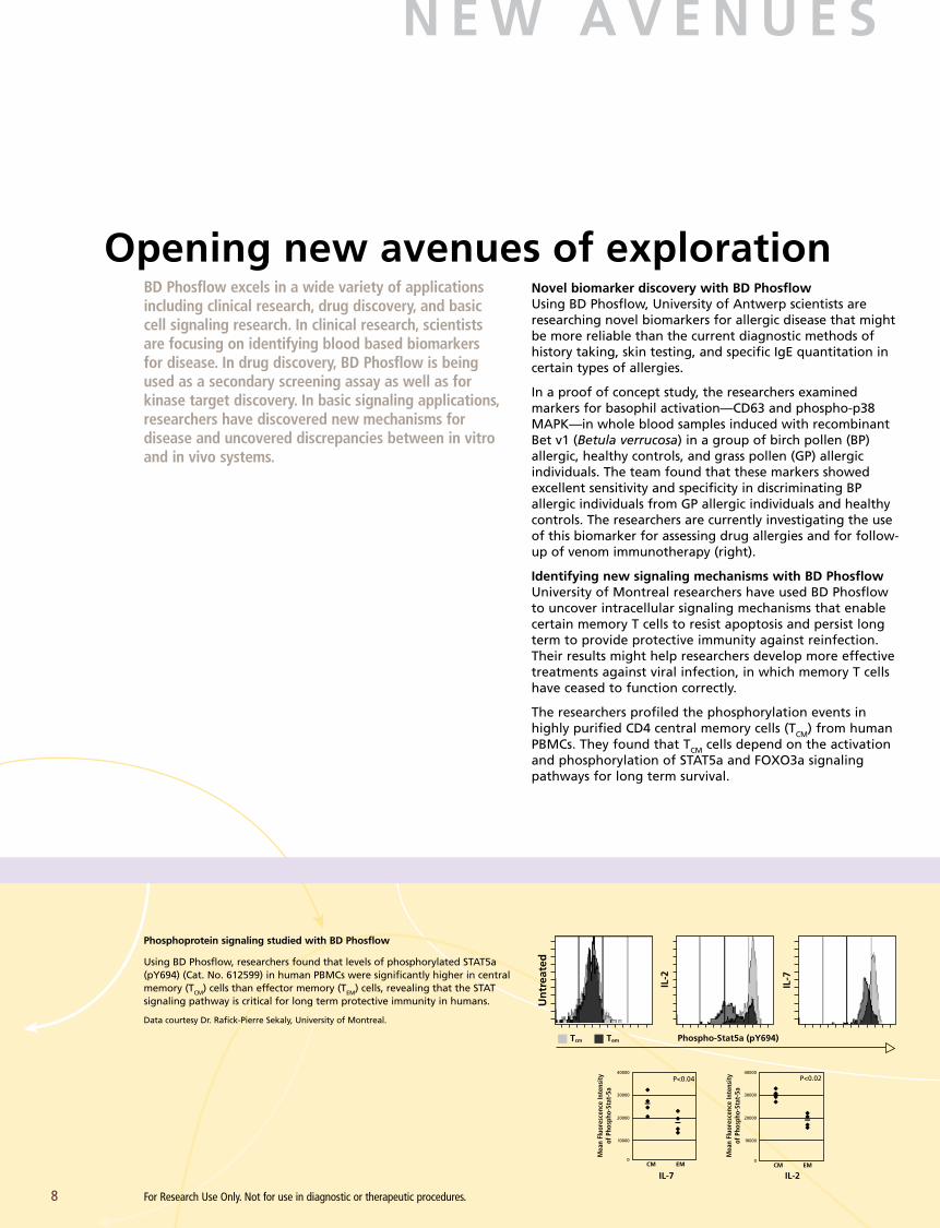
Phosphoprotein signaling studied with BD Phosflow
Using BD Phosflow, researchers found that levels of phosphorylated STAT5a (pY694) (Cat. No. 612599) in human PBMCs were significantly higher in central memory (TCM) cells than effector memory (TEM) cells, revealing that the STAT signaling pathway is critical for long term protective immunity in humans.
Data courtesy Dr. Rafick-Pierre Sekaly, University of Montreal.
Phospho-Stat5a (pY694)
IL-7 IL-2
Un
trea
ted
IL-2
IL-7
CM EM
40000
0
20000
30000
10000
P<0.02
CM EM
P<0.0440000
0
20000
30000
10000
Tcm Tem
Mea
n Fl
uore
scen
ce In
tens
ity
of P
hosp
ho-S
tat-
5a
Mea
n Fl
uore
scen
ce In
tens
ity
of P
hosp
ho-S
tat-
5a
For Research Use Only. Not for use in diagnostic or therapeutic procedures.
AK
T (p
S473
) p
ho
sph
ory
lati
on
(fo
ld in
du
ctio
n)
CM CMEM EM
P<0.003
H202 CD28
16
4
8
12
0
2.5
1
1.5
2
9
BD Phosflow for complex cell-signaling analysis
BD Phosflow analysis reveals that phosphorylation levels of AKT (pS473), a protein known to phosphory-late FOXO3a, are higher in human TCM cells than TEM cells in response to H2O2 and anti-CD28 treatment. CD28 triggered an additional increase in AKT phosphorylation in TCM compared to TEM. Intracellular staining was performed using CD4 (Cat. No. 557871), CD27 (Cat. No. 555441), CD45RA, and AKT (pS473) (Cat. No. 560404) -specific antibodies. The results are represented as the mean fold increase ± SD of five independent experiments, calculated as follows: (MFI of stimulated cells/MFI of unstimulated cells). P values (determined by the two-tailed t test) are shown.
Data courtesy Dr. Rafick-Pierre Sekaly, University of Montreal.
resting basophil activated basophil
upregulationof CD203c
phosphorylation ofp38 MAPK
release ofmediators
expression ofCD63 on membrane
allergenspecific IgE
IgE receptor
p38 MAPK
CD63
CD203c
Intracellular biomarkers for allergic disease
The top right illustration shows how basophils not only secrete particular mediators, but will also upregulate specific activation markers such as CD63 and CD203c upon encountering specific allergens that cross-link membrane bound IgE. The exact mechanism(s) that govern basophil degranulation remain elusive, but it has been demonstrated that phosphorylation of p38 MAPK exerts a pivotal role in cell activation.
The bottom figure illustrates the simultaneous analysis of the surface markers CD63 and CD203c and the novel biomarker, phosphorylated p38 MAPK, in an individual with Cefurim® drug allergy using BD Phosflow. Alexa Fluor® 488-conjugated anti-IgE positive basophils were gated out in a circular region (panel A, upper left). Prewarmed basophils were stimulated for 3 or 20 minutes at 37°C with anti-IgE as a positive control (panel A, lower left), washing solution to measure spontaneous CD63, CD203c, and phosphorylated p38 MAPK expression (negative control, panel A, upper center and upper right), and cefuroxim (Cefurim®, TEVA Pharma, Wilrijk, Belgium) (panel A, lower center and lower right). Note the clear bimodal upregulation of CD63 and the more homogenous upregulation of CD203c and phosphorylation of p38 MAPK in a larger proportion of the cells. This is also clear from the histograms in panel B (open histogram spontaneous expression by resting cells, closed histogram expression after stimulation of cells with cefuroxim).
Ebo DG, Hagendorens MM, Bridts CH, De Clerck LS, Stevens WJ. The basophil activation test in immediate drug allergy. Acta Clinica Belgica. In press.
Sid
e SC
atte
r
CD
63
p-P
38 M
APK
CD
63
p-P
38 M
APK
CD
63Ev
ents
Even
ts
Even
ts
1023
104
103
102
101
100
104
103
102
101
100
basophilsR1
A
B D F
C2 2
80 16
E
100 101 102
lgE CD203c CD63
positive control (20 min) cefuroxim (20 min) cefuroxim (3 min)
negative control (20 min) negative control (3 min)
CD203c
CD63 CD203c p-P38-MAPK
CD203c CD63
103 104
100
100 101 102 103 104
101 102 103 104
100 101 102 103 104 100 101 102 103 104
00
8
100 101 102 103 104
08
100 101 102 103 104
08
199 0
0
104
103
102
101
100
100 101 102 103 104
104
103
102
101
100
100 101 102 103 10410
410
310
210
110
0
731 20
42 141 42
17 4637 1
17
A
B
For Research Use Only. Not for use in diagnostic or therapeutic procedures.

S E R V I C E S A N D S U P P O R T
10
Committed to customer successTechnical application supportBD Biosciences technical application support specialists are available to provide field- or phone-based assistance and advice. Expert in a diverse array of topics, BD technical application support specialists are well equipped to address customers’ needs in both instrument and applications support.
Custom servicesMobilizing technology for research applications requires close collaboration. The Custom Technology Team (CTT) at BD Biosciences works with customers to provide solutions through custom reagents, panels, or assay protocols.
Staffed by leading scientists with both breadth and depth of scientific and technical expertise, the CTT team will coor-dinate with researchers to study the problem at hand, make recommendations, and help implement the solutions. In this way, BD Biosciences technical know-how is translated into practical solutions that allow customers to focus on research.
BD Biosciences is fully committed to the success and satisfaction of its customers. To help scientists take full advantage of our offerings, BD Biosciences products are backed by a world-class service and support organiza-tion with unmatched experience in flow cytometry, cell biology, and antibody reagent development.
A Selection of BD Phosflow Cited Publications
1. Aerts NE, Dombrecht EJ, Bridts CH, et al. Simultaneous flow cytometric detection of baso-phil activation marker CD63 and intracellular phosphorylated p38 mitogen-activated protein kinase in birch pollen allergy. Cytometry B Clin Cytom. 2008;76:8-17. [Epub ahead of print]
2. Ebo DG, Dombrecht EJ, Bridts CH, Aerts NE, de Clerck LS, Stevens WJ. Combined analysis of intracellular signalling and immunophenotype of human peripheral blood basophils by flow cytometry: a proof of concept. Clin Exp Allergy. 2007;37:1668-1675.
3. Krutzik PO, Nolan GP. Fluorescent cell bar coding in flow cytometry allows high throughput drug screening and signaling profiling. Nature Methods. 2006;3:361-368.
4. Kostianovsky AM, Maier LM, Baecher-Allan C, Anderson AC, Anderson DE. Up-regulation of gene related to anergy in lymphocytes is associated with Notch-mediated human T cell suppression. J Immunol. 2007;178:6158-6163.
5. Krutzik PO, Nolan GP. Intracellular phospho- protein staining techniques for flow cytometry: monitoring single cell signaling events. Cytometry A. 2003;55:61-70.
6. Danna EA, Nolan GP. Transcending the biomarker mindset: deciphering disease mechanisms at the single cell level. Curr Opin Chem Biol 2006;10:20-27.
7. Perez OD, Nolan GP. Simultaneous measurement of multiple active kinase states using polychromatic flow cytometry. Nat Biotechnol. 2002;20:155-162.
8. Krutzik PO, Hale MB, Nolan GP. Characterization of the Murine Immunological Signaling Network with Phosphospecific Flow Cytometry. J Immunol. 2005;175:2366-2373.
9. Irish JM, Hovland R, Krutzik PO, et al. Single Cell Profiling of Potentiated Phospho-Protein Networks in Cancer Cells. Cell. 2004;118:217-228.
10. Sachs K, Perez OD, Pe’er D, Lauffenburger DA, Nolan GP. Causal Protein-Signaling Networks Derived from Multiparameter Single-Cell Data. Science. 2005;308:523-529.
11. Perez OD, Krutzik PO, Nolan GP. Flow cytometric analysis of kinase signaling cascades. Methods Mol Biol. 2004;263:67-94.
12. Perez OD, Mitchell D, Jager GC, Nolan GP. LFA-1 signaling through p44/42 is coupled to perforin degranulation in CD56+CD8+ natural killer cells. Blood. 2004;104:1083-1093.
13. Perez OD, Mitchell D, Jager GC, et al. Leukocyte functional antigen 1 lowers T cell activation thresholds and signaling through cytohesin-1 and Jun-activating binding protein 1. Nat Immunol. 2003;4:1083-1092.
14. Krutzik PO, Clutter MR, Nolan GP. Coordinate Analysis of Murine Immune Cell Surface Markers and Intracellular Phosphoproteins by Flow Cytometry. J Immunol. 2005;175:2357-2365.


Asia PacificBD SingaporeTel 65.6861.0633Fax 65.6860.1593
Australia/New ZealandAustraliaToll Free: 1800 656 100Tel 61.2.8875.7000Fax [email protected]
New ZealandToll Free: 0800 572.468 Tel 64.9.574.2468Fax [email protected]
United StatesBD Biosciencesbdbiosciences.com
Customer/Technical ServiceToll Free 877.232.8995
Bioimaging SystemsFax 301.340.9775
Discovery LabwareFax 978.901.7490
Immunocytometry SystemsFax 800.325.9637
PharmingenFax 800.325.9637
JapanNippon Becton DickinsonToll Free 0120.8555.90Tel 81.24.593.5405Fax 81.24.593.5761
Latin America/CaribbeanBD BiosciencesToll Free 0800.771.7157Tel 55.11.5185.9995Fax [email protected]
EuropeBD BiosciencesTel 32.2.400.98.95Fax [email protected]
CanadaBD BiosciencesToll Free 888.259.0187Tel 905.542.8028Fax [email protected]
Regional Offices bdbiosciences.com/offices
BD Biosciences 2350 Qume Drive San Jose, CA 95131 bdbiosciences.com
23-10569-01
Alexa Fluor®, Pacific Blue™, Cascade Blue®, and Texas Red® are trademarks of Molecular Probes, Inc.
Cy™ is a trademark of Amersham Biosciences Corp. Cy™ dyes are subject to proprietary rights of Amersham Biosciences Corp. and Carnegie Mellon University and are made and sold under license from Amersham Biosciences Corp. only for research and in vitro diagnostic use. Any other use requires a commercial sublicense from Amersham Biosciences Corp., 800 Centennial Avenue, Piscataway, NJ 08855-1327, USA.
Gleevec® is a registered trademark of Novartis Pharmaceuticals Corporation.
BD flow cytometers are Class I (1) laser products.
For Research Use Only. Not for use in diagnostic or therapeutic procedures.
© 2009 Becton, Dickinson and Company. All rights reserved. No part of this publication may be reproduced, transmitted, transcribed, stored in retrieval systems, or translated into any language or computer language, in any form or by any means: electronic, mechanical, magnetic, optical, chemical, manual, or otherwise, without prior written permission from BD Biosciences.
Unless otherwise noted, BD, BD Logo and all other trademarks are property of Becton, Dickinson and Company. © 2009 BD