Basic Organization of the Nervous System - Dr. Leichnetz€¦ · Basic Organization of the Nervous...
13
Basic Organization of the Nervous System George R. Leichnetz, Ph.D. OBJECTIVES 1. To introduce some of the basic terminology used throughout the course in describing structures within the central nervous system. 2. To understand the origin and general histology of nervous tissue (cellular components). 3. Review basic neuroembryology, including the differentiation of the neural tube into the central nervous system I. INTRODUCTION A. Central Nervous System (CNS) - consists of the brain and spinal cord B. Peripheral Nervous System (PNS) - peripheral nerves (12 prs. of cranial nerves and 31 prs. of spinal nerves), and emerging peripheral nerves, nerve plexuses (brachial, lumbosacral), dorsal root ganglia and autonomic ganglia II. NEURAL TISSUE Consists of neurons (nerve cells) derived from embryonic neuroblasts, and supportive elements (neuroglia) derived from embryonic glioblasts. While neurons do not undergo mitosis (except for limited neurogenesis in the hippocampus), neuroglia retain the capacity to mitose throughout adult life. Hence most brain tumors are gliomas. Tumors of the meninges (meningiomas) can compress the brain and result in functional deficits, but are not strictly-speaking tumors of the brain itself). CENTRAL NERVOUS SYSTEM (Brain and Spinal Cord) PERIPHERAL NERVOUS SYSTEM (Cranial Nerves, Spinal Nerves, and Ganglia) NEURONS Multipolar only (but various types: granule, pyramidal, basket, Purkinje, alpha motor neurons, etc.) Multipolar - autonomic ganglia Bipolar - special sensory ganglia (cochlear & vestibular ganglia and olfactory mucosa) Unipolar - general sensory ganglia (dorsal root and cranial nerve sensory ganglia) SUPPORTIVE ELEMENTS Astrocytes: fibrous and protoplasmic Oligodendrocytes Microglia --- Schwann cells, Satellite cells --- III. TYPICAL NEURON, CONTENT OF GRAY MATTER AND WHITE MATTER The neuron consists of a cell body (soma, perikaryon) and all of its processes (dendrites, axon). The cell body of the neuron lacks myelin (therefore, gray), but the axon is
Transcript of Basic Organization of the Nervous System - Dr. Leichnetz€¦ · Basic Organization of the Nervous...

Basic Organization of the Nervous System George R. Leichnetz, Ph.D.
OBJECTIVES
1. To introduce some of the basic terminology used throughout the course in describing structures within the central nervous system.
2. To understand the origin and general histology of nervous tissue (cellular components). 3. Review basic neuroembryology, including the differentiation of the neural tube into the
central nervous system
I. INTRODUCTION
A. Central Nervous System (CNS) - consists of the brain and spinal cord
B. Peripheral Nervous System (PNS) - peripheral nerves (12 prs. of cranial nerves and 31 prs. of spinal nerves), and emerging peripheral nerves, nerve plexuses (brachial, lumbosacral), dorsal root ganglia and autonomic ganglia
II. NEURAL TISSUE
Consists of neurons (nerve cells) derived from embryonic neuroblasts, and supportive elements (neuroglia) derived from embryonic glioblasts. While neurons do not undergo mitosis (except for limited neurogenesis in the hippocampus), neuroglia retain the capacity to mitose throughout adult life. Hence most brain tumors are gliomas. Tumors of the meninges (meningiomas) can compress the brain and result in functional deficits, but are not strictly-speaking tumors of the brain itself).
CENTRAL NERVOUS SYSTEM(Brain and Spinal Cord)
PERIPHERAL NERVOUS SYSTEM (Cranial Nerves, Spinal Nerves, and Ganglia)
NEURONS
Multipolar only
(but various types: granule, pyramidal, basket, Purkinje, alpha motor neurons, etc.)
Multipolar - autonomic ganglia Bipolar - special sensory ganglia (cochlear & vestibular ganglia and olfactory mucosa) Unipolar - general sensory ganglia (dorsal root and cranial nerve sensory ganglia)
SUPPORTIVE ELEMENTS
Astrocytes: fibrous and protoplasmic Oligodendrocytes Microglia
--- Schwann cells, Satellite cells ---
III. TYPICAL NEURON, CONTENT OF GRAY MATTER AND WHITE MATTER The neuron consists of a cell body (soma, perikaryon) and all of its processes (dendrites, axon). The cell body of the neuron lacks myelin (therefore, gray), but the axon is

surrounded by a laminated myelin sheath which is yellowish- white in color. Myelinated axons make up the white matter of the central nervous system (ie. tracts, fasciculi, lemnisci, capsules), while cell bodies of neurons make up the gray matter (nucleus or cortex).
Figure 1 * Link to Netter Image 1.1 * Link to Netter Image 1.5
CNS GRAY MATTER
CNS WHITE MATTER
Neuron cell bodies (perikarya) in nucleus or cortex Myelinated (and unmyelinated) axons in tracts, fasciculi, lemnisci, bundles, commissures, capsules, laminae, funiculi, columns
Protoplasmic astrocytes Fibrous astrocytes Few oligodendrocytes (peri-neuronal and juxtavascular) Many oligodendrocytes (esp. interfasicular)

Cytoarchitecture - organization of gray matter in CNS (best seen in Nissl stain, e.g., cresyl violet)
A cluster of neuron cell bodies within the CNS is called a nucleus (e.g., hypoglossal nucleus, caudate nucleus). A cluster of neuron cell bodies in the PNS is called a ganglion. In the cerebrum and cerebellum the surface gray matter has neuron cell bodies in layers (called a cortex).
Myeloarchitecture - organization of white matter in CNS (best seen in myelin stain, e.g., Weigert-Pal) Myelinated axons constitute subcortical white matter, tracts (e.g., spinothalamic tract, medial lemniscus), funiculi (e.g., lateral funiculus of spinal cord), capsules (eg. internal, capsule), and laminae (e.g., internal medullary lamina of the thalamus).
Figure 2
IV. ORIGIN OF NERVOUS TISSUE
A. NEUROEMBRYOLOGY
The nervous system differentiates from the neural ectoderm on the dorsal surface of the embryo. First, a neural groove develops with elevated neural folds on

either side, which then fuse on the midline to form a closed hollow neural tube. The walls of the neural tube differentiate into the entire central nervous system (rostrally, the brain; caudally, the spinal cord). The neurocoel (cavity inside tube) gives rise to the ventricular system of the brain. The neural crest splits off from the dorsolateral aspect of the neural tube and gives rise to the peripheral nervous system (nerves, dorsal root ganglia, autonomic ganglia, cranial sensory ganglia), except bipolar neurons in special sensory ganglia (e.g., cochlear and vestibular ganglia of placode origin) or retina.
Figure 3 * Link to Netter Image 1.68 * Link to Netter Image 1.69
Rostrally, the neural tube undergoes cephalization and three primary brain vesicles develop: the prosencephalon (forebrain), mesencephalon (midbrain), and rhombencephalon (hindbrain), which later divide into five secondary brain

vesicles. The prosencephalon divides into the telencephalon and diencephalon, and the rhombencephalon into metencephalon and myelencephalon. The mesencephalon (midbrain) does not divide. These terms are also applied to the five adult subdivisions of the brain.
Figure 4
Figure 5

B. DIFFERENTIATION OF THE NEURAL TUBE AND NEURAL CREST Three concentric layers of the wall of the neural tube begin to be evident: 1. Ependymal layer - innermost, lines the neurocoel; is a germinal layer in the
embryonic neural tube, continually generating stem cells 2. Mantle layer - intermediate zone; primitive gray matter; contains the cell
bodies of primitive neurons (neuroblasts) and neuroglia (glioblasts) 3. Marginal layer - external zone; primitive white matter; contains the processes
(axons) of developing neurons.
Once an undifferentiated cell (migrates from ependymal layer to mantle layer) becomes a neuroblast, it loses the ability to undergo mitosis. Neuroblasts give rise to the multipolar neurons of the CNS. There is only limited neurogenesis in the adult brain (e.g. hippocampal formation). The glioblasts of the developing nervous system retain the capacity to undergo mitosis throughout adult life. This is why CNS tumors are gliomas. Glioblasts give rise to astrocytes (provide metabolic support to neurons) and oligodendrocytes (myelinization of CNS axons). When the brain is injured, neuroglia divide to replace damaged neural tissue with a glial scar. Neurons DO NOT divide to replace dead neurons. The brain grows in size from neonate to adult by growth of neuronal processes (axons, dendrites), and cell division and growth of neuroglia. The growth in size is not due to an increase in numbers of neurons.
Figure 6 * Link to Netter Image 1.70 * Link to Netter Image 1.71

Neural Crest - neural ectoderm which pinches off the dorsolateral aspect of the neural tube, migrates and ultimately gives rise to neurons and "supportive elements" of the PNS including:
1. Schwann (neurilemmal) cells and satellite cells - in peripheral nerves and ganglia respectively
2. Unipolar neurons - sensory neurons in cranial nerve and dorsal root sensory ganglia
3. Multipolar neurons - autonomic ganglia 4. Chromaffin cells - in adrenal medulla
Note: Bipolar neurons in the PNS are not derived from neural crest.
CENTRAL NERVOUS SYSTEM (NEURAL TUBE)
PERIPHERAL NERVOUS SYSTEM (NEURAL CREST)
Brain and Spinal Cord, and Ventricular System (Neurocoel):
Telencephalon (& Lat. Ventricles) Diencephalon (& Third Ventricle) Mesencephalon (& Cerebral Aqueduct) Metencephalon (& Fourth Ventricle)
Myelencephalon Spinal Cord (& Central Canal)
i.e., Neurons (multipolar) from neuroblasts Neuroglia (astrocytes, oligodendrocytes - from glioblasts)
Cranial sensory ganglia Dorsal root ganglia Autonomic ganglia- (symp. chain, preaortic, and terminal ganglia) Chromaffin cells (adrenal medulla) Schwann cells, satellite cells
Exceptions: bipolar neurons (special sensory) - retina (evag. from CNS): cochlear/vestibular ganglia and olfactory epithelium (placode origin)
C. DIFFERENTIATION OF THE MANTLE LAYER INTO ALAR AND BASAL PLATES The dorsal portion of the mantle layer (gray matter) reorganizes into longitudinal gray columns called alar plates, which later subdivide into cell columns: general somatic afferent (GSA), special somatic afferent (SSA), general visceral afferent (GVA), and special visceral afferent (SVA) cell columns, and which ultimately rise to all of the sensory nuclei of the brain. The ventral portion of the mantle layer reorganizes into longitudinal gray columns called basal plates, which later subdivide into cell columns: general somatic efferent (GSE), general visceral efferent (GVE), and special visceral efferent (SVE) cell columns, and which ultimately give rise to all of the motor nuclei of the brain. The sulcus limitans is an important landmark (groove) in the interior of the developing neural tube separating the alar and basal plates (i.e. separating presumptive sensory and motor columns). In the adult brain the sulcus limitans

can be seen in the floor of the fourth ventricle (rhomboid fossa) separating motor from sensory areas.
D. DEVELOPMENT OF SENSORY AND MOTOR CELL COLUMNS
Sensory Columns Derived From
Alar Plate
• General Somatic Afferent (G.S.A.) - pain, temp., touch, proprioception • Special Somatic Afferent (S.S.A.) - vision, audition, vestibular sense • Special Visceral Afferent (S.V.A.) - taste • General Visceral Afferent (G.V.A.) - visceral sensation
Motor Columns Derived From
Basal Plate
• General Somatic Efferent (G.S.E.) - to voluntary skeletal muscle derived from Derived somites, inc. tongue and extraocular muscles
• General Visceral Efferent (G.V.E.) - to smooth and cardiac muscle From (autonomic) • Special Visceral Efferent (S.V.E.) - to voluntary muscle derived from pharyngeal
(visceral) arch mesoderm
Differentiation of Spinal Cord
Figure 7
Figure 8 * Link to Netter Image 1.78A * Link to Netter Image 1.78B

* Link to Netter Image 1.78C * Link to Netter Image 1.78D * Link to Netter Image 1.79 * Link to Netter Image 1.81A * Link to Netter Image 1.81B * Link to Netter Image 1.81C
Figure 9
The cell columns continue to differentiate, ultimately giving rise to separate sensory and motor nuclei of the CNS.
Cell Column Nuclei of Adult CNS
Sensory
GSA
SSA
GVA
SVA
Trigeminal sensory nuclei, dorsal column nuclei
Auditory and vestibular nuclei
Solitary nucleus (caudal portion)
Solitary nucleus (rostral portion taste)
Motor
GSE
SVE
GVE
Oculomotor, trochlear, abducens and hypoglossal nuclei
Trigeminal and facial motor nuclei and nucleus ambiguus
Edinger-Westphal, superior and inf. salivatory, and dorsal vagal autonomic nuclei
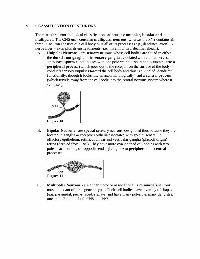
V. CLASSIFICATION OF NEURONS There are three morphological classifications of neurons: unipolar, bipolar and multipolar. The CNS only contains multipolar neurons, whereas the PNS contains all three. A neuron consists of a cell body plus all of its processes (e.g., dendrites, axon). A nerve fiber = axon plus its ensheathments (i.e., myelin or neurilemmal sheath).
A. Unipolar Neurons - are sensory neurons whose cell bodies are found in either the dorsal root ganglia or in sensory ganglia associated with cranial nerves. They have spherical cell bodies with one pole which is short and bifurcates into a peripheral process (which goes out to the receptor on the surface of the body, conducts sensory impulses toward the cell body and thus is a kind of "dendrite" functionally, though it looks like an axon histologically) and a central process (which travels away from the cell body into the central nervous system where it synapses).
Figure 10
B. Bipolar Neurons - are special sensory neurons, designated thus because they are located in ganglia or receptor epithelia associated with special senses, i.e. olfactory epithelium, retina, cochlear and vestibular ganglia (placode origin) retina (derived from CNS). They have more oval-shaped cell bodies with two poles, each coming off opposite ends, giving rise to peripheral and central processes.
Figure 11
C. Multipolar Neurons - are either motor or associational (internuncial) neurons; most abundant of three general types. Their cell bodies have a variety of shapes (e.g. pyramidal, pear-shaped, stellate) and have many poles, i.e. many dendrites, one axon. Found in both CNS and PNS.

Figure 12
Figure 13
VI. NEUROGLIA: SUPPORTIVE ELEMENTS OF THE C.N.S.
A. Astrocytes - fibrous (white matter) and protoplasmic (gray matter) types; located functionally between brain capillaries and neurons. They have processes, known as perivascular end feet, which end on the basement membrane of the capillary endothelium. These cover about 80% of capillary surfaces. Although the brain capillary endothelium represents the principal barrier to free passages of substances into the brain (blood-brain barrier), astrocytes are thought to also play a role. Astrocytes can also take up excess excitatory neurotransmitter, or other metabolites. They surround brain synapses. In the developing brain, these neuroglia can lay down processes that direct neuronal migration (guide neural development).

Figure 14: Noback & Demarest The Human Nervous System * Link to Netter Image 1.4
B. Oligodendrocytes - myelinization of C.N.S. axons; the extension of its cell membrane wraps around a segment (internode) of an axon; successive wrappings create concentric lamellae which constitute myelin. A single oligodendrocyte in the CNS can send out processes that myelinate 50 internodal segments.
Figure 15 * Link to Netter Image 1.7

C. Microglia - of mesodermal origin (not neuralectoderm like glioblasts) they are believed to transform to phagocytes in brain injury; their role and origin has long been in question.
CNS tumors are gliomas, because neuroglia can undergo mitosis (uncontrolled cell division)
Figure 16
VII. SUPPORTIVE ELEMENTS OF THE P.N.S. A. Schwann Cells - myelinization of PNS axons; the cell membrane of the Schwann
cell wraps around the axon in a concentric lamellar fashion producing the myelin sheath. One Schwann cell per internodal segment.
B. Satellite cells - found within dorsal root and cranial sensory ganglia; they surround the spherical cell bodies of sensory neurons in a "satellite" fashion.
Note: Supportive elements in the PNS are not called "neuroglia".
A Self-Assessment is available for this lecture.
* Netter Presenter Image Copyright 2004 Icon Learning Systems. All rights reserved.