NERVOUS SYSTEM I: BASIC STRUCTURE AND FUNCTION
33
CHAPTER 10 NERVOUS SYSTEM I: BASIC STRUCTURE AND FUNCTION OVERVIEW The body uses two systems to coordinate and integrate the functions of body systems so that the internal environment remains stable. These systems are the nervous system and the endocrine system. Chapter 10 begins with a discussion of the general functions of the nervous system, the types of cells that comprise nervous tissue, and the two major groups of nervous system organs (Learning Outcomes 1-3). The chapter continues with discussion of sensory receptors and how they respond to stimuli (Learning Outcomes 4-5). The chapter continues with a detailed discussion of neurons and their component parts and the classification of nervous system cells in both the central and peripheral nervous systems (Learning Outcomes 6-12). Finally, the processes of impulse conduction conclude this chapter on the structure and function of the nervous system (Learning Outcomes 13-18). LEARNING OUTCOMES After you have studied this chapter, you should be able to: 10.1 Introduction (p. 354) I. Describe the general functions of the nervous system. 2. Identify thc two types of cells that comprise nervous tissue. 3. Identify the two major groups of nervous system organs. 10.2 General Functions of the Nervous System (p. 355) 4. List the functions of sensory receptors. 5. Describe how the nervous system responds to stimuli. 10.3 Description ofCeHs of the Nervous System (p. 356) 6. Describe the parts of a neuron. 7. Describe the relationships among myelin, the neurilemma, and nodes of Ranvier. 8. Distinguish between the sources of white matter and gray matter. lOA Classification of Cells of the Nervous System (p. 359) 9. Identify structural and functional differences among neurons. 10. Identify the types of neuroglia in the central nervous system and their functions. II. Describe the Schwann cells in the peripheral nervous system. 10.5 The Synapse (p. 365) 12. Explain how information passes from a presynaptic neuron to a postsynaptic cell. 10.6 Cell Membrane Potential (p. 365) 13. Explain how a cell membrane becomes polarized. 14. Describe the events leading to the conduction of a nerve impulse. 15. Compare nerve impulse conduction in myelinated and unmyelinated neurons. 10.7 Synaptic Transmission (p. 371) 16. Identify the changes in membrane potential associated with excitatory and inhibitory neurotransmitters. 17. Explain what prevents a postsynaptic cell from being continuously stimulated. 10.8 Impulse Processing (p. 374) 18. Describe the basic ways in which the nervous system processes information. FOCUS QUESTION How is the nervous system organized at the cellular level to coordinate and integrate the functions of the other body systems? MASTERY TEST Now take the mastery test. Do not guess. Some questions may have more than one correct answer. As soon as you complete the test, correct it. Note your successes and failures so that you can read the chapter to meet your learning needs. I. The two basic types of cells found in neural tissue are _______ and _______ cells. 2. Nerves are bundles of a. axons. c. axons and dendrites. b. dendrites. 93
Transcript of NERVOUS SYSTEM I: BASIC STRUCTURE AND FUNCTION

CHAPTER 10 NERVOUS SYSTEM I: BASIC STRUCTURE AND FUNCTION
OVERVIEW The body uses two systems to coordinate and integrate the functions of body systems so that the internal environment remains stable. These systems are the nervous system and the endocrine system. Chapter 10 begins with a discussion of the general functions of the nervous system, the types of cells that comprise nervous tissue, and the two major groups of nervous system organs (Learning Outcomes 1-3). The chapter continues with discussion of sensory receptors and how they respond to stimuli (Learning Outcomes 4-5). The chapter continues with a detailed discussion of neurons and their component parts and the classification of nervous system cells in both the central and peripheral nervous systems (Learning Outcomes 6-12). Finally, the processes of impulse conduction conclude this chapter on the structure and function of the nervous system (Learning Outcomes 13-18).
LEARNING OUTCOMES
After you have studied this chapter, you should be able to: 10.1 Introduction (p. 354)
I. Describe the general functions of the nervous system. 2. Identify thc two types of cells that comprise nervous tissue. 3. Identify the two major groups of nervous system organs.
10.2 General Functions of the Nervous System (p. 355) 4. List the functions of sensory receptors. 5. Describe how the nervous system responds to stimuli.
10.3 Description ofCeHs of the Nervous System (p. 356) 6. Describe the parts of a neuron. 7. Describe the relationships among myelin, the neurilemma, and nodes of Ranvier. 8. Distinguish between the sources of white matter and gray matter.
lOA Classification of Cells of the Nervous System (p. 359) 9. Identify structural and functional differences among neurons. 10. Identify the types of neuroglia in the central nervous system and their functions. II. Describe the Schwann cells in the peripheral nervous system.
10.5 The Synapse (p. 365) 12. Explain how information passes from a presynaptic neuron to a postsynaptic cell.
10.6 Cell Membrane Potential (p. 365) 13. Explain how a cell membrane becomes polarized. 14. Describe the events leading to the conduction of a nerve impulse. 15. Compare nerve impulse conduction in myelinated and unmyelinated neurons.
10.7 Synaptic Transmission (p. 371) 16. Identify the changes in membrane potential associated with excitatory and inhibitory neurotransmitters. 17. Explain what prevents a postsynaptic cell from being continuously stimulated.
10.8 Impulse Processing (p. 374) 18. Describe the basic ways in which the nervous system processes information.
FOCUS QUESTION How is the nervous system organized at the cellular level to coordinate and integrate the functions of the other body systems?
MASTERY TEST Now take the mastery test. Do not guess. Some questions may have more than one correct answer. As soon as you complete the test, correct it. Note your successes and failures so that you can read the chapter to meet your learning needs.
I. The two basic types of cells found in neural tissue are _______ and _______ cells.
2. Nerves are bundles of
a. axons. c. axons and dendrites.
b. dendrites.
93
3. The functions of neuroglia include
a. support of neurons. c. sending and receiving messages.
b. filling spaces. d. all of the above
4. The small spaces between neurons are called _.~__~____.
5. Electrochemical messages are carried across synapses by ___ .......... ~____.
6. The nervous system is composed of two groups of organs called the ...........~~~___ nervous system and the ......... ___ _ _~ nervous system.
,., I. Monitoring such phenomena as light, sound, and temperature is a ~__........_~~_ function of the nervous system.
8. The peripheral nervous system has two parts: the nervous system and the ~____ nervous system.
9. The basic unit of structure and function of the nervous system is the ~~~____
10. Another name for the cell body of a neuron is the ~~~____ or ~~~____
II. The cells that give rise to ncw neural tissues are ........_~~~~_~_~ ......_~_~____~
12. Which of the following structures is not common to all nerve cells?
a. cell body c. dendrite
b. axon d. Schwann cells
13. The structure that carries impulses away from the cell body of the neuron is the
a. dendrite. c. axon.
b. neurofibri l. d. neurilemma.
14. The neurilemma is composed of
a. Nissl bodies. c. the cytoplasm and nuclei of Schwann cells.
b. myelin. d. neuron cell bodies.
15. The type of neuron that lies totally within the central nervous system is the
a. sensory neuron. c. interneuron.
b. motor neuron. d. unipolar neuron.
16. The supporting framework of the nervous system is composed of
a. neurons. c. neuroglial cells.
b. dendrites. d. myelin.
17. The neuroglial cell s that can phagocytize bacterial cells and increase when there is inflammation of the brain or spinal cord are
a. astrocytes. c. microglia.
b. oligodendrocytes. d. ependyma.
18. Which of the following injuries to nervous tissue can be repaired?
a. damage to a cell body c. damage to nerve fibers that have a neurilemma
b. damage to nerve fibers that have d. Nerve damage cannot be repaired. myelin sheaths
19. The neuron that brings an impulse to the synapse is a ___ neuron.
20. The difference in electrical charge between the inside and the outside of the membrane in the resting nerve cell is called the _ ........_____~_______'
21. The difference in electrical charge between the inner and outer surfaces of the cell membrane is its
22. The propagation of action potentials along a fiber is called
a. a threshold potential. c. a nerve impulse.
b. repolarization. d. a sensation.
23. The period of total depolarization of the neuron membrane when the neuron cannot respond to a second stimulus is called the period.
24. The refractory period acts to limit the
a. intensity of nerve impulses. c. permeability of nerve cell membranes.
b. rate of conduction of nerve impUlses. d. excitability of nerve fibers.
94
25. In which type of fiber is conduction faster?
a. myelinated
b. unmyelinated
26. A decrease in calcium ions below normal limits will
a. facilitate the movement of sodium c. facilitate the movement of potassium across the across the cell membrane. cell membrane.
b. inhibit the movement of sodium across d. inhibit the movement of potassium across the cell the cell membrane. membrane.
27. The neurotransmitter that stimulates the contraction of skeletal muscles is
a. dopamine. c. gamma-aminobutyric acid.
b. acetylcholine. d. encephalins.
28. The amount of neurotransmitter released at a synapse is controlled by
a. calcium. c. potassium.
b. sodium. d. magnesium.
29. Continuous stimulation of a neuron on the distal side of this junction is prevented by
a. exhaustion of the nerve fiber. c. enzymes within the neural junction.
b. the chemical instability of neurotransmitters. d. rapid depletion of ionized calcium.
30. Neuropeptides that are synthesized by the brain and spinal cord in response to pain are _______
31. The process that allows coordination of incoming impulses that represent information from a variety of receptors is called _______
32. Axons originating from different parts of the nervous system leading to the same neuron exhibit _________
33. The process by which an impulse from a single neuron may be amplified by spreading to other neurons is
STUDY ACTIVITIES
Definition of Word Parts (p. 353)
Define the following word parts used in this chapter.
astr
ax
bi
dendr
ependym
-Iemm
moto
multi
oligo
peri
saltator
sens
syn
uni
95
10.1 Introduction (p. 354)
A. When the nervous system detects changes in the body it can stimulate .......________ and _____..............__ to respond.
B. I. Name the two types of cells that make up neural tissue.
2. Structures that bring input to the cell bodies are . information is carried away from the
neuron by (alan) --c---:---=---~. 3. Nerves are comprised 4. The space between a neuron and the cell with which it communicates is a
C. Name the two divisions of the nervous system and list their component parts.
10.2 General Functions ofthe Nervous System (p. 355)
A. What are the three general functions of the nervous system?
B. I. Where are sensory reccptors located? 2. What is the function of sensory receptors? 3. In what part of the nervous system are sensory receptors integrated and interpreted?
C. Effectors are (inside/outside) the nervous system. D. I. Conscious control of activities is overseen by the ________ nervous system.
2. Involuntary control of body activities is characteristic of the nervous system.
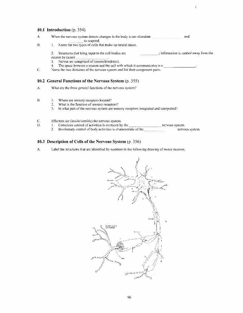
10.3 Description of Cells of the Nervous System (p. 356)
A. Label the structures that are identified by numbers in the following drawing of motor neurons.
96

B. Match the parts of a ncuron in thc first column with the correct description in the second column.
1. neurofibrils a. slender fiber that carries impulses away from the
2. Nissl bodies cell body; this fiber may give off collaterals
3. dendrites b. membranous sacs in the cytoplasm associated with
4. axon the manufacture ofprotein molecules
5. Schwann cells c. celIs of the myelin sheath
d. network offine threads that extend into
nerve fi bers
c. highly branched to provide receptor surfaces to
which processes from other neurons can
communicate
C. Describe how Schwann cells make up the myelin sheath and the neurilemma on the outsides of nerve fibers.
D. What is the composition of white matter in the brain and spinal cord? Of gray matter?
10.4 Classification of Cells of the Nervous System (p. 359)
A. What are two ways in which neurons are classified?
B. Describe each kind of neuron and its location.
bipolar
unipolar
multipolar
c. The neurons in section B are classified according to _______
D. Describe each kind of neuron and its location.
sensory neuron
interneuron
motor neuron
97

E. Fill in the following chart.
Neuroglial Cells
Cell Location Structure Function
Astrocytes
Oligodendrocytes
Microglia
Ependyma
F. When during pregnancy does myelin begin to form?
G. Describe the regeneration of nerve fibers. Include a description of a neuroma.
10.5 The Synapse (p. 365)
A. Label the numbered structures in the accompanying drawing of a synapse.
Direction of }nerve impulse
1 ~\ Synaptic vesicles 2
Ca+2 I Ca+2
Q"":-1J.• <
11 <.. : } 12
~-----3
~----4
~---6
'------9
~------- 10
B. How does a neurotransmitter initiate depolarization? (Include the role of both the presynaptic and the postsynaptic neuron membranes.)
98

10.6 Cell Membrane Potential (p. 365)
A. Describe membrane potential, resting potential, and action potential. Which of these events is a nerve impulse?
B. What happens when a threshold potential is reached?
C. How would hyperpolarization affect the threshold potential?
D. Answer the following concerning impulse conduction.
I. How do the nodes of Ranvier affect nerve impulse conduction? What kind ofconduction is this called?
2. Define refractory period, absolute refractory period, relative refractory period, and all-or-none response in neurons.
3. What is the result of increasing the permeability of the cell membrane to sodium? Of decreasing the permeability?
4. How does calcium affect nerve impulse conduction?
10.7 Synaptic Transmission (p. 371)
A. How do excitatory potentials and inhibitory potentials differ?
B. List the substances that act as neurotransmitters.
C. How is transmission across the synapse halted?
D. What are neuropeptides and what is their function?
10.8 Impulse Processing (p. 374)
A. What is a neuronal pool?
B. Explain the relationship between neuronal pools and facilitation, convergence and divergence.
99

C. Answer the following questions about addiction and the role of receptors.
1. Briefly describe the history of addiction.
2. Define addiction.
3. Describe the role of neurotransmitters and their receptors in the development of addiction.
Clinical Focus Question
Jack, age 24, amputated his finger while cutting the lawn two days ago. The amputated part was brought to the hospital and reattached using microsurgery techniques. Jack is very angry as you meet him this morning. He explains that the surgeon was quite sure that the surgery would be successful but he still has no sensation in the finger. What would you tell Jack?
When you have completed the study activities to your satisfaction, retake the mastery test. If there are still some areas you do not understand, repeat the appropriate study activities.
100

CHAPTER 11 NERVOUS SYSTEM II: DIVISIONS OF THE NERVOUS
SYSTEM
OVERVIEW
This chaptcr continucs thc study of the nervous system. It includes study of the general structure of the brain; its functions; the relationship among the brain, brain stem, and spinal cord; the reflex function ofthe spinal cord; the coverings of the brain and spinal cord; and the formation and function of cerebrospinal fluid (Learning Outcomes 1-10). The chapter continues with a discussion of the structure and function of the peripheral and autonomic nervous systems (Learning Outcomes 11-18). The chapter ends with a discussion of aging-associated changes in the nervous system (Learning Outcome 19).
LEARNING OUTCOMES After you have studied this chapter, you should be able to: 11.1 Introduction (p. 384)
I. Describe the relationship among the brain, brain stem, and spinal cord. 11.2 Meninges (p. 385)
2. Describe the coverings of the brain and spinal cord. 11.3 Ventricles and the Cerebrospinal Fluid (p. 385)
3. Describe the formation and function of cerebrospinal fluid. 11.4 Spinal Cord (p. 387)
4. Describe the structure of the spinal cord and its major functions. 5. Describe a reflex arc and reflex behavior.
11.5 Brain (p. 397) 6. Describe the development of the major parts of the brain and explain the functions of each part. 7. Distinguish among motor, sensory, and association areas of the cerebral cortex. 8. Discuss hemisphere dominance. 9. Explain the stages in memory storage. 10. Explain the functions of the limbic system and the reticular formation.
11.6 Peripheral Nervous System (p. 411) II. Distinguish between the major parts of the peripheral nervous system. 12. Describe the structure of a peripheral nerve and how its fibers are classified. 13. Identify the cranial nerves and list their major functions. 14. Explain how spinal nerves are named and their functions.
11.7 Autonomic Nervous System (p. 424) 15. Characterize the autonomic nervous system. 16. Distinguish between the sympathetic and the parasympathetic divisions of the autonomic nervous system. 17. Describe a sympathetic and a parasympathetic nerve pathway. 18. Explain how the autonomic neurotransmitters differently affect visceral effectors.
11.8 Life-Span Changes (p. 431) 19. Describe aging-associated changes in the nervous system.
FOCUS QUESTION It is noon, and you are just finishing an anatomy assignment. You hear your stomach growling and you realize you are hungry. You make a ham sandwich and pour a glass of milk. After eating, you decide you have been studying for three hours and you should go for a walk. How does the nervous system receive internal and external cues, process incoming information, and decide what action to take?
MASTERY TEST Now take the mastery test. Do not guess. Some questions have more than one correct answer. As soon as you complete the test, correct it. Note your successes and failures so that you can read the chapter to meet your learning needs.
1. The organs of the central nervous system are the _.......______ and the ______________.
101

2. List the parts of the brain.
3. What part of the central nervous system provides two-way communication with the peripheral nervous system?
a. brain stem c. diencephalon
b. cerebellum d. spinal cord
4. The outer membrane covering the brain is composed of fibrous connective tissues and is called the
a. dura mater. c. pia mater.
b. arachnoid mater. d. periosteum.
5. Cerebrospinal fluid is found between the
a. arachnoid mater and the dura mater. c. pia mater and the arachnoid mater.
b. vertebrae and the meninges.
6. Meningitis is most likely to involve inflammation of the
a. dura mater. c. pia mater.
b. arachnoid mater.
7. A series of four interconnected cavities located within the cerebral hemispheres and brain stem are the
a. sucli. c. gyri.
b. ventricles. d. nuclei.
8. Cerebrospinal fluid is secreted by the _______________
9. The function(s) of cerebrospinal tluid (is/are) to
a. supply information about the internal c. prevent infection. environment. d. provide nutrition to central nervous system cells.
b. act as a shock absorber.
10. The spinal cord ends
a. at the sacrum. c. between lumbar vertebrae I and 2.
b. between thoracic vertebrae II and 12. d. at lumbar vertebra 5.
I I. There are pairs of spinal nerves.
12. Which of the following statements is/are true about the white matter in the spinal cord?
a. A cross section of the cord reveals a core of c. The white matter carries sensory stimuli to the brain; white matter surrounded by gray matter. the gray matter carries motor stimuli to the periphery.
b. The white matter is composed of myelinated d. The nerve fibers within spinal tracts arise from cell nerve fibers and makes up nerve pathways, bodies located in the same part of the nervous called tracts. system.
13. The knee-jerk reflex is an example of a
a. reflex that controls involuntary behavior. c. withdrawal reflex.
b. pathologic retlex. d. monosynaptic reflex.
14. An individual who experiences a withdrawal reflex experiences pain at the same time the affected part is removed from the harmful stimulus.
a. True
b. False
15. Damage to the corticospinal tract in an adult may result in alan ________ reflex.
a. biceps-jerk c. ankle-jerk
b. cremasteric d. Babinski
16. Pain impulses are carried from the area stimulated to the brain along the
a. fasciculus gracilis. c. fasciculus cuneatus.
b. spinothalamic tracts. d. spinocerebellar tract.
17. An individual with injury to the spinocerebellar tract is likely to experience
a. loss of a sense of touch. c. involuntary muscle movements.
b. uncoordinated movements. d. severely diminished pain perception.
102

18. An individual suffering from flaccid paralysis has most likely sustained damage to the _______ tract.
a. spinocerebellar c. ribrospinal
b. corticospinal d. reticulospinal
19. Immediate, intensive treatment of spinal cord injuries is important to
a. begin regeneration of severed nerve c. both fibers. d. neither
b. prevent extension of damage secondary to spinal shock.
20. The cerebrum develops from a portion of the
a. forebrain (prosencephalon). c. hindbrain (rhombencephalon).
b. midbrain (mesencephalon).
21. A neural tube defect in the lower posterior portion of the tube results in _______
22. The hemispheres of the cerebrum are connected by nerve fibers called the
a. corpus callosum. c. fissure of Rolando.
b. falx cerebri. d. tentorium.
23. The convolutions on the surface of the cerebrum are called
a. sulci. c. gyri.
b. fissures. d. ganglia.
24. Which of the following statements about the cerebral cortex is/are true?
a. The cortex is the central white portion c. The cortex is the outer gray area of the cerebrum. of the cerebrum. d. The cells in the right hemisphere of the cortex control
b. The cortex has sensory, motor, and the right side of the body. association areas.
25. Match the functions in the first column with the appropriate area of the brain in the second column.
I. hearing a. frontal lobes
2. vision b. parietal lobes
3. recognition of printed words c. temporal lobes
4. control of voluntary muscles d. occipital lobes
5. pain
6. complex problem solving
26. Centers for higher intellectual functions, such as planning and complex problem solving, are located in the ____ lobes.
27. The primary motor centers of the cerebral cortex are located
a. in the frontal lobe. c. in pyramidal shaped cells.
b. posterior to the precentral gyri. d. on the base of the brain near the optic chiasm.
28. Damage to the parietal lobes would impair an individual's ability to
a. hear speech. c. choose appropriate words in speaking.
b. understand speech. d. understand visual cues.
29. The general interpretive area or Wernicke's area is located
a. near Broca's area. c. within the place where the temporal, parietal, and occipital lobes come together. b. in the frontal lobe.
d. within areas common to the cerebrum and cerebellum.
30. In most people, the _______ hemisphere is dominant for verbal and computational skills.
3 l. Some investigators believe that intense, repetitive neuronal activity produces stable changes in nerve pathways to produce ~. memory.
a. short-term c. collective
b. long-term d. unconscious
32. Damage to Broca's area in the cerebral cortex results in the inability to __~~__~~____.
103

33. The function of basal ganglia is to
a. inhibit emotional responses. c. aid in temperature control.
b. facilitate motor functions. d. hormonal function.
34. Which of the following structures is not part of the diencephalon?
a. first and second ventricles c. optic chiasma
b. thalamus d. posterior pituitary gland
35. The part of the brain that controls emotions such as happiness and anger is the
a. thalamus. c. reticular system.
b. limbic system.
36. The cerebral aqueduct is located in the
a. diencephalon. c. midbrain.
b. red nucleus. d. pons.
37. A nonvital control center located in the brain stem is the
a. cardiac center. c. respiratory center.
b. sneezing center. d. vasomotor center.
38. The part of the brain that controls arousal and wakefulness is the
a. hypothalamus. c. basal ganglia.
b. red nucleus. d. reticular formation.
39. The red nucleus ofthe midbrain is the center for
a. color vision. e. postural reflexes.
b. eye reflexes. d. temperature control.
40. The relay station that receives all sensory impulses except smell is the
a. pons. c. basal ganglia.
b. medulla. d. thalamus.
41. The part ofthe brain responsible for the regulation of temperature and heart rate, control of hunger, and regulation of fluid and electrolytes is the
a. thalamus. c. medulla oblongata.
b. hypothalamus. d. pons.
42. The produces emotional reactions of fear, anger, and pleasure.
43. The area of the brain that contains control centers for vital visceral functions is the ~~~~__~
44. REM sleep is also called ~~~__ sleep.
45. With the eyes closed, a person can accurately describe the positions of the various body parts. Which of the following structures serves in this function?
a. proprioceptors c. frontal lobe of the cerebrum
b. pons d. cerebellum
46. An individual who sustains damage to the cerebellum is likely to cxhibit
a. tremors. e. bizarre thought patterns.
b. garbled speech. d. a loss of peripheral vision.
47. The peripheral nervous system has two divisions, the ~~~~~~_nervous system and the ~~~~~~_ nervous system.
48. The nerve fibers that carry motor impulses to smooth muscle structures causing them to contract and to glands causing them to secrete are
a. general somatic afferent tibers. c. general visceral efferent fibers.
b. general somatic efferent fibers. d. general somatic-afferent fibers.
49. There are _~___.... __ pairs of cranial nerves; all but one of these arise from the
lO4

50. The cranial nerve that raises the eyelid and focuses the lens of the eye is the
a. optic nerve. c. abducens nerve.
b. oculomotor nerve. d. facial nerve.
51. In shrugging the shoulders, the sternocleidomastoid and trapezius muscles are'stimulated by
a. the vagus nerve. c. the accessory nerve.
b. the trigeminal nerve. d. the hypoglossal nerve.
52. Sensory fibers of spinal nerves that carry sensory impulses to the brain are found in
a. the dorsal root of spinal nerves. c. the anterior branch or rami of spinal nerves.
b. the ventral root of spinal nerves. d. complex nerve networks called plexuses.
53. The anterior branches of the lower four cervical nerves and the first thoracic nerve give rise to the _______ plexus.
54. Which of the following nerves arises from the lumbosacral plexus?
a. musculocutaneous nerve c. common peroneal nerve
b. femoral nerve d. medial nerve
55. The part of the nervous system that functions without conscious control is the _______ nervous system.
56. Nerves of the sympathetic division leave the spinal cord with spinal nerves in the and
57. Nerves of the parasympathetic division leave the central nervous system within ____......___ nerves and _______ nerves.
58. Match the parts in the first column with the appropriate division in the second column.
1. adrenergic fibers a. sympathetic division
2. cholinergic fibers b. parasympathetic division
3. norepinephrine
4. acetylcholine
59. Which of the following are responses to stimulation by the sympathetic nervous system?
a. increased heart rate c. increased peristalsis
b. increased blood glucose concentration d. increased salivation
60. Which of the following are responses to stimulation of the parasympathetic nervous system?
a. dilation of the bronchioles c. contraction of the gallbladder
b. dilation of the coronary arteries d. contraction of the muscles of the urinary bladder
61. Control of the autonomic nervous system is in the _______ and ___...............__________ integration of autonomic function is in the _______.
STUDY ACTIVITIES
Definition of Word Parts (p. 382)
Define the following word parts used in this chapter.
cephal
chiasm
flacc
funi
gangli
mening
plex
105
11.1 Introduction (p. 384)
A. 1. The central nervous system consists of the _____ and
2. List the parts of the brain.
3. The neurons found in the brain are _____ neurons.
4. The brain communicates with the spinal cord via the ____________.
B. What are the bony coverings of the central nervous system?
11.2 Meninges (p. 384)
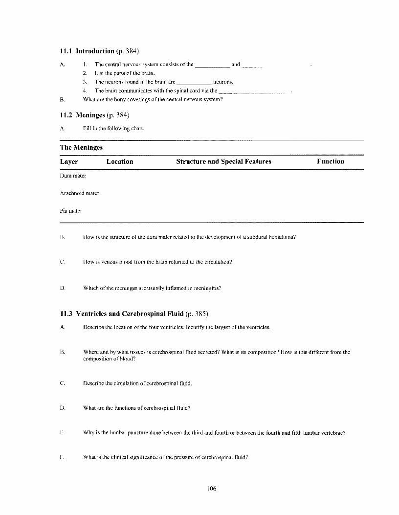
A. Fill in the following chart.
The Meninges
Layer Location Structure and Special Features Function
Dura mater
Arachnoid mater
Pia mater
B. How is the structure of the dura mater related to the development of a subdural hematoma?
C. How is venous blood from the brain returned to the circulation?
D. Which of the meninges are usually inflamed in meningitis?
11.3 Ventricles and Cerebrospinal Fluid (p. 385)
A. Describe the location of the four ventricles. Identify the largest of the ventricles.
B. Where and by what tissues is cerebrospinal fluid secreted? What is its composition? How is this different from the composition of blood?
C. Describe the circulation ofcerebrospinal fluid.
D. What are the functions of cerebrospinal fluid?
E. Why is the lumbar puncture done between the third and fourth or between the fourth and fifth lumbar vertebrae?
F. What is the clinical significance of the pressure of cerebrospinal fluid?
106

11.4 Spinal Cord (pp. 387-396)
A. Answer the following questions about the structure of the spinal cord.
I. The superior boundary of the spinal cord is ..... ____ .... ___. The inferior boundary ofthe spinal cord is
2. How many pairs of spinal nerves are there?
B. Label the numbered items on the accompanying drawing of a cross section of the spinal cord. 1
4
11
14-~~___
12---/ ,,';'~-""*----I't'!'\----- 5
;-~------2
/""""-------3
r?~--~------
.,,-----I-¥~=_~ 6
C. Answer the following questions about the functions of the spinal cord.
I. Answer the following questions about reflex arcs and reflex behavior.
a. What is a reflex arc?
b. What is reflex behavior?
c. Label the numbered parts of the reflex shown in the following drawing.
10
9
2
6
107
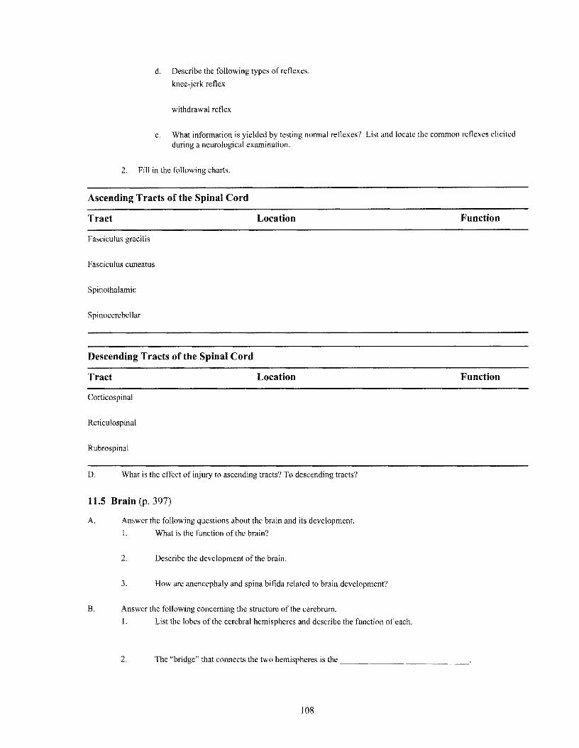
d. Describe the following types of reflexes.
knee-jerk reflex
withdrawal reflex
e. What information is yielded by testing normal reflexes? List and locate the common reflexes elicited during a neurological examination.
2. Fill in the following charts.
Ascending Tracts of the Spinal Cord
Tract Location Function
Fasciculus gracilis
Fasciculus cuneatus
Spinothalamic
Spinocerebellar
Descending Tracts of the Spinal Cord
Tract Location Function
Corticospinal
Reticulospinal
Rubrospinal
D. What is the effect of injury to ascending tracts? To descending tracts?
11.5 Brain (p. 397)
A. Answer the following questions about the brain and its development.
1. What is the function of the brain?
2. Describe the development of the brain.
3. How are anencephaly and spina bifida related to brain development?
B. Answer the following concerning the structure of the cerebrum.
I. List the lobes of the cerebral hemispheres and describe the function of each.
2. The "bridge" that connects the two hemispheres is the ___.. ____ ~____
108

3. The ridges of the hemispheres are _ .... ~__~ _____.__~~......_~~.
4. A shallow groove is a . a deeper groove is a ~- -~~- ~-..... ....
5. The outer layer of the cerebrum is the ~_...~~~~_.
6. The inner layer of the cerebrum is composed
7. Masses of gray matter deep within the cerebrum that inhibit motor activity are the _ .... ~~_ .... ____
C. I. What are the functions of the cerebrum?
2. List the functional areas of the cortex.
a. What is the function of the sensory areas of the cerebrum and where are they located?
b. The sensory area that is not bilateral is What is its function'? c. What is the function of the association areas of the brain and where are they located?
d. The area responsible for concentrating, planning, and complex problem solving is located in the ~~~ .._ ...._~~_ .... lobe.
e. Understanding speech and choosing words to express thought are controlled in the
f. Memories of speech and reading, visual scenes, and other complex sensory patterns are stored in the
-- ..... ..... .....~-.----~--- ~-~-
g. Analyzing visual patterns is accomplished in the _~~..~_____ .____ . D. I. Where are the primary motor areas of the brain located?
2. Describe the function of Broca's area.
E. 1. What is meant by hemisphere dominance? Describe its impact on cerebral function.
2. What are the functions of the nondominant hemisphere?
F. Describe the processes involved in short-term memory and long-term memory.
G. Answer the following about the basal ganglia.
1. Basal ganglia are located within the _~~~_
2. The substance produced by the ganglia is _~~~... __.. '
H. Answer these questions concerning the diencephalon.
I. Locate the diencephalon and describe its structure.
2. Fill in the following chart.
Structure of the Diencephalon
Structure Location Function
Optic chiasma
Posterior pituitary gland
Thalamus
Hypothalamus
3. What structures make up the limbic system? What is the function of this system?
109

I. Answer the following questions about the brain stem.
1. What is the brain stem and what are its component parts?
2. Fill in the following chart.
Functions of the Midbrain
Structure Location Function
Cerebral peduncles
Corpora quadrigemina
Red nucleus
3. Where is the pons located? What is its function?
4. Where is the medulla oblongata located? What vital activities does the medulla control?
5. Describe the location, structure, and function of the reticular formation.
1. Answer the following questions about the cerebellum.
I. What is the function of the cerebellum?
2. The cerebellum communicates with other parts ofthe central nervous system via the ...._____
3. Identify the function of each of the structures in question 1.2.
11.6 Peripheral Nervous System (p. 411)
A. Answer these questions concerning the parts of the peripheral nervous system.
1. What are the parts of the peripheral nervous system?
2. What is the function of each of the following peripheral nerve fibers?
general somatic efferent fibers
general visceral efferent fibers
general somatic afferent fibers
general visceral afferent fibers
110

special somatic efferent fibers
special visceral afferent fibers
special somatic afferent fibers
3. What is the function of the somatic nervous system? The autonomic nervous system?
B. Try the following concerning the cranial nerves.
I. An easy way to memorize the names of the cranial nerves is to use the following sentence:
On old Olympus towering tops a Finn and Gennan viewed some hops.
I II III IV V VI VII VlII IX X XI XII
Note: The vestibulocochlear nerve (VIII) is also known as the acoustic nerve.
You need to be able to identify the cranial nerves by both name and number.
2. Using yourself or a partner, demonstrate how you might test each of the cranial nerves.
3. Fill in the following chart.
Cranial Nerve Fuuctions
Cranial Nerve Sensory, Motor, or Mixed Function
Olfactory
II Optic
III Oculomotor
IV Trochlear
V Trigeminal
VI Abducens
VII Facial
VIII Vestibulocochlear (acoustic)
IX Glossopharyngeal
X Vagus
III

XI Accessory (spinal accessory)
XII Hypoglossal
C. Answer these questions about spinal nerves.
1. How are the spinal nerves identified?
2. What are the structure and function of the dorsal root? Of the ventral root?
3. An area of skin in which a group of sensory nerves leads to a particular dorsal root is called a
4. What structures are innervated by the meningeal branch, the dorsal branch, and the ventral branch of a spinal nerve?
S. What nerves have a visceral branch?
D. Answer the following eoncerning spinal nerve plexuses.
1. What is a plexus?
2. Fill in the following chart.
Spinal Nerve Plexuses
Nerves Involved Structures Innervated
Cervical plexus
Brachial plexus
Lumbosacral plexus
11.7 Autonomic Nervous System (p. 424)
A. What structures make up the autonomic nervous system, and what is the function of this system?
B. How are the nerve pathways of the autonomic division different from those of the somatic division?
C. Identify the structural and funetional differences between the sympathetic and parasympathetic divisions ofthe autonomic nervous system. Be sure to include differenees in neurotransmitters.
D. Describe the interaetion between aeetylcholine and muscarinic and nicotinic cholinergic receptors.
112

E. How does the action of norepinephrine and epinephrine depend on alpha and beta receptors?
F. Describe the mechanisms of control of the autonomic nervous system.
11.8 Life-Span Changes (p. 431)
A. Describe the life-span changes that occur in the nervous system.
B. What is thought to be the pathology of schizophrenia?
C. The expected loss in brain size over a lifetime is
D. Describe noticeable signs of aging in
1. memory. 3. balance.
2. sympathetic function. 4. sleep.
Clinical Focus Questions
A. Your best friend has been hospitalized following a motor vehicle accident in which he sustained injuries to the left temporal and parietal areas. He is right-handed. What results of his injuries do you anticipate?
B. Why is an injury to the first cervical vertebra fatal?
When you have completed the study activities to your satisfaction, retake the mastery test. If there are still some areas you do not understand, repeat the appropriate study activities.
113

CHAPTER 12 NERVOUS SYSTEM III: SENSES
OVERVIEW This chapter deals with specialized parts of the nervous system that allow the body to assess and adjust to the external environment. It describes the differences between the general senses and the special senses (Learning Outcome I). It continues with a discussion of the types of receptors and their locations, structures, and functions in maintaining homeostasis (Learning Outcomes 2-16). The chapter ends by describing aging-associated changes that diminish the senses (Learning Outcome 17).
An understanding of these senses is necessary to knowing how the nervous system receives input and responds to support life.
LEARNING OUTCOMES
After you have studied this chapter, you should be able to: 12.1 Introduction (p. 438)
1. Differentiate between general senses and special senses. 12.2 Reeeptors, Sensation, and Perception (p. 438)
2. Name the five types of receptors and state the function ofeach. 3. Explain how receptors stimulate sensory impulses. 4. Explain sensation production and adaptation.
12.3 General Senses (p. 440) 5. Describe the differences among receptors associated with the senses of touch, pressure, temperature, and
pain. 6. Describe how the sensation ofpain is produced. 7. Explain the importance of stretch receptors in muscles and tendon.
12.4 Special Senses (p. 446) 8. Explain the relationship between the senses of smell and taste. 9. Describe how the sensations of smell and taste are produced and interpreted. 10. Name the parts of the ear and explain the function of each part. II. Distinguish between static and dynamic equilibrium. 12. Describe the roles of the accessory organs to the eye. 13. Name the parts of the eye and explain the function of each part. 14. Explain how the eye refracts light. IS. Explain how the brain perceives depth and distance. 16. Describe the visual nerve pathways.
12.5 Life-Span Changes (p. 476) 17. Describe aging-associated changes that diminish the senses.
FOCUS QUESTION
When you began this chapter at 3:00 r.M., it was 32°F outside, but the sun was pouring into the room. It is now after 5:00 P.M. As you reach to tum on the light, you notice the room has become chilly, so you get a sweater. You smell the supper your roommate is preparing, and you realize that you are hungry. How have your somatic and special senses functioned to process and act on this sensory information?
MASTERY TEST
Now take the mastery test. Do not guess. Some questions may have more than one correct answer. As soon as you complete the test, correct it. Note your successes and failures so that you can read the chapter to meet your learning needs.
1. The function(s) of senses (is/are) to
a. connect us to the outside world. c. help maintain homeostasis.
b. function as an early warning system.
2. Perception occurs in the ~ ........___.~._________
114
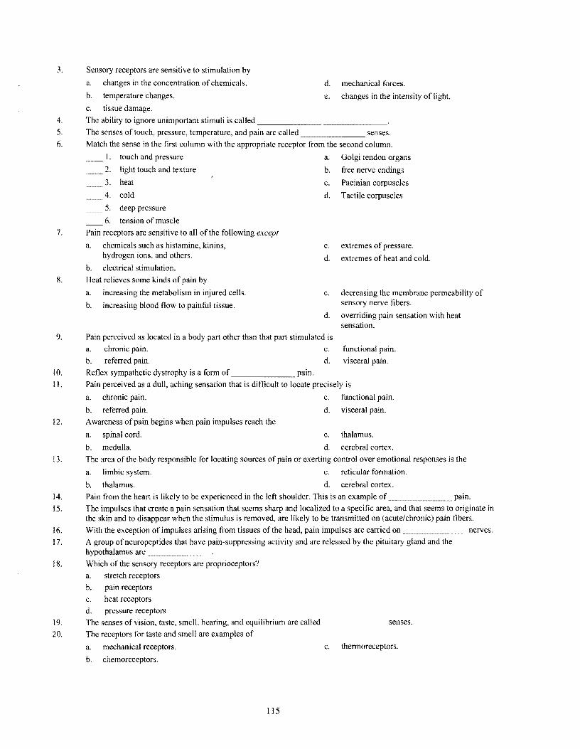
3. Sensory receptors are sensitive to stimulation by
a. changes in the concentration of chemicals. d. mechanical forces.
b. temperature changes. e. changes in the intensity of light.
c. tissue damage. 4. The ability to ignore unimportant stimuli is called ______________
5. The senses of touch, pressure, temperature, and pain are called senses.
6. Match the sense in the first column with the appropriate receptor from the second column.
1. touch and pressure a. Golgi tendon organs
2. light touch and texture b. free nerve endings
3. heat c. Pacinian corpuscles
4. cold d. Tactile corpuscles
5. deep pressure
6. tension of muscle 7. Pain receptors are sensitive to all of the following except
a. chemicals such as histamine, kinins, c. extremes of pressure. hydrogen ions, and others. d. extremes of heat and cold.
b. electrical stimulation. S. Heat relieves some kinds of pain by
a. increasing the metabolism in injured cells. c. decreasing the membrane permeability of sensory nerve fibers. b. increasing blood flow to painful tissue.
d. overriding pain sensation with heat sensation.
9. Pain perceived as located in a body part other than that part stimulated is a. chronic pain. c. functional pain. b. referred pain. d. visceral pain.
10. Reflex sympathetic dystrophy is a form of _______ pain.
II. Pain perceived as a dull, aching sensation that is difficult to locate precisely is
a. chronic pain. c. functional pain.
b. referred pain. d. visceral pain.
12. Awareness of pain begins when pain impulses reach the
a. spinal cord. c. thalamus.
b. medulla. d. cerebral cortex. 13. The area ofthe body responsible for locating sources of pain or exerting control over emotional responses is the
a. limbic system. c. reticular formation.
b. thalamus. d. cerebral cortex. 14. Pain from the heart is likely to be experienced in the left shoulder. This is an example pain. 15. The impulses that create a pain sensation that seems sharp and localized to a specific area, and that seems to originate in
the skin and to disappear when the stimulus is removed, are likely to be transmitted on (acute/chronic) pain fibers.
16. With the exception of impulses arising from tissues of the head, pain impulses are carried on _______ ncrves.
17. A group ofneuropeptides that have pain-suppressing activity and are released by the pituitary gland and the hypothalamus are _______.
IS. Which of the sensory receptors are proprioceptors? a. stretch receptors b. pain receptors c. heat receptors d. pressure receptors
19. The senses of vision, taste, smell, hearing, and equilibrium are called _______ senses.
20. The receptors for taste and smell are examples of
a. mechanical receptors. c. thermoreceptors.
b. chemoreceptors.
lIS
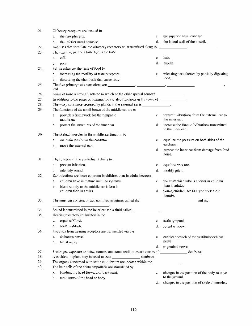
21. Olfactory receptors are located in
a. the nasopharynx. c. the superior nasal conchae.
b. the inferior nasal conchae. d. the lateral wall ofthe nostril.
22. Impulses that stimulate the olfactory receptors are transmitted along the ______________
23. The sensitive part of a taste bud is the taste
a. cell. c. hair.
b. pore. d. papilla.
24. Saliva enhances the taste of food by
a. increasing the motility of taste receptors. c. releasing taste factors by partially digesting food.b. dissolving the chemicals that cause taste.
25. The five primary taste sensations are ____________________________ and _______
26. Sense of taste is strongly related to which of the other special senses? _______
27. In addition to the sense of hearing, the ear also functions in the sense of_______
28. The waxy substance secreted by glands in the external ear is _______
29. The functions of the small bones of the middle ear are to
a. provide a framework for the tympanic c. transmit vibrations from the external ear to membrane. the inner ear.
b. protect the structures of the inner ear. d. increase the force of vibrations transmitted to the inner ear.
30. The skeletal muscles in the middle ear function to
a. maintain tension in the eardrum. c. equalize the pressure on both sides of the eardrum.b. move the external ear.
d. protect the inner ear from damage from loud noise.
31. The function of the eustachian tube is to
a. prevent infection. c. equalize pressure.
b. intensify sound. d. modify pitch.
32. Ear infections are more common in children than in adults because
a. children have immature immune systems. c. the eustachian tube is shorter in children than in adults. b. blood supply to the middle ear is less in
children than in adults. d. young children are likely to suck their thumbs.
33. The inner ear consists of two complex structures called the ______________ and the
34. Sound is transmitted in the inner ear via a fluid called _______
35. Hearing receptors are located in the
a. organ of Corti. c. scala tympani.
b. scala vestibuli. d. round window.
36. Impulses from hearing receptors are transmitted via the
a. abducens nerve. c. cochlear branch of the vestibulocochlear nerve.b. facial nerve.
d. trigeminal nerve.
37. Prolonged exposure to noise, tumors, and some antibiotics are causes of _______ deafness.
38. A cochlear implant may be used to treat deafness.
39. The organs concerned with static equilibrium are located within the _______
40. The hair cells of the crista ampullaris are stimulated by
a. bending the head forward or backward. c. changes in the position of the body relative to the ground. b. rapid turns of the head or body.
d. changes in the position of skeletal muscles.
116

41. The muscle that raises the eyelid is the
a. orbicularis oculi. c. levator palpebrae superioris.
b. superior rectus. d. ciliary muscle. 42. The lacrimal gland is located in the ~~~~___ of the orbit.
a. superior lateral wall c. inferior lateral wall
b. superior medial wall d. inferior medial wall
43. The conjunctiva covers the anterior surface of the eyeball, except for the _______
44. The superior rectus muscle rotates the eye
a. upward and toward the midline. c. away from the midline.
b. toward the midline. d. upward and away from the midline.
45. The orbicularis oculi is innervated by the
a. oculomotor nerve. c. abducens nerve.
b. trochlear nerve. d. facial nerve.
46. The transparency of the cornea is due to
a. the nature of the cytoplasm in the cells of c. the lack ofnuclei with these cells. the cornea. d. keratinization of cells in the cornea.
b. the small number ofcells and the lack of blood vessels.
47. In the posterior wall of the eyeball, the sclera is pierced by the _______.
48. The anterior portion of the middle tunic or vascular tunic of the eye contains the
a. choroid coat. c. ms.
b. ciliary body. d. cornea.
49. The shape ofthe lens changes as the eye focuses on a close object in a process knO\¥ll as
a. accommodation. c. reflection.
b. refraction. d. strabIsmus.
50. The anterior chamber ofthe eye extends from the _______ to the iris.
51. The aqueous humor leaves the anterior chamber via the
a. pupil. c. ciliary body.
b. canal of Schlemm. d. lymphatic system.
52. The part of the eye that controls the amount of light entering it is the ~______
53. The color of the eye is determined by the amount and distribution _______ in the iris.
54. The inner tunic of the eye contains the receptor cells of sight and is called the _______
55. The region associated with the sharpest vision is the
a. macula lutea. c. optic disk.
b. fovea centralis. d. choroid coat.
56. The largest compartment of the eye, which is bounded by the lens, ciliary body, and retina, is filled with
57. The bending of light waves as they pass at an oblique angle from a medium of one optical density to a medium of another optical density is called ______.........
58. The lens loses elasticity with aging, causing a condition called _______
59. There are two types of visual receptors; one has long, thin projections that are called __...._____,. the other has short, blunt projections that are called ~______
60. Match the type ofvision in the first column with the proper receptor from the second column.
I. vision in relatively dim light a. rods
2. color vision b. cones
3. general outlines
4. sharp images
61. The light-sensitive pigment in rods is ~______. In the presence of light, this pigment decomposes to form _____ and ____~ ___.
117
62. The pigments found in cones are ... _______, ___ .............___, and _______<
63. The absence of cone pigments leads to _____---' _______.
64. If the visual cortex is injured, the individual may develop (complete/partial) blindness in (one eye/both eyes).
STUDY ACTIVITIES
Definition of Word Parts (p. 437)
Define the following word parts used in this chapter.
aud
choroid
cochlea
com
iriS
labyrinth
lacri
lut
macula
malle
ocul
olfact
palpebra
photo
scler
therm
tympan
vitre
118

12.1 Introduction (p. 438)
A. How is a feeling produced?
B. Contrast the general senses and the special senses.
C. What characteristics do all senses have in common?
12.2 Receptors, Sensation, and Perception (p. 438)
A. List five groups of sensory receptors and identify the sensations with which they are associated.
B. Describe the transmission of sensory impulses.
C. A feeling that occurs when sensory impulses are recognized by the brain is a _______
D. The process that allows an individual to locate the region of stimulation is called _______
E. The process that makes a receptor ignore an unimportant stimulus unless the strength of that stimulus increases is
12.3 General Senses (p. 440)
A. Fill in the following chart.
Cutaneous Receptors
Type Location Sensation
Free nerve endings (mechanoreceptors)
Tactile (Meissner's) corpuscles (mechanoreceptors)
Lamellated (Pacinian) corpuscles
Thermoreceptors
Free nerve endings (pain receptors)
B. Answer the following concerning pain receptors.
1. In what way do pain receptors differ from the other somatic receptors?
2. Tissue damage is thought to stimulate pain receptors by the release of ~ ______
3. What events trigger visceral pain?
4. What is referred pain'l
119

5. Compare the characteristics of acute pain fibers and chronic pain fibers.
6. How are pain impulses regulated?
7. Neurotransmitters secreted by the spinal cord that inhibit pain impulses are _______
8. Pain suppressants secreted by the pituitary gland are _______
C. Discuss the interaction of stretch receptors in the muscles (muscle spindles) and the tendons (Golgi tendon organs).
12.4 Special Senses (p. 446)
A. Answer the following questions about the sense of smell.
I. The sense of smell supplements the sense
2. On the accompanying illustration, label the structures identified by a letter.
9 h
k
3. How do odors stimulate olfactory receptors?
4. What is synesthesia?
5. Describe the nerve pathways for the sense of smell.
6. Why does the sense of smell diminish in acuity with increasing age?
7. What are the nerve pathways for the sense of smell?
B. Answer the following questions about the sense of taste. 1. Describe the structure of taste receptors and list the five primary taste sensations.
120

2. How does saliva contribute to the perception of taste?
3. Describe the nerve pathways for the sensation of taste.
4. What factors can influence or distort an individual's senses of smell and/or taste?
C. Answer the following questions regarding the sense of hearing.
1. Describe the function of the external ear.
2. Describe the vibration conduction pathway of the ear from the meatus to the temporal lobe of the cerebrum.
3. Why does it help to chew gum while descending in an airplane?
4. How does the structure of the auditory tube and middle ear predispose the middle ear to infection?
5. Why can't the tympanic reflex protect the hearing receptors from the effects of sudden, loud noises?
6. Describe the function of the inner ear.
7. Describe the auditory nerve pathway.
D. Answer the following questions regarding the sense of equilibrium.
I. Distinguish between static and dynamic equilibrium.
2. Describe the function of each of the following structures in maintaining equilibrium.
utricle
saccule
macula
semicircular canals
crista ampullaris
cerebellum
eyes
121

E. Answer the following questions concerning the sense of sight.
1. Answer the following concerning the visual accessory organs.
a. What stmctures are covered by the conjunctiva?
b. Describe the lacrimal apparatus. How does it protect the eye?
c. What is the secretion of the conjunctiva and what is its function?
d. Identify the function ofthe following muscles.
orbicularis oculi
levator palpebrae superioris
superior rectus
inferior rectus
medial rectus
lateral rectus
superior oblique
inferior oblique
122

2. Label the structures identified by letters in the following illustration: lens, iris, suspensory ligaments, vitreous humor, aqueous humor, sclera, optic disk, optic nerve, fovea centralis, posterior cavity, choroid coat, pupil, retina, anterior chamber, posterior chamber, ciliary body. What is the function of each of the labeled structures?
a---~
b----....
c--------:'f
d ---------,f!:--'
e ---------,f,..-
9 ---!:--'
L-------s
3. What characteristics of the cornea contribute to the ease with which it is transplanted?
4. Answer the following questions about functions of the eye.
a. Describe the structure and function of the lens.
b. What is the iris?
c. What is the retina?
5. Answer these statements and questions concerning refraction of light.
a. Define refraction of light.
123

b. Which one of the following illustrates normal refraction? Identify the problems illustrated in the other two drawings.
Point Lens of focus
Light waves
1.
Point of focus
Light waves
lD' '~ .,
'-.
Retina
2.
Point of focus
/Light waves
U
\\ ~i~D
"\.,
3. _______________
6. Answer the following concerning visual receptors.
a. Describe the functions of rods.
b. Describe the location and functions of cones.
c. How are rods related to dark adaptation?
d. Why does vitamin A deficiency affect vision?
e. What mechanism is used by cones to recognize color?
f. Describe colorblindness.
124

7. Answer the following concerning visual nerve pathways.
a. Describe the paths of the right and left optic tracts.
b. Describe the visual deficit that results from damage to either visual cortex.
12.5 Life-Span Changes (p. 476)
Describe the effects of aging on the special senses.
Clinical Focus Question
Based on your knowledge of the way in which the senses interact, explain why a blind person who has had bilateral amputations cannot learn to use artificial legs.
When you have completed the study activities to your satisfaction, retake the mastery test and compare your performance with your initial attempt. If there are still areas you do not understand, repeat the appropriate study activities.
125


