Available online at IJTID Website: ...
6
Vol. 8 No. 3 September–December 2020 Copyright © 2020, IJTID, p-ISSN 2085-1103, e-ISSN 2356-0991 Available online at IJTID Website: https://e-journal.unair.ac.id/IJTID/ Case Report Gastric Perforation Associated with Candidiasis and NSAIDS Febriana Aquaresta 1,2 , Arthur Pohan Kawilarang 1,2 , Pepy Dwi Endraswari 1,2,3 1 Department Clinical Microbiology Faculty of Medicine, Universitas Airlangga Surabaya, 2 Clinical Microbiology Unit Dr Soetomo Academic Hospital Surabaya, 3 Clinical Microbiology Unit, Universitas Airlangga Hospital Received: 11 st November 2019; Revised: 13 rd November 2019; Accepted: 3 rd February 2020 ABSTRACT Invasive candidiasis is an important health-care-associated fungal infection. Candida is often described as an opportunistic pathogen. It is commensal flora in the gastrointestinal tract. Invasive candidiasis can happen usually because of a consequence of increased or abnormal colonization together with a local or generalized defect in host defenses. Candidiasis can occur in patients with HIV, therapy with a broad-spectrum antibiotic, transplant organ, and immunocompromised. Most cases of gastric perforation occur as complications of Peptic Ulcer Disease (PUD), Nonsteroidal Anti-Inflammatory Drugs (NSAIDs) and gastric neoplasms, but candidiasis as a cause of gastric perforation is very rare. This study aims to reveal the correlation between gastric perforation with candidiasis and NSAIDs. It was reported that a 57-year-old East Java Indonesian female presented with severe epigastric pain, generalized peritonitis, fever, nausea also vomiting and had a history of NSAIDs used for five years. The patient was taken to the general surgery of Dr. Sutomo Surabaya Hospital and performed exploratory laparotomy. A gastric perforation was discovered in the antrum. Microbiology culture examination from biopsy gastric tissue revealed an intense fungal growth from sabouraudagar medium and there is no other microorganism that grew in aerobic culture. Candida albicans was identified by VITEK® 2 COMPACT. Histopathological examination from biopsy gastric tissue was performed by Olympus CX-21 microscope, showed invasive Candida albicans consisting of numerous fungal yeasts and pseudohyphae invading and destroying the gastric wall. The patient was subsequently treated with fluconazole anti-fungal and discharge home after nine days postoperative period in good condition. From this result, we suggest using an antifungal treatment for patients who use NSAIDs for long periods to prevent candidiasis. Keywords: Candida albicans, fluconazole, gastric perforation, histopathological, NSAIDs, peritonitis ABSTRAK Kandidiasis invasif adalah infeksi jamur penting yang berhubungan dengan perawatan kesehatan. Candida sering digambarkan sebagai patogen oportunistik. Candida merupakan florakomensal dalam saluran pencernaan. Kandidiasis invasif dapat terjadi sebagai konsekuensi dari peningkatan atau kolonisasi abnormal Candida bersama dengan kurangnya sistem imun lokal atau umum dalam tubuh host. Kandidiasis dapat terjadi pada pasien dengan HIV, terapi dengan antibiotik spektrum-luas, transplantasi organ, dan immunocompromised. Sebagian besar kasus perforasi gastric terjadi sebagai komplikasi dari Penyakit Ulkus Peptikum (PUD), Obat Antiinflamasi Nonsteroid (NSAID) dan neoplasma lambung, tetapi kandidiasis sebagai penyebab perforasi gastric sangat jarang terjadi. Tujuan kami dalam laporan kasus ini adalah mengungkapkan korelasi antara perforasi gastric dengan kandidiasis dan pemakaian NSAIDs. Kami melaporkan seorang wanita Indonesia Jawa Timur berusia 57 tahun yang mengalami nyeri epigastrium parah, peritonitis menyeluruh, demam, mual juga muntah dan memiliki riwayat penggunaan NSAID selama lima tahun. Pasien dirawat di bagian bedah umum Rumah Sakit Dr. Sutomo Surabaya dan dilakukan laparatomi eksplorasi. Perforasi gastric ditemukan di bagian antrum. Pemeriksaan kultur mikrobiologi dari jaringan biopsi menunjukkan pertumbuhan jamur yang banyak pada media sabouraud dextrose agar dan tidak ada mikroorganisme lain yang tumbuh di kultur aerob. Identifikasi di lanjutkan * Corresponding Author: [email protected].
Transcript of Available online at IJTID Website: ...
Vol. 8 No. 3 September–December 2020
Copyright © 2020, IJTID, p-ISSN 2085-1103, e-ISSN 2356-0991
Available online at IJTID Website: https://e-journal.unair.ac.id/IJTID/
Case Report
Gastric Perforation Associated with Candidiasis and NSAIDS
Febriana Aquaresta1,2, Arthur Pohan Kawilarang1,2, Pepy Dwi Endraswari1,2,3
1 Department Clinical Microbiology Faculty of Medicine, Universitas Airlangga Surabaya,2 Clinical Microbiology Unit Dr Soetomo Academic Hospital Surabaya,
3 Clinical Microbiology Unit, Universitas Airlangga Hospital
Received: 11st November 2019; Revised: 13rd November 2019; Accepted: 3rd February 2020
ABSTRACT
Invasive candidiasis is an important health-care-associated fungal infection. Candida is often described as an opportunistic pathogen. It is commensal fl ora in the gastrointestinal tract. Invasive candidiasis can happen usually because of a consequence of increased or abnormal colonization together with a local or generalized defect in host defenses. Candidiasis can occur in patients with HIV, therapy with a broad-spectrum antibiotic, transplant organ, and immunocompromised. Most cases of gastric perforation occur as complications of Peptic Ulcer Disease (PUD), Nonsteroidal Anti-Infl ammatory Drugs (NSAIDs) and gastric neoplasms, but candidiasis as a cause of gastric perforation is very rare. This study aims to reveal the correlation between gastric perforation with candidiasis and NSAIDs. It was reported that a 57-year-old East Java Indonesian female presented with severe epigastric pain, generalized peritonitis, fever, nausea also vomiting and had a history of NSAIDs used for fi ve years. The patient was taken to the general surgery of Dr. Sutomo Surabaya Hospital and performed exploratory laparotomy. A gastric perforation was discovered in the antrum. Microbiology culture examination from biopsy gastric tissue revealed an intense fungal growth from sabouraudagar medium and there is no other microorganism that grew in aerobic culture. Candida albicans was identifi ed by VITEK® 2 COMPACT. Histopathological examination from biopsy gastric tissue was performed by Olympus CX-21 microscope, showed invasive Candida albicans consisting of numerous fungal yeasts and pseudohyphae invading and destroying the gastric wall. The patient was subsequently treated with fl uconazole anti-fungal and discharge home after nine days postoperative period in good condition. From this result, we suggest using an antifungal treatment for patients who use NSAIDs for long periods to prevent candidiasis.
Keywords: Candida albicans, fl uconazole, gastric perforation, histopathological, NSAIDs, peritonitis
ABSTRAK
Kandidiasis invasif adalah infeksi jamur penting yang berhubungan dengan perawatan kesehatan. Candida sering digambarkan sebagai patogen oportunistik. Candida merupakan fl orakomensal dalam saluran pencernaan. Kandidiasis invasif dapat terjadi sebagai konsekuensi dari peningkatan atau kolonisasi abnormal Candida bersama dengan kurangnya sistem imun lokal atau umum dalam tubuh host. Kandidiasis dapat terjadi pada pasien dengan HIV, terapi dengan antibiotik spektrum-luas, transplantasi organ, dan immunocompromised. Sebagian besar kasus perforasi gastric terjadi sebagai komplikasi dari Penyakit Ulkus Peptikum (PUD), Obat Antiinfl amasi Nonsteroid (NSAID) dan neoplasma lambung, tetapi kandidiasis sebagai penyebab perforasi gastric sangat jarang terjadi. Tujuan kami dalam laporan kasus ini adalah mengungkapkan korelasi antara perforasi gastric dengan kandidiasis dan pemakaian NSAIDs. Kami melaporkan seorang wanita Indonesia Jawa Timur berusia 57 tahun yang mengalami nyeri epigastrium parah, peritonitis menyeluruh, demam, mual juga muntah dan memiliki riwayat penggunaan NSAID selama lima tahun. Pasien dirawat di bagian bedah umum Rumah Sakit Dr. Sutomo Surabaya dan dilakukan laparatomi eksplorasi. Perforasi gastric ditemukan di bagian antrum.
Pemeriksaan kultur mikrobiologi dari jaringan biopsi menunjukkan pertumbuhan jamur yang banyak pada media sabouraud dextrose agar dan tidak ada mikroorganisme lain yang tumbuh di kultur aerob. Identifi kasi di lanjutkan
* Corresponding Author:[email protected].
169Febriana Aquaresta, et al.: Gastric Perforation Associated with Candidiasis and NSAIDS
Copyright © 2020, IJTID, p-ISSN 2085-1103, e-ISSN 2356-0991
dengan menggunakan VITEK® 2 COMPACT hasilnya adalah Candida albicans. Pemeriksaan histopatologis dari jaringan biopsi dilakukan dengan mikroskop Olympus CX-21, menunjukkan Candida albicans invasif yang terdiri dari sejumlah yeast dan pseudohypae yang menyerang dan menghancurkan dinding gastric. Pasien kemudian diobati dengan fl ukonazol anti jamur dan pulang ke rumah setelah sembilan hari periode pasca operasi dalam kondisi baik. Dari hasil case report ini, kami menyarankan penggunaan pengobatan antijamur untuk pasien yang menggunakan NSAIDs dalam jangka waktu lama untuk mencegah kandidiasis.
Kata kunci: Candida albicans, fl uconazole, gastric perforasi, histopatological, NSAIDs, peritonitis
How to Cite: Gastric Perforation Associated with Candidiasis and NSAIDS. Aquaresta F., Kawilarang A.P., Endraswari P. D. Indonesian Journal of Tropical and Infectious Disease, 8(3), 168–173.
INTRODUCTION
Invasive candidiasis is an important health- care-associated fungal infection that can be caused by several Candida spp.; the most common species is Candida albicans. The spectrum of disease of invasive candidiasis ranges from minimally symptomatic an candidiasis to fulminant sepsis with associated mortality exceeding 70%.1 Candida albicans are common commensal organisms in the skin and gut microbiota, and disruptions in the cutaneous and gastrointestinal barriers (for example, owing to gastric perforation) that promote invasive disease.1 The common causations of gastric perforation are Peptic Ulcer Disease (PUD), Nonsteroidal Anti-Inflammatory Drugs (NSAIDs), gastric neoplasms and strong antacid consumption, but it is very rare due to candidiasis.1 Candidiasis can occur in patients with HIV, transplant organs, therapy with broad-spectrum antibiotics and immunocompromised.2–4 This case is reportedly rare because in literature there were only two cases that have been published.5, 6 This patient is immunocompetent without other histories of chronic illness, except gout arthritis. The patient had been using NSAIDs to relieve gout pain for almost 5 years. Here, we report the first history associated with invasive candidiasis and review the relevant literature.
CASE REPORT
A 57-years-old woman came to a emergency unit of RSUD Dr Sutomo hospital with a history of abdominal pain since three days earlier.
The pain was epigastric; severe, deep-seated, progressive, non-radiating, not relieved by food intake and vomiting and not associated with diarrhea. Her condition worsened 1 day to presentation with generalized abdominal pain and abdominal wall rigidity. There was abdominal distension, no change in bowel habit, no blood in the stool, no associated fever, weight loss, alcohol binge or trauma. There was no history of changes in pattern of bowel movements, diabetes mellitus, hypertension or Peptic Ulcer Disease (PUD), except the history of using NSAIDs for almost 5 years due to gout arthritis. On clinical examination, an elderly woman was conscious and alert but in obvious painful distress. She was a febrile, not pale, anicteric, not cyanosed, no pedal edema and no dehydrated. On physical examination, blood pressure was 115/80 mmhg, pulse rate 100x beats/minute, respiratory rate 22 x breath/minute, temperature 37.6ºC. The inspection abdomen showed distended, tense, bowel sound decreased, and defense muscular on all abdominal regions. The examination of the liver, spleen, kidneys, and rectum were normal. Diagnosed as peritonitis was made. For initial treatment in the emergency unit, she was given metronidazole, dexamethasone, metamizole, ranitidine and tutofusin as fluid hydration. The preoperative laboratory examinations showed hemoglobin concentration 6.3 g/dl, leucocyte 10.310/ul, hematocrits 23.8%, thrombocyte 457.000/ul, creatinine 1,33 mg/dl, SGOT 30 U/L, SGPT 17 U/L, blood sugar 109 mg/dl, albumin 2.7 g/dl, serum electrolyte concentration sodium (Na+) 137 mmol/L, potassium (K+) 3.4 mmol/L, chloride (Cl−) 101 mmol/L, and non-reactive

170
Copyright © 2020, IJTID, p-ISSN 2085-1103, e-ISSN 2356-0991
Indonesian Journal of Tropical and Infectious Disease, Vol. 8 No. 3 September–December 2020: 168–173
Hbsag examination. Thorax photo showed lung inflammation with minimal right pleural effusion. BOF (Bluch Over Sich) examination showed air-fluid level. The patient was measured 1.57 m tall, weighed 90 kg, and had a body mass index of 36.5 kg/m2. An emergency exploratory laparotomy was performed and found a hole size 2 x 1 cm by 2 x 3 cm of gastric perforation covered with fibrinous exudate within the peritoneum. Gross macroscopy gastric tissue was irregular, weight < 1 gram, greyish-white color, and solid consistency. Microscopic histopathological examination showed pieces of tissue with erosive and ulcerative surfaces coated in necrotic areas and inflammation of neutrophils, lymphocytes and histiocytes. In the muscular layer until serious, fat was obtained in lymphocyte inflammation cells, histiocytes and plasma cells. Gram (Figure 1) and KOH staining from gastric tissue showed yeast and fungal hyphae. There were no Helicobacter pylori-like microorganisms. Microbiology culture used gastric tissue and gastric liquid 3cc specimens. The liquid looked red. Both specimens examined aerobic, anaerobic and Sabouraud Dextrose Agar/fungal culture. Aerobic culture did on Blood Agar Plate, Chocolate and Mac Conkey Agar. They were incubated in 37ºC for 18-24 hours. Anaerobic culture did on Brucella Agar and Cooked Meat Medium, incubated in an anaerobic jar for 48 hours. Fungal culture on Sauboroud Dextrose Agar was incubated in two temperatures 25ºC and 37ºC to differentiate from mold, grew creamy colonies. Lactophenol Cotton Blue staining was also performed to identify yeast from the colony (Figure 2). Three cultures (aerobic, anaerobic and fungal) revealed Candida colonies. The result identification using VITEK® 2 COMPACT was Candida albicans. The histopathological examination was performed using Olympus CX- 21 microscope. It showed numerous yeast and hyphae which invaded tissue gastric.
RESULTS
Gastric tissue was put into formalin and then it was compacted with wax called paraffin
1
Figure 1. Gram staining from direct gastric tissue specimen, showed yeast and
pseudohyphae.
2
Figure 2. Lactophenol Cotton Blue staining from colony, showed fungal yeast
1
Figure 1. Gram Twort Staining 400x arrow (1) Yeast (2) Hyphae
171Febriana Aquaresta, et al.: Gastric Perforation Associated with Candidiasis and NSAIDS
Copyright © 2020, IJTID, p-ISSN 2085-1103, e-ISSN 2356-0991
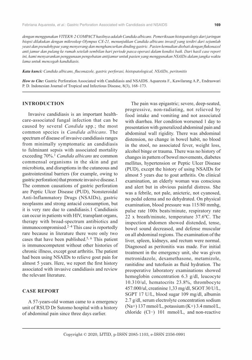
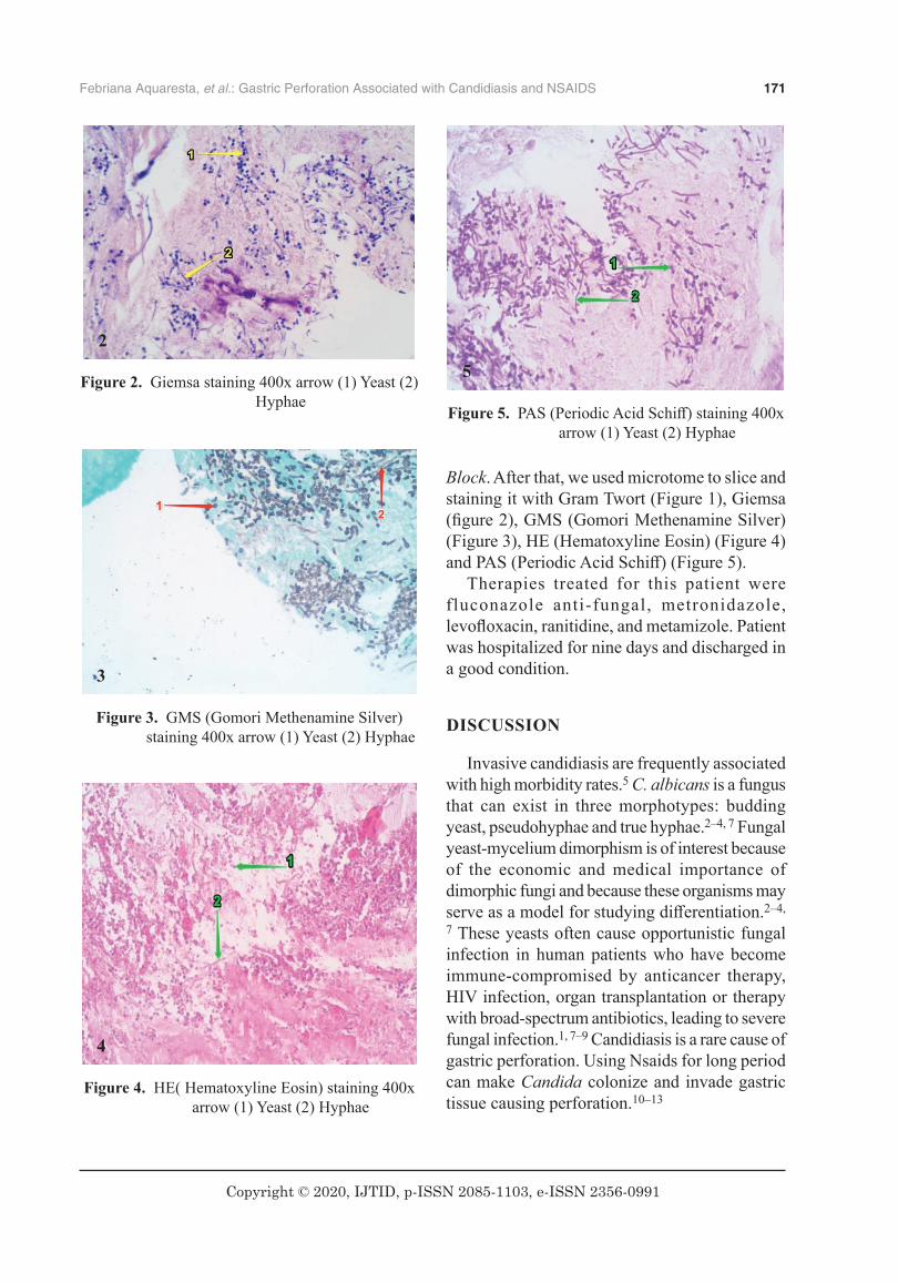
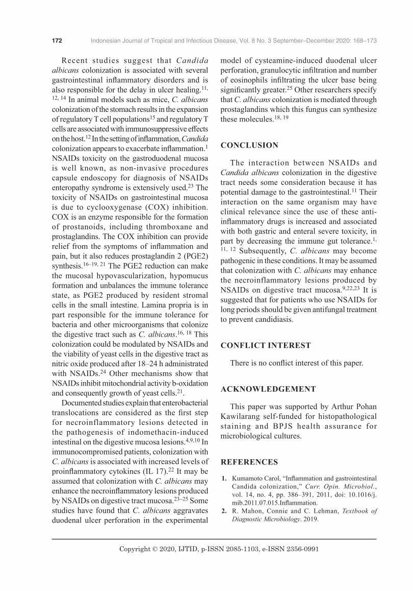
Block. After that, we used microtome to slice and staining it with Gram Twort (Figure 1), Giemsa (fi gure 2), GMS (Gomori Methenamine Silver) (Figure 3), HE (Hematoxyline Eosin) (Figure 4) and PAS (Periodic Acid Schiff) (Figure 5).
Therapies treated for this patient were fluconazole anti-fungal, metronidazole, levofloxacin, ranitidine, and metamizole. Patient was hospitalized for nine days and discharged in a good condition.
DISCUSSION
Invasive candidiasis are frequently associated with high morbidity rates.5 C. albicans is a fungus that can exist in three morphotypes: budding yeast, pseudohyphae and true hyphae.2–4, 7 Fungal yeast-mycelium dimorphism is of interest because of the economic and medical importance of dimorphic fungi and because these organisms may serve as a model for studying differentiation.2–4,
7 These yeasts often cause opportunistic fungal infection in human patients who have become immune-compromised by anticancer therapy, HIV infection, organ transplantation or therapy with broad-spectrum antibiotics, leading to severe fungal infection.1, 7–9 Candidiasis is a rare cause of gastric perforation. Using Nsaids for long period can make Candida colonize and invade gastric tissue causing perforation.10–13
3
Figure 3. GMS (Gomori Methenamine Silver) staining 400x arrow (1) Yeast (2) Hyphae
5
Figure 5. PAS (Periodic Acid Schiff ) staining 400x arrow (1) Yeast (2) Hyphae
2
Figure 2. Giemsa staining 400x arrow (1) Yeast (2) Hyphae
4
Figure 4. HE( Hematoxyline Eosin) staining 400x arrow (1) Yeast (2) Hyphae
172
Copyright © 2020, IJTID, p-ISSN 2085-1103, e-ISSN 2356-0991
Indonesian Journal of Tropical and Infectious Disease, Vol. 8 No. 3 September–December 2020: 168–173
Recent studies suggest that Candida albicans colonization is associated with several gastrointestinal inflammatory disorders and is also responsible for the delay in ulcer healing.11,
12, 14 In animal models such as mice, C. albicans colonization of the stomach results in the expansion of regulatory T cell populations15 and regulatory T cells are associated with immunosuppressive eff ects on the host.12 In the setting of inflammation, Candida colonization appears to exacerbate inflammation.1 NSAIDs toxicity on the gastroduodenal mucosa is well known, as non-invasive procedures capsule endoscopy for diagnosis of NSAIDs enteropathy syndrome is extensively used.23 The toxicity of NSAIDs on gastrointestinal mucosa is due to cyclooxygenase (COX) inhibition. COX is an enzyme responsible for the formation of prostanoids, including thromboxane and prostaglandins. The COX inhibition can provide relief from the symptoms of inflammation and pain, but it also reduces prostaglandin 2 (PGE2) synthesis.16–19, 21 The PGE2 reduction can make the mucosal hypovascularization, hypomucus formation and unbalances the immune tolerance state, as PGE2 produced by resident stromal cells in the small intestine. Lamina propria is in part responsible for the immune tolerance for bacteria and other microorganisms that colonize the digestive tract such as C. albicans.16, 18 This colonization could be modulated by NSAIDs and the viability of yeast cells in the digestive tract as nitric oxide produced after 18–24 h administrated with NSAIDs.24 Other mechanisms show that NSAIDs inhibit mitochondrial activity b-oxidation and consequently growth of yeast cells.21.
Documented studies explain that enterobacterial translocations are considered as the first step for necroinflammatory lesions detected in the pathogenesis of indomethacin-induced intestinal on the digestive mucosa lesions.4,9,10 In immunocompromised patients, colonization with C. albicans is associated with increased levels of proinflammatory cytokines (IL 17).22 It may be assumed that colonization with C. albicans may enhance the necroinflammatory lesions produced by NSAIDs on digestive tract mucosa.23–25 Some studies have found that C. albicans aggravates duodenal ulcer perforation in the experimental
model of cysteamine-induced duodenal ulcer perforation, granulocytic infiltration and number of eosinophils infiltrating the ulcer base being significantly greater.25 Other researchers specify that C. albicans colonization is mediated through prostaglandins which this fungus can synthesize these molecules.18, 19
CONCLUSION
The interaction between NSAIDs and Candida albicans colonization in the digestive tract needs some consideration because it has potential damage to the gastrointestinal.11 Their interaction on the same organism may have clinical relevance since the use of these anti- inflammatory drugs is increased and associated with both gastric and enteral severe toxicity, in part by decreasing the immune gut tolerance.1,
11, 12 Subsequently, C. albicans may become pathogenic in these conditions. It may be assumed that colonization with C. albicans may enhance the necroinflammatory lesions produced by NSAIDs on digestive tract mucosa.9,22,23 It is suggested that for patients who use NSAIDs for long periods should be given antifungal treatment to prevent candidiasis.
CONFLICT INTEREST
There is no conflict interest of this paper.
ACKNOWLEDGEMENT
This paper was supported by Arthur Pohan Kawilarang self-funded for histopathological staining and BPJS health assurance for microbiological cultures.
REFERENCES
1. Kumamoto Carol, “Inflammation and gastrointestinal Candida colonization,” Curr. Opin. Microbiol., vol. 14, no. 4, pp. 386–391, 2011, doi: 10.1016/j. mib.2011.07.015.Inflammation.
2. R. Mahon, Connie and C. Lehman, Textbook of Diagnostic Microbiology. 2019.

173Febriana Aquaresta, et al.: Gastric Perforation Associated with Candidiasis and NSAIDS
Copyright © 2020, IJTID, p-ISSN 2085-1103, e-ISSN 2356-0991
3. P. Tille, Balley & Scott’s: Diagnostic Microbiology. 2017.
4. D. Prieto, I. Correia, J. Pla, and E. Román, “Adaptation of Candida albicans to commensalism in the gut,” Future Microbiol., vol. 11, no. 4, pp. 567–583, 2016, doi: 10.2217/fmb.16.1.
5. N. Gupta, “A rare cause of gastric perforation-Candida infection: A case report and review of the literature,” J. Clin. Diagnostic Res., vol. 6, no. 9, pp. 1564–1565, 2012, doi: 10.7860/JCDR/2012/4632.2563.
6. S. Höfs et al., “Candidiasis, a rare cause of gastric perforation: A case report and review of literature,” Ann. Med. Health Sci. Res., vol. 54, no. 4, p. 314, 2015, doi: 10.4103/2141-9248.160187.
7. G. Pappas, Peter, S. Lionakis, Michail, C. Arendrup, Maiken, L. Zeichner-Ostrosky, and J. Kullberg, Bart, “Invasive candidiasis,” Nat. Rev. Dis. Prim., vol. 4, 2018, doi: 10.1038/nrdp.2018.27.
8. A. da Silva Dantas et al., “Cell biology of Candida albicans–host interactions,” Curr. Opin. Microbiol., vol. 34, pp. 111–118, 2016, doi: 10.1016/j. mib.2016.08.006.
9. S. Höfs, S. Mogavero, and B. Hube, “Interaction of Candida albicans with host cells: virulence factors, host defense, escape strategies, and the microbiota,” J. Microbiol., vol. 54, no. 3, pp. 149–169, 2016, doi: 10.1007/s12275-016-5514-0.
10. T. Yamada, E. Deitch, R. D. Specian, M. A. Perry, R. B. Sartor, and M. B. Grisham, “Mechanisms of acute and chronic intestinal inflammation induced by indomethacin,” Infl ammation, vol. 17, no. 6, pp. 641–662, 1993, doi: 10.1007/BF00920471.
11. G. C. Nadǎş et al., “Erratum to: The Interplay Between NSAIDs and Candida albicans on the Gastrointestinal Tract of Guinea Pigs (Mycopathologia, (2013), 175, (221-230), 10.1007/s11046-013-9613-8),” Mycopathologia, vol. 176, no. 1–2, pp. 171–173, 2013, doi: 10.1007/s11046-013-9659-7.
12. O. Ekenna and R. J. Sherertz, “Factors affecting colonization and dissemination of Candida albicans from the gastrointestinal tract of mice,” Infect. Immun., vol. 55, no. 7, pp. 1558–1563, 1987.
13. M. A. S. Alem and L. J. Douglas, “Effects of Aspirin and Other Nonsteroidal Anti-Inflammatory Drugs on Biofilms and Planktonic Cells of Candida albicans,” Antimicrob. Agents Chemother., vol. 48, no. 1, pp. 41–47, 2004, doi: 10.1128/AAC.48.1.41-47.2004.
14. M. Ilahi, J. Khan, Q. Inayat, and T. S. Abidi, “Histological changes in parts of foregut of rat after indomethacin administration.,” J. Ayub Med. Coll. Abbottabad, vol. 18, no. 3, pp. 29–34, 2006.
15. P. L. Beck et al., “Mechanisms of NSAID-induced gastrointestinal injury defined using mutant mice,” Gastroenterology, vol. 119, no. 3, pp. 699–705, 2000, doi: 10.1053/gast.2000.16497.
16. M. C. Noverr, S. M. Phare, G. B. Toews, M. J. Coffey, and G. B. Huffnagle, “albicans Produce
Immunomodulatory Prostaglandins,” Infect. Immun., vol. 69, no. 5, pp. 2957–2963, 2001, doi: 10.1128/ IAI.69.5.2957.
17. A. H. P. Loh et al., “Multiple indomethacin-induced colonic perforations in an adolescent,” Singapore Med. J., vol. 52, no. 4, pp. 82–84, 2011.
18. R. D. Newberry, J. S. McDonough, W. F. Stenson, and R. G. Lorenz, “ Spontaneous and Continuous Cyclooxygenase-2-Dependent Prostaglandin E 2 Production by Stromal Cells in the Murine Small Intestine Lamina Propria: Directing the Tone of the Intestinal Immune Response ,” J. Immunol., vol. 166, no. 7, pp. 4465–4472,2001,doi: 10.4049/ jimmunol.166.7.4465.
19. [J. L. F. Kock, O. M. Sebolai, C. H. Pohl, P. W. J. Van Wyk, and E. J. Lodolo, “Oxylipin studies expose aspirin as antifungal,” FEMS Yeast Res., vol. 7, no. 8, pp. 1207–1217, 2007, doi: 10.1111/j.1567- 1364.2007.00273.x.
20. J. Kock et al., “Development of a Yeast Bio-Assay to Screen Anti-Mitochondrial Drugs,” Curr. Drug Discov. Technol., vol. 6, no. 3, pp. 186–191,2009,doi: 10.2174/157016309789054960.
21. J. L. F. Kock, C. J. Strauss, C. H. Pohl, and S. Nigam, “The distribution of 3-hydroxy oxylipins in fungi,” Prostaglandins Other Lipid Mediat., vol. 71, no. 3–4, pp. 85–96, 2003, doi: 10.1016/S1098- 8823(03)00046-7.
22. C. Montagnoli et al., “ B7/CD28-Dependent CD4 + CD25 + Regulatory T Cells Are Essential Components of the Memory-Protective Immunity to Candida albicans ,” J. Immunol., vol. 169, no. 11, pp. 6298– 6308, 2002, doi: 10.4049/jimmunol.169.11.6298.
23. Tanaka, S. Hase, T. Miyazawa, and K. Takeuchi, “Up-regulation of cyclooxygenase-2 by inhibition of cyclooxygenase-1: A key to nonsteroidal anti- inflammatory drug-induced intestinal damage,” J. Pharmacol. Exp. Ther., vol. 300, no. 3, pp. 754–761, 2002, doi: 10.1124/jpet.300.3.754.
24. B. J. R. Whittle, “Gastrointestinal effects of nonsteroidal anti-inflammatory drugs,” Fundam. Clin. Pharmacol., vol. 17, no. 3, pp. 301–313, 2003, doi: 10.1046/j.1472- 8206.2003.00135.x.
25. L. Maiden, “Capsule endoscopic diagnosis of nonsteroidal antiinflammatory drug-induced enteropathy,” J. Gastroenterol., vol. 44, no. SUPPL. 19, pp. 64–71, 2009, doi: 10.1007/s00535-008-2248-8.
26. A. Vazquez-Torres, J. Jones-Carson, T. Warner, and E. Balish, “Nitric oxide enhances resistance of scid mice to mucosal candidiasis,” J. Infect. Dis., vol. 172, no. 1, pp. 192–198, 1995, doi: 10.1093/infdis/172.1.192.
27. T. Nakamura et al., “Candida albicans aggravates duodenal ulcer perforation induced by administration of cysteamine in rats,” J. Gastroenterol. Hepatol., vol. 22, no. 5, pp. 749–756, 2007, doi: 10.1111/j.1440- 1746.2006.04353.x.