Automatic segmentation of whole-body bone scintigrams...
10
Automatic segmentation of whole-body bone scintigrams as a preprocessing step for computer assisted diagnostics Tracking number: C0114 Abstract. Bone scintigraphy or whole-body bone scan is one of the most common diagnostic procedures in nuclear medicine used in the last 25 years. Pathological conditions, technically poor quality images and ar- tifacts necessitate that algorithms use sufficient background knowledge of anatomy and spatial relations of bones in order to work satisfactorily. We present a robust knowledge based methodology for detecting reference points of the main skeletal regions that simultaneously works on both anterior and posterior whole-body bone scintigrams. Expert knowledge is represented as a set of parameterized rules which are used to support standard image processing algorithms. Our study includes 467 consec- utive, non-selected scintigrams, which is the biggest number of images ever used in such studies to our knowledge. Automatic analysis of whole- body bone scans using our knowledge based segmentation algorithm gives more accurate and reliable results than previous studies. Obtained refer- ence points are used for automatic segmentation of the skeleton, which is used for automatic (machine learning) or manual (expert physicians) diagnostics. Preliminary experiments show that an expert system based on machine learning closely mimics the results of expert physicians. Keywords: whole-body bone scintigraphy, reference point detection, au- tomatic segmentation, image processing, machine learning 1 Introduction Whole-body scan or bone scintigraphy is a well known clinical routine investi- gation and one of the most frequent diagnostic procedures in nuclear medicine. Indications for bone scintigraphy include benign and malignant diseases, infec- tions, degenerative changes ... [2]). Bone scintigraphy has high sensitivity and the changes of the bone metabolism are seen earlier than changes in bone structure detected on radiograms [15]. The investigator’s role is to evaluate the image, which is of poor resolution due to the physical limitations of gamma camera. There are approximately 158 bones visible on both anterior and posterior whole-body scans [10]. Poor quality and the number of bones to inspect makes it difficult and often tedious work. Some research on automating the process of counting the bone lesions has been done, but only few studies attempted to automatically segment individual bones prior to the computerized evaluation of bone scans [6; 7; 1].
Transcript of Automatic segmentation of whole-body bone scintigrams...

Automatic segmentation of whole-body bonescintigrams as a preprocessing step for computer
assisted diagnostics
Tracking number: C0114
Abstract. Bone scintigraphy or whole-body bone scan is one of themost common diagnostic procedures in nuclear medicine used in the last25 years. Pathological conditions, technically poor quality images and ar-tifacts necessitate that algorithms use sufficient background knowledge ofanatomy and spatial relations of bones in order to work satisfactorily. Wepresent a robust knowledge based methodology for detecting referencepoints of the main skeletal regions that simultaneously works on bothanterior and posterior whole-body bone scintigrams. Expert knowledgeis represented as a set of parameterized rules which are used to supportstandard image processing algorithms. Our study includes 467 consec-utive, non-selected scintigrams, which is the biggest number of imagesever used in such studies to our knowledge. Automatic analysis of whole-body bone scans using our knowledge based segmentation algorithm givesmore accurate and reliable results than previous studies. Obtained refer-ence points are used for automatic segmentation of the skeleton, whichis used for automatic (machine learning) or manual (expert physicians)diagnostics. Preliminary experiments show that an expert system basedon machine learning closely mimics the results of expert physicians.
Keywords: whole-body bone scintigraphy, reference point detection, au-tomatic segmentation, image processing, machine learning
1 Introduction
Whole-body scan or bone scintigraphy is a well known clinical routine investi-gation and one of the most frequent diagnostic procedures in nuclear medicine.Indications for bone scintigraphy include benign and malignant diseases, infec-tions, degenerative changes ... [2]). Bone scintigraphy has high sensitivity and thechanges of the bone metabolism are seen earlier than changes in bone structuredetected on radiograms [15].
The investigator’s role is to evaluate the image, which is of poor resolutiondue to the physical limitations of gamma camera. There are approximately 158bones visible on both anterior and posterior whole-body scans [10]. Poor qualityand the number of bones to inspect makes it difficult and often tedious work.Some research on automating the process of counting the bone lesions has beendone, but only few studies attempted to automatically segment individual bonesprior to the computerized evaluation of bone scans [6; 7; 1].

2 Tracking number: C0114
1.1 Related work
First attempts to automate scintigraphy in diagnostics for thyroid structureand function were made back in 1973 [11]. Most of the research on automaticlocalization of bones has been done at the former Institute of medical informationscience at the University of Hildesheim in Germany from 1994 to 1996. Themain contribution was made by the authors Berning [7] and Bernauer [6] whodeveloped semantic representation of the skeleton and evaluation of the images.Benneke [1] has realized their ideas in 1996.
Yin and Chiu [13] tryied to find lesions using a fuzzy system. Their prepro-cessing of scintigrams includes rough segmentation of six parts with fixed ratiosof the whole skeleton. Those parts are rigid and not specific enough to localizea specific bone. Their approach for locating abnormalities in bone scintigraphyis limited to point-like lesions with high uptake.
When dealing with lesion detection other authors like Noguchi [10] have beenusing merely intensity thresholding and manual lesion counting or manual boneROI (region of interest) labeling. Those procedures are only sufficient for moreobvious pathologies whereas new emerging pathological regions are overlooked.
2 Aim and our approach
The aim of our study was to develop a robust method for segmenting whole-bodybone scans to allow further development of automatic algorithms for bone scandiagnostics of individual bones.
We have developed the algorithm for detecting extreme edges of images(peaks). Here, respective skeletal regions are processed in the following order:shoulders, head, pelvis, thorax and extremities. The experience with automaticprocessing is presented. Several image processing algorithms are used such asbinarization, skeletonization, Hough’s transform, Gaussian filtering [4], leastsquare method and ellipse fitting in combination with background knowledgeof anatomy and scintigraphy specialities.
In everyday practice, when a bone is identified, it is diagnosed by the expertphysician according to several possible pathologies (lesions, malignom, metasta-sis, degenerative changes, inflammation, other pathologies, no pathologies). Thisprocess can be supported by using some machine learning classifier [9] which pro-duces independent diagnoses. As an input it is given a suitably parameterizedbone image, obtained from detected reference points. As an output it assigns thebone to one of the above pathologies. It can therefore be used as a tool to givephysician an additional insight in the problem.
3 Materials and methods
3.1 Patients and images
Retrospective review of 467 consecutive, non-selected scintigraphic images from461 different patients who visited University Medical Centre in Ljubljana from

Automatic segmentation of whole-body bone scintigrams. . . 3
October 2003 to June 2004 was performed. Images were not preselected, so thestudy included standard distribution of patients coming to examination in 9months. 19% of the images were diagnosed as normal, which means no pathol-ogy was detected on the image. 57% of the images were diagnosed with slightpathology, 20% with strong pathology and 2% were classified as super-scans.
Images also contained some artifacts and non-osseous uptake such as urinecontamination and medical accessories (i.e. urinary catheters) [5]. Segmentationwas also complicated by the radiopharmaceutical site of injection. Partial scans(missing a part of the head or upper/lower extremities in the picture) were thecase in 18% of the images. There were also adolescents with growth zones (5%of the images), manifested as increased osteoblastic activity in well delineatedareas with very high tracer uptake.
3.2 Bone scintigraphy
All patients were scanned with gamma camera model Siemens MultiSPECTwith two heads with LEHR (Low Energy High resolution) collimators. Scanspeed was 8cm per minute with no pixel zooming. 99m-Tc-DPD (TechneosR)was used. Bone scintigraphy was obtained about 3h after intravenous injectionof 750 MBq of radiopharmaceutical agent. The whole body field was used torecord anterior and posterior views digitally with resolution of 1024 x 256 pixels.Images represent the counts of detected gamma rays in each spatial unit with16-bit grayscale depth.
3.3 Detection of reference points
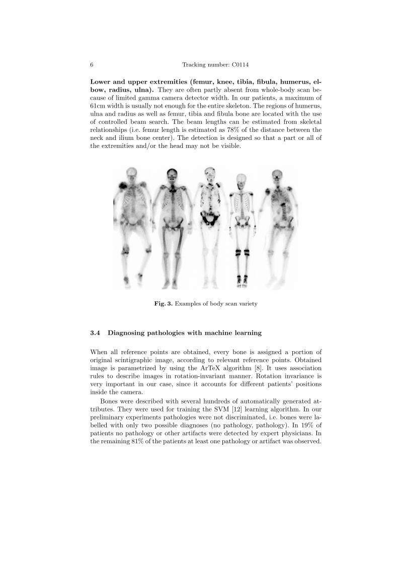
Bone scans are very different (Figure 3) one from another even though the struc-ture and position of bones is more or less the same. In practice many scans areonly partial because only a determined part of the body is observed or due to thescanning time limitations. In our study we have observed that only on two imagesout of 467 the shoulders were not visible. Many other characteristic parts couldhave been missing in images more often (i.e. head, arms, one or both legs). Wehave chosen shoulders as the main reference points to start with, which meansthey are supposed to be visible in the images. Second and the last assumptionis the upward orientation of the image. This assumption is not limiting since allscintigraphies are made with same orientation.
In order to make the detection of reference points faster and more reliable wehave tried to automatically detect intuitive peaks which would represent edgesand would cover roughly also the reference points. With normal Canny edge filtertoo many peaks were obtained. Our approach is based on orthogonal two-wayGaussian filtering [16].
Low image intensities (count level) acquired in typical studies are due to thelimited level of radioactive dosage required to ensure patient’s safety. They makebone scans look distorted. Bone edges are more expressive after we filter imageswith some averaging algorithm (i.e. wavelet based, median filter, Gaussian filter)[4]. We have used Gaussian filter so that the detection of peaks was more reliable.

4 Tracking number: C0114
Both images, anterior and posterior, are simultaneously processed in the samedetection order and in each step the detected reference points visible on bothimages are compared and corrected adequately.
Detected points from the anterior image are mirrored to the posterior andvice versa. Some bones are better visible on anterior and some on posteriorimages due to the varying distances from both collimators. This improves thecalculation of circles, lines and ellipses with least square method (LSM).
The order in which the reference points were detected was determined byusing knowledge of human anatomy as well as physicians’ recommendations.They are represented as a list of parameterized rules. Rule parameters (e.g.thresholds, spatial and intensity ratios, ...) were initially set by physicians andfurther refined on a separate tuning set. More details can be found in [16].
Shoulders. They are the only part of the body that is assumed to be present inevery image in the upper part on both sides. The algorithm just searches for thehighest detected peak on both sides of the image. Next step is to locally shiftthe candidate points with local maximum intensity tracing to the outermostlocation. Only in 5 images out of 467 the shoulders were not found correctly dueto the tilted head position.
Pelvic region (ilium bone, pubis bone, great trochanter of femur). Themost identifiable bone in pelvic region is ilium bone which has higher uptakevalues than it’s neighboring soft tissue. Ilium bone has circular shape in theupper part and therefore it is convenient for circle detection with LSM method.This bone is well described with already detected peaks by as shown in Figure1(b). Ilium position is roughly estimated with regions of interest (ROIs) whichare found on the basis of skeleton’s anticipated ratios and reference points foundup to this step of detection.
The pelvis is located at the end of the spine and has approximately the samewidth as shoulders. In order to find the pelvis, the calculation of the spine posi-tion is required. This is done with a beam search (Figure 1(a)). The anticipatedspine length is determined from the distance between shoulders. Beam startingpoint is the middle point of the shoulders and it’s orientation is perpendicularto the shoulder line. The angle at which the beam covers most peaks, is a roughestimation of spine direction since there is most of the uptake in the vertebraeand hence peaks are dense in that region.
Pubis bone is detected by estimating the pubis ROI using detected iliumlocation, distance between detected ilium circles and their inclination. The ex-perimentally determined ROI’s size is narrowed and additional vertical peaksare added and circles detected as shown in Figure 1(b).
Head and neck. When at least image orientation and the location of theshoulders are known, some part of the neck or even head is visible since theyare between the shoulders. Finding the head is not difficult but its orientationis, especially in cases where a part of the head in scan is not visible. The mostreliable method for determining head orientation and position is ellipse fittingof the head contour determined by thresholding. Neck is found by local vertical

Automatic segmentation of whole-body bone scintigrams. . . 5
(a) Beam search sketch
(b) Detection of bones in the pelvicregion
Fig. 1. Beam search and detection in pelvic region
shifting of a stripe determined by the ellipse’s semiminor axis (position andorientation).Thoracic part (vertebrae, ribs). Vertebrae have more or less constant spa-tial relations, the only problem is that on a bone scintigraphy only a planarprojection of the spine is visible. Since the spine is longitudinally curved, thespatial relations vary due to different longitudinal orientation of the patients.Average vertebrae relations have been experimentally determined from normalskeletons.
Ribs are the most difficult skeleton region to detect since they are quiteunexpressive on bone scans, their formation can vary considerably and theircontours [14] can be disconnected in the case of stronger pathology (Figure 2).
A B C D E
Fig. 2. Rib detection steps example on a skeleton with strong pathology. Rib ROI isbinarized (A), binarized image is skeletonized (B), Hough transform of linear equationis calculated on skeleton points (C), reference points are estimated using results of theHough transform (D), rib contours are individually followed by the contour followingalgorithm (E).
6 Tracking number: C0114
Lower and upper extremities (femur, knee, tibia, fibula, humerus, el-bow, radius, ulna). They are often partly absent from whole-body scan be-cause of limited gamma camera detector width. In our patients, a maximum of61cm width is usually not enough for the entire skeleton. The regions of humerus,ulna and radius as well as femur, tibia and fibula bone are located with the useof controlled beam search. The beam lengths can be estimated from skeletalrelationships (i.e. femur length is estimated as 78% of the distance between theneck and ilium bone center). The detection is designed so that a part or all ofthe extremities and/or the head may not be visible.
Fig. 3. Examples of body scan variety
3.4 Diagnosing pathologies with machine learning
When all reference points are obtained, every bone is assigned a portion oforiginal scintigraphic image, according to relevant reference points. Obtainedimage is parametrized by using the ArTeX algorithm [8]. It uses associationrules to describe images in rotation-invariant manner. Rotation invariance isvery important in our case, since it accounts for different patients’ positionsinside the camera.
Bones were described with several hundreds of automatically generated at-tributes. They were used for training the SVM [12] learning algorithm. In ourpreliminary experiments pathologies were not discriminated, i.e. bones were la-belled with only two possible diagnoses (no pathology, pathology). In 19% ofpatients no pathology or other artifacts were detected by expert physicians. Inthe remaining 81% of the patients at least one pathology or artifact was observed.

Automatic segmentation of whole-body bone scintigrams. . . 7
4 Results
4.1 Segmentation
Approximately half of the available images were used for tuning rule parametersto optimize the recognition of the reference points and another half to test it.All 246 patients examined from October 2003 to March 2004 were used as thetuning set and 221 patients examined from April 2004 to June 2004 were used asthe test set. In the tuning set there were various non-osseous uptakes in 38.9%of the images, 47.5% images with the visible injection point and 6.8% images ofadolescents with the visible growth zones. Similar distribution was found in thetest set (34.5% non-osseous uptakes, 41.0% visible injection points and 2.85%adolescents). Most of the artifacts were minor radioactivity points from urinecontamination in genital region or other parts (81.4% of all artifacts) whereasonly few other types were observed (urinary catheters 13%, artificial hips 4%and lead accessories 1.6%). We have observed that there were no ill-detectedreference points in adolescents with the visible growth zones since all the bonesare homogenous, have good visibility and are clearly divided with growth zones.Results of detecting reference points on the test set are shown in the Table 1.
Table 1. False reference point detection on test set. Both frequencies and percentagesare given.
Bone no slight strong super-scan allpathology pathology pathology
46 133 39 3 221
ilium 0 2 0.9% 6 2.7% 1 0.5% 9 4.1%
pubis 2 0.9% 3 1.4% 2 0.9% 0 7 3.2%
trochanter 0 1 0.5% 0 0 1 0.5%
shoulder 0 0 1 0.5% 0 1 0.5%
extremities 5 2.3% 11 5.0% 0 0 16 7.2%
spine 0 2 0.9% 1 0.5% 0 3 1.4%
ribs 11 5.0% 17 7.7% 3 1.4% 0 31 14.0%
neck 2 0.9% 4 1.8% 0 0 6 2.7%
4.2 Machine learning results
From our complete set of 467 patients, pathologies were thoroughly evaluated byphysicians only for 268 patients. These 268 patients were used for evaluation ofmachine learning approach by using ten-fold cross validation. Results are shownin Table 2. The bones were grouped in ten relevant groups. They are quiteimpressive, given high numbers of different bones (158 visible for an individualadult patient). Classification accuracy was obtained for a two-class problem.

8 Tracking number: C0114
Table 2. Experimental results with machine learning on two-class problem.
Bone group Classification accuracy
Cervical spine 75.94Feet 83.82Skull back 94.74Illium bone 87.31Lumbal spine 71.43Femur and tibia 88.89Pelvic region 92.16Ribs 98.05Scapula 91.42Thoracic spine 81.95
Total 89.93
5 Discussion
The testing showed encouraging results since the detection of proposed referencepoints gave excellent results for all bone regions but the extremities, which wasexpected.
We have payed special attention to the images with partial skeletons since itis often the case in clinical routine (in our study 18% of the images were partialand no particular problem appeared in detecting) and a robust segmentationalgorithm should not fail on such images. The detection of ribs showed to bethe most difficult, yet that was expected. Results show that in 14% to 20% ofimages there were difficulties in detecting the ribs. This usually means one rib ismissed or not followed to the very end which we intend to improve in the future.In the present system such reference points can be manually repositioned by theexpert physicians.
The automatically detected reference points can be used for mapping a stan-dard skeletal reference mask, which is to our belief the best way to find individualbones on scintigrams since bone regions are often not expressive enough to followtheir contour. An example of such mask mapping is shown in Figure 4.
Fig. 4. Example of mapped standard skeletal mask with the detected reference points

Automatic segmentation of whole-body bone scintigrams. . . 9
While our experimental results with machine learning are quite good, onemust bear in mind that they were obtained for a simplified (two class) problem.Simply extending a problem to a multi-class paradigm is not acceptable in ourcase, as the bone may be assigned several different pathologies at the sametime. A proper approach, the one we are currently working on, is to rephrase aproblem to the multi-label learning problem, where each bone will be labelledwith a nonempty subset of all possible labels [17; 3].
6 Conclusion
The presented computer-aided system for bone scintigraphy is a step forward inautomating routine medical procedures. Some standard image processing algo-rithms were tailored and used in combination to achieve the best reference pointdetection accuracy on scintigraphic images which have very low resolution. Poorquality, artifacts and pathologies necessitate that algorithms use as much back-ground knowledge on anatomy and spatial relations of bones as possible in orderto work satisfactorily. This combination gives quite good results and we expectthat further studies on automatic scintigraphy diagnosing using reference pointsfor image segmentation will give more accurate and reliable results than previousstudies, negligent to the segmentation.
This approach opens a new view on automatic scintigraphy evaluation, sincein addition to detection of point-like high-uptake lesions there are also:
– more accurate and reliable evaluation of bone symmetry when looking forskeletal abnormalities. Many abnormalities can be spotted only when thesymmetry is observed (differences in length, girth, curvature etc.),
– detection of lesions with low-uptake or lower activity due to metallic im-plants,
– possibility of comparing uptake ratios among different bones,– more complex pathology detection with combining pathologies of more bones
(i.e. arthritis in joints)– possibility of automatic reporting of bone pathologies in written language.
Machine learning approach in this problem is in a very early stage, so itsusefulness in practice cannot yet be objectively evaluated. However, preliminaryresults are encouraging and switching to the multilabel learning framework maymake them even better.
Acknowledgement
This work was supported by the Slovenian Ministry of Education, Science andSport through the research programme P2-0209. Special thanks to nuclear medicinespecialist Jure Fettich at the University Medical Centre in Ljubljana for his helpand support.

Bibliography
[1] Benneke A. Konzeption und realisierung eines semi-automatischen befundungssys-tems in java und anbindung an ein formalisiertes begriffssystem am beispiel derskelett-szintigraphie. Diplom arbeit, Institut fur Medizinische Informatik, Univer-sitat Hildesheim, mentor Prof. Dr. D.P. Pretschner, 1997.
[2] Hendler A. and Hershkop M. When to use bone scintigraphy. it can reveal thingsother studies cannot. Postgraduate Medicine, 104(5):54–66, 11 1998.
[3] McCallum A. Multi-label text classification with a mixture model trained by em.In Proc. AAAI’99 Workshop on Text Learning, 1999.
[4] Jammal G. and Bijaoui A. Dequant: a flexible multiresolution restoration frame-work. Signal Processing, 84(7):1049–1069, 7 2004.
[5] Weiner M. G., Jenicke L., Mller V., and Bohuslavizki H. K. Artifacts and non-osseous uptake in bone scintigraphy. imaging reports of 20 cases. Radiol Oncol,35(3):185–91, 2001.
[6] Bernauer J. Zur semantischen rekonstruktion medizinischer begriffssysteme. Ha-bilitationsschrift, Institut fur Medizinische Informatik, Univ. Hildesheim, 1995.
[7] Berning K.-C. Zur automatischen Befundung und Interpretation von Ganzkorper-Skelettszintigrammen. PhD thesis, Institut fur Medizinische Informatik, Univer-sitat Hildesheim, 1996.
[8] Bevk M. and Kononenko I. Towards symbolic mining of images with associationrules: preliminary results on textures. In Brito P. and Noirhomme-Fraiture M.,editors, ECML/PKDD 2004: proc. of the workshop W2 on symbolic and spatialdata analysis: mining complex data structures, pages 43–53, 2004.
[9] Kukar M., Kononenko I., Groselj C., Kralj K., and Fettich J. Analysing andimproving the diagnosis of ischaemic heart disease with machine learning. ArtificialIntelligence in Medicine, 16:25–50, 1999.
[10] Noguchi M., Kikuchi H., Ishibashi M., and Noda S. Percentage of the positivearea of bone metastasis is an independent predictor of disease death in advancedprostate cancer. British Journal of Cancer, (88):195–201, 2003.
[11] Maisey M.N., Natarajan T.K., Hurley P.J., and Wagner H.N. Jr. of a rapid com-puterized method of measuring 99mtc pertechnetate uptake for routine assessmentof thyroid structure and function. J Clin Endocrinol Metab, 36:317–322, 1973.
[12] Cristianini N. and Shawe-Taylor J. An introduction to support vector machines(and other kernel-based learning methods). Cambridge University Press, 2000.
[13] Yin T.K. and Chiu N.T. A computer-aided diagnosis for locating abnormalitiesin bone scintigraphy by a fuzzy system with a three-step minimization approach.IEEE Transactions on Medical Imaging, 23(5):639–654, 5 2004.
[14] Kindratenko V. Development and Application of Image Analysis Techniques forIdentification and Classification of Microscopic Particles. PhD thesis, Universi-taire Instelling Antwerpen, Departement Scheikunde, 1997.
[15] Muller V., Steinhagen J., de Wit M., and Bohuslavizki H. K. Bone scintigraphyin clinical routine. Radiol Oncol, 35(1):21–30, 2001.
[16] Sajn L., Kononenko I., Fettich J., and Milcinski M. Automatic segmentation ofwhole-body bone scintigrams. Nuclear Medicine Comm., 2005. To appear.
[17] Shen X., Boutell M., Luo J., and Brown C. Multi-label machine learning and itsapplication to semantic scene classification. In Proceedings of the 2004 Interna-tional Symposium on Electronic Imaging (EI 2004), San Jose, California, 2004.