Automated MRI segmentation for individualized modeling of ...
Automated Whole Brain Segmentation Using FreeSurfer
https://surfer.nmr.mgh.harvard.edu/
FreeSurfer (FS) is a free software package developed at the Martinos Center for Biomedical Imaging used for three main purposes:
(1) creation of computerized models of the brain from magnetic resonance imaging data, (2) measuring various morphometric
properties of the brain including cortical thickness, curvature characteristics, and cortical and sub-cortical regional volumes, (3)
inter-subject cortical spatial normalization based on aligning individual’s cortical folding patterns to a population averaged cortical
folding pattern to establish correspondence between homologous anatomical regions.
Starting with the good quality T1-weighted MR image volume, the first step in the FreeSurfer processing is to perform a motion
correction, affine transformation (12 degrees of freedom) to Talairach image space, non-uniform intensity normalization for
intensity inhomogeneity correction, and removal of non-brain tissues. The remaining brain image volume is intensity normalized to
match the FreeSurfer atlas image intensity histogram, which is followed by a non-linear warping of the atlas brain image to subject
brain image. Warped atlas brain image in the subject image space is utilized in atlas-based tissue segmentation, in labeling the
subcortical structures, brain stem, cerebellum, and cerebral cortex. The next step in FreeSurfer is to generate topologically correct
cortical surface representation per hemisphere. Cortical surface lies either at the WM/GM tissue interface or at the GM/CSF tissue
interface. Each hemisphere’s cortical surface representation is mapped automatically to a standard spherical coordinate system. Key
components of the surface mapping include surface inflation with minimal metric distortion, projection to spherical coordinates,
topology correction, and surface based warping to align anatomically homologous points. Mapping to the standard spherical
coordinate system defined by FreeSurfer atlas brain allows for automated anatomical parcellation of cortex into gyral regions.
Surface parcellation is then extended to GM volume, yielding parcellation of GM tissue sheet and regional cortical volumes.
Although FreeSurfer processing framework allows for manual edits to improve the results, given the large number of subjects in
ADNI data set, to minimize the processing time and to eliminate the inter-rated variability in manual edits, fully-automated approach
was used. On a small sample data set of three scans, several test runs were performed to assess the performance of various optional
FreeSurfer processing flags on ADNI data set. Based on visual inspections, the ones that attained the best skull-stripping and the best
cortical parcellation and subcortical labeling consistently were chosen.
Many papers about FreeSurfer development and methods can be found here:
http://surfer.nmr.mgh.harvard.edu/fswiki/ArticlesSlidesAndPosters
Cross-sectional processing:
For each subject, T1 MR images from all time points were separately processing using cross-sectional workflow with the following
command line:
recon-all -wsatlas -wsless -all -s <ScanCode>
In case of autorecon1 failure, the notal-check and cw256 options were added and re-launched, with a talairach transformation QC
upon completion.
Base-Image creation:
Given an individual subject, FreeSurfer results for all time points were QCed. Base-image was created only if data from at least three
time points were passed cross-sectional processing QC. Base-image creation followed by cross-sectional processing of the base-
image was launched with the following command line:
recon-all -base <BaseCode> -tp <ScanTP1> -tp <ScanTP2> -tp <ScanTP3> … -all
Longitudinal processing:
Once the base-image was created and processed through the cross-sectional workflow, each time point data that passed cross-
sectional QC (and used in base-image creation) was reprocessed with the longitudinal FreeSurfer workflow:
recon-all -long <ScanTP#> <BaseCode> -all
The cross-sectional and longitudinal morphometry statistics (i.e., regional subcortical and cortical volumes, regional average cortical
thickness) reported at the LONI IDA website (http://www.loni.ucla.edu/ADNI/Data/) were based on statistics provided in aseg.stats and
aparc.stats.
Quality Control:
As of FreeSurfer version 4.3, each post-processed image generated by FreeSurfer for analysis is QCed for global success or failure, as
well as regionally for errors in the segmentation process that may occur in specific structures. The segmentation is QCed using the
tkmedit tool by loading the brainmask.mgz volume, along with the raw T1.mgz volume; the lh.white and rh.white surfaces; and the
segmentation.
Example: tkmedit 001_S_0001X1b_10001 brainmask.mgz lh.white -aux T1.mgz -aux-surface rh.white -segmentation
aparc+aseg.mgz
Every post-processed image is rated for two stages of the FreeSurfer process, Autorecon1 and Autorecon3:
1. Autorecon1 (Pass/Fail): The skullstripped brainmask is checked to ensure that no brain has been left out of the mask. Sometimes
the brainmask overestimates into regions of skull and meninges, this is noted later during the regional QC. However, if the
brainmask cuts off any brain area that is more than 3-4 voxel layers thick, it is failed globally; otherwise it is passed for Autorecon1
QC.
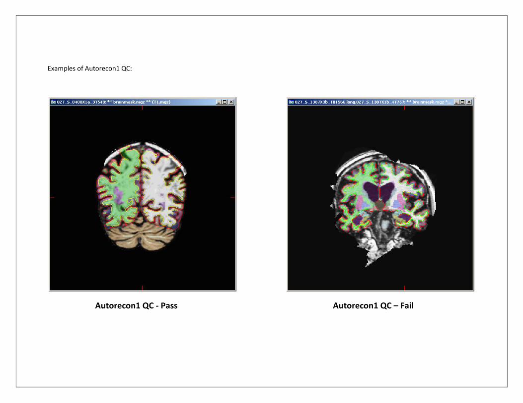
Examples of Autorecon1 QC:
Autorecon1 QC - Pass Autorecon1 QC – Fail
2a. Autorecon3 (Global Pass/Partial/Fail):
The FreeSurfer segmentation and pial surface estimates are checked globally and regionally in the coronal view for regions of
overestimation/underestimation, inconsistency with the structural boundaries, or segmenation regions that don't accurately reflect
the anatomy. The QC procedure accounts only for gross errors; a gross error for these purposes is defined as an area of
over/underestimation that is larger than the cursor, and which occurs on two or more slices. This is to account for inevitable small
errors in segmentation such as those which may be the result of partial voluming. Orientations other than the coronal view may be
used to confirm possible pial border and segmentation gross errors.
The global Autorecon3 QC is given one of three ratings:
-Pass: No significant gross errors.
-Fail: Complete segmentation failure or gross errors throughout all regions of the segmentation.
-Partial: Gross errors in one or more specific regions of the segmentation (see 2b. below).
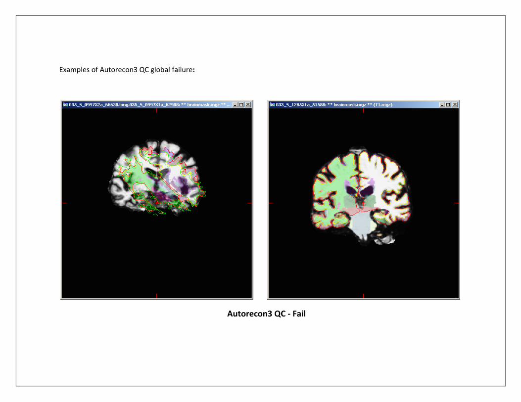
Examples of Autorecon3 QC global failure:
Autorecon3 QC - Fail
2b. Autorecon3 (Regional Pass/Fail):
Certain regions of the FS parcellation are more problematic than others, and may contain gross errors. Each category (Temporal, Superior, Occipital, and Insula) is grouped with a number of associated regions: -Temporal lobes: Often the pial surfaces underestimate regions of the temporal lobes, specifically the temporal pole, fusiform, superior temporal, and inferior temporal. If any of the following regions contain gross errors, TemporalQC is rated as ‘failed’:
LeftTemporalPole
RightTemporalPole
LeftFusiform
RightFusiform
LeftSuperiorTemporal
RightSuperiorTemporal
LeftInferiorTemporal
RightInferiorTemporal -Superior regions: Sometimes if there is excess superior meninges or skull left in the brainmask and the pial surface and segmentation will overestimate beyond the brain. This occurs most frequently in the precentral and postcentral regions, and usually involves other superior regions to varying degrees. If any of the following regions contain gross errors, SuperiorQC is rated as ‘failed’:
LeftPrecentral
RightPrecentral
LeftPostcentral
RightPostcentral
LeftSuperiorParietal
RightSuperiorParietal
LeftSuperiorFrontal
RightSuperiorFrontal
LeftCuneus

RightCuneus
LeftParacentral
RightParacentral
LeftCaudalMiddleFrontal
RightCaudalMiddleFrontal
LeftCerebralWM (SubcorticalVolumes)
RightCerebralWM (SubcorticalVolumes)
-Occipital regions: The grey matter/CSF border can overestimate into meninges in the occipital regions of the brain. If any of the following regions contain gross errors, OccipitalQC is rated as ‘failed’:
LeftLingual
RightLingual
LeftLateralOccipital
RightLateralOccipital -Insula: The insula is occasionally significantly underestimated. If any of the following regions contain gross errors, InsulaQC is rated as ‘failed’:
LeftInsula
RightInsula
If an image receives a "partial" rating, the specific regions above are designated pass/fail to reflect the quality of the segmentation. Note that a scan may fail any or all of the designated regions and still be used for other areas, such as the hippocampus and ventricles. Once image QC is complete, ratings and comments describing the nature of failure and specific regions involved are entered into a QC database for future reference.
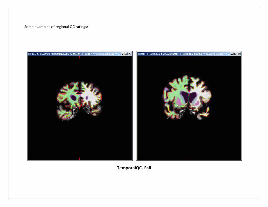
Some examples of regional QC ratings:
TemporalQC- Fail
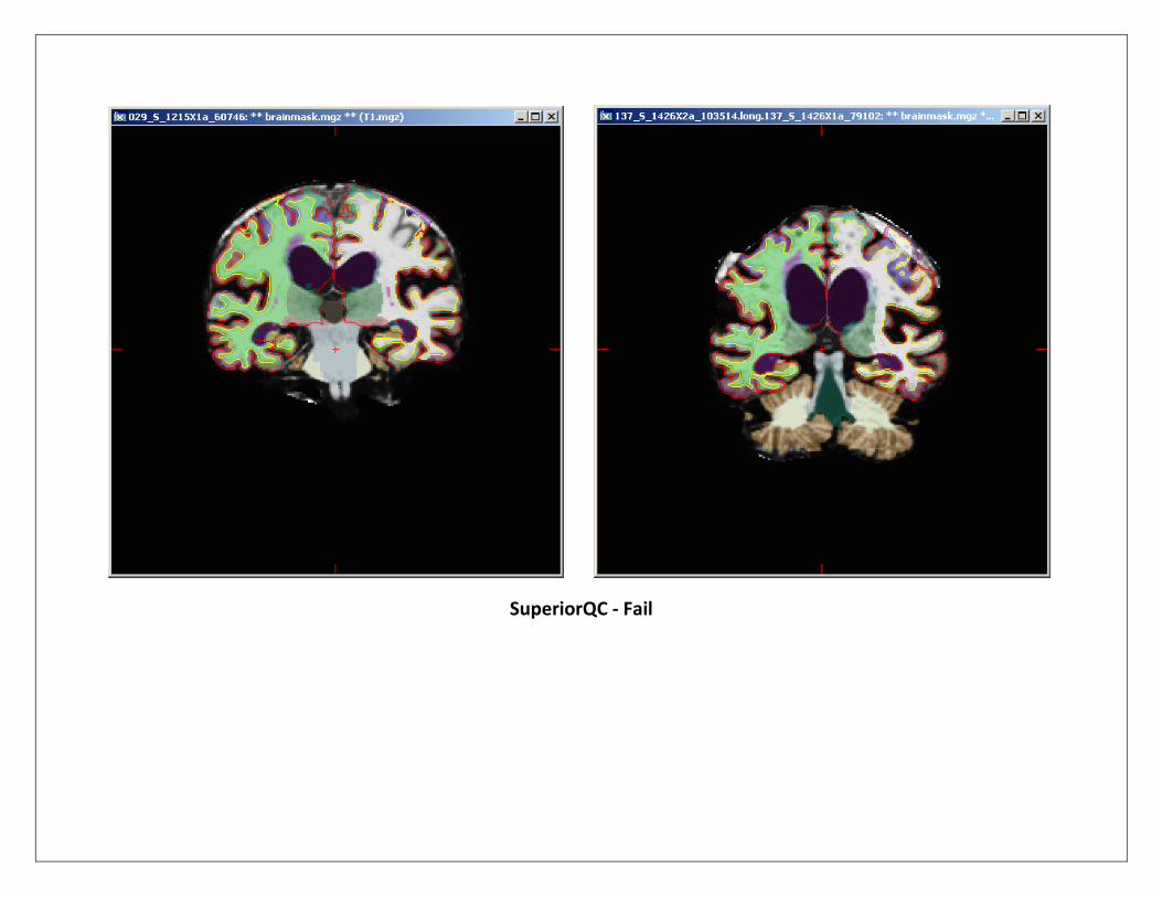
SuperiorQC - Fail
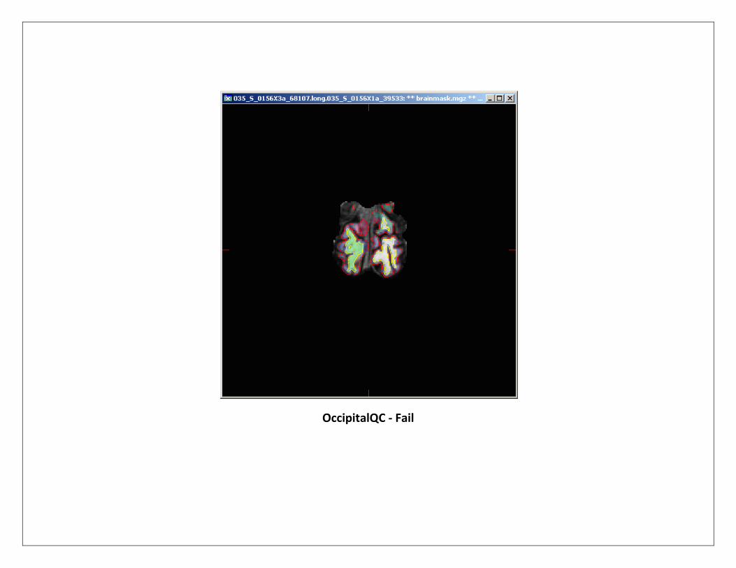
OccipitalQC - Fail
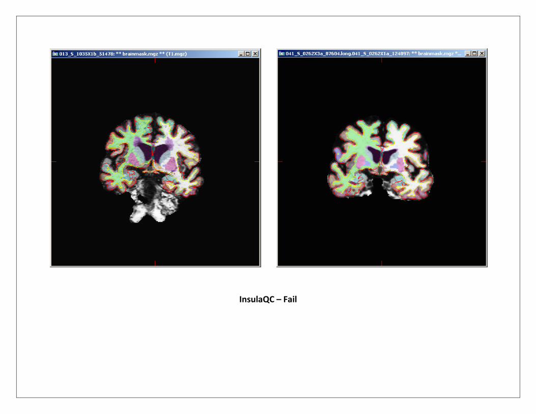
InsulaQC – Fail