Author(s): Matthew Velkey, 2009 - Open.Michigan · PDF fileContraction is involuntary,...
42
Author(s): Matthew Velkey, 2009 License: Unless otherwise noted, this material is made available under the terms of the Creative Commons Attribution – Non-Commercial – Share Alike 3.0 License: http://creativecommons.org/licenses/by-nc-sa/3.0/ We have reviewed this material in accordance with U.S. Copyright Law and have tried to maximize your ability to use, share, and adapt it. The citation key on the following slide provides information about how you may share and adapt this material. Copyright holders of content included in this material should contact [email protected] with any questions, corrections, or clarification regarding the use of content. For more information about how to cite these materials visit http://open.umich.edu/education/about/terms-of-use. Any medical information in this material is intended to inform and educate and is not a tool for self-diagnosis or a replacement for medical evaluation, advice, diagnosis or treatment by a healthcare professional. Please speak to your physician if you have questions about your medical condition. Viewer discretion is advised: Some medical content is graphic and may not be suitable for all viewers.
Transcript of Author(s): Matthew Velkey, 2009 - Open.Michigan · PDF fileContraction is involuntary,...

Author(s): Matthew Velkey, 2009
License: Unless otherwise noted, this material is made available under the terms of the Creative Commons Attribution – Non-Commercial – Share Alike 3.0 License: http://creativecommons.org/licenses/by-nc-sa/3.0/
We have reviewed this material in accordance with U.S. Copyright Law and have tried to maximize your ability to use, share, and adapt it. The citation key on the following slide provides information about how you may share and adapt this material.
Copyright holders of content included in this material should contact [email protected] with any questions, corrections, or clarification regarding the use of content.
For more information about how to cite these materials visit http://open.umich.edu/education/about/terms-of-use.
Any medical information in this material is intended to inform and educate and is not a tool for self-diagnosis or a replacement for medical evaluation, advice, diagnosis or treatment by a healthcare professional. Please speak to your physician if you have questions about your medical condition.
Viewer discretion is advised: Some medical content is graphic and may not be suitable for all viewers.
Citation Key for more information see: http://open.umich.edu/wiki/CitationPolicy
Use + Share + Adapt
Make Your Own Assessment
Creative Commons – Attribution License
Creative Commons – Attribution Share Alike License
Creative Commons – Attribution Noncommercial License
Creative Commons – Attribution Noncommercial Share Alike License
GNU – Free Documentation License
Creative Commons – Zero Waiver
Public Domain – Ineligible: Works that are ineligible for copyright protection in the U.S. (USC 17 § 102(b)) *laws in your jurisdiction may differ
Public Domain – Expired: Works that are no longer protected due to an expired copyright term.
Public Domain – Government: Works that are produced by the U.S. Government. (USC 17 § 105)
Public Domain – Self Dedicated: Works that a copyright holder has dedicated to the public domain.
Fair Use: Use of works that is determined to be Fair consistent with the U.S. Copyright Act. (USC 17 § 107) *laws in your jurisdiction may differ
Our determination DOES NOT mean that all uses of this 3rd-party content are Fair Uses and we DO NOT guarantee that your use of the content is Fair.
To use this content you should do your own independent analysis to determine whether or not your use will be Fair.
{ Content the copyright holder, author, or law permits you to use, share and adapt. }
{ Content Open.Michigan believes can be used, shared, and adapted because it is ineligible for copyright. }
{ Content Open.Michigan has used under a Fair Use determination. }
Muscle Tissue
Matthew Velkey, Ph.D.
Fall 2008
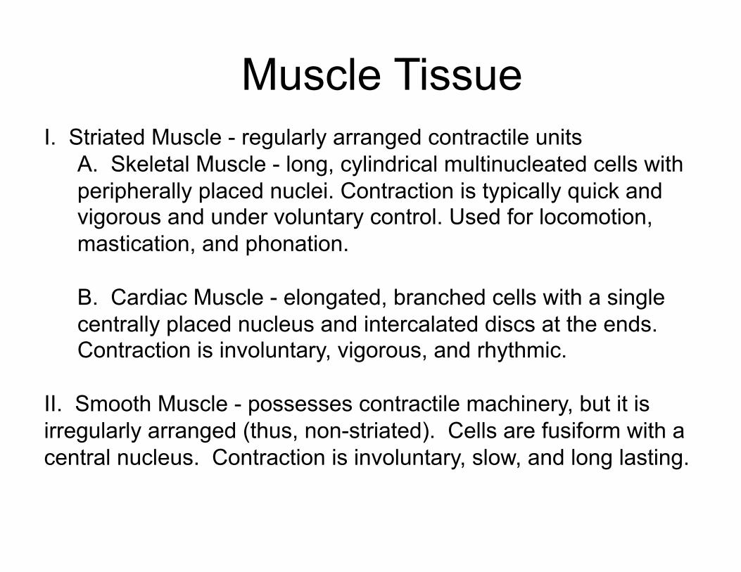
Muscle Tissue I. Striated Muscle - regularly arranged contractile units
A. Skeletal Muscle - long, cylindrical multinucleated cells with peripherally placed nuclei. Contraction is typically quick and vigorous and under voluntary control. Used for locomotion, mastication, and phonation.
B. Cardiac Muscle - elongated, branched cells with a single centrally placed nucleus and intercalated discs at the ends. Contraction is involuntary, vigorous, and rhythmic.
II. Smooth Muscle - possesses contractile machinery, but it is irregularly arranged (thus, non-striated). Cells are fusiform with a central nucleus. Contraction is involuntary, slow, and long lasting.
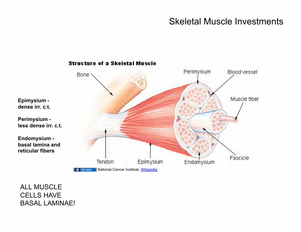
Epimysium - dense irr. c.t.
Perimysium - less dense irr. c.t.
Endomysium - basal lamina and reticular fibers
Skeletal Muscle Investments
ALL MUSCLE CELLS HAVE BASAL LAMINAE!
National Cancer Institute, Wikipedia
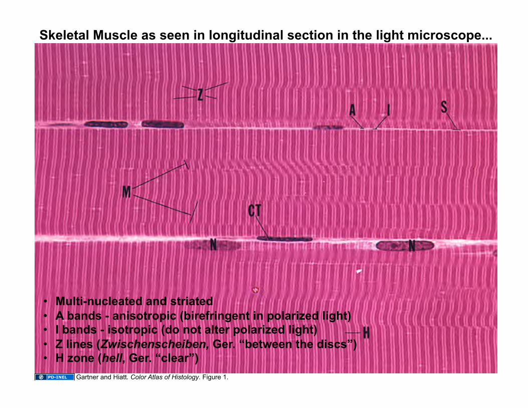
Skeletal Muscle as seen in longitudinal section in the light microscope...
• Multi-nucleated and striated • A bands - anisotropic (birefringent in polarized light) • I bands - isotropic (do not alter polarized light) • Z lines (Zwischenscheiben, Ger. “between the discs”) • H zone (hell, Ger. “clear”)
Gartner and Hiatt. Color Atlas of Histology. Figure 1.
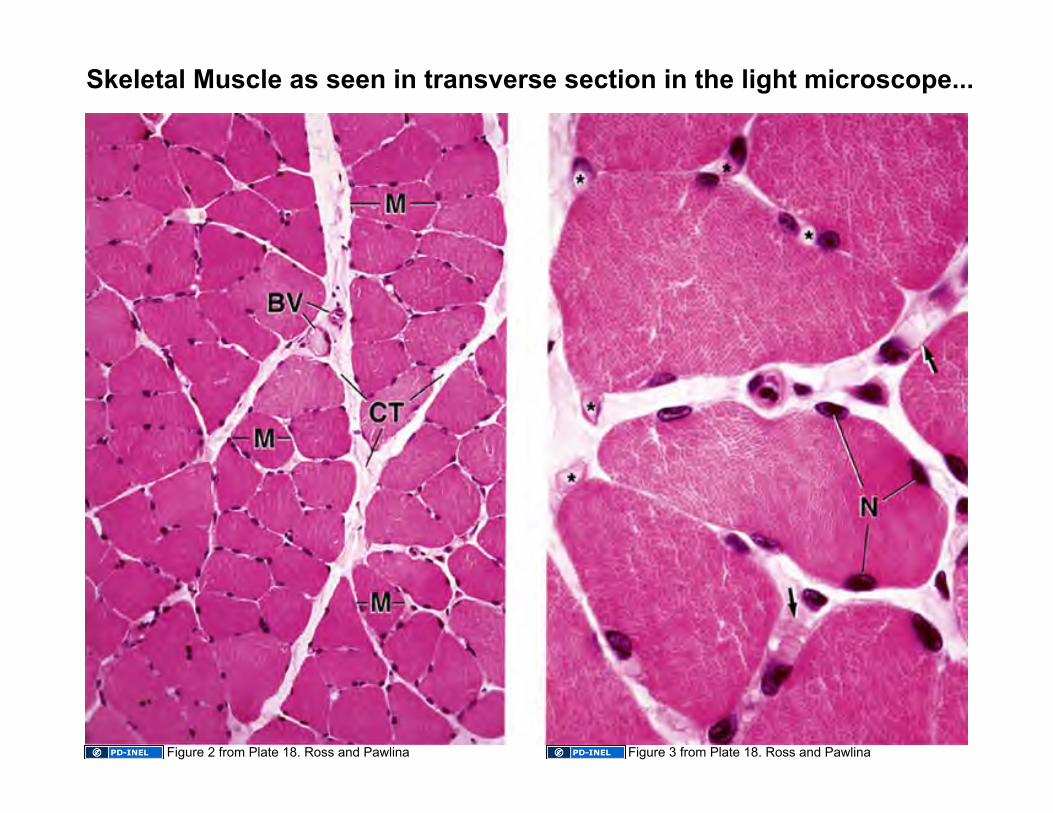
Skeletal Muscle as seen in transverse section in the light microscope...
Figure 2 from Plate 18. Ross and Pawlina Figure 3 from Plate 18. Ross and Pawlina
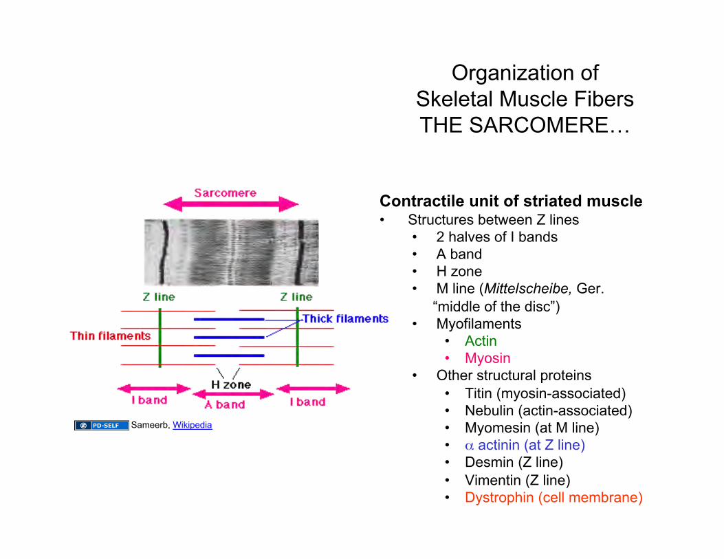
Contractile unit of striated muscle • Structures between Z lines
• 2 halves of I bands • A band • H zone • M line (Mittelscheibe, Ger.
“middle of the disc”) • Myofilaments
• Actin • Myosin
• Other structural proteins • Titin (myosin-associated) • Nebulin (actin-associated) • Myomesin (at M line) • α actinin (at Z line) • Desmin (Z line) • Vimentin (Z line) • Dystrophin (cell membrane)
Organization of Skeletal Muscle Fibers THE SARCOMERE…
Sameerb, Wikipedia
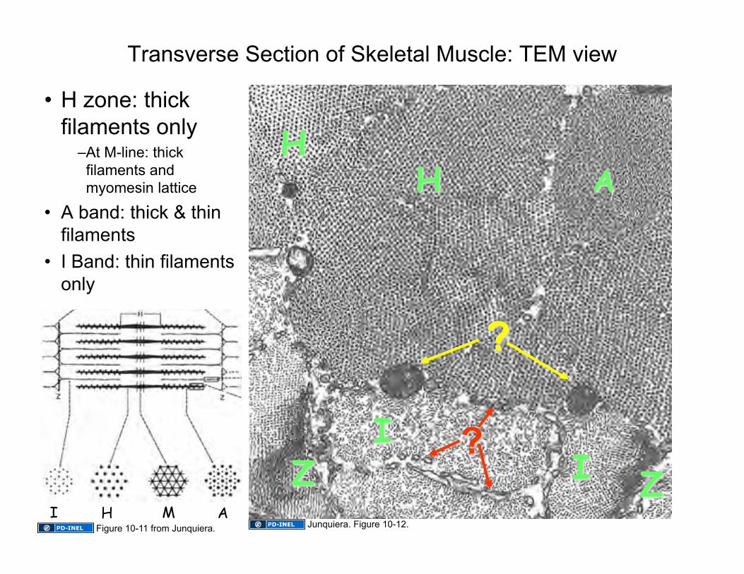
• H zone: thick filaments only
– At M-line: thick filaments and myomesin lattice
• A band: thick & thin filaments
• I Band: thin filaments only
Transverse Section of Skeletal Muscle: TEM view
H I M A Junquiera. Figure 10-12. Figure 10-11 from Junquiera.
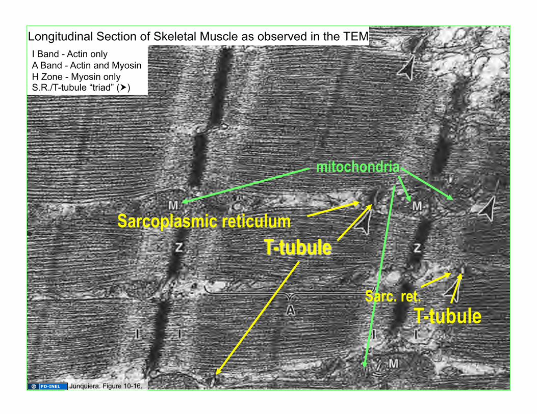
I Band - Actin only A Band - Actin and Myosin H Zone - Myosin only S.R./T-tubule “triad” ()
Longitudinal Section of Skeletal Muscle as observed in the TEM
mitochondria
Sarcoplasmic reticulum
T-tubule Sarc. ret.
Junquiera. Figure 10-16.
T-tubule System: Propagation of the Signal
and Release of Ca2+ T (transverse) Tubules • run perpendicular (transversely)
to myofibrils • conduct membrane
depolarization deep into fibers
Sarcoplasmic Reticulum • smooth ER • site of Ca2+ storage & release • terminal cisternae abut
T-tubules forming triads when myofibrils are viewed in longitudinal section
Image of T-tubule system removed
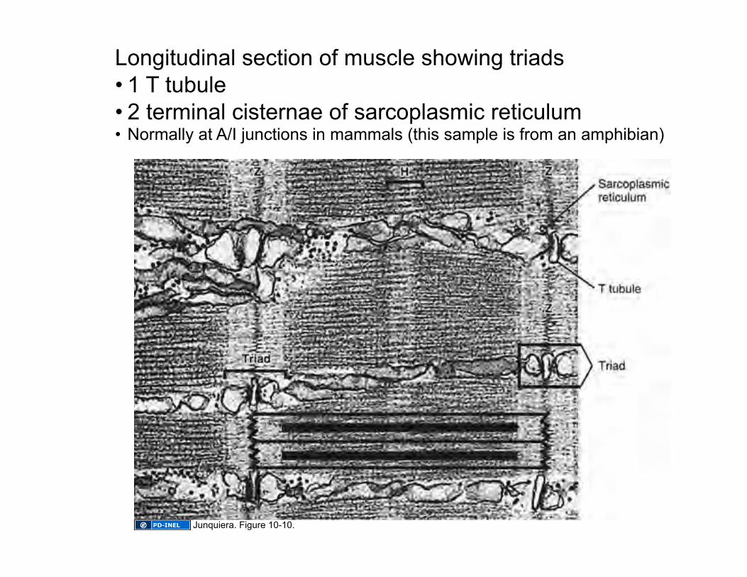
Longitudinal section of muscle showing triads • 1 T tubule • 2 terminal cisternae of sarcoplasmic reticulum • Normally at A/I junctions in mammals (this sample is from an amphibian)
Junquiera. Figure 10-10.
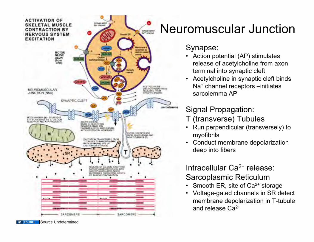
Neuromuscular Junction Synapse: • Action potential (AP) stimulates
release of acetylcholine from axon terminal into synaptic cleft
• Acetylcholine in synaptic cleft binds Na+ channel receptors –initiates sarcolemma AP
Signal Propagation: T (transverse) Tubules • Run perpendicular (transversely) to
myofibrils • Conduct membrane depolarization
deep into fibers
Intracellular Ca2+ release: Sarcoplasmic Reticulum • Smooth ER, site of Ca2+ storage • Voltage-gated channels in SR detect
membrane depolarization in T-tubule and release Ca2+
Source Undetermined
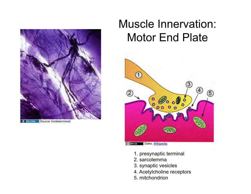
Muscle Innervation: Motor End Plate
Source Undetermined
Dake, Wikipedia
1. presynaptic terminal 2. sarcolemma 3. synaptic vesicles 4. Acetylcholine receptors 5. mitchondrion
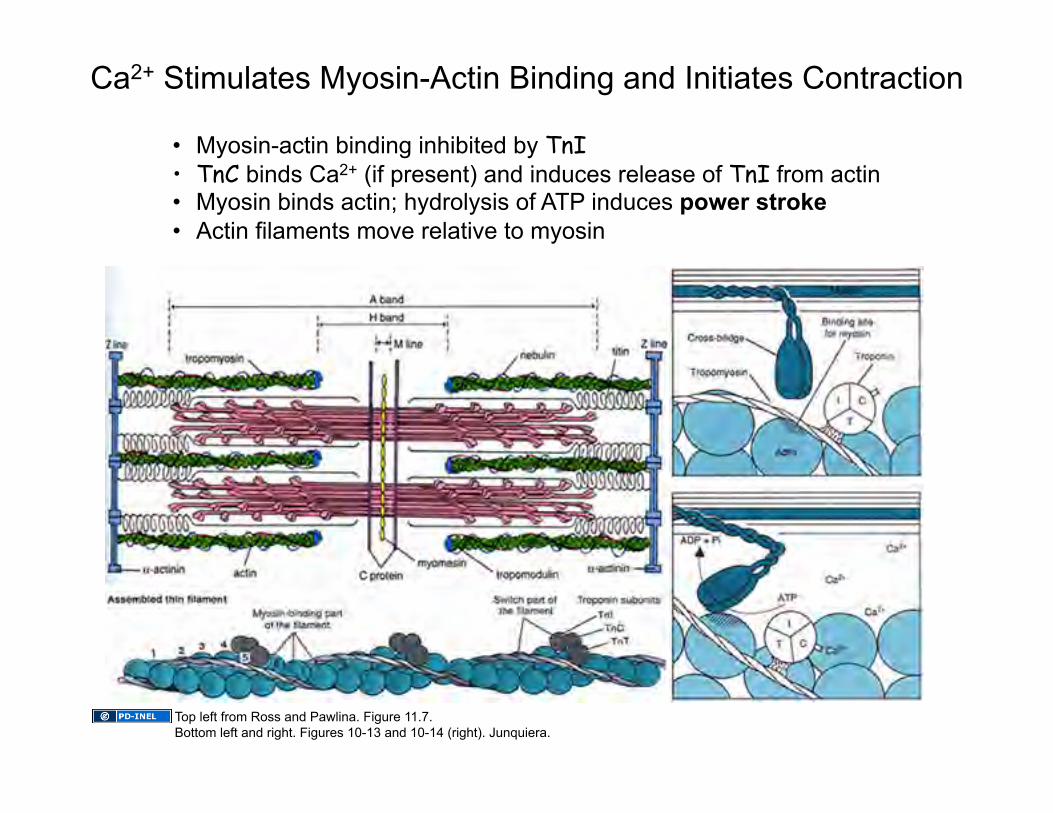
• Myosin-actin binding inhibited by TnI • TnC binds Ca2+ (if present) and induces release of TnI from actin
• Myosin binds actin; hydrolysis of ATP induces power stroke • Actin filaments move relative to myosin
Ca2+ Stimulates Myosin-Actin Binding and Initiates Contraction
Top left from Ross and Pawlina. Figure 11.7. Bottom left and right. Figures 10-13 and 10-14 (right). Junquiera.
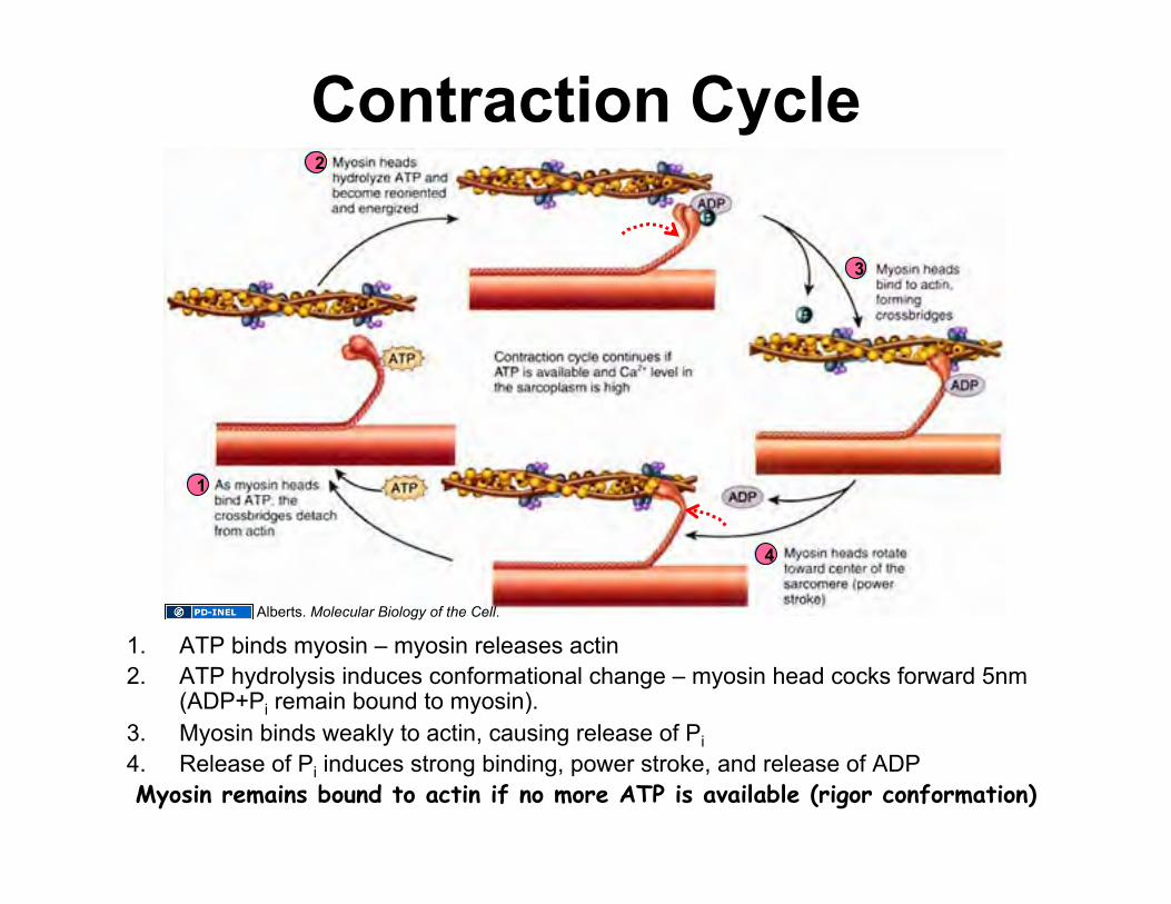
Contraction Cycle
1. ATP binds myosin – myosin releases actin 2. ATP hydrolysis induces conformational change – myosin head cocks forward 5nm
(ADP+Pi remain bound to myosin). 3. Myosin binds weakly to actin, causing release of Pi 4. Release of Pi induces strong binding, power stroke, and release of ADP Myosin remains bound to actin if no more ATP is available (rigor conformation)
1
2
3
4
Alberts. Molecular Biology of the Cell.
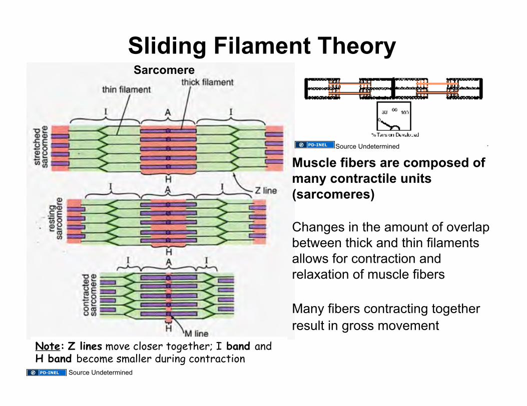
Sliding Filament Theory Sarcomere
Muscle fibers are composed of many contractile units (sarcomeres)
Changes in the amount of overlap between thick and thin filaments allows for contraction and relaxation of muscle fibers
Many fibers contracting together result in gross movement
Note: Z lines move closer together; I band and H band become smaller during contraction
Source Undetermined
Source Undetermined

Cardiac Muscle Tissue Features: • Striated (same contractile machinery) • Self-excitatory and electrically coupled • Rate of contractions modulated by autonomic nervous system
– innervation is neuroendocrine in nature (i.e. no “motor end plates”) Cell Features: • 1 or 2 centrally placed nuclei • Branched fibers with intercalated discs • Numerous mitochondria (up to 40% of cell volume) • Sarcoplasmic reticulum & T-tubules appear as diads at Z lines
– Sarcoplasmic reticulum does not form terminal cisternae – T tubules are about 2x larger in diameter than in skeletal muscle
• transport Ca2+ into fibers
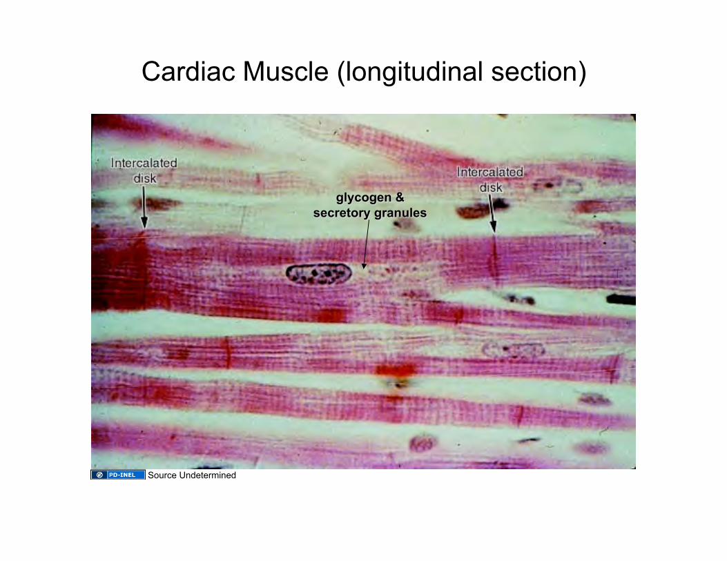
Cardiac Muscle (longitudinal section)
Source Undetermined
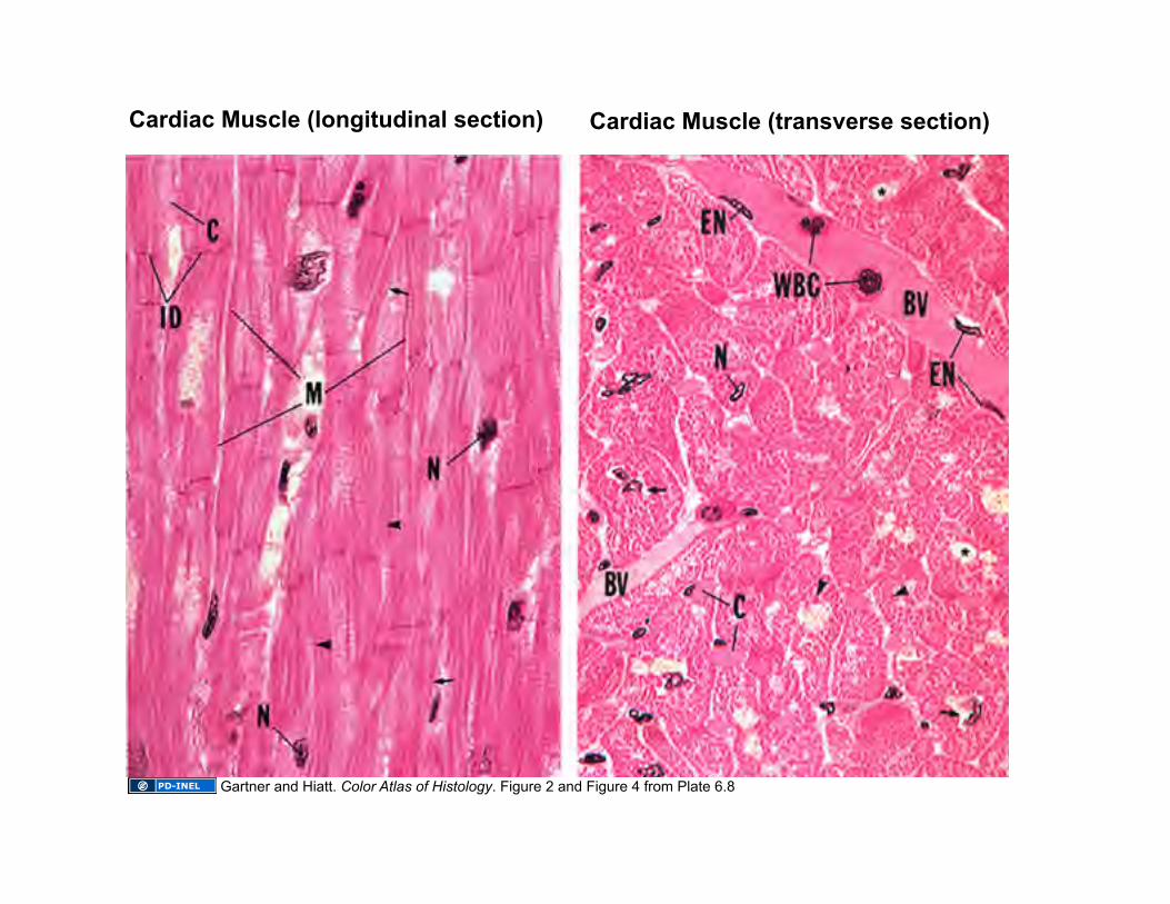
Cardiac Muscle (transverse section) Cardiac Muscle (longitudinal section)
Gartner and Hiatt. Color Atlas of Histology. Figure 2 and Figure 4 from Plate 6.8

Transverse Section of Cardiac Muscle versus Skeletal Muscle
Ross and Pawlina Figure 3 from Plate 18 on right. Left U-M Histology Collection
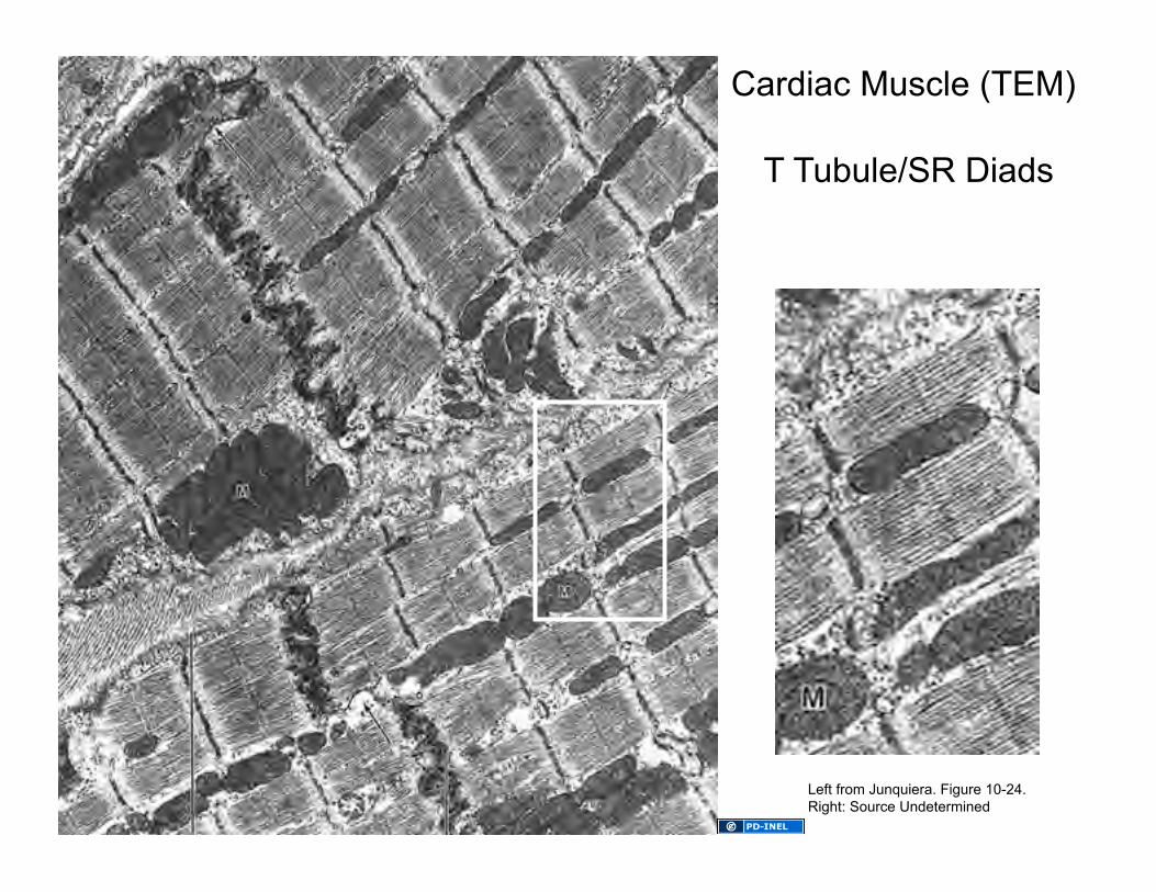
T Tubule/SR Diads
Cardiac Muscle (TEM)
Left from Junquiera. Figure 10-24. Right: Source Undetermined
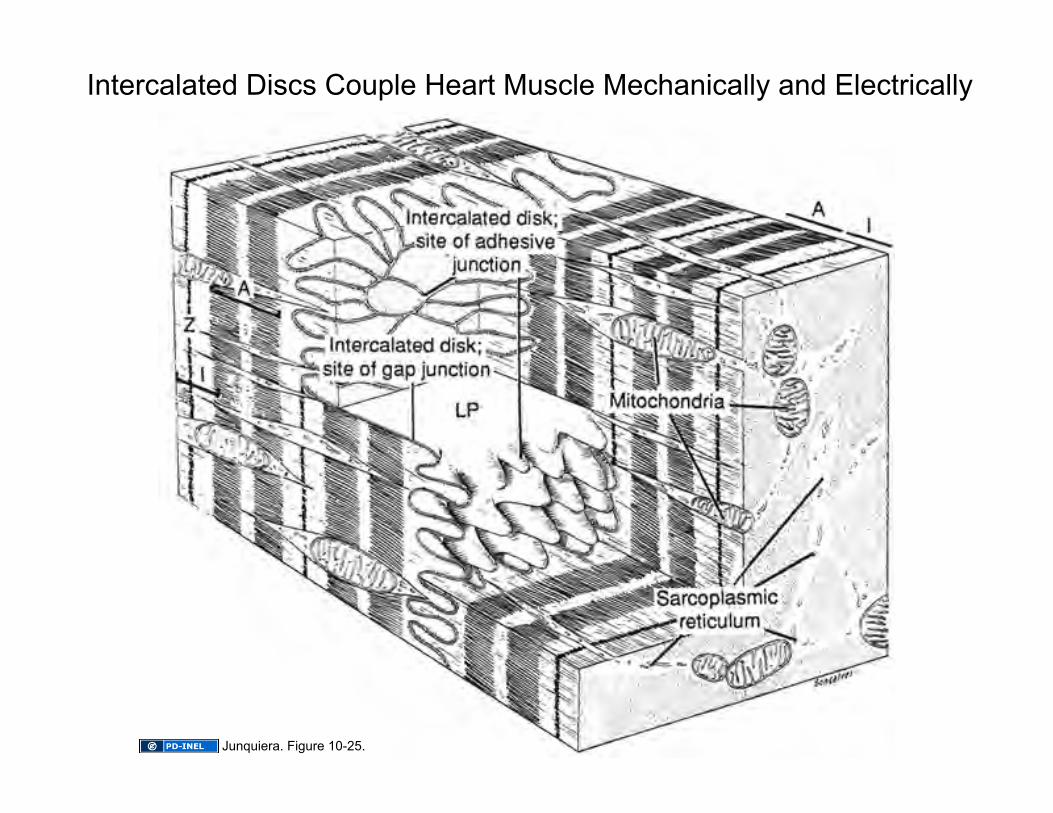
Intercalated Discs Couple Heart Muscle Mechanically and Electrically
Junquiera. Figure 10-25.
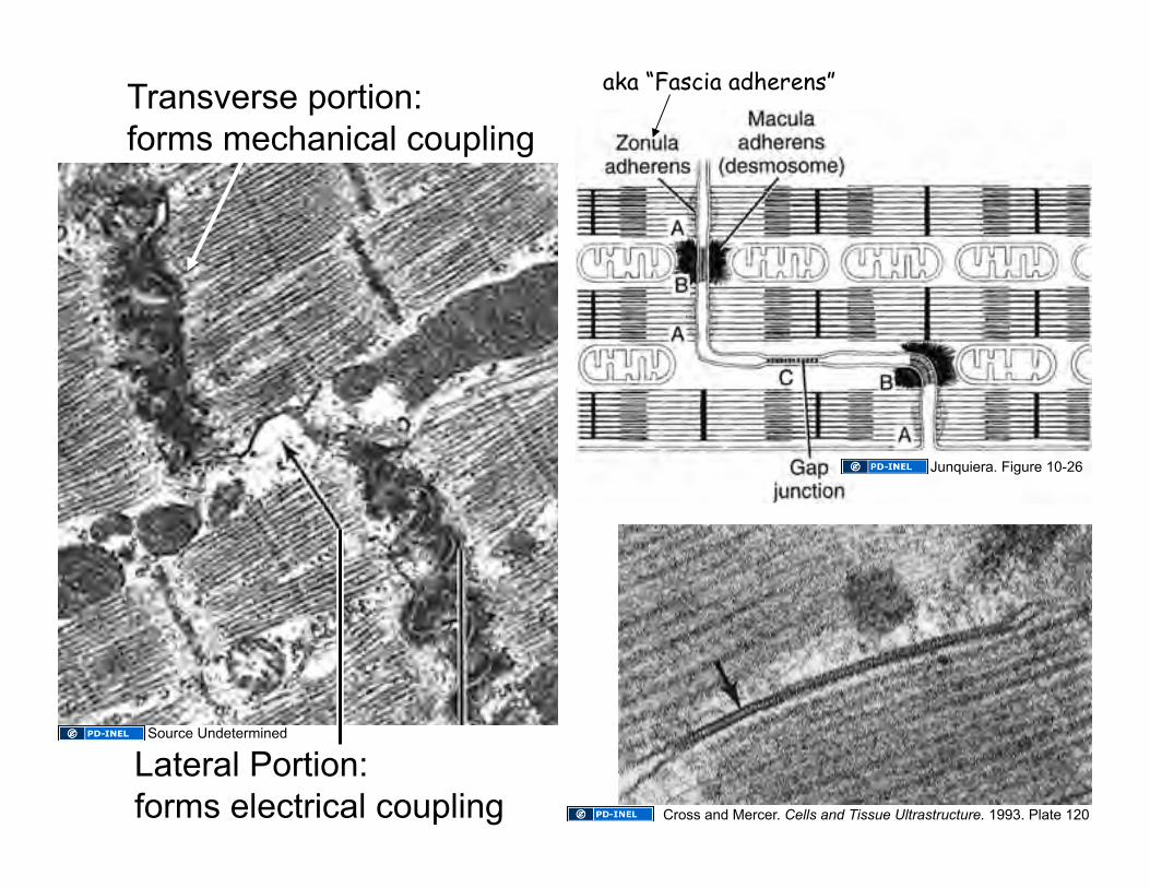
Transverse portion: forms mechanical coupling
Lateral Portion: forms electrical coupling
aka “Fascia adherens”
Source Undetermined
Cross and Mercer. Cells and Tissue Ultrastructure. 1993. Plate 120
Junquiera. Figure 10-26
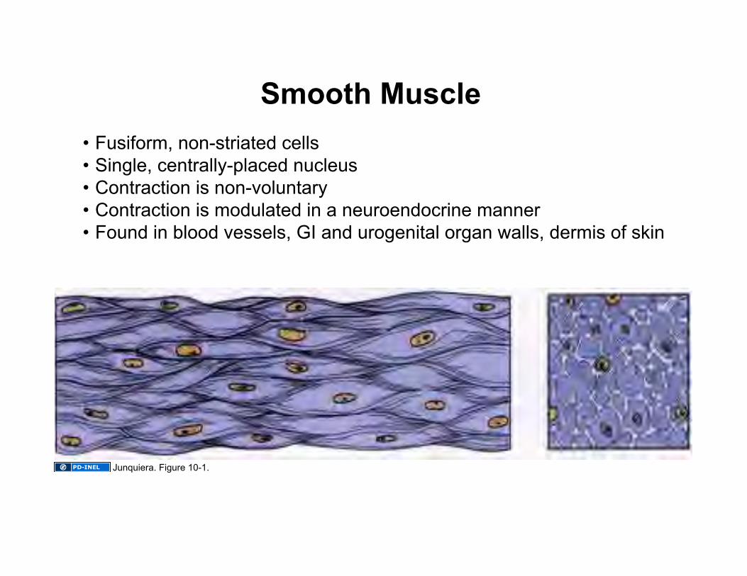
Smooth Muscle • Fusiform, non-striated cells • Single, centrally-placed nucleus • Contraction is non-voluntary • Contraction is modulated in a neuroendocrine manner • Found in blood vessels, GI and urogenital organ walls, dermis of skin
Junquiera. Figure 10-1.
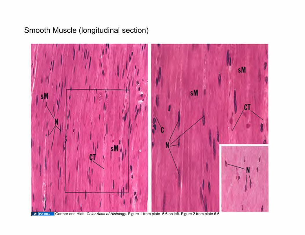
Smooth Muscle (longitudinal section)
Gartner and Hiatt. Color Atlas of Histology. Figure 1 from plate 6.6 on left. Figure 2 from plate 6.6.
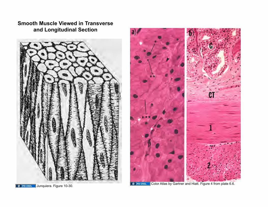
Smooth Muscle Viewed in Transverse and Longitudinal Section
Color Atlas by Gartner and Hiatt. Figure 4 from plate 6.6. Junquiera. Figure 10-30.
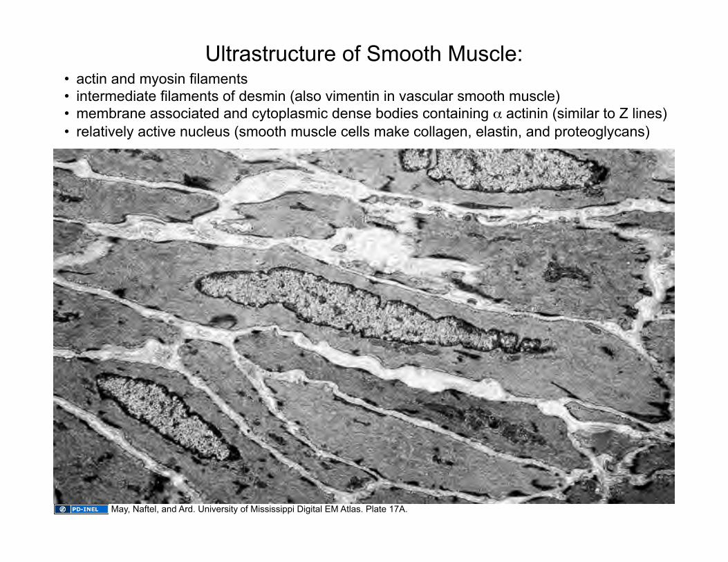
• actin and myosin filaments • intermediate filaments of desmin (also vimentin in vascular smooth muscle) • membrane associated and cytoplasmic dense bodies containing α actinin (similar to Z lines) • relatively active nucleus (smooth muscle cells make collagen, elastin, and proteoglycans)
Ultrastructure of Smooth Muscle:
May, Naftel, and Ard. University of Mississippi Digital EM Atlas. Plate 17A.
*
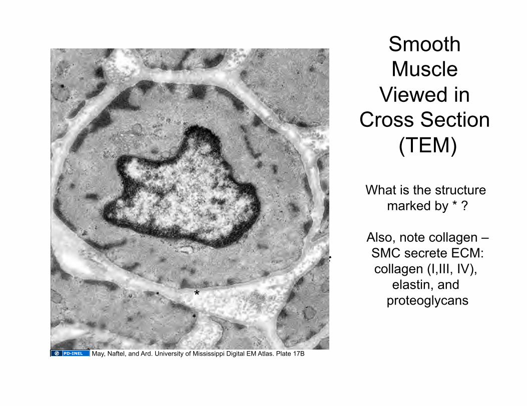
Smooth Muscle
Viewed in Cross Section
(TEM)
What is the structure marked by * ?
Also, note collagen –SMC secrete ECM: collagen (I,III, IV),
elastin, and proteoglycans
May, Naftel, and Ard. University of Mississippi Digital EM Atlas. Plate 17B
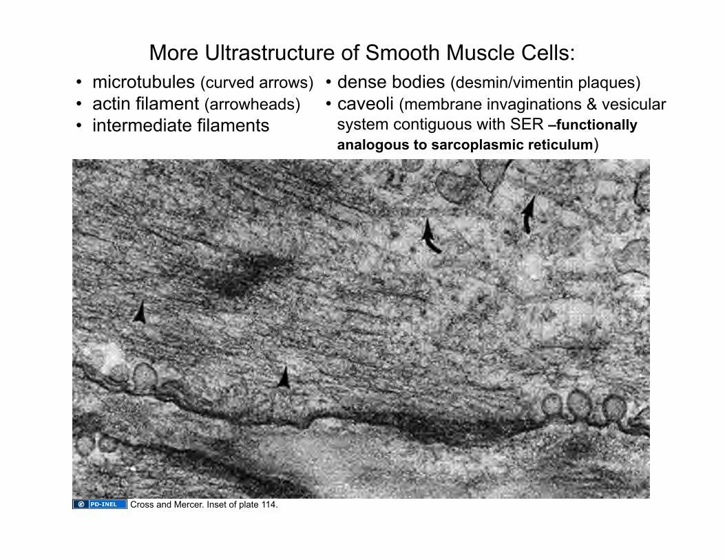
• microtubules (curved arrows) • actin filament (arrowheads) • intermediate filaments
• dense bodies (desmin/vimentin plaques) • caveoli (membrane invaginations & vesicular
system contiguous with SER –functionally analogous to sarcoplasmic reticulum)
More Ultrastructure of Smooth Muscle Cells:
Cross and Mercer. Inset of plate 114.
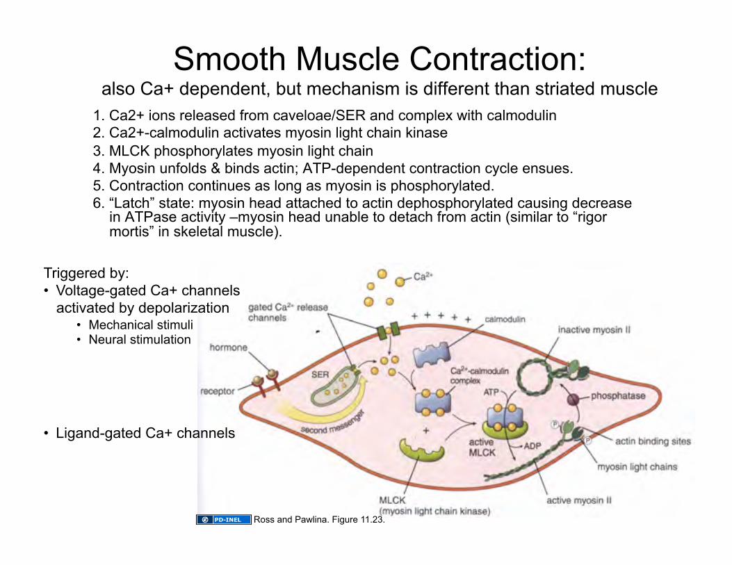
Smooth Muscle Contraction: also Ca+ dependent, but mechanism is different than striated muscle
1. Ca2+ ions released from caveloae/SER and complex with calmodulin 2. Ca2+-calmodulin activates myosin light chain kinase 3. MLCK phosphorylates myosin light chain 4. Myosin unfolds & binds actin; ATP-dependent contraction cycle ensues. 5. Contraction continues as long as myosin is phosphorylated. 6. “Latch” state: myosin head attached to actin dephosphorylated causing decrease
in ATPase activity –myosin head unable to detach from actin (similar to “rigor mortis” in skeletal muscle).
Triggered by: • Voltage-gated Ca+ channels
activated by depolarization • Mechanical stimuli • Neural stimulation
• Ligand-gated Ca+ channels
Ross and Pawlina. Figure 11.23.
Mechanics of Smooth Muscle Contraction
• Dense bodies are analogous to Z lines (plaques into which actin filaments insert)
• Myosin heads oriented in “side polar” arrangement
• Contraction pulls dense bodies together
• Contraction cycle generally about ~10% as fast as skeletal muscle
• Visceral (unitary) smooth muscle cells may be electrically coupled via gap junctions and exhibit either rhythmic or tonic contraction –innervation generally MODIFIES smooth muscle activity rather than initiating it.
• Multiunit smooth muscle cells are innervated individually and can contract rapidly for more precise control.
• Innervation is always at a distance (no motor end plates)
Additional notes:
Image of smooth muscle cell contraction removed

Smooth Muscle (vascular)
Relaxed Contracted Source Undetermined Source Undetermined
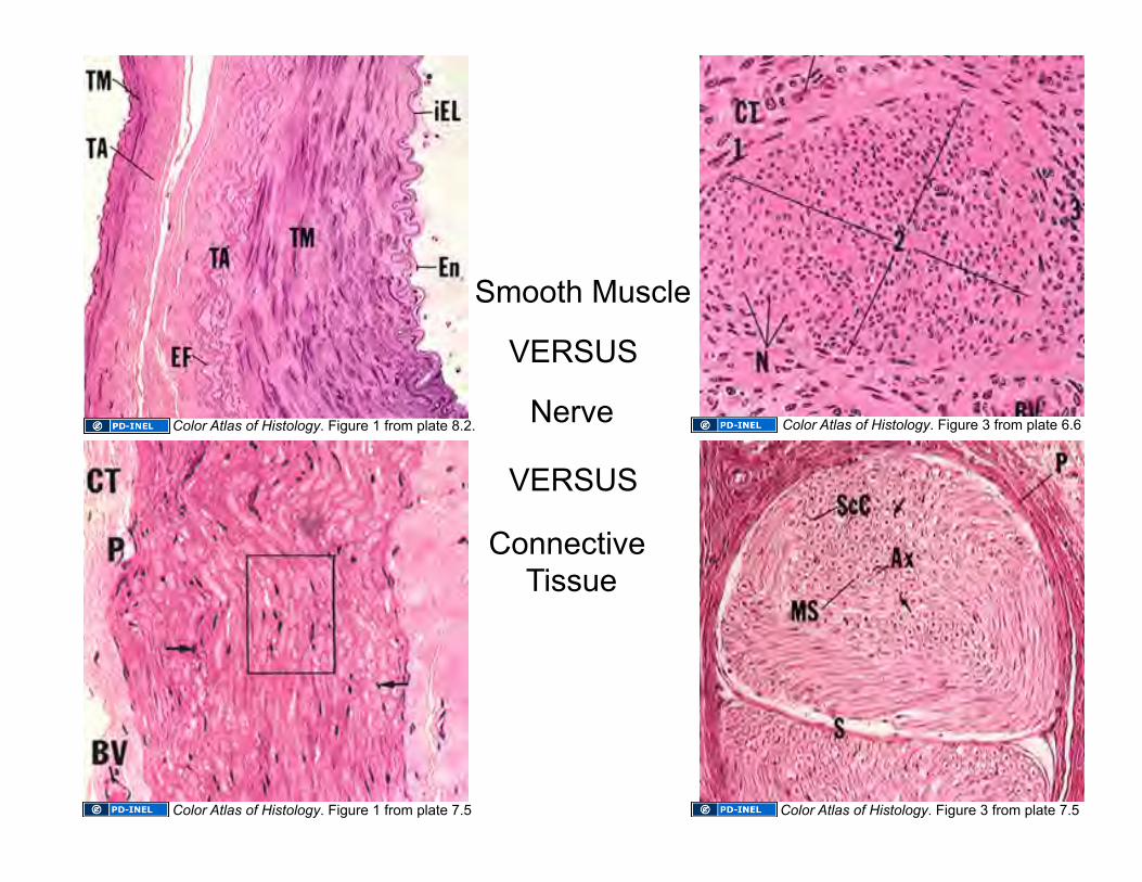
Smooth Muscle
VERSUS
Nerve
VERSUS
Connective Tissue
Color Atlas of Histology. Figure 1 from plate 7.5
Color Atlas of Histology. Figure 1 from plate 8.2.
Color Atlas of Histology. Figure 3 from plate 7.5
Color Atlas of Histology. Figure 3 from plate 6.6
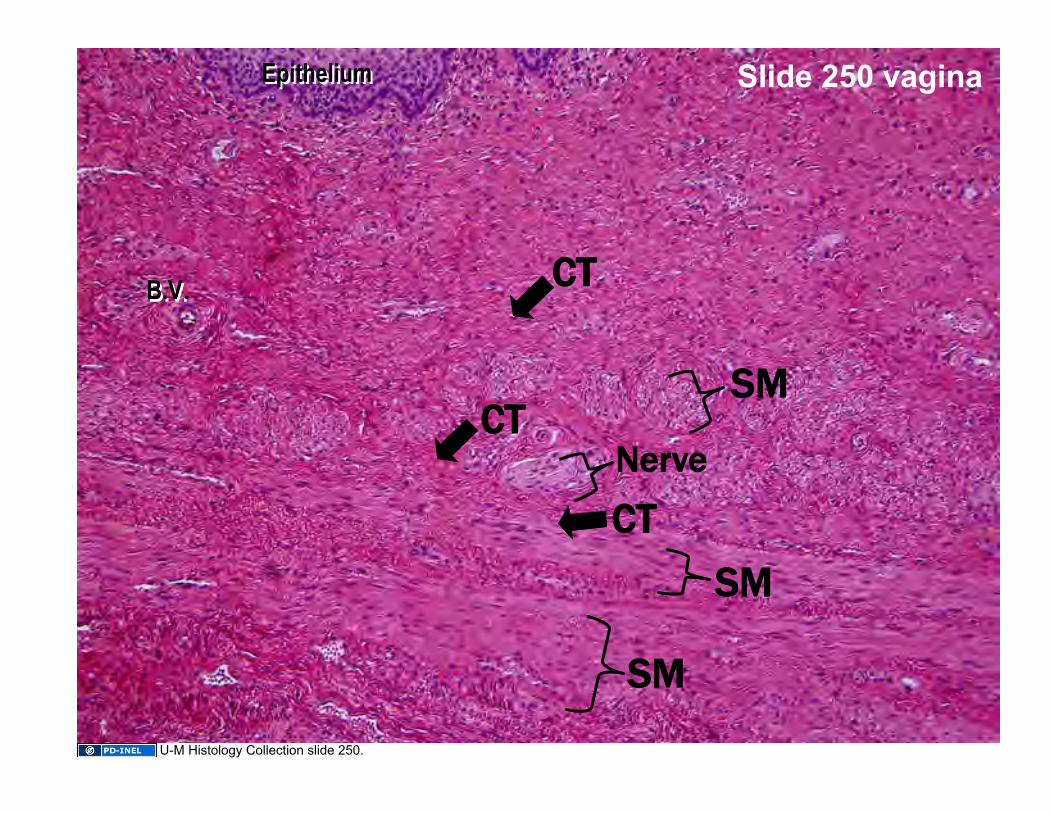
Slide 250 vagina
Nerve
SM
SM
SM
CT
CT
CT
Epithelium Epithelium
B.V. B.V.
U-M Histology Collection slide 250.
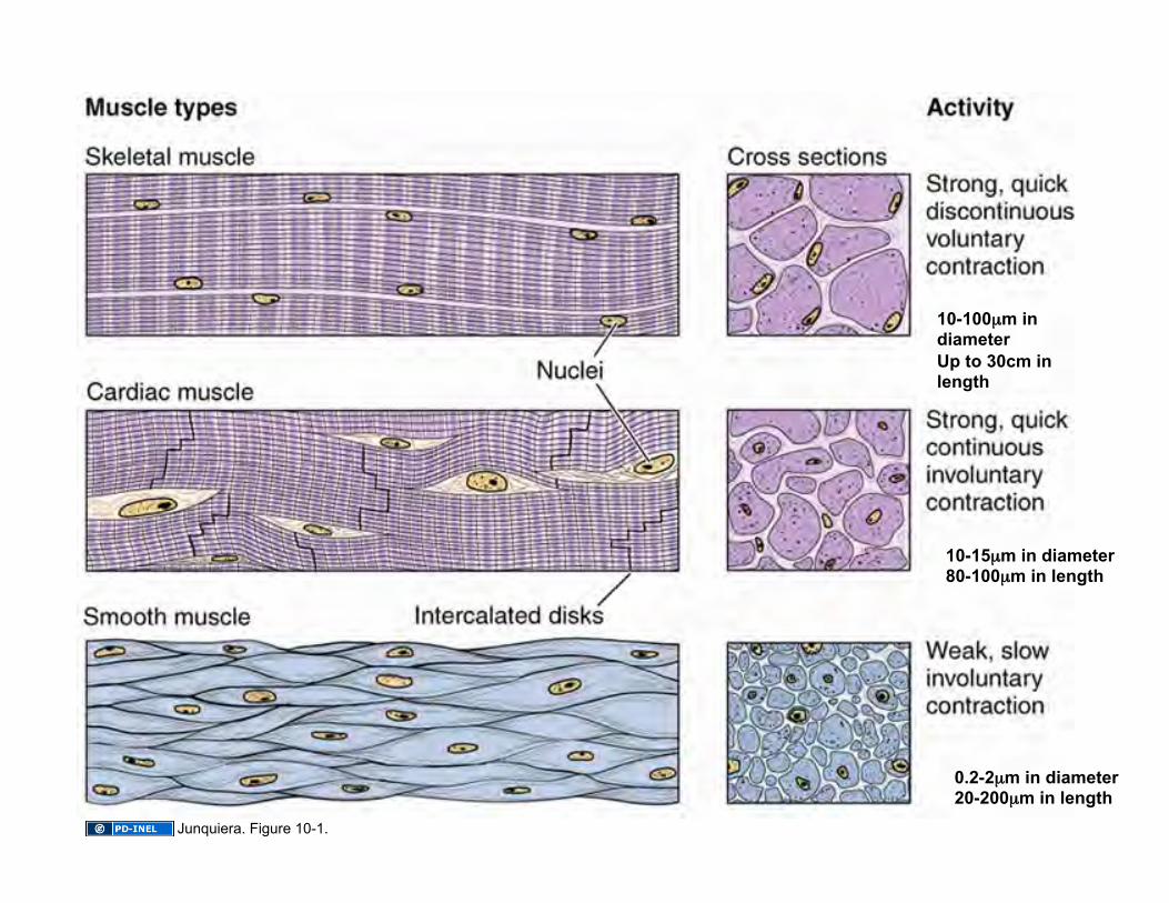
10-100µm in diameter Up to 30cm in length
10-15µm in diameter 80-100µm in length
0.2-2µm in diameter 20-200µm in length
Junquiera. Figure 10-1.
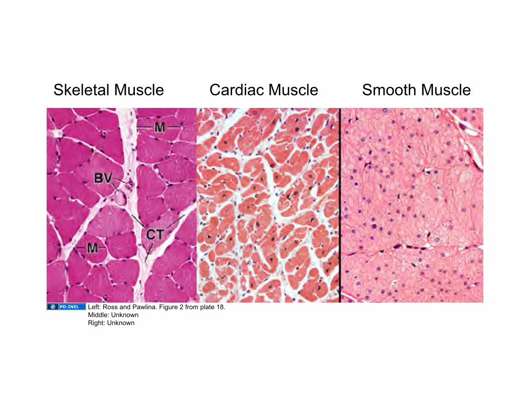
Skeletal Muscle Cardiac Muscle Smooth Muscle
Left: Ross and Pawlina. Figure 2 from plate 18. Middle: Unknown Right: Unknown
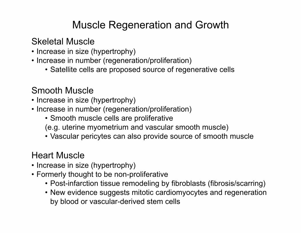
Muscle Regeneration and Growth Skeletal Muscle • Increase in size (hypertrophy) • Increase in number (regeneration/proliferation)
• Satellite cells are proposed source of regenerative cells
Smooth Muscle • Increase in size (hypertrophy) • Increase in number (regeneration/proliferation)
• Smooth muscle cells are proliferative (e.g. uterine myometrium and vascular smooth muscle) • Vascular pericytes can also provide source of smooth muscle
Heart Muscle • Increase in size (hypertrophy) • Formerly thought to be non-proliferative
• Post-infarction tissue remodeling by fibroblasts (fibrosis/scarring) • New evidence suggests mitotic cardiomyocytes and regeneration
by blood or vascular-derived stem cells
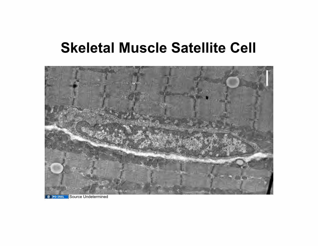
Skeletal Muscle Satellite Cell
Source Undetermined
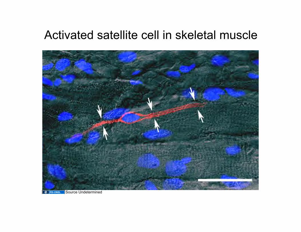
Activated satellite cell in skeletal muscle
Source Undetermined
Learning Objectives 1. Be able to identify the three types of muscle at the light and
electron microscope levels, including distinctive features of each, such as the intercalated disk of cardiac muscle.
2. Be able to describe the structural basis of muscle striation.
3. Know the structural elements that harness muscle contraction (i.e., the shortening of myofibrils) to the movement of a body part (i.e., via connection to bone) as well as the mechanism by which muscle cells contract.
4. Understand the function and organization of the connective tissue in muscle (endo-, peri-, and epiysium).
5. Be familiar with the regenerative potential of each muscle type.

Slide 5: National Cancer Institute, Wikimedia, http://en.wikipedia.org/wiki/File:Illu_muscle_structure.jpg Slide 6: Source Undetermined Slide 7: Source Undetermined Slide 8: Sameerb, Wikipedia, http://en.wikipedia.org/wiki/File:Sarcomere.gif Slide 9: Source Undetermined Slide 10: Source Undetermined Slide 12: Source Undetermined Slide 13: Source Undetermined Slide 14: Source Undetermined; Dake, Wikipedia, http://en.wikipedia.org/wiki/File:Synapse_diag4.png#file Slide 15: Source Undetermined Slide 16: Source Undetermined Slide 17: Source Undetermined Slide 19: Source Undetermined Slide 20: Source Undetermined Slide 21: Source Undetermined Slide 22: Source Undetermined Slide 23: Source Undetermined Slide 24: Source Undetermined Slide 25: Source Undetermined Slide 26: Source Undetermined Slide 27: Source Undetermined Slide 28: Source Undetermined Slide 29: Source Undetermined Slide 30: Source Undetermined Slide 31: Source Undetermined Slide 33: Source Undetermined Slide 34: Source Undetermined Slide 35: Source Undetermined Slide 36: Source Undetermined Slide 37: Source Undetermined Slide 39: Source Undetermined Slide 40: Source Undetermined
Additional Source Information for more information see: http://open.umich.edu/wiki/CitationPolicy