Author(s): Matthew Velkey, 2009 - Open.Michigan · PDF fileMake Your Own Assessment ......
33
Author(s): Matthew Velkey, 2009 License: Unless otherwise noted, this material is made available under the terms of the Creative Commons Attribution – Non-Commercial – Share Alike 3.0 License: http://creativecommons.org/licenses/by-nc-sa/3.0/ We have reviewed this material in accordance with U.S. Copyright Law and have tried to maximize your ability to use, share, and adapt it. The citation key on the following slide provides information about how you may share and adapt this material. Copyright holders of content included in this material should contact [email protected] with any questions, corrections, or clarification regarding the use of content. For more information about how to cite these materials visit http://open.umich.edu/education/about/terms-of-use. Any medical information in this material is intended to inform and educate and is not a tool for self-diagnosis or a replacement for medical evaluation, advice, diagnosis or treatment by a healthcare professional. Please speak to your physician if you have questions about your medical condition. Viewer discretion is advised: Some medical content is graphic and may not be suitable for all viewers.
Transcript of Author(s): Matthew Velkey, 2009 - Open.Michigan · PDF fileMake Your Own Assessment ......
Author(s): Matthew Velkey, 2009
License: Unless otherwise noted, this material is made available under the terms of the Creative Commons Attribution – Non-Commercial – Share Alike 3.0 License: http://creativecommons.org/licenses/by-nc-sa/3.0/
We have reviewed this material in accordance with U.S. Copyright Law and have tried to maximize your ability to use, share, and adapt it. The citation key on the following slide provides information about how you may share and adapt this material.
Copyright holders of content included in this material should contact [email protected] with any questions, corrections, or clarification regarding the use of content.
For more information about how to cite these materials visit http://open.umich.edu/education/about/terms-of-use.
Any medical information in this material is intended to inform and educate and is not a tool for self-diagnosis or a replacement for medical evaluation, advice, diagnosis or treatment by a healthcare professional. Please speak to your physician if you have questions about your medical condition.
Viewer discretion is advised: Some medical content is graphic and may not be suitable for all viewers.
Citation Key for more information see: http://open.umich.edu/wiki/CitationPolicy
Use + Share + Adapt
Make Your Own Assessment
Creative Commons – Attribution License
Creative Commons – Attribution Share Alike License
Creative Commons – Attribution Noncommercial License
Creative Commons – Attribution Noncommercial Share Alike License
GNU – Free Documentation License
Creative Commons – Zero Waiver
Public Domain – Ineligible: Works that are ineligible for copyright protection in the U.S. (USC 17 § 102(b)) *laws in your jurisdiction may differ
Public Domain – Expired: Works that are no longer protected due to an expired copyright term.
Public Domain – Government: Works that are produced by the U.S. Government. (USC 17 § 105)
Public Domain – Self Dedicated: Works that a copyright holder has dedicated to the public domain.
Fair Use: Use of works that is determined to be Fair consistent with the U.S. Copyright Act. (USC 17 § 107) *laws in your jurisdiction may differ
Our determination DOES NOT mean that all uses of this 3rd-party content are Fair Uses and we DO NOT guarantee that your use of the content is Fair.
To use this content you should do your own independent analysis to determine whether or not your use will be Fair.
{ Content the copyright holder, author, or law permits you to use, share and adapt. }
{ Content Open.Michigan believes can be used, shared, and adapted because it is ineligible for copyright. }
{ Content Open.Michigan has used under a Fair Use determination. }
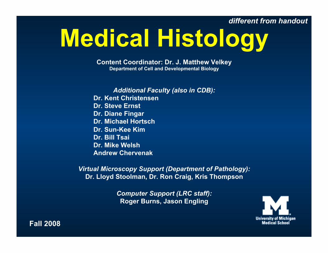
Medical Histology Content Coordinator: Dr. J. Matthew Velkey
Department of Cell and Developmental Biology
Additional Faculty (also in CDB): Dr. Kent Christensen Dr. Steve Ernst Dr. Diane Fingar Dr. Michael Hortsch Dr. Sun-Kee Kim Dr. Bill Tsai Dr. Mike Welsh Andrew Chervenak
Virtual Microscopy Support (Department of Pathology): Dr. Lloyd Stoolman, Dr. Ron Craig, Kris Thompson
Computer Support (LRC staff): Roger Burns, Jason Engling
different from handout
Fall 2008
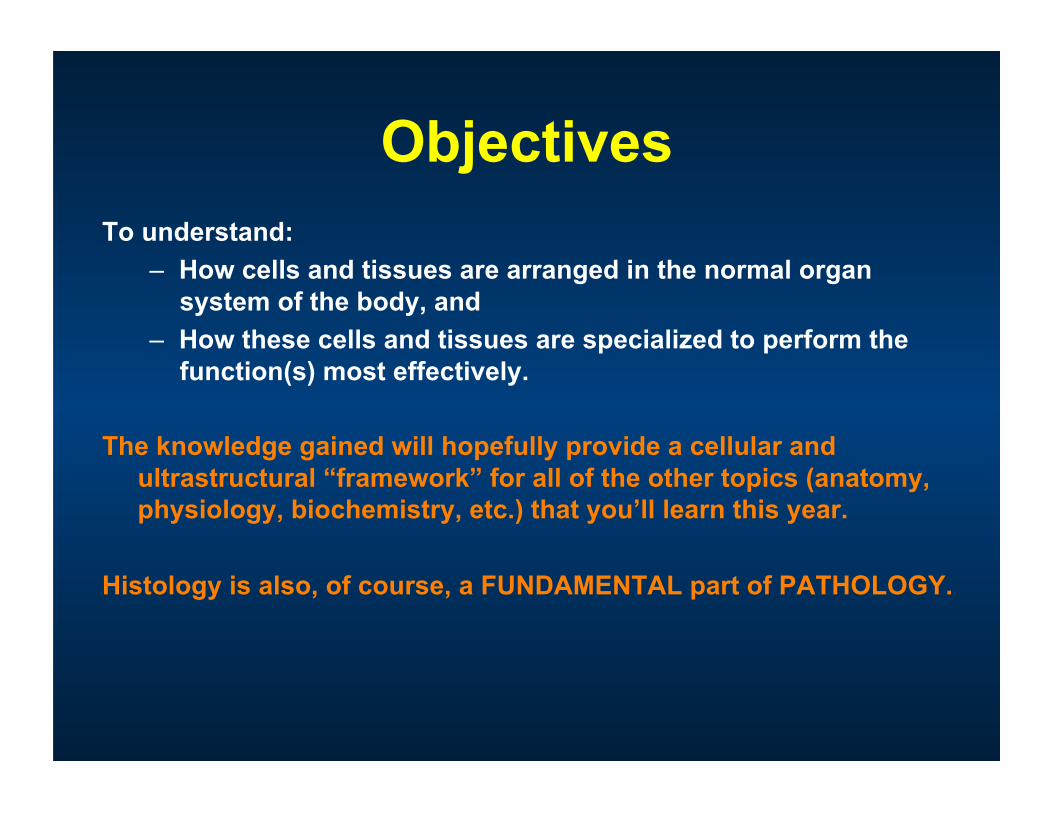
Objectives To understand:
– How cells and tissues are arranged in the normal organ system of the body, and
– How these cells and tissues are specialized to perform the function(s) most effectively.
The knowledge gained will hopefully provide a cellular and ultrastructural “framework” for all of the other topics (anatomy, physiology, biochemistry, etc.) that you’ll learn this year.
Histology is also, of course, a FUNDAMENTAL part of PATHOLOGY.
Correlate
Structure and
Function
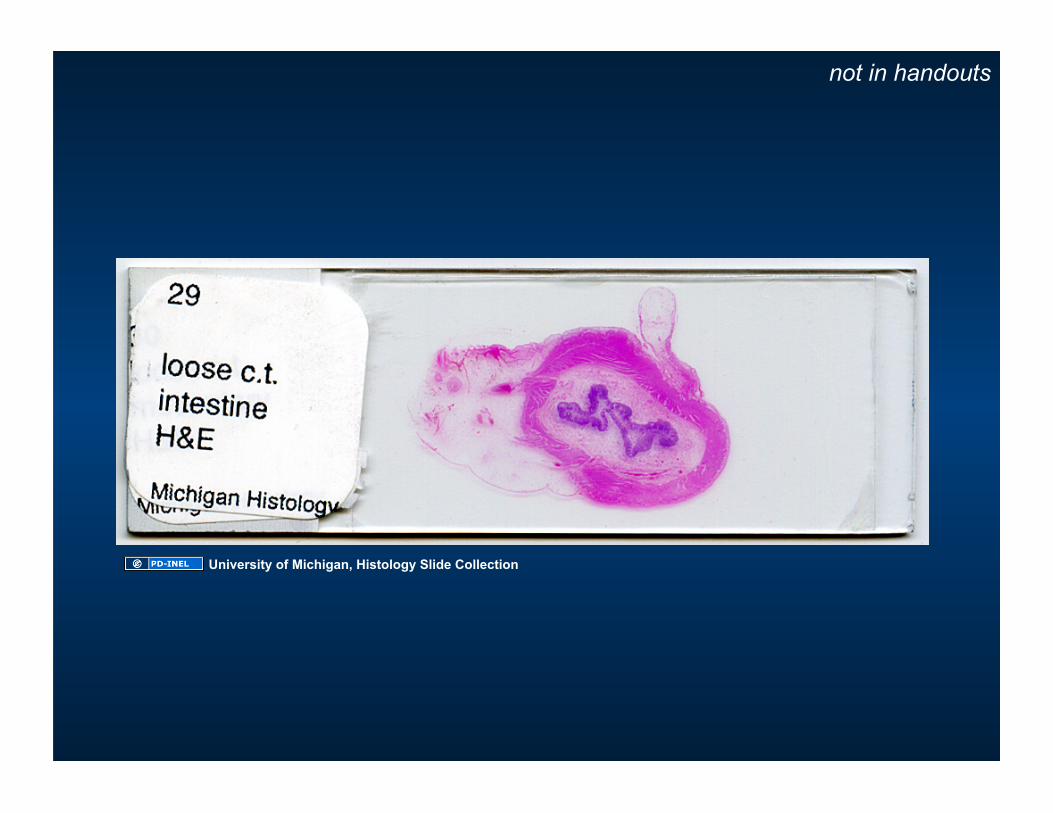
not in handouts
University of Michigan, Histology Slide Collection
HISTOLOGY a.k.a. Micro-anatomy
not in handouts
Cartoon removed
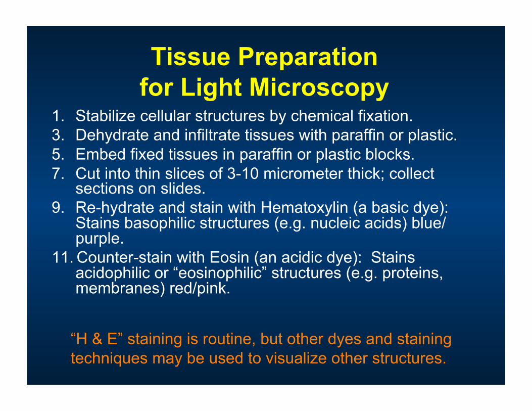
Tissue Preparation for Light Microscopy
1. Stabilize cellular structures by chemical fixation. 3. Dehydrate and infiltrate tissues with paraffin or plastic. 5. Embed fixed tissues in paraffin or plastic blocks. 7. Cut into thin slices of 3-10 micrometer thick; collect
sections on slides. 9. Re-hydrate and stain with Hematoxylin (a basic dye):
Stains basophilic structures (e.g. nucleic acids) blue/purple.
11. Counter-stain with Eosin (an acidic dye): Stains acidophilic or “eosinophilic” structures (e.g. proteins, membranes) red/pink.
“H & E” staining is routine, but other dyes and staining techniques may be used to visualize other structures.
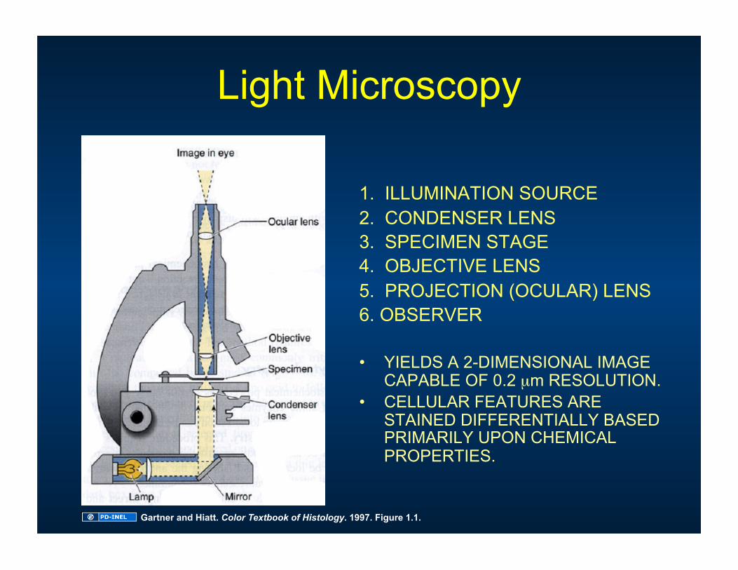
Light Microscopy
1. ILLUMINATION SOURCE 2. CONDENSER LENS 3. SPECIMEN STAGE 4. OBJECTIVE LENS 5. PROJECTION (OCULAR) LENS 6. OBSERVER
• YIELDS A 2-DIMENSIONAL IMAGE CAPABLE OF 0.2 µm RESOLUTION.
• CELLULAR FEATURES ARE STAINED DIFFERENTIALLY BASED PRIMARILY UPON CHEMICAL PROPERTIES.
Gartner and Hiatt. Color Textbook of Histology. 1997. Figure 1.1.
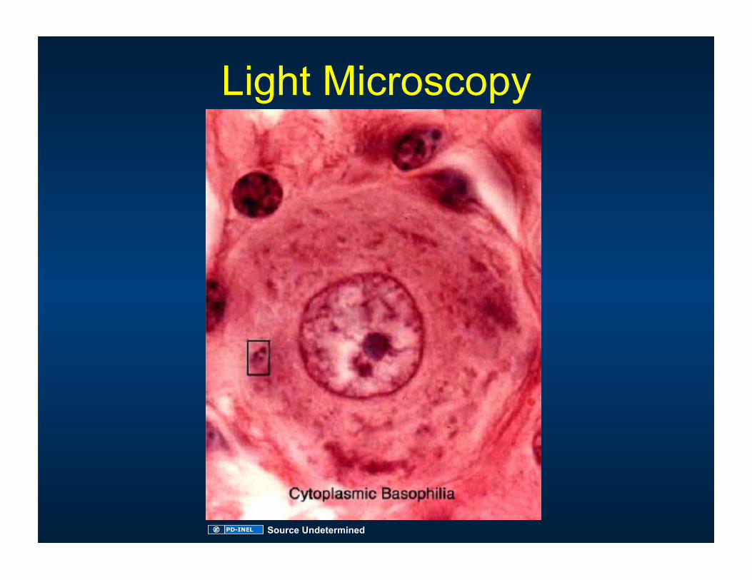
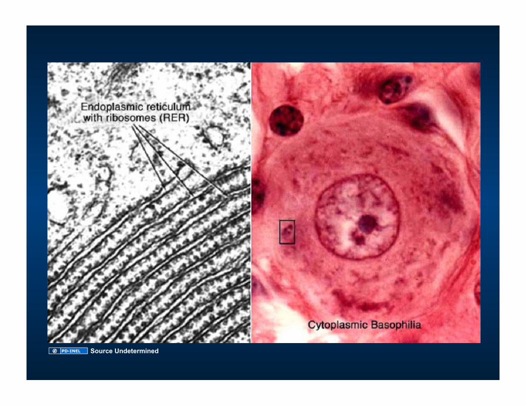
Light Microscopy
Source Undetermined
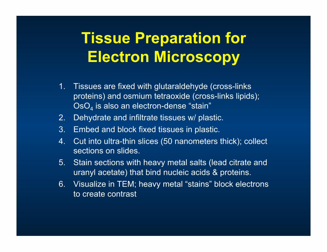
Tissue Preparation for Electron Microscopy
1. Tissues are fixed with glutaraldehyde (cross-links proteins) and osmium tetraoxide (cross-links lipids); OsO4 is also an electron-dense “stain”
2. Dehydrate and infiltrate tissues w/ plastic. 3. Embed and block fixed tissues in plastic. 4. Cut into ultra-thin slices (50 nanometers thick); collect
sections on slides. 5. Stain sections with heavy metal salts (lead citrate and
uranyl acetate) that bind nucleic acids & proteins. 6. Visualize in TEM; heavy metal “stains” block electrons
to create contrast
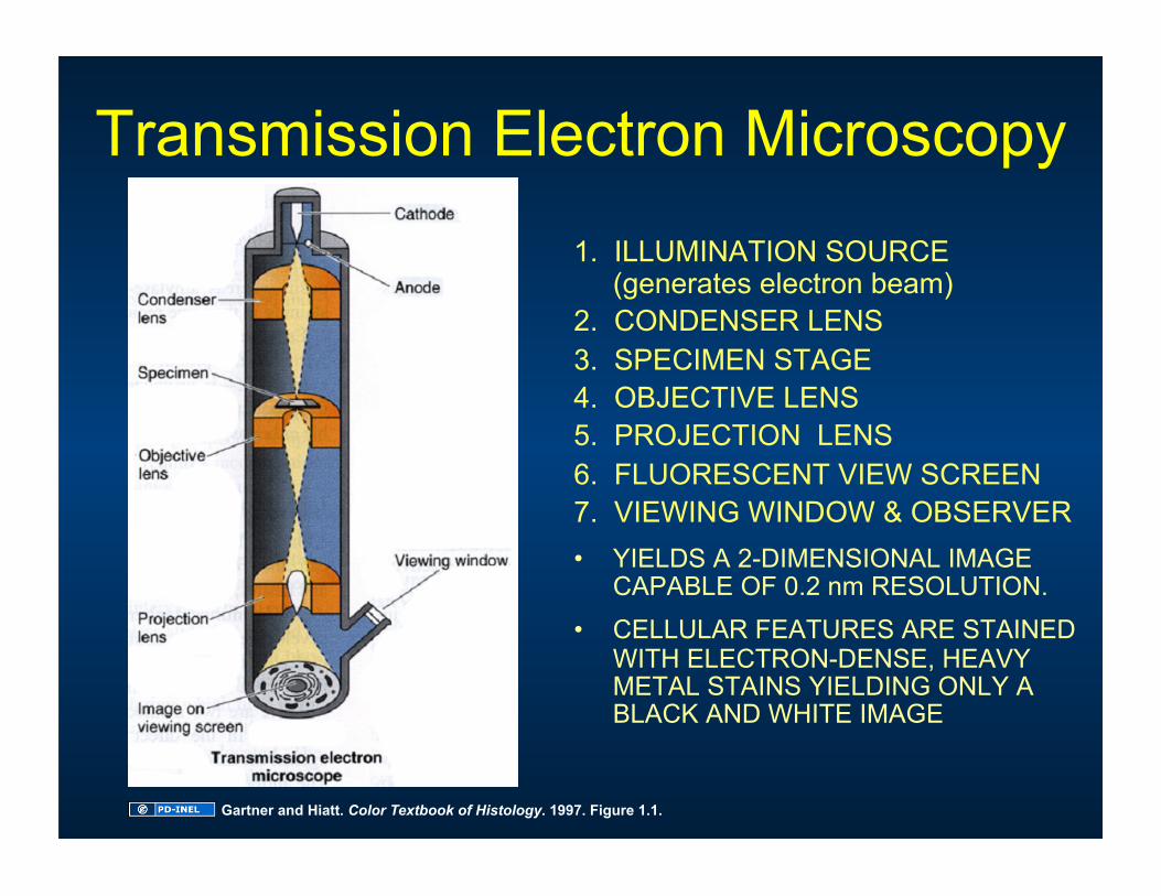
Transmission Electron Microscopy 1. ILLUMINATION SOURCE
(generates electron beam) 2. CONDENSER LENS 3. SPECIMEN STAGE 4. OBJECTIVE LENS 5. PROJECTION LENS 6. FLUORESCENT VIEW SCREEN 7. VIEWING WINDOW & OBSERVER • YIELDS A 2-DIMENSIONAL IMAGE
CAPABLE OF 0.2 nm RESOLUTION. • CELLULAR FEATURES ARE STAINED
WITH ELECTRON-DENSE, HEAVY METAL STAINS YIELDING ONLY A BLACK AND WHITE IMAGE
Gartner and Hiatt. Color Textbook of Histology. 1997. Figure 1.1.
Source Undetermined
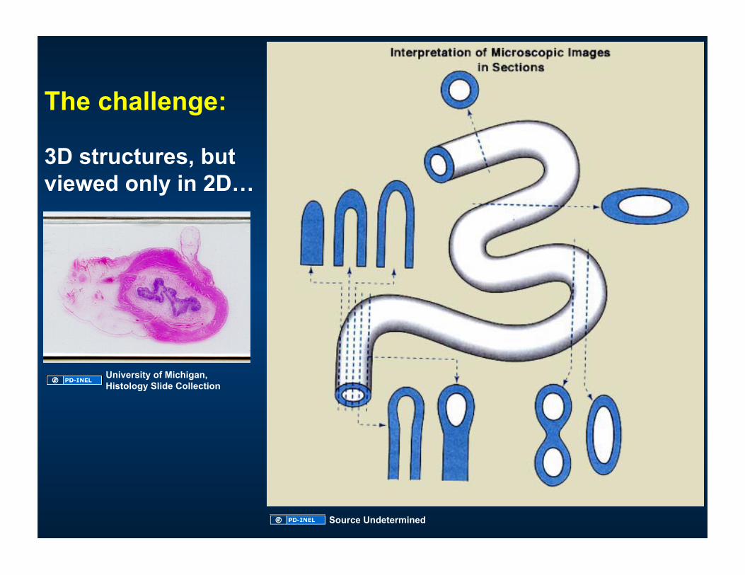
The challenge:
3D structures, but viewed only in 2D…
University of Michigan, Histology Slide Collection
Source Undetermined
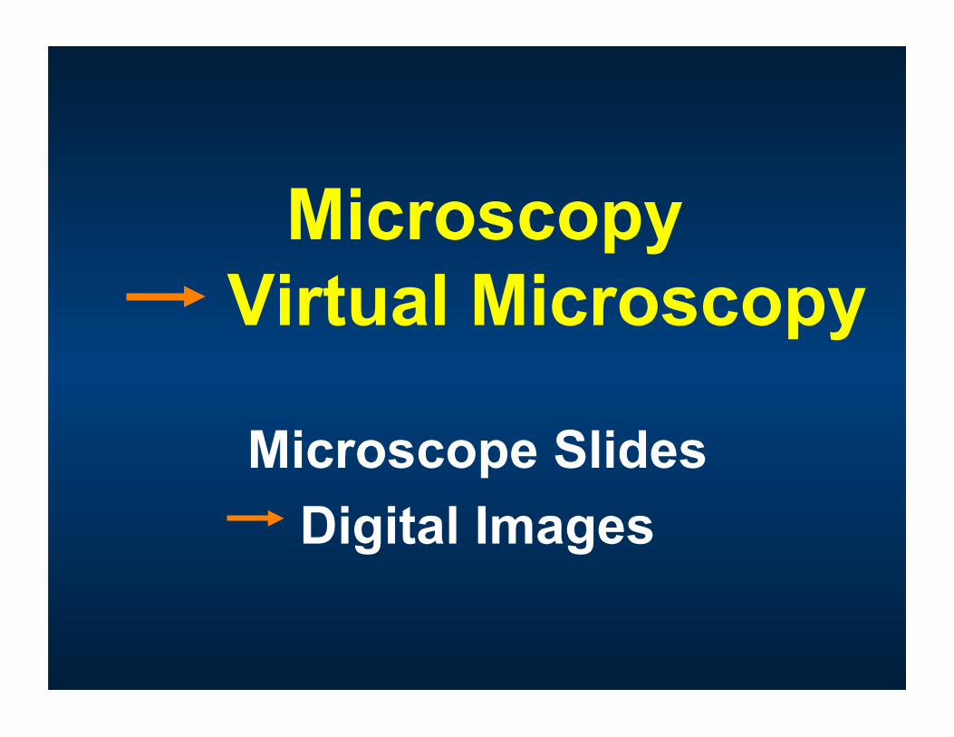
Microscopy Virtual Microscopy
Microscope Slides Digital Images
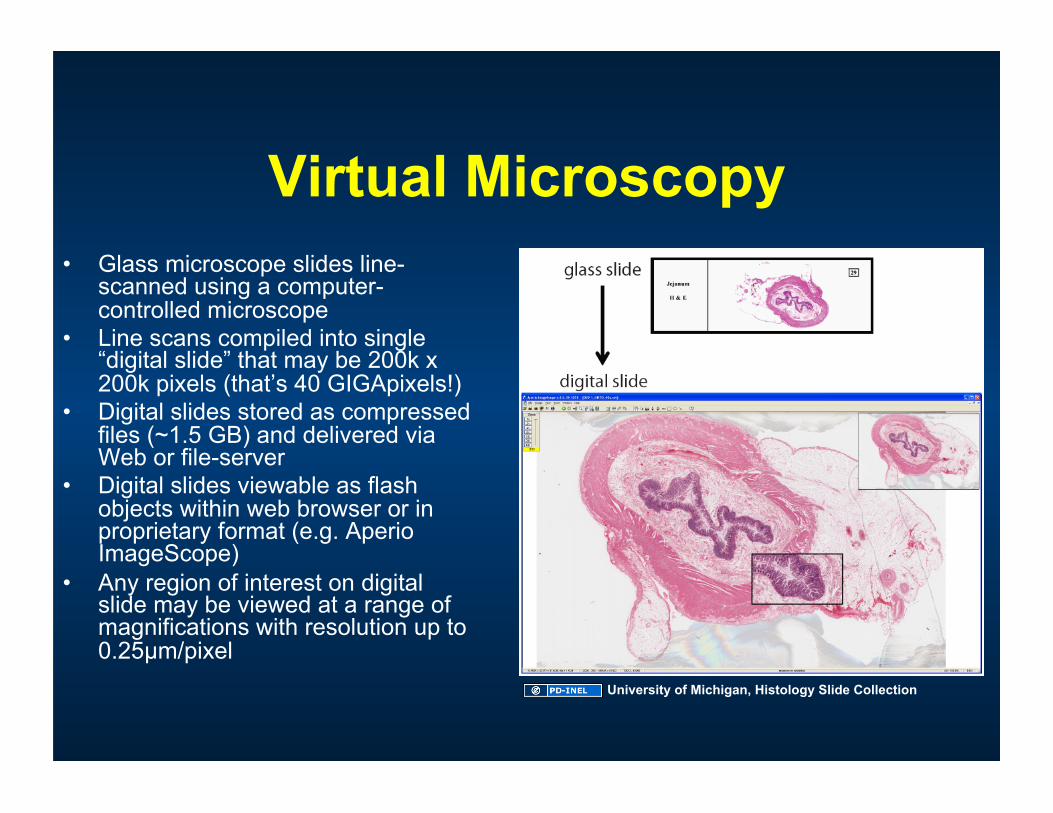
Virtual Microscopy • Glass microscope slides line-
scanned using a computer-controlled microscope
• Line scans compiled into single “digital slide” that may be 200k x 200k pixels (that’s 40 GIGApixels!)
• Digital slides stored as compressed files (~1.5 GB) and delivered via Web or file-server
• Digital slides viewable as flash objects within web browser or in proprietary format (e.g. Aperio ImageScope)
• Any region of interest on digital slide may be viewed at a range of magnifications with resolution up to 0.25µm/pixel
University of Michigan, Histology Slide Collection
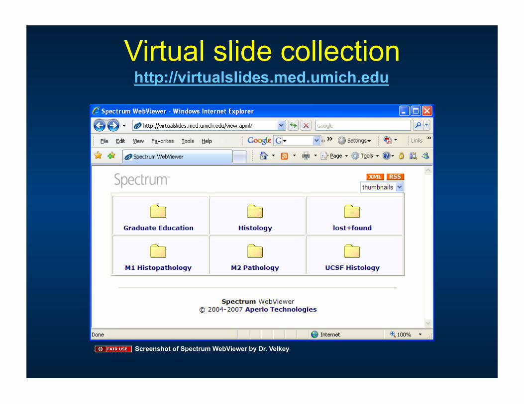
Virtual slide collection http://virtualslides.med.umich.edu
Screenshot of Spectrum WebViewer by Dr. Velkey
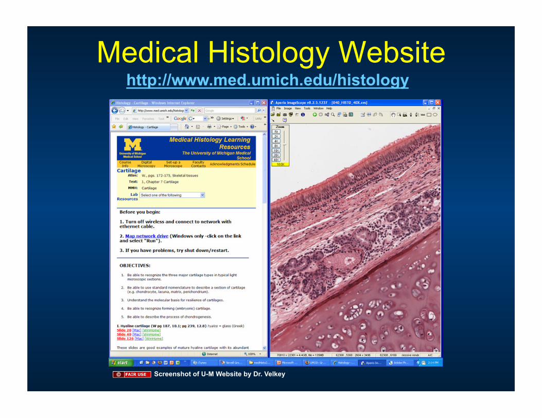
Medical Histology Website http://www.med.umich.edu/histology
Screenshot of U-M Website by Dr. Velkey
not in handouts
Cartoon removed
Microscopes and glass slides still have their place!
• The focal plane and depth-of-field (aperture) of the digital slide is fixed
• The digital slide is only a representative specimen
• Servers crash
• Knowing how to use a microscope has its value.
not in handouts
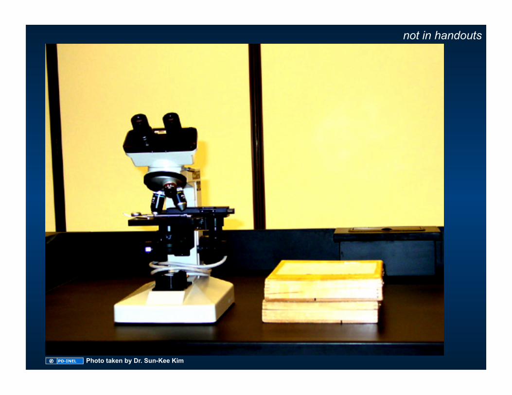
Photo taken by Dr. Sun-Kee Kim
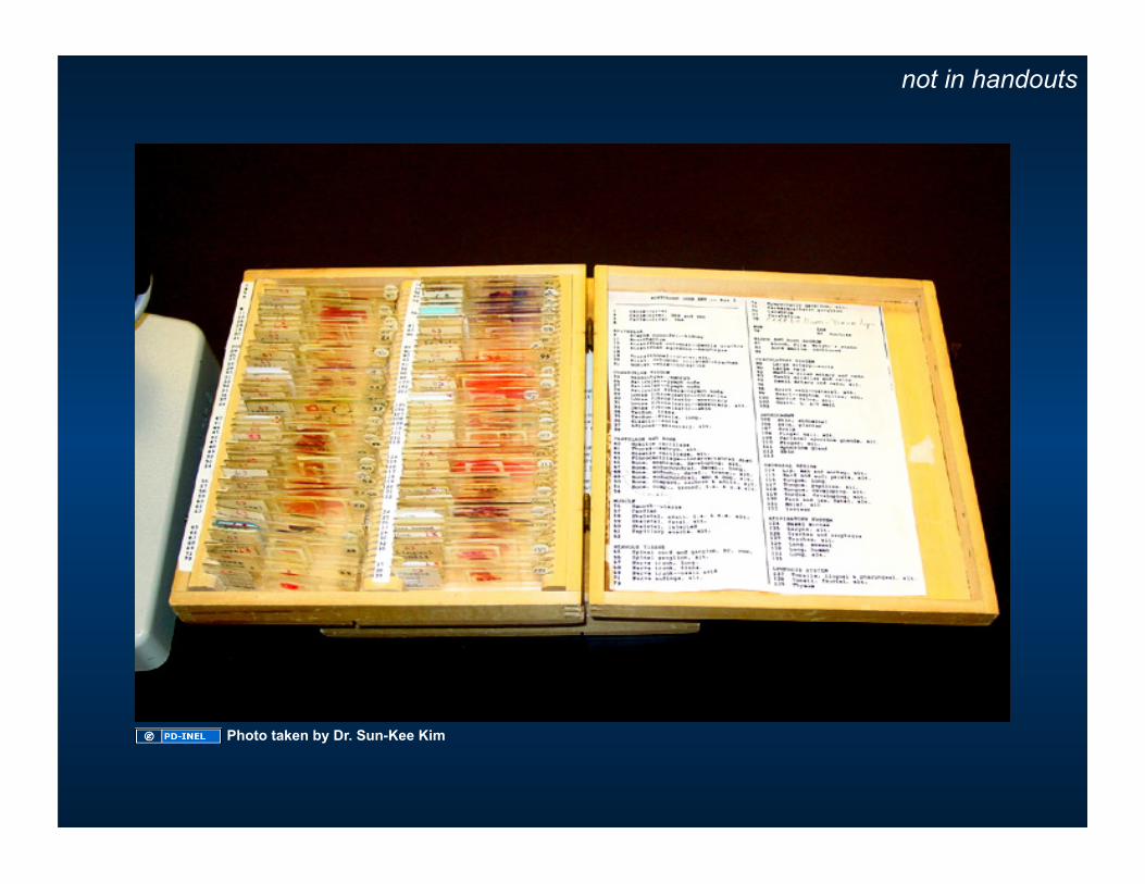
not in handouts
Photo taken by Dr. Sun-Kee Kim
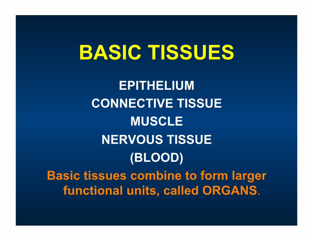
BASIC TISSUES
EPITHELIUM CONNECTIVE TISSUE
MUSCLE NERVOUS TISSUE
(BLOOD) Basic tissues combine to form larger
functional units, called ORGANS.
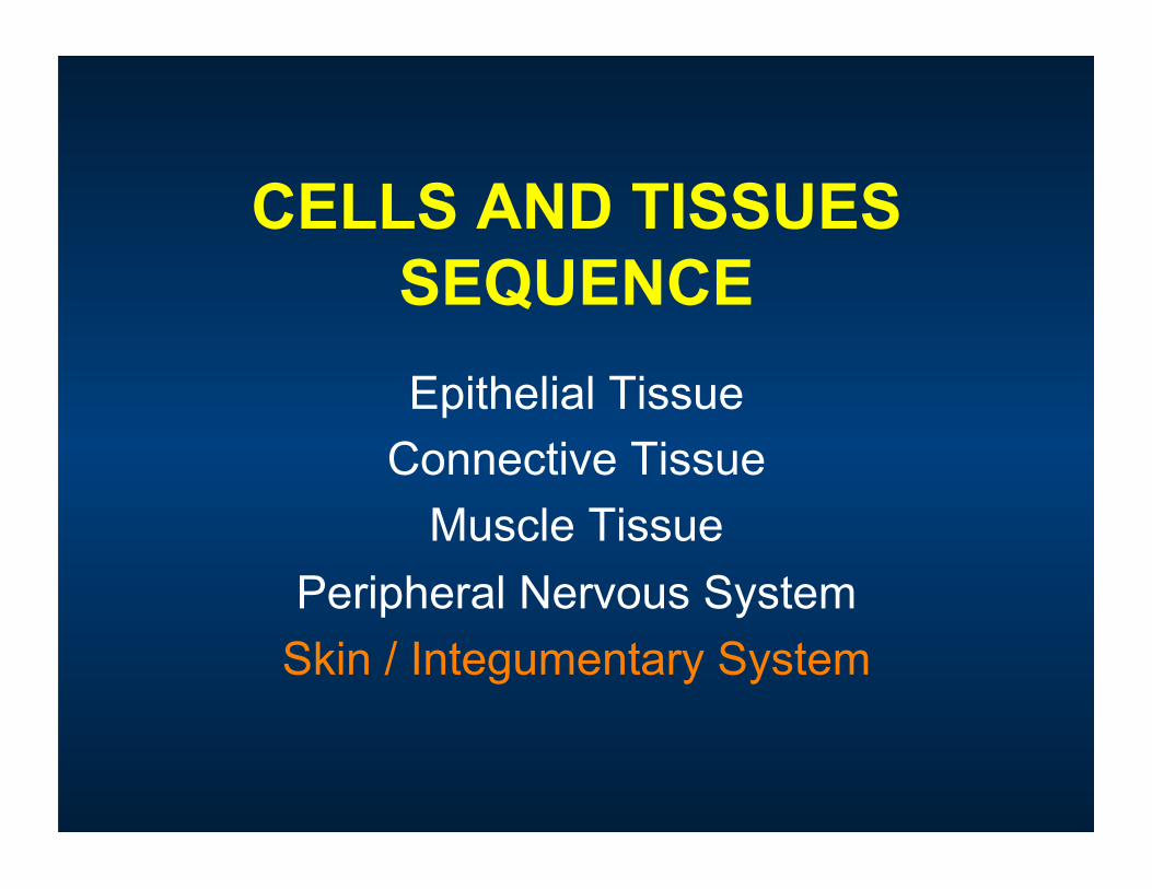
CELLS AND TISSUES SEQUENCE Epithelial Tissue
Connective Tissue Muscle Tissue
Peripheral Nervous System Skin / Integumentary System
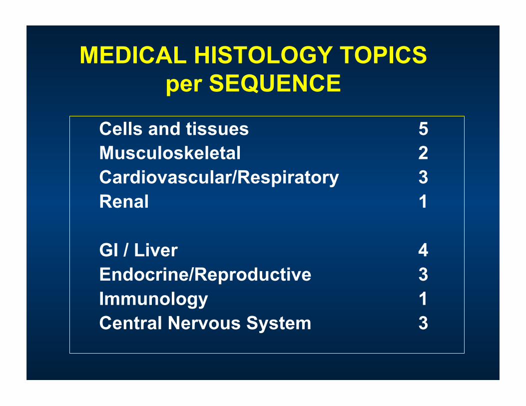
MEDICAL HISTOLOGY TOPICS per SEQUENCE
Cells and tissues 5 Musculoskeletal 2 Cardiovascular/Respiratory 3 Renal 1
GI / Liver 4 Endocrine/Reproductive 3 Immunology 1 Central Nervous System 3
MEDICAL HISTOLOGY Lecture: ~50 minutes
Lecture Handouts in coursepacks Lecture PowerPoints on CTools (also linked from histo web site).
Laboratory: 3 hours Laboratory Guide (hard copy or online) - learning objectives Microscope and slides (“real” and virtual)
Lab Atlas and Text Book: Young, et al.: Wheater’s Functional Histology, 5th ed. –HIGHLY recommended Ross and Pawlina: Histology: A Text and Atlas, 5th ed. -recommended
Michigan Medical Histology CD –not issued this year (won’t work in Mac OS X) Review and Lookalike Images (online) Lab Orientation Presentations (online)
RESOURCES Histo web site: http://www.med.umich.edu/histology
CTools (aka “portal”): https://ctools.umich.edu/portal
different from handout
Quizzes and Exams • Usually a total of 8 questions per session divided
between weekly quizzes and final exam. Questions will weigh equally.
• Weekly quizzes and final exams will all be administered online.
• Multiple choice questions: some straight text, but MOSTLY image-based (LM, EM, or diagram), or virtual slides
• Sample questions may be found in the online syllabus.
different from handout
Issued Histology Materials (in your lockers)
Locker key Microscope* Two Boxes of M1 Histology Microscope Slides* Network Cable No MMH CD issued this year
Sign Loan Agreement Sheet –you acknowledge receipt of EACH item and you agree to return them at the end of the year!
* Shared resources (i.e. MUST stay in locker)
different from handout
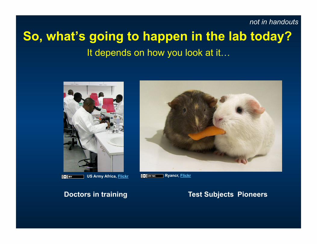
So, what’s going to happen in the lab today?
Doctors in training Test Subjects Pioneers
It depends on how you look at it…
not in handouts
US Army Africa, Flickr Ryancr, Flickr
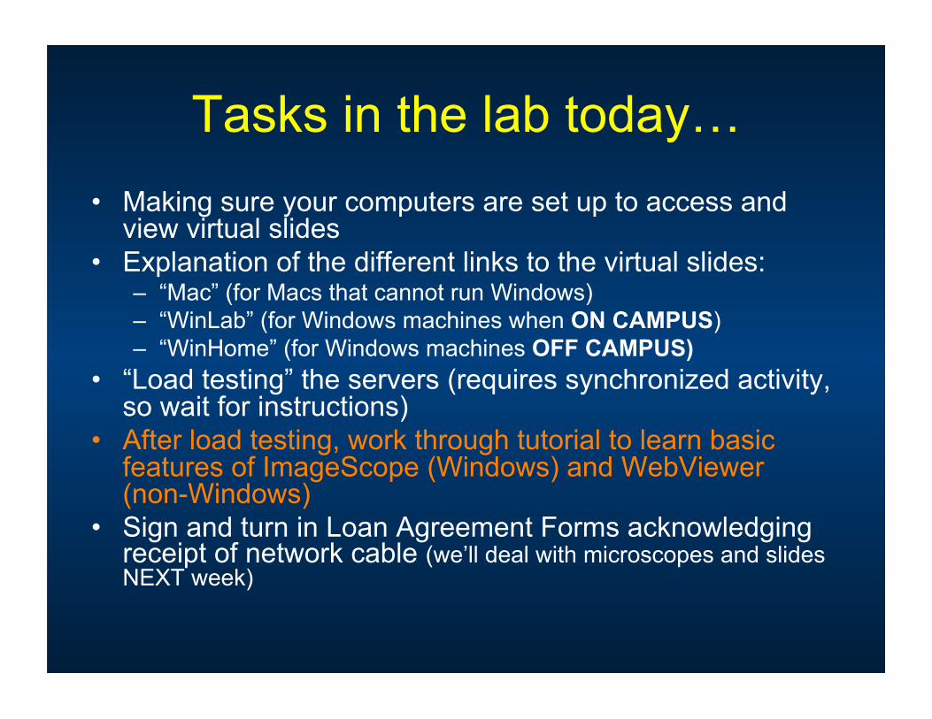
Tasks in the lab today… • Making sure your computers are set up to access and
view virtual slides
• Explanation of the different links to the virtual slides: – “Mac” (for Macs that cannot run Windows) – “WinLab” (for Windows machines when ON CAMPUS) – “WinHome” (for Windows machines OFF CAMPUS)
• “Load testing” the servers (requires synchronized activity, so wait for instructions)
• After load testing, work through tutorial to learn basic features of ImageScope (Windows) and WebViewer (non-Windows)
• Sign and turn in Loan Agreement Forms acknowledging receipt of network cable (we’ll deal with microscopes and slides NEXT week)
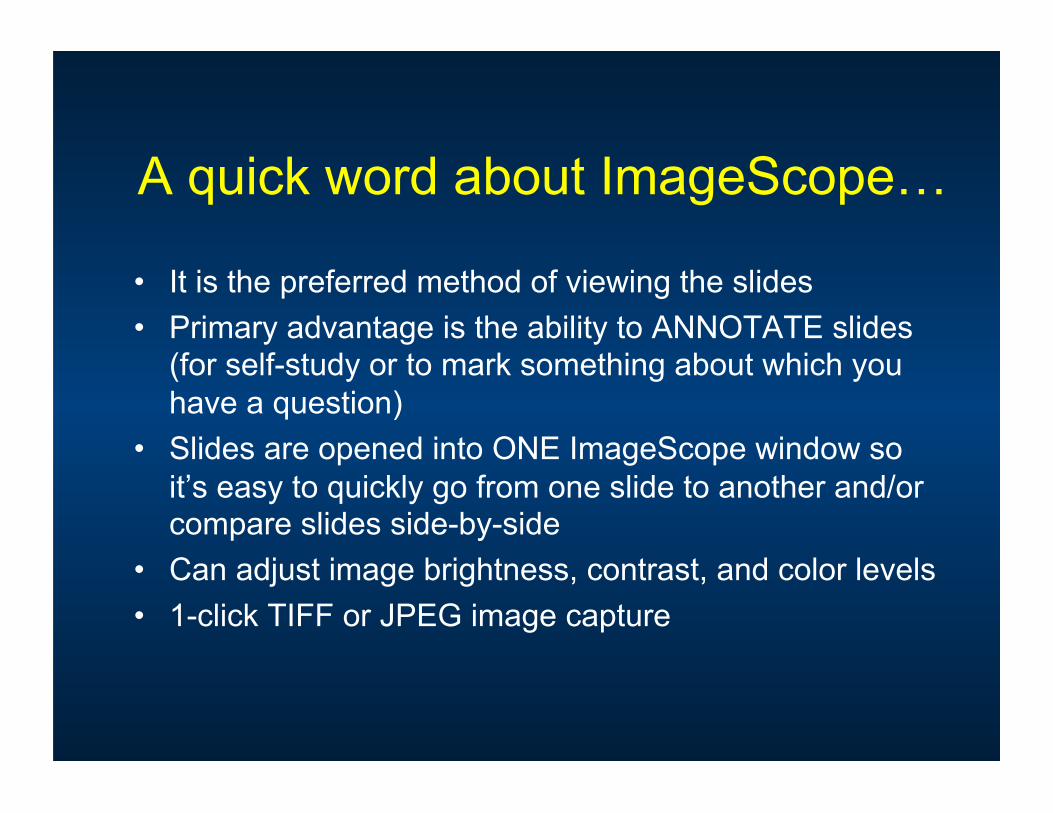
A quick word about ImageScope…
• It is the preferred method of viewing the slides • Primary advantage is the ability to ANNOTATE slides
(for self-study or to mark something about which you have a question)
• Slides are opened into ONE ImageScope window so it’s easy to quickly go from one slide to another and/or compare slides side-by-side
• Can adjust image brightness, contrast, and color levels • 1-click TIFF or JPEG image capture
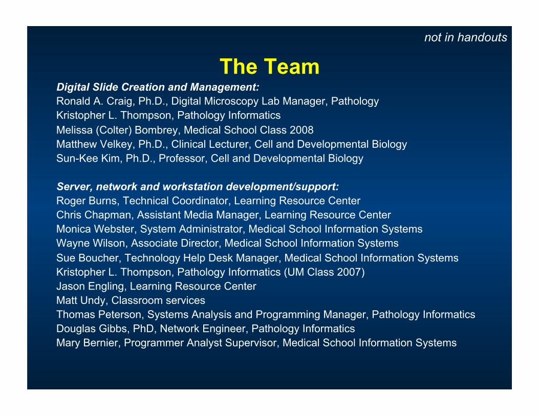
The Team Digital Slide Creation and Management: Ronald A. Craig, Ph.D., Digital Microscopy Lab Manager, Pathology Kristopher L. Thompson, Pathology Informatics Melissa (Colter) Bombrey, Medical School Class 2008 Matthew Velkey, Ph.D., Clinical Lecturer, Cell and Developmental Biology Sun-Kee Kim, Ph.D., Professor, Cell and Developmental Biology
Server, network and workstation development/support: Roger Burns, Technical Coordinator, Learning Resource Center Chris Chapman, Assistant Media Manager, Learning Resource Center Monica Webster, System Administrator, Medical School Information Systems Wayne Wilson, Associate Director, Medical School Information Systems Sue Boucher, Technology Help Desk Manager, Medical School Information Systems Kristopher L. Thompson, Pathology Informatics (UM Class 2007) Jason Engling, Learning Resource Center Matt Undy, Classroom services Thomas Peterson, Systems Analysis and Programming Manager, Pathology Informatics Douglas Gibbs, PhD, Network Engineer, Pathology Informatics Mary Bernier, Programmer Analyst Supervisor, Medical School Information Systems
not in handouts
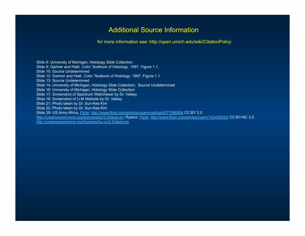
Additional Source Information for more information see: http://open.umich.edu/wiki/CitationPolicy
Slide 6: University of Michigan, Histology Slide Collection Slide 9: Gartner and Hiatt. Color Textbook of Histology. 1997. Figure 1.1. Slide 10: Source Undetermined Slide 12: Gartner and Hiatt. Color Textbook of Histology. 1997. Figure 1.1. Slide 13: Source Undetermined Slide 14: University of Michigan, Histology Slide Collection; Source Undetermined Slide 16: University of Michigan, Histology Slide Collection Slide 17: Screenshot of Spectrum WebViewer by Dr. Velkey Slide 18: Screenshot of U-M Website by Dr. Velkey Slide 21: Photo taken by Dr. Sun-Kee Kim Slide 22: Photo taken by Dr. Sun-Kee Kim Slide 29: US Army Africa, Flickr, http://www.flickr.com/photos/usarmyafrica/4077598908 CC:BY 2.0 http://creativecommons.org/licenses/by/2.0/deed.en; Ryancr, Flickr, http://www.flickr.com/photos/ryanr/142455033/ CC:BY-NC 2.0 http://creativecommons.org/licenses/by-nc/2.0/deed.en