
AUA - Cystography 1
65
Uroradiology Tutorial For Medical Students Lesson 3: Cystography & Urethrography – Part 1 American Urological Association
-
Upload
prashant-bansal -
Category
Health & Medicine
-
view
135 -
download
1
Transcript of AUA - Cystography 1

Uroradiology TutorialFor Medical Students
Lesson 3:
Cystography & Urethrography – Part 1
American Urological Association

Introduction
• Conventional radiography of the urinary tract includes several diagnostic studies:
– Cystogram
– Retrograde urethrogram
– Voiding cystourethrogram
• All of these studies answer questions that are essential to urologic patient management
Voiding Cystourethrogram (VCUG)
• The voiding cystourethrogram is a dynamic test used to define the anatomy and, in part, the function of the lower urinary tract. It is performed by placing a catheter through the urethra into the bladder, filling the bladder with contrast material and then taking x-rays while the patient voids. You can imagine how popular it is among children.

Scout Film
• Several films are taken when performing a VCUG. The first image is a KUB called the scout film. On this film one can evaluate the bones of the spine and pelvis (injury or congenital anomaly such as spina bifida) and the soft tissues (calcifications, foreign bodies, etc.).

• Normal scout image
• What gender?
Scout Film
• Patients with urologic problems (urine infection or incontinence) may have a spinal abnormality that results in abnormal innervation of the bladder. Such anomalies are commonly associated with anomalies of the vertebral column. Let’s look at some spines.

• Here is a spine from a normal KUB or scout film. Notice that the posterior processes of all the vertebrae are intact. You can see the posterior process behind and below each vertebral body.

• Here is another scout film. Notice that the posterior processes are absent below L-4. This patient has lower lumbar spina bifida.

Read This Scout Film
• The bones are normal
• What about soft tissues (bowel, etc.)?
• This child has significant constipation. Notice the variegated pattern of stool and gas in the colon.

Read This Scout Film
• What is the foreign body?

Defining an Unknown Object
• When you see something on an imaging study that you don’t understand, it is helpful systematicaly to describe it.
• Shape:
• Location:
• Density:
• Give it a try with the object on that scout film

Defining an Unknown Object
Shape:
It’s long, thin and it curves
Shape of a catheter or tubing

Defining an Unknown Object
Location:Is it in the child’s body or
external?Of course, a lateral view or an
oblique view would answer that question, but what do you think?
It is internal
Where does it course from and to?
It’s seen in the region of the abdomen and the chest. One end is not seen on the film and the other is near a rib, but coursing from the abdomen to that point.

Defining an Unknown Object
Density:
It’s relatively high density
At least as radiodense as bone
Whoever made that tubing probably wants it to show up on x-ray films because it is so dense

Look carefully at the spine

Compare to this normal scout film

The spine is not fully developed in the film on the right.

• Let’s summarize what we know • Radiodense foreign body (proably a catheter or
tubing) in the region of the chest and the abdomen. One end appears to be in the abdomen, and we don’t see the other end.
• This child has spina bifida.
• It’s a ventriculoperitoneal shunt, used to drain cerebrospinal fluid in patients with hydrocephalus (common in children with spinabifida).
Summary

Filling Phase
• After the scout film is taken, images are obtained while the bladder is being filled with liquid contrast. The bladder should appear smooth and regular and there should be no filling defects except the balloon of the urethral catheter. The edges of the bladder image should be smooth.

Normal Filling Phase

Filling Phase
• This bladder is not smooth. This patient has an obstruction in the urethra. She has spina bifida (see that shunt tubing?). Nerve damage from the spina bifida results in a physiologic obstruction to urine drainage through the urethra. Her bladder responded to the obstruction by detrusor muscle hypertrophy. This thickened muscle caused the irregular border of the bladder.

Filling Phase
• The term we use to describe this irregularity is trabeculation. Note that the bladder isn’t spherical. Some would describe this as a ‘Christmas tree’ bladder. It’s cone shaped and it appears to have ornaments (actually small round diverticulae), a result of high bladder pressures.

Voiding Phase
• Images captured during voiding will demonstrate the urethra (strictures or obstruction) and the bladder, and they will document the presence or absence of vesicoureteral reflux (retrograde leakage of urine from the bladder up the ureters). Unless there is a voiding film, one cannot determine whether the patient has reflux because reflux may only occur with the pressure generated by voiding.

Voiding Phase
• This film shows a normal male urethra; there is no obstruction. The variation seen in the diameter of the urethra is normal.
Indentation at the urethral sphincter (normal)

Voiding Phase
• This film shows a boy with a congenital urethral obstruction. Notice that the bladder is trabeculated. In addition, reflux is present in both ureters.
Urethral obstruction
Reflux

Post-void Film• The post-void image may
demonstrate reflux (contrast seen in the ureter or kidney) or extravasation of urine from the bladder or urethra (such as from a traumatic rupture).
• No reflux and no residual bladder urine is seen in this normal post-void film. Because of the anxiety caused by the catheter placement, it is not unusual for a child not to void completely. There are other, more accurate tests to determine whether a patient has a significant post-void residual.
Normal post-void film

Ready to read some VCUGs?

Case History
• 6 year-old female with a history of at least three urine infections
• Past history: negative
• Family history: mother and sister had urine infections and vesicoureteral reflux
• Exam: completely normal
• Imaging: Pediatrician ordered ultrasound and VCUG

Ultrasound
You can learn to read kidney and
bladder ultrasounds at the AUA Imaging
Site.

Ultrasound Interpretation
• Left Right
• Size (nl = 4 + 6 x .6 cm) 7.7 Normal 8.6 Normal
• Shape Normal Normal
• Parenchyma Normal Normal
• Hydronephrosis No No
• Normal renal / bladder ultrasound

Here is her VCUG
Let’s look at all four phases

Scout Film
• Bones?
– Normal
• Soft tissues?
– Normal except for some constipation
• Foreign bodies?
– No

Filling Phase
• Bladder shape?
– Smooth
• Bladder contour?
– Smooth and regular
• Reflux?
– No
• Extravasation?
– No

Voiding Phase
• Reflux?
– No
• Urethra?
– Normal
• Extravasation?
– No

Post-void
• Reflux?
– No
• Bladder empty?
– Yes
• Extravasation?
– No
• Normal VCUG

Case Summary
• This girl, like the majority of girls with urine infection, has a normal bladder (no reflux) and normal kidneys.
• Her urine infections are likely the result of ascent of bacteria up the urethra from the perineum.
• Do you feel cheated because we showed you a normal VCUG? Most VCUGs in children with urine infection are normal. Get used to it. The important thing is that you’re learning to read the exams.

Case History
• 4 year-old female has had two febrile urine infections (fever to 104, nausea, flank pain and lethargy.
• Exam: Lethargic white female
• T = 103.2, P = 130, BP 78/60
• Abdomen: soft, but tender in left flank and bladder regions. U/A >100 RBCs, WBCs 50-100, bacteria - many

Evaluation & Management
• Full course of antibiotics to treat pyelonephritis
• Why did this girl have a urine infection?
• Renal and bladder ultrasound – normal
• What other abnormalities could raise her risk of pyelonephritis?

Read This VCUG

VCUG Interpretation• Scout:
– Bones?
• Normal
– Soft tissues?
• Normal
– Foreign bodies?
• What is that partially radiodense tubular structure seen in the region of the pelvis?
• It’s the catheter used to fill the bladder with contrast.

Filling phase:
• Bladder shape?
– Normal
• Bladder contour?
– Smooth
• Reflux?
– Bilateral reflux
• Extravasation?
– No

Voiding phase:
• Reflux?
– Bilateral reflux
• Urethra?
– Normal
• Extravasation?
– No

Post void:
• Bladder empty?
– Yes
• Reflux?
– Bilateral reflux
• Extravasation?
– No

Vesicoureteral Reflux
• Urinary tract infections usually result from ascent of bacteria from the perineum up the urethra. That is why bowel flora cause most urine infections.
• Reflux does not cause urine infection, but if reflux is present, ascent of bacteria up the ureter causing pyelonephritis is more likely.

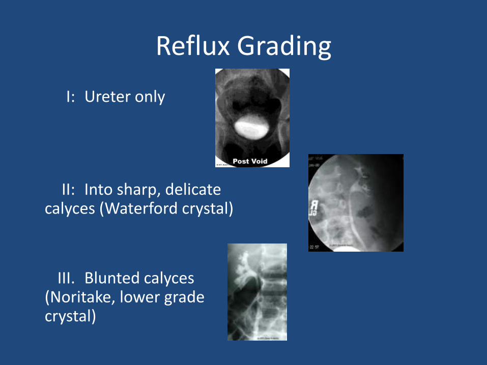
Reflux Grading
A grading system was developed, based on x-ray VCUG findings (primarily the appearance of the calyces) in order to express the severity of reflux. You may find it useful to compare the calyx to a crystal goblet. The calyx (goblet) collects urine draining from the pyramid (area of collecting ducts). The infundibulum (stem of the goblet) drains to the renal pelvis.

Reflux Grading
• The more delicate and fine the calyx (like Waterford crystal), the lower the grade of reflux
Reflux Grading
I: Ureter only
II: Into sharp, delicate calyces (Waterford crystal)
III. Blunted calyces (Noritake, lower grade crystal)

Reflux Grading
IV: Further blunting (remainders table goblet at K-Mart)
V: Dilated, tortuous ureter

Reflux
• 20 – 30% of children with urine infection are found to have reflux. Spontaneous resolution is common among patients with low grade reflux.
• Management: prophylactic antibiotics• Periodic imaging
• You prescribe a daily dose of nitrofurantoin. One year later a follow-up VCUG shows that the reflux is gone. Her parents are so grateful that they name their next child after you.

Case History
• A newborn male is referred to you because prenatal ultrasounds showed bilateral hydronephrosis.
• Exam: healthy, vigorous male. 3.8 Kg. Normal exam except for some vague fullness in the abdomen.
• Ultrasound: Bilateral grade IV hydronephrosis

Read This VCUG
[© 2010, David A. Hatch. Used by permission.]

How did you do? Scout film• Bones?
– Normal
• Soft tissues?
– Why doesn’t the bowel gas pattern extend to the right side of the abdomen?
• What is this? (blue arrow)

Describe the Unknown Object
• Shape?
– It’s long and relatively thin.
– It’s very regular with sharp borders
– Upper end is rounded and the lower end is square

Location?
• Left side of the abdomen
• Is it inside the peritoneal cavity?
– Outside. The lower part of the object extends to a point outside the peritoneal cavity. It’s lateral to the iliac wing.

Density?
• It’s about the density of the adjacent bones (partially calcified in an infant)
• Less dense than mature bone
• So, What Is It?
• Umbilical cord clamp (remember, he’s a newborn)

Read the Filling Phase
• Bilateral reflux
– Blunted calyces
– Tortuous ureters (grade V)
• Urethra
– Wider than normal
– This boy has posterior urethral valves, a congenital obstruction of the proximal urethra

Voiding Phase
• Reflux
• Posterior urethral valves
• What is this (arrow)?
– Location?
– Shape?
– Density?

What is this?• Location?
– Left upper quadrant-renal fossa
• Shape?– Oval, fuzzy, not as well
defined as the ureters or bladder
• Density?– About the same density
as the ureters or the adjacent renal pelvis

Need a hint?
• Does this object appear on the scout film?

Let’s summarize
• Irregular (probably not a cyst or part of the kidney collecting system)
• Intra-abdominal
• Highly radiodense
• Appears only after contrast is dripped into the bladder catheter
• If this is contrast, how did it get into the left renal fossa?

Urine Extravasation
• The fuzzy, ovoid density that is seen only on the voiding film is urine (actually contrast) extravasation. The urethral obstruction was so severe that it caused leakage of urine out of the kidney collecting system into the perinephric space.

Posterior Urethral Valves
• Occurs along a spectrum of severity from mild obstruction to complete urinary obstruction with anhydramnios (no amniotic fluid) and post-natal death from pulmonary insufficiency
• Initial management is usually incision of the valves (performed through a cystoscope) to relieve obstruction

Cystography 1 – Summary
• VCUG
– Serial images: Scout, filling, voiding, post-void
– Contrast only seen outside the bladder (kidney, ureter, etc.) if there is a problem (reflux or extravasation)
• Vesicoureteral reflux
– Grading: I to V

Cystography 1 – Summary
• Posterior urethral valves
– Congenital obstruction of proximal urethra
– Often associated with reflux and sometimes causing urine extravasation
• Evaluation of an unknown – describe it
– Location
– Shape
– Density

Congratulations!
• You have successfully completed the first module on cystography. You are ready to go on to Cystography - 2