Atomic Force Microscopy in Imaging of Viruses and Virus-Infected Cells
18
MICROBIOLOGY AND MOLECULAR BIOLOGY REVIEWS, June 2011, p. 268–285 Vol. 75, No. 2 1092-2172/11/$12.00 doi:10.1128/MMBR.00041-10 Copyright © 2011, American Society for Microbiology. All Rights Reserved. Atomic Force Microscopy in Imaging of Viruses and Virus-Infected Cells Yurii G. Kuznetsov and Alexander McPherson* University of California, Irvine, Department of Molecular Biology and Biochemistry, 560 Steinhaus Hall, Irvine, California 92697-3900 INTRODUCTION .......................................................................................................................................................268 X-RAY DIFFRACTION AND ELECTRON MICROSCOPY ................................................................................268 HOW DOES AFM WORK?.......................................................................................................................................269 VISUALIZATION OF VIRUSES BY AFM .............................................................................................................273 QUANTITATIVE PROPERTIES OF VIRUSES .....................................................................................................274 ICOSAHEDRAL CAPSIDS .......................................................................................................................................274 VIRUSES IN CRUDE SAMPLES AND ON CELLS .............................................................................................276 MUTANT VIRUSES ...................................................................................................................................................277 SPECIALIZED FEATURES ON VIRUSES ............................................................................................................277 DISSECTION OF COMPLEX VIRUSES ...............................................................................................................278 VISUALIZATION OF VIRAL RNA AND DNA ......................................................................................................282 IMMUNE LABELING WITH AFM .........................................................................................................................283 WHAT DOES AFM OFFER THE STRUCTURAL BIOLOGIST? ......................................................................284 REFERENCES ............................................................................................................................................................284 INTRODUCTION There are currently two principal techniques in common use for the direct visualization of virus structure, and these are X-ray diffraction analysis and electron microscopy (EM). X-ray diffraction from single crystals is extraordinary in the resolu- tion of structural detail that it can reveal, approaching the atomic level. In many cases intimate details of the capsid ar- chitecture, as well as, in some cases, the protein-nucleic acid interactions, become clear with X-ray crystallography. It is unsurpassed in this regard, and it is unlikely ever to be chal- lenged. It will remain the technique of choice, whenever it can be applied. X-RAY DIFFRACTION AND ELECTRON MICROSCOPY X-ray diffraction as applied to viruses does, however, have some limitations. First, it requires that the virus be crystallized. This is possible only for viruses having very regular shapes and uniform exterior surfaces, i.e., icosahedral viruses or, conceiv- ably, some virus-like particles (VLP) produced in recombinant systems or reconstructed in vitro. Thus, most complex viruses, incorporating membranes or exhibiting structurally compli- cated external features such as tail assemblies or fibers, are excluded from X-ray analysis. In addition, viruses beyond a certain size limit, probably about 1,000 Å in diameter, exceed our current technologies for recording X-ray data from very large unit cells. Finally, interior components of viruses, espe- cially encapsidated nucleic acid, generally do not share the external symmetry of the particles. Therefore, they are not usually seen in electron density maps. Those components are orientationally randomized in the crystallization process. X-ray diffraction is an “averaging method” that does not reveal the characteristics of individual particles. It assumes that all par- ticles are exactly the same, or it makes them so. In addition to allowing determination of the structures of intact, symmetrical viruses, X-ray diffraction can be useful in another way. It can also be used to determine, very precisely, the structures of individual proteins, and sometimes complexes of proteins, that make up viral substructures. These can then be fitted to lower-resolution models of the particles obtained by other means (electron microscopy, for example), and a “pseudo”-high-resolution structure of a virus can be created (45, 46). This has proven successful in a number of cases. X-ray diffraction analysis is a powerful approach, and can be made even more powerful when it is combined with information from other sources, as it has been with electron microscopy and potentially can be with atomic force microscopy (AFM). Electron microscopy (EM) has been applied to the analysis of virus structure along two principal lines. Transmission EM of viruses spread on substrates, such as carbon-coated grids (13, 14), or of virus-infected cells after thin sectioning is one method. Reconstruction of particles obtained by acquisition of many low-dose images from cryo-electron microscopy (cryo- EM) is the other. While the former once predominated, in more recent times cryo-EM has certainly proven to be the more powerful for elucidating virus architecture (4) and has been shown to be capable of revealing detail at resolutions in the range of 3.5 to 4 Å. In some cases it has even permitted the tracing of a polypeptide chain. Transmission EM is, however, problematic because it gen- erally requires heavy metal staining or shadowing to increase contrast. It usually requires complete dehydration, and it often involves fixatives as well. Finally, it produces projections of entire particles onto the substrate plane. Elimination of one dimension obscures spatial relationships among substructures, * Corresponding author. Mailing address: University of California, Irvine, Department of Molecular Biology and Biochemistry, 560 Stein- haus Hall, Irvine, CA 92697-3900. Phone: (949) 824-1931. Fax: (949) 824-8551. E-mail: [email protected]. 268 Downloaded from https://journals.asm.org/journal/mmbr on 11 November 2021 by 45.169.215.128.
Transcript of Atomic Force Microscopy in Imaging of Viruses and Virus-Infected Cells
MICROBIOLOGY AND MOLECULAR BIOLOGY REVIEWS, June 2011, p. 268–285 Vol. 75, No. 21092-2172/11/$12.00 doi:10.1128/MMBR.00041-10Copyright © 2011, American Society for Microbiology. All Rights Reserved.
Atomic Force Microscopy in Imaging of Viruses andVirus-Infected Cells
Yurii G. Kuznetsov and Alexander McPherson*University of California, Irvine, Department of Molecular Biology and Biochemistry,
560 Steinhaus Hall, Irvine, California 92697-3900
INTRODUCTION .......................................................................................................................................................268X-RAY DIFFRACTION AND ELECTRON MICROSCOPY................................................................................268HOW DOES AFM WORK?.......................................................................................................................................269VISUALIZATION OF VIRUSES BY AFM .............................................................................................................273QUANTITATIVE PROPERTIES OF VIRUSES.....................................................................................................274ICOSAHEDRAL CAPSIDS .......................................................................................................................................274VIRUSES IN CRUDE SAMPLES AND ON CELLS .............................................................................................276MUTANT VIRUSES ...................................................................................................................................................277SPECIALIZED FEATURES ON VIRUSES ............................................................................................................277DISSECTION OF COMPLEX VIRUSES ...............................................................................................................278VISUALIZATION OF VIRAL RNA AND DNA......................................................................................................282IMMUNE LABELING WITH AFM .........................................................................................................................283WHAT DOES AFM OFFER THE STRUCTURAL BIOLOGIST? ......................................................................284REFERENCES ............................................................................................................................................................284
INTRODUCTION
There are currently two principal techniques in common usefor the direct visualization of virus structure, and these areX-ray diffraction analysis and electron microscopy (EM). X-raydiffraction from single crystals is extraordinary in the resolu-tion of structural detail that it can reveal, approaching theatomic level. In many cases intimate details of the capsid ar-chitecture, as well as, in some cases, the protein-nucleic acidinteractions, become clear with X-ray crystallography. It isunsurpassed in this regard, and it is unlikely ever to be chal-lenged. It will remain the technique of choice, whenever it canbe applied.
X-RAY DIFFRACTION AND ELECTRON MICROSCOPY
X-ray diffraction as applied to viruses does, however, havesome limitations. First, it requires that the virus be crystallized.This is possible only for viruses having very regular shapes anduniform exterior surfaces, i.e., icosahedral viruses or, conceiv-ably, some virus-like particles (VLP) produced in recombinantsystems or reconstructed in vitro. Thus, most complex viruses,incorporating membranes or exhibiting structurally compli-cated external features such as tail assemblies or fibers, areexcluded from X-ray analysis. In addition, viruses beyond acertain size limit, probably about 1,000 Å in diameter, exceedour current technologies for recording X-ray data from verylarge unit cells. Finally, interior components of viruses, espe-cially encapsidated nucleic acid, generally do not share theexternal symmetry of the particles. Therefore, they are notusually seen in electron density maps. Those components are
orientationally randomized in the crystallization process. X-raydiffraction is an “averaging method” that does not reveal thecharacteristics of individual particles. It assumes that all par-ticles are exactly the same, or it makes them so.
In addition to allowing determination of the structures ofintact, symmetrical viruses, X-ray diffraction can be useful inanother way. It can also be used to determine, very precisely,the structures of individual proteins, and sometimes complexesof proteins, that make up viral substructures. These can thenbe fitted to lower-resolution models of the particles obtainedby other means (electron microscopy, for example), and a“pseudo”-high-resolution structure of a virus can be created(45, 46). This has proven successful in a number of cases. X-raydiffraction analysis is a powerful approach, and can be madeeven more powerful when it is combined with informationfrom other sources, as it has been with electron microscopyand potentially can be with atomic force microscopy (AFM).
Electron microscopy (EM) has been applied to the analysisof virus structure along two principal lines. Transmission EMof viruses spread on substrates, such as carbon-coated grids(13, 14), or of virus-infected cells after thin sectioning is onemethod. Reconstruction of particles obtained by acquisition ofmany low-dose images from cryo-electron microscopy (cryo-EM) is the other. While the former once predominated, inmore recent times cryo-EM has certainly proven to be themore powerful for elucidating virus architecture (4) and hasbeen shown to be capable of revealing detail at resolutions inthe range of 3.5 to 4 Å. In some cases it has even permitted thetracing of a polypeptide chain.
Transmission EM is, however, problematic because it gen-erally requires heavy metal staining or shadowing to increasecontrast. It usually requires complete dehydration, and it ofteninvolves fixatives as well. Finally, it produces projections ofentire particles onto the substrate plane. Elimination of onedimension obscures spatial relationships among substructures,
* Corresponding author. Mailing address: University of California,Irvine, Department of Molecular Biology and Biochemistry, 560 Stein-haus Hall, Irvine, CA 92697-3900. Phone: (949) 824-1931. Fax: (949)824-8551. E-mail: [email protected].
268
Dow
nloa
ded
from
http
s://j
ourn
als.
asm
.org
/jour
nal/m
mbr
on
11 N
ovem
ber
2021
by
45.1
69.2
15.1
28.

results in loss of chirality, creates artifacts, and produces im-ages that are frequently open to multiple interpretations. Inthe past decade, cryo-electron microscopy has been made in-creasingly powerful by automation in image collection andcataloging and by elaborate mathematical techniques for ana-lyzing the low-dose images and reconstructing viruses fromcollections of particles observed in multiple orientations. Themajor advantage of cryo-EM over X-ray diffraction, of course,is that it does not require that the virus be crystallized.
Cryo-EM (1), however, also has limitations. Like X-ray dif-fraction, it is useful chiefly for analyzing regular viruses havingicosahedral symmetry. It produces the “average” structure forthe entire virus population and does not illuminate eccentric-ities of individuals. More advanced applications of cryo-EM(7) can deal with particles lacking any symmetry (ribosomes,for example), but even here the assumption is that all of theparticles are identical. It is ineffective when the viruses arepleiomorphic and lack architectural uniformity. Cryo-EM isalso likely to be technically limited in its application to verylarge viruses, even icosahedral viruses such as iridoviruses,because of the physical thickness of the particles and the dif-ficulty of simply penetrating their bulk. Just as the power ofX-ray diffraction has been amplified by its combination withcryo-EM, the converse is equally true. It is important to rec-ognize, however, that even when combined, the pictures thesetechniques yield, for the same reasons as for X-ray crystallog-
raphy alone, can still often be incomplete, particularly regard-ing the interior structural features of the viruses, and both canproduce only static images of an averaged structure.
A third, direct imaging technology that promises to have asignificant impact on structural biology and which is, in mostways, complementary to X-ray diffraction and electron micros-copy is atomic force microscopy (AFM). An immediate advan-tage of AFM is that it is based on relatively simple physicalprinciples, unlike X-ray crystallography, and AFM instrumentsare mechanically and electronically rather straightforward, un-like those used for electron microscopy. Unlike both of theother technologies, AFM is fairly inexpensive to institute andapply, even to biological specimens. The microscope itself is asmall device, which is about the size of a common householdcoffee pot.
HOW DOES AFM WORK?
Atomic force microscopy was invented in the mid-1980s (5).The instrument is one of a family of scanning probe micro-scopes that had such a significant impact on imaging and sur-face science that the original incarnation, the scanning tunnel-ing microscope, won Binnig a share of the 1996 Nobel Prize in
FIG. 1. (a) Schematic drawing illustrating the principles of anatomic force microscope. The vertical deflection that the cantilever tipexperiences upon encountering some topological feature on a speci-men is amplified through a reflected laser beam, which is tracked andreported by a split-diode photoelectric detector. Scanning takes placein a fluid-filled (or dry if preferred) cell of about 75 �l in volume. Thesample is translated in a raster manner by piezoelectric positionersupon which the fluid cell is mounted. (b) Scanning electron micro-graphs of high-quality AFM tips etched from silicon.
FIG. 2. Schematic illustration of the convolution of the shape ofthe AFM tip with the shape of the feature or particle being scanned.The side of the cantilever tip contacts the object and begins to producea deflection of the cantilever before the tip apex actually reaches theobject. Similarly, the opposite side of the tip is still in contact with theobject even after the apex itself has passed. Thus, the total deflectionimplies a virtual lateral dimension for the object that is greater than itsactual dimension. The difference between the virtual and actual di-mensions is a function of the width of the cantilever tip. The sharperthe tip, the more accurate the observed dimensions and the greater theresolution attainable.
VOL. 75, 2011 AFM OF VIRUSES 269
Dow
nloa
ded
from
http
s://j
ourn
als.
asm
.org
/jour
nal/m
mbr
on
11 N
ovem
ber
2021
by
45.1
69.2
15.1
28.
Physics. For several years AFM was used almost exclusively tocharacterize the surfaces of nonbiological materials, and evennow its chief applications are in the visualization of microcir-cuits on silicon chips and in nanotechnology. Its principle isremarkably simple, surprisingly so given its acuity, but is madeuseful only by sophisticated electronic image processing andparticularly piezoelectric technology. Its application to biolog-ical materials and systems has been rather slow to develop (2,6). This is curious because it can be used in physiological fluidsas well as air, and its noninvasive and nonintrusive featuresmake it an ideal tool for the study of sensitive, living systems.The chief difficulty with its use on biological materials is thatthey tend to be very soft. AFM is at its best on hard surfaces.
An AFM device and the principles by which it operates areshown schematically in Fig. 1a. AFM instruments can be op-erated in either contact mode or what is referred to as tappingmode. In contact mode, a probe made of silicon or siliconnitride is placed in near contact with the surface of interest, saythe capsid of a virus, and then translated in a systematic rastermode over the surface (or substrate). The AFM probe is asharp stylus, like those pictured in Fig. 1b, similar in functionto a minute phonograph needle. The tip ideally has a singlepoint, with a very small radius of curvature. The probe ismounted at the end of a short cantilever, typically 100 to 250
�m in length, which has a low spring constant to minimize theforce between the tip and the sample.
Scanning is achieved by translating the sample beneath theprobe, using a piezoelectric crystal-positioned x-y stage, alonga continuous sequence of raster lines. As the probe tip passesover the surface, it does not actually touch the structural fea-ture on the surface in a physical sense but interacts through“aggregate atomic forces,” which remain somewhat mysteri-ous. Encounters with substructures cause the probe to be dis-placed vertically as the tip rides across. Exceedingly small dis-placements of the tip are amplified by deflection of a laserbeam that is reflected from the upper surface of the cantilever,and these deflections are detected and tracked by a split pho-todiode. Photoelectric circuitry converts the deflections intoheight information. The resulting scan data, recorded as adigital topographical image, can then be presented in a numberof visual formats.
In contact mode of operation, the data may be acquired aseither “height” or “deflection” information, or the data fromboth modes may be obtained simultaneously. In “height” modethe sample surface is maintained at a constant distance fromthe probe tip by the piezoelectric positioner below, through afeedback mechanism. The cantilever deflection in this case isvery small. In “deflection” mode the sample is stationary and
FIG. 3. AFM images. (a) Condensed mass of brome mosaic virus(BMV), a T � 3 icosahedral virus that infects grasses such as barley.(b) Helical, rod-shaped tobacco mosaic virus (TMV), a ubiquitouspathogen throughout the plant world. (c) Tangles of marine filamen-tous bacteriophage and their broken fragments scattered on the AFMsubstrate. (d) Virions of Tipula iridescent virus, a very large icosahe-dral virus that infects insects. The virions of BMV have a diameter of30 nm, TMV is about 20 nm in diameter and1,000 nm in length, andthe adenovirus and iridovirus have diameters of about 100 nm and 200nm, respectively.
FIG. 4. AFM images. (a) A single virion of Moloney murine leu-kemia virus, an animal retrovirus having a diameter of about 150 nm.Clusters of envelope protein are clearly evident on its surface. (b)Surface of a virion of vaccinia virus, whose largest dimension is 250 to300 nm. (c) A bacteriophage that infects cyanobacteria from marineenvironments. The icosahedral structure of the capsid and the distri-bution of its capsomeres are just becoming evident at this magnifica-tion. The components of the tail assembly are also clearly defined. (d)Mimivirus, the largest virus known in nature; its surface is covered bya forest of fine protein fibers with attached oligosaccharide.
270 KUZNETSOV AND MCPHERSON MICROBIOL. MOL. BIOL. REV.
Dow
nloa
ded
from
http
s://j
ourn
als.
asm
.org
/jour
nal/m
mbr
on
11 N
ovem
ber
2021
by
45.1
69.2
15.1
28.

actual cantilever deflection data are collected. Microfabricatedcantilevers exert a force on the substrate surface of about 10�9
to 10�12 N/m, and, as might be anticipated, the resolution ofthe technique depends on the degree of force employed. Up toa point, the greater the force between probe and surface, themore sensitive the probe is to surface variations. On the otherhand, the greater the force, the more the probe may perturbthe surface.
Sample perturbation and other problems arising from unfa-vorable probe-surface interactions have been obviated to agreat extent by the development of “tapping” mode instru-ments (12). With the tapping mode, the probe tip is not incontinuous contact with the sample surface but rapidly oscil-lates up and down as it is scanned over the surface, essentially“tapping” its way and gently sensing the heights of obstacles itencounters. In tapping mode, the vertical position of the sam-ple is continually adjusted by a feedback mechanism to main-tain the amplitude of the freely oscillating probe constant.Image acquisition times range from 0.5 to 4 min, with shorterscan times usually associated with greater tip-specimen inter-action.
The tapping mode minimizes contact between the probe tipand the sample surface and greatly reduces lateral forces. Aneven more sensitive means of scanning in tapping mode iscalled phase modulation scanning. Here, phase changes areintroduced into the tip oscillations due not only to heightdifferences but also to changes in the nature of the aggregate
atomic interactions, caused in turn by variations in the physicalor chemical properties of the sample surface. This approachhas been shown to be useful for imaging very thin and delicatematerials such as biological membranes (43).
The “tapping mode” approach has been a significant boon inbiological investigations, as it has allowed the characterizationof samples that would otherwise be too soft or too fragile towithstand contact mode examination. Operating with the tap-ping mode in a liquid environment presents some complica-tions due to fluid dynamics, but these are not severe. A con-straint that sometimes presents obstacles during analysis in aliquid medium is that the specimen under study must be fixedto or made to adhere firmly to the substrate surface of the fluidcell, which may be glass, cleaved mica, plastic, or any otherhard material. To achieve this, it may be necessary to treat thesubstrate with various reagents, such as poly-L-lysine, to induceadhesion of samples. If this condition is not met, then thespecimen can move due to interaction with the probe, and nouseful information is gathered.
A particular feature of AFM must be borne in mind when-ever images are interpreted. The one- or two-dimensionaltrace obtained for any object or surface substructure is theconvolution of the tip shape with that of the feature beingscanned. This is illustrated in Fig. 2. An image of an objectscanned with a broad, dull tip is not the same as that acquiredwith a sharper tip. In particular, while the height of the objectwill be the same regardless of the tip shape (because the
FIG. 5. (a and b) Histogram (b) of diameters, determined solely by height measurements, taken from over 250 virions of Moloney murineleukemia virus that were still attached to the surfaces of their 3T3 host cells (a). The very small and very large particles at the extremities of thedistribution are not due to errors in measurements, which are at most a few nanometers, but represent true deviations of particle sizes from thenorm. (c and d) Histogram (d) of similarly measured diameters of the retrotransposon Ty3 from yeast (c). A careful analysis of the data revealedthe presence of T � 7, 4, and 3 icosahedral particles making up the population. These had respective diameters of 155 nm, 140 nm, and 125 nm.
VOL. 75, 2011 AFM OF VIRUSES 271
Dow
nloa
ded
from
http
s://j
ourn
als.
asm
.org
/jour
nal/m
mbr
on
11 N
ovem
ber
2021
by
45.1
69.2
15.1
28.

maximum vertical deflection of the cantilever tip would be thesame), the lateral dimensions will not. The broader tip yields abroader object, and the sharper tip produces a more accuratesize. Because in general, the tip shape being utilized at the timeis not known, the image cannot be easily deconvoluted toprovide the true dimensions. Hence, height information is al-most always trustworthy, but lateral measurements are suspect.The reliability of lateral measurements can, however, be in-creased if some standard having defined spatial features is firstscanned and its known spacings or cell dimensions comparedwith those in the image. Such standards may be etched grids onsilicon or the surfaces of protein crystals (28).
The areas of scanning fields may range from 20 nm2 to 150�m2, with vertical resolutions on biological samples of a frac-tion of a nanometer. Thus, this method provides precise topo-graphical detail over a size range that eludes most other tech-niques. Lateral resolution varies depending on the prominenceof features and the deformability of the specimen. For smallisolated samples on mica, such as macromolecular assembliesand single virus particles, the resolution is most limited by thesharpness and structure of the tip. Commercially available tipshave radii of curvature in the 5- to 20-nm range and provideresolution at fractions of those dimensions. For regular arraysof identical molecules and very small height differences, lateralresolution of a few nanometers can be achieved by imagingwith small tip asperities. AFM application extends over therange of individual macromolecules, which are accessible byX-ray crystallography, to macromolecular assemblies amena-ble to electron microscopy, to living cells, which can just beseen using light microscopy.
On large soft samples, such as living animal cells (25), lateralresolution may be more limited by the motion and deformationof the cell surface in response to tip pressure than by tipstructure or sharpness. Because visualization can be carriedout in a fluid environment, specimens may suffer no dehydra-tion as is generally the case with electron microscopy, and theyrequire no staining. Indeed, specimens can be observed overlong periods, as long as they stay relatively unchanged andimmobilized during a single frame interval. For the most part,even living cells seem oblivious to the presence of the probe tip(25).
Specimens, however, are not always best visualized understrict physiological conditions, particularly when high reso-lution is desired. Because cantilever tip pressure, even in“tapping mode,” may produce deformation (for example, ofa cell membrane), in some cases fixation is the better option.As with light microscopy histological procedures, this usu-ally relies on glutaraldehyde, paraformaldehyde, or osmiumtetroxide fixation, followed by dehydration and imaging inwater-alcohol mixtures or in air. These methods have beendeveloped by microscopists for more than a century to pre-serve the natural morphology of a sample but still allowhigh-resolution imaging. The methods remain applicablewith AFM. While not as ideal as in situ observation, as thecells of course are no longer alive or viruses infective, finedetails of their structures can be visualized that would oth-erwise be obscured by membrane flexion.
Atomic force microscopy (AFM) has both virtues andlimitations, but as already noted, they tend to be comple-mentary to those of X-ray diffraction and cryo-EM. The
resolution of AFM, in the best of cases, is roughly that ofcurrent cryo-EM models (4), and like EM techniques, itdoes not require that the virus be crystallized. It is appliedto individual particles and does not yield an average struc-ture over an entire population. It does not require that thevirus have a symmetrical or uniform architecture or eventhat all particles be the same in structure. Thus, it is equallyapplicable to small icosahedral viruses such as tomato bushystunt virus, to helical viruses such as tobacco mosaic virus,and to completely irregular, complex viruses like vacciniavirus or the retroviruses. There is no size restriction. It hasbeen used to analyze small plant viruses such as turnipyellow mosaic virus (TYMV) (26) and satellite tobacco mo-saic virus (20), massive icosahedral viruses such as the algalvirus Paramecium bursaria Chlorella virus 1 (PBCV-1) (21),and mimivirus (30, 48), the largest virus known.
We must note at the outset that there are other applicationsof AFM to the study of viruses in addition to their visualiza-tion, to which we have restricted the scope of this review. Inparticular, AFM has been utilized to measure mechanical andmaterial properties of particles (11, 15, 41), and these of courseoften have structural implications. This field has recently been
FIG. 6. (a) Raw AFM image of the surface of a crystal of fungallipase, a protein of 30 kDa. The lattice spacings are clearly evident, butlittle more detail is apparent. (b) Fourier transform of the image of thecrystal in panel a. It contains both Bragg reflections arising from theregular features of the image, which fall on a regular lattice, andrandomly distributed intensity arising from noise in the image. (c)Image obtained when only the discrete Bragg reflections (intensities)are included in a reverse Fourier transform, i.e., the noise componentsare filtered out. (d) Enlargement of a small area from panel c in whichindividual protein molecules can be seen as bright globules arranged inthe crystalline lattice.
272 KUZNETSOV AND MCPHERSON MICROBIOL. MOL. BIOL. REV.
Dow
nloa
ded
from
http
s://j
ourn
als.
asm
.org
/jour
nal/m
mbr
on
11 N
ovem
ber
2021
by
45.1
69.2
15.1
28.

the subject of an excellent review (3), which is recommendedas a complement to the one presented here.
VISUALIZATION OF VIRUSES BY AFM
Viruses were first visualized by AFM in their crystallineform, rather than as single isolated particles, in an investigationof the growth of orthorhombic crystals of satellite tobaccomosaic virus (35). Because they were immobilized on the sur-faces of crystals, conditions were suitable for direct imaging ofeven these small, 17-nm-diameter virions. Larger icosahedralplant viruses in crystalline form were studied subsequently (24,34, 37). The first AFM studies of noncrystalline viruses were ofretroviruses on cell surfaces (19, 29), again principally becausethey were immobilized by their association with cell surfaces.Single particles of larger viruses, and helical viruses, were even-tually visualized by AFM, and these included tobacco mosaicvirus, cauliflower mosaic virus, Tipula iridescent virus (24),herpes simplex virus (44), vaccinia virus (17, 36), and the larg-est virus of all, mimivirus (30, 48). Although virus crystals wereinvestigated using both the contact and tapping modes, non-crystalline specimens were imaged exclusively with the tappingmode, both in air and in buffer.
Because AFM images the surfaces of specimens, it might bethought that AFM would be of little use in visualizing theinterior features of viruses or cells. This, however, is not thecase. As has been shown in AFM investigations of a number of
viruses, it is, in fact, an invaluable tool for deducing the interiorarchitecture of virions, regardless of their external form or size.This is because it is possible to systematically strip away layersof structure by chemical, physical, and enzymatic means (16)and to accompany this process of dissection by AFM visual-ization. Using the same strategy as used by conventional anat-omists, it has proven possible to disassemble viral specimens,see what is inside, and ascertain how the components arelinked.
A valuable qualitative result that emerges almost immedi-ately from AFM images is what the virus looks like, its overallarchitecture, and how similar particles are to one another. Arethey uniformly the same in appearance, or are they present ina variety of forms? Thus, even a cursory investigation mayquickly reveal certain general features that allow rapid classi-fication. This is illustrated by the various structural classes ofviruses shown in Fig. 3 and 4. The virions may be spherical,cylindrical, or filamentous. They may have symmetrically ar-ranged capsomeres or other surface units, fibers, protrudingvertices, prolate or icosahedral shapes, unusual morphologies,pleiomorphic character, etc. Tail assemblies may be observeddirectly, as on phages, for example. AFM is therefore a usefultool for simply deducing the kind of virus with which one isdealing, whether more than one kind of virus is present in apopulation, and the general level of contamination that mayaccompany the virus as a consequence (cellular material, de-graded virions, and macromolecular impurities of all sorts).
FIG. 7. (a) A vaccinia virion which was dried and scanned in air. The body of the virus, in shrinking, has retracted from the external membrane,which remains on the mica substrate as a continuous palisade. (b) The icosahedral head and part of the tail assembly of a cyanophage. Thedistribution of capsomeres is clearly evident on the faces. (c) A virion of mimivirus thickly coated with clusters of glycoprotein fibers. (d) Surfaceof a crystal of brome mosaic virus. The protein capsomeres are icosahedrally distributed on the surfaces of the virions and are clearly evident. (e)A virion of human immunodeficiency virus with characteristic clusters of envelope protein (gp 120) distributed in an apparently arbitrary patternover its surface. (f) Large triangular plates assembled to create the capsid of PBCV-1, a large (200-nm-diameter) icosahedral iridovirus that infectsalgae. The regular honeycomb arrangement of capsomeres on the surface can be seen even at a fairly low magnification.
VOL. 75, 2011 AFM OF VIRUSES 273
Dow
nloa
ded
from
http
s://j
ourn
als.
asm
.org
/jour
nal/m
mbr
on
11 N
ovem
ber
2021
by
45.1
69.2
15.1
28.
QUANTITATIVE PROPERTIES OF VIRUSES
A fundamental parameter for virus particles is their diameter ifthey are spherical viruses or their diameter and length if they arehelical. AFM can provide measures of these in both the hydratedand dried states, which also gives an estimate of the degree ofshrinkage they undergo as a result of dehydration. Because of thefinite tip size and tip-to-tip variation in radius of curvature, it isrisky to measure linear dimensions directly by AFM. This wasdiscussed above. It is, however, safe to measure the heights ofobjects above the substrate plane and the distances between thepoints of maximum elevation (e.g., capsomere to capsomere) onparticles or center-to-center distances (e.g., particles in a crystal orin a cluster). This last approach can be applied to noncrystallineor paracrystalline arrays of viruses, including helical, rod-shaped,and spherical viruses.
As has been emphasized already, for spherical and cylindricallysymmetric particles, measurements of particle heights above thesubstrate plane yield accurate values for their diameters, andindividual measurements are usually accompanied by rather mod-est error, generally on the order of 5% or less. By repeatingmeasurements for a number of particles in the field and usingdifferent scan directions (which is facile for AFM), good statisticscan be obtained and histograms of size distributions compiled.Precision of a few angstroms is possible. Histograms of particlesizes are often informative. If the distribution is a simple Gaussianone, then it can be presumed that particles of only one generalmorphology or icosahedra of only one triangulation number arepresent but that their diameters vary to some degree about themean, perhaps due to the physiological state or degree of matu-ration. In Fig. 5a and b is an example where this approach wasquite successful for Moloney murine leukemia virus (MuLV) (18,23). If, on the other hand, a more complex distribution is ob-served, such as one having multiple peaks and shoulders, thenparticles of separate classes may be present. From such a histo-gram analysis (Fig. 5d), it was deduced that Ty3 retrotransposonparticles seen in Fig. 5c existed as icosahedra having triangulationnumbers 3, 4, and 7 (23, 31).
Even if crystals cannot be grown, if the virions can simply beclosely packed so that there are few spaces between them, thencenter-to-center distances may be quite adequate. If the virions,be they spherical or helical, can be crystallized, then the highdegree of periodicity makes crystals ideal specimens for measur-ing particle size. The image of a regular array may be Fouriertransformed, filtered free of non-Bragg intensities, and trans-formed again to yield a clearer image of the specimen (39, 40).The initial transform, in addition, provides accurate values forinterparticle distances and hence diameters. An example of thisfor an ideal case of a protein crystal, that of fungal lipase, is shownin Fig. 6.
The surfaces of virus particles vary topographically as afunction of their composition and architectures. Some exam-ples illustrating this point are shown in Fig. 7 for six differentkinds of viruses. Plant viruses, for example, generally exhibitprotein capsids with few embellishments, and this is true ofmany animal viruses and bacteriophage capsids as well. Theseare generally based on icosahedral architectures, and clustersof coat protein subunits, or capsomeres, are symmetricallydistributed (13; D. L. D. Caspar and A. Klug, presented at theCold Spring Harbor Symposium on Quantitative Biology,
1962). More complex animal viruses, on the other hand,though they may contain an icosahedral capsid in their interior,often have either a lipid membrane over their surface, a cov-ering of protein clusters (the Soc and Hoc proteins of T4bacteriophage), or even a fur-like coating of fibers (mimivirus).These various surfaces are identifiable by AFM, and with theaid of some histological procedures, such as osmium tetroxidefixation or protease treatment, can be delineated with a con-siderable degree of precision.
ICOSAHEDRAL CAPSIDS
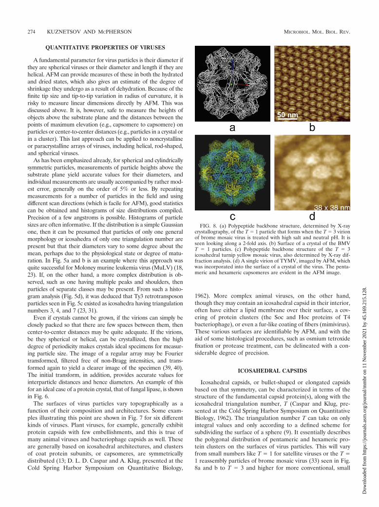
Icosahedral capsids, or bullet-shaped or elongated capsidsbased on that symmetry, can be characterized in terms of thestructure of the fundamental capsid protein(s), along with theicosahedral triangulation number, T (Caspar and Klug, pre-sented at the Cold Spring Harbor Symposium on QuantitativeBiology, 1962). The triangulation number T can take on onlyintegral values and only according to a defined scheme forsubdividing the surface of a sphere (9). It essentially describesthe polygonal distribution of pentameric and hexameric pro-tein clusters on the surfaces of virus particles. This will varyfrom small numbers like T � 1 for satellite viruses or the T �1 reassembly particles of brome mosaic virus (33) seen in Fig.8a and b to T � 3 and higher for more conventional, small
FIG. 8. (a) Polypeptide backbone structure, determined by X-raycrystallography, of the T � 1 particle that forms when the T � 3 virionof brome mosaic virus is treated with high salt and neutral pH. It isseen looking along a 2-fold axis. (b) Surface of a crystal of the BMVT � 1 particles. (c) Polypeptide backbone structure of the T � 3icosahedral turnip yellow mosaic virus, also determined by X-ray dif-fraction analysis. (d) A single virion of TYMV, imaged by AFM, whichwas incorporated into the surface of a crystal of the virus. The penta-meric and hexameric capsomeres are evident in the AFM image.
274 KUZNETSOV AND MCPHERSON MICROBIOL. MOL. BIOL. REV.
Dow
nloa
ded
from
http
s://j
ourn
als.
asm
.org
/jour
nal/m
mbr
on
11 N
ovem
ber
2021
by
45.1
69.2
15.1
28.

icosahedral viruses such as poliovirus or turnip yellow mosaicvirus, which is presented in Fig. 8c, to very large numbers forcomplex viruses such as the iridoviruses (Fig. 3d), algal viruses(Fig. 7f) like PBCV-1 (T � 169), and mimivirus (Fig. 7c).
In many cases the exterior shell of a virus may not be ico-sahedral, but it might possess an inner capsid that is. Forexample, though membrane covered and with a pleiomorphicexternal shape, herpes simplex virus possesses a nucleic acid-containing capsid of icosahedral form T � 16. Mimivirus (Fig.7c) exhibits a complex outer surface coated with a forest offibers, but it too contains an icosahedral core with a triangu-lation number of between T � 324 and 381 (48). The T num-ber, then, contains a good part of the information needed todescribe an icosahedral capsid.
The triangulation numbers of icosahedral viruses can fre-quently be deduced from AFM images. Examples were seen inFig. 7 and 8, but even more strikingly in the case of Ty3retrotransposons, a protoretrovirus, as illustrated by Fig. 9. Itwas found for Ty3 (31) that virions existed in three differentsizes and three different architectures corresponding to T num-bers 7 (the largest fraction of the population), 4, and 3 (thesmallest component). AFM images of the surface capsomeredistribution could be triangulated visually, as in Fig. 9, bydefining the arrangement of hexamers with respect to pentam-ers. A particularly good description of this approach is given by
Rayment (44), where it is illustrated for surface lattices of T �1, 3, 4, and 7, the four lattices with the lowest T number. TheT number was thus defined. Of particular interest were the T �7 particles, which could conceivably exist in either of twoenantiomorphs, d and l. Because height information is pre-served in AFM images, so is handedness. Thus, it was possiblein the case of the T � 7 Ty3 virions to determine that theiractual T number was 7d.
A somewhat different approach must be taken with verylarge icosahedral capsids, like those in Fig. 7c and f, whichinclude mimivirus and PBCV-1. This is achieved by determin-ing the two indices h and k, which define the triangulationnumber (T � h2 � hk � k2), by following a row of hexagonalcapsomeres from one pentagonal vertex to the next icosahedraledge and simply counting the number of capsomeres along oneedge h and the other k (the h and k coordinates of the inter-section point on the icosahedral edge) needed to traverse (8;Caspar and Klug, presented at the Cold Spring Harbor Sym-posium on Quantitative Biology, 1962; Viperdb.scripps.edu).Precisely this method was used to determine the T numbers ofthe iridovirus PBCV-1 (49) and the capsid architecture ofmimivirus (48).
While the T number describes the overall distribution ofcapsomeres on the surface of an icosahedral capsid, a morecomplete description of a virus structure would require the
FIG. 9. The icosahedral symmetry of the Ty3 retrotransposon, a protoretrovirus that infects yeast, was established by AFM. It was furthershown that Ty3 existed in three different icosahedral forms corresponding to T numbers of 3, 4, and 7. (A) The left panel shows a Ty3 particle,and the right panel shows the same particle with a T � 7 icosahedral net overlay. Pentagonal vertices are in red. The center diagram is that of atriangular face of an icosahedron showing positions of pentagonal and hexagonal capsomeres for T � 7. (B) Another, smaller particle of Ty3 andthe equivalent overlay and diagram showing it to have T � 4 icosahedral symmetry.
VOL. 75, 2011 AFM OF VIRUSES 275
Dow
nloa
ded
from
http
s://j
ourn
als.
asm
.org
/jour
nal/m
mbr
on
11 N
ovem
ber
2021
by
45.1
69.2
15.1
28.
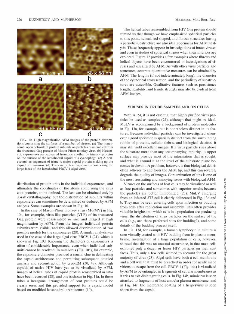
distribution of protein units in the individual capsomeres, andultimately the coordinates of the atoms comprising the viruscoat proteins, to be defined. The last can be obtained only byX-ray crystallography, but the distribution of subunits withincapsomeres can sometimes be determined or deduced by AFManalysis. Some examples are shown in Fig. 10.
In the case of Mason-Pfizer monkey virus (M-PMV) in Fig.10a, for example, virus-like particles (VLP) of its truncatedGag protein were reassembled in vitro and imaged at highmagnification by AFM. From the images, individual proteinsubunits were visible, and this allowed discrimination of twopossible models for the capsomeres (28). A similar analysis wasused in the case of the large algal virus PBCV-1 (21), which isshown in Fig. 10d. Knowing the diameters of capsomeres isoften of considerable importance, even when individual sub-units cannot be resolved. In mimivirus (Fig. 10c), for example,the capsomere diameter provided a crucial clue in delineatingthe capsid architecture and permitting subsequent detailedanalysis and reconstruction by cryo-EM (30, 48). Althoughcapsids of native HIV have yet to be visualized by AFM,images of helical tubes of capsid protein reassembled in vitrohave been recorded (28), and one is shown in Fig. 11a. In thesetubes a hexagonal arrangement of coat proteins could beclearly seen, and this provided support for a capsid modelbased on modified icosahedral architecture (10).
The helical tubes reassembled from HIV Gag protein shouldremind us that though we have emphasized spherical particlesto this point, helical, rod-shaped, and fibrous structures havinga periodic substructure are also ideal specimens for AFM anal-ysis. These frequently appear in investigations of intact virusesand even in studies of spherical viruses when their interiors areexplored. Figure 12 provides a few examples where fibrous andhelical objects have been encountered in investigations of vi-ruses and visualized by AFM. As with other virus particles andstructures, accurate quantitative measures can be obtained byAFM. The lengths (if not indeterminately long), the diameterof the cylindrical cross section, and the periodicity of substruc-tures are accessible. Qualitative features such as persistencelength, flexibility, and tensile strength may also be evident fromAFM images.
VIRUSES IN CRUDE SAMPLES AND ON CELLS
With AFM, it is not essential that highly purified virus par-ticles be used as samples (24), although that might be ideal.MuLV is accompanied by a background of protein moleculesin Fig. 13a, for example, but is nonetheless distinct in its fea-tures. Because individual particles can be investigated when-ever a good specimen is spatially distinct from the surroundingrubble of proteins, cellular debris, and biological detritus, itmay still yield excellent images. If a virus particle rises abovethe substrate more than any accompanying impurity, its uppersurface may provide most of the information that is sought,and what is around it at the level of the substrate plane be-comes irrelevant. A problem, however, is that biological debrisoften adheres to and fouls the AFM tip, and this can severelydegrade the quality of images. Contamination of tips is one ofthe most frustrating and annoying issues with biological AFM.
Viruses on the surfaces of host cells may be visualized as wellas free particles and sometimes with superior results becausethe particles are better immobilized (23). MuLV emergingfrom an infected 3T3 cell is clearly delineated in Fig. 13a andb. They may be seen entering cells upon infection or buddingfrom cells after replication and assembly. This often providesvaluable insights into which cells in a population are producingvirus, the distribution of virus particles on the surface of thecells (e.g., are there preferred sites for budding?), and somedetails of the budding process itself.
In Fig. 13d, for example, a human lymphocyte in culture isseen virtually coated with HIV budding from its plasma mem-brane. Investigation of a large population of cells, however,showed that this was an unusual occurrence, in that most cellsexhibited only a dozen or fewer HIV particles on their sur-faces. Thus, only a few cells seemed to account for the greatmajority of virus (23). Algal cells have both a cell membraneand a cell wall that must be breached in order for newly madevirions to escape from the cell. PBCV-1 (Fig. 14a) is visualizedby AFM to be entangled in fragments of cellular membranes asit tries to exit disintegrating cells. In Fig. 14b, mimivirus is seenattached to fragments of host amoeba plasma membrane, andin Fig. 14c, the membrane coating of a herpesvirus is seenshorn from the capsid.
FIG. 10. High-magnification AFM images of the protein distribu-tions comprising the surfaces of a number of viruses. (a) The honey-comb, open network of protein subunits on particles reassembled fromthe truncated Gag protein of Mason-Pfizer monkey virus. (b) Hexam-eric capsomeres are separated from one another by trimeric proteinson the surface of the icosahedral capsid of a cyanophage. (c) A hon-eycomb arrangement of trimeric major capsid protein making up thecapsid of mimivirus. (d) Trimeric protein capsomeres composing thelarge faces of the icosahedral PBCV-1 algal virus.
276 KUZNETSOV AND MCPHERSON MICROBIOL. MOL. BIOL. REV.
Dow
nloa
ded
from
http
s://j
ourn
als.
asm
.org
/jour
nal/m
mbr
on
11 N
ovem
ber
2021
by
45.1
69.2
15.1
28.

MUTANT VIRUSES
Mutant viruses, naturally occurring or produced in the lab-oratory, can be imaged as well as native virions and VLPcreated in vitro from capsid proteins. In some cases, the phe-notype of the mutant can be revealed by observing infectedhost cells for unique or anomalous features. This was done, asshown in Fig. 11d, in a study of Moloney murine leukemia virus(MuLV)-infected 3T3 cells, where the mutant gPr80gag lackedglycosylated Gag protein (19, 32, 42). Prior evidence suggestedthat such mutants failed in some stage of viral budding. Thiswas amply confirmed by AFM visualization of infected, virus-producing cells.
As seen in Fig. 11d, instead of normal, spherical virus emerg-ing from the cell surface, bullet- and comet-shaped protrusionswere found distributed over all of the plasma membrane ofhost cells. The comets were viruses that were trying to escapebut were unable to terminate association with the host cell.From this it was concluded that the failure of glycosylationproduced a defect in late stages of the budding process.
Other mutations in virus genomes may produce alterationsin external features of virus particles that are readily observ-able by AFM. MuLV particles that failed to make envelopeprotein (gp120 protein), one of which is seen in Fig. 11e, were
examined in another study (23). While normal particles, aswere seen in Fig. 4a and 13a and b, are characterized by acoating of protein tufts, about 100 to 150 in number, mutantparticles were “bald” virions lacking any such protein clusters.Instead, only an outer lipid membrane was visible. A study ofTy3 retrotransposon having a defective protease gene mutantand reverse transcriptase (RT) mutant Ty3 particles was illu-minating in a different way (31). While wild-type virus ap-peared as T � 3, 4, and 7 icosahedral particles, the RT and Prmutants appeared to be exclusively T � 7 particles. This sug-gested that T � 4 and T � 3 particle forms might representmature or late-stage virus forms. All T � 7 particles, no matterwhat their genome, exhibited the same general appearance.
SPECIALIZED FEATURES ON VIRUSES
Some viruses exhibit specialized external structures, or de-viations from their general architectures. A collection of thesedrawn from several different viruses and visualized by AFM areshown in Fig. 15. For example, the MuLV particles in Fig. 15agenerally have, somewhere on their otherwise uniformly cren-ulated surfaces, a single small bump or brief protrusion. Theseare likely to be “budding scars” resulting from breaking away
FIG. 11. Two in vitro, reassembled, virus-like particles produced in bacteria as imaged by AFM. (a) Helical tubes, a product of in vitroself-assembly of the Gag protein of human immunodeficiency virus. (b) Particles reassembled in vitro from a truncated form of the Gag proteinfrom Mason-Pfizer monkey virus. (c to f) Mutant and aberrant virus particles. (c) A mutant cyanophage that lacks any protein sheath about its tailassembly and fully exposes the injection tube normally found in its interior. (d) AFM image of 3T3 cells in culture that are infected with a mutantform (gPr80gag) of Moloney murine leukemia virus. The virus, upon budding, is unable to separate completely from the host cell membrane andforms long, comet-like extensions from the cell surface. (e) A mutant of Moloney murine leukemia virus that, genetically, lacks the capacity to makeenvelope protein. As a consequence, it appears as a “bald” virus that exposes its limiting lipid membrane to the exterior. The undulations andvariations of the membrane surface are an effect produced by local movement of the membrane in response to AFM tip pressure. (f) A crystal ofbrome mosaic virus. Though most of the crystal is composed of conformist T � 3 particles with the standard diameter of 30 nm, the arrows pointout the presence of two exceptionally small virions, probably T � 1 icosahedra lacking RNA, and one exceptionally large virion, probably T � 4or 7, likely containing multiple copies of RNA.
VOL. 75, 2011 AFM OF VIRUSES 277
Dow
nloa
ded
from
http
s://j
ourn
als.
asm
.org
/jour
nal/m
mbr
on
11 N
ovem
ber
2021
by
45.1
69.2
15.1
28.
from the host cell (18). Some MuLV particles, perhaps defec-tive, exhibited small sectors on their surfaces where proteinwas absent and a channel into the interior appeared (19).Other, more prominent features are the thick fibers on thesurfaces of PBCV-1 (22) and the lateral bodies of vacciniavirus, seen in Fig. 15c and f, respectively (17, 36).
PBCV-1, a large algal virus, exhibited a unique pentagonalassembly of proteins (21) at every 5-fold vertex of its icosahe-dral capsid. This is illustrated in Fig. 16a. The assembly had asingle protein in the center that could “push in” and “pull out”as demonstrated by the application of AFM tip pressure. Itsexact function is speculative. Many bacteriophages have tailassemblies of one sort or another for packaging and injectingtheir DNA. Mimivirus is, in a sense, similar to these phagesand has an assembly, seen in Fig. 16b, with a presumablysimilar function at a single unique, 5-fold vertex. This star-shaped structure is analogous to the tail assemblies of phagesand is, as is evident in those figures, a distinctive feature of thevirions. It is a complex structure, presumably composed ofmany proteins, and AFM reveals much of that complexity.
A point that deserves particular emphasis is that all of theparticles within a population of virus are not absolutely iden-tical, and often there are very significant differences in thedetailed features of individual particles. The degree of struc-tural diversity is evident in many of the AFM images presentedhere. This is a point often obscured by the results of X-raycrystallography or cryo-EM reconstructions. Those techniquesrely completely on the assumption of structural conformity.
They produce models that represent the average in time andspace for the individuals that make up the population. AFM,on the other hand, reveals the eccentricities and unique fea-tures of the individuals, and these are instructive. They oftendefine the extremes of what is structurally possible among alarge population of viruses having, presumably, the same ge-nome and the same environment for replication and assembly.What is seen with AFM is that anomalous and aberrant indi-viduals are not only present but common.
DISSECTION OF COMPLEX VIRUSES
It might be thought that because AFM provides images ofthe surfaces of objects and does not peer into their interiors, asdo X-ray diffraction and electron microscopy, AFM would beof little value in delineating the interior structure of viruses,i.e., the layers beneath the external surface. This is not true,however, as we can apply the same technique that has beenused by anatomists for centuries: dissection. With the aid ofchemical, enzymatic, and physical tools, we can systematicallypare a complex entity, including a virus, down to its core, layerby layer. At each stage, AFM may then be used to visualizewhat remains and what has been removed as well.
This approach is particularly effective with large, complexviruses such as vaccinia virus (17, 36), as illustrated in Fig. 17,or mimivirus (30, 48), as illustrated in Fig. 18. For these com-plicated assemblies, ordered and disordered protein shells,lipid membranes, and the nucleic acid within can be revealed
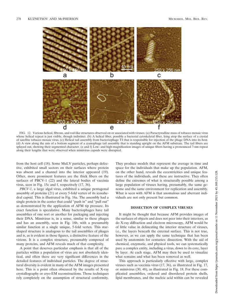
FIG. 12. Various helical, fibrous, and rod-like structures observed on or associated with viruses. (a) Paracrystalline mass of tobacco mosaic viruswhose helical repeat is just visible, though indistinct. (b) A helical fiber, possibly a bacterial cytoskeletal fiber, lying atop the surface of a crystalof satellite tobacco mosaic virus. (c) Helical tail assembly from bacteriophage T4 that is responsible for injection of the phage DNA into its host.(d) A view along the axis of a bottom segment of a cyanophage tail assembly that is standing upright on the AFM substrate. The tail fibers aresplayed out, showing their segmented character. (e and f) Low- and high-magnification images of unique fibers having a pronounced 7-nm repeatalong their lengths that were observed when mimivirus capsids were disrupted.
278 KUZNETSOV AND MCPHERSON MICROBIOL. MOL. BIOL. REV.
Dow
nloa
ded
from
http
s://j
ourn
als.
asm
.org
/jour
nal/m
mbr
on
11 N
ovem
ber
2021
by
45.1
69.2
15.1
28.

and analyzed. By deconstruction, the architecture of particlescan be revealed, and at the same time, the kinds of biochemicalinteractions that maintain each level of structure may be de-lineated as well.
Among the most useful agents for chemical dissection havebeen detergents, usually 0.5 to 2% of some nonionic detergentsuch as NP-40, and reducing agents such as dithiothreitol(DTT) or dithioerythritol (DTE). The former causes the pro-tein structure to gradually unravel, and detergents strip awaythe lipid membrane. The latter reduce disulfide bonds and
liberate polypeptides otherwise bonded to one another. Disul-fide bond reduction appears to be particularly important inlarge, complex viruses where such covalent linkages cross-linkcoat proteins and stabilize capsids (36, 47).
In some cases, nonionic detergents are insufficient to disruptstructure, and more vigorous ionic detergents such as SDSmust be used. There is difficulty with SDS, however. It tends tohave an all-or-none effect, so that upon reaching a concentra-tion sufficient to disrupt viruses, it completely degrades themuncontrollably.
The most effective enzymatic tools have been proteases thatdegrade polypeptides. These are particularly useful becausethey have a range of activities and a spectrum of specificities.As a consequence, a whole variety of proteases have beenemployed, and these include trypsin, bromelin, proteinase K,subtilisin, and mixtures of pancreatic proteases. Viruses areusually exposed to the proteases for anywhere from 15 min toseveral hours, or even overnight, at concentrations of from 0.5mg/ml to as high as 5 mg/ml. The proteases must be washedfrom the virions with buffer or water before imaging, as theyotherwise produce a dense, irregular background that makeimaging problematic, and they foul the cantilever tip.
Physical forces have also been used to disrupt viruses, andoften fortuitous perturbations, resulting simply from prepara-tion and handling, have proven to be structurally illuminating.Heat, for example, was used to open TYMV (26) to release itsencapsidated RNA, and direct physical pressure on mimivirussandwiched between two layers of atomically smooth, cleavedmica was used as well (30). Cycles of freezing and thawing havebeen reported to disrupt viruses in some cases, but to thispoint, freezing has not proven useful in AFM studies. Thereare also instances where “hammering” of individual particleswith the AFM tip has been utilized, taking advantage of thefact that AFM can serve as a tool as well as an imaging device.
In carrying out the dissection of a virus, or even in simplyvisualizing particles spread on a glass, plastic, or mica sub-strate, it is necessary to ensure that the virus particles adherefirmly to the substrate. Failure to do so allows the particles tomove in response to close approach of the AFM tip and ren-ders imaging impossible. Occasionally, altering the charge onthe substrate is sufficient. Mica is negatively charged on itssurface, but exposure to a nickel or magnesium salt such as
FIG. 13. (a) A cluster of Moloney murine leukemia virus buddingfrom the surface of an infected 3T3 cell. (b) A single Moloney murineleukemia virus particle emerging from the protein matrix characteriz-ing the external surface of a 3T3 cell. (c) Newly made cyanophagespilling from a lysed bacterium. (d) Cascade of human immunodefi-ciency virus budding from the surface of a heavily infected humanlymphocyte in culture.
FIG. 14. (a) Five shards of plasma membrane from a disrupted, virus-infected Chlorella cell form a flowerlike aggregate. Attached to themembrane fragments is PBCV-1. (b) Giant mimivirus is seen still attached to fragments of the host amoeba plasma membrane. (c) The white sheetof the membrane coating a herpes simplex virus has “splashed” to one side of the capsid and lies on the substrate plane.
VOL. 75, 2011 AFM OF VIRUSES 279
Dow
nloa
ded
from
http
s://j
ourn
als.
asm
.org
/jour
nal/m
mbr
on
11 N
ovem
ber
2021
by
45.1
69.2
15.1
28.

MgCl2 coats it with divalent ions and leaves it positivelycharged. Some viruses or macromolecules, such as nucleic ac-ids, if they are repelled by a negative surface, may then befirmly held by a positive surface or vice versa.
Charge is, however, frequently insufficient for virions wherethe contact area of the particle is relatively small. To fix mostviruses to a substrate, as well as a wide variety of other bio-logical entities and materials, an effective procedure is to coat
FIG. 15. Distinctive accessory features observed on the surfaces of viruses. (a) A budding scar (arrow), a small protrusion seen on most virionsof Moloney murine leukemia virus that is produced from infected 3T3 cells in culture. The bulges are thought to arise from pinching off the virusfrom the host cell membrane. (b) Helical tail assembly of a cyanophage. The tail fibers have been broken off, but at the head end of the sheathis a unique entity that has pulled from the head along with the tail. Denoted by an arrow and the letter P, this is the portal assembly responsiblefor drawing the phage DNA into the capsid. (c) A virion of PBCV-1 exhibiting several thick fibers, of unknown function, from its surface. (d)Typical example of the protein fibers, usually appearing like hairs from a basal tuft, that thickly coat the outer surface of mimivirus. The lightercolored material that shadows the two fibers is believed to be associated oligosaccharide. (e) A particle reassembled in vitro from truncated Gagprotein of Mason-Pfizer monkey virus. The particles invariably display a number of dislocations and defects on their surfaces. The defects allowthe particles rough sphericity without icosahedral or any other exact symmetry. (f) Air-dried vaccinia virus. Retraction of the membrane and overallshrinkage emphasize the lateral body at center. A unique feature of vaccinia virus is the presence of two such lateral bodies associated with eachvirion.
FIG. 16. Some additional external features that characterize the pentameric vertices of some large icosahedral viruses. (a) A specialized clusterof five proteins, with another protein at the center, that occupy the 12 unique vertices on the surface of PBCV-1. All of the proteins in these clustersare clearly different in structure from the normal capsid protein. (b) Image of the “stargate,” five-vane apparatus found at only a single pentamericvertex of mimivirus. The apparatus provides a mechanism for the release of the encapsidated DNA. (c) AFM image of adenovirus. A noteworthyfeature of adenovirus is that degradation of the virions invariably initiates and proceeds with loss of pentons at the 5-fold vertices, leaving particlesperforated in an icosahedrally symmetric manner.
280 KUZNETSOV AND MCPHERSON MICROBIOL. MOL. BIOL. REV.
Dow
nloa
ded
from
http
s://j
ourn
als.
asm
.org
/jour
nal/m
mbr
on
11 N
ovem
ber
2021
by
45.1
69.2
15.1
28.

the substrate with poly-L-lysine before depositing the virus.Presumably salt bridges between the ε amino groups of thelysines and the glutamic and aspartic acid carboxyl groups onthe particles lock them in place. After such substrate-particleattachment, the substrate can be rinsed with water severaltimes with acceptable loss of sample. The only serious disad-vantage of coating with poly-L-lysine is that it produces a ratherrough and irregular background. As a consequence, molecularobjects, such as lipid membranes or nucleic acids, which riseonly about a nanometer or two above the substrate plane,become difficult to identify and visualize. The method is excel-lent, however, for imaging cells and intact or partially degradedvirions.
It is occasionally unnecessary to actually treat viruses withany chemical or biochemical agents to explore the interior, asthe physical stress of preparation and especially structurallydelicate virus purification may result in damaged or partiallydegraded particles. These may expose internal structural fea-tures otherwise not accessible to the cantilever tip. Retrovi-ruses, in particular, are physically fragile. Some MuLVs, asshown in Fig. 19a, when subjected to the shear forces of evenlow-speed centrifugation lose portions of the shell surroundingthe capsid. This permits direct visualization of the virus corestill embedded within the layers of envelope and matrix protein(18).
HIV is another example where even the mildest procedures
produce some damaged virions. Although the cores of HIVhave not yet been visualized by AFM, likely due to their fra-gility, the remainder of the virus without the cores has been(29). An example can be seen in Fig. 19b. Such partially dis-robed particles, both MuLV and HIV, provide specimens thatcan be subjected to quantitative examination and thereby yieldthe thicknesses of internal layers of structure, and they givesome clues as to their components as well.
Good examples of complete dissections of complex virusesusing AFM are those of vaccinia virus, a poxvirus of about 300nm diameter that is delimited by a lipid membrane (17, 36),and mimivirus (30, 48). Vaccinia virus contains a double-stranded DNA genome bounded by several protein shells. Italso has associated with its inner core two unusual proteinassemblies of still-unknown function, known as lateral bodies.
Vaccinia virus was sequentially degraded with 0.5% NP-40nonionic detergent combined with 0.05 M DTT, followed byexposure to this same mixture but containing either trypsin orproteinase K or to the proteases alone. Four stages in thisprocess are presented in Fig. 17. In the end, the innermost corewas breached and the DNA was exposed. From images of theDNA emerging from the core, it was deduced that while some
FIG. 17. Four stages in the chemical and enzymatic dissection ofvaccinia virus. (a) An intact vaccinia virus virion. (b) The core of thevirus obtained by treatment with a nonionic detergent (NP-40) anddithiothreitol, a reducing agent. (c) The ghosts, or capsules, that re-main after the cores have been treated with proteases, which alsoproduces release of the viral nucleic acid. (d) Masses of DNA releasesonto the AFM substrate by disrupted viral cores. The arrow indicatesa disrupted vaccinia virus core adjacent to the DNA.
FIG. 18. Stages in the enzymatic dissection of mimivirus, whichinfects protozoans and is the largest virus known. (a) A mass of intactvirus still coated with glycoprotein fibers. The “starfish”-ornamentedvertex is visible on many particles in spite of the fiber coating. (b) Thefibrous coat has been removed by treatment with lysozyme and pro-teinase K, and the “starfish” apparatus has become more prominent.The geometric character of the underlying protein capsid is beginningto emerge. (c) The capsid has been further treated with proteases, andthe icosahedral architecture of the protein capsid and the distributionof capsomeres have become evident. (d) The capsid has beenbreached, and the DNA contained within is seen spilling onto the micasubstrate, which also contains many viral proteins released in theprocess.
VOL. 75, 2011 AFM OF VIRUSES 281
Dow
nloa
ded
from
http
s://j
ourn
als.
asm
.org
/jour
nal/m
mbr
on
11 N
ovem
ber
2021
by
45.1
69.2
15.1
28.
portion of the encapsidated DNA was heavily integrated withprotein, the vast majority was largely naked, with only occa-sional associations with protein. The giant mimivirus was sim-ilarly dissected using essentially the same approach. A sam-
pling of the results is presented in Fig. 18. Mimivirus, at 750 nmin diameter, was revealed to be a very complex particle withnumerous layers of structure.
VISUALIZATION OF VIRAL RNA AND DNA
The nucleic acids of viruses, from a structural standpoint,are of considerable interest, in particular with regard to howthey are condensed and packaged inside capsids and cores.Clearly, packaging is accomplished differently by specific fam-ilies of viruses. It is unlikely, for example, that bacteriophagesand poxviruses package their genomic double-stranded DNAsin the same way. The packing densities of the nucleic acid differby more than 10-fold (17). Nor is it likely that large, single-stranded-RNA-containing viruses, such as retroviruses, pack-age their genomes the same way as do T � 1 or T � 3icosahedral plant viruses (38). Certainly, helical and filamen-tous viruses use entirely different mechanisms.
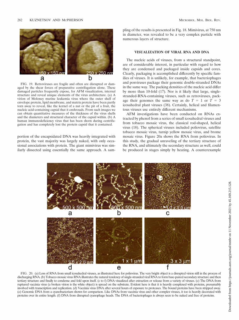
AFM investigations have been conducted on RNAs ex-tracted by phenol from a series of small icosahedral viruses andfrom tobacco mosaic virus, the classical rod-shaped, helicalvirus (18). The spherical viruses included poliovirus, satellitetobacco mosaic virus, turnip yellow mosaic virus, and bromemosaic virus. Figure 20a shows the RNA from poliovirus. Inthis study, the gradual unraveling of the tertiary structure ofthe RNA, and ultimately the secondary structure as well, couldbe produced in stages simply by heating. A counterexample
FIG. 19. Retroviruses are fragile and often are disrupted or dam-aged by the shear forces of preparative centrifugation alone. Thesedamaged particles frequently expose, for AFM visualization, internalstructure and reveal unique elements of the virus architecture. (a) Avirion of Moloney murine leukemia virus where the outer shell ofenvelope protein, lipid membrane, and matrix protein have been partlytorn away to reveal, like the kernel of a nut or the pit of a fruit, thenucleic acid-containing capsid that it enshrouds. From such images wecan obtain quantitative measures of the thickness of the virus shellsand the diameters and structural character of the capsid within. (b) Ahuman immunodeficiency virus that has been shorn during centrifu-gation and has completely lost the protein capsid that it contained.
FIG. 20. (a) Loss of RNA from small icosahedral viruses, as illustrated here for poliovirus. The very bright object is a disrupted virion still in the process ofdischarging RNA. (b) Tobacco mosaic virus RNA illustrates the natural tendency of single-stranded viral RNA to form base-paired secondary structure and thentertiary structure and finally to condense and fold upon itself. (c to f) DNA visualized after extraction or release from a variety of viruses. (c) The DNA fromruptured vaccinia virus (a broken virion is the white object) is spread on the substrate. Evident here is that it is heavily complexed with proteins, presumablyinvolved with transcription and replication. (d) Vaccinia virus DNA after several hours of exposure to proteases. The bound proteins have been stripped away.(e) Genomic DNA from a cyanobacterium shown for comparison. Like DNAs from vaccinia virus and other complex viruses, it too is heavily decorated withproteins over its entire length. (f) DNA from disrupted cyanophage heads. The DNA of bacteriophages is always seen to be naked and free of proteins.
282 KUZNETSOV AND MCPHERSON MICROBIOL. MOL. BIOL. REV.
Dow
nloa
ded
from
http
s://j
ourn
als.
asm
.org
/jour
nal/m
mbr
on
11 N
ovem
ber
2021
by
45.1
69.2
15.1
28.

was provided by tobacco mosaic virus RNA (Fig. 20b), whichappeared initially as a thread, a completely extended moleculelacking any secondary structure. With time, it began forminglocal secondary structural elements and eventually condensedinto forms similar to those seen for the RNAs from the icosa-hedral viruses. The upshot of the study was that the singlestrands of RNA spontaneously condensed as linear arrange-ments of stem-loop substructures following synthesis, that thecondensed RNA bound coat protein to it, and that the twocooperatively coalesced into the completed particle (20). Inthose studies, AFM proved itself an able technique for directlyvisualizing nucleic acid structure, demonstrating its fluidity andsuggesting the mechanisms by which it is encapsidated.
DNA and RNA have quite different appearances in AFMimages, and this can be seen by contrasting the images of RNAin Fig. 20a and b with the images of DNA from various viruses,as well as a bacterial cell, in Fig. 20c to f, which presents bothkinds of nucleic acid. DNA looks like strands and coils of stiffrope lacking any higher levels of structure; RNA appears ascomplicated, linear sequences of self-involved secondary struc-ture. Sometimes, however, the distinction is not entirely clear,and further evidence may be needed to show whether a fila-ment, strand, or complex is DNA or RNA.
A method was devised for additional identification based onexposure of the nucleic acid to high concentrations of bovineRNase A (27). One example of results from that study is shown
in Fig. 21. Intact RNA in Fig. 21c, as seen in Fig. 21d, washydrolyzed to small pieces by RNase A and left only fragmentson the substrate, presumably corresponding to protected stem-loops. DNA, seen in Fig. 21a, on the other hand, becamecoated with the protein, as shown in Fig. 21b, and the resultingstrands exhibited thicknesses two to three times that of nakeddouble-stranded DNA. Thus, it is possible with AFM to prac-tice a kind of crude histology.
IMMUNE LABELING WITH AFM
A second example of histological AFM is the immunolabel-ing of viruses with antibodies specific for certain proteins.Although individual IgGs are not clearly identifiable by AFMwhen bound to a virion, IgGs conjugated with gold particlesgenerally are. In a sense, these are used in the same way as theyare used in transmission electron microscopy immunolabeling,except that instead of visualizing points of high electron den-sity, one images with AFM objects having the size and shape ofthe immunogold particles.
Using gold-IgG conjugate particles against the envelopeprotein, as shown in Fig. 21e and f, it was possible to show thatprotein tufts on the surfaces of MuLV were indeed envelopeprotein (gp120) (19, 23). The major problem to this point withIgG-gold conjugates is that their physical size limits the reso-
FIG. 21. Consequences of treatment of viral nucleic acids with RNase A, an enzyme that binds to both RNA and DNA but degrades only RNA.(a) Two crossing double-stranded helices of DNA before treatment. (b) The same two strands after exposure to a high concentration of RNaseA. The protein completely coats the DNA and increases the thickness of the strands from about 1.5 nm (dry) above the substrate to about 3.5 nmabove the substrate. (c) Single-stranded RNA from poliovirus, showing its many secondary structural domains sequentially arranged along itslength. (d) After treatment with RNase A, only short, disconnected secondary structural domains remain, apparently resistant to attack by virtueof conformational protection. (e and f) Images of two different Moloney murine leukemia viruses decorated with 15-nm-diameter gold particlescoated with antibody whose antigen is the envelope protein of the virus. The arrows indicate the gold particles. The protein-tufted virions are seenbeneath.
VOL. 75, 2011 AFM OF VIRUSES 283
Dow
nloa
ded
from
http
s://j
ourn
als.
asm
.org
/jour
nal/m
mbr
on
11 N
ovem
ber
2021
by
45.1
69.2
15.1
28.
lution of the method. Conjugated gold particles can bind onlyas close as their diameters allow.
WHAT DOES AFM OFFER THESTRUCTURAL BIOLOGIST?
What can AFM visualize that is of value to structural virol-ogy? The answer is almost everything, and at resolutions thatapproach and in some cases surpass those with electron mi-croscopy. At this time, lipid membranes have been identified,both RNA and DNA have been visualized, and large proteinassemblies have been resolved into their component substruc-tures. The capsids of icosahedral viruses and the icosahedralcapsids of nonicosahedral viruses have been seen at high res-olution, in some cases sufficiently high to deduce the arrange-ment of coat protein units in the capsomeres, as well as deter-mine the triangulation number T.
In addition, viruses have been recorded budding from in-fected cells and suffering the consequences of a variety ofstresses. Mutant viruses have been examined and phenotypesdescribed. Unusual structural features have appeared, and veryimportantly, the unexpectedly great amount of structural non-conformity within populations of virus particles has been am-ply documented.
Virus samples may be imaged in air or in fluids having arange of viscosities, including culture medium or buffer, in situon living cell surfaces, or after processing by histological pro-cedures. AFM is nonintrusive and nondestructive, unless theintention is otherwise, and it can be applied to soft biologicalsamples, particularly when the tapping mode is employed. Inprinciple, only a single cell or virion need be imaged to learn ofits structure, though normally images of as many as is practicalare collected. While lateral resolution, limited by the finite sizeof the AFM cantilever tip, is a few nanometers, the heightresolution is exceptional, approximately 0.5 nm. Finally, AFMproduces three-dimensional, topographical images that accu-rately depict the surface features of the object, virus, or cellunder study. In most ways, the images resemble common lightphotographic images and require little interpretation, only un-derstanding.
It has been shown that the structures of viruses observed byAFM are entirely consistent with models derived by X-raycrystallography and cryo-EM. AFM has also been proven ef-fective in visualizing viruses in situ emerging from infected,living cells. Although there are currently no examples, there iscertainly no reason why structural information derived fromX-ray crystallography and/or electron microscopy cannot becombined with AFM images, just as it has been for those lattertwo technologies.
New techniques, reagents, and methods that will furtherenhance the value of AFM to structural biologists are currentlyunder development (3). Finally, the resolution of the technol-ogy and the unique insights into virus structure that it yieldswill be highly influenced by advances in tip acuity and improve-ments in instrument design. A great deal has yet to be done indeveloping sample preparation techniques, methods for chem-ically modifying cantilever tips, and expanding the use of his-tological procedures appropriate to AFM.
REFERENCES
1. Adrian, M., J. Dubochet, J. Lepault, and A. W. McDowall. 1984. Cryoelec-tron microscopy of viruses. Nature 308:32–36.
2. Allen, S., M. C. Davies, C. J. Roberts, S. J. B. Tendler, and P. M. Williams.1997. Atomic force microscopy in analytical biotechnology. Trends Biotech-nol. 15:101–105.
3. Baclayon, M., G. J. L. Wuite, and W. H. Roos. 2010. Imaging and manipu-lation of single viruses by atomic force microscopy. Soft Matter 6:5273–5285.
4. Baker, T. S., N. H. Olson, and S. D. Fuller. 1999. Adding the third dimensionto virus life cycles: three-dimensional reconstruction of icosahedral virusesfrom cryo-electron micrographs. Microbiol. Mol. Biol. Rev. 63:862–922.
5. Binnig, G., and C. F. Quate. 1986. Atomic force microscope. Phys. Rev. Lett.56:930–933.
6. Bustamante, C., and D. Keller. 1995. Scanning force microscopy in biology.Phys. Today 48:32–38.
7. Carragher, B., C. S. Potter, and F. J. Sigworth. 2007. Software tools formacromolecular microscopy. J. Struct. Biol. 157:1–288.
8. Carrillo-Tripp, M., et al. 2009. VIPERdb2: an enhanced and web APIenabled relational database for structural virology. Nucleic Acids Res. 37:D436–D442.
9. Caspar, D. L. D. 1965. Viral and rickettsial infections of man. Lippincott,New York, NY.
10. Ganser, B. K., S. Li, V. Y. Klishko, J. T. Finch, and W. I. Sundquist. 1999.Assembly and analysis of conical models for the HIV-1 core. Science 283:80–82.
11. Goldsbury, C. S., S. Scheuring, and L. Kreplak. 2009. Introduction to atomicforce microscopy (AFM) in biology. Curr. Protoc. Protein Sci. 17.7:1–19.
12. Hansma, P. K. 1994. Tapping mode atomic force microscopy in liquids.Appl. Phys. Lett. 64:1738–1740.
13. Horne, R. W., and P. Wildy. 1961. Symmetry in virus architecture. Virology15:348–373.
14. Horne, R. W., and P. Wildy. 1963. Virus structure revealed by negativestaining. Adv. Virus Res. 10:101–170.
15. Jena, B. P., and J. K. Horber (ed.). 2002. Atomic force microscopy in cellbiology. Methods Cell Biol. 68:1–415.
16. Kaper, J. M. 1975. The chemical basis of virus structure, dissociation andreassembly. Front. Biol. Ser. 39:1–185.
17. Kuznetsov, Y., P. D. Gershon, and A. McPherson. 2008. Atomic force mi-croscopy investigation of vaccinia virus structure. J. Virol. 85:7551–7566.
18. Kuznetsov, Y. G., S. Daijogo, J. Zhou, B. L. Semler, and A. McPherson. 2005.Atomic force microscopy analysis of icosahedral virus RNA. J. Mol. Biol.347:41–52.
19. Kuznetsov, Y. G., et al. 2002. Atomic force microscopy investigation offibroblasts infected with wild-type and mutant murine leukemia virus(MuLV). Biophys. J. 83:3665–3674.
20. Kuznetsov, Y. G., J. J. Dowell, J. A. Gavira, J. D. Ng, and A. McPherson.2010. Biophysical and atomic force microscopy characterization of the RNAfrom satellite tobacco mosaic virus. Nucleic Acids Res. 38:8284–8294.
21. Kuznetsov, Y. G., J. R. Gurnon, J. L. Van Etten, and A. McPherson. 2005.Atomic force microscopy investigation of a chlorella virus, PBCV-1. J. Struct.Biol. 149:256–263.
22. Kuznetsov, Y. G., A. Low, H. Fan, and A. McPherson. 2005. Atomic forcemicroscopy investigation of isolated virions of murine leukemia virus. J. Vi-rol. 79:1970–1974.
23. Kuznetsov, Y. G., A. Low, H. Fan, and A. McPherson. 2004. Atomic forcemicroscopy investigation of wild-type Moloney murine leukemia virus par-ticles and virus particles lacking the envelope protein. Virology 323:189–196.
24. Kuznetsov, Y. G., A. J. Malkin, R. W. Lucas, M. Plomp, and A. McPherson.2001. Imaging of viruses by atomic force microscopy. J. Gen. Virol. 82:2025–2034.
25. Kuznetsov, Y. G., A. J. Malkin, and A. McPherson. 1997. Atomic forcemicroscopy studies of living cells: visualization of motility, division, aggrega-tion, transformation, and apoptosis. J. Struct. Biol. 120:180–191.
26. Kuznetsov, Y. G., and A. McPherson. 2006. Atomic force microscopy inves-tigation of turnip yellow mosaic virus capsid disruption and RNA extrusion.Virology 352:329–337.
27. Kuznetsov, Y. G., and A. McPherson. 2006. Identification of DNA and RNAfrom retroviruses using ribonuclease A. Scanning 28:278–281.
28. Kuznetsov, Y. G., P. Ulbrich, S. Haubova, T. Ruml, and A. McPherson. 2007.Atomic force microscopy investigation of Mason-Pfizer monkey virus andhuman immunodeficiency virus type 1 reassembled particles. Virology 360:434–446.
29. Kuznetsov, Y. G., J. G. Victoria, W. E. Robinson, Jr., and A. McPherson.2003. Atomic force microscopy investigation of human immunodeficiencyvirus (HIV) and HIV-infected lymphocytes. J. Virol. 77:11896–11909.
30. Kuznetsov, Y. G., et al. 2010. Atomic force microscopy investigation of thegiant mimivirus. Virology 404:127–137.
31. Kuznetsov, Y. G., M. Zhang, T. M. Menees, A. McPherson, and S. Sand-meyer. 2005. Investigation by atomic force microscopy of the structure of Ty3retrotransposon particles. J. Virol. 79:8032–8045.
32. Low, A., et al. 2007. Mutation in the glycosylated gag protein of murine
284 KUZNETSOV AND MCPHERSON MICROBIOL. MOL. BIOL. REV.
Dow
nloa
ded
from
http
s://j
ourn
als.
asm
.org
/jour
nal/m
mbr
on
11 N
ovem
ber
2021
by
45.1
69.2
15.1
28.
leukemia virus results in reduced in vivo infectivity and a novel defect in viralbudding or release. J. Virol. 81:3685–3692.
33. Lucas, R. W., Y. G. Kuznetsov, S. B. Larson, and A. McPherson. 2001.Crystallization of brome mosaic virus (BMV) and T�1 brome mosaic virusparticles following a structural transition. Virology 286:290–303.
34. Malkin, A. J., Y. G. Kuznetsov, R. W. Lucas, and A. McPherson. 1999.Surface processes in the crystallization of turnip yellow mosaic virus visual-ized by atomic force microscopy. J. Struct. Biol. 127:35–43.
35. Malkin, A. J., T. A. Land, Y. G. Kuznetsov, A. McPherson, and J. J. DeYoreo.1995. Investigation of virus crystal growth mechanisms by in situ atomic forcemicroscopy. Phys. Rev. Lett. 75:2778–2781.
36. Malkin, A. J., A. McPherson, and P. D. Gershon. 2003. Structure of intra-cellular mature vaccinia virus visualized by in situ AFM. J. Virol. 77:6332–6340.
37. Malkin, A. J., M. Plomp, and A. McPherson. 2002. Application of atomicforce microscopy to studies of surface processes in virus crystallization andstructural biology. Acta Crystallogr. 58:1617–1621.
38. McPherson, A. 2005. Micelle formation and crystallization as paradigms forvirus assembly. Bioessays 27:447–458.
39. McPherson, A., Y. G. Kuznetsov, A. J. Malkin, and M. Plomp. 2004. Mac-romolecular crystal growth investigations using atomic force microscopy. J.Synchrotron Radiat. 11:21–23.
40. McPherson, A., A. J. Malkin, Y. G. Kuznetsov, and M. Plomp. 2001. Atomic
force microscopy applications in macromolecular crystallography. Acta Crys-tallogr. D 57:1053–1060.
41. Morris, V. J., A. R. Kirby, and A. P. Gunning. 2010. Atomic force microscopyfor biologists, 2nd ed. Imperial College Press, London, United Kingdom.
42. Nitta, T., Y. Kuznetsov, A. McPherson, and H. Fan. 2010. Murine leukemiavirus glycosylated Gag (gPr80gag) facilitates interferon-sensitive virus re-lease through lipid rafts. Proc. Natl. Acad. Sci. U. S. A. 107:1190–1195.
43. Plomp, M., M. K. Rice, E. K. Wagner, A. McPherson, and A. J. Malkin. 2002.Rapid visualization at high resolution of pathogens by atomic force micros-copy: structural studies of herpes simplex virus-1. Am. J. Pathol. 160:1959–1966.
44. Rayment, I. 1984. Animal virus structure, vol. 1. Wiley Interscience, NewYork, NY.
45. Rossmann, M. G. 2000. Fitting atomic models into electron-microscopymaps. Acta Crystallogr. 56:1341–1349.
46. Rossmann, M. G., R. Bernal, and S. V. Pletnev. 2001. Combining electronmicroscopic with X-ray crystallographic structures. J. Struct. Biol. 136:190–200.
47. Wikoff, W. R., et al. 2000. Topologically linked protein rings in the bacte-riophage HK97 capsid. Science 289:2129–2133.
48. Xiao, C., et al. 2009. Structural studies of the giant mimivirus. PLoS Biol.7:958–966.
49. Yan, X., et al. 2000. Structure and assembly of large lipid-containing dsDNAviruses. Nat. Struct. Biol. 7:101–103.
VOL. 75, 2011 AFM OF VIRUSES 285
Dow
nloa
ded
from
http
s://j
ourn
als.
asm
.org
/jour
nal/m
mbr
on
11 N
ovem
ber
2021
by
45.1
69.2
15.1
28.