
ASTHMA Inflammation of bronchial smooth muscle in allergic ...
8
ASTHMA Inflammation of bronchial smooth muscle in allergic asthma H Begueret, P Berger, J-M Vernejoux, L Dubuisson, R Marthan, J M Tunon-de-Lara ................................................................................................................................... See end of article for authors’ affiliations ........................ Correspondence to: Dr J M Tunon-de-Lara, Laboratoire de Physiologie Cellulaire Respiratoire, INSERM E356 Universite ´ Bordeaux 2, 146 rue Le ´o Saignat, 33076 Bordeaux, France; manuel. tunondelara@ u-bordeaux2.fr Received 9 March 2006 Accepted 14 July 2006 ........................ Thorax 2007;62:8–15. doi: 10.1136/thx.2006.062141 Background: Recent observations in asthma suggest that bronchial smooth muscle is infiltrated by inflammatory cells including mast cells. Such an infiltration may contribute to airway remodelling that is partly due to an increase in smooth muscle mass. Whether muscle increase is the result of smooth muscle cell hypertrophy remains controversial and has not been studied by ultrastructural analysis. A morphometric analysis of airway smooth muscle (ASM) was undertaken in asthmatic patients using electron microscopy to examine the interactions between ASM cells and inflammatory cells. Methods: ASM specimens were obtained from 14 asthmatic subjects and nine non-asthmatic controls undergoing fibreoptic endoscopy. Inflammatory cell counts were assessed by immunohistochemistry, and ultrastructural parameters were measured using electron microscopy in a blinded fashion on smooth muscle cells and inflammatory cells. Results: ASM from asthmatic patients was infiltrated by an increased number of mast cells and lymphocytes. Smooth muscle cells and their basal lamina were thicker in asthmatic patients (9.5 (0.8) and 1.4 (0.2) mm) than in controls (6.7 (0.4) and 0.7 (0.1) mm). In asthmatics the extracellular matrix was frequently organised in large amounts between ASM cells. Myofibroblasts within smooth muscle bundles were only observed in asthmatics, some of them displaying a close contact with ASM cells. Conclusion: In asthma, airway myositis is characterised by a direct interaction between ASM cells and mast cells and lymphocytes. Smooth muscle remodelling was present, including cell hypertrophy and abnormal extracellular matrix deposition moulding ASM cells. A sthma is an inflammatory disease characterised by bronchial hyperresponsiveness and infiltration of airway mucosa by several cell types including eosinophils and activated mast cell. Several studies have previously reported such an inflammatory response within the airway mucosa and fewer reports have provided ultrastructural data. 1–7 Until recently, however, the study of inflammation was limited to the submucosa whereas the airway smooth muscle (ASM) was strictly considered as an effector that contracts and/or proliferates in response to inflammatory cell products. It has recently been clearly demonstrated that mast cells also infiltrate the smooth muscle layer and that the number of mast cells infiltrating the smooth muscle is closely related to hyperresponsiveness. 8 The mechanism of such mast cell infiltration involves the secretion of chemotactic factors by the ASM cells itself, including transforming growth factor b 1 , 9 CXCL10 (IP10), 10 and CX 3 CL1 (fractalkine). 11 Mast cells can produce a variety of lipid mediators, proteases, and cytokines that may interact with ASM cells, and in vitro studies have shown that mast cell products induce both bronchial hyperre- sponsiveness and ASM cell proliferation. 12 13 Other reports have suggested that T lymphocytes may also interact with ASM cells, and ligands involved in T cell activation have been detected at the surface of the ASM cells obtained from asthmatic tissues. 14 15 Interactions between monocytes and ASM cells have also been studied in vitro and may influence tissue remodelling. 16 Taken together, these findings suggest that, in asthma, the ASM is also infiltrated by a range of inflammatory cells that may directly interact with ASM cells and influence their function. However, a direct cell-cell interaction has not yet been confirmed within the ASM in asthma. Finally, because of difficulties in obtaining proper biopsy specimens, little is known about the ultrastructure of the ASM in asthma or the characteristics of inflammatory cell interactions with ASM cells. Using electron transmission microscopy and morphometric analysis, we provide evidence for the first time that, in asthmatic patients, ASM cells display particular features of remodelling associated with direct interactions with mast cells, lymphocytes, and myofibroblasts. METHODS Tissue specimens Sixteen subjects with asthma and 10 non-asthmatic controls were prospectively recruited to the study. All gave their written informed consent to participate and underwent fibreoptic bronchoscopy. The study received the approval from the local ethics committee (Comite´ Consultatif pour la Protection des Personnes en Recherche Biome´dicale). Asthma was defined according to ATS criteria and hyperre- sponsiveness was confirmed either by a significant reversibility (.15% of baseline forced expiratory volume in 1 second (FEV 1 )) or a positive methacholine challenge (concentration provoking a fall in FEV 1 of 20% or more (PC 20 ) ,4 mg/ml). Asthmatic patients were non-smokers and atopy was defined according to EAACI. 17 Control subjects were non-atopic, non-asthmatic patients with no respiratory history who were undergoing a fibreoptic fibroscopy because of haemoptysis or an abnormal image on the chest radiograph. They were asymptomatic, had no evidence of airway obstruction, and a PC 20 for methacholine .16 mg/ml. Only subjects having a normal fibreoptic investi- gation and a normal bronchial mucosa were selected as controls. All control subjects were currently non-smokers or had stopped smoking for more than 3 years with a smoking history of less than 10 pack-years. The characteristics of the study subjects are shown in table 1. Abbreviations: ASM, airway smooth muscle; FEV 1 , forced expiratory volume in 1 second 8 www.thoraxjnl.com on October 21, 2021 by guest. Protected by copyright. http://thorax.bmj.com/ Thorax: first published as 10.1136/thx.2006.062141 on 22 December 2006. Downloaded from
Transcript of ASTHMA Inflammation of bronchial smooth muscle in allergic ...
ASTHMA
Inflammation of bronchial smooth muscle in allergic asthmaH Begueret, P Berger, J-M Vernejoux, L Dubuisson, R Marthan, J M Tunon-de-Lara. . . . . . . . . . . . . . . . . . . . . . . . . . . . . . . . . . . . . . . . . . . . . . . . . . . . . . . . . . . . . . . . . . . . . . . . . . . . . . . . . . . . . . . . . . . . . . . . . . . . . . . . . . . . . . . . . . . . . . . . . . . . . . . . . . .
See end of article forauthors’ affiliations. . . . . . . . . . . . . . . . . . . . . . . .
Correspondence to:Dr J M Tunon-de-Lara,Laboratoire de PhysiologieCellulaire Respiratoire,INSERM E356 UniversiteBordeaux 2, 146 rue LeoSaignat, 33076 Bordeaux,France; [email protected]
Received 9 March 2006Accepted 14 July 2006. . . . . . . . . . . . . . . . . . . . . . . .
Thorax 2007;62:8–15. doi: 10.1136/thx.2006.062141
Background: Recent observations in asthma suggest that bronchial smooth muscle is infiltrated byinflammatory cells including mast cells. Such an infiltration may contribute to airway remodelling that is partlydue to an increase in smooth muscle mass. Whether muscle increase is the result of smooth muscle cellhypertrophy remains controversial and has not been studied by ultrastructural analysis. A morphometricanalysis of airway smooth muscle (ASM) was undertaken in asthmatic patients using electron microscopy toexamine the interactions between ASM cells and inflammatory cells.Methods: ASM specimens were obtained from 14 asthmatic subjects and nine non-asthmatic controlsundergoing fibreoptic endoscopy. Inflammatory cell counts were assessed by immunohistochemistry, andultrastructural parameters were measured using electron microscopy in a blinded fashion on smooth musclecells and inflammatory cells.Results: ASM from asthmatic patients was infiltrated by an increased number of mast cells and lymphocytes.Smooth muscle cells and their basal lamina were thicker in asthmatic patients (9.5 (0.8) and 1.4 (0.2) mm)than in controls (6.7 (0.4) and 0.7 (0.1) mm). In asthmatics the extracellular matrix was frequently organisedin large amounts between ASM cells. Myofibroblasts within smooth muscle bundles were only observed inasthmatics, some of them displaying a close contact with ASM cells.Conclusion: In asthma, airway myositis is characterised by a direct interaction between ASM cells and mastcells and lymphocytes. Smooth muscle remodelling was present, including cell hypertrophy and abnormalextracellular matrix deposition moulding ASM cells.
Asthma is an inflammatory disease characterised bybronchial hyperresponsiveness and infiltration of airwaymucosa by several cell types including eosinophils and
activated mast cell. Several studies have previously reportedsuch an inflammatory response within the airway mucosa andfewer reports have provided ultrastructural data.1–7 Untilrecently, however, the study of inflammation was limited tothe submucosa whereas the airway smooth muscle (ASM) wasstrictly considered as an effector that contracts and/orproliferates in response to inflammatory cell products.
It has recently been clearly demonstrated that mast cells alsoinfiltrate the smooth muscle layer and that the number of mastcells infiltrating the smooth muscle is closely related tohyperresponsiveness.8 The mechanism of such mast cellinfiltration involves the secretion of chemotactic factors bythe ASM cells itself, including transforming growth factor b1,9
CXCL10 (IP10),10 and CX3CL1 (fractalkine).11 Mast cells canproduce a variety of lipid mediators, proteases, and cytokinesthat may interact with ASM cells, and in vitro studies haveshown that mast cell products induce both bronchial hyperre-sponsiveness and ASM cell proliferation.12 13 Other reports havesuggested that T lymphocytes may also interact with ASM cells,and ligands involved in T cell activation have been detected atthe surface of the ASM cells obtained from asthmatictissues.14 15 Interactions between monocytes and ASM cellshave also been studied in vitro and may influence tissueremodelling.16 Taken together, these findings suggest that, inasthma, the ASM is also infiltrated by a range of inflammatorycells that may directly interact with ASM cells and influencetheir function. However, a direct cell-cell interaction has not yetbeen confirmed within the ASM in asthma. Finally, because ofdifficulties in obtaining proper biopsy specimens, little isknown about the ultrastructure of the ASM in asthma or thecharacteristics of inflammatory cell interactions with ASMcells.
Using electron transmission microscopy and morphometricanalysis, we provide evidence for the first time that, inasthmatic patients, ASM cells display particular features ofremodelling associated with direct interactions with mast cells,lymphocytes, and myofibroblasts.
METHODSTissue specimensSixteen subjects with asthma and 10 non-asthmatic controlswere prospectively recruited to the study. All gave their writteninformed consent to participate and underwent fibreopticbronchoscopy. The study received the approval from the localethics committee (Comite Consultatif pour la Protection desPersonnes en Recherche Biomedicale).
Asthma was defined according to ATS criteria and hyperre-sponsiveness was confirmed either by a significant reversibility(.15% of baseline forced expiratory volume in 1 second(FEV1)) or a positive methacholine challenge (concentrationprovoking a fall in FEV1 of 20% or more (PC20) ,4 mg/ml).Asthmatic patients were non-smokers and atopy was definedaccording to EAACI.17
Control subjects were non-atopic, non-asthmatic patientswith no respiratory history who were undergoing a fibreopticfibroscopy because of haemoptysis or an abnormal image onthe chest radiograph. They were asymptomatic, had noevidence of airway obstruction, and a PC20 for methacholine.16 mg/ml. Only subjects having a normal fibreoptic investi-gation and a normal bronchial mucosa were selected ascontrols. All control subjects were currently non-smokers orhad stopped smoking for more than 3 years with a smokinghistory of less than 10 pack-years. The characteristics of thestudy subjects are shown in table 1.
Abbreviations: ASM, airway smooth muscle; FEV1, forced expiratoryvolume in 1 second
8
www.thoraxjnl.com
on October 21, 2021 by guest. P
rotected by copyright.http://thorax.bm
j.com/
Thorax: first published as 10.1136/thx.2006.062141 on 22 D
ecember 2006. D
ownloaded from
Assessable ASM was identified by a pathologist in a blindedfashion using both morphological characteristics and smoothmuscle a-actin staining. There was assessable ASM in thebiopsy specimens from 14 asthmatics and nine controls. Amean of 10 serial sections per biopsy were examined for eachsubject.
ImmunohistochemistrySpecimens were embedded in glycol methacrylate and pro-cessed as previously described.18 Primary antibodies includedmouse anti-human c-kit (CD117, Dako), anti-tryptase (AA1,Dako), anti-CD3 (Dako), anti-CD68 (Dako), anti-neutrophilelastase (NE, Dako), EG2 antibody against the cleaved form ofeosinophil cationic protein (Pharmacia), anti-Ki-67 (MIB-1,Dako), or the appropriate unrelated antibody. The number ofpositive cells was automatically assessed by Quancoul software(Bordeaux, France) at a magnification of 6200, but only cellsinfiltrating the smooth muscle layer were taken into account bythe pathologist.19 Cell counts were expressed as number of cells/mm2 smooth muscle. The thickness of the subepithelialmembrane was measured 10 times for each subject. Epithelialintegrity, defined as the percentage of length of basementmembrane with intact epithelium and the total area of smoothmuscle layer were assessed20 21 manually in a blinded fashion bya pathologist using Quancoul software at a magnification of6200. The smooth muscle area was normalised to the wholearea of the corresponding biopsy and presented as a percentageof the whole area. The number of ASM cell nuclei was alsoassessed manually by a pathologist at a magnification of 6400and normalised by the surface of the ASM layer.
Electron microscopyBiopsies were fixed in 2.5% glutaraldehyde in cacodylate buffer,post-fixed in 1% osmium tetroxide, dehydrated, and embeddedin Epon. For each biopsy specimen, semi-thin sections (1 mmthick) were cut and stained with alkaline toluidine blue. Thefirst semi-thin section large enough to span from theepithelium to the muscular layer was selected. Ten ultrathinserial sections (60 nm thick) were then cut on diamond knives.Three of these latter sections were subsequently randomlyselected and placed on grids. Staining was performed withuracile acetate and lead citrate. Grids were then scanned bytransmission electron microscopy (EM, Tecnai 12, Philips) and
examined by a pathologist from left to right and from top tobottom to locate every whole nucleated ASM cell andinflammatory cell. Each ultrathin section was examined in itsentirety.
We evaluated the mean width of both the nucleus and wholecell measured on sections at the nucleus level from 10measurements per section. For these measurements, the withinsubject variability was assessed by the coefficient of error (apercentage of standard error of the mean divided by the mean).Each ASM cell is surrounded by an external lamina or basalmembrane that is well identified using electron microscopy. Weassessed both the mean thickness of the basal membrane from10 measurements per section and the mean distance betweentwo ASM cell basal membranes from 15 measurements persection.
Mast cells were located either in the submucosa or within theASM layer—that is, muscular mast cells surrounded by ASMcells. Mast cells identified by electron microscopy were furthersubdivided into normal cells with intact electron dense granulesor degranulated cells either by piecemeal or anaphylacticdegranulation. Piecemeal degranulation was characterised bythe presence of variable losses of dense contents fromgranules.4 22 Anaphylactic degranulation was characterised byextrusion of membrane-free granules into newly formeddegranulation channels in the cytoplasm or through pores inthe plasma membrane to the exterior environment.4 22 Bothmechanisms of mast cell degranulation involved one to allgranules (partially to totally degranulated mast cells). Aspreviously described, the variable ultrastructural patterns ofmast cell granules were classified as cored (homogeneouslydense granules almost impenetrably black), scrolled (scrolledmatrix free of particulate material), particulated (granulecontent varying from finely particulate to stippled or ropey),or mixed (mixtures of these patterns).4 22 Myofibroblasts wereidentified according to morphological criteria described byEyden.23 Specific ultrastructural characteristics are the presenceof fibronexus defined as the cell surface point of convergence ofintracellular myofilaments and extracellular fibronectin fila-ments, cell surface component referred to as basementmembrane-like material, and poorly developed smooth musclemyofilaments.
Computerised photographs and measurements were per-formed in a blinded fashion using the Analysis Soft Imaging
Table 1 Characteristics of study subjects
Asthmatics(n = 14)
Controls(n = 9) p value
Asthma severityGINA 1 (n) 2 0GINA 2 (n) 2 0GINA 3 (n) 2 0GINA 4 (n) 8 0
Asthma history (years)* 25.8 (4.2 ) 0.0 (0.0 )Current treatment�
SABA (n) 14 0ICS (n) 10 0LABA (n) 4 0Pred (n) 2 0
Sex (M/F) 5/9 6/3Age (years)* 43.7 (4.7 ) 47.8 (5.4 ) 0.40Atopy Yes NoFEV1 (%)* 84.7 (7.2) 95.1 (3.8) 0.34FVC (%)* 100.4 (5.6) 96.6 (3.3) 0.70
*Values are mean (SE).�Treatments are short acting b2 agonist (SABA), inhaled corticosteroid (ICS), long acting b2 agonist (LABA), or oralprednisolone 20 mg/day (Pred).GINA, Global Initiative for Asthma. FEV1, forced expiratory volume in 1 second; FVC, forced vital capacity.p values from one way analyses of variance tests are indicated.
Inflammation of bronchial smooth muscle in allergic asthma 9
www.thoraxjnl.com
on October 21, 2021 by guest. P
rotected by copyright.http://thorax.bm
j.com/
Thorax: first published as 10.1136/thx.2006.062141 on 22 D
ecember 2006. D
ownloaded from
System and mast cell granules were counted visually at astandard magnification of 616500.
Statistical analysisSubject characteristics, epithelial integrity, ASM cell nucleiconcentration, and smooth muscle area, expressed as mean(SE), were normally distributed and compared with one wayanalyses of variance (ANOVA) tests. Subepithelial membranethicknesses were measured 10 times for each subject and werecompared with repeated measures ANOVA. Cell counts,expressed as median (interquartile range), were not normallydistributed and were thus compared using the one wayanalyses of variance Kruskal-Wallis tests. Correlations betweenFEV1 and inflammatory cell counts were performed usingSpearman correlation rank tests. Data on ultrastructuralcharacteristics are reported as mean (SE) values and werecompared using one way analyses of variance ANOVA orunpaired t tests. The presence/absence of myofibroblastsbetween ASM cells in asthmatic and non-asthmatic bronchiwere compared using Fisher’s exact test. All analyses wereperformed with NCSS software (NCSS Statistical software,Kaysville, UH, USA).
RESULTSAirway remodellingSpecimens from asthmatic patients were characterised byepithelial damage and a thickened subepithelial membranecompared with control specimens. The epithelial integrity waslower in asthmatics than in control subjects (48.9 (3.5)% v 72.2(2.3)%, p,0.0001), and the subepithelial membrane wasthicker in asthmatics than in controls (8.2 (0.6 mm v 4.7(0.8) mm, p,0.0001).
Specimens from asthmatic patients were also characterisedby a thickened ASM. The normalised smooth muscle areas forasthmatic and control subjects were 39.9 (3.6)% and 20.8(3.8)% of total area, respectively (p = 0.002). The number ofASM cell nuclei/mm2 was not significantly different betweenasthmatics and controls (1081 (129) v 926 (76), p = 0.38).Whereas Ki67 positive cells were found within the epitheliallayer, no Ki67 positive cells were found in the smooth musclelayer from asthmatic patients or control subjects. No aspect ofmitoses was found in the smooth muscle layer. Eosinophilicinfiltration of the submucosa was seen in asthmatic specimenscompared with controls (median (IQR) 19.4 (14.4–45.1) v 0.0(0.0–1.2) cells/mm2, p,0.0001).
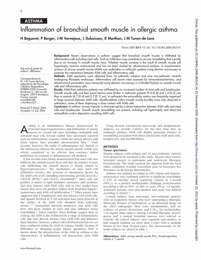
Infil tration of ASM by inflammatory cellsImmunohistochemical analysis revealed the presence of moremast cells and lymphocytes within the ASM layer fromasthmatic patients than from control subjects (table 2). Onlycells surrounded by ASM cells were considered as muscularinflammatory cells (fig 1).
There was no significant correlation between asthma severityaccording to FEV1 and smooth muscle infiltration by anyinflammatory cell types (data not shown). In two samples fromasthmatic patients we detected the presence of eosinophils inthe smooth muscle layer associated with a very high concen-tration of eosinophils in both the epithelium and submucosa.
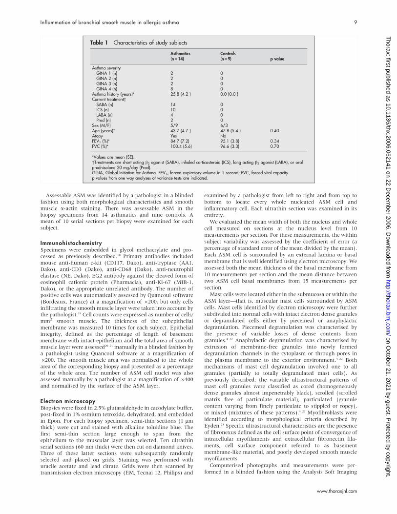
Ultrastructural analysis of ASM cellsAn ultrastructural analysis of the smooth muscle wasperformed in Epon embedded samples from nine asthmaticsubjects and five controls. ASM cells were characterised by aspindle shape, an elongated nucleus with a blunt end, and thepresence of dense bodies within the cytoplasm corresponding tothe organisation of smooth muscle myofilaments (fig 2). Themorphometric parameters of ASM cells from asthmatics andcontrols are shown in table 3. The mean ASM cell size waslarger in asthmatic patients than in control subjects whereasthe within subject variability was similar (2.1% and 2.3%,respectively). We also observed significant variations in ASM
Table 2 Counts of inflammatory cells (per mm2) infiltrating ASM of asthmatic and controlsubjects
Asthmatics(n = 14)
Controls(n = 9) p values
CD117 16.3 (2.5–51.0) 4.4 (0.0–10.8) 0.04AA1 19.6 (0.0–37.6) 4.4 (0.0–10.8) 0.04EG2 0.0 (0.0–0.0) 0.0 (0.0–0.0) 0.57CD3 9.2 (4.7–15.3) 0.0 (0.0–6.0) 0.02CD68 3.9 (0.0–10.1) 0.0 (0.0–3.8) 0.17NE 0.0 (0.0–0.0) 0.0 (0.0–0.0) 0.90
Values shown as median (interquartile range).p values from one way analyses of variance Kruskal-Wallis tests are indicated.
A
ASM
ASM
B
Figure 1 Infiltration of asthmatic airway smooth muscle (ASM) byinflammatory cells. (A) Mast cells are stained with antitryptase antibody. (B)Lymphocytes are stained with anti-CD3 antibody. Arrows indicate cellswithin the ASM layer. Sections are counterstained with Mayer’shaematoxylin. Bars represent 50 mm.
10 Begueret, Berger, Vernejoux, et al
www.thoraxjnl.com
on October 21, 2021 by guest. P
rotected by copyright.http://thorax.bm
j.com/
Thorax: first published as 10.1136/thx.2006.062141 on 22 D
ecember 2006. D
ownloaded from
cell basement membrane thickness and in extracellular matrixdeposition. When comparing both groups, mean ASM cell basallamina thickness was larger in asthmatic patients. The extra-cellular matrix was frequently organised into large numbers ofmoulding ASM cells and displayed non-fibrillar ultrastructuralfeatures (fig 2A, B). These features were not observed inspecimens obtained from non-asthmatic controls (fig 2C, D).
Ultrastructural analysis of cells infi ltrating the smoothmuscle layerAs mentioned above, we analysed various cell types infiltratingthe smooth muscle layer and only considered cells totallysurrounded by ASM cells.
Mast cellsIn asthmatic specimens the muscular mast cells were smallerthan the submucosal mast cells (143.9 (5.3) v 259.9 (22.1) mm2,p = 0.01) whereas their nuclear area was not significantlydifferent (38.7 (2.1) v 67.2 (9.6) mm2, p = 0.09). Muscular mastcells had very few short cytoplasmic processes or pseudopodscompared with submucosal mast cells. Nevertheless, they hadclose contacts with ASM cells (fig 3A) which was also observedin control specimens (fig 3B). Muscular mast cells displayed apattern of degranulation with a mean number of granuleslower than that of submucosal mast cells (19.1 (1.6) v 55.2(8.1) mm2, p = 0.02). Nevertheless, the mean area of thesegranules was similar in both muscular and submucosal mast
A
C D
B Figure 2 Ultrastructural characteristics ofairway smooth muscle (ASM) cells andextracellular matrix deposition. Longitudinalsections show a thicker extracellular matrixdeposition (arrows) surrounding ASM cells in(A) an asthmatic patient than in a controlsubject (C). In transverse sections theextracellular matrix is frequently organisedinto large bland amounts of matrix proteindeposits (arrows) moulding ASM cells inasthmatic patients (B); this feature was notobserved in control subjects (D). Barsrepresent 10 mm.
Table 3 Morphological parameters of ASM cells from asthmatic and control subjects
Whole cell meansize (mm)
Nuclei mean size(mm)
Distance betweenASM cell BM (mm)
ASM cell BMthickness (mm)
Presence of myofibroblastsbetween ASM cells
Asthmatics1 9.92 4.43 1.33 1.61 No2 8.20 6.26 0.81 0.75 Yes3 11.06 3.01 1.57 2.62 Yes4 7.29 3.12 0.52 1.00 Yes5 7.83 4.71 1.14 0.62 Yes6 6.33 7.01 1.39 1.70 No7 14.31 9.53 1.95 1.28 Yes8 9.75 2.19 0.84 0.94 Yes9 10.88 3.68 1.21 1.82 YesMean (SE) 9.5 (0.8) 4.9 (0.8) 1.2 (0.1) 1.4 (0.2)
Controls1 5.60 3.01 1.60 0.60 No2 6.30 3.52 1.56 0.61 No3 7.51 4.61 1.28 0.66 No4 7.76 5.01 1.71 1.01 No5 6.12 3.81 1.34 0.63 NoMean (SE) 6.7 (0.4) 4.0 (0.4) 1.5 (0.1) 0.7 (0.1)p values 0.03 0.43 0.17 0.04 0.02
p values from one way analyses of variance or Fisher’s exact tests are indicated.ASM, airway smooth muscle; BM, basal membrane.
Inflammation of bronchial smooth muscle in allergic asthma 11
www.thoraxjnl.com
on October 21, 2021 by guest. P
rotected by copyright.http://thorax.bm
j.com/
Thorax: first published as 10.1136/thx.2006.062141 on 22 D
ecember 2006. D
ownloaded from
cells (1.5 (0.3) v 2.0 (0.1) mm2, p = 0.11), although theirultrastructural appearance was different. From a total of 523granules, the mean (SE) percentage of cored granules was 75.3
(13.2)% in the smooth muscle layer and only 17.6 (5.2)% in thesubmucosa (p = 0.001). The percentages of scrolled, particu-lated, or mixed granules were not significantly differentbetween the two sites.
A
SM
SM
SM
*
*
B
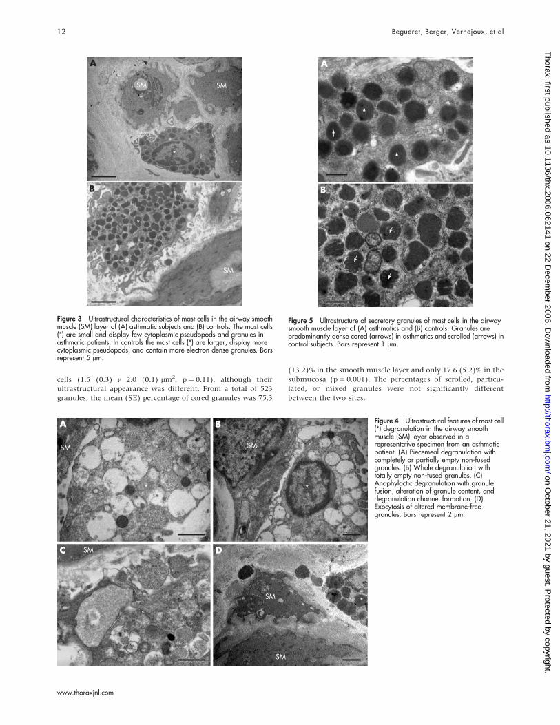
Figure 3 Ultrastructural characteristics of mast cells in the airway smoothmuscle (SM) layer of (A) asthmatic subjects and (B) controls. The mast cells(*) are small and display few cytoplasmic pseudopods and granules inasthmatic patients. In controls the mast cells (*) are larger, display morecytoplasmic pseudopods, and contain more electron dense granules. Barsrepresent 5 mm.
A
C D
B
SM
SM
SM
SM
*
*
**
SM
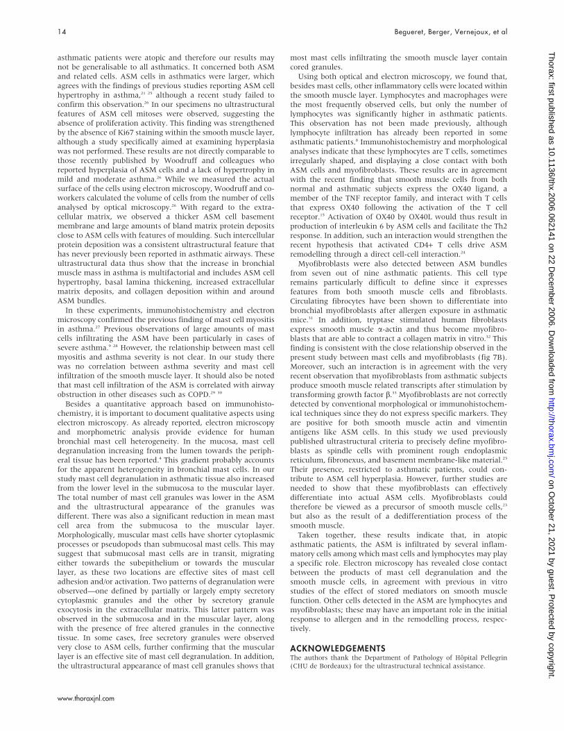
Figure 4 Ultrastructural features of mast cell(*) degranulation in the airway smoothmuscle (SM) layer observed in arepresentative specimen from an asthmaticpatient. (A) Piecemeal degranulation withcompletely or partially empty non-fusedgranules. (B) Whole degranulation withtotally empty non-fused granules. (C)Anaphylactic degranulation with granulefusion, alteration of granule content, anddegranulation channel formation. (D)Exocytosis of altered membrane-freegranules. Bars represent 2 mm.
A
B
Figure 5 Ultrastructure of secretory granules of mast cells in the airwaysmooth muscle layer of (A) asthmatics and (B) controls. Granules arepredominantly dense cored (arrows) in asthmatics and scrolled (arrows) incontrol subjects. Bars represent 1 mm.
12 Begueret, Berger, Vernejoux, et al
www.thoraxjnl.com
on October 21, 2021 by guest. P
rotected by copyright.http://thorax.bm
j.com/
Thorax: first published as 10.1136/thx.2006.062141 on 22 D
ecember 2006. D
ownloaded from
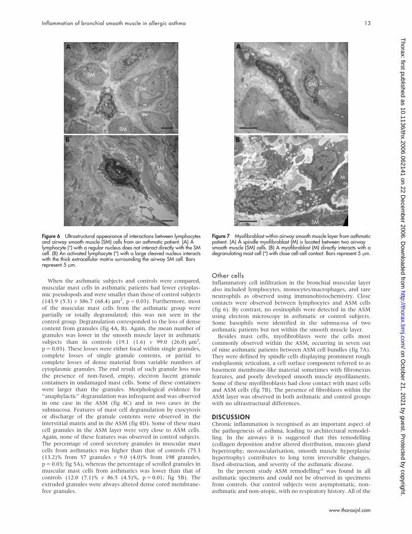
When the asthmatic subjects and controls were compared,muscular mast cells in asthmatic patients had fewer cytoplas-mic pseudopods and were smaller than those of control subjects(143.9 (5.3) v 386.7 (68.4) mm2, p = 0.03). Furthermore, mostof the muscular mast cells from the asthmatic group werepartially or totally degranulated; this was not seen in thecontrol group. Degranulation corresponded to the loss of densecontent from granules (fig 4A, B). Again, the mean number ofgranules was lower in the smooth muscle layer in asthmaticsubjects than in controls (19.1 (1.6) v 99.0 (26.0) mm2,p = 0.03). These losses were either focal within single granules,complete losses of single granule contents, or partial tocomplete losses of dense material from variable numbers ofcytoplasmic granules. The end result of such granule loss wasthe presence of non-fused, empty, electron lucent granulecontainers in undamaged mast cells. Some of these containerswere larger than the granules. Morphological evidence for‘‘anaphylactic’’ degranulation was infrequent and was observedin one case in the ASM (fig 4C) and in two cases in thesubmucosa. Features of mast cell degranulation by exocytosisor discharge of the granule contents were observed in theinterstitial matrix and in the ASM (fig 4D). Some of these mastcell granules in the ASM layer were very close to ASM cells.Again, none of these features was observed in control subjects.The percentage of cored secretory granules in muscular mastcells from asthmatics was higher than that of controls (75.3(13.2)% from 57 granules v 9.0 (4.0)% from 198 granules,p = 0.03; fig 5A), whereas the percentage of scrolled granules inmuscular mast cells from asthmatics was lower than that ofcontrols (12.0 (7.1)% v 86.5 (4.5)%, p = 0.01; fig 5B). Theextruded granules were always altered dense cored membrane-free granules.
Other cellsInflammatory cell infiltration in the bronchial muscular layeralso included lymphocytes, monocytes/macrophages, and rareneutrophils as observed using immunohistochemistry. Closecontacts were observed between lymphocytes and ASM cells(fig 6). By contrast, no eosinophils were detected in the ASMusing electron microscopy in asthmatic or control subjects.Some basophils were identified in the submucosa of twoasthmatic patients but not within the smooth muscle layer.
Besides mast cells, myofibroblasts were the cells mostcommonly observed within the ASM, occurring in seven outof nine asthmatic patients between ASM cell bundles (fig 7A).They were defined by spindle cells displaying prominent roughendoplasmic reticulum, a cell surface component referred to asbasement membrane-like material sometimes with fibronexusfeatures, and poorly developed smooth muscle myofilaments.Some of these myofibroblasts had close contact with mast cellsand ASM cells (fig 7B). The presence of fibroblasts within theASM layer was observed in both asthmatic and control groupswith no ultrastructural differences.
DISCUSSIONChronic inflammation is recognised as an important aspect ofthe pathogenesis of asthma, leading to architectural remodel-ling. In the airways it is suggested that this remodelling(collagen deposition and/or altered distribution, mucous glandhypertrophy, neovascularisation, smooth muscle hyperplasia/hypertrophy) contributes to long term irreversible changes,fixed obstruction, and severity of the asthmatic disease.
In the present study ASM remodelling24 was found in allasthmatic specimens and could not be observed in specimensfrom controls. Our control subjects were asymptomatic, non-asthmatic and non-atopic, with no respiratory history. All of the
A
SM
*
*
SM
SM
SM
B
Figure 6 Ultrastructural appearance of interactions between lymphocytesand airway smooth muscle (SM) cells from an asthmatic patient. (A) Alymphocyte (*) with a regular nucleus does not interact directly with the SMcell. (B) An activated lymphocyte (*) with a large cleaved nucleus interactswith the thick extracellular matrix surrounding the airway SM cell. Barsrepresent 5 mm.
A
M
SM
SM
SM
M
*
B
Figure 7 Myofibroblast within airway smooth muscle layer from asthmaticpatient. (A) A spindle myofibroblast (M) is located between two airwaysmooth muscle (SM) cells. (B) A myofibroblast (M) directly interacts with adegranulating mast cell (*) with close cell-cell contact. Bars represent 5 mm.
Inflammation of bronchial smooth muscle in allergic asthma 13
www.thoraxjnl.com
on October 21, 2021 by guest. P
rotected by copyright.http://thorax.bm
j.com/
Thorax: first published as 10.1136/thx.2006.062141 on 22 D
ecember 2006. D
ownloaded from
asthmatic patients were atopic and therefore our results maynot be generalisable to all asthmatics. It concerned both ASMand related cells. ASM cells in asthmatics were larger, whichagrees with the findings of previous studies reporting ASM cellhypertrophy in asthma,21 25 although a recent study failed toconfirm this observation.26 In our specimens no ultrastructuralfeatures of ASM cell mitoses were observed, suggesting theabsence of proliferation activity. This finding was strengthenedby the absence of Ki67 staining within the smooth muscle layer,although a study specifically aimed at examining hyperplasiawas not performed. These results are not directly comparable tothose recently published by Woodruff and colleagues whoreported hyperplasia of ASM cells and a lack of hypertrophy inmild and moderate asthma.26 While we measured the actualsurface of the cells using electron microscopy, Woodruff and co-workers calculated the volume of cells from the number of cellsanalysed by optical microscopy.26 With regard to the extra-cellular matrix, we observed a thicker ASM cell basementmembrane and large amounts of bland matrix protein depositsclose to ASM cells with features of moulding. Such intercellularprotein deposition was a consistent ultrastructural feature thathas never previously been reported in asthmatic airways. Theseultrastructural data thus show that the increase in bronchialmuscle mass in asthma is multifactorial and includes ASM cellhypertrophy, basal lamina thickening, increased extracellularmatrix deposits, and collagen deposition within and aroundASM bundles.
In these experiments, immunohistochemistry and electronmicroscopy confirmed the previous finding of mast cell myositisin asthma.27 Previous observations of large amounts of mastcells infiltrating the ASM have been particularly in cases ofsevere asthma.9 28 However, the relationship between mast cellmyositis and asthma severity is not clear. In our study therewas no correlation between asthma severity and mast cellinfiltration of the smooth muscle layer. It should also be notedthat mast cell infiltration of the ASM is correlated with airwayobstruction in other diseases such as COPD.29 30
Besides a quantitative approach based on immunohisto-chemistry, it is important to document qualitative aspects usingelectron microscopy. As already reported, electron microscopyand morphometric analysis provide evidence for humanbronchial mast cell heterogeneity. In the mucosa, mast celldegranulation increasing from the lumen towards the periph-eral tissue has been reported.4 This gradient probably accountsfor the apparent heterogeneity in bronchial mast cells. In ourstudy mast cell degranulation in asthmatic tissue also increasedfrom the lower level in the submucosa to the muscular layer.The total number of mast cell granules was lower in the ASMand the ultrastructural appearance of the granules wasdifferent. There was also a significant reduction in mean mastcell area from the submucosa to the muscular layer.Morphologically, muscular mast cells have shorter cytoplasmicprocesses or pseudopods than submucosal mast cells. This maysuggest that submucosal mast cells are in transit, migratingeither towards the subepithelium or towards the muscularlayer, as these two locations are effective sites of mast celladhesion and/or activation. Two patterns of degranulation wereobserved—one defined by partially or largely empty secretorycytoplasmic granules and the other by secretory granuleexocytosis in the extracellular matrix. This latter pattern wasobserved in the submucosa and in the muscular layer, alongwith the presence of free altered granules in the connectivetissue. In some cases, free secretory granules were observedvery close to ASM cells, further confirming that the muscularlayer is an effective site of mast cell degranulation. In addition,the ultrastructural appearance of mast cell granules shows that
most mast cells infiltrating the smooth muscle layer containcored granules.
Using both optical and electron microscopy, we found that,besides mast cells, other inflammatory cells were located withinthe smooth muscle layer. Lymphocytes and macrophages werethe most frequently observed cells, but only the number oflymphocytes was significantly higher in asthmatic patients.This observation has not been made previously, althoughlymphocyte infiltration has already been reported in someasthmatic patients.8 Immunohistochemistry and morphologicalanalyses indicate that these lymphocytes are T cells, sometimesirregularly shaped, and displaying a close contact with bothASM cells and myofibroblasts. These results are in agreementwith the recent finding that smooth muscle cells from bothnormal and asthmatic subjects express the OX40 ligand, amember of the TNF receptor family, and interact with T cellsthat express OX40 following the activation of the T cellreceptor.15 Activation of OX40 by OX40L would thus result inproduction of interleukin 6 by ASM cells and facilitate the Th2response. In addition, such an interaction would strengthen therecent hypothesis that activated CD4+ T cells drive ASMremodelling through a direct cell-cell interaction.24
Myofibroblasts were also detected between ASM bundlesfrom seven out of nine asthmatic patients. This cell typeremains particularly difficult to define since it expressesfeatures from both smooth muscle cells and fibroblasts.Circulating fibrocytes have been shown to differentiate intobronchial myofibroblasts after allergen exposure in asthmaticmice.31 In addition, tryptase stimulated human fibroblastsexpress smooth muscle a-actin and thus become myofibro-blasts that are able to contract a collagen matrix in vitro.32 Thisfinding is consistent with the close relationship observed in thepresent study between mast cells and myofibroblasts (fig 7B).Moreover, such an interaction is in agreement with the veryrecent observation that myofibroblasts from asthmatic subjectsproduce smooth muscle related transcripts after stimulation bytransforming growth factor b.33 Myofibroblasts are not correctlydetected by conventional morphological or immunohistochem-ical techniques since they do not express specific markers. Theyare positive for both smooth muscle actin and vimentinantigens like ASM cells. In this study we used previouslypublished ultrastructural criteria to precisely define myofibro-blasts as spindle cells with prominent rough endoplasmicreticulum, fibronexus, and basement membrane-like material.23
Their presence, restricted to asthmatic patients, could con-tribute to ASM cell hyperplasia. However, further studies areneeded to show that these myofibroblasts can effectivelydifferentiate into actual ASM cells. Myofibroblasts couldtherefore be viewed as a precursor of smooth muscle cells,23
but also as the result of a dedifferentiation process of thesmooth muscle.
Taken together, these results indicate that, in atopicasthmatic patients, the ASM is infiltrated by several inflam-matory cells among which mast cells and lymphocytes may playa specific role. Electron microscopy has revealed close contactbetween the products of mast cell degranulation and thesmooth muscle cells, in agreement with previous in vitrostudies of the effect of stored mediators on smooth musclefunction. Other cells detected in the ASM are lymphocytes andmyofibroblasts; these may have an important role in the initialresponse to allergen and in the remodelling process, respec-tively.
ACKNOWLEDGEMENTSThe authors thank the Department of Pathology of Hopital Pellegrin(CHU de Bordeaux) for the ultrastructural technical assistance.
14 Begueret, Berger, Vernejoux, et al
www.thoraxjnl.com
on October 21, 2021 by guest. P
rotected by copyright.http://thorax.bm
j.com/
Thorax: first published as 10.1136/thx.2006.062141 on 22 D
ecember 2006. D
ownloaded from
Authors’ affiliations. . . . . . . . . . . . . . . . . . . . . . .
H Begueret, P Berger, R Marthan, J M Tunon-de-Lara, Universite Bordeaux2, Laboratoire de Physiologie Cellulaire Respiratoire, F-33076, Bordeaux;Inserm E356 F-23076, Bordeaux, FranceJ M Vernejoux, J M Tunon-de-Lara, CHU de Bordeaux, Service desMaladies Respiratoires, F-33076, Bordeaux, FranceL Dubuisson, Universite Bordeaux 2, Service de Microscopie Electronique,F-33076, Bordeaux, France
This study was supported by a grant PHRC (Programme Hospitalier deRecherche Clinique) 2002 from the CHU de Bordeaux and a grant fromFRM (Fondation Recherche Medicale) DAL2005120574.
Competing interests: none declared.
REFERENCES1 Lamb D, Lumsden A. Intra-epithelial mast cells in human airway epithelium:
evidence for smoking-induced changes in their frequency. Thorax1982;37:334–42.
2 Franconi GM, Rubinstein I, Levine EH, et al. Mechanical removal of airwayepithelium disrupts mast cells and releases granules. Am J Physiol1990;259:L372–7.
3 Djukanovic R, Wilson JW, Britten KM, et al. Quantitation of mast cells andeosinophils in the bronchial mucosa of symptomatic atopic asthmatics andhealthy control subjects using immunohistochemistry. Am Rev Respir Dis1990;142:863–71.
4 Heard BE, Dewar A, Nunn AJ, et al. Heterogeneous ultrastructure of humanbronchial mast cells: morphometric subdivision of cell types and evidence for adegranulation gradient. Am J Respir Cell Mol Biol 1990;3:71–8.
5 Ohashi Y, Motojima S, Fukuda T, et al. Airway hyperresponsiveness, increasedintracellular spaces of bronchial epithelium, and increased infiltration ofeosinophils and lymphocytes in bronchial mucosa in asthma. Am Rev Respir Dis1992;145:1469–76.
6 Gizycki MJ, Adelroth E, Rogers AV, et al. Myofibroblast involvement in theallergen-induced late response in mild atopic asthma. Am J Respir Cell Mol Biol1997;16:664–73.
7 Cokugras H, Akcakaya N, Seckin, et al. Ultrastructural examination of bronchialbiopsy specimens from children with moderate asthma. Thorax 2001;56:25–9.
8 Brightling CE, Bradding P, Symon FA, et al. Mast-cell infiltration of airwaysmooth muscle in asthma. N Engl J Med 2002;346:1699–705.
9 Berger P, Girodet PO, Begueret H, et al. Tryptase-stimulated human airwaysmooth muscle cells induce cytokine synthesis and mast cell chemotaxis. Faseb J2003;17:2139–41.
10 Brightling CE, Ammit AJ, Kaur D, et al. The CXCL10/CXCR3 axis mediateshuman lung mast cell migration to asthmatic airway smooth muscle. Am J RespirCrit Care Med 2005;171:1103–8.
11 El-Shazly A, Berger P, Girodet PO, et al. Fraktalkine produced by airway smoothmuscle cells contributes to mast cell recruitment in asthma. J Immunol2006;176:1860–8.
12 Berger P, Compton SJ, Molimard M, et al. Mast cell tryptase as a mediator ofhyperresponsiveness in human isolated bronchi. Clin Exp Allergy1999;29:804–12.
13 Berger P, Perng DW, Thabrew H, et al. Tryptase and agonists of PAR-2 inducethe proliferation of human airway smooth muscle cells. J Appl Physiol2001;91:1372–9.
14 Lazaar AL, Albelda SM, Pilewski JM, et al. T lymphocytes adhere to airwaysmooth muscle cells via integrins and CD44 and induce smooth muscle cell DNAsynthesis. J Exp Med 1994;180:807–16.
15 Burgess JK, Blake AE, Boustany S, et al. CD40 and OX40 ligand are increasedon stimulated asthmatic airway smooth muscle. J Allergy Clin Immunol2005;115:302–8.
16 Zhu YK, Liu X, Wang H, et al. Interactions between monocytes and smooth-muscle cells can lead to extracellular matrix degradation. J Allergy Clin Immunol2001;108:989–96.
17 Johansson SG, Hourihane JO, Bousquet J, et al. A revised nomenclature forallergy. An EAACI position statement from the EAACI nomenclature task force.Allergy 2001;56:813–24.
18 Berger P, Walls AF, Marthan R, et al. Immunoglobulin E-induced passivesensitization of human airways: an immunohistochemical study. Am J Respir CritCare Med 1998;157:610–6.
19 Berger P, Lavallee J, Rouiller R, et al. Assessment of bronchial inflammation usingan automated cell recognition system based on colour analysis. Eur Respir J1999;14:1394–402.
20 Amin K, Ludviksdottir D, Janson C, et al. Inflammation and structural changes inthe airways of patients with atopic and nonatopic asthma. Am J Respir Crit CareMed 2000;162:2295–301.
21 Benayoun L, Druilhe A, Dombret MC, et al. Airway structural alterationsselectively associated with severe asthma. Am J Respir Crit Care Med2003;167:1360–8.
22 Dvorak AM. Ultrastructural analysis of human mast cells and basophils. ChemImmunol 1995;61:1–33.
23 Eyden B. The myofibroblast: an assessment of controversial issues and adefinition useful in diagnosis and research. Ultrastruct Pathol 2001;25:39–50.
24 Ramos-Barbon D, Presley JF, Hamid QA, et al. Antigen-specific CD4+ T cellsdrive airway smooth muscle remodeling in experimental asthma. J Clin Invest2005;115:1580–9.
25 Ebina M, Takahashi T, Chiba T, et al. Cellular hypertrophy and hyperplasia ofairway smooth muscles underlying bronchial asthma. A 3-D morphometric study.Am Rev Respir Dis 1993;148:720–6.
26 Woodruff PG, Dolganov GM, Ferrando RE, et al. Hyperplasia of smooth musclein mild to moderate asthma without changes in cell size or gene expression.Am J Respir Crit Care Med 2004;169:1001–6.
27 Berger P, Girodet PO, Tunon De Lara JM. Mast cell myositis: a new feature ofallergic asthma? Allergy 2005;60:1238–40.
28 Carroll NG, Mutavdzic S, James AL. Distribution and degranulation of airwaymast cells in normal and asthmatic subjects. Eur Respir J 2002;19:879–85.
29 Tunon-de-Lara JM, Berger P, Begueret H. Mast cells in airway smooth muscle.N Engl J Med 2002;347:1040–1.
30 Berger P, Laurent F, Begueret H, et al. Structure-function of small airways insmokers: relationship between air trapping on CT and airway inflammation.Radiology 2003;228:85–94.
31 Schmidt M, Sun G, Stacey MA, et al. Identification of circulating fibrocytes asprecursors of bronchial myofibroblasts in asthma. J Immunol 2003;171:380–9.
32 Skold CM, Ohkuni Y, Liu XD, et al. Co-cultured human mast cells stimulatefibroblast-mediated contraction of collagen gels. Inflammation 2001;25:47–51.
33 Wicks J, Haitchi HM, Holgate ST, et al. Enhanced upregulation of smooth muscle-related transcripts by TGFb in asthmatic (myo)fibroblasts. Thorax2006;61:313–9.
bmjupdates+
bmjupdates+ is a unique and free alerting service, designed to keep you up to date with themedical literature that is truly important to your practice.bmjupdates+ will alert you to important new research and will provide you with the best newevidence concerning important advances in health care, tailored to your medical interests andtime demands.
Where does the information come from?bmjupdates+ applies an expert critical appraisal filter to over 100 top medical journalsA panel of over 2000 physicians find the few ’must read’ studies for each area of clinical interest
Sign up to receive your tailored email alerts, searching access and more…
www.bmjupdates.com
Inflammation of bronchial smooth muscle in allergic asthma 15
www.thoraxjnl.com
on October 21, 2021 by guest. P
rotected by copyright.http://thorax.bm
j.com/
Thorax: first published as 10.1136/thx.2006.062141 on 22 D
ecember 2006. D
ownloaded from


