Association of Dromedary Camels and Camel Ticks with ...
9
C rimean-Congo hemorrhagic fever virus (CCHFV) is a tickborne nairovirus (order Bunyavirales) that is maintained primarily in Hyalomma ticks (Ixodidae), and various mammalian livestock serve as amplify- ing hosts. Humans might become infected from the bite of an infected tick or during slaughter of a viremic animal, and the infection might lead to severe viral hemorrhagic fever and death. In the Arabian Penin- sula, human cases are sporadically reported and seem to be primarily associated with abattoir work (1,2) or nosocomial human-to-human transmissions (3). CCHFV is genetically diverse and has a relatively wide geographic distribution (4). The virus might be introduced into nonendemic regions through com- mercial trading of livestock (5) or through phoretic transport of ticks on migratory birds (6,7). Compara- tively little is known about the zoonotic transmission of the virus in the United Arab Emirates or whether past outbreaks were only associated with recent im- portations (5). We previously performed a cross-sectional vi- rologic and serologic survey of CCHFV in drom- edary camels (Camelus dromedarius) at various sites throughout the United Arab Emirates (8). We found the highest transmission activity at a large livestock market, in which viral nucleic acids were detected in camel ticks (Hyalomma dromedarii) and camels. On the basis of partial gene sequences from the small and medium (M) RNA gene segments, the virus strain appeared to be a novel reassortant (8). We performed a follow-up study at the same market to test whether other livestock are involved in the transmission of CCHFV and to better characterize the virus strain. The Study During October 10–24, 2019, we sampled camels, cattle, goats, and sheep upon their entry to a live- stock market in the emirate of Abu Dhabi, United Arab Emirates (≈24.16°N, 55.81°E) (Appendix Fig- ure 1, https://wwwnc.cdc.gov/EID/article/27/9/ 21-0299-App1.pdf). All procedures were conducted as part of standard veterinary inspection required for market entry. We obtained 5 mL of blood from each animal, separated serum by centrifugation, and stored serum at –80°C. We tested serum for CCHFV-reactive anti- bodies by using a commercial kit (ID Screen CCHF Double Antigen Multi-species; IDvet, https://www. id-vet.com). Antibodies to CCHFV were found in 72/90 camels, 7/51 cattle, 1/45 goats, and 4/55 sheep (Table). We extracted total nucleic acids from 200 µL of the same serum samples by using a com- mercial kit (QIAamp Viral RNA Mini Kit; QIAGEN, https://www.qiagen.com) and QIAcube or QIAcube Association of Dromedary Camels and Camel Ticks with Reassortant Crimean-Congo Hemorrhagic Fever Virus, United Arab Emirates Jeremy V. Camp, Pia Weidinger, Sathiskumar Ramaswamy, Dafalla O. Kannan, Babiker Mohammed Osman, Jolanta Kolodziejek, Noushad Karuvantevida, AhmadAbou Tayoun, Tom Loney, Norbert Nowotny Emerging Infectious Diseases • www.cdc.gov/eid • Vol. 27, No. 9, September 2021 2471 Author affilations: Medical University of Vienna, Vienna, Austria (J.V. Camp); University of Veterinary Medicine Vienna, Vienna (J.V. Camp, P. Weidinger, J. Kolodziejek, N. Nowotny); Mohammed Bin Rashid University of Medicine and Health Sciences, Dubai, United Arab Emirates (S. Ramaswamy, N. Karuvantevida, A. Abou Tayoun, T. Loney, N. Nowotny); Al Jalila Children’s Hospital, Dubai (S. Ramaswamy, A. Abou Tayoun); Al Ain City Municipality, Al Ain, United Arab Emirates (D.O. Kannan, B.M. Osman) DOI: https;//doi.org/10.3201/eid2709.210299 We previously detected a potentially novel reassortant of Crimean-Congo hemorrhagic fever virus in camels at the largest livestock market in the United Arab Emirates. A broader survey of large mammals at the site indicated zoonotic transmission is associated with dromedaries and camel ticks. Seroprevalence in cattle, sheep, and goats is minimal.
Transcript of Association of Dromedary Camels and Camel Ticks with ...

Crimean-Congo hemorrhagic fever virus (CCHFV) is a tickborne nairovirus (order Bunyavirales) that
is maintained primarily in Hyalomma ticks (Ixodidae), and various mammalian livestock serve as amplify-ing hosts. Humans might become infected from the bite of an infected tick or during slaughter of a viremic animal, and the infection might lead to severe viral hemorrhagic fever and death. In the Arabian Penin-sula, human cases are sporadically reported and seem to be primarily associated with abattoir work (1,2) or nosocomial human-to-human transmissions (3).
CCHFV is genetically diverse and has a relatively wide geographic distribution (4). The virus might be introduced into nonendemic regions through com-mercial trading of livestock (5) or through phoretic transport of ticks on migratory birds (6,7). Compara-tively little is known about the zoonotic transmission of the virus in the United Arab Emirates or whether
past outbreaks were only associated with recent im-portations (5).
We previously performed a cross-sectional vi-rologic and serologic survey of CCHFV in drom-edary camels (Camelus dromedarius) at various sites throughout the United Arab Emirates (8). We found the highest transmission activity at a large livestock market, in which viral nucleic acids were detected in camel ticks (Hyalomma dromedarii) and camels. On the basis of partial gene sequences from the small and medium (M) RNA gene segments, the virus strain appeared to be a novel reassortant (8). We performed a follow-up study at the same market to test whether other livestock are involved in the transmission of CCHFV and to better characterize the virus strain.
The StudyDuring October 10–24, 2019, we sampled camels, cattle, goats, and sheep upon their entry to a live-stock market in the emirate of Abu Dhabi, United Arab Emirates (≈24.16°N, 55.81°E) (Appendix Fig-ure 1, https://wwwnc.cdc.gov/EID/article/27/9/21-0299-App1.pdf). All procedures were conducted as part of standard veterinary inspection required for market entry.
We obtained 5 mL of blood from each animal, separated serum by centrifugation, and stored serum at –80°C. We tested serum for CCHFV-reactive anti-bodies by using a commercial kit (ID Screen CCHF Double Antigen Multi-species; IDvet, https://www.id-vet.com). Antibodies to CCHFV were found in 72/90 camels, 7/51 cattle, 1/45 goats, and 4/55 sheep (Table). We extracted total nucleic acids from 200 µL of the same serum samples by using a com-mercial kit (QIAamp Viral RNA Mini Kit; QIAGEN, https://www.qiagen.com) and QIAcube or QIAcube
Association of Dromedary Camels and Camel Ticks with Reassortant Crimean-Congo Hemorrhagic Fever
Virus, United Arab EmiratesJeremyV.Camp,PiaWeidinger,SathiskumarRamaswamy,DafallaO.Kannan,BabikerMohammedOsman,
JolantaKolodziejek,NoushadKaruvantevida,AhmadAbouTayoun,TomLoney,NorbertNowotny
EmergingInfectiousDiseases•www.cdc.gov/eid•Vol.27,No.9,September2021 2471
Authoraffilations:MedicalUniversityofVienna,Vienna,Austria(J.V.Camp);UniversityofVeterinaryMedicineVienna,Vienna(J.V.Camp,P.Weidinger,J.Kolodziejek,N.Nowotny);MohammedBinRashidUniversityofMedicineandHealthSciences,Dubai,UnitedArabEmirates(S.Ramaswamy,N.Karuvantevida,A.AbouTayoun,T.Loney,N.Nowotny);AlJalilaChildren’sHospital,Dubai(S.Ramaswamy,A.AbouTayoun);AlAinCityMunicipality,AlAin,UnitedArabEmirates(D.O.Kannan,B.M.Osman)
DOI:https;//doi.org/10.3201/eid2709.210299
We previously detected a potentially novel reassortantofCrimean-Congohemorrhagic fevervirus incamelsatthelargestlivestockmarketintheUnitedArabEmirates.Abroadersurveyoflargemammalsatthesiteindicatedzoonotictransmissionisassociatedwithdromedariesandcamel ticks.Seroprevalence incattle,sheep,andgoatsisminimal.

DISPATCHES
HT Extraction Robots (QIAGEN). We tested extracts for CCHFV RNA by using a commercially available quantitative reverse transcription PCR (qRT-PCR) as-say (RealStar CCHFV RT-PCR Kit 1.0; Altona Diag-nostics, https://www.altona-diagnostics.com). Viral nucleic acids were detected in 2 of 90 camels, and in no other species at the market.
During blood collection, we thoroughly searched each animal (≈2 min) and removed attached ticks. Ticks were frozen at −80°C, and adult ticks were mor-phologically identified by using various keys on an ice cold plate. We collected 210 H. dromedarii adults, 4 unidentified Hyalomma sp. adults, and 4 Hyalomma sp. nymphs from 84/90 camels. No ticks were found on any other animal during this sampling session, and it was later confirmed that topical acaricides were rou-tinely used for all animals except camels, where they were applied only sporadically.
We processed frozen ticks to screen for viral nucleic acids by making a parasagittal section using a sterile scalpel and then made homogenized pools containing half-ticks (<5 per pool, pooled per tick species, and per individual host) in a bead mill in buffered saline before adding DNA/RNA Shield (Zy-moResearch, https://www.zymoresearch.com) and performing nucleic acid extraction and qRT-PCR. We detected CCHFV RNA in 3 pools of H. dromedarii ticks taken from 2 camels, 1 of which was seropositive and the other seronegative.
We then extracted nucleic acids from the remaining halves of 3 ticks collected in the previous sampling ses-sion (April 2019) (8) and 2 ticks collected in this sampling session from pools that were positive by qRT-PCR. Af-ter confirming the presence of CCHFV nucleic acid in the individual tick halves, we processed the samples by using a shotgun transcriptomic sequencing approach (Appendix). In brief, we quality-filtered, trimmed, and assembled paired-end reads from Illumina (https://www.illumina.com) sequencing into scaffolds, which we then searched against the National Center for Bio-technology Information nonredundant database using blastn (https://blast.ncbi.nlm.nih.gov).
We identified near-complete genomes of CCHFV, including complete open reading frames of all but 1
gene segment (missing 577 nt from the large segment open reading frame 3′ mRNA end), from all 5 samples (GenBank accession nos. MW548490–504) (Appen-dix). We aligned the sequences to selected reference sequences representing the major genotypes (4). All sequences had high identity to each other (98.8%–100%) and to a recently described CCHFV (98.5%–99.6%) identified in a dromedary from the same livestock market in the emirate of Abu Dhabi, but 4 years earlier, during 2015 (9) (Appendix Table). The M segment was the most variable; it had 52–64 non-synonymous mutations compared with the sample obtained during 2015 from the same place, and 1–40 nonsynonymous mutations among the 5 sequences (Appendix Table).
We constructed phylogenetic trees from the alignments of the respective gene segments by us-ing maximum-likelihood analysis over 500 bootstrap replicates of the general time reversible plus invariant sites plus gamma distribution substitution model and 4 gamma categories. Small segments fit within the previously described genotype from Africa (group III sensu [4] and Africa 3 sensu [9]), and large segments had a common ancestor with sequences from Africa (groups I and III sensu [4], Africa 1/3 sensu [9]), and Europe (group V sensu [4] and Europe 1 sensu [9]) (Appendix Figures 2, 3). The M segment appeared to be a novel lineage of CCHFV (Figure).
ConclusionsWe concur with the findings of Khalafalla et al. (9), who provided additional serologic and virologic evidence that the CCHFV strain in the United Arab Emirates might be specifically associated with cam-els and camel ticks. Our study differs from previous studies, in that our sampling was performed directly at entry to a livestock market, but our previous study was performed after camels had entered the market (range 0–77 d, mean 12.2 d). Moreover, all animals were raised in the United Arab Emirates, although some sheep and goats were imported as young ani-mals from India, Saudi Arabia, and Oman. Com-bined, the evidence suggests that the CCHFV strain has spread throughout the United Arab Emirates, but the livestock market is also a focus of transmission (8).
Although this strain of CCHFV appears to be circulating at least since 2015 in the United Arab Emirates (8,9), there is additional evidence that it might be more widely distributed (10). Evidence of increased exposure of camels to CCHFV at livestock markets in contrast to other locations (e.g., private farms or in tourist/recreational use) increases the potential for the virus to be transported long
2472 EmergingInfectiousDiseases•www.cdc.gov/eid•Vol.27,No.9,September2021
Table.EvidenceofexposuretoCrimean-Congohemorrhagicfevervirusinanimalsatalivestockmarket,UnitedArabEmirates,2019
Species No.sampled No. antibodypositive
No.virusRNApositive
Camels 90 72 2 Cattle 55 7* 0 Goats 45 1 0 Sheep 55 4 0 *Serumwasnotavailablefor4cattle.

ReassortantCCHFV,UnitedArabEmirates
distances through the camel trade (8). We support the suggestion of Khalafalla et al. to increase efforts to characterize CCHFV from camels, camel ticks, and other livestock in a broader geographic region (9). The infection of camels appears to be systemic; virus was detected in blood (8; this study) and the respiratory tract (9). The low CCHFV-reactive sero-prevalence and low tick burden on other livestock
entering the market is probably caused by use of acaricides, which are reportedly used only spo-radically on camels. We therefore recommend in-creased vigilance, including use of acaricides on all livestock, including dromedaries, to limit spillover to humans involved in the camel trade, abattoir workers, and those handling raw meat or consum-ing raw camel milk.
EmergingInfectiousDiseases•www.cdc.gov/eid•Vol.27,No.9,September2021 2473
Figure.MolecularphylogenyofCrimean-CongohemorrhagicfevervirusmediumRNAsegments,UnitedArabEmirates,2019(solidcircles),andreferenceviruses.Virusesfromthisstudywereobtainedfromcamelticks(Hyalomma dromedarii)removedfromdromedarycamelsatalargelivestockmarketintheemirateofAbuDhabi.OthervirussequencesincludedwereselectedasrepresentativesofthemajorsmallandlargeRNAsegmentgenotypesforwhichfull-lengthsequencesofall3viralgenomicsegmentswereavailable.ViruseslistedincludeGenBankaccessionnumberandcountryoforigin.Maximum-likelihoodanalysisofcoding-completesequenceswasperformedbyusingthegeneraltimereversibleplusinvariantsitesplusgammadistributionsubstitutionmodeland4categorieswith>500bootstrapreplicates.Numbersalongbranchesarepercentagesupport,showingonlyvalues>65%,andbranchlengthisrelativetothenumberofsubstitutionspersite,asindicatedbythescalebar.DemRepCongo,DemocraticRepublicoftheCongo;UAE,UnitedArabEmirates.

DISPATCHES
AcknowledgmentsWe thank Matar Mohammed Saif Al Nuaimi and his team for supporting the study, and Athiq Ahmed Wahab and Abubakkar Babuhan for assistance in facilitating the study.
This study was supported by research grants from the College of Medicine, Mohammed Bin Rashid University of Medicine and Health Sciences, Dubai, United Arab Emirates (grant no. MBRU-CM-RG2018-14 to N.N. and grant no. MBRU-CM-RG2019-13 to T.L.).
About the AuthorDr. Camp is a research virologist at the Medical University of Vienna, Vienna, Austria. His primary research interest is the ecology of emerging zoonotic and vectorborne viruses.
References 1. Al-Abri SS, Hewson R, Al-Kindi H, Al-Abaidani I,
Al-Jardani A, Al-Maani A, et al. Clinical and molecular epidemiology of Crimean-Congo hemorrhagic fever in Oman. PLoS Negl Trop Dis. 2019;13:e0007100. https://doi.org/10.1371/journal.pntd.0007100
2. Khan AS, Maupin GO, Rollin PE, Noor AM, Shurie HH, Shalabi AG, et al. An outbreak of Crimean-Congo hemorrhagic fever in the United Arab Emirates, 1994–1995. Am J Trop Med Hyg. 1997;57:519–25. https://doi.org/ 10.4269/ajtmh.1997.57.519
3. Suleiman MN, Muscat-Baron JM, Harries JR, Satti AG, Platt GS, Bowen ET, et al. Congo/Crimean haemorrhagic fever in Dubai. An outbreak at the Rashid Hospital. Lancet. 1980;2:939–41. https://doi.org/10.1016/ S0140-6736(80)92103-0
4. Deyde VM, Khristova ML, Rollin PE, Ksiazek TG, Nichol ST. Crimean-Congo hemorrhagic fever virus genomics and global diversity. J Virol. 2006;80:8834–42. https://doi.org/10.1128/JVI.00752-06
5. Rodriguez LL, Maupin GO, Ksiazek TG, Rollin PE, Khan AS, Schwarz TF, et al. Molecular investigation of a multisource outbreak of Crimean-Congo hemorrhagic fever in the United Arab Emirates. Am J Trop Med Hyg. 1997;57:512–8. https://doi.org/10.4269/ajtmh.1997.57.512
6. Negredo A, de la Calle-Prieto F, Palencia-Herrejón E, Mora-Rillo M, Astray-Mochales J, Sánchez-Seco MP, et al.; Crimean Congo Hemorrhagic Fever@Madrid Working Group. Autochthonous Crimean-Congo hemorrhagic fever in Spain. N Engl J Med. 2017;377:154–61. https://doi.org/10.1056/ NEJMoa1615162
7. Capek M, Literak I, Kocianova E, Sychra O, Najer T, Trnka A, et al. Ticks of the Hyalomma marginatum complex transported by migratory birds into central Europe. Ticks Tick Borne Dis. 2014;5:489–93. https://doi.org/10.1016/j.ttbdis.2014.03.002
8. Camp JV, Kannan DO, Osman BM, Shah MS, Howarth B, Khafaga T, et al. Crimean-Congo hemorrhagic fever virus endemicity in United Arab Emirates, 2019. Emerg Infect Dis. 2020;26:1019–21. https://doi.org/10.3201/eid2605.191414
9. Khalafalla AI, Li Y, Uehara A, Hussein NA, Zhang J, Tao Y, et al. Identification of a novel lineage of Crimean- Congo haemorrhagic fever virus in dromedary camels, United Arab Emirates. J Gen Virol. 2021;102. https://doi.org/10.1099/jgv.0.001473
10. Chisholm K, Dueger E, Fahmy NT, Samaha HA, Zayed A, Abdel-Dayem M, et al. Crimean-Congo hemorrhagic fever virus in ticks from imported livestock, Egypt. Emerg Infect Dis. 2012;18:181–2. https://doi.org/10.3201/eid1801.111071
Address for correspondence: Norbert Nowotny, Institute of Virology, University of Veterinary Medicine Vienna, Veterinaerplatz 1, 1210 Vienna, Austria; email: [email protected]
2474 EmergingInfectiousDiseases•www.cdc.gov/eid•Vol.27,No.9,September2021
Page 1 of 5
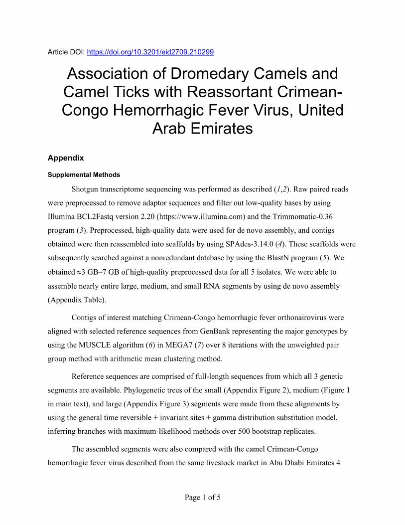
Article DOI: https;//doi.org/10.3201/eid2709.210299
Association of Dromedary Camels and Camel Ticks with Reassortant Crimean-Congo Hemorrhagic Fever Virus, United
Arab Emirates Appendix
Supplemental Methods
Shotgun transcriptome sequencing was performed as described (1,2). Raw paired reads
were preprocessed to remove adaptor sequences and filter out low-quality bases by using
Illumina BCL2Fastq version 2.20 (https://www.illumina.com) and the Trimmomatic-0.36
program (3). Preprocessed, high-quality data were used for de novo assembly, and contigs
obtained were then reassembled into scaffolds by using SPAdes-3.14.0 (4). These scaffolds were
subsequently searched against a nonredundant database by using the BlastN program (5). We
obtained ≈3 GB‒7 GB of high-quality preprocessed data for all 5 isolates. We were able to
assemble nearly entire large, medium, and small RNA segments by using de novo assembly
(Appendix Table).
Contigs of interest matching Crimean-Congo hemorrhagic fever orthonairovirus were
aligned with selected reference sequences from GenBank representing the major genotypes by
using the MUSCLE algorithm (6) in MEGA7 (7) over 8 iterations with the unweighted pair
group method with arithmetic mean clustering method.
Reference sequences are comprised of full-length sequences from which all 3 genetic
segments are available. Phylogenetic trees of the small (Appendix Figure 2), medium (Figure 1
in main text), and large (Appendix Figure 3) segments were made from these alignments by
using the general time reversible + invariant sites + gamma distribution substitution model,
inferring branches with maximum-likelihood methods over 500 bootstrap replicates.
The assembled segments were also compared with the camel Crimean-Congo
hemorrhagic fever virus described from the same livestock market in Abu Dhabi Emirates 4

Page 2 of 5
years earlier during 2015 (9). We provide sequence identities for this reference isolate (Appendix
Table).
References
1. Tayoun AA, Loney T, Khansaheb H, Ramaswamy S, Harilal D, Deesi ZO, et al. Multiple early
introductions of SARS-CoV-2 into a global travel hub in the Middle East. Sci Rep.
2020;10:17720. PubMed https://doi.org/10.1038/s41598-020-74666-w
2. Harilal D, Ramaswamy S, Loney T, Suwaidi HA, Khansaheb H, Alkhaja A, et al. SARS-CoV-2 Whole
genome amplificaton and sequencing for effective population-based surveillance and control of
viral transmission. Clin Chem. 2020;66:1450–8. PubMed
https://doi.org/10.1093/clinchem/hvaa187
3. Bolger AM, Lohse M, Usadel B. Trimmomatic: a flexible trimmer for Illumina sequence data.
Bioinformatics. 2014;30:2114–20. PubMed https://doi.org/10.1093/bioinformatics/btu170
4. Bankevich A, Nurk S, Antipov D, Gurevich AA, Dvorkin M, Kulikov AS, et al. SPAdes: a new
genome assembly algorithm and its applications to single-cell sequencing. J Comput Biol.
2012;19:455–77. PubMed https://doi.org/10.1089/cmb.2012.0021
5. Altschul SF, Gish W, Miller W, Myers EW, Lipman DJ. Basic local alignment search tool. J Mol Biol.
1990;215:403–10. PubMed https://doi.org/10.1016/S0022-2836(05)80360-2
6. Edgar RC. MUSCLE: multiple sequence alignment with high accuracy and high throughput. Nucleic
Acids Res. 2004;32:1792–7. PubMed https://doi.org/10.1093/nar/gkh340
7. Kumar S, Stecher G, Tamura K. MEGA7: Molecular Evolutionary Genetics Analysis version 7.0 for
bigger datasets. Mol Biol Evol. 2016;33:1870–4. PubMed
https://doi.org/10.1093/molbev/msw054
8. Deyde VM, Khristova ML, Rollin PE, Ksiazek TG, Nichol ST. Crimean-Congo hemorrhagic fever
virus genomics and global diversity. J Virol. 2006;80:8834–42. PubMed
https://doi.org/10.1128/JVI.00752-06
9. Khalafalla AI, Li Y, Uehara A, Hussein NA, Zhang J, Tao Y, et al. Identification of a novel lineage of
Crimean-Congo haemorrhagic fever virus in dromedary camels, United Arab Emirates. J Gen
Virol. 2021;102:doi: 10.1099/jgv.0.001473. PubMed https://doi.org/10.1099/jgv.0.001473

Page 3 of 5
Appendix Table. Summary of genomic sequences of Crimean-Congo hemorrhagic fever virus obtained from camel ticks by using a shotgun transcriptomic sequencing approach, United Arab Emirates, 2019* GenBank Accession no. Isolate name Segment Sequence length, bp % nt identity† No. aa changes† MW548490 UAE/CT25M1/2019 S 1,670 99.6 1 MW548495 UAE/CT25M1/2019 M 5,302 98.8 63 MW548500 UAE/CT25M1/2019 L 11,316 99.2 11 MW548491 UAE/CT25M2/2019 S 1,663 99.6 1 MW548496 UAE/CT25M2/2019 M 5,324 98.8 64 MW548501 UAE/CT25M2/2019 L 12,114 99.2 12 MW548492 UAE/CT25M3/2019 S 1,648 99.6 1 MW548497 UAE/CT25M3/2019 M 5,301 98.8 63 MW548502 UAE/CT25M3/2019 L 12,105 99.2 12 MW548493 UAE/CT219M1/2019 S 1,656 99.6 2 MW548498 UAE/CT219M1/2019 M 5,309 99.0 52 MW548503 UAE/CT219M1/2019‡ L 11,924 99.2 9 MW548494 UAE/CT219M3/2019 S 1,663 98.5 11 MW548499 UAE/CT219M3/2019 M 5,286 98.8 64 MW548504 UAE/CT219M3/2019 L 12,122 99.2 8 *L, large; M, medium; S, small; UAE, United Arab Emirates. †Reference sequences: Camel CCHFV/Abu Dhabi/B84, S: MN901927; M: MN901926; L: MN901925; ‡This sequence is not coding-complete; it is missing 577 nt from the 3′ complementary RNA end.
Appendix Figure 1. Map of the United Arab Emirates (gray), neighboring countries (white), and the
Arabian Gulf (blue). Sampling was performed at a livestock market in Al Ain (black star) and in Dubai
(black circle).

Page 4 of 5
Appendix Figure 2. Molecular phylogeny of selected Crimean-Congo hemorrhagic fever virus small RNA
segments. Viruses from this study are indicated by solid circles, and were isolated from camel ticks
(Hyalomma dromedarii) collected from dromedary camels at a large livestock market in United Arab
Emirates, 2019. Maximum-likelihood analysis of coding-complete sequences was performed by using the
general time reversible + invariant sites + gamma distribution substitution model and 4 categories with
>500 bootstrap replicates. Viruses include GenBank accession number and country of origin, and major
lineages are indicated in brackets on the right and colored according to Deyde et al. (8). Numbers along
branches are percentage support, showing only values >65%, and branch length is relative to the number
of substitutions per site, as indicated by the scale bar. Dem Rep Congo, Democratic Republic of the
Congo; UAE, United Arab Emirates.

Page 5 of 5
Appendix Figure 3. Molecular phylogeny of selected Crimean-Congo hemorrhagic fever virus large RNA
segments. Viruses from this study are indicated by solid circles, and were obtained from camel ticks
(Hyalomma dromedarii) collected from dromedary camels at a large livestock market in the emirate of
Abu Dhabi, United Arab Emirates, 2019. Maximum-likelihood analysis of coding-complete sequences
(with the exception of strain Al Ain/CT25M1/2019) was performed by using the general time reversible +
invariant sites + gamma distribution substitution model and 4 categories with >500 bootstrap replicates.
Viruses include GenBank accession number and country of origin, and major lineages are indicated in
brackets on the right and colored according to Deyde et al. (8). Numbers along branches are percentage
support, showing only values >65%, and branch length is relative to the number of substitutions per site,
as indicated by the scale bar. Dem Rep Congo, Democratic Republic of the Congo; UAE, United Arab
Emirates.