Assessment of the microbial safety of the drinking water...
77
Department of Food and Environmental Hygiene Faculty of Veterinary Medicine University of Helsinki Helsinki, Finland Medical School Finnish Defence Forces Lahti, Finland ASSESSMENT OF THE MICROBIAL SAFETY OF DRINKING WATER PRODUCED FROM SURFACE WATER UNDER FIELD CONDITIONS Ari Hörman ACADEMIC DISSERTATION To be presented with the permission of the Faculty of Veterinary Medicine, University of Helsinki, for public examination in Auditorium 1041, Biokeskus 2,Viikinkaari 5, Helsinki, on September 16 th , 2005 at 12 o’clock noon. Helsinki 2005
Transcript of Assessment of the microbial safety of the drinking water...

Department of Food and Environmental Hygiene Faculty of Veterinary Medicine
University of Helsinki Helsinki, Finland
Medical School
Finnish Defence Forces Lahti, Finland
ASSESSMENT OF THE MICROBIAL SAFETY OF DRINKING WATER
PRODUCED FROM SURFACE WATER UNDER FIELD CONDITIONS
Ari Hörman
ACADEMIC DISSERTATION
To be presented with the permission of the Faculty of Veterinary Medicine, University of Helsinki, for public examination in Auditorium 1041, Biokeskus 2,Viikinkaari 5, Helsinki,
on September 16th, 2005 at 12 o’clock noon.
Helsinki 2005

ii
Supervisors Professor Marja-Liisa Hänninen Department of Food and Environmental Hygiene
Faculty of Veterinary Medicine University of Helsinki Helsinki, Finland
Docent Heikki Korpela Central Military Hospital Finnish Defence Forces Helsinki, Finland
Supervising Professor Professor Hannu Korkeala Department of Food and Environmental Hygiene
Faculty of Veterinary Medicine University of Helsinki Helsinki, Finland
Reviewers Doctor Gertjan Medema Kiwa Water Research Nieuwegein, Netherlands Professor Thor-Axel Stenström Department of Parasitology, Mycology and Water
Swedish Institute for Infectious Disease Control Solna, Sweden Opponent Docent Jorma Hirn National Food Agency Helsinki, Finland Cover photos: Ari Hörman ISBN 952-91-9103-0 (paperback) ISBN 952-10-2626-x (PDF) http://ethesis.helsinki.fi/ Yliopistopaino Helsinki 2005

iii
To lake Kärenjärvi in Kaavi, where I learned to swim and became interested in surface waters and their quality.

ii
CONTENTS
ACKNOWLEDGEMENTS ................................................................................................. IV
ABBREVIATIONS ............................................................................................................. VI
ABSTRACT.........................................................................................................................1
LIST OF ORIGINAL PUBLICATIONS.................................................................................2
1. INTRODUCTION .............................................................................................................3
2. REVIEW OF THE LITERATURE.....................................................................................5
2.1 DRINKING WATERBORNE ENTERIC DISEASES IN HUMANS..................................................................... 5 2.1.1 Significant drinking waterborne enteropathogens worldwide and in Finland ................................ 5
2.1.1.1 Noroviruses .............................................................................................................................................. 6 2.1.1.2 Campylobacter spp. ............................................................................................................................... 7 2.1.1.3 Giardia spp. and Cryptosporidium spp. ............................................................................................ 7
2.1.2 Surveillance for drinking waterborne enteric diseases ........................................................................ 9
2.2 ENTEROPATHOGENIC AND INDICATOR MICROBES IN SURFACE WATER ........................................ 10 2.2.1 Enteropathogens in surface water ........................................................................................................... 10 2.2.2 Indicator microbes and water quality ...................................................................................................... 11
2.3 MICROBIOLOGICAL REQUIREMENTS FOR DRINKING WATER QUALITY ............................................ 14
2.4 WATER TREATMENT METHODS UNDER FIELD CONDITIONS................................................................. 15 2.4.1 Overview on water treatment ..................................................................................................................... 15 2.4.1 Thermal treatment......................................................................................................................................... 16 2.4.3 Chemical disinfection .................................................................................................................................. 17 2.4.4 Filtration .......................................................................................................................................................... 20 2.4.5 Other treatment methods ............................................................................................................................ 21
2.5 CONCEPTS OF MICROBIAL RISK ASSESSMENT AND MANAGEMENT OF DRINKING WATER ..... 22 2.5.1 Quantitative microbial risk assessment (QMRA) .................................................................................. 22 2.5.2 Hazard analysis of critical control points (HACCP) and water safety plans (WSP) ..................... 23 2.5.3 Acceptable risk .............................................................................................................................................. 23
2.6 BIOTERRORISM AND INTENTIONAL CONTAMINATION OF DRINKING WATER ................................. 24 2.6.1 Biohazardous agents ................................................................................................................................... 24 2.6.2 Detection of bioterrorism ............................................................................................................................ 26 2.6.3 Protection against bioterrorism ................................................................................................................ 26
3. AIM OF THE STUDY.....................................................................................................28
4. MATERIALS AND METHODS ......................................................................................29
4.1 Enteropathogens and indicators in surface water (I) .................................................................................. 29 4.1.1 Sampling sites and sampling (I) ................................................................................................................ 29 4.1.2 Microbiological and physicochemical analysis (I) ............................................................................... 29
4.2 Assessment of water treatment devices and tests for detection of botulinum toxin, coliform bacteria and Escherichia coli (II-IV) ........................................................................................................................ 30

iii
4.2.1 Natural and seeded water samples (II-IV) ............................................................................................... 30 4.2.2 Testing of the water treatment devices (II, III)........................................................................................ 32 4.2.3 Microbiological and physicochemical analyses (II, III)........................................................................ 32 4.2.4 Tests for detection of botulinum neurotoxin (III) .................................................................................. 34 4.2.5 Tests for detection of coliform bacteria and Escherichia coli (IV) ................................................... 34
4.3 Prevalence and incidence of Giardia spp. and Cryptosporidium spp. infections in the Nordic countries (V) .................................................................................................................................................................. 34
4.3.1 Meta-analysis (V) ........................................................................................................................................... 34 4.3.2 Estimation of annual incidence of Giardia spp. and Cryptosporidium spp. infections in the general population (V) ........................................................................................................................................... 36
4.4 Statistical methods (I-V) ...................................................................................................................................... 36 4.4.1 Enteropathogens in surface water (I)....................................................................................................... 36 4.4.2 Evaluation of the water treatment devices (II, III).................................................................................. 37 4.4.3 Evaluation of tests for detection of coliform bacteria and Escherichia coli (IV) ......................... 37 4.4.4 Prevalence and incidence of Giardia spp. and Cryptosporidium spp. infections in the Nordic countries (V) ............................................................................................................................................................. 38
5. RESULTS......................................................................................................................40
5.1 Enteropathogens and indicators in surface water (I) .................................................................................. 40 5.1.1 Prevalence of enteropathogens and indicators in surface water (I) ................................................ 40 5.1.2 Correlation between enteropathogens and indicators in surface water samples (I) .................. 41
5.2 Evaluation of water treatment devices (II, III) ................................................................................................. 41
5.3 Evaluation of tests for detection of botulinum toxin (III) ............................................................................ 42
5.4 Evaluation of tests for detection of coliform bacteria and Escherichia coli (IV) .................................. 42
5.5 Prevalence and incidence of Giardia spp. and Cryptosporidium spp. infections in the Nordic countries (V) .................................................................................................................................................................. 42
6. DISCUSSION ................................................................................................................44
6.1 Enteropathogens and indicators in surface water (I) .................................................................................. 44
6.2 Evaluation of water treatment devices (II, III) ................................................................................................. 45
6.3 Evaluation of tests for detection of botulinum toxin (III) ............................................................................ 46
6.4 Evaluation of tests for detection of coliform bacteria and Escherichia coli (IV) .................................. 47
6.5 Prevalence and incidence of Giardia spp. and Cryptosporidium spp. infections in the Nordic countries (V) .................................................................................................................................................................. 48
7. CONCLUSIONS ............................................................................................................50
8. REFERENCES ..............................................................................................................52

iv
ACKNOWLEDGEMENTS I would like to express my sincere gratitude to the Department of Food and Environmental Hygiene, Faculty of Veterinary Medicine, University of Helsinki, for providing the opportunity, facilities and stimulating atmosphere for carrying out this research project. Without the crucial co-operation between the Department and the Finnish Defence Forces the project would not have been possible. The head of the Department and supervising professor of this work, Professor Hannu Korkeala deserves a distinction for giving his support to me and to this work. I am most grateful to my primary supervisor Professor Marja-Liisa Hänninen for her willingness and readiness for giving her endless and broad knowledge, support and enthusiasm into supervision of me and this research project. My gratitude comes directly from my heart and it was truly a pleasure to work with Marja-Liisa. I am also grateful to my second supervisor Docent Heikki Korpela for his exemplary work in combining veterinary and human medicine and epidemiology; I also would like to thank Heikki for several stimulating discussions. This work was a successful example of active co-operation between several institutes and individual experts. Without the contribution from each of the co-authors this work would have been very difficult or even impossible to carry out. Thank you for sharing your valuable time and knowledge fellow researchers Ruska Rimhanen-Finne, Mari Nevas and Annamari Heikinheimo, and Kirsti Lahti and Jarkko Rapala. I want to express a warm distinction to Professor Carl-Henrik von Bonsdorff and Doctor Leena Maunula for guiding me into exciting world of viruses. Two co-authors deserve special remarks not only for being collaborators in this work but also being important and close friends of mine. Miia Lindström and Jussi Sutinen, you are truly experts in your fields, but also like a sister and brother to me - in good and in bad. The enjoyable and important co-operation with the Nordic School of Public Health in Gothenburg, Sweden, has given me a lot of knowledge in the field of public health, epidemiology and biostatics. I am most grateful for having had an opportunity to have been taught by Professor Hans Wedel and other professional teachers in Gothenburg. The competent personnel at the Department of Food and Environmental Hygiene have willingly done a lot of practical work for this project in the laboratory and in office. A cordial thank to you all, especially to Urszula Hirvi, Hürol Samaletdin, Hanna Korpunen and Johanna Seppälä. I appreciate the valuable comments from the reviewers of my thesis, Doctor Gertjan Medema and Professor Thor-Axel Stenström. It has been a pleasure and honour to receive guidance from two of the leading experts in the world in the field of assessment of drinking water safety. Thank you for being able to share your valuable and busy time for this work. The Finnish Defence Forces have given me the opportunity to carry out this research. I hope that this work and its results will prove to be worth of the trust and resources that were kindly offered to me. I am most grateful to the organisations and personnel of the Häme Regiment, the Logistic

v
Training Centre and the Medical School for having facilitated this work and having shown a great patience during the last five years. My veterinarian colleagues in the Finnish Defence Forces have encouraged me significantly during this research project. I am thankful for Eeva Sovijärvi for constructive comments during this project. A particular thank goes to the Chief Veterinarian Juhani Tiili for inducing me to the work at the Finnish Defence Forces and leading me into the important and interesting topic of drinking water safety under field conditions. I am also grateful for the great interest and support from the Medical and Logistic sectors and their personnel in the Finnish Defence Forces to this work. It has also been a pleasure to have been able to work with my veterinarian colleagues in the Swedish, Norwegian and Danish Defence Forces, thank you for a fruitful co-operation. My sincere thanks go to James Thompson for the language revision of my texts during this project. I am also very thankful to Anssi Mattila for the great work of designing the visual outlook of numerous posters and other material during this project. No project is possible without an active network of supportive persons and close friends. I am most grateful for having so good friends who have shared their time during the good and bad days during all these years and hopefully also in the future. Warm thank to you, dear Peksu, Köpi, Sanni, Mappe, Heini, Juha, Sönke, Aatu, Marju, Katri, Kari, Satu, Panu, Sampo, Outi, Make and numerous other good friends. Fundamentally thinking, this work would not have been possible without my dear parents Raili and Vilho. You have supported me unselfishly during all my life and this work. Thank you! This work was financially supported by the Finnish Scientific Advisory Board for Defence (MATINE), Ministry of Defence, Finland (study grant Mdd587), by the Finnish Veterinary Foundation (Suomen Eläinlääketieteen Säätiö) and by the Field catering project of the Finnish Defence Forces. This research project was joined in the Graduate School on Environmental Health 2002-2005. I am very honoured and grateful for having received support from these organizations; this support has facilitated the work.

vi
ABBREVIATIONS
ANOVA analysis of variance ATCC American Type Culture Collection B-agent biological agent BHI brain-hearth –infusion BoNT botulinum neurotoxin CCDA charcoal cefoperazone deoxycholate agar CFU colony-forming unit CI confidence interval CT concentration-time DALY disability-adjusted life-year df degrees of freedom DFEH Department of Food and Environmental Hygiene, University of Helsinki, Finland DSL DerSimonian Laird EIA enzyme-immune assay ELISA enzyme-linked immunosorbent assay EPA Environmental Protection Agency (USA) EU European Union FEI Finnish Environment Institute, Helsinki, Finland F-RNA F-specific ribonucleic acid HACCP hazard analysis of critical control points HI Haartman Institute, University of Helsinki, Finland HPC heterotrophic plate count IFA immunofluorescence assay IFR Institute of Food Research, Norwich, UK IMS immunomagnetic separation ISO International Standardization Organization log10 logarithmic LTTC lactose triphenyl tetrazolium chloride MF membrane filtration MPN most probable number NATO North Atlantic Treaty Organization NTU nephlometric turbidity unit NV noroviruses OR odds ratio PCR polymerase chain reaction PFU plaque-forming unit ppb parts per billion ppm parts per million QMRA quantitative microbial risk assessment RE random effects RNA ribonucleic acid RO reverse osmosis

vii
RT-PCR reverse transcriptase PCR SD standard deviation SFP Shahidi Ferguson Perfringens SFS Finnish Standards Association SFS TPGY tryptone-peptone-glucose-yeast extract UK United Kingdom USA United States of America US United States (of America) UV ultraviolet WHO World Health Organization WSP water safety plan

1
ABSTRACT
Treated or untreated surface water is one of the main sources of drinking water under field and emergency conditions. The aims of the present thesis were to determine the prevalence of enteropathogens in surface water in Finland, evaluate the purification capacities of water treatment devices and the methods used for detection of enteropathogens and indicators to obtain data for the assessment and management of microbial risks in drinking water production from surface water. The present study will aid in developing practical plans to improve water safety, especially under field conditions. In all, 41.0% (57/139) of the surface water samples collected during 2000-2001 were positive for at least one of the analysed pathogens: 17.3% positive for campylobacters, 13.7% for Giardia spp., 10.1% for Cryptosporidium spp. and 9.4% for noroviruses (23.0% genogroup I and 77.0% genogroup II). During the winter season, the samples were significantly (p < 0.05) less frequently positive for enteropathogens than during other sampling seasons. No significant differences were found in the prevalences of enteropathogens between rivers and lakes. The presence of thermotolerant coliforms, Escherichia coli and Clostridium perfringens showed significant bivariate, nonparametric, Spearman’s rank order correlation coefficients (p < 0.001), with a sample being positive for one or more of the analysed enteropathogens. No significant correlations were observed between counts or count levels of thermotolerant coliforms, E. coli or presence of F-RNA phages and enteropathogens in the analysed samples. In general, the water treatment devices tested were able to remove bacterial contaminants by 3.6–6.9 log10 units from contaminated raw water, while devices based only on filtration through pores 0.2–0.4 µm or larger failed in viral and chemical purification. Only one device, based on reverse osmosis, was capable of removing F-RNA phages and botulinum neurotoxin (BoNT) at concentrations under the detection limit and microcystins by 2.5 log10 units. Simultaneous testing for various enteropathogenic and indicator microbes was a useful and practical way to obtain data on the purification capacity of commercial small-scale drinking water filters. The m-Endo LES SFS 3016:2001 was the only method showing no statistical differences in E. coli counts compared with the reference method LTTC ISO 9308-1:2000, whereas the Colilert 18 and Readycult methods showed significantly higher counts for E. coli than the LTTC method. Based on this evaluation study, the Colilert 18, Readycult and Water Check methods are all suitable for coliform and E. coli detection both under field conditions and in routine use in the water industry. The two rapid enzyme immunoassay tests intended for the detection of BoNT failed to detect BoNT from aqueous samples containing an estimated concentration of BoNT of 396 000 ng/l. We estimated the prevalence of Giardia infections in the asymptomatic (i.e. no gastroenteric symptoms) general population in the Nordic countries to be 2.97% (95% CI: 2.64; 3.31) and in the symptomatic population 5.81% (95% CI: 5.34; 6.30). For Cryptosporidium the prevalences were 0.99% (95% CI: 0.81; 1.19) and 2.91% (95% CI: 2.71; 3.12), respectively. The vast majority of cases will remain unregistered in the national registers of infectious diseases, since for single registered cases there will be 254-867 cases of Giardia remaining undetected/unregistered and 4072-15 181 cases of Cryptosporidium, respectively.

2
LIST OF ORIGINAL PUBLICATIONS
The present thesis is based on the following original articles referred to in the text by the Roman numerals I to V:
I Hörman, A., Rimhanen-Finne, R., Maunula, L., Bonsdorff von, C. H., Torvela, N.,
Heikinheimo, A. and Hänninen, M. L. 2004. Campylobacter spp., Giardia spp., Cryptosporidium spp., noroviruses, and indicator organisms in surface water in Southwestern Finland, 2000-2001. Appl. Environ. Microbiol. 70:87-95.
II Hörman, A., Rimhanen-Finne, R., Maunula, L., Bonsdorff von, C. H., Rapala, J., Lahti, K.
and Hänninen, M. L. 2004. Evaluation of the purification capacity of nine portable, small-scale water purification devices. Water Sci. Technol. 50(1):179-183.
III Hörman, A., Nevas, M., Lindström, M., Hänninen, M. L. and Korkeala H. 2005.
Elimination of botulinum neurotoxin (BoNT) type B from drinking water by small-scale water purification devices and detection of BoNT in water samples. Appl. Environ. Microbiol. 71:1941-1945.
IV Hörman, A. and Hänninen, M. L. Evaluation of Tergitol-7, m-Endo LES, Colilert-18,
Readycult Coliforms 100, Water Check 100, 3M Petrifilm EC DryCult Coliform tests methods for detection of total coliforms and Escherichia coli in water samples. (submitted).
V Hörman, A., Korpela, H., Sutinen, J., Wedel, H. and Hänninen, M. L. 2004. Meta-analysis
in assessment of the prevalence and annual incidence of Giardia spp. and Cryptosporidium spp. infections in humans in the Nordic countries. Int. J. Parasitol. 34:1337-1346.
The original articles have been reprinted with kind permission from the American Society for Microbiology (I, III), International Water Association (II) and Elsevier Science (V).

3
1. INTRODUCTION Drinking water is worldwide the most important single source of gastroenteric diseases, mainly due to the faecally contaminated raw water, failures in the water treatment process or recontamination of treated drinking water (Medema et al., 2003a; World Health Organization, 2003a). Two thirds of the total drinking water consumed worldwide is derived from various surface water sources (Annan, 2000) that may easily be contaminated microbiologically by sewage discharges or faecal loading by domestic or wild animals or whose microbial quality may be endangered by various weather conditions. In Finland 42% of the total drinking water was produced from surface water in 2001 (Finnish Environment Institute, 2003). Surface waters are also widely used for leisure and recreational activities, and thus unintended ingestion of microbiologically contaminated water poses a potential health risk (Cabelli et al., 1982; Asperen van et al., 1998; Stuart et al., 2003; Schönberg-Norio et al., 2004). Treated or untreated surface water is also one of the main sources of drinking water under field and emergency conditions (Backer, 2002; Townes, 2002; Boulware et al., 2003). A minimum of two litres of safe drinking water should be available per person daily to compensate for the water lost in urine, faeces or perspiration (North Atlantic Treaty Organization, 2002). During physical exercise, compensation for lost fluid is essential to maintain physical and mental activity (Noakes et al., 1988; Armstrong et al., 1997). Unsafe or contaminated drinking water may infect and incapacitate not only individual persons but also large groups, thus prohibiting them from fulfilling their tasks (Blaisdell, 1988; Aho et al., 1989; Sartin, 1993; Cook, 2001; Boulware et al., 2003; Boulware, 2004). Field conditions here refer to those situations without organized, municipal or other piped water supplies. The present work focuses on those field conditions under which individual persons or groups produce their drinking water from various surface fresh water sources for direct consumption. This type of condition is usually encountered by military and aid personnel, hikers or any person in wilderness or emergency situations. Drinking water production, from surface water sources to the consumer, is described as a flow chart in Figure1 from the perspective of microbial safety and security. The term drinking water safety refers here to drinking water hygiene, microbiological hazards, microbial risk assessment and management of risks, whereas security refers to preventive measures for minimizing the risk that drinking water supplies will be tampered with or become targets for bioterrorism (Khan et al., 2001; Rose, 2002; Luthy, 2002). All these activities combined under the concepts of drinking water safety and security help ensure the microbial safety of drinking water. The term microbial pathogens refers here to the waterborne organisms, enteropathogenic bacteria, viruses and protozoa and the toxins produced by them and assessment of microbial safety regarding the possibility of these hazardous agents entering drinking water supplies (World Health Organization, 2003b). Microbial risks associated with the water treatment processes at large water plants, during distribution of treated drinking water to consumers or activities undertaken by the end-user are not included here. These factors play a significant role in the overall microbial safety of drinking water, especially in communities with extensive piped water supply systems. Under field and emergency

4
conditions the main safety and security efforts are focused on selection of the best raw water source available, utilization and control of an effective treatment process and control of security. The significance of drinking water safety and security has increased, especially after the terrorist acts in 2001 (Rose, 2002; Meinhardt, 2005). Although, these acts were not targeted against drinking water supplies, the vulnerability of these supplies as targets of bioterrorism has been a concern of public health authorities and policymakers (Christen, 2001). The international concepts of hazard analysis of critical control points (HACCP) (Dewettinck et al., 2001; Howard, 2003; Westrell et al., 2004) and water safety plans (WSP) by the World Health Organization (WHO) (World Health Organization, 2004) have been introduced to enable the improvement of drinking water safety and security. WSPs include health-based targets, which means that the microbial risks and adverse health effects to which a population is exposed through drinking water should be minimized, be very low and not exceed the tolerable risk suggested by WHO (World Health Organization, 2004). Nationally both civil and military authorities and other organizations have initiated projects to develop plans and measures for ensuring safe drinking water supplies. The present studies will hopefully aid in assessing the microbial safety of drinking water and in developing practical plans to improve water safety, especially in the field.
Watertreatment
Surface water source Water consumers
Outcome of waterbornediseases
-End-pointmonitoring of purified water-Security
-Operational control-Security
-Surveillanceand control of raw water quality-Selection of best source
-Sewage-Animals-Agriculture-Weather conditions-Seasonal factors
-Inefficiency-Malfunction-Carelessness -Errors
-Failures in control and insurveillance
-Doseresponse-Host-related factors
-Intentional contamination(B-terrorism)
-Surveillanceof waterbornediseases
Figure 1. A flow chart showing production of drinking water from surface water, including factors bearing impact on microbial safety and selection of critical control points. Production stages and critical control points bearing major impact under field conditions are underlined.

5
2. REVIEW OF THE LITERATURE 2.1 DRINKING WATERBORNE ENTERIC DISEASES IN HUMANS 2.1.1 Significant drinking waterborne enteropathogens worldwide and in Finland Waterborne gastrointestinal infections remain one of the major causes of morbidity and mortality worldwide (World Health Organization, 2002b; World Health Organization, 2003a). The most important microbes causing infections or epidemics through drinking water include the bacteria Campylobacter spp., Escherichia coli, Salmonella spp., Shigella spp., Vibrio cholerae and Yersinia enterocolitica, viruses such as: adeno-, entero-, hepatitis A- and E-, noro-, sapo- and rotaviruses and the protozoa: Cryptosporidium parvum, Dracunculus medinensis, Cyclospora cayetanensis, Entamoeba histolytica, Giardia duodenalis and Toxoplasma gondii (World Health Organization, 2004). Historically, large waterborne cholera epidemics with numerous casualties in the mid-1800s, the early investigations of cholera epidemics in London by John Snow (1813-1858) and the works of Robert Koch (1843-1910) on V. cholerae have remarkably increased the level of understanding of the epidemiology and prevention of waterborne diseases (Brock, 1999). Worldwide, V. cholerae is still a significant cause of waterborne infections, especially in developing countries where most of the victims are children under five years of age (World Health Organization, 2002b; World Health Organization, 2003a; Ashbolt, 2004a). Epidemiological studies of waterborne outbreaks in Finland have indicated that the most important waterborne pathogens in Finland are noroviruses (NVs; formerly referred to as the Norwalk-like viruses) and campylobacters (Miettinen et al., 2001; Vartiainen et al., 2003; Kuusi, 2004). During 1998-1999, eight of a total of 14 waterborne outbreaks reported were caused by NVs and three by campylobacters (Miettinen et al., 2001). This trend has continued also during a longer surveillance period, in 1980-2001 nine (15%) of a total 61 waterborne outbreaks reported were caused by campylobacters and 17 (27%) by noro- and other viruses, while in 26 (43%) outbreaks the causal agent remained unknown (Johansson et al., 2003). NVs are also the leading causes of gastroenteritis elsewhere in the Western world, causing 60-80% of all gastroenteritis outbreaks (Fankhauser et al., 2002; Lopman et al., 2003b). Under field conditions NV outbreaks are common, especially during military deployments (Sharp et al., 1995; McCarthy et al., 2000; Ahmad, 2002). Campylobacter spp. are the most common bacterial causes of gastroenteritis in the Nordic countries (Rautelin and Hänninen, 2000). A total of 15 000 persons have been estimated to have infected in reported waterborne outbreaks in Finland during 1988-2002 but the true number of infected persons was estimated to be significantly higher (Vartiainen et al., 2003). The leading technical cause of community-based outbreaks in Finland has been faecally contaminated groundwater, either by surface water overflow or by sewage discharge (Miettinen et al., 2001). One large NV epidemic with almost 3000 infected persons occurred when contaminated and untreated surface water was distributed in a community to customers (Kukkula et al., 1999). In Finland one documented campylobacter outbreak occurred under field conditions when drinking of

6
untreated surface water caused severe campylobacter gastroenteritis among military conscripts during a field exercise (Aho et al., 1989). Enteric parasites such as Giardia spp. and Cryptosporidium spp. have not been reported to cause waterborne epidemics in Finland according to the National Register of Infectious Diseases (Finnish National Public Health Institute, 2003), but a small number of sporadic cases are reported annually. However, these protozoa are well recognized as emerging pathogens in drinking water and as being able to cause severe waterborne enteritis even with small doses, especially in immunocompromised persons (Franzen and Muller, 1999; Szewzyk et al., 2000). Giardia spp. and Cryptosporidium spp. are common causes of human diarrhoeal diseases in the developed and developing countries (Marshall et al., 1997; Clark, 1999). Outbreaks associated with contaminated drinking water have occurred, especially in the United States of America (USA) and the United Kingdom (UK). Cryptosporidium parvum infected 403 000 persons, in one of the largest waterborne epidemics ever seen, in Milwaukee, WI, USA in 1993 (MacKenzie et al., 1994). During the 1990s Cryptosporidium was one of the most important pathogenic contaminants found in drinking water, due to its low infective dose (Dillingham et al., 2002), high resistance to the commonly used water disinfectant, chlorine, and to environmental factors such as low temperature (Rose, 1997; Fayer et al., 1998; Payment, 1999). 2.1.1.1 Noroviruses Human NVs, earlier described as Norwalk-like viruses, belong to the genus Caliciviridae, together with the sapoviruses. NVs are small ribonucleic acid (RNA) viruses, with an RNA genome of approximately 7.5-7.7 kb, which enables their high degree of genomic plasticity and capability to adapt to new environmental niches (Radford et al., 2004). NVs were divided recently into five genogroups, genogroups I and II being associated mostly with human infections. Within genogroups there is wide inherent genetic variability and at least 20 genotypes have been recognized (Radford et al., 2004). NV infection is typically a violent vomiting disease with a sudden onset and an incubation period of normally 1-3 days. In addition to vomiting, symptoms may include high fever, diarrhoea and headache. The symptoms are generally self-limited and last 2-3 days (Kaplan et al., 1982a). The infective dose for man is very low: 10-100 virus particles may cause a clinical infection (Green, 1997; Schaub and Oshiro, 2000). Large amounts of viruses, 109-10 virus particles per ml (Bonsdorff von and Maunula, 2003), are excreted in faeces and vomit and the person may be infective during the incubation period and remain infective to 2-3 weeks after the symptoms have ended (Okhuysen et al., 1995; Thornton et al., 2004). NV gastroenteritis is rapidly and effectively spread from person to person, especially in close contacts (Koopmans et al., 2002). In most cases the NV infection does not require medication but some severe cases may need hospitalization and fluid therapy (Kaplan et al., 1982b; Arness et al., 2000). Detection methods of NVs in faecal samples have developed remarkably after molecular methods were applied to virus detection (Koopmans and Duizer, 2004). The most sensitive method for

7
detecting NVs is the reverse transcriptase -polymerase chain reaction (RT-PCR), which is able to detect 1-1000 virus particles per gram, although the less sensitive electron microscopy and enzyme-linked immunosorbent assay (ELISA) are also utilized (Koopmans and Duizer, 2004; Thornton et al., 2004). Before these specific detection methods were available, the causative agents of most viral epidemics and infections remained unspecified or unsolved (Johansson et al., 2003). These molecular methods are useful tools in epidemiological investigations and in tracking of infection routes (Maunula et al., 1999; Bonsdorff von and Maunula, 2003; Lopman et al., 2003a; Kuusi, 2004). 2.1.1.2 Campylobacter spp. Campylobacter enteritis in man is caused mainly by Campylobacter jejuni or C. coli which are zoonotic and carried by wild and domestic animals, especially by birds and poultry (Blaser, 1997). The pathogenic potential of C. jejuni and C. coli was not discovered until the 1970s (Szewzyk et al., 2000). Cambylobacters are microaerophilic and survive for only a few hours in the environment at high temperatures (> 30 °C) but several days at low (4 °C) temperatures (Szewzyk et al., 2000). The infective dose of campylobacters is relatively low: 800-100 000 ingested organisms are needed to cause illness in man (Black et al., 1988). During the 1990s, Campylobacter-like organisms, such as Arcobacter spp. were described, which occur in the environment and possess pathogenic potential (Szewzyk et al., 2000). Campylobacter infection is usually self-limited and characterized by diarrhoea, fever and abdominal cramps (Butzler, 2004). The incubation time can vary from 1 to 10 days, but is usually 2-5 days. Diarrhoea may last for 3-5 days, although abdominal pain and cramps may continue afterwards (Blaser, 1997). Campylobacter infection may lead to severe but rare sequelae, including reactive arthritis (Hannu et al., 2004), Guillain-Barré syndrome (Hughes, 2004; Kuwabara, 2004) or myocarditis (Cunningham and Lee, 2003). Risk for developing Guillan-Barré syndrome is low, less than 1 per 1000 infections (Hughes, 2004; Kuwabara, 2004). Diagnosis of Campylobacter gastroenteritis is traditionally done by bacterial culture of faecal samples in selective media and isolation and detection of typical colonies and by morphological and biochemical tests (Hänninen et al., 2003). Positive isolates can be further subtyped to various serotypes according to the antigens detected; tests for antibiotic resistance can also be applied for subtyping. During recent years, pulsed-field gel electrophoresis has been utilized in typing of Campylobacter strains and this has increased the accuracy of epidemiological investigations (Hänninen et al., 1998; Moore et al., 2001; Hänninen et al., 2003). 2.1.1.3 Giardia spp. and Cryptosporidium spp. The genus Giardia comprises six species that can infect a variety of hosts. Giardia duodenalis (also referred to as G. intestinalis or G. lamblia) is infectious for humans but can also cause infections in other hosts (Monis et al., 2003). The spectrum of clinical giardiasis varies from

8
asymptomatic carriers to severe diarrhoea and malabsorption. Acute giardiasis develops after an incubation period of 1-14 days (mean 7 days) and usually lasts 1-3 weeks. The symptoms include watery, foul-smelling diarrhoea, abdominal pain, bloating, nausea and vomiting. In chronic giardiasis the symptoms are recurrent and malabsorption and debilitation may occur. Occasionally, the illness may last for months, or even years, causing recurrent mild or moderate symptoms such as impaired digestion, especially lactose intolerance, intermittent diarrhoea, tiredness and weakness, and significant weight loss. Giardiasis is diagnosed by the identification of cysts or trophozoites in the faeces, using direct microscopy as well as concentration procedures. Repeated samplings may be necessary, sometimes for 4-5 weeks, to obtain a positive laboratory diagnosis. In addition to faecal samples, samples of the duodenal fluid or a duodenal biopsy may demonstrate trophozoites. Alternative methods for detection include antigen detection tests using enzyme-immuno assays (EIA) and detection of cysts by immunofluorescence assay (IFA), commercial reagents are available for both methods. The genus Cryptosporidium was recently suggested to comprise over 20 species based on morphological, biological and genetic studies (Xiao et al., 2004). These species have several mammalian and nonmammalian hosts and cross-infections may occur between various host species (Dillingham et al., 2002). In humans cryptosporidiosis was first diagnosed in the late 1970s in immunocompromised persons for which Cryptosporidium can cause severe, even fatal disease (Marshall et al., 1997). Later the causal agent C. parvum was noted as a global human enteropathogen. Cryptosporidium parvum is genetically divided into human genotype 1 (C. hominis) and genotype 2, which also infects cattle (Dillingham et al., 2002). The life cycle of Cryptosporidium is more complex than that of Giardia and includes an asexual and a sexual stage inside the host’s intestine and an infective stage outside the host: the oocyst stage (Centers for Disease Control and Prevention, 2001; Dillingham et al., 2002; Centers for Disease Control and Prevention, 2003; Monis and Thompson, 2003). The symptoms of cryptosporidiosis include diarrhoea, loose or watery stools, stomach cramps, upset stomach and a slight fever (Centers for Disease Control and Prevention, 2003). Some infected persons are asymptomatic, while in others the symptoms generally begin after a 2-10 -day incubation period. In persons having average immune systems, the symptoms usually last approximately two weeks. The symptoms may occur in cycles in which the person may appear to recover for a several days, then feel worse, before the illness ends. Although Cryptosporidium can infect all people, some groups are more likely to develop more serious illness. People that have a severely weakened immune system, cancer, transplant patients receiving certain immunosuppressive drugs and those with inherited diseases that affect the immune system are at risk for more serious disease (Keusch et al., 1995; Gerba et al., 1996). In these patients the symptoms may be more severe and could lead to serious, even life-threatening illness. Testing for Cryptosporidium can be difficult and several stool specimens over several days may be needed to detect the oocysts of the parasite. Acid-fast staining methods, with or without stool concentration, are most frequently used in clinical laboratories for detection of Cryptosporidium oocysts. For increased sensitivity and specificity, IFA and EIA are used in some clinical laboratories, while molecular methods in the detection and subtyping are mainly applied for

9
research purposes. However, tests for Cryptosporidium are not routinely done in most clinical laboratories (Nygård et al., 2003). There is no established specific therapy for human cryptosporidiosis (Marshall et al., 1997). Rapid loss of fluids resulting from diarrhoea can be managed by fluid therapy. Nitazoxanide has provided some encouraging results in the management of cryptosporidiosis in immunocompetent patients (White, Jr., 2003). For persons with acquired immunodeficiency syndrome antiretroviral therapy, which improves immune status, will also reduce oocyst excretion and decrease the diarrhoea associated with cryptosporidiosis (Miao et al., 2000; Ives et al., 2001; Kaplan et al., 2002). 2.1.2 Surveillance for drinking waterborne enteric diseases Surveillance of waterborne outbreaks in Finland is the responsibility of the local municipal public health authorities (Statute Book of Finland, 1994a). The health authorities have the duty to investigate suspected waterborne outbreaks and report them further to provincial and governmental authorities, the National Public Health Institute, National Food Agency and Food and Veterinary Research Institute. When the outbreak is investigated the report is sent to the National Food Agency. State authorities, including the National Public Health Institute and National Food Agency, collect the reports and analyse them annually. The present notification system has been in effect since 1997 (Kuusi, 2004). The assumption is that large community-based drinking waterborne epidemics will be reported, even if delayed but mild, single or obscure waterborne infections will probably remain undetected and unreported. Military organizations usually have well-established regular health surveillance systems and single outbreaks, epidemics and severe cases of waterborne infections are usually noted without delay. To be able to recognize and report a waterborne disease, the health care provider or medical personnel must first be contacted by the infected person who develops the symptoms. The symptoms and anamnesis may then guide the medical personnel to suspect a waterborne disease and take the necessary faecal, vomit or other samples. Other possible patients with similar symptoms and anamneses, e.g. time and place of exposure, can provide valuable information for outbreak investigation. The WHO defines a waterborne outbreak as an episode in which two or more persons experience a similar illness after ingestion of the same type of water from the same source and when the epidemiological evidence implicates the water as the source of the illness (Schmidt, 1995). A sufficient number of samples collected from the drinking water consumed at the early stages of investigation are essential to facilitate connection of the exposure with the outbreak. To obtain representative samples may be difficult or even impossible due to the time-lag between the exposure and the time when person has developed symptoms and contacted the health care personnel (Hunter et al., 2003a). The present assumption is that some of waterborne diseases are underdetected and underreported (Kukkula et al., 1999; Leclerc et al., 2002; Vartiainen et al., 2003) especially those caused by Giardia and Cryptosporidium, in official infectious disease registers in the Nordic countries (Nygård et al., 2003). One evident reason for this underestimation is that not all patients have severe symptoms and seek medical care. The clinical symptoms may be masked by other

10
causes and thus faecal samples will not be analysed for the presence of protozoa. Laboratory analysis may also fail to detect these parasites in faecal samples. Underreporting has also been estimated for viruses (Kukkula et al., 1999; Koopmans and Duizer, 2004). Thus the subclinical, asymptomatic or undetected cases may play significant roles in infection transmission and epidemiology in the general population. 2.2 ENTEROPATHOGENIC AND INDICATOR MICROBES IN SURFACE WATER 2.2.1 Enteropathogens in surface water
Enteropathogenic microbes are usually adapted to multiplying in the intestines of humans and animals and surface water is only a niche in their circulation (Figure 1) through the environment and human or animal populations (Medema et al., 2003a). The occurrence of waterborne enteropathogenic microbes in surface water is associated with faecal contamination of surface water sources (Westrell et al., 2003; Ashbolt, 2004a). Environmental factors influence how enteropathogens survive and move in surface water. Faecal contamination can originate from municipal or domestic sewage discharges or from direct release of faecal material into surface water by domestic or wild animals. Enteropathogenic and other microbes can adhere to soil particles and be carried on them (Stenström, 1989). Exceptional weather conditions such as heavy rains and flooding may increase the faecal load in surface water, lakes and rivers, by moving sewage, other waste or contaminated soil into the water (Kistemann et al., 2002; Auld et al., 2004; Chigbu et al., 2004). Surface runoff after snowmelt can also impact surface water quality. The diffuse and single-point pollution sources in the catchment area heavily influence surface water quality in densely populated areas, but remote wilderness waters can also be faecally contaminated and contain human enteropathogens (Welch, 2000; Boulware et al., 2003). Extensively collected and documented monitoring data are available in Finland on the hygienic quality of surface water sources based on faecal indicator microbes, mainly thermotolerant coliforms and to a lesser extent on E. coli counts (Poikolainen et al., 1994; Niemi et al., 1997). According to these monitoring studies coastal rivers tend to have higher counts of thermotolerant coliforms compared with lakes, probably indicating higher loading of faecal contamination in rivers. Modern satellite surveillance technologies have also been applied into monitoring of surface water quality (Harma et al., 2001). Monitoring programmes have not included data on the prevalence of various enteric pathogens in surface water. Few systematic studies were undertaken to analyse simultaneous prevalence of various enteric pathogens in surface water in Finland and elsewhere (Goyal et al., 1977; Arvanitidou et al., 1997; Maunula et al., 1999; Payment et al., 2000; Lee et al., 2002). Possible seasonal or time-related variation in the occurrence of various groups of enteric pathogens in surface water appears to be dependent on the source of contamination and conditions facilitating contaminants discharged into surface water. If the major sources are effluents from sewage plants that treat human wastes, seasonal patterns similar to those found in human infections for a particular pathogen would be detected in effluents and downstream water samples (Kukkula et al., 1999; Nylen et al., 2002; Hänninen et al., 2005). If the watershed is

11
contaminated from discharges stemming from agricultural runoffs, the highest numbers of zoonotic enteric pathogens would be found during the pasture season, after snowmelt, floods and heavy rainfalls (Bodley-Tickell et al., 2002). Most of the human Campylobacter infections in Finland occur during the warm months of the year in July and August, and most of the NV infections in winter and early spring in January, February and March, according to the Register of Infectious Diseases (Finnish National Public Health Institute, 2003) and therefore seasonality would also be expected in surface waters due to sewage loading. The major dissimilating factors between seasons in watersheds in Finland and regions with similar climatic conditions are temperature, ice cover, and solar radiation (Järvinen et al., 2002). Low temperatures (< 5-10 °C) in water during winter and high solar radiation during the summer months (June, July and August) are known to impact the survival and recovery of Campylobacter spp. In studies done in Norway (Brennhovd et al., 1992; Kapperud and Aasen, 1992) and Finland (Korhonen and Martikainen, 1991b), campylobacters in natural waters exhibited seasonal patterns, the number of positive samples being highest in winter and lowest in summer. Campylobacter jejuni and C. coli survive in cold water below 10 °C much longer than in water with temperatures exceeding 18 °C (Korhonen and Martikainen, 1991a; Korhonen and Martikainen, 1991b). A confounding factor in the assessment of campylobacter seasonality in natural water sources is faecal loading caused by waterfowl living in watershed areas that are known to be carriers of C. jejuni, C. lari and C. coli (Waldenström et al., 2002; Hänninen et al., 2003). Recent data has revealed that the protozoan parasites Giardia duodenalis and Cryptosporidium parvum occur in surface water sources in rivers and lakes from the Nordic countries and can pose a potential biohazard for drinking water supplies (Robertson and Gjerde, 2001; Rimhanen-Finne et al., 2002; Hänninen et al., 2005). In Norway the prevalence of Giardia was 7.5% and Cryptosporidium 13.5% in water samples taken from water treatment plants and in raw water samples 9.0% and 13.5%, respectively (Robertson and Gjerde, 2001). A significant association in the occurrence of Giardia and Cryptosporidium was discovered if the turbidity in water samples was ≥ 2 nephlometric turbidity units (NTU) and high numbers of domestic animals were present in the catchment area (Robertson and Gjerde, 2001). Few studies are available on the possible seasonality of the intestinal parasites Giardia spp. and Cryptosporidium spp. in surface waters. Lower numbers of positive samples with these parasites during the cold winter months compared with other seasons have been found in some studies (Wallis et al., 1996). In one study the highest frequencies of positive samples for Giardia spp. and Cryptosporidium spp. were found during autumn and winter in surface waters impacted by agricultural discharges due to heavy rains (Bodley-Tickell et al., 2002), but no clear seasonality was found in some other studies (Robertson and Gjerde, 2001). 2.2.2 Indicator microbes and water quality Since the analysis of various enteropathogens can be laborious and require special analytical techniques, extensive efforts to find or develop an overall indicator of hygienic quality have been

12
undertaken. In the late 1800s the concept of total heterotrophic plate count (HPC) had already been used to assess drinking water quality and > 100 bacteria in a 1-ml sample was noted as unacceptable (Bartram et al., 2003; Medema et al., 2003a). The United States Environmental Protection Agency (US EPA) has suggested that the HPC should not exceed 500 colony-forming units (CFU)/ml, and it was estimated that HPC bacteria of water do not represent a significant fraction of the total bacteria in the average diet in the USA (Stine et al., 2005). The absence of correlation between HPC and pathogenic microbes was found in most studies (Edberg and Smith, 1989; Bartram et al., 2003) and the HPC is no longer used as a faecal indicator of drinking water quality (World Health Organization, 2004). HPC bacteria are considered to be harmless but some studies have proposed that they may constitute a health risk, especially for immunocompromised individuals (Pavlov et al., 2004). To reliably assess the level of faecal contamination of water and thus the possibility for occurrence of enteropathogenic microbes other indicators have been proposed, amongst which the earliest was E. coli (Ashbolt et al., 2001). Research on faecal indicator bacteria in water hygiene was also conducted in Finland (Hirn, 1979) based on research on the relations between the counts of faecal indicator bacteria (Hirn and Raevuori, 1976), faecal indicators and Salmonella (Hirn, 1980) and stability of faecal indicators in water samples (Hirn and Pekkanen, 1977; Hirn et al., 1980). Microbial indicators of drinking water quality and faecal contamination should 1) be absent in unpolluted water and present when a source of pathogenic microorganisms is present, 2) not multiply in the environment, 3) be present in greater numbers than the pathogenic microorganisms, 4) respond to natural environmental conditions and water treatment processes in a manner similar to that of the pathogens and 5) have methods available for their isolation, identification and enumeration (Medema et al., 2003a). Total coliform and E. coli counts are used worldwide as indicators for faecal contamination of drinking and recreational bathing water (Edberg et al., 2000; Havelaar et al., 2001; Rompre et al., 2002; Scott et al., 2002). The focus of debate has concerned the suitability of these organisms as indicators of water quality and contamination, since pathogens may be present in drinking water without the presence of coliforms or E. coli (Payment et al., 1991; Gofti et al., 1999). Some E. coli strains have also been isolated from surface and industrial wastewater without connection to faecal contamination (Niemi et al., 1987). The correlation between the actual coliform or E. coli counts and presence of pathogens has been studied extensively and direct correlation is weak or nonexistent (Grabow, 1996). In addition to coliforms and E. coli other organisms have also been proposed as suitable indicators of the hygienic quality of drinking and bathing water, e.g. faecal enterococci, sulphite-reducing clostridia, Clostridium perfringens and bifidobacteria (Barrell et al., 2000; Ashbolt et al., 2001; Skraber et al., 2004). The spores of C. perfringens exhibit long persistence in water; this persistence is considered to be much longer than that of most enteropathogenic bacteria (Cabelli et al., 1982), thus making these spores candidates for indicators of the presence of Giardia cysts or Cryptosporidium oocysts. Bacteriophages such as somatic coliphages, F-specific RNA (F-RNA) bacteriophages, or phages of Bacteroides fragilis have also been proposed as indicator organisms

13
especially suitable for assessment of viral contamination (Payment and Franco, 1993; Contreras-Coll et al., 2002). To obtain reliable data on a specific enteropathogen in a surface water source, the investigation must make use of adequate sampling and analytical methods. Inadequate sampling may result in failure to detect pathogenic and indicator organisms that may otherwise be present e.g. lack of use of sodium thiosulphate to inactivate chlorine was reported to cause false-negative Legionella and HPC results in chlorinated water samples (Wiedenmann et al., 2001). The ecological and environmental survival characteristics of bacterial, viral and parasitic enteropathogens vary, revealing that most probably no single indicator organism can predict the presence of all enteric pathogens. Furthermore, whether a true correlation exists between pathogens and the indicator organisms generally used, and to which extent and under which circumstances these organisms can be used as reliable determinants in water hygiene are matters that have been frequently discussed (Edberg et al., 2000; Leclerc et al., 2001; Tillett et al., 2001; Duran et al., 2002). However, E. coli is still considered to be superior as an indicator of faecal contamination and hygienic quality of drinking water (Edberg et al., 2000; Havelaar et al., 2001). Escherichia coli is abundant in human and animal faeces; in fresh faeces it can be present at concentrations of 109
CFU/g (Payment et al., 2003). To some extent coliforms or E. coli can also be used as process indicators when water treatment processes and water purification devices are tested (Grabow et al., 1999). Coliform bacteria are defined as Gram-negative, nonspore-forming, oxidase-negative, rod-shaped, facultative anaerobic bacteria that ferment lactose and β-galactosidase to acid and gas within 24-48 h at 36 ± 2 °C (Ashbolt et al., 2001). Thermotolerant coliforms are those coliforms that produce acid and gas from lactose at 44.5 ± 0.2 °C within 24 ± 2h. In addition E. coli as a thermotolerant coliform that produce indole from tryptophan at 44.5 ± 0.2 °C (Ashbolt et al., 2001). E. coli have been defined also as thermotolerant coliform that produce indole but also as a coliform that produce β-glucuronidase (Ashbolt et al., 2001). Detecting and counting of total coliforms and E. coli have traditionally been based either on the multiple-tube fermentation method, using the most probable number (MPN) estimation of the bacterial count or membrane filtration (MF) methods (Ashbolt et al., 2001; Rompre et al., 2002). The reference method used in the European Union (EU) for detection of E. coli in drinking water samples is an MF method of the International Organization for Standardization (ISO) 9308-1:2000 (International Organization for Standardization, 2000) based on cultivating the membrane filter on lactose triphenyl tetrazolium chloride Tergitol-7 (LTTC) agar (Council of the European Union, 1998). Furthermore, an MF method based on the use of m-Endo agar LES method of the Finnish Standards Association (SFS) 3016:2001 (Finnish Standards Association, 2001) has been used in Finland and in many other European countries. According to the present legislation, ISO 9308-1:2000 is the official reference method for E. coli from drinking water samples, but methods that show equivalent results can also be used (Statute Book of Finland, 2000).

14
Since traditional cultivation-based methods require a minimum of 24 h of incubation followed by a confirmation procedure lasting 24-48 h, the need for rapid test methods has increased especially in the water industry and in emergency situations (International Water Association, 2000b). During recent decades new chromogenic or fluorogenic, defined-substrate methods based on β-galactosidase (total coliforms) or β-glucuronidase (E. coli) and ready-made culture media have been introduced. Numerous comparative studies have shown these tests to give results comparable to those of the MF LTTC or m-Endo Agar LES methods (Edberg et al., 1988; Edberg and Edberg, 1988; Clark et al., 1991; Edberg et al., 1991; Clark and el Shaarawi, 1993; Eckner, 1998; Ashbolt et al., 2001; Rompre et al., 2002; Schets et al., 2002). Due to differences in the test principles, the outcome of different test methods may vary in the numbers of organisms detected and the tests may also detect metabolically different types of organisms (Ashbolt et al., 2001; Rompre et al., 2002). One explanation may be the apparent differences in sensitivity and specificity due to the various selective or confirmation components used in the test media or procedures in the confirmation tests, e.g. the production of indole versus β-glucuronidase in E. coli detection. 2.3 MICROBIOLOGICAL REQUIREMENTS FOR DRINKING WATER QUALITY Drinking water or water intended for human consumption is defined in the EU legislation as all water intended for drinking, cooking, food preparation or other domestic purposes or water used in food production (Statute Book of Finland, 1994a; Council of the European Union, 1998). The western military organizations, North Atlantic Treaty Organization (NATO) and national defence forces define drinking water as water to be used for all hydration, quenching of thirst and nutritional purposes, as well as food preparation (North Atlantic Treaty Organization, 2002). In most developed countries, drinking water is ranked as food, and high standards are set for its quality and safety (Szewzyk et al., 2000) The WHO has established revised guidelines for drinking water quality that can be applied to national standards and legislation, taking into account the national climatic, geographic, socioeconomic and infrastructural characteristics, as well as national health-based targets (World Health Organization, 2004). The national legislation regulating drinking water quality in Finland (Statute Book of Finland, 2000) and in the other member states of the EU is implemented from directive 98/83/EC (Council of the European Union, 1998). The directive and national legislation follow the guidelines given by the WHO. In general, water intended for human consumption “must be free from any micro-organisms and parasites and from any substances which, in numbers or concentrations, constitute a potential danger to human health” at the point of compliance (Council of the European Union, 1998). Although not written in the directive, to fulfil this requirement necessitates a risk assessment for microbiological and chemical hazards in a particular drinking water production process or plant. The specific parametric values for microbiological quality require that E. coli or enterococci may not be detected in a 100-ml sample using the accepted detection methods. Similar requirements are also effective outside the EU (Havelaar et al., 2001). NATO as well as the Finnish and Swedish defence forces have established their own requirements for drinking water quality under field and emergency conditions, the requirements of which comply generally with the civil legislation (Swedish Defense Forces, 1998; North Atlantic Treaty

15
Organization, 2002; Tiili et al., 2004). However, the use of water not fulfilling the microbiological requirements (e.g. untreated surface water) can be consumed only in extreme situations where microbiologically approved water or treatment is by no means available and the lack of water would lead to more severe consequences. The fundamental objective in these regulations is to prevent waterborne diseases that could incapacitate the personnel immediately or shortly after water consumption and prohibit them from fulfilling their tasks. Historically, military as well as civilian requirements have been more detailed, including statements on the highest accepted numbers of viruses, spores and cysts in drinking water (North Atlantic Treaty Organization, 1994). However, there were and still are no means to reliably investigate viruses or parasites under field conditions, and these regulations have had to be modified. The effective NATO standard states that microbiologically approved drinking water may not have coliforms detected in a 100-ml sample, but offers no reference method for analysing coliform bacteria (North Atlantic Treaty Organization, 2002). European legislation sets requirements for the quality of surface water intended for the production of drinking water (Council of the European Communities, 1975; Statute Book of Finland, 1994b). The legislation gives instructions for the minimum treatments required for production of drinking water from surface water according to surface water quality. Surface waters are divided into three quality categories based on various microbiological and physicochemical parameters. The microbiological parameters include the counts of coliform and thermotolerant coliform bacteria, faecal streptococci and Salmonella in water samples. Military NATO regulations instruct in the use of the best surface water source and treatment method available for drinking water production (North Atlantic Treaty Organization, 1996; North Atlantic Treaty Organization, 2002). In general, the consumption of untreated surface water in the field is not permitted except in a life-threatening situation (Tiili et al., 2004). 2.4 WATER TREATMENT METHODS UNDER FIELD CONDITIONS 2.4.1 Overview on water treatment The general purpose of water treatment under field conditions is to make water potable by removing or inactivating the pathogenic organisms and toxins from drinking water entirely or to a level at which no harmful effects will occur to the consumer (Backer, 2002). Disinfection is a process in which harmful microbes are inactivated, chemically or physiologically, while purification refers to removal of harmful substances from drinking water. The terms treatment, disinfection and purification are commonly used interchangeably. In general, the purpose of drinking water treatment is not to sterilize the water but only to destroy or remove harmful microbes and substances (Backer, 2002). The concept of multiple barriers is essential in water treatment, since only in exceptional cases is a single treatment method capable of removing or inactivating all different types of pathogenic microbes under all conditions (Stanfield et al., 2003; LeChevallier and Au, 2004). In practice, the multiple barrier concept means a combination of two or more treatment methods or steps in drinking water production. Having multiple barriers lessens the possibility that harmful microbes or

16
toxins will enter the drinking water through failure in one of the treatment steps (World Health Organization, 2004). The multiple barrier concept can also make use of steps beyond the actual treatment process, such as selection of the best possible raw water source and protection of the treated water (LeChevallier and Au, 2004). Treatment methods suitable for field conditions can basically be the same as those utilized in large community-based water treatment plants, limited only by their flexibility, mobility, robustness and source of energy. Several methods are available that are easy to implement, require no energy and are robust. Chemical disinfection methods are suitable under various conditions for a single person but also for larger groups of persons under field or emergency conditions and these methods also protect the water after the treatment. Some methods, e.g. thermal treatment and small-scale filtration, are best suited for single persons or small groups under primitive conditions. Several small-scale devices, usually various filters, from different manufacturers are commercially available for drinking water purification in the field. These devices, mostly based on filtration through ceramic or membrane filters, are needed especially by soldiers, hikers or workers of aid organizations operating in primitive wilderness or under disaster conditions (Backer, 2002). Similar filters are also marketed for point-of-use in single households. Some methods, such as ultraviolet (UV) radiation and reverse osmosis (RO), require a source of energy and are more suitable for larger groups. For the treatment of drinking water for large groups (several hundreds or thousands of persons), technical products are available from several manufacturers; these products are without exception based on the multiple barrier concept. To assess the purification capacity of a treatment method or device it is essential to perform evaluation tests in which the method or device is challenged against microbial and/or chemical substances (Monjour et al., 1990; Eisenberg et al., 2001). Data are available on the purification capacity of basic treatment methods, e.g. thermal and chemical treatments and some treatment devices. Usually, however, these data are based on the capacity of these treatments to remove or destroy only selected microbial organisms, e.g. E. coli, coliforms or Cryptosporidium oocysts (Raynor et al., 1984; Grabow et al., 1999; Schlosser et al., 2001), rather than simultaneously removing or destroying several types of microbes. There are some reports in which water purification devices or techniques were tested for their capacity to eliminate microbial toxins, mainly cyanobacterial toxins (Rapala et al., 2002b) and botulinum neurotoxins (BoNT) produced by Clostridium botulinum (Wannemacher et al., 1993; Josko, 2004). 2.4.2 Thermal treatment Thermal treatment, i.e. letting the water boil at 100 °C for < 1 min, is the oldest means of disinfecting water and is a simple way to treat smaller (less than a few tens of litres) amounts of water under field and emergency conditions when a heat source is available (Backer, 2002). The ‘boil water’ advice is also a common practice in communities when drinking water is suspected of having been contaminated or temporal quality problems have occurred. Intervention studies done on the population level in developing countries have shown that boiling-water campaigns improve the quality of drinking water and reduce the incidence of childhood diarrhoea (Mclennan, 2000). To

17
heat the water until ‘too hot to touch’, which is approximately 60 °C or less, is inadequate for safe drinking water purposes (Groh et al., 1996). Under field conditions the most reliable way is to ensure that all the water volume boils at any altitude and no thermometer is needed, although special indicator strips have been developed to indicate heating temperatures of 65 °C or 70 °C (Iijima et al., 2001; Qazi et al., 2003). In desperate situations an adequate temperature can be reached in hot, sunny climates using a solar oven or reflectors (Backer, 2002). The destructive effect of heat on microbes is based on the irreversible denaturation of DNA or RNA molecules and intra- and extracellular proteins. In practice all vegetative bacteria and protozoa and viruses begin to be inactivated at temperatures above 50-60 °C, with final inactivation depending on the temperature and length of heating time. Heat inactivation of microbes is exponential and thermal death occurs in less time at higher temperatures (Backer, 2002). Some mathematical models have been designed to estimate the level of thermal inactivation (Lambert, 2003). At a temperature of 100 °C all pathogenic vegetative bacteria, protozoa and viruses are destroyed; only microbial spores, e.g. spores of Clostridium and Bacillus, and heat resistant toxins such as some cyanobacterial toxins, survive or maintain their toxicity (Backer, 2002). Studies done on coliform and thermotolerant coliform bacteria such as E. coli, Salmonella typhimurium and Streptococcus faecalis in water have shown that 3 logarithmic (log10) units inactivation is obtained once water is heated at 65 °C (Fjendbo Jorgensen et al., 1998). Vibrio cholerae was inactivated at 60 °C in 10 min and at 100 °C in 10 s (Rice and Johnson, 1991). Another experiment showed no inactivation of E. coli viability when the temperature was 50 °C, but total inactivation occurred in 5 min at 60 °C, in 1 min at 70 °C and in any time at 100 °C (Groh et al., 1996). Giardia cysts were destroyed when water was heated at 72 °C for 10 min (Ongerth et al., 1989) and Cryptosporidium oocysts at 72 °C for over 1 min (Fayer, 1994). Hepatitis A virus was totally inactivated at 98 °C in 1 min (Krugman et al., 1970) and caliciviruses by 3 log10 units at 71.3 °C in 1 min (Duizer et al., 2004). Inactivation of a heat-sensitive BoNT was shown when water was heated at 80 °C for 30 min (Josko, 2004). For some purposes water may need to be distilled, i.e. the water molecules are transformed at boiling temperature from a liquid to a gaseous phase and separated from the remaining liquid and substances. This is a method for producing pure water and the temperature at normal air pressure is also effective against microbes and heat-sensitive toxins. Vacuum distillation is a method for distilling the water under negative pressure; the temperature needed to boil the water may be as low as 50 °C (Al-Kharabshed and YogiGoswami, 2003). This method is used especially to produce drinking water from salty seawater but it is not considered as effective against pathogenic microbes. 2.4.3 Chemical disinfection Chemical treatment of drinking water includes the use of various forms of the halogens chlorine or iodine, or of silver or ozone. All of these compounds can be used in the field, although the ozone generation requires technical equipment. Chemical treatment, especially with halogens, is the only

18
method that ensures some protection for treated drinking water after the treatment. The efficiency of chemical treatment is a function of dose, contact time, temperature and pH (Stanfield et al., 2003). The practical application of this kinetics is the concentration time (CT) concept, which is a product of the residual chemical concentration in milligrams per litre and the contact time in minutes (Stanfield et al., 2003). The antimicrobial effect of a chemical is depending on the microbe’s susceptibility; a given CT value can be applied when a required inactivation of a certain microbe in log10 units is estimated. The treatment efficiency of all chemicals is reduced by solvents and organic material such as humic acids in the water, since a proportion of the added chemical (also referred to as chemical demand) is bound to the organic material and cannot act against microbes; only the free residual chemical is effective in microbial inactivation. All chemicals are most efficient at moderate temperature (15-20 °C) and at a pH of 6-9 (Backer, 2002). In addition to the antimicrobial effect, chemicals can also oxidize and remove some harmful chemicals from drinking water. Drinking water chlorination was first used in 1800, but it was not until the early 1900s that chlorination became widely used in water treatment, after which it dramatically reduced the number of waterborne outbreaks (Beck, 2000). Today chlorination is the most widely used method of chemical water treatment for inactivation of pathogenic microbes. Chlorination can be performed using liquefied chlorine gas, sodium hypochlorite solution or calcium hypochlorite granules, sodium dichloroisocyanurat, chloramines and chlorine dioxide, each having different disinfection properties (Stanfield et al., 2003; World Health Organization, 2004). Chloramine has lower disinfection activity than chlorine but is more stable. Chlorine dioxide has greater effectiveness against protozoa but is not so as stable as chlorine. The wide use of chlorination has raised the question of possible side effects and chlorine has been shown to form mutagenic compounds when reacting with organic material, especially humic acids. However, the benefits of drinking water chlorination have been estimated to exceed tremendously the negative side effects of by-products (Ashbolt, 2004b). The formation of by-product can be minimized by filtering the cloudy water before chlorination and by not using excessive concentrations of chlorine (World Health Organization, 2004). In general, chlorination is effective against bacteria and viruses but less effective or ineffective against protozoa and algae at the concentrations normally used in drinking water, e.g. 0.5-1 mg/l (parts per million, ppm) of free residual chlorine. In addition to the use of chlorination for drinking water treatment, it can be used as shock chlorination at high doses of 10-50 mg/l for disinfecting drinking water pipelines or storage tanks. Another halogen, iodine, can also be used as a water treatment chemical and its effects are mainly similar to those of chlorine, but there are some physiological concerns, e.g. its effects on the thyroid, potential toxicity and allergenicity (Backer and Hollowell, 2000; Goodyer and Behrens, 2000). However, in short-term use iodine is considered to be safe except for persons with thyroid dysfunction, iodine allergy or pregnancy. One of iodination’s benefits is its more acceptable taste compared with chlorination. Iodine, like chlorine, is also applied to products for use under emergency and field conditions (Gerba et al., 1997). Some CT values of chlorination and iodination against various microbes and chemical compounds are presented in Table 1.

19
Table 1. Concentration time (CT) values needed for chlorine and iodine to attain 2 log10 units (99%) reduction in counts of various microbes or concentration of various chemicals in water at pH 6-9. Conditions during experiment Halogen type, Concen-
tration Contact time
CT value
Temper-ature
Reference
organism mg/l (ppma)
min min x mg/l
°C
Chlorine dioxide
0.18 20 (LeChevallier et al., 1988) Escherichia coli 0.38 15 (LeChevallier et al., 1988)
Chlorine
1.02 60 60 25 (Rose et al., 2005) Bacillus anthracis 0.88 216 190 5 (Rose et al., 2005)
Campylobacter spp. 0.3 0.5 0.15 25 (Blaser et al., 1986) E. coli 0.1 0.16 0.016 5 (White, 1992) Calicivirus (CaCV48) 300 10 3000b 20 (Duizer et al., 2004) Hepatitis A virus 0.5 5 2.5b 5 (Sobsey, 1975) Norwalk-like virus (human
noroviruses) 5-6 30 150-
180c 25 (Keswick et al., 1985)
Giardia spp. cysts 2.5 60 150 5 (Rice et al., 1982) 10 720 7200 20 (Carpenter et al., 1999) Cryptosporidium spp.,
oocysts 80 90 7200 (White, 1992) Clostridium botulinum,
neurotoxin 5 30 150b (Wannemacher et al.,
1993) Iodine E. coli 1.3 1 1.3 2-5 (Backer, 2002)
4 120 480 5 (Fraker et al., 1992) Giardia spp., cysts 13 20 260 20 (Gerba et al., 1997)
Cryptosporidium spp., oocysts
13 240 3120d 20 (Gerba et al., 1997)
a ppm, parts per million of free residual halogen. b End point > 3 or 4 log10 units reduction. c Result obtained from clinical responses in volunteers, one of eight developed clinical symptoms. d End point < 1 log10 unit reduction, iodine considered not effective against Cryptosporidium spp. oocysts. The silver ion has bactericidal effects at low doses (≤ 100 µg/l, parts per billion, ppb), but the effect is strongly moderated by adsorption onto the surface of any container as well as by any substances in the water. The data covering its effect on viruses and cysts are scant (National Academy of Sciences, 1980). Therefore the use of silver ion products is better suited as a water preservative for previously treated water, not for disinfection of surface water (Backer, 2002). The silver ion is used in many filter devices as a coating of filter media to reduce bacterial growth on media (Backer, 1995). Ozone is a powerful oxidant and effective against bacteria, viruses and even protozoa. In general the CT values needed to reduce protozoa are much lower than those of chlorine or iodine; e.g. the CT value for reducing Giardia cysts by 2 log10 units at 5 °C is 0.5-0.6 (LeChevallier and Au, 2004). In addition to microbes, ozonation is also effective against cyanobacterial toxins, e.g. microcystins, at concentrations of 1.5 mg/l for 9 min (Hoeger et al., 2002; LeChevallier and Au, 2004). However,

20
ozone production needs special technical equipment and therefore ozonation is not readily available for small-scale water treatment in the field, but large-scale mobile water treatment plants have been constructed. Another limitation for ozonation in the field is that to achieve its the full effect the ozone needs a prolonged reaction time in addition to the actual contact time (Hoeger et al., 2002). Ozonation may also produce bromate as a harmful by-product in water (World Health Organization, 2004). 2.4.4 Filtration Filtration is a physical removal method for organisms and other particulate matter from drinking water based on particle and sieve size. The various filtration methods (Figure 2) have their own effective removal ranges according to the pore size of the filter media (Stanfield et al., 2003; LeChevallier and Au, 2004). Particle (also referred to as granular media) filtration is the most widely used filtration process in drinking water treatment, usually combined with coagulation, flocculation and sedimentation. Filter media usually consist of fine-grained sand or other similar material. Slow sand filters are used in rural areas and attempts at modelling microbial contamination removal have been done (Rooklidge et al., 2005). Sand filtration was shown to be effective in removal of bacteriophages and the cyanobacterial toxin microcystin in some experiments (Rapala et al., 2002b). For field conditions there are no commercial sand filters available but an experimental sand filter can be easily constructed, e.g. from a bucket and fine-grained, heated and washed sand. Primitive filters can be constructed e.g. from woven fabric, in which the removal efficiency is dependent on the number of fabric layers, density and material. In India a four-times-folded used sari fabric was shown to reduce the V. cholerae counts from water by 99% (2 log10 units), probably because the V. cholerae was attached to plankton particles (Huo et al., 1996). Simple particle filters are especially useful under primitive conditions in which surface water containing cloudy, organic material is treated to reduce the amount of solvents and turbidity e.g. prior to the chemical treatment. In addition to particle granular media, filtration media can be constructed of ceramics or special membranes. Ceramic filters, of which the first were already developed during the late 1800s (Beck, 2000), are usually filter devices in which mechanical pressure or gravity forces the water through porous filter media. Usually the removal capacity of ceramic filters is ≥ 0.2 µm according to the material. A smaller pore size and thus removal of smaller particles can be achieved using membrane technology. The pore size utilized in ultra- and nanofiltration and reverse osmosis (RO) is such that passage of water molecules and separation from the remaining substances must be achieved by high pressure over a range of 15-50 atmospheres (1.5-5 x 10³ kPa) pressure (World Health Organization, 2004). However, although various MF methods are effective in removal of microorganisms, contamination and microbial fouling of filter media can nevertheless lead to a breakthrough of organisms and failure in water treatment (Daschner et al., 1996; Kimura et al., 2004). The number of sporadic cryptosporidiosis cases declined during 1996-2002 in England in two governmental districts where MF was installed (Goh et al., 2004).

21
The RO technique is also effective for removal of monovalent ions and organic compounds of molecular weight > 50 (World Health Organization, 2004). RO is the most commonly used application for desalination of seawater; several manufacturers offer MF devices for use in the field.
Figure 2. Pore size (dashed lines) and range of removal capacity (solid lines) of various filtration methods and general size range of microbial particles (dodded lines). 2.4.5 Other treatment methods Ultraviolet (UV) radiation can be categorized as UV-A, UV-B, UV-C or vacuum-UV, with wavelengths between 40 and 400 nm, The UV-A and UV-B bands are effective against microorganisms and the maximum effective wavelength is approximately 265 nm (LeChevallier and Au, 2004). The permeability of UV radiation is reduced by substances, e.g. organic material and humic acids (Huovinen et al., 2000); in water and cloudy waters treatment with UV radiation is not considered effective (LeChevallier and Au, 2004). The dose of UV radiation is calculated as the total amount of UV energy incident on a certain area during a certain period of time. The unit of UV dosage is joules per unit area (J/cm2 or J/m2), which is defined as the irradiance rate of the UV radiation (in watts) multiplied by the time the material was exposed to such radiation (in seconds) per unit area. The limiting factor for use of UV radiation in the field is usual lack of a source of electric power. Under primitive conditions solar UV radiation can be utilized for drinking water treatment, e.g. by exposing the water bottles to direct sunlight (McGuigan et al., 1998).
10-8 10-7 10-6 10-5 10-4 10-3 10-2 m 10-9
mm nm µm
Reverse osmosis
Particle filtration
Microfiltration
Ultrafiltration
Nanofiltration
VIRUSES
BACTERIA
PROTOZOAN CYSTS
Pore and particle size and removal capacity range
22
Typical UV (250-275 nm) doses for a 4-log10 unit reduction of bacteria in laboratory experiments with clear water (turbidity < 1 NTU) range from 30 J/m2 for V. cholerae to 80 J/m2 for E. coli (LeChevallier and Au, 2004). For virus inactivation higher doses are required: e.g. for animal caliciviruses 340, human rotavirus 500 and human adenovirus 1210 J/m2 (Duizer et al., 2004). A UV dose of 10 J/m2 was shown to be effective for inactivation of Giardia cysts (Linden et al., 2002) and 20 J/m2 for Cryptosporidium oocysts (Linden et al., 2001), although in one surface water pilot study 500 J/m2 was needed to destroy 3.9 log10 units of Cryptosporidium oocysts (Betancourt and Rose, 2004). Activated carbon is used as a compound, e.g. in water filters, usually in either powdered or granular form (World Health Organization, 2004). Activated carbon is produced by the controlled thermalization of carbonaceous material such as wood. The activation produces a porous material with a large surface area and a high affinity for organic compounds (World Health Organization, 2004). Activated carbon loses its ability to absorb compounds once saturated; the carbon can be reactivated by thermalization. Activated carbon is used for removal of taste and odour compounds, cyanobacterial toxins and other organic chemicals (World Health Organization, 2004). Removal of microbes is only minimal through adhesion on the surface of activated carbon (Backer, 1995). 2.5 CONCEPTS OF MICROBIAL RISK ASSESSMENT AND MANAGEMENT OF DRINKING WATER 2.5.1 Quantitative microbial risk assessment (QMRA) The purpose of the QMRA approach is to calculate the risk of disease in the population from what is known, or can be inferred, of the concentration of specific pathogens in the water supply and the infectivity of these pathogens in humans (Hunter et al., 2003b). The formal steps involved in QMRA are: 1) problem formalization and hazard identification, 2) dose-response analysis, 3) exposure assessment and 4) risk characterization. To perform a reliable QMRA for a certain pathogen in the drinking water supply and for a given population requires knowledge of the concentrations of a pathogen in the source water, removal or inactivation efficiency of the treatment process, consumption of drinking water and any special characteristics in a population. Host related factors and single-hit models at low doses may complicate the dose-response analysis and the outcome of QMRA (Teunis et al., 2000). Some published studies have performed QMRA for Cryptosporidium in surface water treatment (Medema et al., 2003b) and for biofilms, amoebae and Legionella pneumophila in distributed water (Storey et al., 2004) The QMRA approach can also be used to rank or scale various hazardous scenarios by assessing the likelihood, consequences and scale of effects of these scenarios (Westrell et al., 2003). QMRA in relation to drinking water has several practical benefits: it can 1) predict the burden of waterborne diseases in the community under outbreak and non-outbreak conditions, 2) aid in setting microbial standards for the drinking water supply, 3) identify the most cost-effective option to reduce microbial health risks, 4) aid in determining the optimum treatment of water and 5)

23
provide a conceptual framework for understanding the nature and risk from use of water and how these risks can be minimized (Hunter et al., 2003b). 2.5.2 Hazard analysis of critical control points (HACCP) and water safety plans (WSP) The HACCP approach was first introduced for food production with the aim of producing safe food for astronauts, but the framework has since also been found acceptable for risk management processes in the water supply (Deere et al., 2001; Dewettinck et al., 2001; Howard, 2003; Westrell et al., 2004). The formal principles for HACCP are: 1) identification of hazards and preventive measures, 2) identification of critical control points, 3) establishment of critical limits, 4) identification of monitoring procedures, 5) establishment of corrective action procedures, 6) validation and verification of HACCP plans and 7) establishment of documentation and record-keeping. The approach to assessing and managing the risks in drinking water production as related to HACCP is the WSPs introduced by the WHO (World Health Organization, 2004). The WSPs draw on many of the principles and concepts from other risk management approaches, in particular from the multibarrier approach and from HACCP. The general principles of the WSPs should be developed and implemented for individual drinking water system. The key steps in the WSPs are similar to those of HACCP. For drinking water supplies under emergency or field conditions, the basic principles can be applied but to establish a full-scale WSP may not be realistic (World Health Organization, 2004). However, the principles provide a suggestive framework for assessing and managing microbial risks in any circumstance. 2.5.3 Acceptable risk The purpose of drinking water treatment and drinking water hygiene is to minimize the adverse health effects of hazards on the consumer, although in practice it is impossible to reduce the risks to zero under all circumstances (Hunter and Fewtrell, 2001). Therefore, some risk must be accepted or tolerated and several approaches can be applied to estimate what the acceptable level of risk may be in a given situation. A risk may be acceptable when 1) it falls below an arbitrarily defined probability, a level that is already tolerated, an arbitrarily defined attributable fraction of the total disease burden in the community, 2) the cost of reducing the risk would exceed the costs saved, including those saved when the costs of suffering are also factored in, 3) the opportunity costs would be better spent on other, more pressing, public health problems or 4) public health professionals, the general public (whether they say or not) or politicians say the risk is acceptable (Hunter and Fewtrell, 2001). Each of these approaches could lead to a different definition of the acceptable risk, even in the same population. The acceptability of risk is dependent on the given population, circumstances and time; a risk accepted somewhere is not necessarily accepted elsewhere. In the field, it may be acceptable to risk developing diarrhoea from unsafe drinking water if the risk of developing even more severe

24
health effects is probable due to severe thirst. The opposite extreme would be the near-zero tolerance to developing a disease from drinking water for astronauts or combat pilots during flight operations. The US EPA requires that the microbial risk of drinking water is less than one infection per 10 000 persons annually, using Giardia as a reference organism (Macler and Regli, 1993). The logic behind this requirement is that Giardia is more resistant to drinking water disinfection than other microbial pathogens. Requirement is based on the numbers of annually reported cases of giardiasis in USA at present. The US EPA is so far the only internationally known authority that has defined quantitatively the acceptable level of microbial risk for drinking water (Hunter and Fewtrell, 2001). Acceptable risk can be defined also using a disability-adjusted life-year (DALY) –concept as suggested by WHO (World Health Organization, 2004). DALY is the sum of years lost by premature mortality and years lived with a disability using the severity weights of a particular disease or disability. WHO has suggested a reference level of tolerable loss of healthy live of 10-6 DALYs per person-year (World Health Organization, 2004). 2.6 BIOTERRORISM AND INTENTIONAL CONTAMINATION OF DRINKING WATER 2.6.1 Biohazardous agents Water sources, drinking water supply systems and treated drinking water can become contaminated with naturally occurring microbes or toxins but may also be targets of bioterrorism, sabotage and intentional contamination (Mobley, 1995; Christopher et al., 1997; Burrows and Renner, 1999; Khan et al., 2001; Salem, 2003; Meinhardt, 2005). The United Nations Biological Weapons Convention has since 1972 prohibited member states from developing, producing and using bioweapons, but monitoring compliance with the Convention has proven to be difficult. Even the definition of a bioweapon or biological agent (B-agent) has proven to be elusive, since the Convention prohibits only the use of bioweapons and not the B-agent itself. The intentional destruction of drinking water resources leading to waterborne diseases without deliberate microbial contamination with a specific B-agent can also be viewed as a type of biological warfare (White, 2002). Moreover, bioweapons are most probable used by terrorist organizations and disturbed individuals rather than by governments or nations. They may also target the civilian population to create panic and threaten civil order (World Health Organization, 2002a). B-agents that may be utilized in intentional contamination of drinking water include naturally occurring human enteropathogenic microbes, eradicated or uncommon pathogens, genetically modified organisms or microbial toxins (Black, III, 2003; Meinhardt, 2005). In theory, any microbe or microbial toxin capable of causing illness or disorder in man can be used as a B-agent against a target population through the drinking water supply. Table 2 lists some microbes and microbial toxins that have been identified as possible B-agents that pose a direct threat to public health (Burrows and Renner, 1999; Hickman, 1999; Salem, 2003; Meinhardt, 2005).

25
The most frightening B-agents include those microbes and microbial toxins that have low infective, incapacitating or lethal doses, high contagiousness, and for which there is no acquired immunity in the population and no medication or preventive means available. To infect or intoxicate through drinking water, the organism or toxin must be able to survive in the aquatic environment and tolerate other unfavourable environmental conditions. Intentional contamination of drinking water with microbes or toxins that are colourless, odourless and tasteless presents a serious threat; this threat cannot be assessed by sensory testing of water. BoNTs produced by Clostridium botulinum represent this type of severe threat and are the most potent biotoxins known (Gill, 1982; Atlas, 1998; Schechter and Arnon, 2000; Lindström et al., 2005). Table 2. Microbes and microbial toxins that can potentially be used as biohazardous and bioterrorism agents in drinking water supplies. Sources: Burrows and Renner, 1999; Hickman, 1999; Salem, 2003; Meinhardt, 2005, if not otherwise stated. Agent Weaponized Infectious or effective
dose (per os for adult if not otherwise stated)
Survival or stability in water (without free chlorine)
Bacteria Bacillus anthracis Yes 6000 spores 2 years (spores) Brucella spp. Yes 10 000 bacteria 20-72 days Clostridium perfringens Probable ~500 000 spores Weeks Francisella tularensis Yes 25 bacteria < 90 days Salmonella spp. Unknown 10 000 bacteria 8 days Shigella spp. Unknown 10-100 bacteriaa 2-5 days Vibrio cholerae Unknown 1000 bacteria Weeks Yersinia pestis Probable 500 bacteria 16 days Rickettsia Coxiella burnetii (Q fever) Yes 25 particles Unknown Viruses Hepatitis A Unknown 30 plaque forming
units (PFU) Unknown
Variola major and V. minor (smallpox)
Possible 10 PFU Unknown
Protozoa Cryptosporidium parvum Unknown 130 oocysts Days-weeks Microbial toxin Aflatoxin Yes 2 mg Probably stable Botulinum neurotoxin Yes 70 ngb Stable Ricin Yes > 500 ngb Unknown Saxitoxin Possible 0.3 mg Stable Staphylococcus aureus
enterotoxins Probable 20-50 µg (intravenous
for monkey) b Probably stable
a Source Kothary and Babu, 2001. b Source Gill, 1982.

26
2.6.2 Detection of bioterrorism To distinguish between naturally occurring disease or outbreaks and intentionally spread disease may be extremely difficult. The uncommon symptoms, high infectivity, severity or other abnormal factors may direct suspicions towards bioterrorism. Surveillance for infectious diseases and early notice of single cases and outbreaks of emerging diseases is essential for prevention of further cases in the population (Hugh-Jones, 2003). Unfortunately, the surveillance systems so far evaluated are insufficient for detecting possible intentional release of B-agents (Ashford et al., 2003; Bravata et al., 2004). Rapid and sensitive diagnostic tests are needed for detection of B-agents and biotoxins as well as for rapid screening for suspected samples (Blazes et al., 2002). Some devices have already been developed for use and an expanding market has been predicted for the detection industry (Alocilja and Radke, 2003). Real-time PCR methods have provided some promising results for detection of Francisella tularensis (McAvin et al., 2004), E. coli and Bacillus anthracis (Higgins et al., 2003a). Some multiplex diagnostic platforms have also been described (Cirino et al., 2004). However, the fundamental differences in detection principles between microbes and inorganic chemicals probably mean that real-time detection methods that are similar and routine for both microbes and chemicals will not be developed in the foreseeable future (Green et al., 2003). Currently, the few rapid tests available for BoNT detection have not shown sensitivity or specificity sufficient to replace the standard mouse bioassay, which remains the only standard method available for BoNT detection (Arnon et al., 2001; Whitby et al., 2002). Apart from being time-consuming, the mouse bioassay poses ethical, economic and safety concerns. Some EIA tests are available that show sensitivity similar to that of the mouse bioassay (Doellgast et al., 1993; Doellgast et al., 1994; Hallis et al., 1996; Wictome et al., 1999; Liu et al., 2004). 2.6.3 Protection against bioterrorism All activities taken to ensure drinking water safety and security are also effective against bioterrorism and vice versa (Christen, 2001). There are only limited means available to specifically protect against B-agents used to intentionally contaminate drinking water supplies, in addition to controlling all critical control points, guaranteeing treatment efficiency and securing the treated drinking water from manipulation. The most effective general protection against B-agents is to maintain high general and drinking water hygiene and to use adequate treatment coupled with common sense (Berger and Shapiro, 1997). Probiotic microbes, e.g. Lactobacillus spp., are well known to have beneficial effects on stabilizing intestinal disturbances (Isolauri, 2001; Ried, 2004), but may provide only limited protection against less severe B-agents in the field. Vaccination and immunization can be implemented in cases of special threat, e.g. against B. anthracis, smallpox or BoNT (Arnon et al., 2001; Grabenstein, 2003; Grabenstein and Winkenwerder, Jr., 2004; Hunter et al., 2004). If exposure or suspected infection has occurred or

27
clinical disease has developed, specific medical treatment with appropriate antibiotics or antidotes can be initiated after consulting the medical experts. To protect the population against bioterrorism or intentional contamination of drinking water is a multi- and interdisciplinary challenge, in which close collaboration and cooperation between veterinary, public health and medical professionals together with experts on security, water engineering and communication is essential (Mossel, 1990; Hartung, 1992; Balogh de et al., 2002; Rose, 2002; Meinhardt, 2005).
28
3. AIM OF THE STUDY The aims of the present thesis were to determine the prevalence of enteropathogens in surface water in Finland, evaluate the purification capacities of water treatment devices and methods and methods used for detection of enteropathogens to obtain data for the assessment and management of microbial risks in drinking water production from surface water, especially under field conditions. The specific aims were as follows:
1. To investigate simultaneously the occurrence of various enteropathogens belonging to different microbial groups, including Campylobacter spp., Giardia spp., Cryptosporidium spp., and noroviruses in diverse types of surface water in south-western Finland during several consecutive seasons (I).
2. To analyse the correlation between these enteropathogens and selected indicator
parameters, including counts of thermotolerant coliforms and Escherichia coli, the presence of Clostridium perfringens and F-RNA bacteriophages and turbidity (I).
3. To obtain data on the purification capacity of several water treatment devices to
simultaneously remove indicator bacteria, Cryptosporidium oocysts, F-RNA bacteriophages and cyanobacterial microcystins and to assess the evaluation procedure (II).
4. To obtain data on the capacities of commercial water treatment devices to eliminate
botulinum neurotoxin (BoNT) from experimentally contaminated drinking water and to evaluate commercially available rapid enzyme immuno assays for BoNT detection in comparison to the standard mouse bioassay (III).
5. To compare some commercially available tests for detection of coliform bacteria and
Escherichia coli with traditional reference membrane filtration methods and to assess the sensitivity and specificity of these tests for E. coli detection (IV).
6. To use meta-analysis in prevalence studies of Giardia spp. and Cryptosporidium spp.
infections, to estimate the prevalence and annual incidence of these infections in the asymptomatic and symptomatic human populations of the Nordic countries and compare these estimates with official national surveillance data on infectious diseases (V).

29
4. MATERIALS AND METHODS 4.1 Enteropathogens and indicators in surface water (I) 4.1.1 Sampling sites and sampling (I) A total of 139 surface water samples were collected at 30 different sites on five separate sampling occasions within a 2-3-week period during consecutive seasons in 2000-2001. The sampling sites were selected to cover rivers and lakes representing various types of catchment area in southwestern and coastal Finland. Primary selection was made on a macro (lake or river) and micro (sampling site) level (International Water Association, 2000a) using map reconnaissance. The sites included seven lakes, 13 rivers and a further 10 separate sites from two rivers (five sites at each): the Aura and Kokemäki rivers. These two rivers were included in this study due to their importance as local drinking water sources and differences in total flow rates, water volumes and catchment areas. The first sampling was performed in September-October (autumn) 2000, followed by February-March (winter), May (spring), August (summer), and the final sampling in October (autumn) 2001. During the first period 19 samples were collected from 19 sites (Aura and Kokemäki rivers not included) and during the four subsequent periods the number of samples and sampling sites was increased to 30. Sampling consisted of four separate subsamples from each site and time: three separate 1-l samples for bacteriological, virological and physicochemical analysis and one 10-l grab sample for parasitological analysis. The samples were taken from the nearshore areas of lakes or rivers at 0.5-1-m depths (20-30 cm beneath the surface), avoiding the sediment that would contaminate the sample. During the winter season sampling was done beneath the ice cover by vacuuming the sample with a pump through a hole made in the ice. The water samples were transported within 24 h after sampling to the laboratories and stored there cooled (at 5-8 °C) prior to analysis, which was begun within 24 h. 4.1.2 Microbiological and physicochemical analysis (I) Microbiological analysis and a summary of the analytical methods used are described in Table 3. The physicochemical parameters temperature (°C), pH and conductivity (µS/cm) were measured with portable devices immediately after sampling at each site (temperature: Delta Ohm HD8601P, Delta Ohm; Padua, Italy; pH: Eutech Cybernetics pHScanWP2, Eutech Instruments; Singapore and conductivity: HACH Model C0150 conductivity meter, HACH Company; Loveland, CO, USA). Turbidity was measured in the laboratory for each thoroughly stirred sample on the day after sampling, using a Eutech CyberScan WL Turbimeter TB1000 (Eutech) and the result was given in NTUs.

30
Table 3. Summary of the microbiological analysis parameters, methods and sample volumes for 139 surface water samples collected from southwestern Finland in 2000-2001. Analysis Methoda Sample
volume Reference
Escherichia coli MPN method. Colilert-18/Quanti-Tray 2000 test
(IDEXX Laboratories, Inc; Westbrook, ME, USA). 100 ml
Thermotolerant coliform bacteria
MF, m-FC broth with rosolic acid impregnated on cellulose pad (Millipore Corporation; Bedford, MA, USA); incubation at 44.5 ± 0.5 °C for 24 ± 2 h.
100 ml
Clostridium perfringens
Heat treatment at 70 ± 2.0 °C for 10 min. MF, SFP agar (Becton Dickinson Microbiology System; Franklin Lakes, NJ, USA); anaerobic incubation at 44 ± 0.5 °C for 24 ± 2 h.
100 ml ISO 6461-1:1986 (International Organization for Standardization, 1986)
Campylobacter spp. MF, liquid Bolton Campylobacter enrichment broth
(LabM; Bury Lancs, UK), incubation in microaerobic atmosphere at 37 ± 1.0 °C for 6 h followed by further incubation for 24 h at 42 ± 0.5 °C after addition of LAB M X131 supplement (LabM), inoculation of 10 µl onto CCDA agar plates (Oxoid Ltd; Hampshire, UK), incubation microaerobically at 37 ± 1.0 °C for 48 h.
100 ml Hänninen et al., 2003
Noroviruses Selective filtration and concentration, RT-PCR 1000 ml Gilgen et al., 1997;
Kukkula et al., 1999; Maunula et al., 1999
F-RNA bacteriophages
Culture of the sample with host strain Salmonella typhimurium WG49 on semisolid TPGY-based agar.
1 ml ISO 10705-1:1995 (International Organization for Standardization, 1995)
Giardia spp. and Cryptosporidium spp.
MF, IMS, IFA microscopy and PCR 10 l Rimhanen-Finne et al., 2002
a MPN, most probable number; MF, membrane filtration; SFP, Shahidi Ferguson Perfringens; CCDA, charcoal cefoperazone deoxycholate agar; RT-PCR, reverse transcriptase polymerase chain reaction; TPGY, tryptone-peptone-glucose-yeast extract; IMS, immunomagnetic separation; IFA, immunofluorescence assay (IFA) 4.2 Assessment of water treatment devices and tests for detection of botulinum toxin, coliform bacteria and Escherichia coli (II-IV)) 4.2.1 Natural and seeded water samples (II-IV) Lake water was taken into a plastic 700-l container (II) and treated tap water in a plastic 80-l container (III) for the assessment of water treatment devices. The contaminants (Table 4) were

31
added and mixed into the containers during filling and this intentionally seeded water was used as influent water for the water treatment devices. The concentrations of the contaminants were adjusted to represent microbially heavily polluted surface water with high nutrient level and cyanobacterial blooming (II) or high concentration of type B BoNT (III). Table 4. Microbial strains and toxins used in the assessment of water treatment devices and tests for detection of coliform bacteria, Escherichia coli and botulinum neurotoxin. Microbe Strain (alternative code) Origina Sourceb Study Aeromonas hydrophila ATCC 7966 NK ATCC IV Clostridium botulinum ATCC 7949 (Hegarty 213 B) Canned onions ATCC III (7 strains) ATCC 17841 (McClung 1347 B) NK ATCC III 93/24 (FT243) NK IFR III 93/36 (4 B) NK IFR III M15/18 Soil DFEH III M18/2 Soil DFEH III M46/15 Soil DFEH III Clostridium perfringens ATCC 12916 NK ATCC II Citrobacter freundii 173 NK DFEH IV Escherichia coli ATCC 25922 NK ATCC II, IV (2 strains) 36 NK DFEH II Enterobacter chloacae 87 NK DFEH II, IV Hafnia alvei ATCC 29927 NK ATCC IV Klebsiella pneumoniae 26 NK DFEH II (2 strains) 46 NK DFEH IV Cryptosporidium parvum oocysts
NK Waterborne II
F-RNA bacteriophages MS2 NK HI II Microcystin-producing cyanobacters, Anabaena spp. and Microcystis spp.
Aquatic environment, Finland
FEI II
a NK, not known. b ATCC, American Type Culture Collection; IFR, Institute of Food Research, Norwich, UK; DFEH, Department of Food and Environmental Hygiene, University of Helsinki, Finland; Waterborne, New Orleans, LA, USA; HI, Haartman Institute, University of Helsinki, Finland; FEI, Finnish Environment Institute, Helsinki, Finland. The bacterial strains were cultured separately in sterile 200-ml glass bottles in liquid brain-heart -infusion (BHI) media and incubated at 35 °C for 24 ± 2 h either in aerobic or anaerobic (C. perfringens) conditions (II). The C. botulinum strains III were cultured separately in 100 ml tryptone-peptone-glucose-yeast extract (TPGY) liquid broth medium (Oxoid) anaerobically at 37 °C for 72 ± 2 h, followed by subculture at 37 °C for 16 h (III). The coliform bacterial strains and one

32
noncoliform bacterial strain were cultured separately on Brucella agar (Oxoid Ltd.; Basingstoke, Hampshire, UK) plates aerobically at 37 °C for 24 h followed by subculture in a liquid Brucella broth (Oxoid) at 37 °C for 18-22 h (IV). For evaluation of tests for detection of coliform bacteria and E. coli a total of 110 samples were randomly taken from various surface water sites (75 samples) and from drinking water supply systems or private wells (35 samples) between February and April 2004. A 1-l sample was taken from each site in a sterile 1-l plastic bottle, delivered to the laboratory within the same day and stored refrigerated before analysis. Bacteriological analysis for coliforms and E. coli was initiated within 24 h after sampling. 4.2.2 Testing of the water treatment devices (II, III) The devices were selected among products commercially available, based on their suitability for field operation. It was not intended to include all commercially available devices but to obtain a representative selection of various types of filter or other devices and thus a general overview of their purification capacities. All devices tested (Table 5) were portable (weight < 10 kg) and functioned without electricity or chemical supplementation, except for the UV device in III. In addition, an experimental sand filter was developed for the botulinum test in III. The devices tested were used manually, following instructions by the manufacturer. Before treatment of the seeded raw water (influent), 1-2 l of sterile water were rinsed through each device. A total of 15 l (II) or 2 l (III) of purified water was produced, or less if filter blockage occurred. The purified water was collected in a sterile glass or plastic container. Samples for analyses were taken from this processed water (effluent). 4.2.3 Microbiological and physicochemical analyses (II, III) All bacteriological (coliforms, E. coli and sulphite-reducing clostridia) and physicochemical parameters (turbidity, pH, conductivity, KMnO4 count, nitrate and nitrite concentrations, free and total chlorine) were analysed using national SFS standards (Finnish Standards Association, 1995). C. parvum oocysts and F-RNA bacteriophages were analysed as described earlier in Table 3 (International Organization for Standardization, 1995; Rimhanen-Finne et al., 2002). Samples for microcystins were analysed using a commercial ELISA test (EnviroGuard Microcystins Plate Kit, Strategic Diagnostics Inc.; Newark, DE, USA) as described elsewhere (Lahti et al., 2001; Rapala et al., 2002a). BoNT was analysed using the mouse bioassay (Nordic Committee on Food Analysis, 1991; Food and Drug Administration, 2001) with permission of the State Provincial Office of Southern Finland.

33
Table 5. Water treatment devices tested for elimination or inactivation of various microbes and microbial toxins, including microcystin and botulinum toxin type B from drinking water. Device (Manufacturer)
Treatment techniquea Production capacitya
l/h
Study
Katadyn Combi (Katadyn Products Inc.; Wallisellen, Switzerland)
1. Ceramic filter (pores 0.2 µm) 2. Activated carbon
72 II, III
Katadyn Pocket (Katadyn Products Inc.)
1. Ceramic filter (pores 0.2 µm) 72 II, III
Katadyn Survivor MROD-35 (Katadyn Products Inc.)
1. Reverse osmosis 4.5 II, III
Nerox 02 Filter (Nerox Filter Oy; Tampere, Finland/Plastec AS; Gjovik, Norway)
1. Membrane (pores 0.4 µm) 5 II, III
Safe Water In-line Hydration Filter (SafeWater Anywhere; Ashland, OR, USA)
1. Metal prefilter (pores 25 µm) 2. Silver-coated primary filter (pores 2 µm)
5 II
Sand filter (Experimental device)
1. Sand filtration (0.6-1.2 -mm silica sand granules, column height 20 cm, column diameter 20 cm)
30 III
Survival Straw (Ingram Water and Air; Paducah, KY, USA)
1. Filter 2. Bacteriostatic release
5 II
WalkAbout Microfilter (Sweet Water, Cascade Designs, Inc.; Seattle, WA, USA)
1. Filter cartridge (pores 0.2 µm) 54 II
WaterMiniWorks (Mountain Safety Research; Seattle, WA, USA)
1. Ceramic filter (pores 0.3 µm) 2. Activated carbon
40 II
WaterWorksII (Mountain Safety Research)
1. Ceramic filter (pores 0.3 µm) 2. Activated carbon 3. Membrane (pores 0.2 µm)
40 II, III
Wedeco Aquada 4 (Wedeco AG Water Technology; Düsseldorf, Germany)
1. Low-pressure ultraviolet lamp (254-nm radiation)
3870 III
a Data according to the manufacturer.

34
4.2.4 Tests for detection of botulinum neurotoxin (III) Two commercial rapid EIA tests (Bot Tox BioThreat AlertTM Test Strip, Tetracore, Inc.; Gaithersburg, MD, USA and BADD BoNT Rapid Detection Kit, Osborn Scientific Group; Lakeside, AZ, USA) were used to analyse the water samples seeded with type B toxic C. botulinum strains and samples taken from water treated with the water treatment devices. The tests were qualitative, immunochromatographic and based on the use of dye-labelled anti-BoNT antibodies, which in the presence of BoNT appear as visible coloured lines. Both tests were performed according to the manufacturers’ instructions and the results were compared with a reference mouse bioassay. 4.2.5 Tests for detection of coliform bacteria and Escherichia coli (IV) For the evaluation of tests (Table 6) for detection of coliform bacteria and E. coli -seeded water, surface and drinking water samples were analysed using each of the test methods simultaneously, following the test manufacturers’ procedures and/or the reference method. The MF method ISO 9308-1:2000 with LTTC agar (Oxoid) was taken as the reference method for coliform and E. coli detection (International Organization for Standardization, 2000). 4.3 Prevalence and incidence of Giardia spp. and Cryptosporidium spp. infections in the Nordic countries (V) 4.3.1 Meta-analysis (V) The meta-analysis was conducted with the MEDLINE/PubMed database for searching articles published before 2004, using each of the following search words: Giardia, giardiasis, Cryptosporidium and cryptosporidiosis with each of the following geographical search words: Nordic countries, Denmark, Finland, Norway and Sweden. The search was also conducted using geographical search words: Baltic countries, Estonia, Latvia, and Lithuania to obtain data from these countries. The search was not restricted by other means and articles published in any language were included in the search. After the primary search the secondary search was conducted, reviewing the reference lists of the articles found in the primary search. The primary search resulted in 76 articles and the secondary search a further four articles; thus the total number of articles was 80. No article was found using any of the Baltic countries (Estonia, Latvia or Lithuania) as the geographical search words and none publishing duplicated data were noted. All the articles found were reviewed primarily to exclude those not focusing on human giardiasis or cryptosporidiosis. Of the articles 29 were rejected at this stage, 14 articles dealt with parasites in animals, nine with parasites in food items or the water supply and six with parasite taxonomy, diagnostics or pathogenesis. The remaining 51 articles were critically reviewed to locate studies
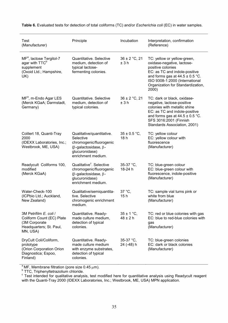
35
Table 6. Evaluated tests for detection of total coliforms (TC) and/or Escherichia coli (EC) in water samples.
Test (Manufacturer)
Principle Incubation Interpretation, confirmation (Reference)
MFa, lactose Tergitol-7 agar with TTCb supplement (Oxoid Ltd.; Hampshire, UK)
Quantitative. Selective medium, detection of typical lactose- fermenting colonies.
36 ± 2 °C, 21 ± 3 h
TC: yellow or yellow-green, oxidase-negative, lactose- positive colonies EC: as TC and indole-positive and forms gas at 44.5 ± 0.5 °C. ISO 9308-1:2000 (International Organization for Standardization, 2000)
MFa, m-Endo Agar LES (Merck KGaA; Darmstadt, Germany)
Quantitative. Selective medium, detection of typical colonies.
36 ± 2 °C, 21 ± 3 h
TC: dark or black, oxidase- negative, lactose-positive colonies with metallic shine EC: as TC and indole-positive and forms gas at 44.5 ± 0.5 °C. SFS 3016:2001 (Finnish Standards Association, 2001)
Colilert 18, Quanti-Tray 2000 (IDEXX Laboratories, Inc.; Westbrook, ME, USA)
Qualitative/quantitative. Selective chromogenic/fluorogenic (β-galactosidase, β-glucuronidase) enrichment medium.
35 ± 0.5 °C, 18 h
TC: yellow colour EC: yellow colour with fluorescence (Manufacturer)
Readycult Coliforms 100, modified (Merck KGaA)
Qualitativec. Selective chromogenic/fluorogenic (β-galactosidase, β-glucuronidase) enrichment medium.
35-37 °C, 18-24 h
TC: blue-green colour EC: blue-green colour with fluorescence, indole-positive (Manufacturer)
Water-Check-100 (ICPbio Ltd.; Auckland, New Zealand)
Qualitative/semiquantita-tive. Selective chromogenic enrichment medium.
37 °C, 15 h
TC: sample vial turns pink or white from blue (Manufacturer)
3M Petrifilm E. coli / Coliform Count (EC) Plate (3M Corporate Headquarters; St. Paul, MN, USA)
Quantitative. Ready-made culture medium, detection of typical colonies.
35 ± 1 °C, 48 ± 2 h
TC: red or blue colonies with gas EC: blue to red-blue colonies with gas (Manufacturer)
DryCult Coli/Coliform, prototype (Orion Corporation Orion Diagnostica; Espoo, Finland)
Quantitative. Ready-made culture medium with enzyme substrates, detection of typical colonies.
35-37 °C, 24 (-48) h
TC: blue-green colonies EC: dark or black colonies (Manufacturer)
a MF, Membrane filtration (pore size 0.45 µm). b TTC, Triphenyltetrazolium chloride. c Test intended for qualitative analysis, test modified here for quantitative analysis using Readycult reagent with the Quanti-Tray 2000 (IDEXX Laboratories, Inc.; Westbrook, ME, USA) MPN application.

36
focusing on epidemiology, prevalence or incidence of giardiasis and/or cryptosporidiosis in any of the Nordic countries. The following a priori-decided inclusion criteria were used at this stage: the study population must have been adult with both genders included, with the study using random and fully described sampling of subjects, the latter of which had to be either symptomatic or asymptomatic. Furthermore, the case definition of giardiasis or cryptosporidiosis must have included microscopic analysis of stool samples, with the number of analysed stool samples per subject described and the number of study subjects and positive cases reported. After this stage of critical review, 13 articles were included in the meta-analysis. 4.3.2 Estimation of annual incidence of Giardia spp. and Cryptosporidium spp. infections in the general population (V) The annual incidences of Giardia and Cryptosporidium infections in the general population of the Nordic countries were estimated per 100 000 population, using the random effects (RE) model prevalences obtained from meta-analysis. The estimation included the following: a) the point prevalence of gastroenteric symptoms due to any cause in the general population is 11% (Herikstad et al., 2002), b) the estimate for mean duration of excretion of Giardia cysts in faeces during the eventual symptomatic infection and asymptomatic shedding phase was 50 days (Centers for Disease Control and Prevention, 2001) and c) the estimate for excretion of Cryptosporidium oocysts was 35 days (Centers for Disease Control and Prevention, 2003). The estimated number of annual incidences per 100 000 population were compared with the mean annual numbers of cases reported to the various national infectious disease registers between 1995 and 2002 in study countries where data were available. 4.4 Statistical methods (I-V) 4.4.1 Enteropathogens in surface water (I) All individual results were recorded using MS Excel 2002 software (Microsoft Corporation; Redmond, WA, USA) and the statistical analysis was performed with the Statistical Package for Social Sciences 11.5 for Windows (SPSS Inc.; Chicago, IL, USA) software. The point prevalences were calculated for each analysed microbe and arithmetic means with standard deviations (SDs) for the MPNs of E. coli, turbidity counts and temperature separately for each sampling time and type of sampling site (all lakes plus the Kokemäki, Aura and other rivers). Analysis of variance (ANOVA) was used to determine possible significant differences at the 0.05 level (p < 0.05) in prevalences and means between different sampling times and sites. If significant differences were obtained, the Duncan’s Post Hoc test was performed to determine which values differed from all other values. All samples were grouped according to the thermotolerant and E. coli counts at four different levels and the prevalence of enteropathogens and indicator microbes at each level was analysed, using ANOVA and Duncan’s Post Hoc test.

37
A nonparametric Spearman’s rank order correlation coefficient with two-tailed p value was calculated for cross-correlations between different indicator parameters (coliform and thermotolerant coliform counts, MPNs of E. coli, turbidity value, and presence/absence of C. perfringens and F-RNA bacteriophages). Spearman’s correlation coefficient was also computed for bivariate correlations occurring between indicator parameters and pathogen findings. The odds ratio (OR) with 95% confidence interval (95% CI) was calculated for each sample, being positive for analysed pathogens according to the outcome of various indicator parameters. All the samples were divided into two groups: pathogen absent or pathogen present (i.e. the sample was either negative for Campylobacter spp., Giardia spp., Cryptosporidium spp., and NVs or was positive for at least one of the analysed pathogens). ‘Pathogen absent’ was taken as a dependent variable in the logistic multivariable regression model. In this model various indicator parameters (i.e. levels of E. coli and thermotolerant coliforms and the presence of C. perfringens and F-RNA bacteriophages) were taken as independent variables and their predictive values for the dependent variable were analysed by computing the coefficient estimates (B values), p values for the B values, and ORs with 95% CIs from the B values. 4.4.2 Evaluation of the water treatment devices (II, III) The purification capacities were calculated as log10 reductions (Equation 1) of analysed parameters between the concentrations in raw water (influent) and purified water (effluent). The raw water concentrations were calculated as the arithmetic mean of three separate samples from each analysis. Purification capacity ( )ei NN10log= , (Equation 1)
where Ni = concentration in raw water before treatment (influent) and Ne = concentration in purified water after treatment (effluent).
If no target organism or substance was detected in purified water, the log10 reduction was calculated assuming one organism or unit of concentration of the substance of the detection limit in purified water per analysed volume. 4.4.3 Evaluation of tests for detection of coliform bacteria and Escherichia coli (IV) All individual results were recorded using MS Excel 2002 software (Microsoft) and the statistical analysis was performed with the Statistical Package for Social Sciences 11.5 for Windows (SPSS Inc.) software. All 110 natural surface and drinking water samples were recorded according to the outcome in various tests as positive or negative for E. coli. To determine the sensitivity and specificity of a particular test for detection of E. coli, MF LTTC method was used as a reference (Table 6).

38
A total of 69 samples were excluded from statistical analyses of the mean E. coli counts, bivariate correlations between various tests and proportion of confirmed E. coli counts, 21 due to uncertainty in the total coliform counts (CFUs of ≥ 1000/100 ml in any of the tests) and 48 because no coliforms were consistently detected in a sample with the tests. Arithmetic means for the E. coli counts with 95% CIs as detected with each test method were calculated and compared with the E. coli counts detected with the LTTC method. To analyse the statistical significance of differences in mean E. coli counts between tests, the paired sample t test and its p value were computed. A nonparametric Spearman’s rank order correlation coefficient with two-tailed p value was calculated for bivariate cross-correlations within the total coliform and E. coli counts detected with each test method. The mean proportions of confirmed E. coli counts in the total coliform count detected in the samples for each test were computed together with the 95% CI. 4.4.4 Prevalence and incidence of Giardia spp. and Cryptosporidium spp. infections in the Nordic countries (V) The statistical analyses were performed with the Statistical Package for Social Sciences 11.5 for Windows (SPSS Inc.), EpiInfo v6.04c PLUS for DOS (Centers for Disease Control and Prevention (CDC); Atlanta, GA, USA) and MS Excel 2002 (Microsoft) software. The statistical heterogeneity of the combined studies included in the meta-analysis were tested separately in four subsets (Giardia and Cryptosporidium in both asymptomatic and symptomatic subjects), using the chi-square (χ2) test (Equation 2) with the Poisson approximation (Potthoff and Whittinghill, 1966a; Potthoff and Whittinghill, 1966b; Böhning et al., 2002). The cases and number of study subjects in each subset were pooled and meta-analytic techniques were applied, using the RE model (Equation 3) with the DerSimonian-Laird (DSL, Equation 4) estimator (Bailar, 1995; Chalmers and Altman, 1995; Welch, 2000; Böhning et al., 2002). The prevalences of Giardia and Cryptosporidium with the 95% exact binomial CIs were the primary outcome of the meta-analysis. The statistical heterogeneity between different studies was statistically significant if the p value for the χ2 test was < 0.05 at specific degrees of freedom (df). To further assess the statistical heterogeneity between studies of each subset (column), a test value H (Higgins et al., 2003b) was calculated as the square root of the χ2 test value divided by its df value and the 95% CI for H using Equation 5. Furthermore, a test value I2 was calculated (Equation 6) to estimate the percentage of total variation across studies that was due to statistical heterogeneity rather than chance (Higgins et al., 2003b).
Chi-square value, 2
1
2 ∑=
∧∧
⎟⎟⎠
⎞⎜⎜⎝
⎛−=
k
i i
iinx
µωχ , (Equation 2)
where
∑
∑
=
=∧
= k
ii
k
ii
n
x
1
1µ and
1
2
11
−∧∧
∧
∧
⎟⎟⎟⎟
⎠
⎞
⎜⎜⎜⎜
⎝
⎛⎟⎠⎞
⎜⎝⎛ −
==i
in
µµ
υω
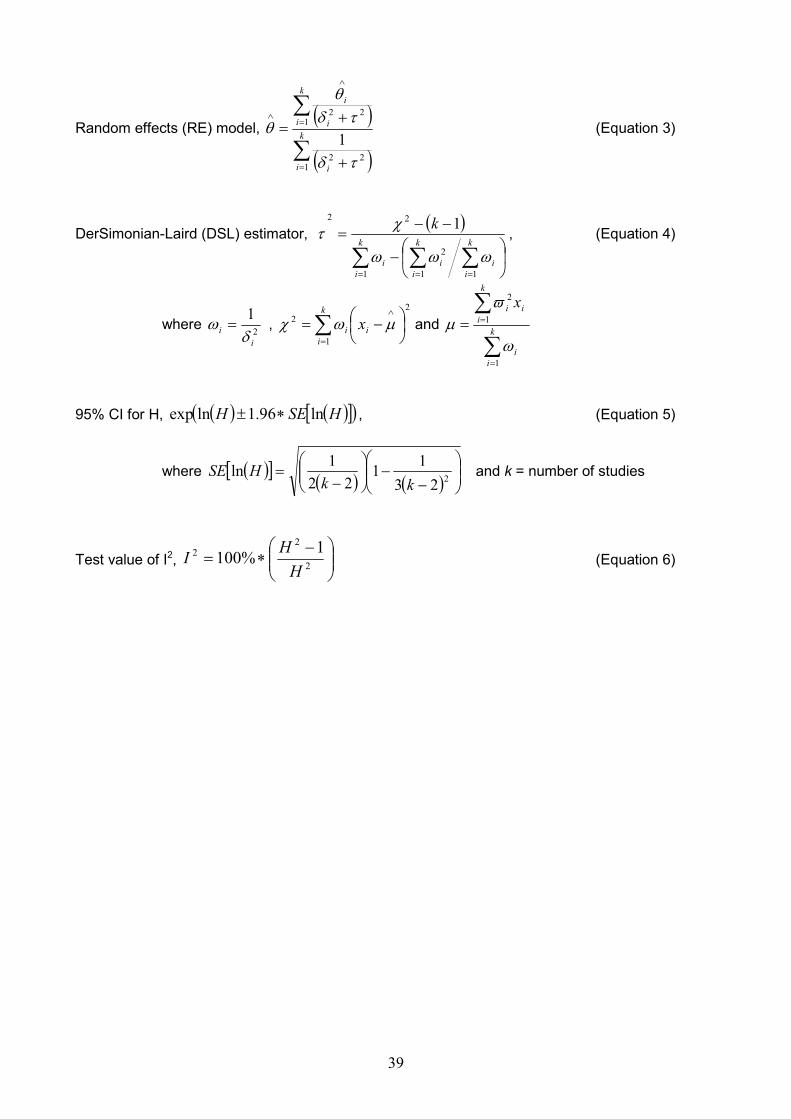
39
Random effects (RE) model, ( )
( )∑
∑
=
=
∧
∧
+
+= k
i i
k
i i
i
122
122
1τδ
τδθ
θ (Equation 3)
DerSimonian-Laird (DSL) estimator, ( )
∑ ∑∑= ==
⎟⎠
⎞⎜⎝
⎛−
−−=
k
i
k
ii
k
iii
k
1 11
2
22 1
ωωω
χτ , (Equation 4)
where 2
1
ii δ
ω = , ∑=
∧
⎟⎠⎞
⎜⎝⎛ −=
k
iii x
1
22 µωχ and
∑
∑
=
== k
ii
k
iii x
1
1
2
ω
ϖµ
95% CI for H, ( ) ( )[ ]( )HSEH ln96.1lnexp ∗± , (Equation 5)
where ( )[ ] ( ) ( ) ⎟⎟⎠
⎞⎜⎜⎝
⎛
−−⎟⎟
⎠
⎞⎜⎜⎝
⎛−
= 22311
221ln
kkHSE and k = number of studies
Test value of I2, ⎟⎟⎠
⎞⎜⎜⎝
⎛ −∗= 2
22 1%100
HHI (Equation 6)

40
5. RESULTS 5.1 Enteropathogens and indicators in surface water (I) 5.1.1 Prevalence of enteropathogens and indicators in surface water (I) A total of 57 samples out of 139 (41.0%) were positive for at least one or more of the analysed enteropathogens (Campylobacter spp., Giardia spp., Cryptosporidium spp., or NVs). Of these 57 positive samples, 46 had one of the analysed pathogens, while two pathogens were isolated from 10 samples and three pathogens from one sample. The pathogens were isolated less frequently (3/30; 10.0%) during the winter (p < 0.05) than during other sampling periods. There was no significant difference (p > 0.05) found in prevalences of positive samples for pathogens between different types of sampling site: lakes or the Aura, Kokemäki, or other rivers. Of 30 individual sampling sites only one was negative for all enteropathogens studied in all five samplings. Ten sites were positive at one sampling only, 10 during two and nine sites during three samplings. None of the sites was positive for one or more of the analysed pathogens in all five samplings. Campylobacter spp. were isolated from 24 samples (17.3%) and were the most frequently isolated enteropathogens in this study. From 24 campylobacter-positive samples, 11 (45.8%) were typed to C. jejuni, six (25.0%) to C. lari and one (4.2%) to C. coli, while six (25.0%) of the strains were undetermined. The samples were positive for campylobacters more often during May 2001 (43.3% of samples positive) than during other samplings (p < 0.05). Lakes and the Aura River were significantly (p < 0.05) more often positive for campylobacters than the Kokemäki or other rivers. NVs were detected in 13 samples (9.4%), three of which were typed to genogroup I and 10 to genogroup II. There were no differences in prevalence of NV findings between sampling times, but the Kokemäki River was more frequently (p < 0.05) positive for NVs than other sampling site types. NVs were the only pathogens analysed that showed significant differences in occurrence among 30 sampling sites; two separate river sites were more frequently (p < 0.01) positive for NVs than the remaining 28 sites. Giardia spp. were isolated from 19 (13.7%) and Cryptosporidium spp. from 14 (10.1%) of the 139 samples. Both Giardia spp. and Cryptosporidium spp. were found more frequently during summer 2001 and less frequently during winter 2001 than during the other sampling times (p < 0.05). No significant differences between proportions of positive samples at different types of sampling site were detected. The MPNs of E. coli were below the detection limit of the test used (one microbe per 100 ml) in 13 samples (9.3%), while a total of 110 samples (79.1%) had an MPN for E. coli of 100 CFUs/100 ml or less. Only five samples (3.6%) had MPNs > 1000/100 ml. The MPNs of E. coli varied widely between individual samples; thus there were no significant differences (p > 0.05) in mean MPNs between sampling times or sites. However, the MPNs tended to be higher in the Aura and most other rivers than in the lakes and the Kokemäki River. A total of 44 samples (31.7%) were positive for C. perfringens and 47 (33.8%) for F-RNA bacteriophages. Clostridium perfringens was isolated

41
most frequently during autumn 2000 and spring 2001 (p < 0.05). No sample from the Kokemäki River was positive for C. perfringens. There were no differences (p > 0.05) in occurrence of F-RNA bacteriophages between different sampling sites, but phages were more often isolated during winter 2001 and less frequently during autumn 2001 (p < 0.05). 5.1.2 Correlation between enteropathogens and indicators in surface water samples (I) The indicator parameters used in this study (turbidity, counts of thermotolerant coliforms, MPNs of E. coli and presence/absence of C. perfringens) showed significant cross-correlation (correlation coefficients 0.30-0.86, p < 0.05). However, the presence/absence of F-RNA phages showed no significant correlation (p > 0.05) with other parameters, except with turbidity and the MPNs of E. coli, in which there was low significant correlation (coefficients 0.30 and 0.21, p < 0.05). The presence of any analysed pathogen did not correlate significantly with the presence of other pathogens (p > 0.05 for Spearman’s correlation coefficients). Significant (p < 0.05) bivariate, nonparametric, Spearman’s rank order correlation coefficients and ORs were detected between various indicator parameters and pathogens in the samples. Based on these bivariate correlations, four variables (levels of E. coli and thermotolerant coliform counts, absence of C. perfringens and F-RNA bacteriophages) were selected for use in a multivariate logistic regression model in which the absence of E. coli, thermotolerant coliforms and C. perfringens showed a significant (p < 0.05) predictive value (ORs 1.15 x 10⁸, 7.57 and 2.74, respectively) for a sample being negative for any of the analysed pathogens. 5.2 Evaluation of water treatment devices (II, III) In general, the devices tested were able to remove the bacterial contaminants E. coli, Klebsiella pneumoniae, Enterobacter cloacae and sulphite-reducing clostridia from raw water, but failed in removal of bacteriophages used as models for viruses and in removal of microcystins. Water treatment based purely on physical filtration was not able to remove viral contaminants or cyanobacterial microcystins from heavily polluted raw water. Removal of C. parvum oocysts showed results similar to those shown in removal of bacteria, but some devices failed in purification and passed some oocysts into the purified water. Two of the water treatment devices tested were able to eliminate some or all of the BoNT type B from the inoculated influent water. The device based on RO removed > 2.3 log10 units of BoNT to the level below the detection limit of the mouse bioassay, and the experimental sand filter reduced the level of toxin by 0.3-1.3 log10 units. Those devices based merely on physical filtration through ceramic or membrane filters through 0.2-0.4-µm pores were not able to remove BoNT from the inoculated influent water (reduction < 0.1 log10 units), nor was the UV radiation from the low-pressure lamp able to destroy the toxin. All treatment devices, except for the sand filter, reduced the level of total aerobic count from the inoculated water by > 3.3 log10 units (sand filter by 0.8 log10 units).

42
5.3 Evaluation of tests for detection of botulinum toxin (III) Both commercial rapid EIA test kits determined that all the samples that were positive in the mouse bioassay were negative for BoNT; all samples negative in the mouse bioassay were also negative in the rapid EIA tests. The BoNT concentrations in influent and effluent water samples were extrapolated from the mouse bioassay results to be in the range of 3960-5985 ng/l in undiluted inoculated influent water and 100-fold higher in the TPGY broth used for inoculating the influent water. 5.4 Evaluation of tests for detection of coliform bacteria and Escherichia coli (IV) The tests detected typical coliform bacteria as coliforms and detected no noncoliform species, e.g. Aeromonas hydrophila, as coliforms except on LTTC agar in which A. hydrophila reproduced as a yellow colony. All the tests detected the reference E. coli strain as E. coli. The m-Endo LES SFS 3016:2001 was the only method showing no statistically significant (p < 0.05) differences between E. coli counts compared with the LTTC method ISO 9308-1:2000, whereas the Colilert 18 and Readycult showed significantly higher counts for E. coli than the LTTC. In general, those tests based on the analysis of a 1-ml sample (Petrifilm EC and DryCult Coli) showed weak sensitivity (39.5-52.5%) but high specificity (90.9-78.8%). When the results of E. coli detection were compared between test methods, the highest agreement (both tests either negative or positive) with the LTTC was calculated for the m-Endo LES (83.6%), followed by the Colilert 18 (82.7%), Water-Check (81.8%) and Readycult (78.4%), whereas the Petrifilm EC (70.6%) and DryCult Coli (68.9%) showed the weakest agreement. 5.5 Prevalence and incidence of Giardia spp. and Cryptosporidium spp. infections in the Nordic countries (V) The prevalences of both Giardia and Cryptosporidium infections were significantly (p < 0.05) lower in the asymptomatic than in the symptomatic population, with prevalences of 2.97% (95% CI: 2.64; 3.31) and 5.81% (95% CI: 5.35; 6.30) for Giardia and 0.99% (95% CI: 0.81; 1.19) and 2.91% (95% CI: 2.71; 3.12) for Cryptosporidium. Thirteen studies were excluded from the meta-analysis, since these studies did not meet the a priori-defined inclusion criteria for random sampling or study subjects recruited from the general population. The studies showed significant methodological and clinical differences and thus attempts to generate any subgroup meta-analysis, e.g. within hospitalized patients or children in daycare, were not feasible. Even though remarkable variation was shown in prevalences between the various studies, the latter also displayed higher prevalences of both parasites in subpopulations compared with the general population in meta-analysis. Even though the prevalences of both Giardia and Cryptosporidium were lower in the asymptomatic compared with the symptomatic population, the asymptomatic cases of Giardia and

43
Cryptosporidium represented a major proportion of the total annual cases (19 300 asymptomatic and 4670 symptomatic cases of Giardia per 100 000 population, for Cryptosporidium 9190 and 3340, respectively. The difference between the asymptomatic and symptomatic cases was statistically significant (p < 0.05). For each registered Giardia case an estimated 867 unregistered symptomatic cases can be expected to occur annually per 100 000 population in Finland, 634 in Norway and 254 in Sweden. For Cryptosporidium the ratio was 1:15 181 in Finland and 1:4072 in Sweden.

44
6. DISCUSSION 6.1 Enteropathogens and indicators in surface water (I) Our results showed that all the enteropathogens analysed could be detected in surface waters from lakes and rivers located in southwestern Finland. This was the first systematic study done in Finland in which enteropathogens representing different microbial groups: NVs as a model for enteric viruses, campylobacters as enteric bacterial pathogens, and Giardia spp. and Cryptosporidium spp. as models for resistant protozoan parasites, were analysed from diverse surface water sites during several consecutive seasons. The point prevalences of the various enteropathogens in our study as well as in all biological and environmental studies were dependent on sampling time and site, but the findings can serve as rough general estimates of their prevalences in surface water as a whole and give qualitative data for risk assessment. Possible seasonal or time-related variation in the occurrence of various groups of enteric pathogens in surface water appears to be dependent on the source of contamination and conditions facilitating contaminants discharged into surface water. If the major sources are sewage plants that treat human wastes, seasonal patterns similar to those found in human infections for a particular pathogen would be detected in effluents and downstream water samples (Kukkula et al., 1999). If the watershed is contaminated by discharges stemming from agricultural runoffs, the highest numbers of zoonotic enteric pathogens would be found during the pasture season after snowmelt, floods and heavy rainfalls (Bodley-Tickell et al., 2002). Even though most of the NV infections in Finland occur, according to the Finnish Infection Register, in winter and early spring (Finnish National Public Health Institute, 2003), no clear winter peak in the number of samples positive for NVs was detected in our studies, but positive samples were nevertheless detected in all seasons. No evidence that enteric pathogens would be more frequent in certain types of surface water in Finland was obtained. There was, however, a trend for higher E. coli counts in the Aura and other rivers compared with lakes and the Kokemäki River, thus confirming the results of previous monitoring studies (Poikolainen et al., 1994; Niemi et al., 1997). There was also a tendency for higher turbidity counts in rivers (except the Kokemäki River) compared with lakes; this result reflects the general appearance of coastal rivers with high loads of inorganic material and clay in river waters combined with relatively low flow rates. Occurrence of the various pathogens did not significantly correlate with traditionally used faecal indicator parameters, counts or count levels for E. coli or thermotolerant coliforms per 100 ml, or turbidity value. In some studies a significant correlation was found between the level of thermotolerant coliforms and number of samples positive for certain pathogens (Arvanitidou et al., 1997; Payment et al., 2000). However, poor correlation similar to that shown in the present study was also demonstrated elsewhere (Carter et al., 1987; Geldreich, 1996; Leclerc et al., 2001; Tillett et al., 2001). The presence/absence of correlation between faecal indicators and pathogens could reflect the occasional appearance of enteropathogens in surface waters and the varying survival and recovery rates of these pathogens compared with faecal indicators. One possible factor

45
affecting this low correlation could also be varying microbial densities in the original contamination source and therefore the failure to detect pathogens due to too small sampling volumes (Geldreich, 1996; Hänninen et al., 2003). For detection of Giardia spp. and Cryptosporidium spp. a 10-l, for NVs a 1-l, and for Campylobacter spp., E. coli and thermotolerant coliforms a 100-ml volume was used. However, a significant predictive value was found for a sample being positive for one or more of the analysed pathogens if E. coli or thermotolerant coliforms were detected in the sample. In multivariable analysis a significant positive predictive value was also found for a sample being negative for all of the analysed pathogens if no E. coli or thermotolerant coliforms were detected in 100-ml volumes. Therefore the presence/absence of E. coli and thermotolerant coliforms appears to be a better predictor for occurrence of enteropathogens than the specific microbial CFU levels of these microbes. A positive correlation between the presence of C. perfringens and a sample being positive for one or more analysed pathogens was detected, as was a positive predictive value for a sample being negative for all of the analysed pathogens if there were no C. perfringens detected. However, a considerable number of samples with absence of C. perfringens were simultaneously positive for some of the analysed pathogens and vice versa, which decreases the practical usability of C. perfringens as a reliable indicator for the presence of enteropathogens. The presence of F-RNA bacteriophages could not be linked with the presence of NVs or any other enteric pathogen in a sample, and therefore they cannot serve as reliable indicator organisms. There are some earlier studies on the correlation of F-RNA phages with NVs that have suggested bacteriophages as suitable indicators for enteric viruses (Havelaar et al., 1993; Duran et al., 2002). The occurrence of enteropathogens in surface waters is linked directly to potential contamination sources, while environmental conditions affect only the survival of these microbes in water. The presence of traditionally used faecal indicators, including thermotolerant coliforms and E. coli, has significant predictive value for the presence of the enteropathogens studied but no significant correlation was found between the specific CFU level of indicators and the presence of pathogens. Microbial monitoring of raw water using only faecal indicator organisms is not sufficient for assessment of the presence of a particular enteropathogen. 6.2 Evaluation of water treatment devices (II, III) With regard to the treatment techniques of individual filters, the results coincided with their theoretical purification capacities and with the results of earlier studies on other drinking water treatment filters (Schlosser et al., 2001). Testing of water treatment filters simultaneously with indicator bacteria and bacteriophages as viral markers, protozoal oocysts and microbial toxins resulted in valuable information for safety assessment purposes. Comparison of marketing information with the test results revealed that some devices were advertised as being more efficient in removal of microbes and toxins than was found in our studies, while information from some manufacturers coincided closely with the test results. Some manufacturers stated in the marketing information that their products were able to decrease the counts of all bacteria (including E. coli) by 2-6 log10 units and parasites (including C. parvum oocysts) by 2-4 log10 units. Others

46
gave only a general statement of removal capacity or stated that the device was not able to remove viruses. Only one device based on RO was advertised as able to remove “harmful viruses and chemicals from raw water”. This device indeed was the only one that removed F-RNA phages under the detection limit and microcystins under the guide value 1 µg/l for microcystin in drinking water (World Health Organization, 2004). In the present study the construction or robustness of each device was not tested. Information on this aspect could be obtained through operational tests under various climatic and environmental conditions. In addition to the fundamental treatment technique, the construction of devices and cleaning and maintenance procedures may play important roles in the final purification capacity. Testing of the water treatment devices resulted in information crucial to the assessment of drinking water safety and security. Based on the present studies the only technique available for portable devices capable of eliminating BoNT from drinking water is RO. To some extent sand filtration could be effective in reducing biotoxins, as suggested here and in studies on microcystin removal, but this most probably is strongly dependent on the thickness of the sand bed and properties of the sand used (Lahti et al., 2001; Rapala et al., 2002b). In the present study, the 254-nm UV radiation produced by the low-pressure lamp was not able to degrade BoNT in the water. Some studies indicated that direct sunlight can degrade BoNT totally in 3 h (U.S. Army Medical Research Institute of Infectious diseases, 2001). Probably the degradation process required broad-spectrum UV radiation coupled with the oxidative spectrum produced by high-pressure UV lamps. Activated carbon combined with filtration through ceramic filters did not affect toxin removal in this study (III). A single previous study showed activated carbon to be effective against BoNT type A in water samples (Gomez et al., 1995), but the removal was apparently due to the amount and type of activated carbon used as well as to the flow rate of the water and contact time with the carbon. In the present study, the activated carbon was either in the form of a thin layer or in a relatively small cartridge. 6.3 Evaluation of tests for detection of botulinum toxin (III) The rapid EIA tests for detection of BoNT showed poor performance compared with the results of the standard mouse bioassay. Even the primary growth medium with toxic C. botulinum cultures at estimated BoNT concentrations of 396 000-598 500 ng/l appeared to be negative for BoNT when evaluated with the rapid EIA tests. It can be estimated from the toxicological data that only 1.2-1.8 ml of this broth constitutes the oral lethal dose for a 70-kg human being (Gill, 1982; Schechter and Arnon, 2000; Arnon et al., 2001). Intentional contamination of drinking water and water supply systems will apparently result in concentrations remarkably lower in distributed drinking water than in pure bacterial culture (Burrows and Renner, 1999). However, the total intake of the toxin can still cause symptoms or death due to the total amount of water ingested. Therefore, the usefulness of these rapid tests is very limited due to failure to detect lethal concentrations of toxin in drinking water. The negative test results will be misleading and may result in casualties if intentional release of BoNT into drinking water supplies has been committed.

47
6.4 Evaluation of tests for detection of coliform bacteria and Escherichia coli (IV) The present results with the MF m-Endo LES method SFS 3016:2001 were consistent with those obtained with the LTTC method ISO 9308-1:2000 for coliforms and E. coli detection in water samples. The defined-substrate Colilert 18, Readycult and Water Check methods attained levels of sensitivity and specificity in E. coli detection similar to those in the LTTC method. However, the Colilert 18 and Readycult methods detected higher E. coli counts in E. coli-positive samples compared with the LTTC method. The higher E. coli counts detected with these tests were most probably due to the ability of the test media to induce recovery of injured and stressed coliforms and E. coli in the samples (McFeters et al., 1993; McFeters et al., 1995). The MF procedure is also known to reduce the recovery of the target organisms (Standridge and Delfino, 1982; Brenner and Rankin, 1990). The bivariate correlation between the Colilert 18 and Readycult methods was highly significant both for detected total coliforms and E. coli in the water samples. To our knowledge, this study was the first to describe the Readycult method modified for use as an MPN method. These results are consistent with the methodological principles of test methods that have identical detection targets. Both MF methods are based on the same confirmation procedures and both the Colilert 18 and Readycult methods are based on the same defined substrates, e.g. the production of indole versus β-glucuronidase used in E. coli detection. Thus, the MF and defined-substrate tests do not detect the metabolically exact same organisms, and therefore detailed comparison of the equivalence between these two methods is not possible (Niemi et al., 2001; Niemelä et al., 2003; International Organization for Standardization, 2004). However, we were able to show that the defined-substrate methods Colilert 18 and Readycult were able to attain at least the same sensitivity as the reference MF methods used in E. coli detection. Those tests based on use of a ready-made culture medium and analysis of a 1-ml sample failed to attain the level of sensitivity in E. coli detection shown in the reference MF method. Furthermore, these tests showed no significant correlation in coliform or E. coli detection between each other or between other evaluated tests. These results were most probably due to the uncertainty caused by analysis of the small 1-ml volumes compared with the 100-ml reference volume (Rompre et al., 2002; Vail et al., 2003). Based on this evaluation study, the Colilert 18, Readycult and Water Check methods are all suitable for coliform and E. coli detection both under field conditions and in routine use in the water industry. These tests are labour-saving, easy to perform and able to give final results within 24 h. When the numbers of E. coli counts detected in water samples are compared, the results given by the Colilert 18 and Readycult methods are equal to, if not greater than those given by the reference MF ISO 9308-1:2000 method.

48
6.5 Prevalence and incidence of Giardia spp. and Cryptosporidium spp. infections in the Nordic countries (V) This is the first study to give a general estimate for the prevalences of Giardia and Cryptosporidium in the general population from the Nordic countries of Denmark, Finland, Norway and Sweden. The prevalences obtained were relatively low, both in the asymptomatic and symptomatic populations in all countries. However, the annual numbers of Giardia and Cryptosporidium cases were higher than would be expected based on these low prevalences. Furthermore, the majority of annual cases remain undetected and unregistered in the national surveillance registers of infectious diseases. Meta-analytical studies have been widely used for analysis of the relationships between various environmental exposures and clinical or other outcomes (Halvorsen et al., 1992). Our study, in which we used separate prevalence studies to generate general estimates for the prevalence and incidence of the protozoan enteropathogens Giardia and Cryptosporidium in the Nordic countries, is an application of the meta-analytical approach for which there are a very limited number of published studies. Caution must be observed when comparing or combining epidemiological studies due to potential biases caused by heterogeneity in the study population, case definitions, or laboratory diagnostics, as well as time-related differences. The prevalences obtained for Giardia and Cryptosporidium in the meta-analysis were relatively low, both in the asymptomatic and symptomatic members of the general population, although both parasites were statistically more prevalent in the latter. Similar prevalences have been detected in a sentinel study in Netherlands (Wit de et al., 2001). Epidemiological studies performed in immunocompromised or other subpopulations show a tendency toward higher prevalences in these populations compared with the general population (Castor, 1981; Ranki et al., 1985; Jokipii et al., 1985b; Benzeguir et al., 1999). When parasitological screening tests or monitoring surveys are planned, the number of subjects needed to be screened to find a positive case in the general asymptomatic population can be estimated to be 30-38 for Giardia and 84-123 for Cryptosporidium. In screening of immunocompromised subjects, the number screened can be estimated to be as low as 3-34 for Giardia and 9-108 for Cryptosporidium. One factor bearing an impact on estimation is the prevalence of persons having gastrointestinal symptoms at certain time points in the general population. The estimate used in this study was obtained from a study undertaken in the USA (Herikstad et al., 2002), and with reasonable presumption this estimate could also be utilized in the Nordic countries. This factor bears impact especially on the estimated number of annual Cryptosporidium cases: the higher the proportion of the symptomatic subpopulation the higher the annual number of Cryptosporidium cases in the general population. The other factor impacting the annual number of estimated incidences of cases is the duration of (oo)cyst excretion. In the present study an estimate of excretion included a combination of (oo)cyst excretion during the symptomatic phase followed by a 2-week asymptomatic excretion phase (Centers for Disease Control and Prevention, 2001; Centers for Disease Control and Prevention, 2003). A significant proportion of Giardia and Cryptosporidium infections in the general population will be asymptomatic. The estimate of annual incidence of

49
giardiasis and cryptosporidiosis is here based on the assumption that the point prevalence of these diseases is constant throughout the year. Registration and surveillance systems in the Nordic countries vary and Giardia and Cryptosporidium are not included in the national lists of notifiable diseases in all countries. Diagnosis of cryptosporidiosis requires specific methods such as modified acid-fast or immunofluorescence staining of the oocysts (Marshall et al., 1997), which are not included in routine microbiological analysis of patients with diarrhoea but instead must be specifically requested. As estimated in our study, the majority of Giardia and Cryptosporidium infections in the general population remains undetected and unregistered and only a small number will annually be reported in national registers for infectious diseases. A remarkable finding is that despite the lower prevalence of Cryptosporidium, a relatively smaller proportion of Cryptosporidium cases will be detected and registered. This finding is consistent with a previous study done in New York in which it was estimated that for every three cases registered, 10 000 cases remained undetected and unregistered in the general population (Perz et al., 1998). This factor of underreporting was also discussed in other studies (Marshall et al., 1997; Wheeler et al., 1999; Nygård et al., 2003). Domestic outbreaks reported in the Nordic countries as caused by Giardia or Cryptosporidium are rare (Kettis and Thoren, 1974; Pohjola et al., 1986b; Neringer et al., 1987; Ravn et al., 1991; Ljungström and Castor, 1992; Tjernström et al., 1992), and this finding supports the relatively low prevalence of these parasites. However, Giardia and Cryptosporidium appear to be endemic to the Nordic countries and since domestic epidemics do occur, however infrequently, parasites can be isolated in the Nordic environment, such as sewage sludge (Rimhanen-Finne et al., 2001; Ottoson and Stenström, 2003), surface water (Robertson and Gjerde, 2001; Rimhanen-Finne et al., 2002), and in animal reservoirs (Viring et al., 1993; Laakkonen et al., 1994; Iburg et al., 1996; Enemark et al., 2002), and not all sporadic cases can be linked to known risk factors, such as travelling abroad or animal contacts (Wester et al., 1963; Jokipii et al., 1983; Jokipii et al., 1985a; Pohjola et al., 1986a). We were able to provide data for estimation of the importance and risk for public health caused by Giardia and Cryptosporidium in the Nordic countries. Both these parasites can cause significant morbidity with low infective doses, even for fully immunocompetent persons (Petersen, 1972; Dillingham et al., 2002), but especially for young, old, pregnant and immunocompromised persons (Dillingham et al., 2002). Only recently has medication been available against cryptosporidiosis in humans (White, Jr., 2003). Due to their capacity for survival in the environment and for zoonotic spreading, these parasites can pose a significant risk for public health, especially through contaminated drinking water. Most cases of giardiasis and cryptosporidiosis in human populations are assumed to be caused through contaminated drinking or recreational water (Marshall et al., 1997; Slifko et al., 2000). When a sensitive subpopulation increases, especially the elderly in society, the role these parasites play may become more prominent in the general population and the disease burden in the general population caused by these parasites may increase.

50
7. CONCLUSIONS
1. In all, 41.0% (57/139) of the surface water samples were positive for at least one of the analysed pathogens: 17.3% positive for campylobacters (45.8% Campylobacter jejuni, 25.0% C. lari, 4.2% C. coli and 25.0% Campylobacter spp.), 13.7% for Giardia spp., 10.1% for Cryptosporidium spp. and 9.4% for noroviruses (23.0% genogroup I and 77.0% genogroup II). During the winter season the samples were significantly (p < 0.05) less frequently positive for enteropathogens than during other sampling seasons. No significant differences were found in the prevalences of enteropathogens between rivers and lakes.
2. The presence of thermotolerant coliforms, Escherichia coli and Clostridium perfringens
showed significant bivariate, nonparametric, Spearman’s rank order correlation coefficients (p < 0.001), with a sample being positive for one or more of the analysed enteropathogens. Absence of these indicators in a logistic regression model had significant predictive value (ORs 1.15 x 10⁸, 7.57 and 2.74 respectively, p < 0.05) for a sample being negative for the analysed enteropathogens. No significant correlations were observed between counts or count levels of thermotolerant coliforms, E. coli or presence of F-RNA phages and enteropathogens in the analysed samples.
3. In general, the water treatment devices tested were able to remove bacterial contaminants
by 3.6–6.9 log10 units from contaminated raw water. Those devices based only on filtration through pores 0.2–0.4 µm or larger failed in viral and chemical purification. Only one device, based on reverse osmosis, was capable of removing F-RNA phages at concentrations under the detection limit and microcystins by 2.5 log10 units. Simultaneous testing for various enteropathogenic and indicator microbes and microcystins was a useful and practical way to obtain data on the purification capacity of commercial small-scale drinking water filters.
4. Water treatment devices based on filtration through ceramic or membrane filters with pore
size of 0.2-0.4 µm or radiation from low-pressure UV lamp (254 nm) failed to remove botulinum neurotoxin (BoNT) from raw water (reduction < 0.1 log10 units). A single device based on reverse osmosis was capable of removing the BoNT to a level below the detection limit of the mouse bioassay (reduction > 2.3 log10 units). The two rapid enzyme-immune assay tests intended for the detection of BoNT failed to detect BoNT from aqueous samples containing an estimated concentration of BoNT of 396 000 ng/l.
5. The m-Endo LES SFS 3016:2001 was the only method showing no statistical difference in
E. coli counts compared with the reference, lactose Tergitol-7 (LTTC) ISO 9308-1:2000, whereas the Colilert 18 and Readycult methods gave significantly higher counts for E. coli than the LTTC. When the results of E. coli detection were compared between test methods, the highest agreement (both tests negative or positive) with the LTTC was calculated for the m-Endo LES (83.6%), followed by the Colilert 18 (82.7%), Water-Check (81.8%) and Readycult (78.4%), whereas the Petrifilm EC (70.6%) and DryCult Coli (68.9%) showed the weakest agreement. In general, those tests based on the analysis of a 1-ml sample

51
(Petrifilm EC and DryCult Coli) showed weak sensitivity (39.5-52.5%) but high specificity (90.9-78.8%).
6. In combining the data of 13 clinically and methodologically nonheterogeneous studies
published before 2004, using the random effects model with the DerSimonian-Laird estimator, we estimated the prevalence of Giardia cases in the asymptomatic (i.e. no gastroenteric symptoms) general population in the Nordic countries to be 2.97% (95% CI: 2.64; 3.31) and in the symptomatic population 5.81% (95% CI: 5.34; 6.30). For Cryptosporidium the prevalences were 0.99% (95% CI: 0.81; 1.19) and 2.91% (95% CI: 2.71; 3.12), respectively. In analysing the data, we estimated that there will be 4670 (95% CI: 4300; 5060) symptomatic cases of Giardia and 3340 (95% CI: 3110; 3580) of Cryptosporidium annually per 100 000 general population in the Nordic countries. The vast majority of cases will remain unregistered in the national registers of infectious diseases, since for single registered cases there will be 254-867 cases of Giardia undetected/unregistered and 4072-15 181 cases of Cryptosporidium.

52
8. REFERENCES
Ahmad, K. 2002. Norwalk-like virus attacks troops in Afghanistan. Lancet Infect. Dis. 2:391.
Aho, M., Kurki, M., Rautelin, H. and Kosunen, T. U. 1989. Waterborne outbreak of Campylobacter enteritis after outdoors infantry drill in Utti, Finland. Epidemiol. Infect. 103:133-141.
Al-Kharabshed, S. and YogiGoswami, D. 2003. Analysis of an innovative water desalination system using low-grade solar heat. Desalination 156:323-332.
Alocilja, E. C. and Radke, S. M. 2003. Market analysis of biosensors for food safety. Biosens. Bioelectron. 18:841-846.
Annan, K. 2000. We the Peoples - The Role of the United Nations in the 21st Century. Millennium Report of the Secretary-General. United Nations, New York, NY, USA.
Armstrong, L. E., Maresh, C. M., Gabaree, C. V., Hoffman, J. R., Kavouras, S. A., Kenefick, R. W., Castellani, J. W. and Ahlquist, L. E. 1997. Thermal and circulatory responses during exercise: effects of hypohydration, dehydration, and water intake. J. Appl. Physiol. 82:2028-2035.
Arness, M. K., Feighner, B. H., Canham, M. L., Taylor, D. N., Monroe, S. S., Cieslak, T. J., Hoedebecke, E. L., Polyak, C. S., Cuthie, J. C., Fankhauser, R. L., Humphrey, C. D., Barker, T. L., Jenkins, C. D. and Skillman, D. R. 2000. Norwalk-like viral gastroenteritis outbreak in U.S. Army trainees. Emerg. Infect. Dis. 6:204-207.
Arnon, S. S., Schechter, R., Inglesby, T. V., Henderson, D. A., Bartlett, J. G., Ascher, M. S., Eitzen, E., Fine, A. D., Hauer, J., Layton, M., Lillibridge, S., Osterholm, M. T., O'Toole, T., Parker, G., Perl, T. M., Russell, P. K., Swerdlow, D. L. and Tonat, K. 2001. Botulinum toxin as a biological weapon: medical and public health management. JAMA 285:1059-1070.
Arvanitidou, M., Papa, A., Constantinidis, T. C., Danielides, V. and Katsouyannopoulos, V. 1997. The occurrence of Listeria spp. and Salmonella spp. in surface waters. Microbiol. Res. 152:395-397.
Ashbolt, N. J. 2004a. Microbial contamination of drinking water and disease outcomes in developing regions. Toxicology 198:229-238.
Ashbolt, N. J. 2004b. Risk analysis of drinking water microbial contamination versus disinfection by-products (DBPs). Toxicology 198:255-262.
Ashbolt, N. J., Grabow, W. O. and Snozzi, M. 2001. Indicators of microbial water quality, p. 289-316. In: Fewtrell, L. and Bartram, J. (eds.), Water Quality: Guidelines, Standards and Health. World Health Organization and IWA Publishing, London, UK.
Ashford, D. A., Kaiser, R. M., Bales, M. E., Shutt, K., Patrawalla, A., McShan, A., Tappero, J. W., Perkins, B. A. and Dannenberg, A. L. 2003. Planning against biological terrorism: lessons from outbreak investigations. Emerg. Infect. Dis. 9:515-519.
Asperen van, I. A., Medema, G., Borgdorff, M. W., Sprenger, M. J. and Havelaar, A. H. 1998. Risk of gastroenteritis among triathletes in relation to faecal pollution of fresh waters. Int. J. Epidemiol. 27:309-315.
Atlas, R. M. 1998. The medical threat of biological weapons. Crit. Rev. Microbiol. 24:157-168.
Auld, H., MacIver, D. and Klaassen, J. 2004. Heavy rainfall and waterborne disease outbreaks: the Walkerton example. J. Toxicol. Environ. Health A 67:1879-1887.
Backer, H. 1995. Field water disinfection, p. 1060-1110. In: Auerbach, P. (ed.), Wilderness Medicine. Mosby, St. Louis, MO, USA.
Backer, H. 2002. Water disinfection for international and wilderness travelers. Clin. Infect. Dis. 34:355-364.
Backer, H. and Hollowell, J. 2000. Use of iodine for water disinfection: iodine toxicity and maximum recommended dose. Environ. Health Perspect. 108:679-684.

53
Bailar, J. C. 1995. The practice of meta-analysis. J. Clin. Epidemiol. 48:149-157.
Balogh de, K. K., Schouten, M. C. and Lipman, L. J. 2002. [Bioterrorism: the role of veterinarians in detection and prevention]. Tijdschr. Diergeneeskd. 127:616-619.
Barrell, R. A., Hunter, P. R. and Nichols, G. 2000. Microbiological standards for water and their relationship to health risk. Commun. Dis. Public Health 3:8-13.
Bartram, J., Cotruvo, J., Exner, M., Fricker, C. and Glasmacher, A. 2003. Heterotrophic Plate Counts and Drinking-water Safety: The Significance of HPCs for Water Quality and Human Health. IWA Publishing, London, UK.
Beck, R. 2000. A Chronology of Microbiology in Historical Context. ASM Press, Washington, D.C., USA.
Benzeguir, A. K., Capraru, T., Aust-Kettis, A. and Björkman, A. 1999. High frequency of gastrointestinal parasites in refugees and asylum seekers upon arrival in Sweden. Scand. J. Infect. Dis. 31:79-82.
Berger, S. and Shapiro, I. 1997. Re-emergence of cholera vaccine. J. Travel. Med. 4:58-60.
Betancourt, W. Q. and Rose, J. B. 2004. Drinking water treatment processes for removal of Cryptosporidium and Giardia. Vet. Parasitol. 126:219-234.
Black, J. L., III. 2003. Genome projects and gene therapy: gateways to next generation biological weapons. Mil. Med. 168:864-871.
Black, R. E., Levine, M. M., Clements, M. L., Hughes, T. P. and Blaser, M. J. 1988. Experimental Campylobacter jejuni infection in humans. J. Infect. Dis. 157:472-479.
Blaisdell, F. W. 1988. Medical advances during the Civil War. Arch. Surg. 123:1045-1050.
Blaser, M. J. 1997. Epidemiologic and clinical features of Campylobacter jejuni infections. J. Infect. Dis. 176 Suppl 2:S103-S105.
Blaser, M. J., Smith, P. F., Wang, W. L. and Hoff, J. C. 1986. Inactivation of Campylobacter jejuni by chlorine and monochloramine. Appl. Environ. Microbiol. 51:307-311.
Blazes, D. L., Lawler, J. V. and Lazarus, A. A. 2002. When biotoxins are tools of terror. Early recognition of intentional poisoning can attenuate effects. Postgrad. Med. 112:89-98.
Bodley-Tickell, A. T., Kitchen, S. E. and Sturdee, A. P. 2002. Occurrence of Cryptosporidium in agricultural surface waters during an annual farming cycle in lowland UK. Water Res. 36:1880-1886.
Bonsdorff von, C. H. and Maunula, L. 2003. Microbiology and the Investigation of Waterborne Outbreaks: Typing of Norwalk-Like Virus, p. 79-85. In: Hunter, P., Waite, M. and Ronchi, E. (eds.), Drinking Water and Infectious Disease: Establishing the Links. IWA Publishing, London, UK.
Boulware, D. R. 2004. Influence of hygiene on gastrointestinal illness among wilderness backpackers. J. Travel Med. 11:27-33.
Boulware, D. R., Forgey, W. W. and Martin, W. J. 2003. Medical risks of wilderness hiking. Am. J. Med. 114:288-293.
Bravata, D. M., McDonald, K. M., Smith, W. M., Rydzak, C., Szeto, H., Buckeridge, D. L., Haberland, C. and Owens, D. K. 2004. Systematic review: surveillance systems for early detection of bioterrorism-related diseases. Ann. Intern. Med. 140:910-922.
Brenner, K. P. and Rankin, C. C. 1990. New screening test to determine the acceptability of 0.45-micron membrane filters for analysis of water. Appl. Environ. Microbiol. 56:54-64.
Brennhovd, O., Kapperud, G. and Langeland, G. 1992. Survey of thermotolerant Campylobacter spp. and Yersinia spp. in three surface water sources in Norway. Int. J. Food Microbiol. 15:327-338.

54
Brock, T. 1999. Robert Koch: a Life in Medicine and Bacteriology. American Society for Microbiology, Washington, DC, USA.
Burrows, W. D. and Renner, S. E. 1999. Biological warfare agents as threats to potable water. Environ. Health Perspect. 107:975-984.
Butzler, J. P. 2004. Campylobacter, from obscurity to celebrity. Clin. Microbiol. Infect. 10:868-876.
Böhning, D., Malzahn, U., Dietz, E., Schlattmann, P., Viwatwongkasem, C. and Biggeri, A. 2002. Some general points in estimating heterogeneity variance with the DerSimonian-Laird estimator. Biostatistics 3:445-457.
Cabelli, V. J., Dufour, A. P., McCabe, L. J. and Levin, M. A. 1982. Swimming-associated gastroenteritis and water quality. Am. J. Epidemiol. 115:606-616.
Carpenter, C., Fayer, R., Trout, J. and Beach, M. J. 1999. Chlorine disinfection of recreational water for Cryptosporidium parvum. Emerg. Infect. Dis. 5:579-584.
Carter, A. M., Pacha, R. E., Clark, G. W. and Williams, E. A. 1987. Seasonal occurrence of Campylobacter spp. in surface waters and their correlation with standard indicator bacteria. Appl. Environ. Microbiol. 53:523-526.
Castor, B. 1981. [Giardiasis in 3 Swedish institutions]. Läkartidningen 78:950-951.
Centers for Disease Control and Prevention. 2001. Giardiasis infection fact sheet. Centers for Disease Control and Prevention Division of Parasitic Disease, Atlanta, GA, USA. [Online.] http://www.cdc.gov/ ncidod/dpd/parasites/giardiasis/factsht_giardia.htm. Accessed 15 May 2004.
Centers for Disease Control and Prevention. 2003. Cryptosporidiosis fact sheet. Centers for Disease Control and Prevention Division of Parasitic Disease, Atlanta, GA, USA. [Online.] http://www.cdc.gov/ ncidod/dpd/parasites/cryptosporidiosis/factsht_cryptosporidiosis.htm. Accessed 6 November 2003.
Chalmers, I. and Altman, D. G. 1995. Systematic Reviews. The BMJ Publishing Group, London, UK.
Chigbu, P., Gordon, S. and Strange, T. 2004. Influence of inter-annual variations in climatic factors on fecal coliform levels in Mississippi Sound. Water Res. 38:4341-4352.
Christen, K. 2001. Bioterrorism and waterborne pathogens: how big is the threat? Environ. Sci. Technol. 35:396A-397A.
Christopher, G. W., Cieslak, T. J., Pavlin, J. A. and Eitzen, Jr., E. M. 1997. Biological warfare. A historical perspective. JAMA 278:412-417.
Cirino, N. M., Musser, K. A. and Egan, C. 2004. Multiplex diagnostic platforms for detection of biothreat agents. Expert. Rev. Mol. Diagn. 4:841-857.
Clark, D. L., Milner, B. B., Stewart, M. H., Wolfe, R. L. and Olson, B. H. 1991. Comparative study of commercial 4-methylumbelliferyl-beta-D-glucuronide preparations with the standard methods membrane filtration fecal coliform test for the detection of Escherichia coli in water samples. Appl. Environ. Microbiol. 57:1528-1534.
Clark, D. P. 1999. New insights into human cryptosporidiosis. Clin. Microbiol. Rev. 12:554-563.
Clark, J. A. and el Shaarawi, A. H. 1993. Evaluation of commercial presence-absence test kits for detection of total coliforms, Escherichia coli, and other indicator bacteria. Appl. Environ. Microbiol. 59:380-388.
Contreras-Coll, N., Lucena, F., Mooijman, K., Havelaar, A., Pierz, V., Boque, M., Gawler, A., Holler, C., Lambiri, M., Mirolo, G., Moreno, B., Niemi, M., Sommer, R., Valentin, B., Wiedenmann, A., Young, V. and Jofre, J. 2002. Occurrence and levels of indicator bacteriophages in bathing waters throughout Europe. Water Res. 36:4963-4974.

55
Cook, G. C. 2001. Influence of diarrhoeal disease on military and naval campaigns. J. R. Soc. Med. 94:95-97.
Council of the European Communities. 1975. Council directive concerning the quality required of surface water intended for the abstraction of drinking water in the Member States (75/440/EEC). Official Journal of the European Communities L194:26-31.
Council of the European Union. 1998. Council directive on the quality on the water intended for human consumption (98/83/EC). Official Journal of the European Communities L330:32-54.
Cunningham, C. and Lee, C. H. 2003. Myocarditis related to Campylobacter jejuni infection: a case report. BMC. Infect. Dis. 3:16.
Daschner, F. D., Ruden, H., Simon, R. and Clotten, J. 1996. Microbiological contamination of drinking water in a commercial household water filter system. Eur. J. Clin. Microbiol. Infect. Dis. 15:233-237.
Deere, D., Stevens, M., Davison, A., Helm, G. and Dufour, A. 2001. Management strategies, p. 257-288. In: Fewtrell, L. and Bartram, J. (eds.), Water Quality: Guidelines, Standards and Health: Assessment of risk and risk management for water-related infectious disease. IWA Publishing, London, UK.
Dewettinck, T., Van Houtte, E., Geenens, D., Van Hege, K. and Verstraete, W. 2001. HACCP (Hazard Analysis and Critical Control Points) to guarantee safe water reuse and drinking water production - a case study. Water Sci. Technol. 43(12):31-38.
Dillingham, R. A., Lima, A. A. and Guerrant, R. L. 2002. Cryptosporidiosis: epidemiology and impact. Microbes. Infect. 4:1059-1066.
Doellgast, G. J., Beard, G. A., Bottoms, J. D., Cheng, T., Roh, B. H., Roman, M. G., Hall, P. A. and Triscott, M. X. 1994. Enzyme-linked immunosorbent assay and enzyme-linked coagulation assay for detection of Clostridium botulinum neurotoxins A, B, and E and solution-phase complexes with dual-label antibodies. J. Clin. Microbiol. 32:105-111.
Doellgast, G. J., Triscott, M. X., Beard, G. A., Bottoms, J. D., Cheng, T., Roh, B. H., Roman, M. G., Hall, P. A. and Brown, J. E. 1993. Sensitive enzyme-linked immunosorbent assay for detection of Clostridium botulinum neurotoxins A, B, and E using signal amplification via enzyme-linked coagulation assay. J. Clin. Microbiol. 31:2402-2409.
Duizer, E., Bijkerk, P., Rockx, B., De Groot, A., Twisk, F. and Koopmans, M. 2004. Inactivation of caliciviruses. Appl. Environ. Microbiol. 70:4538-4543.
Duran, A. E., Muniesa, M., Mendez, X., Valero, F., Lucena, F. and Jofre, J. 2002. Removal and inactivation of indicator bacteriophages in fresh waters. J. Appl. Microbiol. 92:338-347.
Eckner, K. F. 1998. Comparison of membrane filtration and multiple-tube fermentation by the colilert and enterolert methods for detection of waterborne coliform bacteria, Escherichia coli, and enterococci used in drinking and bathing water quality monitoring in southern Sweden. Appl. Environ. Microbiol. 64:3079-3083.
Edberg, S. C., Allen, M. J. and Smith, D. B. 1988. National field evaluation of a defined substrate method for the simultaneous enumeration of total coliforms and Escherichia coli from drinking water: comparison with the standard multiple tube fermentation method. Appl. Environ. Microbiol. 54:1595-1601.
Edberg, S. C., Allen, M. J. and Smith, D. B. 1991. Defined substrate technology method for rapid and specific simultaneous enumeration of total coliforms and Escherichia coli from water: collaborative study. J. Assoc. Off. Anal. Chem. 74:526-529.
Edberg, S. C. and Edberg, M. M. 1988. A defined substrate technology for the enumeration of microbial indicators of environmental pollution. Yale J. Biol. Med. 61:389-399.

56
Edberg, S. C., Rice, E. W., Karlin, R. J. and Allen, M. J. 2000. Escherichia coli: the best biological drinking water indicator for public health protection. Symp. Ser. Soc. Appl. Microbiol. 29:106S-116S.
Edberg, S. C. and Smith, D. B. 1989. Absence of association between total heterotrophic and total coliform bacteria from a public water supply. Appl. Environ. Microbiol. 55:380-384.
Eisenberg, D., Soller, J., Sakaji, R. and Olivieri, A. 2001. A methodology to evaluate water and wastewater treatment plant reliability. Water Sci. Technol. 43(10):91-99.
Enemark, H. L., Ahrens, P., Lowery, C. J., Thamsborg, S. M., Enemark, J. M., Bille-Hansen, V. and Lind, P. 2002. Cryptosporidium andersoni from a Danish cattle herd: identification and preliminary characterisation. Vet. Parasitol. 107:37-49.
Fankhauser, R. L., Monroe, S. S., Noel, J. S., Humphrey, C. D., Bresee, J. S., Parashar, U. D., Ando, T. and Glass, R. I. 2002. Epidemiologic and molecular trends of "Norwalk-like viruses" associated with outbreaks of gastroenteritis in the United States. J. Infect. Dis. 186:1-7.
Fayer, R. 1994. Effect of high temperature on infectivity of Cryptosporidium parvum oocysts in water. Appl. Environ. Microbiol. 60:2732-2735.
Fayer, R., Trout, J. M. and Jenkins, M. C. 1998. Infectivity of Cryptosporidium parvum oocysts stored in water at environmental temperatures. J. Parasitol. 84:1165-1169.
Finnish Environment Institute. 2003. Water services in Finland. The Finnish Environment Institute, Helsinki, Finland. [Online.] http://www.vyh.fi/eng/environ/state/ waterre/waterser/waterser.htm. Accessed 30 June 2003.
Finnish National Public Health Institute. 2003. Infection register 1995-1999. The Finnish National Public Health Institute, Helsinki, Finland. [Online.] www.ktl.fi/en/publications. Accessed 30 June 2003.
Finnish Standards Association. 1995. Methods for Microbiological Water Analysis, 2. edition. Finnish Standards Association SFS, Helsinki, Finland.
Finnish Standards Association. 2001. Water Quality. Membrane filter technique for the enumeration of total coliform bacteria (SFS 3016:2001). Finnish Standards Association SFS, Helsinki, Finland.
Fjendbo Jorgensen, A. J., Nohr, K., Sorensen, H. and Boisen, F. 1998. Decontamination of drinking water by direct heating in solar panels. J. Appl. Microbiol. 85:441-447.
Food and Drug Administration. 2001. Bacteriological Analytical Manual Online. Food and Drug Administration, Center for Food and Safety and Applied Nutrition, USA. [Online] http://www.cfsan.fda.gov/~ebam/bam-toc.html. Accessed 15 May 2004.
Fraker, L., Gentile, D., Krivoy, D., Condon, M. and Backer, H. 1992. Giardia cyst inactivation by iodine. J. Wilderness Med. 3:351-358.
Franzen, C. and Muller, A. 1999. Cryptosporidia and microsporidia--waterborne diseases in the immunocompromised host. Diagn. Microbiol. Infect. Dis. 34:245-262.
Geldreich, E. E. 1996. Pathogenic agents in freshwater resources. Hydrol. Processes 10:315-333.
Gerba, C. P., Johnson, D. C. and Hasan, M. N. 1997. Efficacy of iodine water purification tablets against Cryptosporidium oocysts and Giardia cysts. Wilderness Environ. Med. 8:96-100.
Gerba, C. P., Rose, J. B. and Haas, C. N. 1996. Sensitive populations: who is at the greatest risk? Int. J. Food Microbiol. 30:113-123.
Gilgen, M., Germann, D., Luthy, J. and Hubner, P. 1997. Three-step isolation method for sensitive detection of enterovirus, rotavirus, hepatitis A virus, and small round structured viruses in water samples. Int. J. Food Microbiol. 37:189-199.
Gill, D. M. 1982. Bacterial toxins: a table of lethal amounts. Microbiol. Rev. 46:86-94.

57
Gofti, L., Zmirou, D., Seigle, M. F., Hartemann, P. and Potelon, J. L. 1999. [Waterborne microbiological risk assessment: a state of the art and perspectives]. Rev. Epidemiol. Sante Publique 47:61-73.
Goh, S., Reacher, M., Casemore, D. P., Verlander, N. Q., Chalmers, R., Knowles, M., Williams, J., Osborn, K. and Richards, S. 2004. Sporadic cryptosporidiosis, North Cumbria, England, 1996-2000. Emerg. Infect. Dis. 10:1007-1015.
Gomez, H. F., Johnson, R., Guven, H., McKinney, P., Phillips, S., Judson, F. and Brent, J. 1995. Adsorption of botulinum toxin to activated charcoal with a mouse bioassay. Ann. Emerg. Med. 25:818-822.
Goodyer, L. and Behrens, R. H. 2000. Safety of iodine based water sterilization for travelers. J. Travel Med. 7:38.
Goyal, S. M., Gerba, C. P. and Melnick, J. L. 1977. Occurrence and distribution of bacterial indicators and pathogens in canal communities along the Texas coast. Appl. Environ. Microbiol. 34:139-149.
Grabenstein, J. D. 2003. Anthrax vaccine: a review. Immunol. Allergy Clin. North Am. 23:713-730.
Grabenstein, J. D. and Winkenwerder, Jr., W. 2004. Bioterrorism and compulsory vaccination: United States continues vaccinating to keep troops healthy. BMJ 329:977.
Grabow, W. O. 1996. Waterborne diseases: Update on water quality assessment and control. Water SA 22:193-202.
Grabow, W. O., Clay, C. G., Dhaliwal, W., Vrey, M. A. and Muller, E. E. 1999. Elimination of viruses, phages, bacteria and Cryptosporidium by a new generation Aquaguard point-of-use water treatment unit. Zentralbl. Hyg. Umweltmed. 202:399-410.
Green, K. Y. 1997. The role of human caliciviruses in epidemic gastroenteritis. Arch. Virol. Suppl 13:153-165.
Green, U., Kremer, J. H., Zillmer, M. and Moldaenke, C. 2003. Detection of chemical threat agents in drinking water by an early warning real-time biomonitor. Environ. Toxicol. 18:368-374.
Groh, C. D., MacPherson, D. W. and Groves, D. J. 1996. Effect of heat on the sterilization of artificially contaminated water. J. Travel Med. 3:11-13.
Hallis, B., James, B. A. and Shone, C. C. 1996. Development of novel assays for botulinum type A and B neurotoxins based on their endopeptidase activities. J. Clin. Microbiol. 34:1934-1938.
Halvorsen, K. T., Burdick, E., Colditz, G. A., Frazier, H. S. and Mosteller, F. 1992. Combining results from independent investigations. Meta-analysis in clinical research. p. 413-426. In: Bailar, J. C. and Mosteller, F. (eds.), Medical Uses of Statistics. NEJM Books, Boston, Massachusetts, USA.
Hannu, T., Kauppi, M., Tuomala, M., Laaksonen, I., Klemets, P. and Kuusi, M. 2004. Reactive arthritis following an outbreak of Campylobacter jejuni infection. J. Rheumatol. 31:528-530.
Harma, P., Vepsäläinen, J., Hannonen, T., Pyhälahti, T., Kamari, J., Kallio, K., Eloheimo, K. and Koponen, S. 2001. Detection of water quality using simulated satellite data and semi-empirical algorithms in Finland. Sci. Total Environ. 268:107-121.
Hartung, J. 1992. [Environmental hygiene tasks of the veterinarian in practice]. Dtsch. Tierarztl. Wochenschr. 99:279-281.
Havelaar, A. H., Blumenthal, U., Strauss, M., Kay, D. and Bartram, J. 2001. Guidelines: the current position. p. 17-42. In: Fewtrell, L. and Bartram, J. (eds.), Water Quality: Guidelines, Standards and Health. World Health Organization and IWA Publishing, London, UK.
Havelaar, A. H., Olphen van, M. and Drost, Y. C. 1993. F-specific RNA bacteriophages are adequate model organisms for enteric viruses in fresh water. Appl. Environ. Microbiol. 59:2956-2962.

58
Herikstad, H., Yang, S., Van Gilder, T. J., Vugia, D., Hadler, J., Blake, P., Deneen, V., Shiferaw, B. and Angulo, F. J. 2002. A population-based estimate of the burden of diarrhoeal illness in the United States: FoodNet, 1996-7. Epidemiol. Infect. 129:9-17.
Hickman, D. C. 1999. A Chemical and Biological Warfare Threat: USAF Water Systems at Risk. The Counterproliferation Papers Future Warfare Series No.3. Air War Collage, Air University, Maxwell, AL, USA.
Higgins, J. A., Nasarabadi, S., Karns, J. S., Shelton, D. R., Cooper, M., Gbakima, A. and Koopman, R. P. 2003a. A handheld real time thermal cycler for bacterial pathogen detection. Biosens. Bioelectron. 18:1115-1123.
Higgins, J. P., Thompson, S. G., Deeks, J. J. and Altman, D. G. 2003b. Measuring inconsistency in meta-analyses. BMJ 327:557-560.
Hirn, J. 1979. Feacal Indicator Bacteria in Water Hygiene. Academic dissertation. Department of Food Hygiene, College of Veterinary Medicine, Helsinki, Finland.
Hirn, J. 1980. Indicator bacteria and Salmonella in food-processing and domestic effluent. J. Water Pollut. Control Fed. 52:48-52.
Hirn, J. and Pekkanen, T. 1977. The stability of simulated water samples for the purpose of bacteriological quality control. Vatten 3:318-323.
Hirn, J. and Raevuori, M. 1976. Relations between the counts of faecal streptococci and other indicator bacteria in certain Finnish surface water. Aqua Fennica 6:34-40.
Hirn, J., Viljamaa, H. and Raevuori, M. 1980. The effect of physicochemical, phytoplankton and seasonal factors on faecal indicator bacteria in northern brackish water. Water Res. 14:279-285.
Hoeger, S. J., Dietrich, D. R. and Hitzfeld, B. C. 2002. Effect of ozonation on the removal of cyanobacterial toxins during drinking water treatment. Environ. Health Perspect. 110:1127-1132.
Howard, G. 2003. Water safety plans for small systems: a model for applying HACCP concepts for cost-effective monitoring in developing countries. Water Sci. Technol. 47(3):215-220.
Hugh-Jones, M. 2003. Surveillance for emergencies. J. Vet. Med. Educ. 30:148-151.
Hughes, R. 2004. Campylobacter jejuni in Guillain-Barré syndrome. Lancet Neurol. 3:644.
Hunter, D., Zoutman, D., Whitehead, J., Hutchings, J. and MacDonald, K. 2004. Health effects of anthrax vaccination in the Canadian forces. Mil. Med. 169:833-838.
Hunter, P., Andersson, Y., Bonsdorff von, C. H., Chalmers, R., Cifuentes, E., Deere, D., Endo, T., Kadar, M., Krogh, T., Newport, L., Prescott, A. and Robertson, W. 2003a. Surveillance and investigation of contamination incidents and waterborne outbreaks. p. 205-236. In: Dufour, A., Snozzi, M., Koster, W., Bartram, J., Ronchi, E. and Fewtrell, L. (eds.), Assessing Microbial Safety of Drinking Water: Improving approaches and methods. IWA Publishing, London, UK.
Hunter, P. and Fewtrell, L. 2001. Acceptable risk. p. 207-228. In: Fewtrell, L. and Bartram, J. (eds.), Water Quality: Guidelines, Standards and Health: Assessment of risk and risk management for water-related infectious disease. IWA Publishing, London, UK.
Hunter, P., Payment, P., Ashbolt, J., B and tram, J. 2003b. Assessment of risk. p. 79-109. In: Dufour, A., Snozzi, M., Koster, W., Bartram, J., Ronchi, E. and Fewtrell, L. (eds.), Assessing Microbial Safety of Drinking Water: Improving approaches and methods. IWA Publishing, London, UK.
Huo, A., Xu, B., Chowdhury, M. A., Islam, M. S., Montilla, R. and Colwell, R. R. 1996. A simple filtration method to remove plankton-associated Vibrio cholerae in raw water supplies in developing countries. Appl. Environ. Microbiol. 62:2508-2512.

59
Huovinen, P. S., Penttilä, H. and Soimasuo, M. R. 2000. Penetration of UV radiation into Finnish lakes with different characteristics. Int. J. Circumpolar Health 59:15-21.
Hänninen, M. L., Haajanen, H., Pummi, T., Wermundsen, K., Katila, M. L., Sarkkinen, H., Miettinen, I. and Rautelin, H. 2003. Detection and typing of Campylobacter jejuni and Campylobacter coli and analysis of indicator organisms in three waterborne outbreaks in Finland. Appl. Environ. Microbiol. 69:1391-1396.
Hänninen, M. L., Niskanen, M. and Korhonen, L. 1998. Water as a reservoir for Campylobacter jejuni infection in cows studied by serotyping and pulsed-field gel electrophoresis (PFGE). Zentralbl. Veterinarmed. B 45:37-42.
Hänninen, M. L., Hörman, A., Rimhanen-Finne, R., Vahtera, H., Malmberg, S., Herve, S. and Lahti, K. 2005. Monitoring of Cryptosporidium and Giardia in the Vantaa river basin, southern Finland. Int. J. Hyg. Environ. Health 208:163-171.
Iburg, T., Gasser, R. B. and Henriksen, S. A. 1996. First record of Giardia in cattle in Denmark. Acta Vet. Scand. 37:337-341.
Iijima, Y., Karama, M., Oundo, J. O. and Honda, T. 2001. Prevention of bacterial diarrhea by pasteurization of drinking water in Kenya. Microbiol. Immunol. 45:413-416.
International Organization for Standardization. 1986. Water Quality - Detection and enumeration of the spores of sulfite-reducing anaerobes (clostridia). Part 2: Method by membrane filtration (ISO 6461-2:1986). International Organization for Standardization, Geneva, Switzerland.
International Organization for Standardization. 1995. Water Quality - Detection and enumeration of bacteriophages. Part 1. Enumeration of F-specific RNA bacteriophages (ISO 10705-1:1995). International Organization for Standardization, Geneva, Switzerland.
International Organization for Standardization. 2000. Water Quality - Detection and enumeration of Escherichia coli and coliform bacteria. Part 1. Membrane filtration method (ISO 9308-1:2000). International Organization for Standardization, Geneva, Switzerland.
International Organization for Standardization. 2004. Water Quality - Criteria for the establishment of equivalence between microbiological methods (ISO 17994:2004). International Organization for Standardization, Geneva, Switzerland.
International Water Association. 2000a. Quality Monitoring of Surface Water Resources for Drinking Water Production. International Water Association, London, UK.
International Water Association. 2000b. Rapid Microbiological Monitoring Methods: The status quo. International Water Association, London, UK.
Isolauri, E. 2001. Probiotics in human disease. Am. J. Clin. Nutr. 73:1142S-1146S.
Ives, N. J., Gazzard, B. G. and Easterbrook, P. J. 2001. The changing pattern of AIDS-defining illnesses with the introduction of highly active antiretroviral therapy (HAART) in a London clinic. J. Infect. 42:134-139.
Johansson, T., Markkula, A., Hatakka, M., Oivanen, L. and Maijala, R. 2003. Opas Elintarvikkeiden ja Talousveden Mikrobiologista Vaaroista. [Manual of Microbiological Hazards Connected with Food and Drinking Water]. National Food Agency and National Veterinary and Food Research Institute, Helsinki, Finland.
Jokipii, L., Pohjola, S. and Jokipii, A. M. 1983. Cryptosporidium: a frequent finding in patients with gastrointestinal symptoms. Lancet 2:358-361.
Jokipii, L., Pohjola, S. and Jokipii, A. M. 1985a. Cryptosporidiosis associated with traveling and giardiasis. Gastroenterology 89:838-842.
Jokipii, L., Pohjola, S., Valle, S. L. and Jokipii, A. M. 1985b. Frequency, multiplicity and repertoire of intestinal protozoa in healthy homosexual men and in patients with gastrointestinal symptoms. Ann. Clin. Res. 17:57-59.

60
Josko, D. 2004. Botulin toxin: a weapon in terrorism. Clin. Lab. Sci. 17:30-34.
Järvinen, M., Rask, M., Ruuhijärvi, J. and Arvola, L. 2002. Temporal coherence in water temperature and chemistry under the ice of boreal lakes (Finland). Water Res. 36:3949-3956.
Kaplan, J. E., Feldman, R., Campbell, D. S., Lookabaugh, C. and Gary, G. W. 1982a. The frequency of a Norwalk-like pattern of illness in outbreaks of acute gastroenteritis. Am. J. Public Health 72:1329-1332.
Kaplan, J. E., Gary, G. W., Baron, R. C., Singh, N., Schonberger, L. B., Feldman, R. and Greenberg, H. B. 1982b. Epidemiology of Norwalk gastroenteritis and the role of Norwalk virus in outbreaks of acute nonbacterial gastroenteritis. Ann. Intern. Med. 96:756-761.
Kaplan, J. E., Masur, H. and Holmes, K. K. 2002. Guidelines for preventing opportunistic infections among HIV-infected persons--2002. Recommendations of the U.S. Public Health Service and the Infectious Diseases Society of America. MMWR Recomm. Rep. 51:1-52.
Kapperud, G. and Aasen, S. 1992. Descriptive epidemiology of infections due to thermotolerant Campylobacter spp. in Norway, 1979-1988. APMIS 100:883-890.
Keswick, B. H., Satterwhite, T. K., Johnson, P. C., DuPont, H. L., Secor, S. L., Bitsura, J. A., Gary, G. W. and Hoff, J. C. 1985. Inactivation of Norwalk virus in drinking water by chlorine. Appl. Environ. Microbiol. 50:261-264.
Kettis, A. A. and Thoren, G. 1974. Inter- and intrafamilial distribution of Giardia lamblia infection. Scand. J. Infect. Dis. 6:349-353.
Keusch, G. T., Hamer, D., Joe, A., Kelley, M., Griffiths, J. and Ward, H. 1995. Cryptosporidia--who is at risk? Schweiz. Med. Wochenschr. 125:899-908.
Khan, A. S., Swerdlow, D. L. and Juranek, D. D. 2001. Precautions against biological and chemical terrorism directed at food and water supplies. Public Health Rep. 116:3-14.
Kimura, K., Hane, Y., Watanabe, Y., Amy, G. and Ohkuma, N. 2004. Irreversible membrane fouling during ultrafiltration of surface water. Water Res. 38:3431-3441.
Kistemann, T., Classen, T., Koch, C., Dangendorf, F., Fischeder, R., Gebel, J., Vacata, V. and Exner, M. 2002. Microbial load of drinking water reservoir tributaries during extreme rainfall and runoff. Appl. Environ. Microbiol. 68:2188-2197.
Koopmans, M. and Duizer, E. 2004. Foodborne viruses: an emerging problem. Int. J. Food Microbiol. 90:23-41.
Koopmans, M., Bonsdorff von, C. H., Vinje, J., de Medici, D. and Monroe, S. 2002. Foodborne viruses. FEMS Microbiol. Rev. 26:187-205.
Korhonen, L. K. and Martikainen, P. J. 1991a. Comparison of the survival of Campylobacter jejuni and Campylobacter coli in culturable form in surface water. Can. J. Microbiol. 37:530-533.
Korhonen, L. K. and Martikainen, P. J. 1991b. Survival of Escherichia coli and Campylobacter jejuni in untreated and filtered lake water. J. Appl. Bacteriol. 71:379-382.
Kothary, M. and Babu, U. 2001. Infective dose of foodborne pathogens in volunteers: A review. J. Food Safety 21:49-74.
Krugman, S., Giles, J. P. and Hammond, J. 1970. Hepatitis virus: effect of heat on the infectivity and antigenicity of the MS-1 and MS-2 strains. J. Infect. Dis. 122:432-436.
Kukkula, M., Maunula, L., Silvennoinen, E. and Bonsdorff von, C. H. 1999. Outbreak of viral gastroenteritis due to drinking water contaminated by Norwalk-like viruses. J. Infect. Dis. 180:1771-1776.
Kuusi, M. 2004. Investigating Outbreaks of Waterborne Gastroenteritis. Application of Modern Epidemiological and Microbiological Methods. Academic Dissertation. National Public Health Institute, Department of Infectious Disease Epidemiology, Helsinki, Finland.

61
Kuwabara, S. 2004. Guillain-Barré syndrome: epidemiology, pathophysiology and management. Drugs 64:597-610.
Laakkonen, J., Soveri, T. and Henttonen, H. 1994. Prevalence of Cryptosporidium spp. in peak density Microtus agrestis, Microtus oeconomus and Clethrionomys glareolus populations. J. Wildl. Dis. 30:110-111.
Lahti, K., Rapala, J., Kivimäki, A. L., Kukkonen, J., Niemelä, M. and Sivonen, K. 2001. Occurrence of microcystins in raw water sources and treated drinking water of Finnish waterworks. Water Sci. Technol. 43(12):225-228.
Lambert, R. J. 2003. A model for the thermal inactivation of micro-organisms. J. Appl. Microbiol. 95:500-507.
LeChevallier, M. W. and Au, K. 2004. Water Treatment and Pathogen Control: Process Efficiency in Achieving Safe Drinking Water. IWA Publishing, London, UK.
LeChevallier, M. W., Cawthon, C. D. and Lee, R. G. 1988. Factors promoting survival of bacteria in chlorinated water supplies. Appl. Environ. Microbiol. 54:649-654.
Leclerc, H., Mossel, D. A., Edberg, S. C. and Struijk, C. B. 2001. Advances in the bacteriology of the coliform group: their suitability as markers of microbial water safety. Annu. Rev. Microbiol. 55:201-234.
Leclerc, H., Schwartzbrod, L. and Dei-Cas, E. 2002. Microbial agents associated with waterborne diseases. Crit. Rev. Microbiol. 28:371-409.
Lee, S. H., Levy, D. A., Craun, G. F., Beach, M. J. and Calderon, R. L. 2002. Surveillance for waterborne-disease outbreaks--United States, 1999-2000. MMWR Surveill. Summ. 51:1-47.
Linden, K. G., Shin, G. A., Faubert, G., Cairns, W. and Sobsey, M. D. 2002. UV disinfection of Giardia lamblia cysts in water. Environ. Sci. Technol. 36:2519-2522.
Linden, K. G., Shin, G. A. and Sobsey, M. D. 2001. Comparative effectiveness of UV wavelengths for the inactivation of Cryptosporidium parvum oocysts in water. Water Sci. Technol. 43(12):171-174.
Lindström, M., Nevas, M. and Korkeala, H. 2005. Clostridium botulinum. p. 266-269. In: Fuchs, J. and Podda, M. (eds.), Encyclopedia of Medical Genomics and Proteomics. Marcel Dekker, Inc., New York, NY, USA.
Liu, B., Li, S. and Hu, J. 2004. Technological advances in high-throughput screening. Am. J. Pharmacogenomics 4:263-276.
Ljungström, I. and Castor, B. 1992. Immune response to Giardia lamblia in a water-borne outbreak of giardiasis in Sweden. J. Med. Microbiol. 36:347-352.
Lopman, B. A., Adak, G. K., Reacher, M. H. and Brown, D. W. 2003a. Two epidemiologic patterns of norovirus outbreaks: surveillance in England and Wales, 1992-2000. Emerg. Infect. Dis. 9:71-77.
Lopman, B. A., Reacher, M. H., Van Duijnhoven, Y., Hanon, F. X., Brown, D. and Koopmans, M. 2003b. Viral gastroenteritis outbreaks in Europe, 1995-2000. Emerg. Infect. Dis. 9:90-96.
Luthy, R. G. 2002. Bioterrorism and water security. Environ. Sci. Technol. 36:123A.
MacKenzie, W. R., Hoxie, N. J., Proctor, M. E., Gradus, M. S., Blair, K. A., Peterson, D. E., Kazmierczak, J. J., Addiss, D. G., Fox, K. R. and Rose, J. B. 1994. A massive outbreak in Milwaukee of Cryptosporidium infection transmitted through the public water supply. N. Engl. J. Med. 331:161-167.
Macler, B. A. and Regli, S. 1993. Use of microbial risk assessment in setting US drinking water standards. Int. J. Food Microbiol. 18:245-256.
Marshall, M. M., Naumovitz, D., Ortega, Y. and Sterling, C. R. 1997. Waterborne protozoan pathogens. Clin. Microbiol. Rev. 10:67-85.

62
Maunula, L., Piiparinen, H. and Bonsdorff von, C. H. 1999. Confirmation of Norwalk-like virus amplicons after RT-PCR by microplate hybridization and direct sequencing. J. Virol. Methods 83:125-134.
McAvin, J. C., Morton, M. M., Roudabush, R. M., Atchley, D. H. and Hickman, J. R. 2004. Identification of Francisella tularensis using real-time fluorescence polymerase chain reaction. Mil. Med. 169:330-333.
McCarthy, M., Estes, M. K. and Hyams, K. C. 2000. Norwalk-like virus infection in military forces: epidemic potential, sporadic disease, and the future direction of prevention and control efforts. J. Infect. Dis. 181 Suppl 2:S387-S391.
McFeters, G. A., Broadaway, S. C., Pyle, B. H., Pickett, M. and Egozy, Y. 1995. Comparative performance of Colisure (TM) and accepted methods in the detection of chlorine-injured total coliforms and E. coli. Water Sci. Technol. 31(5-6):259-261.
McFeters, G. A., Pyle, B. H., Gillis, S. J., Acomb, C. J. and Ferrazza, D. 1993. Chlorine injury and the comparative performance of Colisure (TM), ColiLert (TM) and ColiQuik (TM) for the enumeration of coliform bacteria and E. coli in drinking water. Water Sci. Technol. 27(3-4):261-265.
McGuigan, K. G., Joyce, T. M., Conroy, R. M., Gillespie, J. B. and Elmore-Meegan, M. 1998. Solar disinfection of drinking water contained in transparent plastic bottles: characterizing the bacterial inactivation process. J. Appl. Microbiol. 84:1138-1148.
Mclennan, J. D. 2000. To boil or not: drinking water for children in a periurban barrio. Soc. Sci. Med. 51:1211-1220.
Medema, G. J., Hoogenboezem, W., Veer van der, A. J., Ketelaars, H. A., Hijnen, W. A. and Nobel, P. J. 2003b. Quantitative risk assessment of Cryptosporidium in surface water treatment. Water Sci. Technol. 47(3):241-247.
Medema, G. J., Payment, P., Dufour, A., Robertson, W., Waite, M., Hunter, P., Kirby, R. and Andersson, Y. 2003a. Safe drinking water: An ongoing challenge. p. 11-45. In: Dufour, A., Snozzi, M., Koster, W., Bartram, J., Ronchi, E. and Fewtrell, L. (eds.), Assessing Microbial Safety of Drinking Water: Improving approaches and methods. IWA Publishing, London, UK.
Meinhardt, P. L. 2005. Water and Bioterrorism: Preparing for the Potential Threat to U.S. Water Supplies and Public Health. Annu. Rev. Public Health 26:213-237.
Miao, Y. M., Awad-El-Kariem, F. M., Franzen, C., Ellis, D. S., Muller, A., Counihan, H. M., Hayes, P. J. and Gazzard, B. G. 2000. Eradication of cryptosporidia and microsporidia following successful antiretroviral therapy. J. Acquir. Immune Def. Syndr. 25:124-129.
Miettinen, I. T., Zacheus, O., Bonsdorff von, C. H. and Vartiainen, T. 2001. Waterborne epidemics in Finland in 1998-1999. Water Sci. Technol. 43(12):67-71.
Mobley, J. A. 1995. Biological warfare in the twentieth century: lessons from the past, challenges for the future. Mil. Med. 160:547-553.
Monis, P. T., Andrews, R. H., Mayrhofer, G. and Ey, P. L. 2003. Genetic diversity within the morphological species Giardia intestinalis and its relationship to host origin. Infect. Genet. Evol. 3:29-38.
Monis, P. T. and Thompson, R. C. 2003. Cryptosporidium and Giardia-zoonoses: fact or fiction? Infect. Genet. Evol. 3:233-244.
Monjour, L., Volta, C., Uwechue, N. and de Lorenzi, G. 1990. Evaluation of traditional filters for water purification in Burkina Faso. Ann. Soc. Belg. Med. Trop. 70:311-315.
Moore, J., Caldwell, P. and Millar, B. 2001. Molecular detection of Campylobacter spp. in drinking, recreational and environmental water supplies. Int. J. Hyg. Environ. Health 204:185-189.
Mossel, D. A. 1990. [The veterinarian active in the field of veterinary public health, towards the year 2000]. Tijdschr. Diergeneeskd. 115:115-124.
63
National Academy of Sciences. 1980. The disinfection of drinking water. Drinking Water Health 2:5-139.
Neringer, R., Andersson, Y. and Eitrem, R. 1987. A water-borne outbreak of giardiasis in Sweden. Scand. J. Infect. Dis. 19:85-90.
Niemelä, S. I., Lee, J. V. and Fricker, C. R. 2003. A comparison of the International Standards Organisation reference method for the detection of coliforms and Escherichia coli in water with a defined substrate procedure. J. Appl. Microbiol. 95:1285-1292.
Niemi, J. S., Niemi, R. M., Malin, V. and Poikolainen, M.-L. 1997. Bacteriological quality of Finnish rivers and lakes. Environ. Toxicol. Water Quality 12:15-21.
Niemi, R. M., Heikkilä, M. P., Lahti, K., Kalso, S. and Niemelä, S. I. 2001. Comparison of methods for determining the numbers and species distribution of coliform bacteria in well water samples. J. Appl. Microbiol. 90:850-858.
Niemi, R. M., Niemelä, S., Mentu, J. and Siitonen, A. 1987. Growth of Escherichia coli in a pulp and cardboard mill. Can. J. Microbiol. 33:541-545.
Noakes, T. D., Adams, B. A., Myburgh, K. H., Greeff, C., Lotz, T. and Nathan, M. 1988. The danger of an inadequate water intake during prolonged exercise. A novel concept re-visited. Eur. J. Appl. Physiol. Occup. Physiol. 57:210-219.
Nordic Committee on Food Analysis. 1991. Botulinum Toxin. Detection in foods, blood and other test materials. Method no. 80, 2nd ed. Nordic Committee on Food Analysis, Espoo, Finland.
North Atlantic Treaty Organization. 1994. Minimum Standards of Water Potability During Field Operations and in Emergency Situations. STANAG 2136 (Edition 3). North Atlantic Treaty Organization, Military Agency for Standardization, Brussels, Belgium.
North Atlantic Treaty Organization. 1996. Emergency Supply of Water in War. STANAG 2885 (Edition 3). North Atlantic Treaty Organization, Military Agency for Standardization, Brussels, Belgium.
North Atlantic Treaty Organization. 2002. Minimum Standards of Water Potability During Field Operations and in Emergency Situations. STANAG 2136 (Edition 4). NATO Standardization Agency, Brussels, Belgium.
Nygård, K., Vold, L., Robertson, L. and Lassen, J. 2003. [Are domestic Cryptosporidium and Giardia infections in Norway underdiagnosed?]. Tidsskr. Nor. Laegeforen. 123:3406-3409.
Nylen, G., Dunstan, F., Palmer, S. R., Andersson, Y., Bager, F., Cowden, J., Feierl, G., Galloway, Y., Kapperud, G., Megraud, F., Molbak, K., Petersen, L. R. and Ruutu, P. 2002. The seasonal distribution of Campylobacter infection in nine European countries and New Zealand. Epidemiol. Infect. 128:383-390.
Okhuysen, P. C., Jiang, X., Ye, L., Johnson, P. C. and Estes, M. K. 1995. Viral shedding and fecal IgA response after Norwalk virus infection. J. Infect. Dis. 171:566-569.
Ongerth, J. E., Johnson, R. L., Macdonald, S. C., Frost, F. and Stibbs, H. H. 1989. Back-country water treatment to prevent giardiasis. Am. J. Public Health 79:1633-1637.
Ottoson, J. and Stenström, T. A. 2003. Faecal contamination of greywater and associated microbial risks. Water Res. 37:645-655.
Pavlov, D., de Wet, C. M., Grabow, W. O. and Ehlers, M. M. 2004. Potentially pathogenic features of heterotrophic plate count bacteria isolated from treated and untreated drinking water. Int. J. Food Microbiol. 92:275-287.
Payment, P. 1999. Poor efficacy of residual chlorine disinfectant in drinking water to inactivate waterborne pathogens in distribution systems. Can. J. Microbiol. 45:709-715.

64
Payment, P., Berte, A., Prevost, M., Menard, B. and Barbeau, B. 2000. Occurrence of pathogenic microorganisms in the Saint Lawrence River (Canada) and comparison of health risks for populations using it as their source of drinking water. Can. J. Microbiol. 46:565-576.
Payment, P. and Franco, E. 1993. Clostridium perfringens and somatic coliphages as indicators of the efficiency of drinking water treatment for viruses and protozoan cysts. Appl. Environ. Microbiol. 59:2418-2424.
Payment, P., Richardson, L., Siemiatycki, J., Dewar, R., Edwardes, M. and Franco, E. 1991. A randomized trial to evaluate the risk of gastrointestinal disease due to consumption of drinking water meeting current microbiological standards. Am. J. Public Health 81:703-708.
Payment, P., Waite, M. and Dufour, A. 2003. Introducing parameters for the assessment of drinking water quality. p. 47-77. In: Dufour, A., Snozzi, M., Koster, W., Bartram, J., Ronchi, E. and Fewtrell, L. (eds.), Assessing Microbial Safety of Drinking Water: Improving approaches and methods. IWA Publishing, London, UK.
Perz, J. F., Ennever, F. K. and Le Blancq, S. M. 1998. Cryptosporidium in tap water: comparison of predicted risks with observed levels of disease. Am. J. Epidemiol. 147:289-301.
Petersen, H. 1972. Giardiasis (lambliasis). Scand. J. Gastroenterol. Suppl. 14:1-44.
Pohjola, S., Jokipii, A. M. and Jokipii, L. 1986a. Sporadic cryptosporidiosis in a rural population is asymptomatic and associated with contact to cattle. Acta Vet. Scand. 27:91-102.
Pohjola, S., Oksanen, H., Jokipii, L. and Jokipii, A. M. 1986b. Outbreak of cryptosporidiosis among veterinary students. Scand. J. Infect. Dis. 18:173-178.
Poikolainen, M.-L., Niemi, J. S., Niemi, R. M. and Malin, V. 1994. Fecal contamination of Finnish fresh waters in 1962-1984. Water Air Soil Poll. 81:37-47.
Potthoff, R. F. and Whittinghill, M. 1966a. Testing for homogeneity. I. The binomial and multinomial distributions. Biometrika 53:167-182.
Potthoff, R. F. and Whittinghill, M. 1966b. Testing for homogeneity. II. The Poisson distribution. Biometrika 53:183-190.
Qazi, I. A., Awan, M. A. and Baig, M. A. 2003. Development of thermochromic strips as a water pasteurization indicator. J. Environ. Sci. (China) 15:863-864.
Radford, A. D., Gaskell, R. M. and Hart, C. A. 2004. Human norovirus infection and the lessons from animal caliciviruses. Curr. Opin. Infect. Dis. 17:471-478.
Ranki, A., Valle, S. L., Antonen, J., Suni, J., Jokipii, L., Jokipii, A. M., Saxinger, C. and Krohn, K. 1985. Immunosuppression in homosexual men seronegative for HTLV-III. Cancer Res. 45:4616s-4618s.
Rapala, J., Erkomaa, K., Kukkonen, J., Sivonen, K. and Lahti, K. 2002a. Detection of microcystins with protein phosphatase inhibition assay, high-performance liquid chromatography-UV detection and enzyme-linked immunosorbent assay. Comparison of methods. Anal. Chim. Acta 466:213-231.
Rapala, J., Lahti, K., Räsänen, L. A., Esala, A. L., Niemelä, S. I. and Sivonen, K. 2002b. Endotoxins associated with cyanobacteria and their removal during drinking water treatment. Water Res. 36:2627-2635.
Rautelin, H. and Hänninen, M. L. 2000. Campylobacters: the most common bacterial enteropathogens in the Nordic countries. Ann. Med. 32:440-445.
Ravn, P., Lundgren, J. D., Kjaeldgaard, P., Holten-Anderson, W., Hojlyng, N., Nielsen, J. O. and Gaub, J. 1991. Nosocomial outbreak of cryptosporidiosis in AIDS patients. BMJ 302:277-280.
Raynor, T. H., White, E. L., Cheplen, J. M., Sherrill, J. M. and Hamm, T. E., Jr. 1984. An evaluation of a water purification system for use in animal facilities. Lab. Anim. 18:45-51.

65
Rice, E. W., Hoff, J. C. and Schaefer, F. W., III. 1982. Inactivation of Giardia cysts by chlorine. Appl. Environ. Microbiol. 43:250-251.
Rice, E. W. and Johnson, C. H. 1991. Cholera in Peru. Lancet 338:455.
Ried, K. 2004. Gastrointestinal health. The role of pro- and pre-biotics in standard foods. Aust. Fam. Physician 33:253-255.
Rimhanen-Finne, R., Hörman, A., Ronkainen, P. and Hänninen, M. L. 2002. An IC-PCR method for detection of Cryptosporidium and Giardia in natural surface waters in Finland. J. Microbiol. Methods 50:299-303.
Rimhanen-Finne, R., Ronkainen, P. and Hänninen, M. L. 2001. Simultaneous detection of Cryptosporidium parvum and Giardia in sewage sludge by IC-PCR. J. Appl. Microbiol. 91:1030-1035.
Robertson, L. J. and Gjerde, B. 2001. Occurrence of Cryptosporidium oocysts and Giardia cysts in raw waters in Norway. Scand. J. Public Health 29:200-207.
Rompre, A., Servais, P., Baudart, J., de Roubin, M. R. and Laurent, P. 2002. Detection and enumeration of coliforms in drinking water: current methods and emerging approaches. J. Microbiol. Methods 49:31-54.
Rooklidge, S. J., Burns, E. R. and Bolte, J. P. 2005. Modeling antimicrobial contaminant removal in slow sand filtration. Water Res. 39:331-339.
Rose, J. B. 1997. Environmental ecology of Cryptosporidium and public health implications. Annu. Rev. Public Health 18:135-161.
Rose, J. B. 2002. Water quality security. Environ. Sci. Technol. 36:246A-250A.
Rose, L. J., Rice, E. W., Jensen, B., Murga, R., Peterson, A., Donlan, R. M. and Arduino, M. J. 2005. Chlorine inactivation of bacterial bioterrorism agents. Appl. Environ. Microbiol. 71:566-568.
Salem, H. 2003. Issues in chemical and biological terrorism. Int. J. Toxicol. 22:465-471.
Sartin, J. S. 1993. Infectious diseases during the Civil War: the triumph of the "Third Army". Clin. Infect. Dis. 16:580-584.
Schaub, S. A. and Oshiro, R. K. 2000. Public health concerns about caliciviruses as waterborne contaminants. J. Infect. Dis. 181 Suppl 2:S374-S380.
Schechter, R. and Arnon, S. S. 2000. Extreme potency of botulinum toxin. Lancet 355:237-238.
Schets, F. M., Nobel, P. J., Strating, S., Mooijman, K. A., Engels, G. B. and Brouwer, A. 2002. EU Drinking Water Directive reference methods for enumeration of total coliforms and Escherichia coli compared with alternative methods. Lett. Appl. Microbiol. 34:227-231.
Schlosser, O., Robert, C., Bourderioux, C., Rey, M. and de Roubin, M. R. 2001. Bacterial removal from inexpensive portable water treatment systems for travelers. J. Travel Med. 8:12-18.
Schmidt, K. 1995. WHO surveillance programme for control of foodborne infections and intoxications in Europe. Sixth report. 1990-92. BgVV, Berlin, Germany.
Schönberg-Norio, D., Takkinen, J., Hänninen, M. L., Katila, M. L., Kaukoranta, S. S., Mattila, L. and Rautelin, H. 2004. Swimming and Campylobacter infections. Emerg. Infect. Dis. 10:1474-1477.
Scott, T. M., Rose, J. B., Jenkins, T. M., Farrah, S. R. and Lukasik, J. 2002. Microbial source tracking: current methodology and future directions. Appl. Environ. Microbiol. 68:5796-5803.
Sharp, T. W., Hyams, K. C., Watts, D., Trofa, A. F., Martin, G. J., Kapikian, A. Z., Green, K. Y., Jiang, X., Estes, M. K. and Waack, M. 1995. Epidemiology of Norwalk virus during an outbreak of acute gastroenteritis aboard a US aircraft carrier. J. Med. Virol. 45:61-67.
66
Skraber, S., Gassilloud, B. and Gantzer, C. 2004. Comparison of coliforms and coliphages as tools for assessment of viral contamination in river water. Appl. Environ. Microbiol. 70:3644-3649.
Slifko, T. R., Smith, H. V. and Rose, J. B. 2000. Emerging parasite zoonoses associated with water and food. Int. J. Parasitol. 30:1379-1393.
Sobsey, M. 1975. Enteric viruses and drinking water supplies. J. Am. Water Works Assoc. 67:414-418.
Standridge, J. H. and Delfino, J. J. 1982. Underestimation of total-coliform counts by the membrane filter verification procedure. Appl. Environ. Microbiol. 44:1001-1003.
Stanfield, G., LeChevallier, M. and Snozzi, M. 2003. Treatment efficiency. p. 159-178. In: Dufour, A., Snozzi, M., Koster, W., Bartram, J., Ronchi, E. and Fewtrell, L. (eds.), Assessing Microbial Safety of Drinking Water: Improving approaches and methods. IWA Publishing, London, UK.
Statute Book of Finland. 1994a. Terveydensuojelulaki 763/1994 [Health Protection Act]. Parliament of Finland, Helsinki, Finland.
Statute Book of Finland. 1994b. Valtioneuvoston päätös juomaveden valmistamiseen tarkoitetun pintaveden laatuvaatimuksista ja tarkkailusta 366/1994 [Council of State Decision on the use and monitoring of surface water for production of drinking water]. The Government of Finland, Helsinki, Finland.
Statute Book of Finland. 2000. Sosiaali- ja terveysministeriön asetus talousveden laatuvaatimuksista ja valvontatutkimuksista 461/2000 [Decree of the Ministry of Social Affairs and Health Relating to the Quality and Monitoring of Water Intended for Human Consumption]. Ministry of Social Affairs and Health, Helsinki, Finland.
Stenström, T. A. 1989. Bacterial hydrophobicity, an overall parameter for the measurement of adhesion potential to soil particles. Appl. Environ. Microbiol. 55:142-147.
Stine, S. W., Pepper, I. L. and Gerba, C. P. 2005. Contribution of drinking water to the weekly intake of heterotrophic bacteria from diet in the United States. Water Res. 39:257-263.
Storey, M. V., Ashbolt, J. and Stenström, T. A. 2004. Biofilms, thermophilic amoebae and Legionella pneumophila -a quantitative risk assessment for distributed water. Water Sci. Technol. 50(1):77-82.
Stuart, J. M., Orr, H. J., Warburton, F. G., Jeyakanth, S., Pugh, C., Morris, I., Sarangi, J. and Nichols, G. 2003. Risk factors for sporadic giardiasis: a case-control study in southwestern England. Emerg. Infect. Dis. 9:229-233.
Swedish Defense Forces. 1998. Generalläkarens föreskrifter om livsmedelskontroll inom Försvarsmakten FFS 1998:11 [Chief medical officers decision of food control in the Swedish Defence Forces]. Högkvarteret, Swedish Defence Forces, Stockholm, Sweden.
Szewzyk, U., Szewzyk, R., Manz, W. and Schleifer, K. H. 2000. Microbiological safety of drinking water. Annu. Rev. Microbiol. 54:81-127.
Teunis, P. and Havelaar A. 2000. The Beta Poisson dose-response model is not a single-hit model. Risk Anal. 20:513-520.
Thornton, A. C., Jennings-Conklin, K. S. and McCormick, M. I. 2004. Noroviruses: agents in outbreaks of acute gastroenteritis. Disaster Manag. Response 2:4-9.
Tiili, J., Sovijärvi, E. and Hörman, A. 2004. Kenttähygieniakäsikirja (1 ed.). [Manual of the Field Hygiene]. Pääesikunta, Lääkintähuolto-osasto, Edita Prima Oy, Helsinki.
Tillett, H. E., Sellwood, J., Lightfoot, N. F., Boyd, P. and Eaton, S. 2001. Correlations between microbial parameters from water samples: expectations and reality. Water Sci. Technol. 43(12):19-22.
Tjernström, K., Alvin, A., Christenson, B. and Ryd, G. 1992. [An outbreak of giardiasis among children in family day nurseries]. Läkartidningen 89:764-765.

67
Townes, D. A. 2002. Wilderness medicine. Prim. Care 29:1027-1048.
U.S. Army Medical Research Institute of Infectious diseases. 2001. Botulinum. In: AMRIID’s medical management of biological casualties handbook. U.S. Army Medical Research Institute of Infectious diseases, Fort Detrick, Maryland, USA. [Online.] http://www.usamriid.army.mil/education/bluebook.htm. Accessed 04 August 2005.
Vail, J. H., Morgan, R., Merino, C. R., Gonzales, F., Miller, R. and Ram, J. L. 2003. Enumeration of waterborne Escherichia coli with petrifilm plates: comparison to standard methods. J. Environ. Qual. 32:368-373.
Vartiainen, T., Miettinen, I. and Zacheus, O. 2003. Waterborne outbreak - an emerging problem. Suom. Lääkäril. 58:3193-3195.
Viring, S., Olsson, S. O., Alenius, S., Emanuelsson, U., Jacobsson, S. O., Larsson, B., Linde, N. and Uggla, A. 1993. Studies of enteric pathogens and gamma-globulin levels of neonatal calves in Sweden. Acta Vet. Scand. 34:271-279.
Waldenström, J., Broman, T., Carlsson, I., Hasselquist, D., Achterberg, R. P., Wagenaar, J. A. and Olsen, B. 2002. Prevalence of Campylobacter jejuni, Campylobacter lari, and Campylobacter coli in different ecological guilds and taxa of migrating birds. Appl. Environ. Microbiol. 68:5911-5917.
Wallis, P. M., Erlandsen, S. L., Isaac-Renton, J. L., Olson, M. E., Robertson, W. J. and Keulen van, H. 1996. Prevalence of Giardia cysts and Cryptosporidium oocysts and characterization of Giardia spp. isolated from drinking water in Canada. Appl. Environ. Microbiol. 62:2789-2797.
Wannemacher, R. W., Dinterman, R. E., Thompson, W. L., Schimdt, M. O. and Burrows, W. D. 1993. Treatment for removal of biotoxins from drinking water. Technical Report 9120. US Army Biomedical Research and Development Laboratory, Fort Detrick, Frederick, MD, USA.
Welch, T. P. 2000. Risk of giardiasis from consumption of wilderness water in North America: a systematic review of epidemiologic data. Int. J. Infect. Dis. 4:100-103.
Wester, P., Molin, L. and Bengtsson, E. 1963. Giardiasis in Sweden. Nord. Med. 69:670-671.
Westrell, T., Bergstedt, O., Stenström, T. A. and Ashbolt, N. J. 2003. A theoretical approach to assess microbial risks due to failures in drinking water systems. Int. J. Environ Health Res. 13:181-197.
Westrell, T., Schonning, C., Stenström, T. A. and Ashbolt, N. J. 2004. QMRA (quantitative microbial risk assessment) and HACCP (hazard analysis and critical control points) for management of pathogens in wastewater and sewage sludge treatment and reuse. Water Sci. Technol. 50(2):23-30.
Wheeler, J. G., Sethi, D., Cowden, J. M., Wall, P. G., Rodrigues, L. C., Tompkins, D. S., Hudson, M. J. and Roderick, P. J. 1999. Study of infectious intestinal disease in England: rates in the community, presenting to general practice, and reported to national surveillance. The infectious intestinal disease study executive. BMJ 318:1046-1050.
Whitby, M., Street, A. C., Ruff, T. A. and Fenner, F. 2002. Biological agents as weapons 1: smallpox and botulism. Med. J. Aust. 176:431-433.
White, Jr., A. C. 2003. Nitazoxanide: an important advance in anti-parasitic therapy. Am. J. Trop. Med. Hyg. 68:382-383.
White, F. 2002. Water: life force or instrument of war? Lancet 360 Suppl:s29-s30.
White, G. 1992. Handbook of Chlorination (3rd ed.). Van Nostrand Reinhold, New York, NY, USA.
Wictome, M., Newton, K., Jameson, K., Hallis, B., Dunnigan, P., Mackay, E., Clarke, S., Taylor, R., Gaze, J., Foster, K. and Shone, C. 1999. Development of an in vitro bioassay for Clostridium botulinum type B neurotoxin in foods that is more sensitive than the mouse bioassay. Appl. Environ. Microbiol. 65:3787-3792.

68
Wiedenmann, A., Langhammer, W. and Botzenhart, K. 2001. A case report of false negative Legionella test results in a chlorinated public hot water distribution system due to the lack of sodium thiosulfate in sampling bottles. Int. J. Hyg. Environ. Health 204:245-249.
Wit de, M., Koopmans, M., Kortbeek, L., Leeuwen van, N., Vinjé, J. and Duynhoven, T. 2001. Etiology of gastroenteritis in sentinel general practices in the Netherlands. Clin. Inf. Dis. 33:280-288.
World Health Organization. 2002a. Terrorist Threats to Food : Guidance for Establishing and Strengthening Prevention and Response Systems. World Health Organization, Geneva, Switzerland.
World Health Organization. 2002b. The World Health Report 2002. World Health Organization, Geneva, Switzerland.
World Health Organization. 2003a. Emerging Issues in Water and Infectious Disease. World Health Organization, Geneva, Switzerland.
World Health Organization. 2003b. Hazard Characterization for Pathogens in Food and Water: Guidelines. World Health Organization, Geneva, Switzerland.
World Health Organization. 2004. Guidelines for Drinking-water Quality. Volume 1. Recommendations (Third ed.). World Health Organization, Geneva, Switzerland.
Xiao, L., Fayer, R., Ryan, U. and Upton, S. J. 2004. Cryptosporidium taxonomy: recent advances and implications for public health. Clin. Microbiol. Rev. 17:72-97.