
Assessment of natural and synthetic wollastonite as source for bioceramics preparation · ·...
12
Assessment of natural and synthetic wollastonite as source for bioceramics preparation R. G. Carrodeguas, 1 A. H. De Aza, 2 P. N. De Aza, 3 C.Baudı´n, 2 J. Jime ´nez, 4 A. Lo ´ pez-Bravo, 4 P. Pena, 2 S. De Aza 2 1 Centro de Biomateriales, Universidad de La Habana, Ave. Universidad s/n e/G y Ronda, Apdo. Postal 6130, 10600, La Habana, Cuba 2 Departamento de Cera ´mica, Instituto de Cera ´mica y Vidrio, Consejo Superior de Investigaciones Cientı ´ficas (CSIC), Campus de Cantoblanco, Calle Kelsen 5, 28049 Madrid, Spain 3 Instituto de Bioingenierı ´a, Universidad Miguel Herna ´ndez, Avenida Universidad s/n, Edif. Vinalopo ´, 03202 Elche, Alicante, Spain 4 Hospital Provincial de A ´ vila, C/ Jesu ´s del Gran Poder 42, 05003 A ´ vila, Spain Received 8 September 2006; accepted 27 November 2006 Published online 14 May 2007 in Wiley InterScience (www.interscience.wiley.com). DOI: 10.1002/jbm.a.31216 Abstract: Pseudowollastonite ceramics (b-CaSiO 3 ) from synthetic and natural sources were assessed with regard to their properties relevant to biomedical applications. Syn- thetic and natural CaSiO 3 powders, with average particle size of 1.6 and 13.2 mm, respectively, were first employed. Powders were pressed and sintered at 14008C for 2 h. Pseu- dowollastonite was the only crystalline phase in sintered materials. Glassy phase, eight times more abundant in sin- tered natural wollastonite (SNW) than in the synthetic one (SSW), was observed in grain boundaries and in triple points. Larger grains and bigger and more abundant pores were present in SNW, resulting in lower diametral tensile strength (26 MPa), than in SSW (42 MPa). However, by mill- ing the natural wollastonite starting powder to a particle size of 2.0 mm and sintering (SNW-M), the microstructure became finer and less porous, and diametral tensile strength increased (48 MPa). Weibull modulus of SNW and SNW-M samples was twice that of the SSW. All the samples released Si and Ca ions, and removed phosphate ions from simulated body fluid in similar amounts and were completely coated by apatite-like spherules after soaking in simulated body fluid for 3 wk. The aqueous extracts from all samples stud- ied were not cytotoxic in a culture of human fibroblastic cells. No differences in fibroblast-like human cells adhesion and proliferation were observed between samples. Accord- ing to the obtained results, properly processed pseudowol- lastonite bioceramics, obtained from the natural source, ex- hibit the same in vitro behavior and better performance in terms of strength and reliability than do the more expensive synthetic materials. Ó 2007 Wiley Periodicals, Inc. J Biomed Mater Res 83A: 484–495, 2007 Key words: wollastonite; mechanical properties; in vitro test; cytotoxicity; cell adhesion INTRODUCTION Glasses composed of SiO 2 , Na 2 O, CaO, and P 2 O 5 , which were developed by Hench et al., were the first materials where in vitro bioactivity and tight bond to living bone were observed. 1 After this, several phos- phorus-containing materials have also been de- scribed as bioactive, which exhibit quite different na- ture. Apatite-wollastonite glass-ceramics from the system MgO-CaO-SiO 2 -P 2 O 5 -CaF 2 2 and some calcium phosphate ceramics were soon established as bioac- tive materials, and they are currently used in clinical practice as fillers for bone defects, middle ear, verte- bral, and dental implants. 3 However, phosphorus-containing ceramics, bio- glasses, and glass-ceramics are not the only bioactive materials. Ohura et al. also observed the formation of an apatite layer on the surface of glasses of the system CaO-SiO 2 exposed to a simulated body fluid (SBF). 4 More recently, synthetic crystalline com- pounds in the systems CaO-MgO-SiO 2 and CaO- SiO 2 , for example, diopside (CaMgSi 2 O 6 ) and pseu- dowollastonite (b-CaSiO 3 ), have also been found to be bioactive. 5–8 Pseudowollastonite ceramics quickly react in SBF. Calcium ions from the pseudowollas- tonite network are exchanged for H þ from the SBF Correspondence to: S. De Aza; e-mail: [email protected] Contract grant sponsor: Projects CICYT MAT2003-08331- C02-01 and 02 Contract grant sponsor: CYTED Network VIII.J ‘‘Bioma- teriales para la Salud’’ Contract grant sponsor: National Program for Mobility of Foreign Researchers of the Ministry of Education and Science of Spain; contract grant number: SAB2005-0015 ' 2007 Wiley Periodicals, Inc.
Transcript of Assessment of natural and synthetic wollastonite as source for bioceramics preparation · ·...

Assessment of natural and synthetic wollastonite assource for bioceramics preparation
R. G. Carrodeguas,1 A. H. De Aza,2 P. N. De Aza,3 C. Baudın,2 J. Jimenez,4 A. Lopez-Bravo,4
P. Pena,2 S. De Aza21Centro de Biomateriales, Universidad de La Habana, Ave. Universidad s/n e/G y Ronda, Apdo. Postal 6130,10600, La Habana, Cuba2Departamento de Ceramica, Instituto de Ceramica y Vidrio, Consejo Superior de Investigaciones Cientıficas (CSIC),Campus de Cantoblanco, Calle Kelsen 5, 28049 Madrid, Spain3Instituto de Bioingenierıa, Universidad Miguel Hernandez, Avenida Universidad s/n, Edif. Vinalopo,03202 Elche, Alicante, Spain4Hospital Provincial de Avila, C/ Jesus del Gran Poder 42, 05003 Avila, Spain
Received 8 September 2006; accepted 27 November 2006Published online 14 May 2007 in Wiley InterScience (www.interscience.wiley.com). DOI: 10.1002/jbm.a.31216
Abstract: Pseudowollastonite ceramics (b-CaSiO3) fromsynthetic and natural sources were assessed with regard totheir properties relevant to biomedical applications. Syn-thetic and natural CaSiO3 powders, with average particlesize of 1.6 and 13.2 mm, respectively, were first employed.Powders were pressed and sintered at 14008C for 2 h. Pseu-dowollastonite was the only crystalline phase in sinteredmaterials. Glassy phase, eight times more abundant in sin-tered natural wollastonite (SNW) than in the synthetic one(SSW), was observed in grain boundaries and in triplepoints. Larger grains and bigger and more abundant poreswere present in SNW, resulting in lower diametral tensilestrength (26 MPa), than in SSW (42 MPa). However, by mill-ing the natural wollastonite starting powder to a particlesize of 2.0 mm and sintering (SNW-M), the microstructurebecame finer and less porous, and diametral tensile strengthincreased (48 MPa). Weibull modulus of SNW and SNW-M
samples was twice that of the SSW. All the samples releasedSi and Ca ions, and removed phosphate ions from simulatedbody fluid in similar amounts and were completely coatedby apatite-like spherules after soaking in simulated bodyfluid for 3 wk. The aqueous extracts from all samples stud-ied were not cytotoxic in a culture of human fibroblasticcells. No differences in fibroblast-like human cells adhesionand proliferation were observed between samples. Accord-ing to the obtained results, properly processed pseudowol-lastonite bioceramics, obtained from the natural source, ex-hibit the same in vitro behavior and better performance interms of strength and reliability than do the more expensivesynthetic materials. � 2007 Wiley Periodicals, Inc. J BiomedMater Res 83A: 484–495, 2007
Key words: wollastonite; mechanical properties; in vitrotest; cytotoxicity; cell adhesion
INTRODUCTION
Glasses composed of SiO2, Na2O, CaO, and P2O5,which were developed by Hench et al., were the firstmaterials where in vitro bioactivity and tight bond toliving bone were observed.1 After this, several phos-phorus-containing materials have also been de-scribed as bioactive, which exhibit quite different na-
ture. Apatite-wollastonite glass-ceramics from thesystem MgO-CaO-SiO2-P2O5-CaF2
2 and some calciumphosphate ceramics were soon established as bioac-tive materials, and they are currently used in clinicalpractice as fillers for bone defects, middle ear, verte-bral, and dental implants.3
However, phosphorus-containing ceramics, bio-glasses, and glass-ceramics are not the only bioactivematerials. Ohura et al. also observed the formationof an apatite layer on the surface of glasses of thesystem CaO-SiO2 exposed to a simulated body fluid(SBF).4 More recently, synthetic crystalline com-pounds in the systems CaO-MgO-SiO2 and CaO-SiO2, for example, diopside (CaMgSi2O6) and pseu-dowollastonite (b-CaSiO3), have also been found tobe bioactive.5–8 Pseudowollastonite ceramics quicklyreact in SBF. Calcium ions from the pseudowollas-tonite network are exchanged for Hþ from the SBF
Correspondence to: S. De Aza; e-mail: [email protected] grant sponsor: Projects CICYT MAT2003-08331-
C02-01 and 02Contract grant sponsor: CYTED Network VIII.J ‘‘Bioma-
teriales para la Salud’’Contract grant sponsor: National Program for Mobility
of Foreign Researchers of the Ministry of Education andScience of Spain; contract grant number: SAB2005-0015
' 2007 Wiley Periodicals, Inc.

medium and an amorphous silica layer is formed.Because of this exchange, the pH, just at the interfacepseudowollastonite/SBF, increases up to 10.5 andpartial dissolution of the amorphous silica and pre-cipitation on the pseudowollastonite surface of anapatite-like phase take place.9 In vivo test in rat tibiashowed that osteoblasts migrate to the surface ofpseudowollastonite implants and deposit new wo-ven bone directly onto their surface, which graduallytransforms into mature bone.10,11 Synthetic pseudo-wollastonite ceramic was not cytotoxic,12 and on thecontrary, dissolution products from pseudowollas-tonite ceramics induced calcification in osteoblastcultures in vitro.13–15 In spite of the availability ofhigh purity and cheap commercial natural wollas-tonite,16 only synthetic pseudowollastonites havebeen studied and evaluated as precursors for prepar-ing bioactive pseudowollastonite ceramics.
Thus, the present research is aimed to comparepseudowolllastonite ceramics from synthetic andnatural sources with regard to their properties rele-vant to biomedical applications.
MATERIALS AND METHODS
Materials
Synthetic polycrystalline pseudowollastonite powder wasprepared by solid state reaction between CaCO3 (ProbusAG) and SiO2 (washed Belgian sand) in a molar ratio equalto 1, as elsewhere described, and grinded to an average par-ticle size of 1.6 mm.17 Powdered natural wollastonite, Van-sil1 W40, was supplied by R.T. Vanderbilt, with an averageparticle size of 13.2 mm. Part of this material was milled in
isopropanol by attrition milling to an average particle size of2.0 mm. Qualitative X-ray diffraction analysis showed thatthe major crystalline phase was b-CaSiO3 (JCPDF 31-0300) inthe synthetic powder and a-CaSiO3, also called CaSiO3-Tc(JCPDF 43-1460), in the natural one. Minor amounts ofprehnite (2CaO�Al2O3�3SiO2�H2O, JCPDF 29-0290) and diop-side (Ca(Mg,Al)2(Si,Al)2O6 JCPDF 41-1370) were also foundin the natural wollastonite (Fig. 1).
The results of chemical analysis by X-ray fluorescence,for both materials, are shown in Table I.
Sintered ceramics were prepared by one of the followingprocedures:
a. Powders were axially pressed in a stainless steelmould at 500 MPa and sintered at 14008C for 2 h,with heating and cooling rates of 6008C/h. Sinteredcylinders were 4.0–5.0 mm in diameter and 3.2–3.6mm in height.
b. Powders were isostatically pressed at 200 MPa andsintered as described earlier. Discs of 10 mm diameterand 1 mm thickness were cut with a diamond saw.
Hereafter, the terms SSW (sintered synthetic wollaston-ite), SNW (sintered natural wollastonite), and SNW-M willbe employed to designate ceramics prepared from syn-thetic pseudowollastonite and from not-milled and millednatural wollastonite, respectively.
X-Ray diffraction
X-Ray diffraction analysis was performed on powderedspecimens in a diffractometer D5000 with a Kristalloflex go-niometer (Siemens) and Cu-target. Diffractograms wererecorded employing Ni-filtered radiation (l ¼ 1.5406 A), andanodic voltage and current of 50 kV and 30 mA, respectively.The step size was 0.058 and the time/step ratio was 1.5 s.
Microstructure characterization
The microstructure was studied on specimens polishedwith diamond down to 1 mm and chemically etched, when
Figure 1. X-ray diffraction patterns of synthetic and natu-ral wollastonite powders. (a) Synthetic; (b) Natural.
TABLE IChemical Composition of the Starting Materials (wt %)
ComponentNatural
WollastoniteSynthetic
Pseudowollastonite
CaO (%) 44.3 6 0.3 48.2 6 0.3SiO2 (%) 50.1 6 0.3 51.3 6 0.3Al2O3 (%) 0.75 6 0.01 0.18 6 0.01MgO (%) 1.99 6 0.01 0.13 6 0.01Na2O (%) 0.24 6 0.002 0.019 6 0.002K2O (%) 0.069 6 0.001 0.076 6 0.001MnO (%) 0.026 6 0.003 0.029 6 0.003Fe2O3 (%) 0.14 6 0.004 0.042 6 0.004TiO2 (%) 0.019 6 0.002 n.d.SrO (%) 0.041 6 0.002 0.013 6 0.002BaO (%) 0.043 6 0.002 n.d.ZnO (%) 0.012 6 0.002 n.d.P2O5 (%) 0.077 6 0.003 0.029 6 0.003SO3 (%) 0.015 6 0.002 n.d.L.O.I. (%) 2.13 6 0.03 n.d.SiO2/CaO (molar ratio) 1.056 6 0.001 0.994 6 0.008
NATURAL AND SYNTHETIC WOLLASTONITE 485
Journal of Biomedical Materials Research Part A DOI 10.1002/jbm.a

required, with diluted acetic acid (1:5). Gold-coated sampleswere examined in the scanning electron microscope (SEM).
Scanning electron microscopy and energydispersive spectroscopy
SEM analysis was performed using a field emissionscanning electron microscope Hitachi S-4700 (Hitachi, Ja-pan). For elemental microanalysis and mapping, an X-Rayenergy dispersive spectroscopy (EDS) module was em-ployed coupled to the microscope. Samples were goldcoated, except those intended to phosphorus microanalysisor mapping, which were coated with graphite.
In vitro bioactivity
To assess in vitro bioactivity, discs, prepared by proce-dure b, were immersed in Kokubo’s conventional Simu-lated Body Fluid18 at 36.58C using a ratio of volume of SBFto area of ceramic of 0.5 cm3/mm2. SBF was replaced ev-ery 2 days. Discs were removed at 1, 2, and 3 days, and 1,2, and 3 wk of immersion, and rinsed with distilled waterand acetone and let dry in air.
Ion release
The calcium, silicon, and phosphorus ions release profilesin SBF at 36.58C were determined for SSW and SNW. Cylin-ders prepared by uniaxial pressing and sintering wereimmersed in Kokubo’s conventional SBF18 at 36.58C, using aratio of volume of SBF to area of ceramic of 0.5 cm3/mm2.The SBF was removed and replaced with a fresh portion af-ter several periods of immersion. Calcium, silicon, and phos-phorus were determined in the removed SBF by inductivelycoupled plasma atomic emission spectrometry.12
Ceramic/SBF interfacial pH
An ion-sensitive field-effect transistor (ISFET-Meter) ofSi3N4 type9 (built at the Centro Nacional de Microelectron-ica CNM-CSIC, Universidad Autonoma de Barcelona.08193 Bellaterra, Barcelona, Spain) was used to measurethe pH exactly at the ceramic/SBF interface. A disc of theSNW-M sample prepared by the procedure b was clampedjust onto the microelectrode and immersed in SBF solutionat 378C (Fig. 2). The pH was continuously monitored bymeans of a potentiometer, an X–Y recorder, and an Ag/AgCl reference electrode.
Diametral compression strength
Disc specimens fabricated by uniaxial pressing and sin-tering were used for strength determinations by DiametralCompression (DCDT), also known as Brazilian or BrittleRing Test. This test was initially proposed for concretes19,20
and has been proved to be a simple and practical methodfor measuring the tensile strength of brittle materials even
with relatively large amounts of porosity (30–60 vol %).21
Specimens were placed between two stainless steel loadingplates using carton pads to distribute load. Load wasapplied using 5 mm/min rate for the displacement of theloading frame in a universal testing machine (Microtest,Spain). Fifteen specimens of each composition were testedand the strengths were calculated using Eq. (1):
s ¼ 2P=pDT ð1Þ
where P is the applied load, and D and T are the diameterand the thickness of the disc, respectively.
DCDT is based upon the state of stress developed when acylindrical specimen is compressed between two oppositegenerators of its surface. Maximum tensile stresses, propor-tional to the applied load, are developed normal to the load-ing direction across the diametrical plane of the specimenparallel to the loading direction. Fracture has to be initiatedby these tensile stresses for the test to yield useful resultsand for Eq. (1) to be valid to calculate stress. Additional highshear and compressive stresses develop close to the ends ofthe specimen in contact with the loading plates, especially inhigh Young’s modulus materials, that should be reduced iffailure is to initiate in tension. Therefore, all fractured sam-ples were analyzed to ensure that fracture was not origi-nated by extensive local crushing close to the loading points.
Weibull reliability analysis
The distribution of strength values of brittle materials iswell described by the Weakest Link Theory, which can beacceptably represented by the Weibull function.22 The Wei-bull parameters were determined by the procedure ofENV-843-5. This standard implies the use of the simplestform of the Weibull function,22 Eq. (2), where Pf (r) is theprobability of failure at the stress r, r0 is a scaling con-stant, also called characteristic strength, that correspondsto the stress at which the probability of failure is 63.2 %,and m is the Weibull modulus.
PfðsÞ ¼ 1� exp � sm
s0m
8>:
9>; ð2Þ
According to this standard, the probabilities of failurewere calculated using Eq. (3):
Figure 2. Scheme of the experimental device for interfa-cial pH measurement.
486 CARRODEGUAS ET AL.
Journal of Biomedical Materials Research Part A DOI 10.1002/jbm.a
Pf ¼n � 0:5
Nð3Þ
where N is the total number of specimens tested and nis the specimen rank in ascending order of failurestress. For this simple form of the Weibull function,the threshold stress below which no failure occurs inthe material is taken as zero, as adequate for brittleceramics. This Weibull function has been proven todescribe adequately the strength distribution of po-rous materials (16–60 vol %).20–23
From a linear adjustment by the maximum likelihoodmethod of the plot ln (ln (1/(1�Pf)) versus ln(rf), charac-teristic strength and Weibull modulus were estimated foreach material. Upper and lower 90% confidence limitswere calculated for the Weibull modulus and the charac-teristic strength to ensure significance of the results. Thefracture surfaces of tested specimens were observed bySEM.
Indirect cytotoxicity by the 3-(4,5-dimethylthiazol-2-yl)-2,5-diphenyltetrazolium bromide(MTT) method
For indirect cytotoxicity assay (MTT method), discs ofeach material (10 mm diameter � 1 mm thickness), fabri-cated by isostatic pressing and sintering, were sterilized byethylene oxide and submitted to indirect cytotoxicity test bythe MTT method.24 Thermanox (TMX) discs were used asnegative control and a 0.5 wt/vol % solution of poly(ethyl-eneglycol octylphenyl ether) (Triton X-100, TTN) in the cul-ture medium was the positive control. The cells employedwere a primary cell culture of human fibroblasts. The culturemedium was minimal essential medium Eagle (MEM),modified with HEPES, and supplemented with 10% of fetalbovine serum, 0.5% of 200 mM L-glutamine, and 1% of peni-cillin/streptomycin solution 100 U/mL each.
The discs of the experimental materials and positivecontrol were incubated in 5 mL of culture medium at (376 1)8C on a roller mixer. The culture medium, containingthe soluble extracts from the test materials, was removedafter 1 day and replaced by a fresh portion. The procedurewas repeated after 2 and 7 days from the starting of theexperiment. All the extracts were obtained under sterileconditions and frozen. The cells suspended in culture me-dium (1.1 � 105 cells/mL) were seeded in 96-well plates(100 mL/well) and incubated for 24 h at (37 6 1)8C. Then,the culture medium was removed and replaced with100 mL of the soluble extract of each test material, THX,and TTN, and incubated at (37 6 1)8C for 24 h. A controltest was run with 100 mL of culture medium. A stock solu-tion (5 mg/mL) of MTT in warm phosphate-buffered sa-line was prepared and diluted at 0.5 mg/mL with culturemedium. The extracts were removed from the wells and100 mL of MTT diluted solution were added and incubatedat (37 6 1)8C for 3.5 h. MTT diluted solution was removedand 100 mL of dimethyl sulfoxide was added and shakenfor 20 s, so as to breakdown the cell membrane and to dis-solve the dark blue formazan crystals formed inside theviable cells. As blank, a test without material extract or
seeded cells was run. The optical density (OD) was mea-sured with a Biotek ELX808IU detector using a test wave-length of 570 nm and a reference wavelength of 630 nm.The values of OD were corrected for the mean absorbanceof blank. In all cases the number of replica was 16.
Direct cytotoxicity by the Alamar Blue method
For direct cytotoxicity assay (Alamar Blue method),discs of each material (10 mm diameter � 1 mm thick-ness), fabricated by isostatic pressing and sintering, weresterilized by ethylene oxide and submitted to direct cyto-toxicity test by the Alamar Blue method.25 The sterilematerials and THX as negative control were placed in 24-well plates (n ¼ 4) and 1 mL of a cell suspension in com-plete MEM (1.4 3 105 cells/mL), prepared from a primaryculture of human fibroblasts, was added to each well. Af-ter incubating at 37 6 18C for 1 day, the medium and su-pernatant cells were removed. One milliliter of AlamarBlue solution, prepared by 1:10 dilution of Alamar Blue so-lution, Serotec, BUFO12A, with complete MEM withoutphenol red, was added and plates were incubated for 4 hat (37 6 1)8C. A blank test was run in the same way, butwithout using any material and replacing cell suspensionby 1 mL of Alamar Blue solution. Four aliquots of 100 lLwere extracted from each well and trespassed to a 96-wellplate. In all cases, the final number of replica was 16. Thewells with the testing materials were washed with phos-phate-buffered saline and 1 mL of fresh complete MEMwas added and incubation at (37 6 1)8C was continued tothe next reading time, and the procedure repeated. TheOD was measured as previously described for MTT assay.Readings were taken at 1, 2, 4, 7, 14, and 21 days from theseeding of cells. The values of absorbance were correctedfor the mean absorbance of blank.
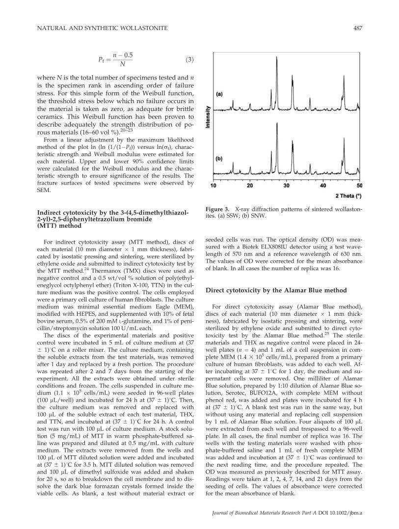
Figure 3. X-ray diffraction patterns of sintered wollaston-ites. (a) SSW; (b) SNW.
NATURAL AND SYNTHETIC WOLLASTONITE 487
Journal of Biomedical Materials Research Part A DOI 10.1002/jbm.a

Statistics
The statistical analysis of the results was performed underStatistica for Windows (StatSoft, 1999). In all statistical hy-pothesis tests a confidence level of a ¼ 0.05 was considered.
RESULTS
Phase composition and microstructure
Pseudowollastonite (b-CaSiO3, JCPDF 31-0300), thehigh temperature form of wollastonite, was the onlycrystalline phase detected by X-ray diffraction inSSW as previously reported [Fig. 3(a)].8
The wollastonite-Tc, the main crystalline phase ini-tially present in the natural powder, transformed
into pseudowollastonite during sintering, resultingin the same crystalline phase composition in SNWand SNW-M as in SSW [Fig. 3(b)].
However, polished sections of the sintered materi-als exhibited significant microstructural differences,mainly in pseudowollastonite crystal sizes and in theamount and distribution of the glassy phase (Fig. 4).SSW [Fig. 4(a,b)] presented smaller grain size, lesseramount of intergranular glassy phase, and smallerand more disperse pores than SNW [Fig. 4(c,d)]. Bygrinding the starting natural powder to reduce its par-ticle size from 13.2 to 2.0 mm, reduction in pore andgrain size, the first more accented, were reached in theSNW-M. A more homogeneous distribution of inter-granular glassy phase was also observed [Fig. 4(e,f)].
The results of semiquantitative analysis of theglassy phase in the sintered materials, performed byEDS, are shown in Table II. The pseudowollastonite
Figure 4. SEM images of the polished surfaces of the sintered samples. (a,b) SSW etched with 1:5 HAc for 2 s; (c,d) SNWno-etched; (e,f) SNW-M no-etched.
488 CARRODEGUAS ET AL.
Journal of Biomedical Materials Research Part A DOI 10.1002/jbm.a

crystals did not show any evidence of solid solutionin any of the samples studied. Therefore, the minorelements in the starting powders were concentratedin the glass formed during sintering of the samples.Consequently, the amount of glassy phase, in all thesamples, was roughly estimated from the Al2O3 con-tent in the glass, as determined by EDS, and fromthe Al2O3 content in the starting materials, as deter-mined by X-ray fluorescence, corrected for L.O.I.when required (Table I).
Fracture behavior
In Figure 5, characteristic fracture surfaces of the sin-tered materials are displayed. It was not possible to dis-cern any large defect as obvious fracture origin. At lowmagnification [Fig. 5(a,c)], fracture was much more tor-tuous in SNW samples than in the other ones. In the for-mer, large (section % 100 mm) zones surrounded bycracks were observed. Fracture was mostly transgranu-lar in SSW and SNW-M specimens [Fig. 5(d,f)] andmixed transgranular/intergranular in SNW [Fig. 5(b,e)].In all surfaces, distinguishable pores with sizes obeyingthe order SNW> SNW-M> SSWwere observed.
Characteristic diametral compressionstrength and Weibull module
The Weibull parameters are summarized in Table IIIand the diametral compression strength distributions
TABLE IIResults of the Semiquantitative Chemical
Analysis of the Glassy Phase
Element (wt %) SiO2 CaO Al2O3 Fe2O3 MgO Na2O K2O Glass
SSW 52 32 8 2 4 1 1 %2SNW 51 39 4.5 0.8 7 0.6 – %7SNW-M 57 22 5 3 13 0.9 – %15
Figure 5. SEM images of the fracture surfaces of the sintered samples: (a,d) SSW; (b,e) SNW; (c,f) SNW-M.
NATURAL AND SYNTHETIC WOLLASTONITE 489
Journal of Biomedical Materials Research Part A DOI 10.1002/jbm.a

calculated from data in Table III are plotted in Fig-ure 6, together with the experimental values for thethree studied materials.
Characteristic strength for SNW was, for a confi-dence level of 90%, significantly lower than for theother two materials, for which no significant differ-ences were found. For Weibull modulus, only signif-icant differences were found for SSW, which waslower than for both SNW materials.
Ions release and interfacial pH
The release profiles of Si, Ca, and P ions in SBF at36.58C for SSW and SNW are displayed in Figure 7.Both ceramic materials released Si and Ca ions, andremoved P ions from SBF in similar way andamounts. The specific concentration of Ca in SBFincreased from 100 to 230 mg L�1 cm�2 when SSWand SNW were incubated at 36.58C for 504 h (21days). In addition, SBF was enriched in Si up to avalue of 78 mg L�1 cm�2 for the same incubation pe-riod. However, P ions were removed from SBF byboth sintered materials, and its concentrationsdecreased from 31 to 19 and 5 mg L�1 cm�2 for SSWand SNW, respectively.
The pH at the SBF/ceramic interface continuouslyincreased from a value of 7.4 to over 13.0 after moni-toring for 20 h. However, the pH in the SBF bulk so-lution remained almost steady during the monitor-ing period (Fig. 8). Exactly the same behavior wasobserved for SSW and SNW-M.
In vitro bioactivity
The SEM surface images of SSW and SNW afterimmersion in SBF at 36.58C for 1 and 3 wk are dis-played in Figure 9. The surface of both ceramicswere eroded by the dissolution of the wollastonitegrains in the SBF during the first periods of immer-sion and a porous structure, apparently composedof the intergranular vitreous phase, was revealed
[Fig. 9(a,c)]. The superficial erosion was quickly fol-lowed for the precipitation of globular aggregates oftiny apatite-like crystals that covered the surface ofthe samples completely. The surface morphologiesobserved were very similar for all samples and allthe immersion periods.
The microanalysis and elemental mapping per-formed on polished cross-sections of the studiedmaterials after immersion in SBF at 36.58C for 3 wkrendered the results shown in Figure 10. In bothmaterials an external Ca- and P-rich layer wasobserved, followed by a Si-rich intermediate layer,coating the pseudowollastonite inner bulk material.
TABLE IIICharacteristic Diametral Compression Strength
and Weibull Modulusa
MaterialWeibull
Modulus, m
CharacteristicDiametral TensileStrength, s0 (MPa)
SSW 3.52 þ1.05 42.17 þ6.53�1.27 �5.58
SNW 6.47 þ1.93 25.55 þ2.09�2.34 �1.90
SNW-M 7.30 þ2.18 48.00 þ3.48�2.60 �3.16
aUpper and lower limits are given for a confidence levelof 90%.
Figure 6. Cumulative distribution for probability of fail-ure versus diametral tensile strength. Symbols correspondto experimental data and solid lines to the Weibull distri-bution calculated using the values of m and r0 of Table III.
Figure 7. Si, Ca, and P ion release profiles in SBF at36.58C.
490 CARRODEGUAS ET AL.
Journal of Biomedical Materials Research Part A DOI 10.1002/jbm.a

Indirect cytotoxicity
MTT [3-(4,5-dimethylthiazol-2-yl)-2,5-diphenylte-trazolium bromide] assay, first described by Mos-mann in 1983, is based on the ability of a mitochon-drial dehydrogenase enzyme from viable cells tocleave the tetrazolium rings of the pale yellow MTTand form dark blue formazan crystals which arelargely impermeable to cell membranes, thus result-ing in its accumulation within healthy cells. Theaddition of dimethyl sulfoxide results in the disinte-gration of the cell membrane and the liberation ofthe crystals which are quickly dissolved. The num-ber of surviving cells is directly proportional to the
level of the formazan product created, and the ODof its solution can be measured and related to theamount of viable cells.
MTT assay results obtained for extracts from theexperimental materials, THX (noncytotoxic) andTTN (cytotoxic), are displayed in Figure 11. Theresult for a quality control test is also shown in thefigure. The value of O:D obtained for the positivecontrol, TTN, was normal. The OD of the qualitycontrol test was also normal but slightly lower thanthe values of THX for the periods of extraction of 1and 2 days, according to the T-test for independentsamples. At 7 days of extraction there was no signifi-cant difference between control and THX, whichsupported the validity of the method. The ANOVAand post hoc comparison, using the Tukey’s honestsignificant difference test, revealed no significant dif-ference between SNW and SSW for any period ofextraction; more else, the extracts from experimentalmaterials and negative control, THX, had no signifi-cant difference for periods of extraction of 1 and 2days, and were significantly less cytotoxic than THXat 7 days of extraction.
Direct cytotoxicity
As employed in this work, Alamar Blue assay wasaimed to quantitatively measure cell proliferation,
Figure 8. pH evolution at the SBF/ceramic interface andin the bulk solution.
Figure 9. SEM images of the samples surfaces after immersion in SBF at 36.58C for different periods of time: (a,b) SSW, 1and 3 wk; (c,d) SNW, 1 and 3 wk.
NATURAL AND SYNTHETIC WOLLASTONITE 491
Journal of Biomedical Materials Research Part A DOI 10.1002/jbm.a

cytotoxicity, and viability, directly on the materialsunder study. The method is based on the incorpora-tion of resazurin and resarfurin as colorimetric redoxindicators. These indicators respond to chemicalreduction resulting from cell metabolism by chang-ing color. This color change may be measured bymonitoring the absorbance of the reduced form ofthe indicator (corrected for the background corre-sponding to the oxidized form), which is propor-tional to the amount of viable cells attached to thesurface of the tested material.
The results of the Alamar Blue assay performedon the experimental materials and the negative con-trol are represented in Figure 12. Thermanox, thenegative control, exhibited higher OD than the ex-perimental materials for all times of culture. The ODcorresponding to the experimental materials alwaysreached 80 to 60% of the Thermanox. The ANOVApointed out significant effects of the material nature,the culture time, and their interaction on OD. Therewere no significant differences between the ODs ofSSW and SNW-M, but both were significantly lower
than that of Thermanox for any culture time accord-ing to the post hoc comparison of means using theTukey’s honest significant difference test.
DISCUSSION
According to the results of chemical analysis(Table I), both precursor powders had similaramounts of major constituents. However, significantdifferences were found in the amounts of minor con-stituents, Al2O3, MgO, Na2O, and Fe2O3, which weresignificantly higher in the natural precursor. Theyshould be related to the minor crystalline phasesdetected in the natural powder as shown in theX-ray diffractogram of Figure 1(b).
The observed SiO2/CaO molar ratio in naturalwollastonite is slightly grater than the theoretical1.0 while in synthetic wollastonite is slightly lower(Table I), indicating a slight silica deficiency.
The main crystalline phases in the powders wereb-CaSiO3, also called pseudowollastonite, in the syn-thetic powder and a-CaSiO3 or wollastonite-Tc in the
Figure 10. Element mapping and EDS microanalysis of cross-sections of the samples after 3 wk in SBF at 36.58C. [Colorfigure can be viewed in the online issue, which is available at www.interscience.wiley.com.]
Figure 11. Results of MTT indirect cytotoxicity test. Errorbars represent the mean confidence interval (n ¼ 16, a ¼0.05).
Figure 12. Results of the Alamar Blue test. Error bars rep-resent the mean confidence interval (n ¼ 16, a ¼ 0.05).
492 CARRODEGUAS ET AL.
Journal of Biomedical Materials Research Part A DOI 10.1002/jbm.a

natural one. By heating, over 11258C, during the sin-tering, the low temperature form of wollastonite (a-CaSiO3), initially present in the natural powder,transformed reconstructively into b-CaSiO3.
26 At thecooling rate employed in this work, the b?a rever-sion expected at 11258C did not take place and thehigh temperature form was retained at room temper-ature in both SSW and SNW (Fig. 3).
According to the phase equilibrium diagram of thesystem CaO-SiO2,
27 no liquid should exist at the sin-tering temperature of 14008C; however, a glassy phasewas observed at the grain boundaries in SNW andSNW-M samples and mostly in triple points in SSWas shown in the micrographs of Figure 4. The amountof glassy phase developed in SNW and SNW-M sam-ples was about eight times greater that in SSW (TableII), and it was directly related to the content of impur-ities in the starting powder (Tables I). The grain sizedepended on the particle size of the starting powder.The large grains observed in SNW considerablydiminished in SNW-M [Fig. 4(e,f)]. A wide grain sizedistribution was observed in SNW [Fig. 4(c,d)].
In any of the fracture surfaces analyzed there wereno large singular defects. The main defects observedin the microstructure of the materials were pores,and sizes were the smallest for material SSW [2–10 mm, Fig. 5(a,d)], intermediate for material SNW-M[20–40 mm, Fig. 5(c,f)], and the largest for materialSNW [30–110 mm, Fig. 5(b,e)]. Therefore, large poresshould act as critical flaws in the studied materials,and this would explain the lowest values of charac-teristic strength found for material SNW (Table III),with the largest pore size [Fig. 5(b,e)].
Applying the same approach to SSW and SNW-M,the greatest strength values should be expected forSSW, with the smallest pores [Fig. 5(a–c,f)]. How-ever, the strength values of SSW specimens weresimilar or even lower than those of SNW-M (Fig. 6)and there were no significant differences betweenthe characteristic strengths of both materials for 90%confidence limits (Table III).
The three studied materials were constituted by asingle crystalline phase, pseudowollastonite, andglass. The chemical compositions of the glasses pres-ent in the materials (Table II) were close to that ofwollastonite. Moreover, negligible residual stresses,due to thermal expansion mismatch, will develop atthe boundaries of the two phases (pseudowollas-tonite and glass), both low Young’s modulus28,29 andthermal coefficients being30 very similar. (Table IV).Therefore, strong boundaries between the two phaseswould be expected from a physicochemical stand-point. In fact, fracture was mostly transgranular inspecimens SSW and SNW-M [Fig. 5(a,c,d,f)]. In mate-rial SNW fracture was mixed trans/intergranular,probably owing to the large glass areas found sur-rounding some crystalline zones [Fig. 4(c,d)].
The characteristic of the grain boundaries canexplain the fact that material SSW, with the finestmicrostructure [Fig. 4(a,b)], presented lower charac-teristic strength than did SNW (Fig. 6, Table III).According to Table II, the amount of glassy phase(15 wt %) in SNW-M was considerably larger thanin SSW (2 wt %), but it was homogeneously distrib-uted surrounding wollastonite grains [Fig. 4(f)].Therefore, the higher strength values of SNW-Mspecimens will be due to the role of the glass actingas effective cement between them. The lowest valueof Weibull modulus (Table III) and the bimodalshape of the probability of failure curve observed forSSW (Fig. 6) can be attributed to the small amountof vitreous phase, unevenly distributed in triplepoints, as well as to a broad distribution of grainsizes [Table II, Fig. 4(b)].
From data in Table III, the best mechanical per-formance, not only in terms of strength levels butalso in terms of reliability, corresponded to the mate-rial prepared from natural wollastonite previouslymilled to a particle size of 2.0 mm (SNW-M).
In the triclinic b-CaSiO3 network, the Ca2þ ionsare disposed in columns between chains of SiO4
4�
tetrahedrons. This special disposition of atoms pro-vides the crystalline network with lability and capa-bility to exchange Ca2þ for H3O
þ ions.6–9 No differ-ences were noticed in the ion release profiles andinterfacial pH (Figs. 7 and 8) for SSW and SNW sam-ples. All materials reacted in the same way and atthe same rate when were immersed in SBF at 36.58C,simulating the body environment. In previousreports, the maximum interfacial pH observed after20 days in SBF for a synthetic pseudowollastonite ce-ramic was 10.5.9 However, in this work, pH valuesover 13.0 were obtained after 20 h in SBF. The valuesof pH obtained, apparently unusual, can be due tothe lower liquid to area ratio (0.5 cm3/mm2) and theexperimental assembly here employed.
During the reaction of the ceramics in SBF a fastsurface dissolution of pseudowollatonite grains took
TABLE IVYoung’s Modules and Thermal Expansion
Coefficients of the Different Phases
Material
Young’sModulus(E) (GPa)
Coefficient ofExpansion(�10�6 C�1)
Pseudowollastonitesintered at 14008C/2 h27 94 7.0
Glass in sample SSW 84a 6.1b
Glass in sample SNW 86a 6.3b
Glass in sample SNW-M 81a 4.5b
aCalculated according to Navarro28 and Appen et al.,29
using the glass composition in Table II.bCalculated assuming ternary SiO2-CaO-Na2O glasses,
according to Miller and White.30
NATURAL AND SYNTHETIC WOLLASTONITE 493
Journal of Biomedical Materials Research Part A DOI 10.1002/jbm.a

place during the first week. The result of the attackwas a very porous surface structure constituted bythe remainder glassy phase, mainly located at triplepoints in sample SSW and grain boundaries in SNW[Fig. 9(a,c)]. As the dissolution progressed, the Ca2þ,OH�, and PO4
3� activities overcame the solubilityproduct of apatite and this starts to precipitate withthe typical morphology of globules constituted byaggregates of very fine acicular crystals [Fig. 9(b,d)].
The mechanism of the formation of the apatitelayer on pseudowollatonite was previously describedby some of the present authors6,8,9 and can be sum-marize in the following steps:
1. Exchange of Ca2þ, from the pseudowollastonitenetwork, by H3O
þ, from the SBF, and pH in-creasing at the pseudowollastonite/SBF interface;
2. Formation of a surface layer of hydrated silicagel, which partially dissolves at the high inter-facial pH;
3. Increasing of Ca2þ, OH�, HPO42� ionic activities at
the neighborhood of the reacting surface until theyovercome the solubility product of the apatite;
4. Nucleation of apatite on the remainder hydratedsilica gel layer; and
5. Growing of the apatite nuclei and growing ofthe apatite layer as well.
This mechanism is common to most of bioactivesilicon-containing materials1,3,9 and was confirmedfor SSW and SNW by the results of the elementalmapping and microanalysis performed on polishedcross sections of these materials submitted to immer-sion in SBF for 3 wk (Fig. 10).
In previous reports it has been clearly establishedthat synthetic pseudowollastonite is not cytotoxic, bio-compatible, and osteogenic.10–15 However, no studyon the cytotoxicity of pseudowollastonite ceramicobtained from natural precursors has been performed.SNW contain certain impurities that are absent in SSWand could induce a different response on in vitro cellcultures. However, the results of the MTT test dis-played in Figure 11 demonstrated that the solubleextracts from SNW and SSW exerted the same effecton the culture of human fibroblasts, and both werecompletely innocuous as evidenced by comparisonwith the results for the negative control, Thermanox.
From the results of the Alamar Blue test displayedin Figure 12, SNW and SSW exhibited the samebehavior again. However, the cell adhesion on SSWand SNW were significantly lower than on the nega-tive control, Thermanox. This situation may be a con-sequence of the fast surface remodeling that takesplace when the materials are immersed in MEM.MEM has an ionic composition and pH similar toSBF; thus, the same surface reactions observed in SBFshould occur when the ceramics are immersed in
MEM. The fast dissolution of the surface and thelocal pH increasing prior to the formation of the sta-ble apatite layer could hinder cell adhesion duringthe first periods of immersion. The formation of thestable apatite layer should provide a more ‘‘comforta-ble’’ bed to cell adhesion. According to this hypothe-sis, direct cell adhesion tests on bioactive materialsthat suffer a fast surface remodeling by immersion inthe body environment, as pseudowollastonite, shouldbe better carried out after a previous immersion pe-riod in the culture medium to stabilize their surface.The favorable results of in vivo tests conducted onsynthetic pseudowollastonite ceramics and previ-ously reported10,11 show that the material is perfectlybiocompatible, bioactive, and osteoconductive, inspite of the apparently negative results obtained inthe Alamar Blue test conducted in this work.
CONCLUSIONS
According to the obtained results, polycrystallinepsedowollastonite ceramics properly processed, frompure powder precursors of natural origin, exhibitedchemical and phase compositions, in vitro reactivity,bioactivity, and cytotoxicity very similar to those ofpseudowollastonite ceramic obtained by the syn-thetic route, and even better performance, in termsof strength and reliability, than the more expensivesynthetic material.
The results support the safe and successful use, inbiomedical applications, of pseudowollastonite bio-ceramics obtained from pure natural precursors.
References
1. Hench LL, Splinter RJ, Allen WC, Geenle TK. Bonding mech-anisms at the interface of ceramic prosthetic materials.J Biomed Mater Res 1972;2:1117–1141.
2. Kokubo T, Ito S, Sakka S, Yamamuro T. Formation of high-strength bioactive glass-ceramic in the system MgO-CaO-SiO2-P2O5. J Mater Sci 1986;21:536–540.
3. Hench LL, Wilson J, editors. An Introduction to Ceramics.Advanced Series in Ceramics, Vol. 1. Singapore: World Scien-tific; 1993.
4. Ohura K, Nakamura T, Yamamuro T, Kokubo T, Ebisawa T,Kotoura T. Bone bonding ability of P2O5-free CaO-SiO2
glasses. J Biomed Mater Res 1991;25:357–365.5. Nonami T, Tsutsumi S. Study of diopside ceramics for bio-
materials. J Mater Sci Mater Med 1999;10:475–479.6. De Aza PN, Guitian F, De Aza S. Bioactivity of wollastonite
ceramics: In vitro evaluation. Scr Metall Mater 1994;31:1001–1005.7. De Aza PN, Lublinska ZB, Anseau MR, Guitian F, De Aza S.
Bioactivity of pseudowollastonite in human saliva. J Dent1999;27:107–113.
8. De Aza PN, Lublinska ZB, Anseau M, Guitian F, De Aza S.Morphological studies of pseudowollastonite for biomedicalapplications. J Microsc 1996;182(Pt. 1):24–31.
9. De Aza PN, Guitian F, Merlos A, Lora-Tamayo E, De Aza S.Bioceramics-simulated body fluid interfaces: pH and its influ-
494 CARRODEGUAS ET AL.
Journal of Biomedical Materials Research Part A DOI 10.1002/jbm.a

ence on hydroxyapatite formation. J Mater Sci Mater Med1996;7:399–402.
10. De Aza PN, Lublinska ZB, Martinez A, Anseau MR, GuitianF, De Aza S. Morphological and structural study of pseudo-wollastonite implants in bone. J Microsc 2000;197(Pt. 1):60–67.
11. De Aza PN, Luklinska ZB, Anseau MR, Guitian F, De Aza S.Transmission electron microscopy of the interface between boneand pseudowollastonite implant. J Microsc 2001;201(Pt. 1):33–43.
12. Dufrane D, Delloye C, Mckay IJ, De Aza PN, De Aza S,Schneider YJ, Anseau M. Indirect cytotoxicity evaluation ofpseudowollastonite. J Mater Sci Mater Med 2003;14:33–38.
13. Sarmento C, Luklinska ZB, Brown L, Anseau M, De Aza PN,De Aza S, Hughes FJ, McKay IJ. The in vitro behaviour ofosteoblastic cell cultured in the presence of pseudowollaston-ite ceramic. J Biomed Mater Res 2004;69A:351–358.
14. Brown L, Lublinska ZB, De Aza PN, De Aza S, Anseau M,Hughes FJ, McKay IJ. Proceedings of the 7th World Biomate-rials Congress, Sydney, Australia, May 17–21 (2004).
15. Brown L, Lublinska ZB, De Aza PN, De Aza S, Anseau M,Hughes FJ, McKay IJ. Silicon released from bioactive ceramicmaterials enhances osteoblastic differentiation. Biomaterials.Submitted for publication.
16. O’Driscoll M. Wollastonite. Ceramics remains top consumer. I.M.Glass&Ceramics Survey. IndMinerals 1993 (Special Issue):77–83.
17. De Aza PN, Guitian F, De Aza S, Valle FJ. Analytical controlof wollastonite for biomedical applications by use of atomicabsorption spectrometry and inductively coupled plasmaatomic emission spectrometry. Analyst 1998;123:681–685.
18. Kim H-M, Miyazaki T, Kokubo T, Nakamura T. Revisedsimulated body fluid. Key Eng Mater 2001;192–195:47–50.
19. Carneiro FLLB, Barcellos A. Resistance a la traction desbetons. Bull RILEM(I) 1953;13:97–108.
20. Rudnick A, Hunter AR, Holden FC. An Analysis of the Dia-metral-Compression Test. Mater Res Stand 1963;4:283–289.
21. Villora JM, Callejas P, Barba MF, Baudın C. Statistical analy-sis of the fracture behaviour of porous ceramic raschig rings.J Eur Cer Soc 2004;24:589–594.
22. Weibull W. A statistical distribution function of wide applic-ability. J Appl Mech 1951;18:292–297.
23. Ferrari B, Gonzalez S, Moreno R, Baudın C. Strength analysisof self-supported films produced by aqueous electrophoreticdeposition. J Am Cer Soc 2005;88:2645–2648.
24. Mosmann T. Rapid colorimetric assay for cellular growth andsurvival: Application to proliferation and cytotoxicity assays.J Immunol Methods 1983;65:55–63.
25. Nakayama GR, Caton MC, Nova MP, Parandoosh Z. J Immu-nol Methods 1997;204:205–208.
26. Deer WA, Howie RA, Zussman J. Rock Forming Minerals,2nd ed. Vol. 2A. Single-Chain Silicates. London: Longmans;1978. p 547–563.
27. Phillips B, Muan A. Phase equilibria in the system CaO–ironoxide–SiO2, in air. J Am Ceram Soc 1959;42:413–423.
28. Fernandez Navarro JM. El Vidrio. Madrid: Sociedad Espanolade Ceramica y Vidrio; 1993.
29. Appen AA, Kozlovskaya EI, Fu-Si H. Study of the elastic andacoustic properties of silicates glasses. J Appl Chem (U.S.S.R.)1961;34:975–981.
30. Miller GH, White D. Composition-thermal expansion rela-tionships in ternary glass systems. Glass Technol 1977;18:113–116.
NATURAL AND SYNTHETIC WOLLASTONITE 495
Journal of Biomedical Materials Research Part A DOI 10.1002/jbm.a


