BIOCERAMICS. - Alumina - Zirconia - Carbon - Hydroxyapatite - glasses (vetroceramics, bioglasses)

CHAPTER 15BIOCERAMICS
David H. KohnUniversity of Michigan,Ann Arbor, Michigan
1
15.1 INTRODUCTION 1
15.2 BIOINERT CERAMICS 3
15.3 BIOACTIVE CERAMICS 7
15.4 CERAMICS FOR TISSUE ENGINEERINGAND BIOLOGICAL THERAPIES 14
15.5 SUMMARY 21
ACKNOWLEDGEMENTS 21
REFERENCES 21
15.1 INTRODUCTION
The clinical goal when using ceramic biomaterials, as is the case with any biomaterial, is to replacelost tissue or organ structure and/or function. The rationale for using ceramics in medicine and den-tistry was initially based upon the relative biological inertness of ceramic materials compared tometals. However, in the past 25 years, this emphasis has shifted more toward the use of bioactiveceramics, materials that elicit normal tissue formation and also form an intimate bond with bone tis-sue through partial dissolution of the material surface. In the last decade, bioceramics have also beenutilized in conjunction with more biological therapies. In other words, the ceramic, usually resorbable,facilitates the delivery and function of a biological agent (i.e., cells, proteins, and/or genes), with anend-goal of eventually regenerating a full volume of functional tissue.
Ceramic biomaterials are processed to yield one of four types of surfaces and associated mecha-nisms of tissue attachment (Kohn and Ducheyne, 1992): (1) fully dense, relatively inert crystallineceramics that attach to tissue by either a press fit, tissue growth onto a roughened surface, or via agrouting agent; (2) porous, relatively inert ceramics, where tissue grows into the pores, creating amechanical attachment between the implant and tissue; (3) fully dense, surface reactive ceramics,which attach to tissue via a chemical bond; and (4) resorbable ceramics that integrate with tissue andeventually are replaced by new or existing host tissue. Ceramics may therefore be classified by theirmacroscopic surface characteristics (smooth, fully dense, roughened, or porous) or their chemicalstability (inert, surface reactive, or bulk reactive/resorbable). The integration of biological (i.e., inductive)agents with ceramics further expands the clinical potential of these materials.
Relatively inert ceramics elicit minimal tissue response and lead to a thin layer of fibrous tissueadjacent to the ceramic surface. Surface-active ceramics are partially soluble, resulting in ionexchange and the potential to lead a direct chemical bond with tissue. Bulk bioactive ceramics arefully resorbable, have greater solubility than surface-active ceramics, and may ultimately bereplaced by an equivalent volume of regenerated tissue. The relative level of bioactivity mediatesthe thickness of the interfacial zone between the biomaterial surface and host tissue (Fig. 15.1).
15-Kutz-V-I_c015_001-000.qxd 3/5/09 10:01 AM Page 1

There are, however, no standardized measures of “reactivity,” but the most common are pHchanges, ion solubility, tissue reaction, and any number of assays that assess some parameter ofcell function.
Five main ceramic materials are used for musculoskeletal reconstruction and regeneration: carbon(Christel et al., 1987; Haubold et al., 1981; Huttner and Huttinger, 1984), alumina (Al2O3) (Kohnand Ducheyne, 1992; Hulbert et al., 1970; Boutin et al., 1988; Heimke et al., 1978; Websteret al., 2000; Zreiqat et al., 1999; Tohma et al., 2006; Nizard et al., 2008), zirconia (ZrO2) (Kohn andDucheyne, 1992; Cales and Stefani, 1995; Christel et al., 1989; Filiaggi, et al., 1996), bioactiveglasses and glass ceramics (Kohn and Ducheyne, 1992; Ducheyne, 1985; El-Ghannam et al., 1997;Radin et al., 2005; Reilly et al., 2007; Gross and Strunz, 1980; Hench et al., 1972; Nakamura et al.,1985) and calcium phosphates (Kohn and Ducheyne, 1992; Murphy et al., 2000a; Shin et al., 2007;Ducheyne 1987; Ducheyne et al., 1980; Koeneman et al., 1990; Van Raemdonck et al., 1984).Carbon, alumina, and zirconia are considered “bioinert,” whereas glasses and calcium phosphates arebioactive.
In this chapter, three types of bioceramics (bioinert, surface bioactive, bulk bioactive) are dis-cussed, with a focus on musculoskeletal applications. A material science approach is taken to addressdesign issues of importance to a biomedical engineer; the processing-structure-composition-propertysynergy is discussed for each material, then properties important to the design and clinical successof each class of bioceramic are presented. Within the framework of discussing the processing-composition-structure synergy, issues of material selection, service conditions, fabrication routes andcharacterization methodologies are discussed.
2 BIOMATERIALS
30
20
40
60
80
100
10 100Implantation time (days)
Perc
enta
ge o
f in
terf
acia
l bon
e tis
sue
Rel
ativ
ebi
orea
ctiv
ity
1000
Bioceramics
GFEDC
BA
A
A
BC
DE F G
A. 45SS bioglass
Type 4 (resorbable)
Type 3 bioactive
B. KGS ceravitalC. 55S4.3 bioglassD. A-W glass ceramicE. Hydroxylapatite (HA)F. KGX ceravitalG. Al2O3, Si3N4
B
Type 2 porous ingrowth
Type 1 nearly inert
FIGURE 15.1 Bioactivity spectra for selected bioceramics: (a) relative magnitudes and rates of bioactivity,(b) time dependence of bone formation at bioceramic surface and ceramic/bone bonding. [From Hench andBest (2004), with permission.]
15-Kutz-V-I_c015_001-000.qxd 3/5/09 10:01 AM Page 2

15.2 BIOINERT CERAMICS
Ceramics are fully oxidized materials and are therefore chemically stable and less likely to elicit anadverse biological response than metals, which only oxidize at their surface. Three types of “inert”ceramics are of interest in musculoskeletal applications: carbon, alumina, and zirconia.
15.2.1 Carbon
The benign biological reaction to carbon-based materials, along with the similarity in stiffness andstrength between carbon and bone, made carbon a candidate material for musculoskeletal recon-struction almost 40 years ago (Bokros et al., 1972). Carbon has a hexagonal crystal structure that isformed by strong covalent bonds. Graphite has a planar hexagonal array structure, with a crystal sizeof approximately 1000 Å (Bokros, 1978). The carbon-carbon bond energy within the planes is large(114 kcal/mol), whereas the bond between the planes is weak (4 kcal/mol) (Hench and Ethridge,1982). Therefore, carbon derives its strength from the strong in-plane bonds, whereas the weak bondingbetween the planes results in a low modulus, near that of bone (Bokros, 1978).
Isotropic carbon, on the other hand, has no preferred crystal orientation and therefore possessesisotropic material properties. There are three types of isotropic carbon: pyrolytic, vitreous, and vapordeposited. Pyrolytic carbons are formed by the deposition of carbon from a fluidized bed onto a sub-strate. The fluidized bed is formed from pyrolysis of hydrocarbon gas between 1000 to 2500°C(Hench and Ethridge, 1982). Low temperature isotropic (LTI) carbons are formed at temperaturesbelow 1500°C. LTI pyrolytic carbon possesses good frictional and wear properties, and incorpora-tion of silicon can further increase hardness and wear resistance (Bokros, 1978). Vitreous carbon isa fine-grained polycrystalline material formed by slow heating of a polymer. Upon heating, the morevolatile components diffuse from the structure and only carbon remains (Hench and Ethridge, 1982).Since the process is diffusion mediated and potentially volatile, heating must be slow and dimensionsof the structure are limited to approximately 7 mm (Bokros, 1978). Salient properties of all threeforms of carbon are summarized in Table 15.1.
Deposition of LTI coatings onto metal substrates is limited by the brittleness of the coatings andpropensity for coating fracture and coating/substrate debonding (Hench and Ethridge, 1982). Carbonmay also be vapor deposited onto a substrate by the evaporation of carbon atoms from a high-temperature source and subsequent condensation onto a low temperature substrate (Hench andEthridge, 1982). Vapor-deposited coatings are approximately 1 μm thick, allowing properties of thesubstrate to be retained. More recently, diamondlike carbon (DLC) coatings have been studied, as ameans of improving fixation to bone (Koistinen et al., 2005, Reikeras et al., 2004) and wear resis-tance (Allen et al., 2001). Carbon-based thin films are produced from solid carbon or liquid/gaseoushydrocarbon sources, using ion beam or plasma deposition techniques, and have properties interme-diate to those of graphite and diamond (Allen et al., 2001).
With the advent of nanotechnology, interest in carbon has been rekindled, in the form of carbonnanotubes (CNTs). CNTs have been proposed as scaffolds to support osteoconductivity (Zanelloet al., 2006) and as a second phase in polymer scaffolds (Shi et al., 2006).
15.2.2 Alumina
High-density, high-purity, polycrystalline alumina is used for femoral stems, femoral heads, acetabularcomponents, and dental implants (Kohn and Ducheyne, 1992; Boutin et al., 1988; Heimke et al., 1978;Tohma et al., 2006; Nizard et al., 2008). More recently, ion-modified and nanostructured Al2O3 havebeen synthesized, to make these bioceramics stronger and more bioactive (Webster et al., 2000; Zreiqatet al., 1999). In addition to chemical stability and relative biological inertness, other attributes of alu-mina are hardness and wear resistance. Therefore, a main motivation for using alumina in orthopaedicsurgery is to increase tribological properties, and many total hip replacements are now designed asmodular devices, that is, an alumina femoral head is press-fit onto the neck of a metal femoral stem.
BIOCERAMICS 3
15-Kutz-V-I_c015_001-000.qxd 3/5/09 10:01 AM Page 3

High-purity alumina powder is typically isostatically compacted and shaped. Subsequent sintering at1600 to 1800°C transforms a preform into a dense polycrystalline solid having a grain size of less than5 μm (Boutin et al., 1988). Addition of trace amounts of MgO aids in sintering and limits grain growth.If processing is kept below 2050°C, α-Al2O3, which is the most stable phase, forms. Alternatively,single crystals (sapphire) may be grown by feeding powder onto a seed and allowing buildup.
The physical and mechanical properties (e.g., ultimate strength, fatigue strength, fracture toughness,wear resistance) of α-alumina are a function of purity, grain size, grain size distribution, porosity, andinclusions (Kohn and Ducheyne, 1992; Boutin et al., 1988; Dorre and Dawihl, 1980) (Table 15.1). Theelastic modulus of dense alumina is two-to-four fold greater than that of metals used in bone and jointreconstruction. Both grain size (d) and porosity (P, 0 ≤ P ≤ 1) affect strength (σ) via power law andexponential relations, respectively [Eqs. (15.1) and (15.2)], where σ0 is the strength of the denseceramic, A, n, and B are material constants, experimentally determined, and n is approximately 0.5.
4 BIOMATERIALS
TABLE 15.1 Physical and Mechanical Properties of Bioceramics
Porosity, Density, Modulus, Compressive Tensile Flexural KIc, Material % mg/m3 (GPa) strength, MPa strength, MPa strength, MPa MPa.m1/2
Graphite 7 1.8 25 – – 140 – (isotropic) 12 1.8 20–24 65–95 24–30 45–55 –
16–20 1.6–1.85 6–13.4 18–58 8–19 14–27 – 30 1.55 7.1 – – – – – 0.1–0.5 – 2.5–30 – – –
Pyrolytic 2.7 2.19 28–41 – – – – graphite, LTI – 1.3–2 17–28 900 200 340–520 –
– 1.7–2.2 17–28 – – 270–550 –
Glassy (vitreous) – 1.4–1.6 – – – 70–205 – carbon – 1.45–1.5 24–28 700 70–200 150–200 –
– 1.38–1.4 23–29 – – 190–255 – ≤50 <1.1 7–32 50–330 13–52 – –
Bioactive – – – – 56–83 – – ceramics and – 2.8 – 500 – 100–150 – glass ceramics 31–76 0.65–1.86 2.2–21.8 – – 4–35 –
Hydroxyapatite 0.1–3 3.05–3.15 7–13 350–450 38–48 100–120 – 10 2.7 – – – – – 30 – – 120–170 – – – 40 – – 60–120 – 15–35 –
2.8–19.4 2.55–3.07 44–48 310–510 – 60–115 – 2.5–26.5 – 55–110 ≤800 – 50–115 –
Tetracalcium Dense 3.1 – 120–200 – – – phosphate
Tricalcium Dense 3.14 – 120 – – – phosphate
Other calcium Dense 2.8–3.1 – 70–170 – – – phosphates
Al2O3 0 3.93–3.95 380–400 4000–5000 350 400–500 5–625 2.8–3.0 150 500 – 70 – 35 – – 200 – 55 –
50–75 – – 80 – 6–11.4 –
ZrO2, stabilized 0 4.9–5.6 150–200 1750 – 150–900 4–12(~3% Y2O3) 1.5 5.75 210–240 – – 280–450 –
5 – 150–200 – – 50–500 – 28 3.9–4.1 – < 400 – 50–65 –
Source: Modified from Kohn and Ducheyne (1992), with permission.
15-Kutz-V-I_c015_001-000.qxd 3/5/09 10:01 AM Page 4

σ = Ad−n (15.1)
σP = σ0 e−BP (15.2)
For example, decreasing the grain size of Al2O3 from 4 to 7 μm increases strength by approximately20 percent (Dorre and Dawihl, 1980). With advances in ceramic processing, it is now possible tofabricate alumina with grain sizes approximately 1 μm and small grain size distributions, materialcharacteristics that increase strength.
The amount of wear in alumina-alumina bearing couples can be as much as 10 times less than inmetal-polyethylene systems (Davidson, 1993; Kumar et al., 1991; Lusty et al., 2007). The coeffi-cients of friction of alumina-alumina and alumina-polyethylene are less than that of metal-polyethylene, because of alumina’s low surface roughness and wettability (Boutin et al., 1988;Semlitsch et al., 1977).
The major limitations of alumina are its low tensile and bending strengths and fracture toughness.As a consequence, alumina is sensitive to stress concentrations and overloading. Clinically retrievedalumina total hip replacements exhibit damage caused by fatigue, impact, or overload (Walter andLang, 1986). Many ceramic failures can be attributed to materials processing or design deficiencies,and can be minimized through better materials choice and quality control.
15.2.3 Zirconia
Yttrium oxide partially stabilized zirconia (YPSZ) is an alternative to alumina, and there are approx-imately 150,000 zirconia components in clinical use (Christel et al., 1989; Cales and Stefani, 1995).YPSZ has a higher toughness than alumina, since it can be transformation toughened, and is used inbulk form or as a coating (Filiaggi et al., 1996).
At room temperature, pure zirconia has a monoclinic crystal symmetry. Upon heating, it trans-forms to a tetragonal phase at approximately 1000 to 1100°C, and then to a cubic phase at approxi-mately 2000°C (Fig. 15.2). A partially reversible volumetric shrinkage (density increase) of 3 to 10percent occurs during the monoclinic to tetragonal transformation (Christel et al., 1989). The volu-metric changes resulting from the phase transformations can lead to residual stresses and cracking.Furthermore, because of the large volume reduction, pure zirconia cannot be sintered. However, sin-tering and phase transformations can be controlled via the addition of stabilizing oxides. Yttriumoxide (Y2O3) serves as a stabilizer for the tetragonal phase such that upon cooling, the tetragonalcrystals are maintained in a metastable state and do not transform back to a monoclinic structure.The tetragonal to monoclinic transformation and volume change are also prevented by neighboringgrains inducing compressive stresses on one another (Christel et al., 1989).
The modulus of partially stabilized zirconia is approximately half that of alumina, while the bendingstrength and fracture toughness are 2 to 3 and 2 times greater, respectively (Table 15.1). The relativelyhigh strength and toughness are a result of transformation toughening, a mechanism that manifests itselfas follows (Fig. 15.3): crack nucleation and propagation lead to locally elevated stresses and energy inthe tetragonal crystals surrounding the crack tip. The elevated energy induces the metastable tetragonalgrains to transform into monoclinic grains in this part of the microstructure. Since the monoclinic grainsare larger than the tetragonal grains, there is a local volume increase, compressive stresses are induced,more energy is needed to advance the crack, and crack blunting occurs.
The wear rate of YPSZ on UHMWPE can be 5 times less than the wear rate of alumina onUHMWPE, depending on experimental conditions (Kumar et al., 1991; Davidson, 1993; Derbyshireet al., 1994). Wear resistance is a function of grain size, surface roughness, and residual compressivestresses induced by the phase transformation. The increased mechanical and tribological propertiesof zirconia may allow for smaller diameter femoral heads to be used in comparison to alumina.
Partially stabilized zirconia is typically shaped by cold isostatic pressing and then densified bysintering. Sintering may be performed with or without a subsequent hot isostatic pressing (HIP-ing)cycle. The material is usually presintered until approximately 95 percent dense and then HIP-ed toremove residual porosity (Christel et al., 1989). Sintering can be performed without inducing graingrowth, and final grain sizes can be less than 1 μm.
BIOCERAMICS 5
15-Kutz-V-I_c015_001-000.qxd 3/5/09 10:01 AM Page 5

15.2.4 Critical Properties of Bioinert Ceramics
Properties of bioinert ceramics important for their long-term clinical function include stiffness,strength, toughness, wear resistance, and biological response. Stiffness represents one gauge of themechanical interaction between an implant and its surrounding tissue, it is one determinant of themagnitude and distribution of stresses in a biomaterial and tissue, and dictates, in part, the potentialfor stress shielding (Kohn and Ducheyne, 1992; Ko et al., 1995). Load-bearing biomaterials mustalso be designed to ensure that they maintain their structural integrity, that is, designed to be fail-safeat stresses above peak in-service stresses for a lifetime greater than the expected service life of theprosthesis. Thus, the static (tensile, compressive, and flexural strength), dynamic (high-cycle fatigue)and toughness properties of ceramics, in physiological media, under a multitude of loading condi-tions and rates must be well-characterized.
Although knowledge of these properties is an important aspect of bioceramic design, the mechani-cal integrity of a bioceramic is also dependent on its processing, size, and shape. Failure of ceramics
6 BIOMATERIALS
0
0
500
1000
1500
2000
2500
3000
5 10
Tem
pera
ture
(°C
)
Mole % YO1.5
Monoclinic CubicNontransformable
tetragonal (T')
Mono- clinic(M)
Cubic (F)
T + F
L + FLiquid (L)
15 20
Transformabletetragonal(T)
M + T
FIGURE 15.2 Schematic phase diagram of the ZrO2−Y2O3 system. [FromCales and Stefani (1995), with permission.]
15-Kutz-V-I_c015_001-000.qxd 3/5/09 10:01 AM Page 6

usually initiates at a critical defect, at a stress level that depends on the geometry of the defect. Toaccount for these variables and minimize the probability of failure, fracture mechanics and statis-tical distributions are used to predict failure probability at different load levels (Soltesz andRichter, 1984).
15.3 BIOACTIVE CERAMICS
The concept of bioactivity originated with bioactive glasses via the hypothesis that the biocompati-bility of an implant is optimal if it elicits the formation of normal tissues at its surface, and if it estab-lishes a contiguous interface capable of supporting the loads which occur at the site of implantation(Hench et al., 1972). Under appropriate conditions, three classes of ceramics may fulfill theserequirements: bioactive glasses and glass ceramics, calcium phosphate ceramics, and composites ofglasses and ceramics. Incorporation of inductive factors into each of these ceramics may enhancebioactivity. These different classes of ceramics (and biological constituents) are used in a variety ofapplications, including: bulk implants (surface active), coatings on metal or ceramic implants (sur-face active), permanent bone augmentation devices/scaffold materials (surface active), temporaryscaffolds for tissue engineering (surface or bulk active), fillers in cements or scaffolds (surface orbulk active), and drug delivery vehicles (bulk active).
The nature of the biomaterial/tissue interface and reactions (e.g., ion exchange) at the ceramicsurface and adjacent tissues dictate the resultant mechanical, chemical, physical, and biologicalproperties. Four factors determine the long-term effect of bioactive ceramic implants: (1) the site ofimplantation, (2) tissue trauma, (3) the bulk and surface properties of the material, and (4) the relative
BIOCERAMICS 7
FIGURE 15.3 Schematic of microstructure in yttria partially stabilized zirconia (YPSZ) bioceramicundergoing transformation toughening at a crack tip. (From Miller et al. (1981), with permission.]
Propagatingcrack
Tetragonal grain
Transformed monoclinic grain
Compressive stress ahead of the crack
15-Kutz-V-I_c015_001-000.qxd 3/5/09 10:01 AM Page 7

motion at the implant/tissue interface (Ducheyne et al., 1987). For resorbable materials, additionaldesign requirements include: the need to maintain strength and stability of the material/tissueinterface during material degradation and host tissue regeneration; material resorption and tissuerepair/regeneration rates should be matched; and the resorbable material should consist only of meta-bolically acceptable species.
15.3.1 Bioactive Glasses and Glass Ceramics
Bioactive glasses are used as bulk implants, coatings on metal or ceramic implants, and scaffolds forguiding biological therapies (Kohn and Ducheyne, 1992; Hench et al., 1972; El-Ghannam et al., 1997;Gross and Strunz, 1980; Nakamura et al., 1985; Radin et al., 2005; Reilly et al., 2007) (Table 15.2).Chemical reactions are limited to the surface (~300 to 500 μm) of the glass, and bulk properties arenot affected by surface reactivity. The degree of activity and physiologic response are dependent onthe chemical composition of the glass, and may vary by over an order of magnitude. For example,the substitution of CaF for CaO decreases solubility, while addition of B2O3 increases solubility(Hench and Ethridge, 1982).
Ceravital, a variation of Bioglass, is a glass ceramic. The seed material is quench-melted to forma glass, then heat-treated to form nuclei for crystal growth and transformation from a glass to aceramic. Ceravital has a different alkali oxide concentration than bioglass—small amounts of alka-line oxides are added to control dissolution rates (Table 15.2)—but the physiological response toboth glasses is similar (Gross and Strunz, 1980). A glass ceramic containing crystalline oxyapatite,fluorapatite, and β-Wollastonite in a glassy matrix, denoted glass-ceramic A-W, is another bioactiveglass ceramic (Kitsugi et al., 1986; Kokubo et al., 1990a, 1990b; Nakamura et al., 1985). A-W glass-ceramic bonds to bone through a thin calcium and phosphorus-rich layer, which is formed at the sur-face of the glass ceramic (Kitsugi et al., 1986; Nakamura et al., 1985). In vitro, if the physiologicalenvironment is correctly simulated in terms of ion concentration, pH, and temperature, this layer con-sists of small carbonated hydroxyapatite (HA) crystallites with a defective structure, and the com-position and structural characteristics are similar to those of bone (Kokubo et al., 1990a).
Glass and glass-ceramics interface with the biological milieu because ceramics are susceptible tosurface changes in an aqueous media. Lower valence ions segregate to surfaces and grain boundaries,leading to concentration gradients and ion exchange. These reactions are dependent on the local pH andreactive cellular constituents (Hench and Ethridge, 1982), and can be biologically beneficial or adverse.Therefore, the surface reactions of glass ceramics should be well-controlled and characterized.
When placed in physiological media, bioactive glasses leach Na+ ions, and subsequently K+,Ca2+, P5+, Si4+, and Si-OH. These ionic species are replaced with H3O
+ ions from the media through
8 BIOMATERIALS
TABLE 15.2 Composition (Weight Percent) of Bioactive Glasses and Glass Ceramics
45S5 45S5-F 45S5-B5 52S4.6 Stabilized A-W GlassMaterial Bioglass Bioglass Bioglass Bioglass Ceravital ceravital ceramic
SiO2 45.0 45.0 45.0 52.0 40–50 40–50 34.2P2O5 6.0 6.0 6.0 6.0 10–15 7.5–12.0 16.3CaO 24.5 12.3 24.5 21.0 30–35 25–30 44.9Na2O 24.5 24.5 24.5 21.0 5–10 3.5–7.5 –B2O3 – – 5.0 – – – –CaF2 – 12.3 – – – 0.5K2O – – – – 0.5–3.0 0.5–2.0 –MgO – – – – 2.5–5.0 1.0–2.5 4.6Al2O3 – – – – – 5.0–15.0 –TiO2 – – – – – 1.0–5.0 –Ta2O5 – – – – – 5.0–15.0 –
Source: From Kohn and Ducheyne (1992), with permission.
15-Kutz-V-I_c015_001-000.qxd 3/5/09 10:01 AM Page 8

an ion-exchange reaction which produces a silica-rich gel surface layer (Hench and Ethridge, 1982).In an in vitro setting at least, the depletion of H+/H3O
+ ions in solution causes a pH increase, whichfurther drives dissolution of the glass surface. With increasing time of exposure to media, the high-surface-area silica-rich surface gel chelates calcium and phosphate ions, and a Ca-P-rich, amorphousapatite layer forms on top of the silica-rich layer. This Ca-P-rich layer may form after as little as1 hour in physiological solution (Hench and Ethridge, 1982). The amorphous Ca-P layer eventuallycrystallizes and CO3
2− substitutes for OH− in the apatite lattice, leading to the formation of a car-bonated apatite layer. Depending on animal species, anatomic, site and time of implantation, thesteady-state thickness of the Ca-P-rich and Si-rich zones can range from 30 to 70 μm and 60 to 230 μm,respectively (Hench and Ethridge, 1982).
In parallel with these physical/chemical-mediated reactions, in an in vivo setting, proteinsadsorb/desorb from the silica gel and carbonate layers. The bioactive surface and preferential proteinadsorption that can occur on the surface can enhance attachment, differentiation, and proliferation ofosteoblasts and secretion of an extracellular matrix (ECM). Crystallization of carbonated apatitewithin an ordered collagen matrix leads to an interfacial bond.
The overall rate of change of the glass surface R is quantified as the sum of the reaction rates ofeach stage of the reaction (Hench and Best, 2004):
R = −k1t0.5 − k2t
1.0 + k3t1.0 + k4t
y + k5tz (15.3)
where ki is the rate constant for each stage, i and represents, respectively, the rate of exchange betweenalkali cations in glass and H+/H3O
+ in solution (k1), interfacial SiO2 network dissolution (k2), repoly-merization of SiO2 (k3), carbonate precipitation and growth (k4), and other precipitation reactions (k5).Using these rates, the following design criterion may be established: the kinetics of each stage, especiallystage 4, should match the rate of biomineralization in vivo. For R >> in vivo rates, resorption will occur,whereas if R << in vivo rates, the glass will be nonbioactive (Hench and Best, 2004).
The degree of activity and physiological response (e.g., rates of formation of the Ca-P surface andglass/tissue bond) therefore depends on the glass composition and time, and is mediated by the bio-material, solution, and cells. The dependence of reactivity and rate of bond formation on glass com-position is defined by the ratio of network former to network modifier: SiO2/[CaO + Na2O + K2O](Hench and Clark, 1982). The higher this ratio is, the less soluble is the glass, and the slower is therate of bone formation. A SiO2-Na2O-CaO ternary diagram (Fig. 15.4) is useful to quantify the rela-tionship between composition and biological response (Hench and Best, 2004). The diagram may bedivided into three zones: zone A—bioactive bone bonding: glasses are characterized by CaO/P2O5ratios > 5 and SiO2/[CaO + Na2O] < 2; zone B—nearly inert: bone bonding does not occur (onlyfibrous tissue formation occurs), because the SiO2 content is too high and reactivity is too low—these high SiO2 glasses develop only a surface hydration layer or too dense of a silica-rich layer toenable further dissolution and ion exchange; zone C—resorbable glasses: no bone bonding occursbecause reactivity is too high and SiO2 undergoes rapid selective alkali ion exchange with protonsor H3O
+, leading to a thick but porous unprotected SiO2-rich film that dissociates at a high rate. The level of bioactivity is related to bone formation via an index of bioactivity IB, which is related
to the amount of time it takes for 50 percent of the interface to be bonded (Hench and Best, 2004):
IB = 100/t0.5BB (15.4)
The compositional dependence of the biological response may be understood by iso-IB contourssuperposed onto the ternary diagram (Fig. 15.4). The cohesion strength of the glass/tissue interfacewill be a function of surface area, thickness, and stiffness of the interfacial zone, and is optimum forIB ~ 4 (Hench and Best, 2004).
15.3.2 Calcium-Phosphate Ceramics
Calcium-phosphate (Ca-P) ceramics are ceramics with varying calcium-to-phosphate ratios. Amongthe Ca-Ps, the apatites, defined by the chemical formula M10(XO4)6Z2, have been studied most andare most relevant to biomaterials Apatites form a range of solid solutions as a result of ion substitution
BIOCERAMICS 9
15-Kutz-V-I_c015_001-000.qxd 3/5/09 10:01 AM Page 9

at the M2+, XO43−, or Z− sites. Apatites are usually nonstoichiometric and contain less than 10 mol of
M2+ ions, less than 2 mol of Z− ions, and exactly 6 mol of XO43− ions (Van Raemdonck et al., 1984).
The M2+ species is typically a bivalent metallic cation, such as Ca2+, Sr2+ or Ba2+, Pb2+, or Cd2+, theXO4
3− species is typically PO43−, VO4
3−, CrO43− or MnO4
3, and the monovalent Z− ions are usuallyOH−, F−, or Br− (Van Raemdonck et al., 1984).
More complex ionic structures may also exist. For example, replacing the two monovalentZ− ions with a bivalent ion, such as CO3
2−, results in the preservation of charge neutrality, but oneanionic position becomes vacant. Similarly, the M2+ positions may also have vacancies. In this case,charge neutrality is maintained by vacancies at the Z− positions or by substitution of trivalent PO4
3−
ions with bivalent ions (Van Raemdonck et al., 1984).The most common apatite used in medicine and dentistry is hydroxyapatite, a material with the
chemical formula Ca10(PO4)6(OH)2, denoting that 2 formula units are represented within each unitcell (Fig. 15.5). HA has ideal weight percents of 39.9 percent Ca, 18.5 percent P, and 3.38 percentOH, and an ideal Ca/P ratio of 1.67. The crystal structure and crystallization behavior of HA areaffected by ionic substitutions.
The impetus for using synthetic HA as a biomaterial stems from the hypothesis that a materialsimilar to the mineral phase in bone and teeth will have superior binding to mineralized tissues andis, therefore, advantageous for replacing these tissues. Additional advantages of bioactive ceramicsinclude low thermal and electrical conductivity, elastic properties similar to those of bone, control ofin vivo degradation rates through control of material properties, and the potential for ceramic to func-tion as a barrier when coated onto a metal substrate (Koeneman et al., 1990).
The HA in bone is nonstoichiometric, has a Ca/P ratio less than 1.67, and also contains carbonate,sodium, magnesium, fluorine, and chlorine (Posner, 1985a). Most synthetic hydroxyapatites containsubstitutions for the PO4
3− and/or OH− groups and therefore vary from the ideal stoichiometry andCa/P ratios. Oxyhydroxyapatite, tricalcium phosphate, tetracalcium phosphate, and octocalciumphosphate have all been detected in commercially available apatite implants (Table 15.3) (Kohn andDucheyne, 1992; Ducheyne et al., 1986, 1990; Koch et al., 1990).
10 BIOMATERIALS
FIGURE 15.4 Ternary diagram (SiO2-Na2O-CaO, at fixed 6 percent P2O5) showingthe compositional dependence (in weight percent) of bone bonding and fibrous tissuebonding to the surfaces of bioactive glasses and glass ceramics: zone A: bioactive bonebonding ceramics; zone B: nearly inert ceramics—bone bonding does not occur at theceramic surface, only fibrous tissue formation occurs; zone C: resorbable ceramics—no bone bonding occurs because reactivity is too high; IB = index of bioactivity forbioceramics in zone A. [From Hench and Best (2004), with permission.]
A-W GC (high P2O5)
CaO Na2O
A
D
C
SiO2Bioglass 45S5
Ceravital
55S4.3
Soft tissue bonding
100
B
IB =t 0.5 bb
IB = 5
IB = 10IB = 8
IB = 0
IB = 2
15-Kutz-V-I_c015_001-000.qxd 3/5/09 10:01 AM Page 10

BIOCERAMICS 11
FIGURE 15.5 Schematic of hydroxyapatite crystal structure: (a) hexagonal, (b) monoclinic. [From Kohnand Ducheyne (1992), with permission.]
OH
O
Ca(I)Ca(II)P
A
BOH
O
Ca(I)Ca(II)P
TABLE 15.3 Calcium-Phosphate Phases with Corresponding Ca/P Ratios
Name Formula Ca/P Ratio
Hydroxyapatite (HA) Ca10(PO4)6(OH)2 1.67Fluorapatite Ca10(PO4)6F2 1.67Chlorapatite Ca10(PO4)6Cl2 1.67A-type carbonated apatite (unhydroxylated) Ca10(PO4)6CO3 1.67B-type carbonated hydroxyapatite (dahllite) Ca10-x[(PO4)6-2x(CO3)2x](OH)2 ≥1.67Mixed A and B-type carbonated apatites Ca10-x[(PO4)6-2x(CO3)2x]CO3 ≥1.67HPO4 containing apatite Ca10-x[(PO4)6-x(HPO4)x](OH)2-x ≤1.67Monohydrate calcium phosphate (MCPH) Ca(H2PO4)2H2O 0.50Monocalcium phosphate (MCP Ca(H2PO4)2 0.50Dicalcium phosphate dihydrate (DCPD) Ca(HPO4)
.2H2O 1.00Tricalcium phosphate (TCP) α and β-Ca3(PO4)2 1.50Octacalcium phosphate (OCP) Ca8H(PO4)6
.5H2O 1.33
Source: Adopted from Segvich et al. (2008c), with permission.
15-Kutz-V-I_c015_001-000.qxd 3/5/09 10:01 AM Page 11

Synthetic apatites are processed via hydrolysis, hydrothermal synthesis and exchange, sol-geltechniques, wet chemistry, and conversion of natural bone and coral (Koeneman et al., 1990).Differences in the structure, chemistry, and composition of apatites arise from differences in pro-cessing techniques, time, temperature, and atmosphere. Understanding the processing-composition-structure-processing synergy for calcium phosphates is therefore critical to understanding the in vivofunction of these materials. For example, as stoichiometric HA is heated from room temperature, itbecomes dehydrated. Between 25 and 200°C, adsorbed water is reversibly lost. Between 200 and400°C, lattice-bound water is irreversibly lost, causing a contraction of the crystal lattice. At tem-peratures above 850°C, reversible weight loss occurs, indicating another reversible dehydrationreaction. Above 1050°C, HA may decompose into β-TCP and tetracalcium phosphate (VanRaemdonck et al., 1984), and at temperatures above 1350°C, β-TCP transforms into α-TCP.Analogous reactions occur with nonstoichiometric HA, but the reaction products differ, as a functionof the Ca/P ratio (Van Raemdonck et al., 1984).
The mechanism of biological bonding to calcium phosphates is as follows (de Bruijn et al., 1995).Differentiated osteoblasts secrete a mineralized matrix at the ceramic surface, resulting in a narrow,amorphous electron-dense band approximately 3 to 5 μm thick. Collagen bundles form between thiszone and cells. Bone mineral crystals nucleate within this amorphous zone in the form of an octo-calcium phosphate precursor phase and, ultimately, undergo a conversion to HA. As the healing sitematures, the bonding zone shrinks to about 0.05 to 0.2 μm, and bone attaches through a thinepitaxial layer as the growing bone crystals align with apatite crystals of the material.
Calcium-phosphate-based bioceramics have also been used as coatings on dense implantsand porous surface layers to accelerate fixation to tissue (Kohn and Ducheyne, 1992; Cooket al., 1992; Ducheyne et al., 1980; Oonishi et al., 1994). Bond strength to bone, solubility, andin vivo function vary, suggesting a window of material variability in parallel with a window ofbiological variability.
Processing techniques used to bond Ca-P powders to substrates include plasma and thermal-spraying (de Groot et al., 1987; Koch et al., 1990), sintering (de Groot, 1983; Ducheyne, et al., 1986,1990), ion-beam, and other sputter techniques (Ong et al., 1991; Wolke et al., 1994), electrophoreticdeposition (Ducheyne et al., 1986,1990), sol-gel techniques (Chai et al., 1998), pulsed laser deposi-tion (Garcia et al., 1998), and chemical vapor deposition (Gao et al., 1999).
Different structures and compositions of Ca-P coatings result from different processing approaches,and modulate biological reactions. For example, increased Ca/P ratios, fluorine and carbonate con-tents, and degree of crystallinity lead to greater stability of the Ca-P (Posner, 1985b, Van Raemdoncket al., 1984). Calcium phosphates with Ca/P ratios in the range 1.5 to 1.67 yield the most beneficialtissue response.
15.3.3 Bioactive Ceramic Composites
Bioactive ceramics typically exhibit low strength and toughness. The design requirement of bioac-tivity supercedes any mechanical property requirement and, as a result, mechanical properties arerestricted. Bioceramic composites have therefore been synthesized as a means of increasing themechanical properties of bioactive materials. Three approaches are used in developing bioceramiccomposites: (1) utilize the beneficial biological response to bioceramics, but reinforce the ceramicwith a second phase as a strengthening mechanism; (2) utilize bioceramic materials as the secondphase to achieve desirable strength and stiffness; and (3) synthesize transient scaffold materials fortissue (re)generation (Ducheyne, 1987).
Bioactive glass composites have been synthesized via thermal treatments that create a secondphase (Gross and Strunz, 1980, 1985; Kitsugi et al., 1986). By altering the firing temperature andcomposition of the bioactive glass, stable multiphase bioactive glass composites have been produced.Adding oxyapatite, fluorapatite, β-Wollastonite, and/or β-Whitlockite results in bending strengths2 to 5 times greater than that of unreinforced bioactive glasses (Kitsugi et al., 1986). Calcium phos-phates have been strengthened via incorporation of glasses, alumina, and zirconia (Ioku et al., 1990;Knowles and Bonfield, 1993; Li et al., 1995).
12 BIOMATERIALS
15-Kutz-V-I_c015_001-000.qxd 3/5/09 10:01 AM Page 12
15.3.4 Critical Properties of Bioactive Ceramics
Important needs in bioactive ceramics research and development include characterization of theprocessing-composition-structure-property synergy, characterization of in vivo function, and estab-lishing predictive relationships between in vitro and in vivo outcomes. Understanding reactions atthe ceramic surface and improving the ceramic/tissue bond depend on (Ducheyne, 1987) (1) charac-terization of surface activity, including surface analysis, biochemistry, and ion transport; (2) physicalchemistry, pertaining to strength and degradation, stability of the tissue/ceramic interface and tissueresorption; and (3) biomechanics, as related to strength, stiffness, design, wear, and tissue remodeling.These properties are time dependent and should be characterized as functions of loading and envi-ronmental history.
Physical/chemical properties that are important to characterize and relate to biological responseinclude powder particle size and shape, pore size, shape and distribution, specific surface area,phases present, crystal structure and size, grain size, density, coating thickness, hardness, and surfaceroughness.
Starting powders may be identified for their particle size, shape, and distribution, via sifting tech-niques or quantitative stereology. Pore size, shape, and distribution, important properties with respectto strength and bioreactivity, may be quantified via stereology and/or SEM. Specific surface area,important in understanding the dissolution and precipitation reactions at the ceramic/fluid interface,may be characterized by B.E.T. Phase identification may be accomplished via XRD and FTIR. Grainsizes may be determined through optical microscopy, SEM, or TEM. Auger electron spectroscopy(AES) and x-ray photoelectron spectroscopy (XPS) may also be utilized to determine surface andinterfacial compositions. Chemical stability and surface activity may be analyzed via XPS and mea-surements of ionic fluxes and zeta potentials.
An additional factor that should be considered in evaluating chemical stability and surface activityof bioceramics is the aqueous microenvironment and how closely it simulates the in vivo environ-ment. The type and concentration of electrolytes in solution and the presence of proteins or cells mayinfluence how the ceramic surface changes when it interacts with a solution. For example, a solutionwith constituents, concentrations and pH equivalent to human plasma most accurately reproducessurface changes observed in vivo, whereas more standard buffers do not reproduce these changes(Kokubo et al., 1990b).
The integrity of a biomaterial/tissue interface is dependent on both the implant and tissue.Therefore, both of these constituents should be well characterized: the implant surface should beanalyzed and the species released into the environment and tissues should also be determined.Surface analyses can be accomplished with solution chemical methods, such as atomic absorptionspectroscopy; physical methods, such as thin film XRD, electron microprobe analysis (EMP), energydispersive x-ray analysis (EDXA), FTIR; and surface-sensitive methods, such as AES, XPS, and sec-ondary ions mass spectroscopy (SIMS) (Fig. 15.6). The integrity of an implant/tissue interface alsodepends on the loading pattern, since loading may alter the chemical and mechanical behavior of theinterface.
The major factors limiting expanded use of bioactive ceramics are their low-tensile strength andfracture toughness. The use of bioactive ceramics in bulk form is therefore limited to functions inwhich only compressive loads are applied. Approaches that may allow ceramics to be used in sitessubjected to tensile stresses include (1) use of the bioactive ceramic as a coating on a metal or ceramicsubstrate (Ducheyne et al., 1980), (2) strengthening the ceramic, such as via crystallization of glass(Gross et al., 1981), (3) use fracture mechanics as a design approach (Ritter et al., 1979), and(4) reinforcing the ceramic with a second phase (Ioku et al., 1990; Kitsugi et al., 1986; Knowles andBonfield, 1993; Li et al., 1995).
No matter which of these strategies is used, the ceramic must be stable, both chemically andmechanically, until it fulfills its intended function(s). The property requirements depend upon theapplication. For example, if a metallic total hip prosthesis is to be fixed to bone by coating the stemwith a Ca-P coating, then the ceramic/metal bond must remain intact throughout the service life ofthe prosthesis. However, if the coating will be used on a porous coated prosthesis with the intent ofaccelerating ingrowth into the pores of the metal, then the ceramic/metal bond need only be stableuntil tissue ingrowth is achieved. In either scenario, mechanical testing of the ceramic/metal bond,
BIOCERAMICS 13
15-Kutz-V-I_c015_001-000.qxd 3/5/09 10:01 AM Page 13

which is the weak link in the system (Kohn and Ducheyne, 1992), is critical (Filiaggi et al., 1991,Mann et al., 1994). A number of interfacial bond tests are available, including pull-out, lap-shear,3 and 4 point bending, double cantilever beam, double torsion, indentation, scratch tests, and inter-facial fracture toughness tests (Koeneman et al., 1990, Filiaggi et al., 1991).
15.4 CERAMICS FOR TISSUE ENGINEERING AND BIOLOGICAL THERAPIES
An ideal tissue substitute would possess the biological advantages of an autograft and supply advan-tages of an allograft (Laurencin et al., 1996), but alleviate the complications each of these grafts issubject to. Such a construct would also satisfy the following design requirements (Yaszemski et al.,1996): (1) biocompatibility, (2) osteoconductivity—it should provide an appropriate environment forattachment, proliferation, and function of osteoblasts or their progenitors, leading to secretion of anew bone ECM, (3) ability to incorporate osteoinductive factors to direct and enhance new bonegrowth, (4) allow for ingrowth of vascular tissue to ensure survival of transplanted cells and regen-erated tissue, (5) mechanical integrity to support loads at the implant site, (6) degradability, with con-trolled, predictable, and reproducible rate of degradation into nontoxic species that are easilymetabolized or excreted, and (7) be easily processed into irregular three-dimensional shapes.Particularly difficult is the integration of criteria (4) and (5) into one design, since transport is typi-cally maximized by maximizing porosity, while mechanical properties are frequently maximized byminimizing porosity.
One strategy to achieve these design goals is to create a composite graft in which autogenous orallogenic cells (primary cells, cell lines, genetically modified cells, or stem cells) are seeded into adegradable biomaterial (scaffold) that serves as an ECM analogue and supports cell adhesion, pro-liferation, differentiation, and secretion of a natural ECM. Following cell-seeding, cell/scaffold constructs
14 BIOMATERIALS
0–50 Å
1.5 μm
0.5 μm
Surface layer
Bulk material
SIMSISSAES
ESCA FTIRSEM-EDXA
EMP
FIGURE 15.6 Schematic of sampling depths for different surface analysistechniques used to characterize bioceramics. [From Kohn and Ducheyne (1992),with permission.]
15-Kutz-V-I_c015_001-000.qxd 3/5/09 10:01 AM Page 14

may be immediately implanted or cultured further and then implanted. In the latter case, the cells prolif-erate and secrete new ECM and factors necessary for tissue growth, in vitro, and the biomaterial/tissueconstruct is then implanted as a graft. Once implanted, the scaffold is also populated by cells fromsurrounding host tissue. Ideally, for bone regeneration, secretion of a calcified ECM by osteoblastsand subsequent bone growth occur concurrently with scaffold degradation. In the long term, a func-tional ECM and tissue are regenerated, and are devoid of any residual synthetic scaffold.
Bone regeneration can be achieved by culturing cells capable of expressing the osteoblast pheno-type onto synthetic or natural materials that mimic aspects of natural ECMs. Bioceramics that satisfythe design requirements listed above, include bioactive glasses and glass ceramics (Ducheyne et al.,1994; El-Ghannam et al., 1997; Radin et al., 2005; Reilly et al., 2007), HA, TCP, and coral (Ohgushiet al., 1990; Krebsbach et al., 1997, 1998; Yoshikawa et al., 1996; Redey et al., 2000; Kruyt et al., 2004;Holtorf et al., 2005), HA and HA/TCP + collagen (Kuznetsov et al., 1997; Krebsbach et al., 1997,1998), and polymer/apatite composites (Murphy et al., 2000a; Shin et al., 2007; Segvich et al., 2008a;Hong et al., 2008; Attawia et al., 1995; Thomson et al., 1998). An important consideration is that vary-ing the biomaterial, even subtly, can lead to a significant variation in biological effect in vitro (e.g.,osteoblast or progenitor cell attachment and proliferation, collagen and noncollagenous protein syn-thesis, RNA transcription) (Kohn et al., 2005; Leonova et al., 2006; Puleo et al., 1991; Ducheyneet al., 1994; El-Ghannam et al., 1997; Thomson et al., 1998; Zreiqat et al., 1999; Chou et al., 2005).The nature of the scaffold can also significantly affect in vivo response (e.g., progenitor cell differentiationto osteoblasts, amount and rate of bone formation, intensity or duration of any transient or sustainedinflammatory response) (Kohn et al., 2005; Ohgushi et al., 1990; Kuznetsov et al., 1997; Krebsbach et al.,1997, 1998; James et al., 1999; Hartman et al., 2005).
15.4.1 Biomimetic Ceramics
Through millions of years of evolution, the skeleton has evolved into a near-optimally designed sys-tem that performs the functions of load bearing, organ protection, and chemical balance efficientlyand with a minimum expenditure of energy. Traditional engineering approaches might have accom-plished these design goals by using materials with greater mass. However, nature designed the skeletonto be relatively lightweight, because of the elegant design approaches used. First is the ability toadapt to environmental cues, that is, physiological systems are “smart.” Second, tissues are hierar-chical composites consisting of elegant interdigitations of organic and inorganic constituents that aresynthesized via solution chemistry under benign conditions. Third, nature has optimized the orien-tation of the constituents and developed functionally graded materials, that is, the organic and inor-ganic phases are heterogeneously distributed to accommodate variations in anatomic demands.
Biomimetic materials, or man-made materials that attempt to mimic biology by recapitulatingsome of nature’s design rules, are hypothesized to lead to a superior biological response. Comparedto synthetic materials, natural biominerals reflect a remarkable level of control in their composition,size, shape, and organization at all levels of hierarchy (Weiner, 1986; Lowenstein and Weiner, 1989).A biomimetic mineral surface could therefore promote preferential absorption of biological mole-cules that regulate cell function, serving to promote events leading to cell-mediated biomineraliza-tion. The rationale for using biomimetic mineralization as a material design strategy is based on themechanisms of biomineralization (Weiner, 1986; Lowenstein and Weiner, 1989; Mann et al., 1988;Mann and Ozin, 1996) and bioactive material function (Sec. 15.3). Bioactive ceramics bond to bonethrough a layer of bonelike apatite, which forms on the surfaces of these materials in vivo, and ischaracterized by a carbonate-containing apatite with small crystallites and defective structure(Ducheyne, 1987; Nakamura et al., 1985; Combes and Rey, 2002; Kokubo and Takadama, 2006).This type of apatite is not observed at the interface between nonbioactive materials and bone and ithas been suggested, but not universally agreed upon, that nonbioactive materials do not exhibitsurface-dependent cell differentiation (Ohgushi and Caplan, 1999). It is therefore hypothesized thata requirement for a biomaterial to bond to bone is the formation of a biologically active bonelikeapatite layer (Kohn and Ducheyne, 1992; Ducheyne, 1987; Nakamura et al., 1985; Combes and Rey,2002; Kokubo and Takadama, 2006).
BIOCERAMICS 15
15-Kutz-V-I_c015_001-000.qxd 3/5/09 10:01 AM Page 15

A bonelike apatite layer can be formed in vitro at STP conditions (Murphy et al., 2000a; Shinet al., 2007; Abe et al., 1990; Li et al., 1992; Bunker et al., 1994; Campbell et al., 1996; Tanahashiet al., 1995; Yamamoto et al., 1997; Wu et al., 1997; Wen et al., 1997), providing a way to controlthe in vivo response to a biomaterial. The basis for synthesizing bonelike mineral in a biomimeticfashion lies in the observation that in nature, organisms use macromolecules to control mineral nucle-ation and growth (Weiner, 1986; Bunker et al., 1994). Macromolecules usually contain functionalgroups that are negatively charged at the crystallization pH (Weiner, 1986), enabling them to chelateions present in the surrounding media which stimulate crystal nucleation (Bunker et al., 1994). Thekey requirement is to chemically modify a substrate to induce heterogeneous nucleation of mineralfrom a solution (Bunker et al., 1994). Biomimetic processes are guided by the pH and ionic con-centration of the microenvironment, and conditions conducive to heterogeneous nucleation willsupport epitaxial growth of mineral (Fig. 15.7). To drive heterogeneous precipitation, the net energybetween a nucleated precursor and the substrate must be less than the net energy of the nucleatedprecursor within the ionic solution (Bunker et al., 1994).
16 BIOMATERIALS
Biomimetic Material Design
Soluble
Saturation limit
Heterogeneous nucleation/ film formation
Homogeneous nucleation/precipitation
ΔG = –RT ln S + σclAcl + (σcl – σsl)Acs
Log
[M
]
pH
FIGURE 15.7 Schematic of a design space for biomimetic mineralization ofmaterials. Variations in ionic concentration and pH modulate mineral nucleation.Heterogenous nucleation of mineral onto a substrate is the thermodynamicallydriven design goal. The free energy for crystal nucleation ΔG is a function of thedegree of solution supersaturation S, temperature T, crystal interfacial energy σ,crystal surface area A. Subscripts c, s, and l denote interfaces involving the crys-tal, solid substrate and liquid, respectively.
Surface functionalization may be achieved via grafting, self-assembled monolayers, irradiation,alkaline treatment, or simple hydrolysis (Murphy et al., 2000a; Shin et al., 2007; Segvich et al., 2008a;Tanahashi et al., 1995; Yamamoto et al., 1997; Wu et al., 1997; Hanawa et al., 1998). This biomimeticstrategy has been used with metals to accelerate osseointegration (Kohn, 1998; Abe et al., 1990;Campbell et al., 1996; Wen et al., 1997; Hanawa et al., 1998) and, more recently, with glasses, ceramics,and polymers (Murphy et al., 2000a; Shin et al., 2007; Segvich et al., 2008a; Hong et al., 2008;Tanahashi et al., 1995; Yamamoto et al., 1997; Wu et al., 1997; Kamei et al., 1997; Du et al., 1999;Taguchi et al., 1999; Chou et al., 2005).
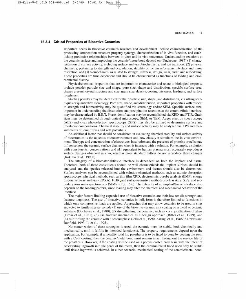
As an example of this biomimetic strategy, porous polyester scaffolds incubated in a simulatedbody fluid (SBF, a supersaturated salt solution with a composition and ionic concentrations approx-imating those of plasma), exhibit coordinated surface functionalization, nucleation and growth of acontinuous bonelike apatite layer on the polymer surfaces and within the pores (Fig. 15.8) after rel-atively short incubation times (Murphy et al., 2000a; Shin et al., 2007; Segvich et al., 2008a). FTIR
15-Kutz-V-I_c015_001-000.qxd 3/5/09 10:01 AM Page 16
analyses confirm the nature of the bonelike mineral, and ability to control mineral composition viacontrolling the ionic activity product (IP) of the SBF (Fig. 15.9). As IP increases, more mineralgrows on the scaffold pore surfaces, but the apatite is less crystalline and the Ca/P molar ratiodecreases. Since mineral composition and structure affect cell function, the IP of the mineralizationsolution is an important modulator of material properties, potentially leading to enhanced control ofcell function. Mineralization of the polymer substrate also results in a fivefold increase in compres-sive modulus, without a significant reduction in scaffold porosity (Murphy et al., 2000a). Theincrease in mechanical properties with the addition of only a thin bonelike mineral is important inlight of the competing design requirements of transport and mechanics, which frequently may onlybe balanced by choosing an intermediate porosity.
The self-assembly of mineral within the pores of a polymer scaffold enhances cell adhesion, pro-liferation, and osteogenic differentiation, as well as modulates cytoskeletal organization and cell motilityin vitro (Kohn et al., 2005; Leonova et al., 2006). When progenitor cells are transplanted on thesematerials, a larger and more spatially uniform volume of bone is regenerated, compared to unminer-alized templates (Kohn et al., 2005; Rossello, 2007). An additional benefit of the biomimetic pro-cessing conditions (e.g., room temperature, atmospheric pressure) is that incorporation of growthfactors is achievable, without concern for denaturing, thus enabling a dual conductive/inductiveapproach (Fig. 15.10) (Murphy et al., 2000b; Luong et al., 2006; Segvich et al., 2008a). Therefore,biomineralized materials can serve as a platform for conductive, inductive, and cell transplantationapproaches to regeneration, and fulfill the majority of the design requirements outlined above.
BIOCERAMICS 17
FIGURE 15.8 Images of 85:15 polylactide/glycolide scaffolds incubated in a simulated body fluid (SBF). (a) Microcomputedtomography image of whole scaffold showing mineralization through the thickness of the scaffold; (b) Localized SEM image ofa scaffold cross-section, showing mineralization of a pore wall; (c) SEM image of mineral nucleation on hydrolyzed PLGA; (d) SEM image of continuous mineral grown on the PLGA—a conglomerated granular structure with needle-shaped precipitatesis visible; (e) higher magnification SEM image of elongated platelike hexagonal crystals extending out of the plane of the granu-lar structure. [(a), From Segvich et al. (2008a), with permission; (b), from Murphy et al., (2000a), with permission; (d),(e), fromHong et al. 2008, with permission.]
A2 mm B
C D E
15-Kutz-V-I_c015_001-000.qxd 3/5/09 10:01 AM Page 17

15.4.2 Inorganic/Organic Hybrid Biomimetics
Advancements in understanding biomineralization have also resulted in the synthesis of mineral-organic hybrids, consisting of bonelike apatites combined with inductive factors, to control cell pro-liferation, differentiation, and bone formation (Murphy et al., 2000b; Luong et al., 2006; Segvich et al.,2008a; Liu et al., 2001). The method of combining inorganic mineral with organic factors caninfluence the resultant release profile, and therefore, influence the biological response of cells. Themost basic method of incorporating proteins into ceramics is adsorption, where the factor is looselybound to the ceramic surface by submersion or pipetting. A second way of incorporating protein withapatite is to create microcarriers that allow HA crystals to form in the presence of protein or allowprotein to adsorb to the HA (Ijntema et al., 1994; Barroug and Glimcher, 2002; Matsumoto et al., 2004).A third method of protein incorporation is coprecipitation, in which protein is added to SBF andbecomes incorporated into bonelike apatite during calcium-phosphate precipitation. Organic/inorganichybrids show promise in combining the osteoconductive properties provided by the apatite with theosteoinductive potential provided by growth factors, DNA, and peptides.
Through coprecipitation, BMP-2 has been incorporated into biomimetic coatings deposited ontitanium, and biological activity has been retained (Liu et al., 2004). Biomolecules can be incorpo-rated at different stages of calcium-phosphate nucleation and growth (Fig. 15.10) (Luong et al.,2006; Azevedo et al., 2005), enabling spatial localization of the biomolecule through the apatitethickness, and allowing the controlled release of the biomolecule. With spatial localization, there isalso the potential for delivery of multiple biomolecules.
18 BIOMATERIALS
FIGURE 15.9 FTIR spectra of the mineralized pore surfaces of 85:15 PLGA scaffolds incubated in simulated bodyfluids (SBF) of varying ionic activity products (IP) for 16 days. Inset = bands within the boxes stacked and enlarged tobetter show changes in CO3
2−. Band intensities of phosphate and carbonate increased with increasing IP. [From Shinet al. (2007), with permission.]
400600
2000 1800 1700 1600 1500 1400 900850
8001000120014001600180020002200Wave number (cm–1)
Abs
orba
nce
1900
: carbonate
: phosphate
0.75 SBF1.00 SBF1.25 SBF1.50 SBF1.75 SBF2.00 SBF
Control
15-Kutz-V-I_c015_001-000.qxd 3/5/09 10:01 AM Page 18

Techniques used to incorporate growth factors into bonelike mineral can also be used to incor-porate genes. One of the most common methods of gene delivery is to encapsulate DNA within aCa-P precipitate (Jordan et al., 1996). This method protects DNA from degradation and encouragescellular uptake, but DNA is released in a burst, which is not always the desired release kinetics. Byutilizing coprecipitation to incorporate plasmid DNA into a biomimetic apatite layer, osteoconduc-tivity and osteoinductivity are combined into a single approach that has the ability to transfect hostcells. The mineral increases substrate stiffness, which also enhances cellular uptake of plasmid DNA(Kong et al., 2005).
BIOCERAMICS 19
FIGURE 15.10 Images through the thickness of a mineral layer containing FITC-labeled BSA taken using confocalmicroscopy. Spatial distribution of the protein through the thickness of the mineral is exhibited for the following proteinincorporation techniques: (1) 6 days of mineral/BSA coprecipitation; (2) 3 days of mineralization followed by 3 days ofprotein adsorption; (3) 3 days of mineralization followed by 3 days of mineral/BSA coprecipitation; (5) 3 days of min-eralization, followed by 2 days of mineral/BSA coprecipitation, followed by 1 day of mineralization. [From Luong et al.(2006), with permission from Elsevier.]
Top ofmineral layer
(external surface)
Preliminarymineral
Top layerof mineral(no FITC)Se
quen
ce o
f im
ages
Preliminarymineral
(1) 6 d cop. (2) 3 d min., 3 d ads.
(3) 3 d min., 3 d cop.
(5) 3 d min., 2 d cop., 1 d min.
Bottom of film(below mineral
layer)
15-Kutz-V-I_c015_001-000.qxd 3/5/09 10:01 AM Page 19

20 BIOMATERIALS
Not only is the method of protein incorporation an integral part of developing an effective deliverysystem, but the interaction between the biological factor and mineral is also important. Biologicalfactors can alter nucleation, growth, and biomineral properties (e.g., crystal phase, morphology, crystalgrowth habit, orientation, chirality) (Wen et al., 1999; Azvedo et al., 2005; Liu et al., 2003; Uchidaet al., 2004; Combes et al., 1999), changing the osteoconductive capacity of the mineral. Whenorganic constituents are introduced into the mineralizing solution, the dynamics of mineralizationchange due to changes in pH, interactions between the biological factor and ions in solution, andinteractions with the substrate. These dynamics can enhance or inhibit the heterogeneous depositionof mineral onto the substrate.
Following coprecipitation, the release of biological factors and resultant biological responses areinfluenced by many variables, including the concentration of the factor, the expression of the recep-tors that are affected by the presence of the factor, the physical characteristics of the delivery sub-strate and mineral/organic coating, and the site of implantation. Release kinetics can be controlledvia diffusion of the biological factor, dissolution/degradation of the carrier and/or osmotic effects.For delivery systems based on coprecipitation of a biological molecule with a biomineral, the disso-lution mechanisms of mineral are the most important.
Mineral dissolution is controlled by factors associated with the solution (pH, saturation), bulk solid(solubility, chemical composition), and surface (adsorbed ions, phases). The apatite that is typicallyformed from a supersaturated ionic solution is carbonated (Murphy et al., 2000a; Shin et al., 2007).The presence of carbonate in an apatite lattice influences crystallinity and solubility (Tang et al., 2003;Ito et al., 1997; Krajewski et al., 2005). The dissolution rate of carbonated HA depends pH, and occurswith the protonation of the carbonate or phosphate group to form either carbonic acid or phosphoricacid (Hankermeyer et al., 2002). Thus, when experimental conditions change, the dissolution proper-ties of mineral and release kinetics of any biomolecules incorporated into the mineral also change.
Apatite that has protein simply adsorbed to its surface undergoes a burst effect, releasing most ofthe protein within the first 6 hours, whereas less than 1 percent of the protein incorporated withinbonelike apatite is released after 5 days (Liu et al., 2001). With coprecipitation, a small burst occursdue to a small amount of protein that is adsorbed to the surface. The resultant sustained release ishypothesized to be due to the incorporation of protein within the apatite matrix, rather than just asuperficial association (Liu et al., 2001). The affinity a protein has for apatite influences the disso-lution rate of the mineral and, therefore, the release rate. Since protein release is proportional toapatite dissolution, the possibility of temporally controlling the release profile, as well as developingmultifactor delivery systems is possible due the ability to spatially localize the protein within the bio-mimetically nucleated mineral (Luong et al., 2006).
In addition to trying to control cell function via biomolecular incorporation within apatite, anotherstrategy is to present biomolecules on a biomimetic surface. While the objective of coprecipitation is tocontrol spatial and temporal release of biomolecules, the objective of presenting peptides with confor-mational specificity on a material surface is to recruit a population of cells that can initiate the earlystages of bone regeneration. Proteins, growth factors, and peptides have been ionically or covalentlyattached to biomaterial surfaces to increase cell adhesion, and ultimately, the amount of bone growth.While specific proteins that enhance cell adhesion have been identified, proteins, in general, are subjectto isolation and prone to degradation (Hersel et al., 2003). Proteins can also change conformation or ori-entation because they possess sections with varying hydrophobicities that address cellular functionsother than adhesion. On the other hand, peptides can effectively mimic the same response as a proteinwhile being smaller, cheaper, and less susceptible to degradation. Peptides have a greater potential forcontrolling initial biological activity, because they can contain specific target amino acid sequences andcan permit control of hydrophilic properties through sequence design (Ladner et al., 2004).
Identification of cell recognition sequences has motivated the development of bioactive materialsthat can recruit a desired cell population to adhere to a material surface via specific integrin-mediatedbonding. One peptide sequence that interacts with a variety of cell adhesion receptors, includingthose on osteoblasts, is the RGD (Arg-Gly-Asp) sequence. Other peptide sequences have been designedto mimic sections of the ECM proteins bone sialoprotein, osteopontin, fibronectin, statherin, elastin,and osteonectin (Fujisawa et al., 1996, 1997; Gilbert et al., 2000; Simionescu et al., 2005). Peptidesequences with preferential affinity to HA and bonelike mineral have been discovered using phagedisplay libraries (Segvich et al., 2008b).
15-Kutz-V-I_c015_001-000.qxd 3/5/09 10:01 AM Page 20

15.5 SUMMARY
In summary, bioceramics have a long clinical history, especially in skeletal reconstruction and regen-eration. Bioceramics are classified as relatively inert (a minimal tissue response is elicited and a layerof fibrous tissue forms adjacent to the implant), surface active (partially soluble, resulting in surfaceion exchange with the microenvironment and leading to a direct chemical bond with tissue), and bulkbioactive (fully resorbable, with the potential to be completely replaced with de novo tissue).Ceramics are processed via conventional materials science strategies, as well as strategies inspiredby nature. The biomimetic approaches discussed in Section 15.4, along with all other strategies toreproduce the design rules of biological systems, do not completely mimic nature. Instead, justselected biological aspects are mimicked. However, if the selected biomimicry is rationally designedinto biomaterial, then the biological system will be able to respond in a more controlled, predictable,and efficient manner, providing an exciting new arena for biomaterials research and development.
ACKNOWLEDGEMENTS
Parts of the author’s research discussed in this chapter were supported by NIH/NIDCR R01 DE013380 and R01 DE015411.
REFERENCES
Abe, Y., Kokubo, T., Yamamuro, T., J. Mater. Sci.: Mater. Med. 1:233–238, 1990.
Allen, M., Myer, B., Rushton, N., J. Biomed. Mater. Res. (Appl. Biomat.) 58:319–328, 2001.
Attawia, M. A., Devin, J. E., Laurencin, C. T., J. Biomed. Mater. Res. 29:843–848, 1995.
Azevedo, H. S., Leonor, I. B., Alves, C. M., Reis, R. L., Mat. Sci. Eng. C.—Bio. S. 25:169, 2005.
Barroug, A., Glimcher, M. J., J. Orthop. Res. 20:274, 2002.
Bokros, J. C., Trans. Biomed. Mater. Res. Symp. 2:32–36, 1978.
Bokros, J. C., LaGrange, L. D., Schoen, G. J., In: Chemistry and Physics of Carbon, Vol. 9, Walker, P. L., (ed.),New York, Dekker, pp. 103–171, 1972.
Boutin, P., Christel, P., Dorlot, J. M., Meunier, A., de Roquancourt, A., Blanquaert, D., Herman, S., Sedel, L.,Witvoet, J., J. Biomed. Mater. Res. 22:1203–1232, 1988.
Bunker, B. C., Rieke, P. C,, Tarasevich, B. J., Campbell, A. A., Fryxell, G. E., Graff, G. L., Song, L., Liu, J.,Virden, J. W., McVay, G. L., Science 264:48–55, 1994.
Cales, B., Stefani, Y., In: Biomedical Engineering Handbook, Bronzino, J. D., (ed.), Boca Raton, FL, CRC Press,pp. 415–452, 1995.
Campbell, A. A., Fryxell, G. E., Linehan, J. C., Graff, G. L., J. Biomed. Mater. Res. 32:111–118, 1996.
Chai, C. S., Gross, K. A., Ben-Nissan, B., Biomat 19:2291–2296, 1998.
Chou, Y. F., Huang, W., Dunn, J. C. Y., Miller, T. A., Wu, B. M., Biomat 26:285–295, 2005.
Christel, P., Meunier, A., Heller, M., Torre, J. P., Peille, C. N., J. Biomed. Mater. Res. 23:45–61, 1989.
Christel, P., Meunier, A., Leclercq, S., Bouquet, P., Buttazzoni, B., J. Biomed. Mater. Res.: Appl. Biomat.21(A2):191–218, 1987.
Combes, C., Rey, C., Biomat 23:2817–2823, 2002.
Combes, C., Rey, C., Freche, M., J. Mater. Sci. Mater. Med. 10:153, 1999.
Cook, S. D., Thomas, K. A., Dalton, J. E., Volkman, T. K., Whitecloud, T. S., III, Kay, J. F., J. Biomed. Mater.Res. 26:989–1001, 1992.
Davidson, J. A., Clin. Orthop. 294:361–178, 1993.
de Bruijn, J. D., van Blitterswijk, C. A., Davies, J. E., J. Biomed. Mater. Res. 29: 89–99, 1995.
BIOCERAMICS 21
15-Kutz-V-I_c015_001-000.qxd 3/5/09 10:01 AM Page 21

de Groot, K., (ed.), Bioceramics of Calcium Phosphate, Boca Raton, FL, CRC Press, 1983.
de Groot, K., Geesink, R. G. T., Klein, C. P. A. T., Serekian, P., J. Biomed. Mater. Res. 21:1375–1381, 1987.
Derbyshire, B., Fisher, J., Dowson, D., Hardaker, C., Brummitt, K., Med. Eng. Phys. 16:229–236, 1994.
Dorre, E., Dawihl, W., In: Mechanical Properties of Biomaterials, Hastings, G. W., Williams, D. F., (eds.),New York, Wiley, pp. 113–127, 1980.
Du, C., Cui, F. Z., Zhu, X. D., de Groot, K., J. Biomed. Mater. Res. 44:407–415, 1999.
Ducheyne, P., J. Biomed. Mater. Res. 19:273–291, 1985.
Ducheyne, P., J. Biomed. Mater. Res.: Appl. Biomat. 21(A2):219–236, 1987.
Ducheyne, P., Hench, L. L., Kagan, A., II, Martens, M., Bursens, A., Mulier, J. C., J. Biomed. Mater. Res.14:225–237, 1980.
Ducheyne, P., El-Ghannam, A., Shapiro, I., J. Cell Biochem. 56:162–167, 1994.
Ducheyne, P., Radin, S., Heughebaert, M., Heughebaert, J. C., Biomat. 11:244–254, 1990.
Ducheyne, P., Van Raemdonck, W., Heughebaert, J. C., Heughebaert, M., Biomat. 7:97–103, 1986.
El-Ghannam, A., Ducheyne, P., Shapiro, I. M., J. Biomed. Mater. Res. 36:167–180, 1997.
Filiaggi, M. J., Coombs, N. A., Pilliar, R. M., J. Biomed. Mater. Res. 25:1211–1229, 1991.
Filiaggi, M. J., Pilliar, R. M., Yakubovich, R., Shapiro, G., J. Biomed. Mater. Res. (Appl. Biomat.) 33:225–238,1996.
Fujisawa, R., Mizuno, M., Nodasaka, Y., Kuboki, Y., Matrix Biol. 16:21, 1997.
Fujisawa, R., Wada, Y., Nodasaka, Y., Kuboki, Y., Biochim. Biophys. Acta 1292:53, 1996.
Gao, Y., In: Biomedical Materials—Drug Delivery, Implants and Tissue Engineering, Neenan, T ., Marcolongo,M., Valentini, R. F., (eds.), Materials Research Society, Warrendale, PA, pp. 361–366, 1999.
Garcia, F., Arias, J. L., Mayor, B., Pou, J., Rehman, I., Knowles, J., Best, S., Leon, B., Perez-Amor, M., Bonfield,W., J. Biomed. Mater. Res. (Appl. Biomat.) 43:69–76, 1998.
Gilbert, M., Shaw, W. J., Long, J. R., Nelson, K., Drobny, G. P., Giachelli, C. M., Stayton, P. S., J. Biol. Chem.275:16213, 2000.
Gross, U., Brandes, J., Strunz, V., Bab, I., Sela, J., J. Biomed. Mater. Res. 15:291–305, 1981.
Gross, U., Strunz, V., J. Biomed. Mater. Res. 14:607–618, 1980.
Gross, U., Strunz, V., J. Biomed. Mater. Res. 19:251–271, 1985.
Hanawa, T., Kon, M., Ukai, H., Murakami, K., Miyamoto, Y., Asaoka, K., J. Biomed. Mater. Res. 41:227–236,1998.
Hankermeyer, C. R., Ohashi, K. L., Delaney, D. C., Ross, J., Constantz, B. R., Biomaterials 23:743, 2002.
Hartman, E. H. M, Vehof, J. W. M., Spauwen, P. H. M., Jansen, J. A., Biomaterials 26:1829–1835, 2005.
Haubold, A. D., Shim, H. S., Bokros, J. C., In: Biocompatibility of Clinical Implant Materials, Vol. II, Williams,D. F., (ed.), Boca Raton, FL, CRC Press, pp. 3–42, 1981.
Heimke, G., Jentschura, G., Werner, E., J. Biomed. Mater. Res. 12:57–65, 1978.
Hench, L. L., Best, S., In: Biomaterials Science: An Introduction to Materials in Medicine, 2nd ed, Ratner, B. D.,Hoffman, A. S., Schoen, F. J., Lemons, J. E., (eds.), San Diego, Elsevier Academic Press, pp. 153–169, 2004.
Hench, L. L, Clark, A. E., In: Biocompatibility of Orthopaedic Implants, Vol. II, Williams, D. F., (ed.), BocaRaton, FL, CRC Press, pp. 129–170, 1982.
Hench, L. L., Ethridge, E. C., Biomaterials An Interfacial Approach, New York, Academic Press, 1982.
Hench, L. L., Splinter, R. J., Allen, W. C., Greenlee, T. K., Jr., J. Biomed. Mater. Res. Symp. 2:117–141, 1972.
Hersel, U., Dahmen, C., Kessler, H., Biomaterials 24:4385, 2003.
Holtorf, H. L., Sheffield, T. L., Ambrose, C. G., Jansen, J. A., Mikos, A. G., Ann. Biomed. Eng. 33:1238–1248,2005.
Hong, S. I., Lee, K. H., Outslay, M. E., Kohn, D. H., J. Mater. Res. 23:478–485, 2008.
Hulbert, S. F., Young, F. A., Mathews, R. S., Klawitter, J. J., Talbert, C. D., Stelling, F. H., J. Biomed. Mater. Res.4:433–456, 1970.
Huttner, W., Huttinger, K. J., In: The Cementless Fixation of Hip Endoprostheses, Morscher, E., (ed.), Berlin,Springer-Verlag, pp. 81–94, 1984.
22 BIOMATERIALS
15-Kutz-V-I_c015_001-000.qxd 3/5/09 10:01 AM Page 22

Ijntema, K., Heuvelsland, W. J. M., Dirix, C., Sam, A. P., Int. J. Pharm. 112:215, 1994.
Ioku, K., Yoshimura, M., Somiya, S., Biomaterials 11:57–61, 1990.
Ito, A., Maekawa, K., Tsutsumi, S., Ikazaki, F., Tateishi, T., J. Biomed. Mater. Res. 36:522, 1997.
James, K., Levene, H., Parson, J. R., Kohn, J., Biomaterials 20:2203–2212, 1999.
Jordan, M., Schallhorn, A., Wurm, F. M., Nucleic. Acids Res. 24:596, 1996.
Kamei, S., Tomita, N., Tamai, S., Kato, K., Ikada, Y., J. Biomed. Mater. Res. 37:384–393, 1997.
Kitsugi, T., Yamamuro, T., Nakamura, T., Higashi, S., Kakutani, Y., Hyakuna, K., Ito, S., Kokubo, T., Takagi, M.,Shibuya, T., J. Biomed. Mater. Res. 20:1295–1307, 1986.
Knowles, J. C., Bonfield, W., J. Biomed. Mater. Res. 27:1591–1598, 1993.
Ko, C. C., Kohn, D. H., Hollister, S. J., J. Mater. Sci.: Mater. Med. 7:109–117, 1995.
Koch, B., Wolke, J. G. C., de Groot, K., J. Biomed. Mater. Res. 24:655–667, 1990.
Koeneman, J., Lemons, J., Ducheyne, P., Lacefield, W., Magee, F., Calahan, T., Kay, J., J. Appl. Biomat. 1:79–90, 1990.
Kohn, D. H., Curr. Opin. Solid State Mater. Sci. 3:309–316, 1998.
Kohn, D. H., Ducheyne, P., “Materials for Bone, Joint and Cartilage Replacement,” In: Medical and DentalMaterials, Williams, D. F. (ed.), VCH Verlagsgesellschaft, FRG, pp. 29–109, 1992.
Kohn, D. H., Shin, K., Hong, S. I., Jayasuriya, A. C., Leonova, E. V., Rossello, R. A., Krebsbach, P. H., In: Proc.8th Int. Conf. on the Chemistry and Biology of Mineralized Tissues, Landis, W. J., Sodek, J., (eds.),University of Toronto Press, pp. 216–219, 2005.
Koistinen, A., Santavirta, S. S., Kroger, H., Lappalainen, R., Biomaterials 26:5687–5694, 2005.
Kokubo, T., Ito, S., Huang, Z. T., Hayashi, T., Sakka, S., Kitsugi, T., Yamamuro, T., J. Biomed. Mater. Res.24:331–343, 1990a.
Kokubo, T., Kushitani, H., Sakka, S., Kitsugi, T., Yamamuro, T., J. Biomed. Mater Res. 24:721–734, 1990b.
Kokubo, T., Takadama, H., Biomaterials 27:2907–2915, 2006.
Kong, H. J., Liu, J. D., Riddle, K., Matsumoto, T., Leach, K., Mooney, D. J., Nat. Mater. 4:460, 2005.
Krajewski, A., Mazzocchi, M., Buldini, P. L., Ravaglioli, A., Tinti, A., Taddei, P., Fagnano, C., J. Mol. Struct.744:221, 2005.
Krebsbach, P. H., Kuznetsov, S. A., Satomura, K., Emmons, R. V. B., Rowe, D. W., Gehron-Robey, P.,Transplantation 63:1059–1069, 1997.
Krebsbach, P. H., Mankani, M. H., Satomura, K., Kuznetsov, S. A., Gehron-Robey, P., Transplantation66:1272–1278, 1998.
Kruyt, M. C., Dhert, W. J. A., Yuan, H., Wilson, C. E., van Blitterswijk, C. A., Verbout, A. J., de Bruijn, J. D.,J. Orthop. Res. 22:544–551, 2004.
Kumar, P., Oka, M., Ikeuchi, K., Shimizu, K., Yamamuro, T., Okumura, H., Kotoura, Y., J. Biomed. Mater. Res.25:813–828, 1991.
Kuznetsov, S. A., Krebsbach, P. H., Satomura, K., Kerr, J., Riminucci, M., Benayahu, D., Gehron-Robey, P.,J. Bone Min. Res. 12:1335–1347, 1997.
Ladner, R. C., Sato, A. K., Gorzelany, J., de Souza, M., Drug Discov. Today 9:525, 2004.
Laurencin, C. T., El-Amin, S. F., Ibim, S. E., Willoughby, D. A., Attawia, M., Allcock, H. R., Ambrosio, A. A.,J. Biomed. Mater. Res. 30:133–138, 1996.
Leonova, E. V., Pennington, K. E., Krebsbach, P. H., Kohn, D. H., J. Biomed. Mater. Res. Part A 79A:263–270,2006.
Li, J., Fartash, B., Hermansson, L., Biomaterials 16:417–422, 1995.
Li, P., Ohtsuki, C., Kokubo, T., Nakanishi, K., Soga, N., Nakamura, T., Yamamuro, T., J. Am. Ceram. Soc.75:2094–2097, 1992.
Liu, Y., Hunziker, E. B., Randall, N. X., de Groot, K., Layrolle, P., Biomaterials 24:65, 2003.
Liu, Y. L., Hunziker, E. B., Layrolle, P., de Bruijn, J. D., de Groot, K., Tissue Eng 10:101–108, 2004.
Liu, Y. L., Layrolle, P., de Bruijn, J., van Blitterswijk, C., de Groot, K., J. Biomed. Mater. Res. 57:327, 2001.
Lowenstein, H. A., Weiner, S., On Biomineralization, Oxford University Press, Oxford, 1989.
Luong, L. N., Hong, S. I., Patel, R. J., Outslay, M. E., Kohn, D. H., Biomaterials 27:1175–1186, 2006.
BIOCERAMICS 23
15-Kutz-V-I_c015_001-000.qxd 3/5/09 10:01 AM Page 23
Lusty, P. J., Watson, A., Tuke, M. A., Walter, W. L., Walter, W. K., Zicat, B., J. Bone Joint Surg. 89B:1158–1164,2007.
Mann, K. A., Edidin, A. A., Kinoshita, R. K., Manley, M. T., J. Appl. Biomat. 5:285–291, 1994.
Mann, S., Heywood, B. R., Rajam, S., Birchall, J. D., Nature 334:692–695, 1988.
Mann, S., Ozin, G. A., Nature 382:313–318, 1996.
Matsumoto, T., Okazaki, M., Inoue, M., Yamaguchi, S., Kusunose, T., Toyonaga, T., Hamada, Y., Takahashi,J., Biomaterials 25:3807, 2004.
Miller, R. A., Smialek, R. G., Garlick, In: Advances in Ceramics, Vol. 3, Science and Technology of Zirconia,Westerville, OH, American Ceramic Society, p. 241, 1981.
Murphy, W. L., Kohn, D. H., Mooney, D.J., J. Biomed. Mater. Res. 50:50–58, 2000a.
Murphy, W. L., Peters, M. C., Kohn, D. H., Mooney, D. J., Biomaterials 21:2521–2527, 2000b.
Nakamura, T., Yamamuro, T., Higashi, S., Kokubo, T., Ito, S., J. Biomed. Mater. Res. 19:685–698, 1985.
Nizard, R., Pourreyron, D., Raould, A., Hannouche, D., Sedel, L., Clin. Orthop. Rel. Res. 466:317–323, 2008.
Ohgushi, H., Caplan, A. I., J. Biomed. Mater. Res. (Appl. Biomat.) 48:913–927, 1999.
Ohgushi, H., Okumura, M., Tamai, S., Shors, E. C., Caplan, A. I., J. Biomed. Mater. Res. 24:1563–1570, 1990.
Ong, J. L., Harris, L. A., Lucas, L. C., Lacefield, W. R., Rigney, E. D., J. Am. Ceram. Soc. 74:2301–2304, 1991.
Oonishi, H., Noda, T., Ito, S., Kohda, A., Ishimaru, H., Yamamoto, M., Tsuji, E., J. Appl. Biomat. 5:23–37, 1994.
Posner, A. S., Clin. Orthop. 200:87–99, 1985a.
Posner, A. S., J. Biomed. Mater. Res. 19:241–250, 1985b.
Puleo, D. A., Holleran, L. A., Doremus, R. H., Bizios, R., J. Biomed. Mater. Res. 25:711–723, 1991.
Redey, S. A., Nardin, M., Bernache-Assolant, D., Rey, C., Delannoy, P., Sedel, L., Marie, P. J., J. Biomed. Mater.Res. 50:353–364, 2000.
Radin, S., Reilly, G., Bhargave, G., Leboy, P. S., Ducheyne, P., J. Biomed. Mater. Res. 73A:21–29, 2005.
Reikeras, O., Johansson, C. B., Sundfeldt, M., J. Long Term Effects of Med. Impl. 14:443–454, 2004.
Reilly, G. C., Radin, S., Chen, A. T., Dycheyne, P., Biomaterials 28:4091–4097, 2007.
Ritter, J. E., Jr., Greenspan, D. C., Palmer, R. A., Hench, L. L., J. Biomed. Mater. Res. 13:251–263, 1979.
Rossello, R. A., Ph.D. Dissertation, University of Michigan, 2007.
Segvich, S. J., Biswas, S., Becker, U., Kohn, D. H., Cells Tissues Organs, in press, 2008b.
Segvich, S. J., Luong, L. N., Kohn, D. H., “Biomimetic Approaches to Synthesize Mineral and Mineral/OrganicBiomaterials,” In: Biomaterials and Biomedical Engineering, Ahmed, W., Ali, N., Öchsner, A., (eds.), TransTech Publications, Ltd, UK, 2008c.
Segvich, S. J., Smith, H. C., Luong, L. N., Kohn, D. H., J. Biomed. Mater. Res., Part B, 84B:340–349, 2008a.
Semlitsch, M., Lehmann, M., Weter, H., Dorre, E., Willert, H. G., J. Biomed. Mater. Res. 11:537, 1977.
Shi, X., Hudson, J. L., Spicer, P. P., Tour, J. M., Krishnamoorti, R., Mikos, A. G., Biomacromol 7:2237–2242, 2006.
Shin, K., Jayasuriya, A. C., Kohn, D. H., J. Biomed. Mater. Res. Part A, 83A:1076–1086, 2007.
Simionescu, A., Philips, K., Vyavahare, N., Biochem Biophys Res Commun, 334:524–532, 2005.
Soltesz, U., Richter, H., In: Metal and Ceramic Biomaterials Volume II Strength and Surface, Ducheyne, P.,Hastings, G. W., (eds.), Boca Raton, FL, CRC Press pp. 23–61, 1984.
Taguchi, T., Shiraogawa, M., Kishida, A., Arashi, M., J. Biomater. Sci. Polymer Edn. 10:19–31, 1999.
Tanahashi, M., Yao, T., Kokubo, T., Minoda, T., Miyamoto, T., Nakamura, T., Yamamuro, T., J. Biomed. Mater.Res. 29:349–357, 1995.
Tang, R. K., Henneman, Z. J., Nancollas, G. H., J. Cryst. Growth 249:614, 2003.
Thomson, R. C., Yaszemski, M. J., Powers, J. M., Mikos, A. G., Biomaterials 19:1935–1943, 1998.
Tohma, Y., Tanaka, Y., Ohgushi, H., Kawate, K., Taniguchi, A., Hayashi, K., Isomoto, S., Takakura, Y., J. Orthop.Res. 24:595–603, 2006.
Uchida, M., Oyane, A., Kim, H. M., Kokubo, T., Ito, A., Adv, Mater. 16:1071, 2004.
Van Raemdonck, W., Ducheyne, P., De Meester, P., In: Metal and Ceramic Biomaterials Volume II Strength andSurface, Ducheyne, P., Hastings, G. W., (eds.), Boca Raton, FL, CRC Press, pp. 143–166, 1984.
24 BIOMATERIALS
15-Kutz-V-I_c015_001-000.qxd 3/5/09 10:01 AM Page 24
Walter, A., Lang, W., In: Biomedical Materials—Mater. Res. Soc. Symp. Proc. Vol. 55, Williams, J. M., Nichols,M. F., Zingg, W., (eds.), Pittsburgh, PA, Materials Research Society, pp. 181–190, 1986.
Webster, T. J., Ergun, C., Doremus, R. H., Siegel, R. W., Bizios, R., J. Biomed. Mater. Res. 51:475–483, 2000.
Weiner, S., CRC Crit Rev Biochem. 20:365–408, 1986.
Wen, H. B., de Wijn, J. R., Cui, F. Z., de Groot, K., J. Biomed. Mater. Res. 35:93–99, 1997.
Wen, H. B., de Wijn, J. R., van Blitterswijk, C. A., de Groot, K., J. Biomed. Mater. Res. 46:245, 1999.
Wolke, J. G. C., van Dijk, K., Schaeken, H. G., de Groot, K., Jansen, J. A., J. Biomed. Mater. Res. 28:1477–1484,1994.
Wu, W., Zhuang, H., Nancollas, G. H., J. Biomed. Mater. Res. 35:93–99, 1997.
Yamamoto, M., Kato, K., Ikada, Y., J. Biomed. Mater. Res. 37:29–36, 1997.
Yaszemski, M. J., Payne, R. G., Hayes, W. C., Langer, R. S., Mikos, A. G., Biomaterials 17:175–185, 1996.
Yoshikawa, T., Ohgushi, H., Tamai, S., J. Biomed. Mater. Res. 32:481–492, 1996.
Zanello, L. P., Zhao, B., Hu, H., Haddon, R. C., Nano Letters 6:562–567, 2006.
Zreiqat, H., Evans, P., Howlett, C. R., J. Biomed. Mater. Res. 44:389–396, 1999.
BIOCERAMICS 25
15-Kutz-V-I_c015_001-000.qxd 3/5/09 10:01 AM Page 25

15-Kutz-V-I_c015_001-000.qxd 3/5/09 10:01 AM Page 26