Antioxidant Activity of Puha (Sonchus oleraceus L.) as Assessed by the Cellular Antioxidant Activity...
7
Antioxidant Activity of Puha (Sonchus oleraceus L.) as Assessed by the Cellular Antioxidant Activity (CAA) Assay Arlene McDowell, 2 * Scott Thompson, 1,2 Mirjam Stark, 2 Zong-Quan Ou 2 and Kevin S. Gould 3 1 Department of Botany, University of Otago, P.O. Box 56, Dunedin 9015, New Zealand 2 School of Pharmacy, University of Otago, P.O. Box 913, Dunedin 9015, New Zealand 3 School of Biological Sciences, Victoria University of Wellington, P.O. Box 600, Wellington 6140, New Zealand There is considerable interest in antioxidant dietary components that can be protective against degenerative diseases in humans. Puha (Sonchus oleraceus L.) is a rich source of polyphenols, and exhibits strong antioxidant activity as measured by the 2,2-diphenylpicrylhydrazyl (DPPH) assay. However, the potential of puha to protect against degenerative diseases requires that low molecular weight antioxidants (LMWA) are absorbed by, and active in, human cells. The cellular antioxidant activity (CAA) assay was used to investigate the antioxidant activity of puha leaf extracts. Preparation methods of freezing and freeze-drying reduced the total polyphenolic content compared with fresh puha, but did not affect the LMWA potential as determined by the DPPH assay. The IC 50 values were 0.012 0.003 mg/mL and 0.010 0.005 mg/mL for freeze-dried and fresh puha leaves, respectively. Using the CAA assay, it was shown that LMWAs from foliar extracts of puha were effectively absorbed into HepG2 cells, and exerted antioxidant activity at levels comparable to those of extracts from blue- berry fruits, the much-touted antioxidant superfood. Methylene blue staining of HepG2 cells indicated that puha extracts were not cytotoxic at concentrations below 100 mg DW/mL. The data indicate the potential of puha as a nutraceutical supplement for human health. Copyright © 2011 John Wiley & Sons, Ltd. Keywords: antioxidant; Sonchus oleraceus; HepG2; cellular antioxidant activity assay. INTRODUCTION Antioxidants prevent oxidative damage to lipids, pro- teins, enzymes and DNA that occurs when there is an excess of free radicals and other reactive oxygen species (ROS) in the body (Ratnam et al., 2006). Such oxidative damage of these molecules has been implicated in many chronic diseases including cancer, cardiovascular disease, as well as neurogenerative and inflammatory disorders (Cao et al., 2008; Fresco et al., 2006; Ratnam et al., 2006; Temple, 2000). The consumption of plant- derived antioxidants in foodstuffs or as nutritional supplements has been associated with the prevention of chronic diseases and disorders caused by oxidative damage due to free radicals (Kelsey et al., 2010; Ratnam et al., 2006; Sun et al., 2002). However, many of these studies are based on epidemiological evidence and some data are contradictory (Jan et al., 2010; Li et al., 2010). Plants generally contain a panoply of low molecular weight antioxidants (LMWAs) including flavonoids, terpenoids and phenolic acids, and have been used as traditional herbal remedies in many cultures (Netzel et al., 2006). Specifically, plant polyphenolics, caroten- oids and vitamins (e.g. vitamin E and C) can scavenge a variety of ROS, and so act in the body as antioxidants (Wolfe and Liu, 2007). There are several in vitro chem- ical assays to assess LMWA activity in plant extracts and isolated phytochemicals, including the oxygen radical absorbance capacity (ORAC) (Cao et al., 1993) and the DPPH (2,2-diphenylpicrylhydrazyl) radical scavenging method (Brand-Williams et al., 1995). How- ever, the accuracy of such assays to predict in vivo antioxidant activity has been called into question (Frankel and Meyer, 2000; Wolfe and Liu, 2007). These chemical assays fail to mimic appropriate physiological conditions, and they do not take into account the bio- availability, uptake and metabolism of the antioxidant compounds (Liu and Finley, 2005). A recent advance in the area of antioxidant research has been the devel- opment of the cellular antioxidant activity (CAA) assay to quantify antioxidant potential in a biological system (Wolfe and Liu, 2007). The method measures the ability of absorbed extracts or isolated compounds to prevent the formation of the fluorescent 2′,7′-dichlorofluorescin (DCF) within the HepG2 human liver cell lines. The CAA assay is considered a superior indicator of in vivo activity compared with in vitro assays because it quanti- fies the uptake of exogenously applied LMWA into cells and involves the exposure of the antioxidants to the complexity of biological substrates under physio- logical conditions (Frankel and Meyer, 2000). The sow thistle (Sonchus oleraceus L. Asteraceae) is a herb native to Europe and central Asia that now has a worldwide distribution (Holm et al., 1977). The vegeta- tive shoots of this herb have been used by many cultures to treat conditions including diarrhoea, pneumonia and hepatitis (Yin et al., 2008). Puha is the Maori name for species in the genus Sonchus and continues to be a significant part of the traditional Maori diet in * Correspondence to: Arlene McDowell, School of Pharmacy, University of Otago, P.O. Box 913, Dunedin 9015, New Zealand. E-mail: [email protected] PHYTOTHERAPY RESEARCH Phytother. Res. 25: 1876–1882 (2011) Published online 19 September 2011 in Wiley Online Library (wileyonlinelibrary.com) DOI: 10.1002/ptr.3648 Copyright © 2011 John Wiley & Sons, Ltd. Received 07 April 2010 Revised 09 July 2011 Accepted 11 July 2011
-
Upload
arlene-mcdowell -
Category
Documents
-
view
221 -
download
3
Transcript of Antioxidant Activity of Puha (Sonchus oleraceus L.) as Assessed by the Cellular Antioxidant Activity...

* Correspof Otago,E-mail: arl
PHYTOTHERAPY RESEARCHPhytother. Res. 25: 1876–1882 (2011)Published online 19 September 2011 in Wiley Online Library(wileyonlinelibrary.com) DOI: 10.1002/ptr.3648
Copyright
AntioxidantActivity of Puha (Sonchus oleraceusL.)as Assessed by the Cellular Antioxidant Activity(CAA) Assay
Arlene McDowell,2* Scott Thompson,1,2 Mirjam Stark,2 Zong-Quan Ou2 and Kevin S. Gould31Department of Botany, University of Otago, P.O. Box 56, Dunedin 9015, New Zealand2School of Pharmacy, University of Otago, P.O. Box 913, Dunedin 9015, New Zealand3School of Biological Sciences, Victoria University of Wellington, P.O. Box 600, Wellington 6140, New Zealand
There is considerable interest in antioxidant dietary components that can be protective against degenerativediseases in humans. Puha (Sonchus oleraceus L.) is a rich source of polyphenols, and exhibits strong antioxidantactivity as measured by the 2,2-diphenylpicrylhydrazyl (DPPH) assay. However, the potential of puha to protectagainst degenerative diseases requires that low molecular weight antioxidants (LMWA) are absorbed by, andactive in, human cells. The cellular antioxidant activity (CAA) assay was used to investigate the antioxidantactivity of puha leaf extracts. Preparation methods of freezing and freeze-drying reduced the total polyphenoliccontent compared with fresh puha, but did not affect the LMWA potential as determined by the DPPH assay.The IC50 values were 0.012� 0.003mg/mL and 0.010� 0.005mg/mL for freeze-dried and fresh puha leaves,respectively. Using the CAA assay, it was shown that LMWAs from foliar extracts of puha were effectivelyabsorbed into HepG2 cells, and exerted antioxidant activity at levels comparable to those of extracts from blue-berry fruits, the much-touted antioxidant superfood. Methylene blue staining of HepG2 cells indicated that puhaextracts were not cytotoxic at concentrations below 100mg DW/mL. The data indicate the potential of puha as anutraceutical supplement for human health. Copyright © 2011 John Wiley & Sons, Ltd.
Keywords: antioxidant; Sonchus oleraceus; HepG2; cellular antioxidant activity assay.
INTRODUCTION
Antioxidants prevent oxidative damage to lipids, pro-teins, enzymes and DNA that occurs when there is anexcess of free radicals and other reactive oxygen species(ROS) in the body (Ratnam et al., 2006). Such oxidativedamage of these molecules has been implicated inmany chronic diseases including cancer, cardiovasculardisease, as well as neurogenerative and inflammatorydisorders (Cao et al., 2008; Fresco et al., 2006; Ratnamet al., 2006; Temple, 2000). The consumption of plant-derived antioxidants in foodstuffs or as nutritionalsupplements has been associated with the preventionof chronic diseases and disorders caused by oxidativedamage due to free radicals (Kelsey et al., 2010; Ratnamet al., 2006; Sun et al., 2002). However, many of thesestudies are based on epidemiological evidence and somedata are contradictory (Jan et al., 2010; Li et al., 2010).Plants generally contain a panoply of low molecular
weight antioxidants (LMWAs) including flavonoids,terpenoids and phenolic acids, and have been used astraditional herbal remedies in many cultures (Netzelet al., 2006). Specifically, plant polyphenolics, caroten-oids and vitamins (e.g. vitamin E and C) can scavengea variety of ROS, and so act in the body as antioxidants(Wolfe and Liu, 2007). There are several in vitro chem-ical assays to assess LMWA activity in plant extracts
ondence to: Arlene McDowell, School of Pharmacy, UniversityP.O. Box 913, Dunedin 9015, New [email protected]
© 2011 John Wiley & Sons, Ltd.
and isolated phytochemicals, including the oxygenradical absorbance capacity (ORAC) (Cao et al., 1993)and the DPPH (2,2-diphenylpicrylhydrazyl) radicalscavenging method (Brand-Williams et al., 1995). How-ever, the accuracy of such assays to predict in vivoantioxidant activity has been called into question(Frankel and Meyer, 2000; Wolfe and Liu, 2007). Thesechemical assays fail to mimic appropriate physiologicalconditions, and they do not take into account the bio-availability, uptake and metabolism of the antioxidantcompounds (Liu and Finley, 2005). A recent advancein the area of antioxidant research has been the devel-opment of the cellular antioxidant activity (CAA) assayto quantify antioxidant potential in a biological system(Wolfe and Liu, 2007). The method measures the abilityof absorbed extracts or isolated compounds to preventthe formation of the fluorescent 2′,7′-dichlorofluorescin(DCF) within the HepG2 human liver cell lines. TheCAA assay is considered a superior indicator of in vivoactivity compared with in vitro assays because it quanti-fies the uptake of exogenously applied LMWA intocells and involves the exposure of the antioxidants tothe complexity of biological substrates under physio-logical conditions (Frankel and Meyer, 2000).
The sow thistle (Sonchus oleraceus L. Asteraceae) is aherb native to Europe and central Asia that now has aworldwide distribution (Holm et al., 1977). The vegeta-tive shoots of this herb have been used by many culturesto treat conditions including diarrhoea, pneumoniaand hepatitis (Yin et al., 2008). Puha is the Maoriname for species in the genus Sonchus and continuesto be a significant part of the traditional Maori diet in
Received 07 April 2010Revised 09 July 2011
Accepted 11 July 2011

1877ANTIOXIDANTACTIVITY OF PUHA ASSESSED BY THE CAA ASSAY
New Zealand (Whyte et al., 2001). The vegetativeshoots of puha are potentially an excellent source ofdietary LMWAs, containing high levels of ascorbicacid (Guil-Guerrero et al., 1998; Thomson and Shaw,2002) and carotenoids compared with other leafyvegetables (Guil-Guerrero et al., 1998; Thomson andShaw, 2002), as well as flavonoids (Thomson andShaw, 2002), including apigenin and luteolin, essentialfatty acids (Guil-Guerrero et al., 1998) and a-tocopherol(Zeghichi et al., 2003). Gould et al. (2006) have demon-strated using the DPPH assay that puha leaf extractsheld at least three times the LMWA activity of blue-berries, the much-touted antioxidant superfood. Thepuha plant also ranked fifth strongest of 18 native NewZealand plant species tested in the same study (Gouldet al., 2006). Similarly, in a study of 27 non-cultivatedspecies used for food in Southern Italy, puha was foundto be the third richest source of LMWAs (Pieroni et al.,2002). Yin et al. (2007) have suggested that the antioxi-dant activity of Sonchus oleraceus extracts is attributableto phenolic compounds present in the leaves. Theseauthors have investigated the most efficient solventfor the extraction of antioxidants and found that 70%methanol yields extracts with the greatest in vitroantioxidant activity as assessed by the DPPH assay(Yin et al., 2007).There is current interest in the development of
LMWA ingredients as nutritional supplements and fordisease prevention. Preformulation studies are essentialin pharmaceutical product development to characterizephysical and chemical properties of bioactive compound(s). Preformulation studies typically investigate featuressuch as the stability and solubility of the active ingredient,and also the impact that processing conditions mayhave on the properties of the product (Wells, 1998). Suchstudies can identify potential stability problems that willneed to be addressed through formulation strategies toensure a safe and efficacious product. Just as the demon-stration of antioxidant activity is important, so too issafety, and therefore toxicity to living cells and tissuesmust be investigated.Currently there is a paucity of information available
on preformulation studies for antioxidant products. Fora nutritional supplement that is taken orally in tablet orcapsule form, formulation of the bioactive is crucial inorder to achieve a biological effect. Oral delivery of anti-oxidant compounds is challenging due to their instability,poor solubility and poor membrane permeability that allcontribute to the low bioavailability of dietary LMWAs(Ratnam et al., 2006; Zhang et al., 2004). Consequently,formulations strategies may be required to improvethe bioavailability of ingested antioxidant supplements.The objectives of this study were to investigate the
antioxidant potential of puha plant extracts using theCAA assay and so whether LMWAs can enter humancells and retain their antioxidant activity. As part ofpreformulation studies, the effect of processing (freeze-drying) on the antioxidant potential of puha leaves wasalso investigated.
MATERIALS AND METHODS
Chemicals. 2,2-Diphenylpicrylhydrazyl (DPPH), 2,2′-azobis(2-methylpropionamididine) dihydrochloride (ABAP),2′,7′-dichlorofluorescin diacetate (DCFH-DA), ascorbic
Copyright © 2011 John Wiley & Sons, Ltd.
acid, bisbenzimide H33258, dimethyl sulfoxide, Dulbecco’smodified Eagle’s medium (DMEM) (high glucose,L-glutamine and sodium pyruvate, without sodium bicar-bonate), Folin-Ciocalteu reagent, gallic acid, glutaralde-hyde, Hank’s balanced salt solution (HBSS), methyleneblue, methanol, phosphate buffered saline solution(PBS), quercetin dihydrate and sodium carbonate werepurchased fromSigma-Aldrich, Inc. (St Louis,MO). Fetalbovine serum (FBS), GlutaMAX, penicillin–streptomycin,trypan blue stain and trypsin were purchased fromInvitrogen NZ Ltd. Methanol was purchased fromBiolab (Scoresby, Australia) Ltd. Human hepatocellularcarcinoma (HepG2) cells were gratefully obtainedfrom the Pathology Department at the University ofOtago, Dunedin, New Zealand. Naturstoff reagent Awas supplied from Sigma (Germany) and dissolved inethanol at a concentration of 5% and diluted in PBSfor application.
Plant samples. Puha (Sonchus oleraceus) seeds werecollected from the wild in the Otago region of southernNew Zealand. Voucher specimens (OTA 061166 andOTA 061167) are lodged at the Otago Regional Herbariumin the Department of Botany at the University of Otago,New Zealand. Seeds were germinated and grown in50:50 potting mix and sand in a glasshouse maintainedat 16 �C and watered daily with water and grown forapproximately 2months. Fresh blueberry fruits (Vacciniumsp.) were purchased from a local supermarket (packedby Gourmet Blueberries, Hastings, New Zealand).
Preparation of fruit and chemical solutions. Puhaextracts were prepared by harvesting fresh, expandedleaves (pooled from different plants) and either extract-ing LMWAs immediately, freezing at �20 �C prior toextraction, or freeze-drying leaves for 48 h prior toextraction. The moisture content of the leaves, deter-mined from a subsample of the harvest following ovendrying, was 85.7%� 1.8 (n= 20 leaves). Soluble LMWAextracts were prepared by grinding 1.0 g fresh plantmaterial, or equivalent dried leaf, in a mortar with10mL of methanol:water 70:30 (v/v) solution. The slurryof ground plant material was then separated by centrifu-gation at 4500 rpm for 10min. The resulting supernatantwas aspirated and the solvent removed by rotary evap-oration at 40 �C under nitrogen gas for 10–15min untilall the liquid had been evaporated. The remainingextract was dissolved in 1mL of ethanol. Workingphytochemical solutions were prepared on the sameday of use. Quercetin dihydrate was dissolved in DMSOand gallic acid was dissolved in 100% methanol andadded to the wells 1:1 with DCFH-DA (20mM to givefinal concentrations of 5, 10, 15 and 20 mM quercetinand 50, 100, 250 and 500 mM gallic acid.
Total polyphenolic content. Amounts of total polyphe-nolics in puha leaf extracts were measured using theFolin-Ciocalteu colorimetric method based on themethod of (Wolfe and Liu, 2007), using gallic acid as astandard, for which a calibration curve was obtainedwith solutions of 0, 50, 100, 150, 250 and 500mg/L ofgallic acid (y= 0.0006x+ 0.0058, R² = 0.9972). For eachcalibration solution, 20 mL of sample or blank (water)was added to 1.58mL of distilled water and 100 mL of10% Folin-Ciocalteu reagent. A 300 mL aliquot of 20%sodium carbonate solution was added, the solution
Phytother. Res. 25: 1876–1882 (2011)

1878 A. MCDOWELL ET AL.
mixed and left at room temperature for 30min. Theabsorbance was measured at 765 nm using a spectropho-tometer. The results were expressed as mean (� SD) mMof gallic acid equivalents (GAE) per 100 g of freshmaterial.
Antioxidant activity. Antioxidant activity was measuredin fresh, frozen and freeze-dried puha leaves usingthe DPPH assay modified after Philpott et al. (2003).Duplicate 1:2 serial dilutions of puha extracts or 1mMascorbic acid standard were made in the wells of a96-well plate. A 200 mL aliquot of 100 mM DPPH inmethanol was added to all wells except those in the lastrow, which comprised only the methanol/water extrac-tion solution and no antioxidant sample. The platewas held in the dark on a bench-top shaker for 30minbefore the absorbance was read at 515 nm. The volumeof sample or standard required to halve the DPPHabsorbance was calculated (IC50).
Cell culture. Human HepG2 cells were grown in growthmedium (DMEM supplemented with 10% heat inacti-vated FBS, 0.2M GlutaMAX and 0.1mg/mL penicillin–streptomycin) and were maintained in 75 cm² flasksat 37 �C in a humidified atmosphere with 5% CO2.Cells were routinely passaged every two to threedays, upon reaching 70% to 90% confluence. The cellswere passaged by removing media and incubating withtrypsin for 15min and fresh media was added. The cellsused for subsequent experiments were between passages12 and 20.
Cytotoxicity. Cytotoxicity was measured using themethod of Wolfe and Liu (2007) with one modification:wherever the treatment medium had been used in theoriginal study, growth medium was used in its place inthis study. HepG2 cells were seeded at 4� 104/well ona 96-well plate in 100 mL of DMEM and maintained at37 �C. 24 h after seeding, the DMEM was removed andthe cells were washed with 100 mL PBS. Treatments ofpuha extracts in 100 mL of DMEM were applied to thecells and the plates were incubated again at 37 �C foranother 24 h. The growth medium was then removedand the cells washed with PBS. Methylene blue stainingsolution (98% HBSS, 0.67% glutaraldehyde, 0.6%methylene blue) was applied as 50 mL aliquots to eachwell, and the plate was incubated at 37 �C for 1 h.Methylene blue was removed and the plate immersedin fresh deionized water three times, plates were air-dried and 100 mL of elution solution (49% PBS, 50%ethanol, 1% acetic acid) added to each well. The micro-plate was then placed on a bench-top shaker for 20min.The absorbance was read at 570 nm with blank subtrac-tion (Beckman-Coulter LD 400). Concentrations oftreatment solutions that decreased the absorbance bymore than 10% when compared with the control wereconsidered cytotoxic.
Cellular antioxidant activity (CAA) assay. Based on themethod of Wolfe and Liu (2007), human HepG2 cellswere seeded at 6� 104/well on a 96-well flat-bottomplate in 100 mL of DMEM, excluding the outside wells,and incubated for 24 h. The DMEM was then removedand the wells were washed with PBS. Wells were treatedfor 1 h with 100mL of quercetin, blueberry extract or puha
Copyright © 2011 John Wiley & Sons, Ltd.
extract plus 25mM DCFH-DA dissolved in DMEM. Analiquot of 600 mM ABAP was applied to the cells in100 mL of HBSS. The ABAP is an exogenous source ofperoxyl radicals to oxidize DCFH to fluorescent DCF.Cells treated with extracts that have antioxidant activitywill have lower fluorescence compared with cells that arenot treated with antioxidants. Fluorescence was readwith emission at 538 nm and excitation at 485 nm every5min for 1 h (Fluoroskan Ascent FL plate-reader) at37 �C. To obtain the concentrations of plant extractsused in the CAA assay, the extracts were diluted withgrowth medium and then 50 mL of the diluted extractswas applied with 50 mL of DCFH-DA to give finalconcentrations of 1, 3, 10 and 30 mM.
Quantification of the CAA assay. Fluorescence valuesfor the blank samples and initial fluorescence valueswere subtracted from the sample fluorescence readings.The area under the fluorescence verses time curve wasintegrated at each time point to calculate the CAA unitusing the following equation:
CAA unit ¼ 100� RSA� R
CA� �� 100
whereRSA and
RCA is the integrated area under the
fluorescence versus time curve for sample and controlcurves, respectively (Wolfe and Liu, 2007).
Confocal laser scanning microscopy. The method usedfollows that of Ernst et al. (2010). The HepG2 cells werecultured on cover slips at a density of 0.5� 106 cells/wellfor at least 24 h. An aliquot of 100 mL of puha leafextract was applied at a concentration of 60mg/mL(fresh weight basis). Following 1 h incubation, the cellswere washed with PBS and then incubated with 0.2%Naturstoff reagent A in PBS for 5min. The cells wereagain washed and the cover slips seeded with cells wereplaced upside down on glass microscope slides andviewed using a confocal laser scanning microscope(Zeiss, LSM 510, Auckland, New Zealand). An Argonlaser was used with an excitation wavelength of 488 nmand an emission wavelength of 505–530 nm.
Statistical analysis. Comparisons between groups weremade using an unpaired Student’s t-test using the soft-ware Microsoft Excel. Differences between means wereconsidered to be significant when p< 0.05.
RESULTS AND DISCUSSION
Total polyphenolic content
The total polyphenolic content in methanol-extractedpuha leaves was greatest for fresh leaf samples (Fig. 1).Both freezing at �20 �C and freeze-drying led to sig-nificant reductions in the total polyphenolic contentcompared with the fresh material, but levels did not varysignificantly between these two processing methods(Fig. 1). Freeze-drying is used widely to preserve bio-active plant constituents, yet it has the potential toalter their chemistry and so potentially the therapeuticproperties of plant leaves. In their review of theliterature on the effects of freeze-drying on activitiesof medicinal compounds extracted from a variety of
Phytother. Res. 25: 1876–1882 (2011)

Figure 1. Total phenolic content of puha leaves with different processing conditions. Values are mean�SD (n=3) of fresh leaves.GAE, gallic acid equivalents. ***p<0.05.
1879ANTIOXIDANTACTIVITY OF PUHA ASSESSED BY THE CAA ASSAY
plant species, Abascal et al. (2005) reported thatthere were no clear trends for the fate of leaf phenoliccompounds upon freeze-drying. Consistent with ourstudy, the freeze-drying of birch (Betula pendula) leavesresulted in a reduction in concentrations of a number ofphenolic compounds compared with fresh material(Keinänen and Julkunen-Tiitto, 1996). Similarly, freezingkradonbok (Careya sphaerica Roxb.) leaves at �18 �Calso reduced the total phenolic content (Maisuthisakuland Pongsawatmanit, 2004).
Antioxidant activity
The difference in the total phenolic acid content betweenfresh and freeze-dried puha leaves did not correlate withdifferences in antioxidant potential as measured by theDPPH assay. The IC50 values of 0.012� 0.003mg/mLand 0.010� 0.005mg/mL were obtained for freeze-driedand fresh puha leaves, respectively. Although the anti-oxidant activity was assessed using techniques that weredifferent to the DPPH assay used here, other authorsalso report no correlation between changes in phenoliclevels on storage and measured antioxidant activityfor cloudberries (Rubus chamaemorus) (Laine et al.,2008) or raspberries (Rubus idaeus) (Mullen et al.,2002). Our data may indicate that the phenolic com-pounds extracted from puha leaves and detected by theFolin-Ciocalteu assay may differ from the compoundsthat have DPPH scavenging ability. This suggestionhas also been made by Xiangfei et al. (2005) in theirstudy of lettuce grown under different environmentalconditions. Plants grown with higher temperatures andstronger light exhibited higher antioxidant activity com-pared with plants grown at low temperature and light,
Figure 2. Cytotoxicity of puha extracts and methanol (70%) when incub
Copyright © 2011 John Wiley & Sons, Ltd.
even though the total phenolic content, as determinedby the Folin-Ciocalteu assay, was not significantly differ-ent. However, Yin et al. (2008) found a strong positivecorrelation (r= 0.89) between total phenolics andin vitro antiradical activity as assessed by the DPPHassay for methanol extracts of Sonchus oleraceus leaves.With no apparent difference in antioxidant activitybetween fresh and freeze-dried puha leaves, freeze-driedleaves were used in the subsequent experimentsreported in the present study.
Cytotoxicity
Cytotoxicity was variable over the range of concentra-tions tested and puha extracts were not considered tobe cytotoxic below a concentration of 100mg DW/mL(Fig. 2). These values are consistent with those fromextracts of blueberry and apple fruit (60 and> 100mg/mL, respectively) reported in other studies (Wolfe andLiu, 2007).
Cellular antioxidant activity assay
The only published data currently available for theCAA assay are those of Wolf and colleagues (Wolfeet al., 2008; Wolfe and Liu, 2007 and 2008) for extractsfrom 25 types of different edible fresh fruits. Thepresent study is the first to use the CAA method to in-vestigate the antioxidant potential of leaf material. Phy-tochemicals present in the leaf extracts of puha wereobserved to be absorbed by the HepG2 cells used in thisstudy over the same 1 h time period that cells wereexposed to puha extracts in the CAA assay (Fig. 3).
ated with HepG2 cells. Data are mean�SD (n=3).
Phytother. Res. 25: 1876–1882 (2011)
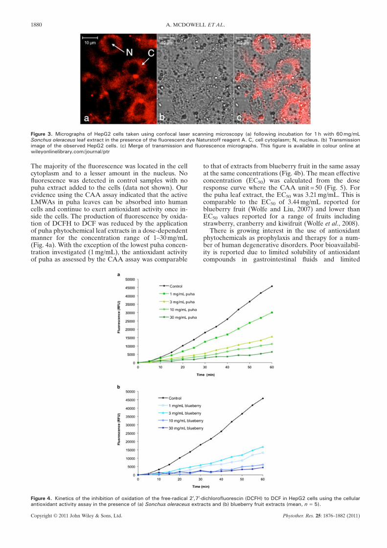
Figure 3. Micrographs of HepG2 cells taken using confocal laser scanning microscopy (a) following incubation for 1h with 60mg/mLSonchus oleraceus leaf extract in the presence of the fluorescent dye Naturstoff reagent A. C, cell cytoplasm; N, nucleus. (b) Transmissionimage of the observed HepG2 cells. (c) Merge of transmission and fluorescence micrographs. This figure is available in colour online atwileyonlinelibrary.com/journal/ptr
1880 A. MCDOWELL ET AL.
The majority of the fluorescence was located in the cellcytoplasm and to a lesser amount in the nucleus. Nofluorescence was detected in control samples with nopuha extract added to the cells (data not shown). Ourevidence using the CAA assay indicated that the activeLMWAs in puha leaves can be absorbed into humancells and continue to exert antioxidant activity once in-side the cells. The production of fluorescence by oxida-tion of DCFH to DCF was reduced by the applicationof puha phytochemical leaf extracts in a dose-dependentmanner for the concentration range of 1–30mg/mL(Fig. 4a). With the exception of the lowest puha concen-tration investigated (1mg/mL), the antioxidant activityof puha as assessed by the CAA assay was comparable
Figure 4. Kinetics of the inhibition of oxidation of the free-radical 2′,7′-antioxidant activity assay in the presence of (a) Sonchus oleraceus extra
Copyright © 2011 John Wiley & Sons, Ltd.
to that of extracts from blueberry fruit in the same assayat the same concentrations (Fig. 4b). The mean effectiveconcentration (EC50) was calculated from the doseresponse curve where the CAA unit = 50 (Fig. 5). Forthe puha leaf extract, the EC50 was 3.21mg/mL. This iscomparable to the EC50 of 3.44mg/mL reported forblueberry fruit (Wolfe and Liu, 2007) and lower thanEC50 values reported for a range of fruits includingstrawberry, cranberry and kiwifruit (Wolfe et al., 2008).
There is growing interest in the use of antioxidantphytochemicals as prophylaxis and therapy for a num-ber of human degenerative disorders. Poor bioavailabil-ity is reported due to limited solubility of antioxidantcompounds in gastrointestinal fluids and limited
dichlorofluorescin (DCFH) to DCF in HepG2 cells using the cellularcts and (b) blueberry fruit extracts (mean, n=5).
Phytother. Res. 25: 1876–1882 (2011)

Figure 5. Dose-response curve for the inhibition of oxidation of thefree-radical 2′,7′-dichlorofluorescin (DCFH) to DCF in HepG2 cellsusing the cellular antioxidant activity assay in the presence ofSonchus oleraceus extracts (mean�SD, n=3). R2=0.9419.
1881ANTIOXIDANTACTIVITY OF PUHA ASSESSED BY THE CAA ASSAY
permeability (Ratnam et al., 2006; Sun et al., 2002).If these antioxidants are to be consumed in an oralsupplement form, there is a need to formulate the
Copyright © 2011 John Wiley & Sons, Ltd.
antioxidants based on their physicochemical and bio-pharmaceutical properties to ensure product efficacy.Using the CAA assay it was demonstrated that antioxi-dants in foliar extracts of Sonchus oleraceus wereabsorbed into HepG2 cells and had antioxidant activitywithin human cells.
Acknowledgements
The research was supported by funding from a University of OtagoStrategic Research Grant and a Laurenson Award. We are gratefulto Susan Mackenzie for growing the puha plants and to Sarah Wilson,Rhodri Harfoot and Julie Clearwater for their assistance with cellculture and Dr Ivan Sammut for use of the plate reader and valuablediscussions.
Conflict of Interest
The authors have declared that there is no conflict of interest.
REFERENCES
Abascal K, Ganora L, Yarnell E. 2005. The effect of freeze-dryingand its implications for botanical medicine: A review.Phytother Res 19: 655–660.
Brand-Williams W, Cuvelier M, Berset C. 1995. Use of a freeradical method to evaluate antioxidant activity. Food Sci Tech28: 25–30.
Cao G, Alessio HM, Cutler RG. 1993. Oxygen-radical absorbancecapacity assay for antioxidants. Free Radic Biol Med 14:303–311.
Cao X, Tsukamoto T, Seki Tet al. 2008. 4-Vinyl-2,6-dimethoxyphenol(canolol) suppresses oxidative stress and gastric carcinogensisin Helicobacter pylori-infected carcinogen-treated Mongoliangerbil. Int J Cancer 122: 1445–1454.
Ernst IMA, Wagner AE, Lipinski S et al. 2010. Celllar uptake,stability, visualization by ‘Naturstoff reagent A’ and multidrugresistance protein 1 gene-regulation activity of cyanidin inhuman keratinocytes. Pharmacol Res 61: 253–258.
Frankel EN, Meyer AS. 2000. The problems of using one-dimensionalmethods to evaluate multifunctional food and biologicalantioxidants. J Sci Food Agric 80: 1925–1941.
Fresco P, Borges F, Diniz C, Marques M. 2006. New insights on theanticancer properties of dietary polyphenols. Med Res Rev22: 747–766.
Gould KS, Thodey K, Philpott M, Ferguson LR. 2006. Antioxidantactivities of extracts from traditional Māori food plants. NZJ Bot 44: 1–4.
Guil-Guerrero JL, Gimenez-Gimenez A, Rodriguez-Garcia I, Torija-Isasa ME. 1998. Nutritional composition of Sonchus species(S. asper L, S. oleraceus L and S. tenerrimus L). J Sci FoodAgric 76: 628–632.
Holm LG, Plucknett DL, Pancho JV, Herbeger JP. 1977. World’sworst weeds. Distribution and biology. University Press ofHawaii: Honolulu.
Jan AT, Kamli MR, Murtaza I, Singh JB, Ali A, Haq QMR. 2010.Dietary flavonoid quercetin and associated health benefits –an overview. Food Rev Int 26: 302–317.
Keinänen M, Julkunen-Tiitto R. 1996. Effect of sample preparationmethod on birch (Betula pendula Roth) leaf phenolics. J AgricFood Chem 44: 2724–2727.
Kelsey NA, Wilkins HM, Linseman DA. 2010. Nutraceuticalantioxidants as novel neuroprotective agents. Molecules 15:7792–7814.
Laine P, Kylli P, Heinonen M, Jouppila K. 2008. Storage stabilityof microencapsulated cloudberry (Rubus chamaemorus)phenolics. J Agric Food Chem 56: 11251–11261.
Li L, Chen CYO, Aldini G et al. 2010. Supplementation with luteinor lutein plus green tea extracts does not change oxidativestress in adequately nourished older adults. J Nutr Biochem21: 544–549.
Liu R, Finley J. 2005. Potential cell culture models for antioxidantresearch. J Agric Food Chem 53: 4311–4314.
Maisuthisakul P, Pongsawatmanit R. 2004. Effect of samplepreparation methods and extraction time on yield and antioxi-dant activity from Kradonbok (Careya sphaerica Roxb.) leaves.Kasetsart J (Nat Sci) 38: 8–14.
Mullen W, Stewart A, Lean M, Gardner P, Duthie G, Crozier A.2002. Effect of freezing and storage on the phenolics,ellagitannins, flavonoids, and antioxidant capacity of red rasp-berries. J Agric Food Chem 50: 5197–5201.
Netzel M, Netzel G, Tian Q, Schwartz S, Konczak I. 2006. Sourcesof antioxidant activity in Australian native fruits. Identificationand quantification of anthocyanins. J Agric Food Chem54: 9820–9826.
Philpott M, Gould KS, Markham KR, Lewthwaite SL, Ferguson LR.2003. Enhanced coloration reveals high antioxidant poten-tial in new sweet potato cultivars. J Sci Food Agric 83:1076–1082.
Pieroni A, Janiak V, Dürr C, Lüdeke S, Trachsel E, Heinrich M.2002. In vitro antioxidant activity of non-cultivated vege-tables of ethnic Albanians in southern Italy. Phytother Res16: 467–473.
Ratnam DV, Ankola DD, Bhardwaj V, Sahana DK, Kumar M.2006. Role of antioxidants in prophylaxis and therapy: Apharmaceutical perspective. J Control Release 113: 189–207.
Sun J, Chu Y-F, Wu X, Liu RH. 2002. Antioxidant and antiprolifera-tive activities of common fruits. J Agric Food Chem 50:7449–7454.
Temple N. 2000. Antioxidants and disease: more questions thananswers. Nutr Res 20: 449–459.
Thomson B, Shaw I. 2002. A comparison of risk and protectivefactors for colorectal cancer in the diet of New Zealand Maoriand non-Maori. Asian Pac J Cancer Prev 3: 319–324.
Wells J. 1998. Pharmaceutical preformulation - the physicochem-ical properties of drug substances. Taylor and Francis: London.
Whyte R, Hudson J, Gray M, O’Rielly R. 2001. TraditionalMaori food preparation methods and food safety. Int J FoodMicrobiol 69: 183–190.
Wolfe KL, Kang X, He X, Dong M, Zhang Q, Liu RH. 2008. Celluarantioxidant activity of common fruits. J Agric Food Chem56: 8418–8426.
Wolfe KL, Liu RH. 2007. Cellular antioxidant activity (CAA) assayfor assessing antioxidants, foods and dietary supplements.J Agric Food Chem 55: 8896–8907.
Wolfe KL, Liu RH. 2008. Structure-activity relationships offlavonoids in the cellular antioxidant activity assay. J AgricFood Chem 56: 8404–8411.
Xiangfei L, Ardob S, Bunninga M et al. 2005. Total phenoliccontent and DPPH radical scavenging activity of lettuce
Phytother. Res. 25: 1876–1882 (2011)

1882 A. MCDOWELL ET AL.
(Lactuca sativa L.) grown in Colorado. LWT - Food Sci Technol40: 552–557.
Yin J, Heo S, Jung M, Wang M. 2008. Antioxidant activity offractions from 70% methanolic extract of Sonchus oleraceusL. Food Sci Technol 17: 1299–1304.
Yin J, Kwon GJ, Wang WH. 2007. The antioxidant and cytotoxicactivities of Sonchus oleraceus L. extracts. Nutr Res Pract 1:189–194.
Copyright © 2011 John Wiley & Sons, Ltd.
Zeghichi S, Kallithraka S, Simopoulos AP, Kypriotakis Z. 2003.Nutritional composition of selected wild plants in the diet ofCrete. In Plants in Human Health and Nutritional Policy.Simopoulos AP. Copalan C. (eds). Karger: Basel, 22–40.
Zhang L, Zheng Y, Chow MSS, Zuo Z. 2004. Investigationof intestinal absorption and disposition of green teacatechins by Caco-2 monolayer model. Int J Pharm 287:1–12.
Phytother. Res. 25: 1876–1882 (2011)