Antibiotic-eluting bioresorbable composite fibers for ...meitalz/Articles/B2.pdf ·...
12
Antibiotic-eluting bioresorbable composite fibers for wound healing applications: Microstructure, drug delivery and mechanical properties Jonathan J. Elsner, Meital Zilberman * Department of Biomedical Engineering, Faculty of Engineering, Tel-Aviv University, Tel-Aviv 69978, Israel Received 19 October 2008; received in revised form 26 March 2009; accepted 7 April 2009 Available online 21 April 2009 Abstract Novel antibiotic-eluting composite fibers designed for use as basic wound dressing elements were developed and studied. These struc- tures were composed of a polyglyconate core and a porous poly(DL-lactic-co-glycolic acid) shell loaded with one of three antibiotic drugs: mafenide acetate, gentamicin sulphate and ceftazidime pentahydrate. The shell was prepared by the freeze-drying of inverted emulsions. The fiber investigation focused on the effects of the emulsion’s formulation on the shell microstructure and on the resulting profile of drug release from the fibers. Albumin was found to be the most effective surfactant for stabilizing the inverted emulsions and also to have a beneficial holdup effect on the release kinetics of the hydrophilic antibiotic drugs, especially mafenide acetate, probably through a specific interaction. An increase in the organic:aqueous phase ratio, polymer content or molecular weight of the host polymer resulted in a decrease in the burst release and a more moderate release profile due to changes in shell microstructure. The first two parameters were found to be more effective than the third. The diverse release profiles obtained in the current study and the good mechanical properties indicate that our new composite fibers have good potential for use in wound healing applications. Ó 2009 Acta Materialia Inc. Published by Elsevier Ltd. All rights reserved. Keywords: Wound dressings; Fiber; Antibiotic; Poly-(DL-lactic-co-glycolic acid); Controlled drug delivery 1. Introduction Wound dressings aim to restore the milieu required for skin regeneration and to protect the wound from environ- mental threats and penetration of bacteria. Although tradi- tional gauze dressings offer some protection against bacteria, this protection is lost when the outer surface of the dressing becomes moistened by wound exudate or external fluids. Furthermore, traditional gauze dressings do not greatly restrict moisture evaporation and may cause dehydration of the wound bed. This can lead to adhesion of the dressing, particularly as wound fluid production diminishes, causing pain and discomfort to the patient when removed [1]. Most modern dressings are designed according to the well-accepted bilayer structure concept, i.e. an upper dense ‘‘skin” layer to prevent bacterial pene- tration and a lower spongy layer designed to adsorb wound exudates and accommodate newly formed tissue [2,3]. Unfortunately, dressing material which has absorbed wound discharge provides conditions that are also favor- able for bacterial growth. This has encouraged the develop- ment of a new generation of wound dressings with improved curative attributes that provide an antimicrobial effect by eluting various germicidal compounds. These dressings still require frequent changing, which may be painful to the patient, harm the vulnerable underlying skin and increase the risk of secondary contamination. Biore- sorbable dressings successfully address this shortcoming, since they do not need to be removed from the wound surface once they have fulfilled their role. Film dressings made of lactide–caprolactone copolymers such as Topkin Ò (Biomet, Europe) and Oprafol Ò (Lohmann & Rauscher, Germany) are currently available [4]. Biodegradation of 1742-7061/$ - see front matter Ó 2009 Acta Materialia Inc. Published by Elsevier Ltd. All rights reserved. doi:10.1016/j.actbio.2009.04.007 * Corresponding author. Tel.: +972 3 6405842; fax: +972 3 6407939. E-mail address: [email protected] (M. Zilberman). Available online at www.sciencedirect.com Acta Biomaterialia 5 (2009) 2872–2883 www.elsevier.com/locate/actabiomat
Transcript of Antibiotic-eluting bioresorbable composite fibers for ...meitalz/Articles/B2.pdf ·...
Available online at www.sciencedirect.com
Acta Biomaterialia 5 (2009) 2872–2883
www.elsevier.com/locate/actabiomat
Antibiotic-eluting bioresorbable composite fibers for woundhealing applications: Microstructure, drug delivery
and mechanical properties
Jonathan J. Elsner, Meital Zilberman *
Department of Biomedical Engineering, Faculty of Engineering, Tel-Aviv University, Tel-Aviv 69978, Israel
Received 19 October 2008; received in revised form 26 March 2009; accepted 7 April 2009Available online 21 April 2009
Abstract
Novel antibiotic-eluting composite fibers designed for use as basic wound dressing elements were developed and studied. These struc-tures were composed of a polyglyconate core and a porous poly(DL-lactic-co-glycolic acid) shell loaded with one of three antibiotic drugs:mafenide acetate, gentamicin sulphate and ceftazidime pentahydrate. The shell was prepared by the freeze-drying of inverted emulsions.The fiber investigation focused on the effects of the emulsion’s formulation on the shell microstructure and on the resulting profile of drugrelease from the fibers. Albumin was found to be the most effective surfactant for stabilizing the inverted emulsions and also to have abeneficial holdup effect on the release kinetics of the hydrophilic antibiotic drugs, especially mafenide acetate, probably through a specificinteraction. An increase in the organic:aqueous phase ratio, polymer content or molecular weight of the host polymer resulted in adecrease in the burst release and a more moderate release profile due to changes in shell microstructure. The first two parameters werefound to be more effective than the third. The diverse release profiles obtained in the current study and the good mechanical propertiesindicate that our new composite fibers have good potential for use in wound healing applications.� 2009 Acta Materialia Inc. Published by Elsevier Ltd. All rights reserved.
Keywords: Wound dressings; Fiber; Antibiotic; Poly-(DL-lactic-co-glycolic acid); Controlled drug delivery
1. Introduction
Wound dressings aim to restore the milieu required forskin regeneration and to protect the wound from environ-mental threats and penetration of bacteria. Although tradi-tional gauze dressings offer some protection againstbacteria, this protection is lost when the outer surface ofthe dressing becomes moistened by wound exudate orexternal fluids. Furthermore, traditional gauze dressingsdo not greatly restrict moisture evaporation and may causedehydration of the wound bed. This can lead to adhesionof the dressing, particularly as wound fluid productiondiminishes, causing pain and discomfort to the patientwhen removed [1]. Most modern dressings are designedaccording to the well-accepted bilayer structure concept,
1742-7061/$ - see front matter � 2009 Acta Materialia Inc. Published by Else
doi:10.1016/j.actbio.2009.04.007
* Corresponding author. Tel.: +972 3 6405842; fax: +972 3 6407939.E-mail address: [email protected] (M. Zilberman).
i.e. an upper dense ‘‘skin” layer to prevent bacterial pene-tration and a lower spongy layer designed to adsorb woundexudates and accommodate newly formed tissue [2,3].Unfortunately, dressing material which has absorbedwound discharge provides conditions that are also favor-able for bacterial growth. This has encouraged the develop-ment of a new generation of wound dressings withimproved curative attributes that provide an antimicrobialeffect by eluting various germicidal compounds. Thesedressings still require frequent changing, which may bepainful to the patient, harm the vulnerable underlying skinand increase the risk of secondary contamination. Biore-sorbable dressings successfully address this shortcoming,since they do not need to be removed from the woundsurface once they have fulfilled their role. Film dressingsmade of lactide–caprolactone copolymers such as Topkin�
(Biomet, Europe) and Oprafol� (Lohmann & Rauscher,Germany) are currently available [4]. Biodegradation of
vier Ltd. All rights reserved.

J.J. Elsner, M. Zilberman / Acta Biomaterialia 5 (2009) 2872–2883 2873
these films occurs via hydrolysis of the copolymer into lac-tic acid and 6-hydroxycaproic acid. However, film dress-ings are better suited for small wounds, since they lackabsorbence and are impermeable to water vapor and gases,both of which cause accumulation of wound fluids on lar-ger wound surfaces.
Some recently reported work has focused as an alterna-tive on the development of more complex biodegradablefiber-based wound dressings with antibiotic delivery [5–8].Fiber-based dressings composed of either continuous fibersthat form a non-woven fiber mesh or a fabric made fromwoven fibers offer a high surface area for controlled release,absorbency and pliability. However, the main challenge indesigning a device for the release of low molecular weight(MW) hydrophilic antibiotics is to overcome the rapid dis-charge of the drug from the device. This drawback has alsobeen reported for other antibiotic-eluting devices such asperiodontal devices [9,10] and vascular grafts [11,12]. Thelocal antibiotic release profile should exhibit a considerableinitial release rate in order to respond to the elevated risk ofinfection from bacteria introduced during the initialtrauma, followed by a release of antibiotics at an effectivelevel long enough to inhibit latent infection [13]. The loca-tion, size and degree of injury, as well as the rate of tissueregeneration (depending on the patient’s age and otherparameters), affect the wound healing process. Hence, char-acteristic healing has been reported to take 3–7 weeks [14].Common strategies that have been described in an attemptto overcome the problem of rapid drug release include theentrapment of the hydrophilic drug within a hydrophobicsubstance as a means to delay water penetration and out-ward drug diffusion [15,16], or enhancement of drug bond-ing to the carrying matrix [11,12,17]. The latter can beachieved either by selecting or modifying a matrix materialto support the formation of covalent bonds, Van der Waalsdispersion forces, hydrogen bonds, or ionic interactionsbetween the drug and the matrix. Vascular grafts sealedwith albumin and gelatin have been shown to promote suchinteractions and therefore demonstrate a reduced burstrelease of antibiotics compared to uncoated grafts [11,12].A wound dressing based on succinylated collagen, whichbehaves as an anion after swelling, has been shown to delaythe release of the cationic drug ciprofloxacin via ionic inter-actions [17].
Incorporation of antibiotics in the process of fiber spin-ning (e.g. electrospinning or melt and solution spinning) isassociated with the disadvantages of poor mechanicalproperties due to drug incorporation and limitations indrug loading. Furthermore, many drugs and proteins donot tolerate melt processing and organic solvents. Themain goal of the current study was therefore to developand study new fiber structures loaded with antibiotics.Our composite fibers combine a dense polymer core fiberand a drug-loaded porous shell structure, i.e. the drug islocated in a separate compartment (a ‘‘shell”) around amelt spun ‘‘core” fiber. The shell is prepared using freeze-drying of inverted emulsions. This fabrication process uses
mild processing conditions and is designed to produce astructure with good mechanical properties as well as thedesired drug-release profile. These new fibers are ideal forforming thin, delicate, biomedically important structuressuch as wound dressings. Our fibers were loaded with oneof three antibiotic drugs: gentamicin sulphate, ceftazidimepentahydrate and mafenide acetate. The first two antibac-terial drugs are broad-spectrum antibiotics which can beused systemically or locally, whereas the third is typicallyused in burn dressings. The drugs’ physicochemical proper-ties and antibacterial spectra are presented in Table 1. Theeffects of the emulsion’s formulation parameters on theshell microstructure and on the resulting drug-release pro-file and mechanical tensile properties are presented.
2. Materials and methods
2.1. Materials
MaxonTM polyglyconate monofilament sutures with adiameter of 0.20–0.25 mm (United States Surgical Inc.,USA) were used as core fibers.
Bioresorbable porous structures (the shell coating) weremade of 75/25 poly(DL-lactic-co-glycolic acid) (PDLGA),inherent viscosity (i.v.) = 0.4, 0.65 and 1.13 dl g�1 (in CHCl3at 30 �C), MW approximately 50, 100, 240 kDa, respectively(Absorbable Polymer Technologies, Inc., USA).
2.1.1. Drugs
Gentamicin sulfate (cell-culture tested), 590 lg gentami-cin base per mg of salt (Sigma, G-1264).
4-Aminomethylbenzenesulfonamide acetate salt (mafe-nide acetate) (Sigma, A-3305).
Ceftazidime hydrate, 90–105% (Sigma, C-3809).
2.1.2. Surface active agents
Bovine serum albumin (BSA), MW = 66,000 Da (Sigma,A-4503).
Poly(vinyl alcohol) (PVA), 87–89% hydrolyzed,MW = 13,000–23,000 Da (Aldrich, 36,317-0).
2.1.3. Reagents
Isopropyl alcohol (propanol) was purchased fromFrutarom, Israel.
1,1,1,3,3,3-hexafluoro-2-propanol (H1008) was pur-chased from Spectrum Chemical Mfg. Corp.
2.2. Preparation of core–shell fiber structures
2.2.1. Fiber surface treatment
The sutures were surface-treated in order to dispose ofthe original fiber coating and to enhance adhesion betweenthe core fiber and the coating. The MaxonTM fibers weregently wrapped around flexible Teflon frames and dippedin a Petri dish containing 1,1,1,3,3,3-hexafluoro-2-propa-nol for 40 s. The fibers were then washed with 70% ethanoland dried.

Table 1Physicochemical properties of the antibiotics used in the study [30].
Antibiotic agent Molecularweight(g mol�1)
Watersolubility(mg ml�1)
Serumproteinbinding*
Antibacterial spectrum
Gentamicin sulphate 477.6 100 0–30%[31]
Effective against a broad spectrum of Gram-positive andGram-negative bacteria
Ceftazidime pentahydrate 546.6 5 0–10%[31]
A third-generation cephalosporin which displays a broadspectrum activity against Gram-positive and Gram-negativebacteria. Unlike most third-generation agents, it is activeagainst Pseudomonas aeruginosa
Mafenide acetate 246.3 250 20–90%[29]
Bacteriostatic for many Gram-negative and Gram-positiveorganisms, including Pseudomonas aeruginosa and certainstrains of anaerobes. Mafenide is highly soluble and diffusesinto and through eschar producing a marked reduction inthe number of bacteria present, even in avascular tissue ofsecond- and third-degree burns
* Albumin binds drugs at varying degrees, depending on the specific drug’s affinity. The bound portion may act as a reservoir or depot from which thedrug is slowly released as the unbound form.
2874 J.J. Elsner, M. Zilberman / Acta Biomaterialia 5 (2009) 2872–2883
2.2.2. Emulsion formation
A known amount of PDLGA was dissolved in chloro-form to form an organic solution. A known amount ofthe drug was dissolved in double-distilled water and thenpoured into the organic phase (in a test tube). Homogeni-zation of this emulsion was performed using a KinematicaPT-3100 Polytron homogenizer operating at 16,000 rpm(medium rate which was found to be optimal) for 2 min.An emulsion formulation containing 15% w/v PDLGA(100 kDa) polymer in the organic solution, 5% w/w drugin the aqueous medium (relative to the polymer load),and an organic to aqueous (O:A) phase ratio of 6:1 v/vwas used as the reference formulation. Additional formula-tions included variations in polymer content (17.5% and20%), drug loading (1% and 2.5%), O:A phase ratios (2:1,8:1, 10:1 and 12:1) and polymer MW (50 and 240 kDa).The effect of BSA and PVA as surfactants (0.5% and 1%w/v in the aqueous phase) was also studied. These surfac-tants are well known as stabilizers for emulsions [18,19].In each studied series, only one parameter was changedand the others remained the same as in the referenceformulation.
2.2.3. Core–shell fiber structure formationThe treated MaxonTM core fibers were dip-coated (while
placed on holders) in fresh emulsions and then frozenimmediately in a liquid nitrogen bath. The holders + sam-
ples were then placed in a pre-cooled (�105 �C) freeze-dryer (Virtis 101 equipped with a nitrogen trap) capableof working with organic solvents and freeze-dried in orderto preserve the microstructure of the emulsion-based core–shell fiber structures. Drying was performed in two stages:
(i) The freeze-dryer chamber pressure was reduced to100 mTorr while the temperature remained at�105 �C.
(ii) The condenser was turned off after 5 h and its platetemperature gradually increased to room temperaturewhile the pressure was monitored between 100 and700 mTorr. During this step the liquid nitrogen trapcondensed the excess water and solvent vapors. Thesamples were stored in desiccators until use.
2.3. In vitro drug-release studies
The composite core–shell fiber structures (triplicate sam-ples, 15 cm each) were immersed in phosphate-buffered sal-ine (PBS, pH 7.0) at 37 �C for 60 days in order to determinethe various drug-release kinetics from these structures. Therelease studies were conducted in closed glass vessels con-taining 1.5 ml PBS medium. The medium was removed(completely) periodically, at each sampling time (6 h, 1, 2,3, 7, 14, 21, 28, 35, 42, 49 and 56 days), and fresh mediumwas introduced.

J.J. Elsner, M. Zilberman / Acta Biomaterialia 5 (2009) 2872–2883 2875
2.3.1. Gentamicin assay
Determination of the medium’s gentamicin content wascarried out using an Abbott Therapeutic Drug MonitoringSystem (TDX, Abbott Laboratories) according to themanufacturer’s instructions. This machine enables thedetermination of the gentamicin concentration based on apolarization fluoroimmunoassay using fluorescein as atracer. Briefly, the latter is excited by polarized light. Polar-ization of the emitted light is dependent on molecule size.Free and labeled drug compete for binding sites. The drugconcentration in the sample is proportional to the scatter ofpolarized light caused by free labeled drug. The measurableconcentration range without dilution is 0.0–10.0 lg ml�1.Higher drug concentrations were measured after carryingout manual dilution.
2.3.2. Mafenide assay
The medium’s mafenide content was determined using aJasco high performance liquid chromatography (HPLC)system with a UV 2075 plus detector and a reverse-phase col-umn (Interstil� ODS-3 V 5 lm, inner diameter d = 4.6 mm,length = 250 mm), kept at 25 �C. The mobile phase con-sisted of a mixture of PBS and acetonitrile at a flow rate of1 ml min�1 with a quaternary gradient pump (PU 2089plus), gradient t = 0 min, 100/0 (v/v), t = 1.5 min, 90/10(v/v), t = 4 min, 100/0 (v/v). Thirty microliters of sampleswere injected with an autosampler (AS 2057 Plus). The col-umn effluent was eluted for 9 min and detected at 267 nm.The area of each eluted peak was integrated using EZstartsoftware, v. 3.1.7. A calibration curve was prepared for con-centrations ranging from 1.0 to 200.0 lg ml�1 (n = 8, corre-lation coefficient >0.999, slope 0.0002295).
2.3.3. Ceftazidime assay
The medium’s ceftazidime content was determined usinga Jasco HPLC system with a UV 2075 plus detector and areverse-phase column (Interstil� ODS-3 V 5 lm, innerdiameter d = 4.6 mm, length = 250 mm), kept at 25 �C.The mobile phase consisted of a mixture of PBS and aceto-nitrile (95/5, v/v) at a flow rate of 1 ml min�1 with a qua-ternary gradient pump (PU 2089 plus) without gradient.Twenty microliters of samples were injected with an auto-sampler (AS 2057 Plus). The column effluent was elutedfor 22 min and detected at 254 nm. The area of each elutedpeak was integrated using EZstart software, v. 3.1.7. A cal-ibration curve was prepared for concentrations rangingfrom 1.0 to 200.0 lg ml�1 (n = 8, correlation coefficient>0.999, slope 0.0000318).
2.4. Residual drug recovery from composite fibers
Residual drug recovery from the composite fibers wasmeasured as follows: the fibers were placed in 1 ml methy-lene chloride in order to dissolve the remaining PDLGAcoating. Two milliliters of water were then added in orderto dissolve the hydrophilic drug residues. The materialswere vortexed for 30 s and then left to stand until phase
separation occurred. The aqueous phase was filtered inorder to dispose of polymer particles. The drug concentra-tion was estimated using the above-described assays. Theexperiments were performed in triplicate.
2.5. Morphological characterization
The morphology of the composite core–shell fiber struc-tures (cross-section of cryogenically fractured fibers) wasobserved by scanning electron microscopy (SEM) using aJEOL JSM-6300 at an accelerating voltage of 5 kV. TheSEM samples were Au sputtered prior to observation.The mean pore diameter (n = 80 pores) and porosity ofthe observed morphologies for each SEM fractograph wereanalyzed using Sigma Scan Pro software. Porosity wasdetermined as the area occupied by the pores divided bythe total area.
2.6. Microbiological evaluation
Agar diffusion tests (Kirby–Bauer method) were con-ducted to confirm the bioactivity of the antibiotics encap-sulated in the composite fibers. Small discs (n = 3, d =10 mm) were cut from plain-woven Maxon fibers coatedwith the reference formulation. The discs were then placeddry on Muller–Hinton agar plates, inoculated with relevantbacteria (one strain on each plate): Staphylococcus aureus,Staphylococcus epidermidis and Pseudomonas aeruginosa.Bacterial inhibition zones around the dressings wereassessed after 24 h of incubation. Similar dressings, pre-pared without drug, were used as controls.
2.7. Tensile mechanical properties
The fibers’ tensile mechanical properties were measuredat room temperature, under unidirectional tension at a rateof 50 mm min�1 (ASTM D 3379), using a 5500 Instronmachine. The tensile strength was defined as the maximumstrength in the stress–strain curve. The maximal strain wasdefined as the breaking strain. Young’s modulus was definedas the slope of the stress–strain curve in the elastic (linear)region. Six samples were tested for each type of specimen.
2.8. Statistics
Statistics were calculated using SPSS 10 software. Alldata are expressed as means ± standard deviation (SD).ANOVA (Tukey–Kramer) was used for group comparisonand P values less than 0.05 were considered significant.
3. Results
3.1. Morphological characterization
Composite fiber structures with a core diameter ofapproximately 280 lm and a shell thickness of 20–50 lmwere produced and studied. A SEM fractograph showing
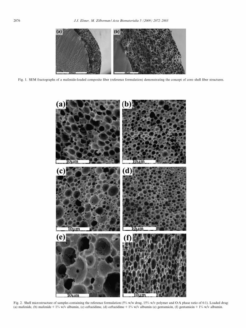
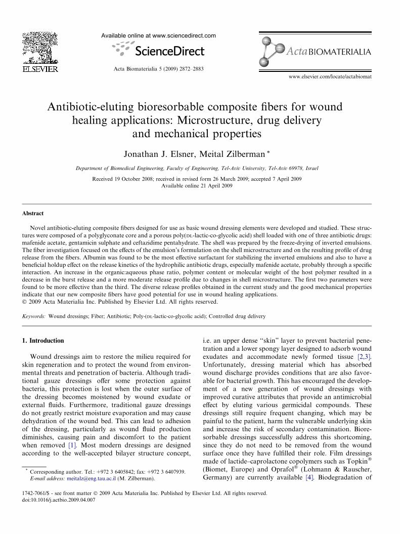
Fig. 1. SEM fractographs of a mafenide-loaded composite fiber (reference formulation) demonstrating the concept of core–shell fiber structures.
Fig. 2. Shell microstructure of samples containing the reference formulation (5% w/w drug, 15% w/v polymer and O:A phase ratio of 6:1). Loaded drug:(a) mafenide, (b) mafenide + 1% w/v albumin, (c) ceftazidime, (d) ceftazidime + 1% w/v albumin (e) gentamicin, (f) gentamicin + 1% w/v albumin.
2876 J.J. Elsner, M. Zilberman / Acta Biomaterialia 5 (2009) 2872–2883
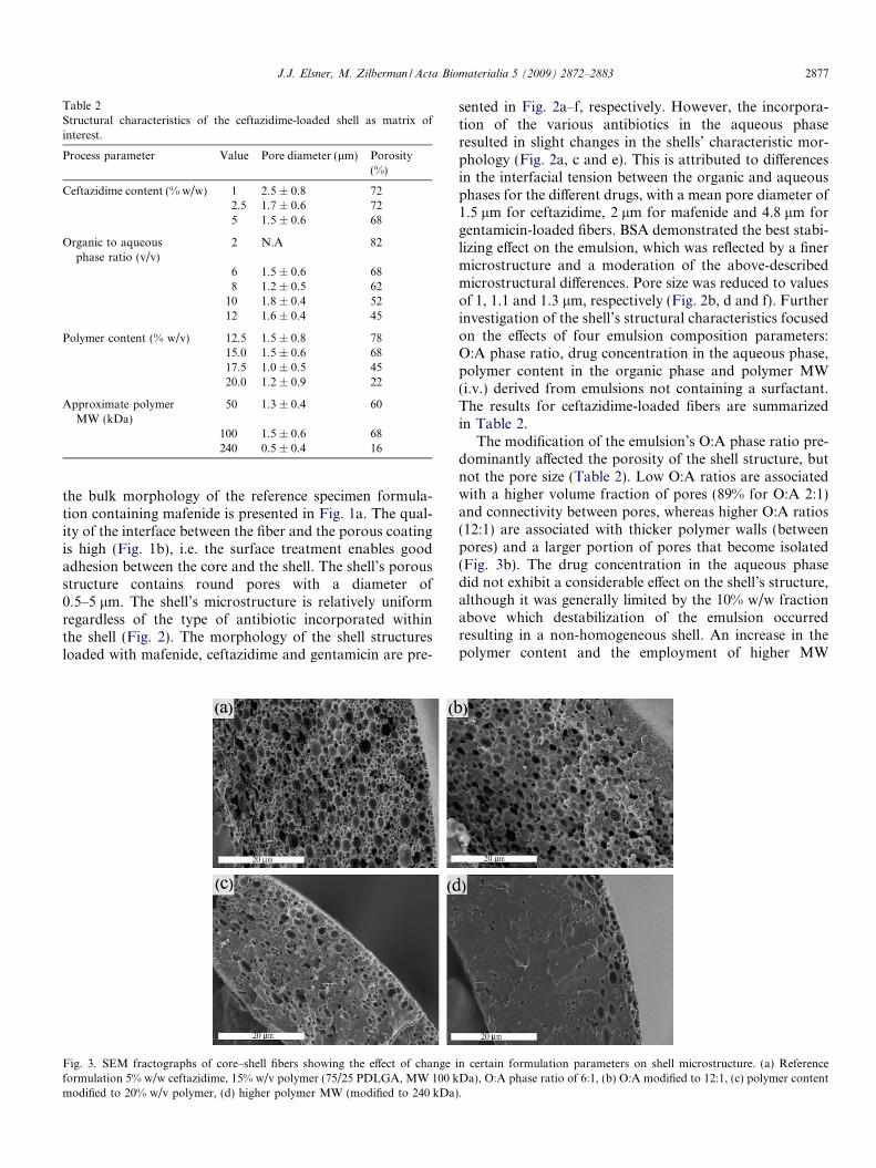
Table 2Structural characteristics of the ceftazidime-loaded shell as matrix ofinterest.
Process parameter Value Pore diameter (lm) Porosity(%)
Ceftazidime content (% w/w) 1 2.5 ± 0.8 722.5 1.7 ± 0.6 725 1.5 ± 0.6 68
Organic to aqueousphase ratio (v/v)
2 N.A 82
6 1.5 ± 0.6 688 1.2 ± 0.5 62
10 1.8 ± 0.4 5212 1.6 ± 0.4 45
Polymer content (% w/v) 12.5 1.5 ± 0.8 7815.0 1.5 ± 0.6 6817.5 1.0 ± 0.5 4520.0 1.2 ± 0.9 22
Approximate polymerMW (kDa)
50 1.3 ± 0.4 60
100 1.5 ± 0.6 68240 0.5 ± 0.4 16
J.J. Elsner, M. Zilberman / Acta Biomaterialia 5 (2009) 2872–2883 2877
the bulk morphology of the reference specimen formula-tion containing mafenide is presented in Fig. 1a. The qual-ity of the interface between the fiber and the porous coatingis high (Fig. 1b), i.e. the surface treatment enables goodadhesion between the core and the shell. The shell’s porousstructure contains round pores with a diameter of0.5–5 lm. The shell’s microstructure is relatively uniformregardless of the type of antibiotic incorporated withinthe shell (Fig. 2). The morphology of the shell structuresloaded with mafenide, ceftazidime and gentamicin are pre-
Fig. 3. SEM fractographs of core–shell fibers showing the effect of changeformulation 5% w/w ceftazidime, 15% w/v polymer (75/25 PDLGA, MW 100 kmodified to 20% w/v polymer, (d) higher polymer MW (modified to 240 kDa)
sented in Fig. 2a–f, respectively. However, the incorpora-tion of the various antibiotics in the aqueous phaseresulted in slight changes in the shells’ characteristic mor-phology (Fig. 2a, c and e). This is attributed to differencesin the interfacial tension between the organic and aqueousphases for the different drugs, with a mean pore diameter of1.5 lm for ceftazidime, 2 lm for mafenide and 4.8 lm forgentamicin-loaded fibers. BSA demonstrated the best stabi-lizing effect on the emulsion, which was reflected by a finermicrostructure and a moderation of the above-describedmicrostructural differences. Pore size was reduced to valuesof 1, 1.1 and 1.3 lm, respectively (Fig. 2b, d and f). Furtherinvestigation of the shell’s structural characteristics focusedon the effects of four emulsion composition parameters:O:A phase ratio, drug concentration in the aqueous phase,polymer content in the organic phase and polymer MW(i.v.) derived from emulsions not containing a surfactant.The results for ceftazidime-loaded fibers are summarizedin Table 2.
The modification of the emulsion’s O:A phase ratio pre-dominantly affected the porosity of the shell structure, butnot the pore size (Table 2). Low O:A ratios are associatedwith a higher volume fraction of pores (89% for O:A 2:1)and connectivity between pores, whereas higher O:A ratios(12:1) are associated with thicker polymer walls (betweenpores) and a larger portion of pores that become isolated(Fig. 3b). The drug concentration in the aqueous phasedid not exhibit a considerable effect on the shell’s structure,although it was generally limited by the 10% w/w fractionabove which destabilization of the emulsion occurredresulting in a non-homogeneous shell. An increase in thepolymer content and the employment of higher MW
in certain formulation parameters on shell microstructure. (a) ReferenceDa), O:A phase ratio of 6:1, (b) O:A modified to 12:1, (c) polymer content.

2878 J.J. Elsner, M. Zilberman / Acta Biomaterialia 5 (2009) 2872–2883
polymers (until the verge of instability—to be discussedlater) resulted in a similar trend of decrease in pore size(Fig. 3c and d, respectively), since high viscosities associ-ated with the increase in these two parameters help reduceflocculation and coalescence of droplets by reducing thetendency of droplets to move. Similar trends were observedalso for gentamicin- and mafenide-loaded samples.
3.2. In vitro drug-release studies
Typical total drug loadings for fibers based on the refer-ence formulation for the three studied drugs were similar:gentamicin 9.1 ± 0.4 lg cm�1, ceftazidime 8.1 ± 0.4 lg cm�1
and mafenide 8.1 ± 0.2 lg cm�1. In vitro drug-release pro-files from these (reference) fibers present a burst release of85–95% after 6 h, with almost total release of the drugwithin a week. There is practically no difference in therelease kinetics of the three drugs. Nonetheless, a rangeof different release profiles which vary in their initial burstrelease and total duration of release were obtained withvariations of the reference formulation.
Of the three studied antibiotics, the release of mafenidewas most affected by the incorporation of surfactants. We
Fig. 4. Effect of the incorporation of surfactants on the in vitro releaseprofile of mafenide from core–shell fiber structures containing: 5% w/wmafenide, 15% w/v polymer, O:A phase ratio of 6:1: (a) Samplescontaining albumin; (b) samples containing PVA (N no surfactant, �0.5% w/v surfactant, j 1% w/v surfactant).
therefore chose to present the effects of surfactants on com-posite fibers loaded with mafenide (Fig. 4). The incorpora-tion of 1% albumin in the reference formulation resulted inthe lowest burst release (53% compared to 72% with 1%PVA, and 85% without surfactant) and yielded controlledrelease of the drug over a period of 50 days. The effect ofalbumin as a surfactant on reference fibers loaded withthe three antibiotic drugs is presented in Fig. 5b. It canbe seen that refinement of the microstructure by stabiliza-tion with albumin helps reduce the burst release to somedegree for gentamicin and ceftazidime, and more notablyfor mafenide, probably due to specific interactions withalbumin. Other modifications of the formulation weremade to promote better restraint of outward drug diffu-sion. These included an increase in the O:A phase ratio,polymer concentration and polymer MW. The effect ofeach of these formulation parameters was studied individ-ually without adding a surfactant, and is presented inFig. 6. An increase in the O:A phase ratio from 6:1 to12:1 reduced the burst release of ceftazidime from 95% to44%, although total release of the encapsulated drug stilloccurred within 1 week. The effect of an increase in
Fig. 5. In vitro release of antibiotics from core–shell fiber structurescontaining 15% w/w polymer (75/25 PDLGA, MW 100 kDa), O:A phaseratio of 6:1, 5% w/w drug: (a) samples not containing albumin; (b) samplescontaining 1% w/v albumin (j mafenide, N ceftazidime, d gentamicin).
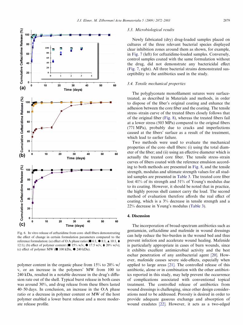
Fig. 6. In vitro release of ceftazidime from core–shell fibers demonstratingthe effect of change in certain formulation parameters compared to thereference formulation: (a) effect of O:A phase ratio (j 6:1, d 8:1, N 10:1, �12:1); (b) effect of polymer content (j 15% w/v, d 17.5 w/v, � 20% w/v);(c) effect of polymer MW (j 100 kDa, d 240 kDa).
J.J. Elsner, M. Zilberman / Acta Biomaterialia 5 (2009) 2872–2883 2879
polymer content in the organic phase from 15% to 20% w/v, or an increase in the polymers’ MW from 100 to240 kDa, resulted in a notable decrease in the drug’s diffu-sion rate out of the shell. Typical burst release in both caseswas around 30%, and drug release from these fibers lasted40–50 days. In conclusion, an increase in the O:A phaseratio or a decrease in polymer content or MW of the hostpolymer enabled a lower burst release and a more moder-ate release profile.
3.3. Microbiological results
Newly fabricated (dry) drug-loaded samples placed oncultures of the three relevant bacterial species displayedclear inhibition zones around them as shown, for example,in Fig. 7 (left) for ceftazidime-loaded samples. Conversely,control samples coated with the same formulation withoutthe drug, did not demonstrate any bactericidal effect(Fig. 7, right). All three bacterial strains demonstrated sus-ceptibility to the antibiotics used in the study.
3.4. Tensile mechanical properties
The polyglyconate monofilament sutures were surface-treated, as described in Materials and methods, in orderto dispose of the fiber’s original coating and enhance theadhesion between the core fiber and the coating. The tensilestress–strain curve of the treated fibers closely follows thatof the original fiber (Fig. 8), whereas the treated fibers failat a lower stress (503 MPa) compared to the original fibers(771 MPa), probably due to cracks and imperfectionscaused at the fibers’ surface as a result of the treatment,which lead to earlier failure.
Two methods were used to evaluate the mechanicalproperties of the core–shell fibers: (i) using the total diam-eter of the fiber; and (ii) using an effective diameter which isactually the treated core fiber. The tensile stress–straincurves of fibers coated with the reference emulsion accord-ing to both methods are presented in Fig. 8, and the tensilestrength, modulus and ultimate strength values for all stud-ied samples are presented in Table 3. The treated core fiberlost 41% of its strength and 51% of Young’s modulus dueto its coating. However, it should be noted that in practice,the highly porous shell cannot carry the load. The secondmethod of evaluation therefore affords the real effect ofcoating, which is a 3% decrease in tensile strength and a22% decrease in Young’s modulus (Table 3).
4. Discussion
The incorporation of broad-spectrum antibiotics such asgentamicin, ceftazidime and mafenide in wound dressingscan help reduce the bio-burden in the wound bed and thusprevent infection and accelerate wound healing. Mafenideis particularly appropriate in cases of burn wounds, sinceit exhibits excellent antimicrobial activity and the besteschar penetration of any antibacterial agent [20]. How-ever, mafenide causes severe side-effects, especially whenapplied to large areas [21]. The controlled release of thisantibiotic, alone or in combination with the other antibiot-ics reported in this study, may help prevent the occurrenceof complications associated with conventional topicaltreatment. The controlled release of antibiotics fromwound dressings is challenging, since other design consider-ations need to be addressed. Porosity is desired in order toprovide adequate gaseous exchange and absorption ofwound exudates [22]. However, it acts as a two-edged

Fig. 7. Demonstration of bioactivity of the drug released from a composite fiber-based dressing against relevant bacterial strains: (a) Pseudomonas
aeruginosa, (b) Staphylococcus aureus and (c) Staphylococcus epidermidis. Ceftazidime-loaded dressings demonstrate clear inhibition zones around them(left), whereas unloaded controls do not (right).
2880 J.J. Elsner, M. Zilberman / Acta Biomaterialia 5 (2009) 2872–2883
sword since rapid water penetration typically leads to arapid release of the active agent within several hours to sev-eral days.
The process of freeze-drying is unique in its ability topreserve the liquid structure in solids. The microstructureof the freeze-dried inverted emulsion, our fibers’ shell com-partment, can therefore serve as a good measure for theemulsions’ stability. Separating a two-phase system gener-ates a large surface area per drop, leading to a high excessGibbs energy per drop and thus to a tendency in the direc-tion of decreasing the Gibbs energy through several typesof interaction patterns such as flocculation, coalescence,Ostwald ripening and creaming [23–25]. Flocculation, forexample, would be manifest in the shells’ microstructureas clustered pores separated by thin polymer walls, whereas
Fig. 8. Tensile stress–strain curves of polyglyconate fibers. Fiber 1,original MaxonTM suture; Fiber 2, surface-treated core fiber; Fiber 3,reference formulation core–shell fiber structure containing mafenide (totaldiameter is considered); Fiber 4, reference formulation core–shell fiberstructure containing mafenide (effective diameter is considered).
coalescence would be apparent from the larger averagepore size and a greater deviation in pore sizes (higher stan-dard deviation). The process–structure release profileeffects of our system are discussed below.
4.1. Effect of drug type on shell microstructure and drug-
release profile
The presence of a hydrophilic drug in the emulsion’saqueous phase modifies the emulsion’s hydrophobic–hydrophilic balance. An increase in the drugs’ charge orconcentration corresponds to an increase in the interfacialtension between the aqueous and organic phases and istherefore expected to result in lower emulsion stability.This phenomenon was confirmed even when a moderatedrug loading of 5% w/w was used. The pore shape rangedfrom completely round pores separated by clear polymerboundaries for ceftazidime-loaded samples (Fig. 2c) to lar-ger, somewhat irregular pores for gentamicin-loaded sam-ples (Fig. 2e). It should be noted that gentamicin, ahighly charged polycation, displays the largest charge(+3.5, pH 7.4 [26]). Differences in shell morphology werethus obtained due to differences in the drugs’ hydrophilicityand charge.
Table 3The tensile mechanical properties of the fibers.
Fiber type Strength(MPa)
Modulus(MPa)
Ultimatestrain (%)
1 MaxonTM suture 771 ± 63 478 ± 54 47 ± 42 Treated MaxonTM 503 ± 44a 476 ± 39 39 ± 1a
3 Composite fiber A* 294 ± 18a,b 233 ± 18a,b 42 ± 1a
4 Composite fiber B** 487 ± 32a 371 ± 14a,b 42 ± 1a
* Composite fiber A: evaluation based on the total fiber cross-sectionalarea.** Composite fiber B: evaluation based on the effective fiber cross-sec-
tional area.a Statistically different (P < 0.05) from the MaxonTM suture.b Statistically different (P < 0.05) from the surface-treated MaxonTM
suture.
J.J. Elsner, M. Zilberman / Acta Biomaterialia 5 (2009) 2872–2883 2881
The adverse effect attributed to the drugs’ physiochemi-cal properties was investigated by the introduction of var-ious surfactants. Surfactants located at the interfacebetween the aqueous phase and the organic phase reducethe interfacial tension between the two phases and act asemulsion stabilizers. Greater uniformity and a reducedpore size (P < 0.05) were observed when albumin wasintroduced into the formulation (Fig. 2). All three drugswere very similar in form (Fig. 2b, d and f), with pore sizesconverging to 1–1.3 lm, thus suggesting that albumin is aneffective surfactant for stabilizing our system. Albuminmay contribute to the achievement of more than merely astabilizing effect, since its ability to bind various drugsthrough specific interactions is well-known. Albumin,which contains approximately 100 acidic groups, 86 basicgroups and 56% hydrophobic residues, is considered thepredominant drug-binding protein in the body [27]. Itsinteraction with acidic or basic drugs by Van der Waals dis-persion forces, hydrogen bonds and ionic interactions [28]may be utilized to counteract drug depletion. Attempts toemploy this principle in albumin-sealed vascular graftssoaked in antibiotics have already been reported [11,12].
Sulfonamides (mafenide) have been found to bind toserum proteins and in particular to albumin from 20% tomore than 90% [29], i.e. to a greater extent than ceftazidimeand gentamicin (Table 1). This is the most reasonableexplanation why fibers containing mafenide in combinationwith albumin display a significantly lower burst release(P < 0.05) and a moderate release rate compared to analo-gous formulations containing PVA as surfactant (Fig. 4) orformulations containing albumin in combination with thetwo other antibiotics (Fig. 5). It should be noted that themicrostructural differences between the three drugs arereduced when albumin is used (Fig. 2b, d and f) and theircontribution can therefore be ruled out.
4.2. Effect of the O:A phase ratio on shell microstructure and
drug-release profile
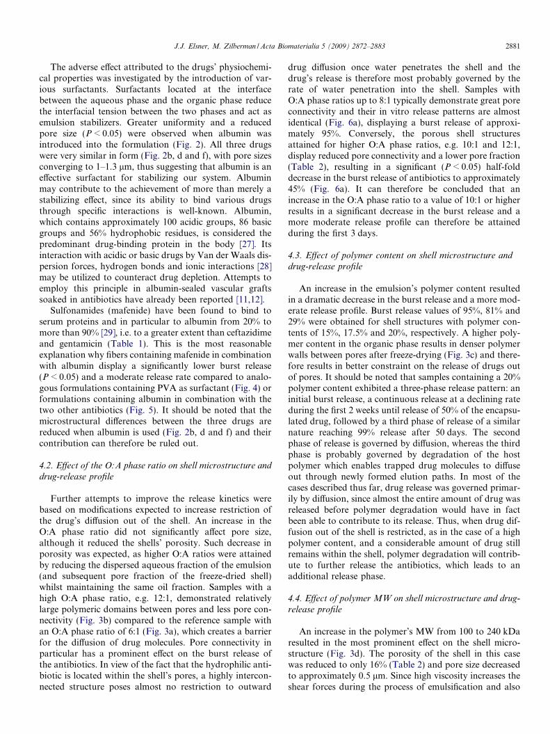
Further attempts to improve the release kinetics werebased on modifications expected to increase restriction ofthe drug’s diffusion out of the shell. An increase in theO:A phase ratio did not significantly affect pore size,although it reduced the shells’ porosity. Such decrease inporosity was expected, as higher O:A ratios were attainedby reducing the dispersed aqueous fraction of the emulsion(and subsequent pore fraction of the freeze-dried shell)whilst maintaining the same oil fraction. Samples with ahigh O:A phase ratio, e.g. 12:1, demonstrated relativelylarge polymeric domains between pores and less pore con-nectivity (Fig. 3b) compared to the reference sample withan O:A phase ratio of 6:1 (Fig. 3a), which creates a barrierfor the diffusion of drug molecules. Pore connectivity inparticular has a prominent effect on the burst release ofthe antibiotics. In view of the fact that the hydrophilic anti-biotic is located within the shell’s pores, a highly intercon-nected structure poses almost no restriction to outward
drug diffusion once water penetrates the shell and thedrug’s release is therefore most probably governed by therate of water penetration into the shell. Samples withO:A phase ratios up to 8:1 typically demonstrate great poreconnectivity and their in vitro release patterns are almostidentical (Fig. 6a), displaying a burst release of approxi-mately 95%. Conversely, the porous shell structuresattained for higher O:A phase ratios, e.g. 10:1 and 12:1,display reduced pore connectivity and a lower pore fraction(Table 2), resulting in a significant (P < 0.05) half-folddecrease in the burst release of antibiotics to approximately45% (Fig. 6a). It can therefore be concluded that anincrease in the O:A phase ratio to a value of 10:1 or higherresults in a significant decrease in the burst release and amore moderate release profile can therefore be attainedduring the first 3 days.
4.3. Effect of polymer content on shell microstructure and
drug-release profile
An increase in the emulsion’s polymer content resultedin a dramatic decrease in the burst release and a more mod-erate release profile. Burst release values of 95%, 81% and29% were obtained for shell structures with polymer con-tents of 15%, 17.5% and 20%, respectively. A higher poly-mer content in the organic phase results in denser polymerwalls between pores after freeze-drying (Fig. 3c) and there-fore results in better constraint on the release of drugs outof pores. It should be noted that samples containing a 20%polymer content exhibited a three-phase release pattern: aninitial burst release, a continuous release at a declining rateduring the first 2 weeks until release of 50% of the encapsu-lated drug, followed by a third phase of release of a similarnature reaching 99% release after 50 days. The secondphase of release is governed by diffusion, whereas the thirdphase is probably governed by degradation of the hostpolymer which enables trapped drug molecules to diffuseout through newly formed elution paths. In most of thecases described thus far, drug release was governed primar-ily by diffusion, since almost the entire amount of drug wasreleased before polymer degradation would have in factbeen able to contribute to its release. Thus, when drug dif-fusion out of the shell is restricted, as in the case of a highpolymer content, and a considerable amount of drug stillremains within the shell, polymer degradation will contrib-ute to further release the antibiotics, which leads to anadditional release phase.
4.4. Effect of polymer MW on shell microstructure and drug-
release profile
An increase in the polymer’s MW from 100 to 240 kDaresulted in the most prominent effect on the shell micro-structure (Fig. 3d). The porosity of the shell in this casewas reduced to only 16% (Table 2) and pore size decreasedto approximately 0.5 lm. Since high viscosity increases theshear forces during the process of emulsification and also

2882 J.J. Elsner, M. Zilberman / Acta Biomaterialia 5 (2009) 2872–2883
reduces the tendency of droplets to move, it is expressed ina finer shell microstructure with a smaller pore size.Although an increase in viscosity can be achieved by anincrease in the polymers’ content as described above, itseffect in this case is more evident. These changes in micro-structure reduced the burst release of the trapped antibiot-ics to approximately 30% and enabled a continuous releaseover a period of 1 month.
In the current study, it was demonstrated that relativelyhigh O:A ratios, high polymer contents and high MWs hada beneficial effect on the release profiles by reducing theburst release and extending the release span (Fig. 5). Thesechanges exhibited release profiles suitable for antibioticrelease systems, i.e. with a considerable initial release thatwill combat the elevated bacterial concentrations duringthe initial shock, followed by a long-term relatively lowrelease rate. It is important to report that similar effectsof O:A phase ratio, polymer content and MW were alsoobtained for core–shell fiber structures loaded with theother two drugs, gentamicin and mafenide. We chose todemonstrate these typical effects on ceftazidime-loaded sys-tems. It should be noted that the release of all three antibi-otics from fibers equivalent to a 1 cm2 square dressingsuccessfully addresses the need to exceed the minimuminhibitory concentration (MIC) of the relevant bacteria(S. epidermidis and P. aeruginosa) within 6 h of immersionin PBS.
4.5. Tensile mechanical properties
MaxonTM sutures were chosen as carrying fibers as theycombine relatively high tensile strength with good flexibil-ity. Furthermore, their degradation rate is lower than thatof the porous PDLGA coating. The mechanical propertieswe report for the MaxonTM sutures are in good agreementwith previous studies [32].
The stress–strain curve of the surface-treated suturesclosely followed that of the original fiber (Fig. 8), whereasthe treated fibers failed at a lower stress (503 MPa) com-pared to the original fibers (771 MPa), probably due tocracks and imperfections caused at the fibers’ surface as aresult of the treatment. This is still acceptable. The deteri-oration in mechanical properties can, however, be avoidedin the future if a carrying fiber is extruded specifically forthis application instead of using a commercial suture mate-rial which requires surface treatment. The results of themechanical testing of the coated fibers demonstrate thatthe process of fiber coating, which includes exposure tothe emulsion, quenching in liquid nitrogen and freeze-dry-ing, results in an actual decrease in tensile strength andmodulus (Table 3). Nevertheless, our composite fibersloaded with 5% drug exhibited a tensile strength of487 MPa. The insignificant decrease in tensile strength,compared to the treated fiber (Table 3), shows that the fiberremains strong and still possesses superior mechanicalproperties compared to monolithic or reservoir fibers.For example, the recently reported monolithic poly(capro-
lactone) fibers containing 5–18% gentamicin exhibited ten-sile strengths of 4.2–7.4 MPa [7], a decrease of 4–45%compared to the unloaded fiber. Our antibiotic-elutingcore–shell fiber structures can therefore in addition towound dressings also be used for other biomedical applica-tions which require good initial mechanical properties, e.g.as meshes for hernia repair.
5. Conclusions
New antibiotic-eluting bioresorbable core–shell fiberstructures were developed and studied. These structureswere composed of a polyglyconate core and a porousPDLGA shell loaded with the antibiotics mafenide acetate,ceftazidime and gentamicin. These structures are designedfor use as basic elements of drug-eluting wound dressings.The shell structures were prepared using the technique offreeze-drying an inverted emulsion. This technique isadvantageous because it results in a combination of goodmechanical properties with the desired drug-release profileand preservation of the drug’s activity. The fiber investiga-tion focused on the effects of the emulsion’s composition(formulation) on the shell microstructure and on the result-ing drug-release profile from the fibers. The mechanicalproperties were also studied. In general, porous ‘‘shell”structures (mean porosities of 16–82% and mean pore sizesof 0.5–5 lm) were obtained with good adhesion to the corefiber. Albumin was found to be an effective surfactant forstabilizing the inverted emulsions and to have a beneficialholdup effect on the release of hydrophilic antibiotic drugs,especially mafenide, probably through specific interactions.The release profiles commonly exhibited an initial bursteffect accompanied by a decrease in release rates with timeover periods ranging from several days to 50 days, depend-ing on the formulation. Higher O:A ratios, polymer con-tents and MWs reduced the burst release of antibioticsfrom the fibers and prolonged their release due to changesin the shell structure. A higher MW and polymer contentdemonstrated a larger effect on the microstructure andrelease profile than the O:A phase ratio. In practice, awound dressing can be woven from a combination of sev-eral types of fibers to create a resultant release profile whichis the product of several release profiles or drug types. Thediverse profiles achieved in this study with higher and lowerburst release rates and with varying elution spans mayserve as a good basis for further in vivo examination ofthe fibers in order to create the ideal profile for a particularwound healing application.
Acknowledgments
We would like to thank Ms. Moran Shohat, Tel-AvivUniversity, for her assistance with the drug-release studies.The authors are grateful to the Israel Science Foundation(ISF, Grant No. 1312/07), the Israel Ministry of Health(Grant number 3-3943) and to the Ela Kodesz foundation,Tel-Aviv University, for supporting this research.

J.J. Elsner, M. Zilberman / Acta Biomaterialia 5 (2009) 2872–2883 2883
References
[1] Boateng JS, Matthews KH, Stevens HN, Eccleston GM. Woundhealing dressings and drug delivery systems: a review. J Pharm Sci2007;97:2892.
[2] Martineau L, Shek PN. Evaluation of a bi-layer wound dressing forburn care: I. Cooling and wound healing properties. Burns2006;32:70.
[3] Jones I, Currie L, Martin R. A guide to biological skin substitutes. BrJ Plast Surg 2002;55:185.
[4] Jurgens C, Schulz AP, Porte T, Faschingbauer M, Seide K.Biodegradable films in trauma and orthopedic surgery. Eur J Trauma2006;2:160.
[5] Knill CJ, Kennedy JF, Mistry J, Miraftab M, Smart G, GroocockMR, et al. Alginate fibres modified with unhydrolysed and hydrolysedchitosans for wound dressings. Carbohydr Polym 2004;55:65.
[6] Ignatova M, Starbova K, Markova N, Manolova N, Rashkov I.Electrospun nano-fibre mats with antibacterial properties fromquaternised chitosan and poly(vinyl alcohol). Carbohydr Res2006;341:2098.
[7] Chang HI, Lau YC, Yan C, Coombes AG. Controlled release of anantibiotic, gentamicin sulphate, from gravity spun polycaprolactonefibers. J Biomed Mater Res A 2008;84:230.
[8] Ruszczak Z, Friess W. Collagen as a carrier for on-site delivery ofantibacterial drugs. Adv Drug Deliv Rev 2003;55:1679.
[9] Southard GL, Godowski KC. Subgingival controlled release ofantimicrobial agents in the treatment of periodontal disease. Int JAntimicrob Agents 1998;9:239.
[10] Steinberg D, Friedman M, Soskolne A, Sela MN. A new degradablecontrolled release device for treatment of periodontal disease: in vitrorelease study. J Periodontol 1990;61:393.
[11] Galdbart JO, Branger C, Andreassian B, Lambert-Zechovsky N,Kitzis M. Elution of six antibiotics bonded to polyethylene vasculargrafts sealed with three proteins. J Surg Res 1996;66:174.
[12] Strachan CJ, Newsom SW, Ashton TR. The clinical use of anantibiotic-bonded graft. Eur J Vasc Surg 1991;5:627.
[13] Wu P, Grainger DW. Drug/device combinations for local drugtherapies and infection prophylaxis. Biomaterials 2006;27:2450.
[14] Roenigk RK, Roenigk HH. Dermatologic surgery: principles andpractice. New York: Marcel Dekker; 1989.
[15] Krasko MY, Golenser J, Nyska A, Nyska M, Brin YS, Domb AJ.Gentamicin extended release from an injectable polymeric implant. JControl Release 2007;117:90.
[16] Blanchemain N, Haulon S, Martel B, Traisnel M, Morcellet M,Hildebrand HF. Vascular PET prostheses surface modification withcyclodextrin coating: development of a new drug delivery system. EurJ Vasc Endovasc Surg 2005;29:628.
[17] Sripriya R, Kumar MS, Sehgal PK. Improved collagen bilayerdressing for the controlled release of drugs. J Biomed Mater Res2004;70:389.
[18] van de Weert M, Hoechstetter J, Hennink WE, Crommelin DJ. Theeffect of a water/organic solvent interface on the structural stability oflysozyme. J Control Release 2000;68:351.
[19] Bouissou C, Rouse JJ, Price R, van der Walle CF. The influence ofsurfactant on PLGA microsphere glass transition, water sorption:remodeling the surface morphology to attenuate the burst release.Pharm Res 2006;23:1295.
[20] Stefanides Sr MM, Copeland CE, Kominos SD, Yee RB. In vitropenetration of topical antiseptics through eschar of burn patients.Ann Surg 1976;183:358.
[21] Noronha C, Almeida A. Local burn treatment—topical antimicrobialagents. Ann Burns Fire Disasters 2000;XIII:4.
[22] Mi FL, Shyu SS, Wu YB, Lee ST, Shyong JY, Huang RN.Fabrication and characterization of a sponge-like asymmetric chito-san membrane as a wound dressing. Biomaterials 2001;22:165.
[23] Becher P. Encyclopedia of emulsion technology. New York: MarcelDekker; 1985.
[24] Bibette J, Calderon FL, Poulin P. Emulsions: basic principles. RepProg Phys 1999;62:64.
[25] Sjoblom J. Emulsions and emulsion stability. New York: MarcelDekker; 1996.
[26] Abraham AM, Walubo A. The effect of surface charge on thedisposition of liposome-encapsulated gentamicin to the rat liver,brain, lungs and kidneys after intraperitoneal administration. Int JAntimicrob Agents 2005;25:392.
[27] Metz P, Kohlhepp SJ, Gilbert DN. Study of different off-line sampleprocessing procedures and the measurement of antibiotic andantiviral levels in human serum by high-performance liquid chroma-tography. J Chromatogr 2002;773:159.
[28] Foye W. Principles of medicinal chemistry. 3rd ed. Philadelphia,PA: Lea and Febiger; 1998.
[29] Hall JB, Schmidt GA, Wood LDH, editorsPrinciples of criticalcare. New York: McGraw-Hill; 2005.
[30] Budavari S. The Merck index: an encyclopedia of chemicals, drugs,and biologicals. Rahway, NJ: Merck & Co.; 1989.
[31] Wishart DS, Knox C, Guo AC, Shrivastava S, Hassanali M, StothardP, et al. DrugBank: a comprehensive resource for in silico drugdiscovery and exploration. Nucleic Acids Res 2006;34(Databaseissue):D668–72.
[32] Kangas J, Paasimaa S, Makela P, Leppilahti J, Tormala P, WarisT, et al. Comparison of strength properties of poly-L/D-lactide(PLDLA) 96/4 and polyglyconate (Maxon) sutures: in vitro, in thesubcutis, and in the achilles tendon of rabbits. J Biomed Mater Res2001;58:121.