Research Student Development Programme and the ePortfolio Dr. Richy Hetherington.
Upload
jonah-shortCategory
view
219download
0
Poster Presentation
Anthony V MoormanRoy RussellRichy HetheringtonElizabeta Mukaetova-Ladinska


Overview of Session
• Interactive section▫ Review posters in 9 groups of 5 people; focussing on different
elements of the posters around the room (15-20min) Identify 3-4 key issues with respect to the element in question
▫ Short group presentation (2-3 minutes per group)• Talk (45-50mins)
▫ Review communication modes in science▫ Advantages and disadvantages of posters▫ Features of a good poster▫ Tips and hints
• Questions

Look at posters critically and consider one of these elements:
• Impact Is the poster eye-catching?
Is it tidy? Is it easy to follow the sequence?
• Title Short, punchy, informative?
• Identity Is it clear who the presenter is?
Is there "Corporate Visual Identity"?
• Readability Are print size, font etc. appropriate?
• Background Is introduction informative to all readers?
• Aims/Objectives Are Aims clearly stated?
• Methods Are these appropriately presented?
• Results Have table, graphs and figures been used well?
Is it easy for the reader to understand data?
• Conclusions How are they presented? Do they match aims?

Modes of Communication in Science
• Written (i.e. paper / article) – good for detail; time to digest; possible to include complex material Clear beginning, middle and end; Reader can combine with other sources of information to clarify anything that is not clear.
• Oral/Spoken/Verbal – good for explaining; can get feedback from listener(s)Limited attention span but possible to repeat or explain technical jargon appropriate to audience.
• Visual/Poster – initial impact; memorable imageLittle or no personal contact; limited viewing time; competing for attention.

When are posters used?
•Meetings/Conferences▫General, Specialist, Clinical▫Large, Small▫National, International
•Open days ▫Entertaining fundraisers▫Attracting staff, students▫ Informing general public
•Site Visits / Visits from Funding Bodies•Corridor decoration•Coursework

Pros and Cons of the Spoken Presentation
PROS Large audience High visibility of speaker Opportunity to stress specific points Easier to prepare Last-minute changes possible (not always an
advantage) Slides reusable
CONS Nerve-wracking Not all audience interested Often few questions/little discussion

Pros and Cons of the Poster PresentationPROS
Good for certain kinds of data Attracts interested viewers Opportunity for discussion (2 way) Good if you are a nervous speaker Good if English is not your first language Displayed all day / whole conference – more
exposureCONS
Time consuming to make (?) Cost May miss viewing parallel posters (?) Not as glamorous as talks

Poster Sessions What is in it for the viewer?
•Can be selective•Opportunity to meet presenter
- ask detailed questions- ask simple questions- chance to mention your own interests
•Can eavesdrop / join in discussions •Time to think about content•Can take notes, get methods, references

A good poster will rapidly allow the reader to answer the questions:
• What is this about? Title, picture(s)• What are they doing? Aims• What is the bottom line ? Conclusion
• How did they do it? Methods, Results• Who are these guys? Names, Presenter • How can I find out more? Contact details,
references
Grab attention | Give information | Memorable message

Poster Style – things to consider
•Visual impact•Title•Layout•Readability
▫Use bullet points

Title
• Possibly the most important aspect of the poster.
• First thing viewers will read.
• Short but needs to capture the essence of the poster
• Choose your wording carefully and remember that the
more words you have, the more space they will take up.

An Investigation Into The Contributory Factors That Improve The Visual Presentation Of Scientific Results In A Conference Scenario.
An Investigation Into The Contributory Factors That Improve The Visual Presentation Of Scientific Results In A Conference Scenario.AN INVESTIGATION INTO THE CONTRIBUTORY FACTORS THAT IMPROVE THE VISUAL PRESENTATION OF SCIENTIFIC RESULTS
IN A CONFERENCE SCENARIO.
An investigation into the contributory factors that improve the visual presentation of scientific results in a conference scenario

An investigation into the contributory factors that improve the visual presentation of scientific results in a conference scenario
Factors that improve visual presentation of scientific results
(note how you can now make the text bigger)
Remove redundant words

Factors that improve visual presentation
FACTORS THAT IMPROVE VISUAL PRESENTATION
The case for lower-case

0
2
4
6
8
10
12
2 8 14
20
26
32
38
44
50
Re
ad
ab
ility
Font size
Relationship between size of font used in a figure and the ease with which it can be read at a distance of 1 m assessed using a ten-point scale (readability )
Size matters

Poster Layout•Landscape better than portrait
3 columns – reduces line length

Know your audience
•Expert, semi-expert, patients, fund-raisers, general public
•THINK about content, text and terminology▫Avoid acronyms – even ones that seems
obvious to you▫No jargon / Do not assume knowledge▫Always ask yourself - do they need to know
this piece of information in order to understand my message?

Tables – keep them simple!

Tables – Focus on the key data.
Abnormality5 year OS(95% CI)
Hazard Ratio(95% CI), p value
Total 44% (39-48%) -IKZF1 35% (24-46%) 1.54 (1.11,2.15), p=0.010
PAX5 56% (41-69%) 0.68 (0.43,1.07), p=0.097
ETV6 57% (37-72%) 0.61 (0.34,1.07), p=0.085
EBF1 14% (1-46%) 2.53 (1.11,5.73), p=0.027

Keep figures simple too
Figure 3. Distribution of the percentage of positive nuclei in patients with recurrent
IGH@ partner genesFigure 2. Percentage of positive nuclei scored in patients with recurrent IGH@ translocations.When comparing the percentages of positive nuclei observed in patients with recurrent IGH@ translocations (known or unknown partner genes), the involvement of certain partner genes was present in the majority of cells: BCL2, CEBPA, CEBPB and CRLF2. Together patients with involvement of these genes had a median translocation population of 79%. This was reduced in patients with ID4 involvement to 65% and further reduced to 30% in patients with IGH@-CEBPE. Patients with IGH@-CEBPD translocations tended to have a wind range of translocation positive nuclei with some as low as 21% and one at 100%.Boxes denote the range of positive nuclei scored, with extending thin black lines marking outliers. The thick black line within the boxes denotes the median percentage of positive nuclei scored per partner gene.
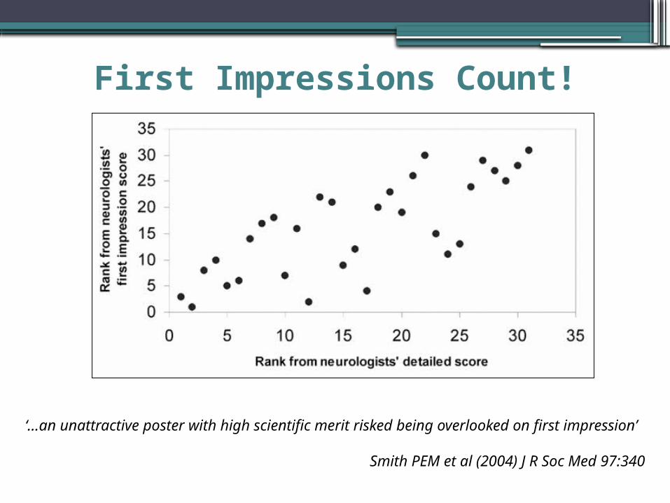
‘…an unattractive poster with high scientific merit risked being overlooked on first impression’
Smith PEM et al (2004) J R Soc Med 97:340
First Impressions Count!

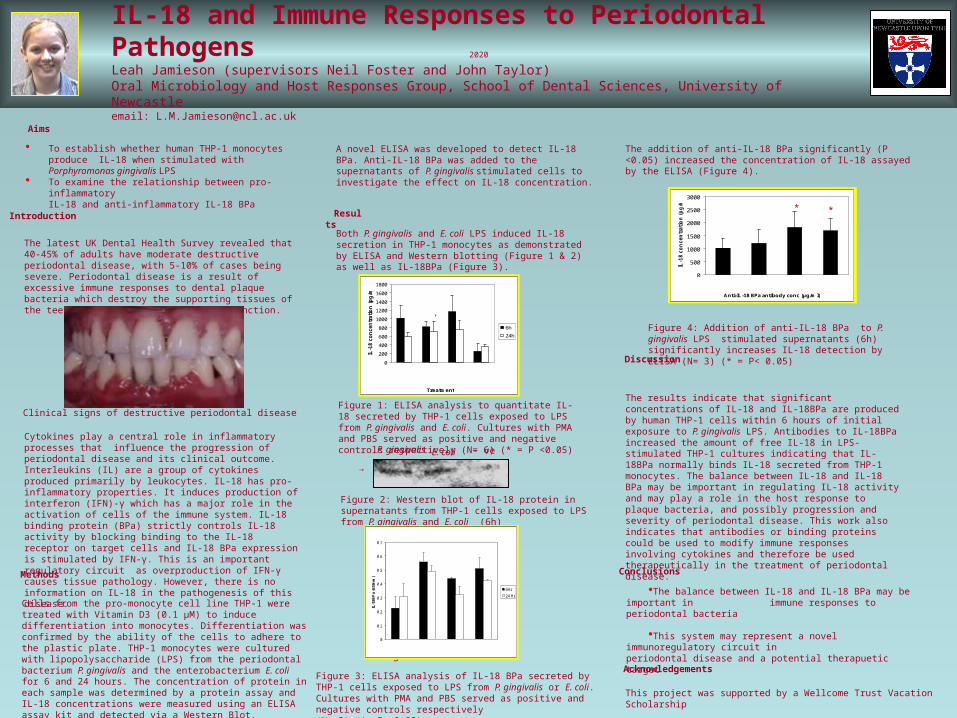
The results indicate that significant concentrations of IL-18 and IL-18BPa are produced by human THP-1 cells within 6 hours of initial exposure to P. gingivalis LPS. Antibodies to IL-18BPa increased the amount of free IL-18 in LPS-stimulated THP-1 cultures indicating that IL-18BPa normally binds IL-18 secreted from THP-1 monocytes. The balance between IL-18 and IL-18 BPa may be important in regulating IL-18 activity and may play a role in the host response to plaque bacteria, and possibly progression and severity of periodontal disease. This work also indicates that antibodies or binding proteins could be used to modify immune responses involving cytokines and therefore be used therapeutically in the treatment of periodontal disease.
2020IL-18 and Immune Responses to Periodontal PathogensLeah Jamieson (supervisors Neil Foster and John Taylor)Oral Microbiology and Host Responses Group, School of Dental Sciences, University of Newcastleemail: [email protected]
0
200
400
600
800
1000
1200
1400
1600
1800
Treatment
IL-1
8 c
on
cen
trati
on
(p
g/m
l)
6h
24h
Pg Ec PMA Unstimulated control
* *
* * *
*
0
500
1000
1500
2000
2500
3000
Anti-IL-18 BPa antibody conc (μg/ml)
IL-1
8 c
on
cen
trati
on
(p
g/m
l)
0 0.15 1.5 15
• To establish whether human THP-1 monocytes produce IL-18 when stimulated with Porphyromonas gingivalis LPS
• To examine the relationship between pro-inflammatory IL-18 and anti-inflammatory IL-18 BPa
Discussion
Conclusions
Acknowledgements
•The balance between IL-18 and IL-18 BPa may be important in immune responses to periodontal bacteria
•This system may represent a novel immunoregulatory circuit in periodontal disease and a potential therapuetic
target
Introduction
Aims
Methods
Results
This project was supported by a Wellcome Trust Vacation Scholarship
The latest UK Dental Health Survey revealed that 40-45% of adults have moderate destructive periodontal disease, with 5-10% of cases being severe. Periodontal disease is a result of excessive immune responses to dental plaque bacteria which destroy the supporting tissues of the teeth, leading to loss of dental function.
Cytokines play a central role in inflammatory processes that influence the progression of periodontal disease and its clinical outcome. Interleukins (IL) are a group of cytokines produced primarily by leukocytes. IL-18 has pro-inflammatory properties. It induces production of interferon (IFN)-γ which has a major role in the activation of cells of the immune system. IL-18 binding protein (BPa) strictly controls IL-18 activity by blocking binding to the IL-18 receptor on target cells and IL-18 BPa expression is stimulated by IFN-γ. This is an important regulatory circuit as overproduction of IFN-γ causes tissue pathology. However, there is no information on IL-18 in the pathogenesis of this disease.
Figure 1: ELISA analysis to quantitate IL-18 secreted by THP-1 cells exposed to LPS from P. gingivalis and E. coli. Cultures with PMA and PBS served as positive and negative controls respectively (N= 6) (* = P <0.05)
P. gingivalis
E. coli - ve
→
Figure 2: Western blot of IL-18 protein in supernatants from THP-1 cells exposed to LPS from P. gingivalis and E. coli (6h)
Figure 3: ELISA analysis of IL-18 BPa secreted by THP-1 cells exposed to LPS from P. gingivalis or E. coli. Cultures with PMA and PBS served as positive and negative controls respectively (N =3) (*= P< 0.05)
Figure 4: Addition of anti-IL-18 BPa to P. gingivalis LPS stimulated supernatants (6h) significantly increases IL-18 detection by ELISA (N= 3) (* = P< 0.05)
Cells from the pro-monocyte cell line THP-1 were treated with Vitamin D3 (0.1 μM) to induce differentiation into monocytes. Differentiation was confirmed by the ability of the cells to adhere to the plastic plate. THP-1 monocytes were cultured with lipopolysaccharide (LPS) from the periodontal bacterium P. gingivalis and the enterobacterium E. coli for 6 and 24 hours. The concentration of protein in each sample was determined by a protein assay and IL-18 concentrations were measured using an ELISA assay kit and detected via a Western Blot.
* *
A novel ELISA was developed to detect IL-18 BPa. Anti-IL-18 BPa was added to the supernatants of P. gingivalis stimulated cells to investigate the effect on IL-18 concentration.
Both P. gingivalis and E. coli LPS induced IL-18 secretion in THP-1 monocytes as demonstrated by ELISA and Western blotting (Figure 1 & 2) as well as IL-18BPa (Figure 3).
The addition of anti-IL-18 BPa significantly (P <0.05) increased the concentration of IL-18 assayed by the ELISA (Figure 4).
Clinical signs of destructive periodontal disease
0
0.1
0.2
0.3
0.4
0.5
0.6
0.7
IL-1
8BP
a (6
20n
m)
6Hr
24 Hr
PBS PMAPg Ec
*
**
*
*
The results indicate that significant concentrations of IL-18 and IL-18BPa are produced by human THP-1 cells within 6 hours of initial exposure to P. gingivalis LPS. Antibodies to IL-18BPa increased the amount of free IL-18 in LPS-stimulated THP-1 cultures indicating that IL-18BPa normally binds IL-18 secreted from THP-1 monocytes. The balance between IL-18 and IL-18 BPa may be important in regulating IL-18 activity and may play a role in the host response to plaque bacteria, and possibly progression and severity of periodontal disease. This work also indicates that antibodies or binding proteins could be used to modify immune responses involving cytokines and therefore be used therapeutically in the treatment of periodontal disease.
2020
IL-18 and Immune Responses to Periodontal PathogensLeah Jamieson (supervisors Neil Foster and John Taylor)Oral Microbiology and Host Responses Group, School of Dental Sciences, University of Newcastleemail: [email protected]
0
200
400
600
800
1000
1200
1400
1600
1800
Treatment
IL-1
8 c
on
cen
trati
on
(p
g/m
l)
6h
24h
Pg Ec PMA Unstimulated control
* *
* * *
*
0
500
1000
1500
2000
2500
3000
Anti-IL-18 BPa antibody conc (μg/ml)
IL-1
8 c
on
cen
trati
on
(p
g/m
l)
0 0.15 1.5 15
• To establish whether human THP-1 monocytes produce IL-18 when stimulated with Porphyromonas gingivalis LPS
• To examine the relationship between pro-inflammatory IL-18 and anti-inflammatory IL-18 BPa
Discussion
Conclusions
Acknowledgements
•The balance between IL-18 and IL-18 BPa may be important in immune responses to periodontal bacteria
•This system may represent a novel immunoregulatory circuit in periodontal disease and a potential therapuetic target
Introduction
Aims
Methods
Results
This project was supported by a Wellcome Trust Vacation Scholarship
The latest UK Dental Health Survey revealed that 40-45% of adults have moderate destructive periodontal disease, with 5-10% of cases being severe. Periodontal disease is a result of excessive immune responses to dental plaque bacteria which destroy the supporting tissues of the teeth, leading to loss of dental function.
Cytokines play a central role in inflammatory processes that influence the progression of periodontal disease and its clinical outcome. Interleukins (IL) are a group of cytokines produced primarily by leukocytes. IL-18 has pro-inflammatory properties. It induces production of interferon (IFN)-γ which has a major role in the activation of cells of the immune system. IL-18 binding protein (BPa) strictly controls IL-18 activity by blocking binding to the IL-18 receptor on target cells and IL-18 BPa expression is stimulated by IFN-γ. This is an important regulatory circuit as overproduction of IFN-γ causes tissue pathology. However, there is no information on IL-18 in the pathogenesis of this disease.
Figure 1: ELISA analysis to quantitate IL-18 secreted by THP-1 cells exposed to LPS from P. gingivalis and E. coli. Cultures with PMA and PBS served as positive and negative controls respectively (N= 6) (* = P <0.05) P.
gingivalisE. coli - ve
→
Figure 2: Western blot of IL-18 protein in supernatants from THP-1 cells exposed to LPS from P. gingivalis and E. coli (6h)
Figure 3: ELISA analysis of IL-18 BPa secreted by THP-1 cells exposed to LPS from P. gingivalis or E. coli. Cultures with PMA and PBS served as positive and negative controls respectively (N =3) (*= P< 0.05)
Figure 4: Addition of anti-IL-18 BPa to P. gingivalis LPS stimulated supernatants (6h) significantly increases IL-18 detection by ELISA (N= 3) (* = P< 0.05)
Cells from the pro-monocyte cell line THP-1 were treated with Vitamin D3 (0.1 μM) to induce differentiation into monocytes. Differentiation was confirmed by the ability of the cells to adhere to the plastic plate. THP-1 monocytes were cultured with lipopolysaccharide (LPS) from the periodontal bacterium P. gingivalis and the enterobacterium E. coli for 6 and 24 hours. The concentration of protein in each sample was determined by a protein assay and IL-18 concentrations were measured using an ELISA assay kit and detected via a Western Blot.
* *
A novel ELISA was developed to detect IL-18 BPa. Anti-IL-18 BPa was added to the supernatants of P. gingivalis stimulated cells to investigate the effect on IL-18 concentration.
Both P. gingivalis and E. coli LPS induced IL-18 secretion in THP-1 monocytes as demonstrated by ELISA and Western blotting (Figure 1 & 2) as well as IL-18BPa (Figure 3).
The addition of anti-IL-18 BPa significantly (P <0.05) increased the concentration of IL-18 assayed by the ELISA (Figure 4).
Clinical signs of destructive periodontal disease
0
0.1
0.2
0.3
0.4
0.5
0.6
0.7
IL-1
8BP
a (6
20n
m)
6Hr
24 Hr
PBS
PMA
Pg Ec
*
**
*
*

The results indicate that significant concentrations of IL-18 and IL-18BPa are produced by human THP-1 cells within 6 hours of initial exposure to P. gingivalis LPS. Antibodies to IL-18BPa increased the amount of free IL-18 in LPS-stimulated THP-1 cultures indicating that IL-18BPa normally binds IL-18 secreted from THP-1 monocytes. The balance between IL-18 and IL-18 BPa may be important in regulating IL-18 activity and may play a role in the host response to plaque bacteria, and possibly progression and severity of periodontal disease. This work also indicates that antibodies or binding proteins could be used to modify immune responses involving cytokines and therefore be used therapeutically in the treatment of periodontal disease.
2020IL-18 and Immune Responses to Periodontal PathogensLeah Jamieson (supervisors Neil Foster and John Taylor)Oral Microbiology and Host Responses Group, School of Dental Sciences, University of Newcastleemail: [email protected]
0
200
400
600
800
1000
1200
1400
1600
1800
Treatment
IL-1
8 c
on
cen
trati
on
(p
g/m
l)
6h
24h
Pg Ec PMA Unstimulated control
* *
* * *
*
0
500
1000
1500
2000
2500
3000
Anti-IL-18 BPa antibody conc (μg/ml)
IL-1
8 c
on
cen
trati
on
(p
g/m
l)
0 0.15 1.5 15
• To establish whether human THP-1 monocytes produce IL-18 when stimulated with Porphyromonas gingivalis LPS
• To examine the relationship between pro-inflammatory IL-18 and anti-inflammatory IL-18 BPa
Discussion
Conclusions
Acknowledgements
•The balance between IL-18 and IL-18 BPa may be important in immune responses to periodontal bacteria
•This system may represent a novel immunoregulatory circuit in periodontal disease and a potential therapuetic
target
Introduction
Aims
Methods
Results
This project was supported by a Wellcome Trust Vacation Scholarship
The latest UK Dental Health Survey revealed that 40-45% of adults have moderate destructive periodontal disease, with 5-10% of cases being severe. Periodontal disease is a result of excessive immune responses to dental plaque bacteria which destroy the supporting tissues of the teeth, leading to loss of dental function.
Cytokines play a central role in inflammatory processes that influence the progression of periodontal disease and its clinical outcome. Interleukins (IL) are a group of cytokines produced primarily by leukocytes. IL-18 has pro-inflammatory properties. It induces production of interferon (IFN)-γ which has a major role in the activation of cells of the immune system. IL-18 binding protein (BPa) strictly controls IL-18 activity by blocking binding to the IL-18 receptor on target cells and IL-18 BPa expression is stimulated by IFN-γ. This is an important regulatory circuit as overproduction of IFN-γ causes tissue pathology. However, there is no information on IL-18 in the pathogenesis of this disease.
Figure 1: ELISA analysis to quantitate IL-18 secreted by THP-1 cells exposed to LPS from P. gingivalis and E. coli. Cultures with PMA and PBS served as positive and negative controls respectively (N= 6) (* = P <0.05)
P. gingivalis
E. coli - ve
→
Figure 2: Western blot of IL-18 protein in supernatants from THP-1 cells exposed to LPS from P. gingivalis and E. coli (6h)
Figure 3: ELISA analysis of IL-18 BPa secreted by THP-1 cells exposed to LPS from P. gingivalis or E. coli. Cultures with PMA and PBS served as positive and negative controls respectively (N =3) (*= P< 0.05)
Figure 4: Addition of anti-IL-18 BPa to P. gingivalis LPS stimulated supernatants (6h) significantly increases IL-18 detection by ELISA (N= 3) (* = P< 0.05)
Cells from the pro-monocyte cell line THP-1 were treated with Vitamin D3 (0.1 μM) to induce differentiation into monocytes. Differentiation was confirmed by the ability of the cells to adhere to the plastic plate. THP-1 monocytes were cultured with lipopolysaccharide (LPS) from the periodontal bacterium P. gingivalis and the enterobacterium E. coli for 6 and 24 hours. The concentration of protein in each sample was determined by a protein assay and IL-18 concentrations were measured using an ELISA assay kit and detected via a Western Blot.
* *
A novel ELISA was developed to detect IL-18 BPa. Anti-IL-18 BPa was added to the supernatants of P. gingivalis stimulated cells to investigate the effect on IL-18 concentration.
Both P. gingivalis and E. coli LPS induced IL-18 secretion in THP-1 monocytes as demonstrated by ELISA and Western blotting (Figure 1 & 2) as well as IL-18BPa (Figure 3).
The addition of anti-IL-18 BPa significantly (P <0.05) increased the concentration of IL-18 assayed by the ELISA (Figure 4).
Clinical signs of destructive periodontal disease
0
0.1
0.2
0.3
0.4
0.5
0.6
0.7
IL-1
8BP
a (6
20n
m)
6Hr
24 Hr
PBS PMAPg Ec
*
**
*
*

Corporate Visual IdentityNewcastle University Policy and
Regulations
http://www.ncl.ac.uk/cvi-support/
Corporate Visual IdentityNewcastle University Policy and Regulations

Conference layout

Conference layout

400 posters
90 mins
13.5 sec/poster
0.225 min/poster
Look at Me!!!!!


Typical Instructions for Poster Presentations • One poster board will be provided for you. The poster board is
approximately 4 feet high and 8 feet wide.
• We will provide a printed “number,” identifying each poster board. Push pins will also be provided.
• One or two authors should be in attendance at each poster during the entire presentation time.
• Do not allow yourself to be monopolized for an inordinate period of time by a single individual.
• Please remove your materials from the poster board immediately after the session. Materials left on the boards after the session will be discarded.
• The only handouts allowed in the Poster Hall include exact copies of your poster or business cards; all other types of handouts are not allowed in the Poster Hall.

Final Points …
•Why are you doing a poster?- to communicate results- to advertise your work, yourself, your
department
•Be proactive - ▫identify yourself ▫(photo on poster, presenter badge, add
poster number to badge)
•Be remembered - ▫first name on poster, email address, take-
away handouts, reprints