
Annual reproductive cycle of the male pocket gopher...
80
ANNUAL REPRODUCTIVE CYCLE OF THE MALE POCKET GOPHER (Geomys pinetis) By KATHERINE CARTER EWEL A DISSERTATION PRESENTED TO THE GRADUATE COUNCIL OF THE UNIVERSITY OF FLORIDA IN PARTIAL FULFILLMENT OF THE REQUIREMENTS FOR THE DEGREE OF DOCTOR OF PHILOSOPHY UNIVERSITY OF FLORIDA 1970
Transcript of Annual reproductive cycle of the male pocket gopher...

ANNUAL REPRODUCTIVE CYCLE OF THE
MALE POCKET GOPHER (Geomys pinetis)
By
KATHERINE CARTER EWEL
A DISSERTATION PRESENTED TO THE GRADUATE COUNCIL OF
THE UNIVERSITY OF FLORIDA
IN PARTIAL FULFILLMENT OF THE REQUIREMENTS FOR THE
DEGREE OF DOCTOR OF PHILOSOPHY
UNIVERSITY OF FLORIDA
1970

UNIVERSITY OF FLORIDA
3 1262 08552 2950
ACKNOWLEDGMENTS
I am indebted to Drs. David W. Johnston, John H. Kaufmann, E. G.
Franz Sauer, and Daniel B. Ward for their encouragement and helpful
criticism during the course of this project. In addition, Dr. John
Thornby offered considerable advice on the statistical analysis of the
data; Thomas Krakauer assisted me several times in trapping; Robert
NcFarlane gave assistance in the preparation of photographs; Dr. Gordon
K, Prine provided environmental data from the Agronomy Farm Weather
Station; and Dr. John F. Gerber assisted in the interpretation of soil
moisture data.
Permission to trap on Stengel Field was kindly granted by Wr.
A. Arano.
The University of Florida Computing Center provided the facilities
for the statistical analysis
•

TABLE OF CONTENTS
ACKNOiVLEDGMEMTS ii
LIST 01' TABLES iv
LIST OF FIGURES v
A33TRACT vii
INTRODUCTION 1
Pocket Gopher Environment and Physiological Adaptations 2
Effects of EnvironiTient on Activity Cycle 3
Reproductive Cycles in Pocket Gophers 3
The Reproductive Cycle of Geomys pinetis 5
MATERIALS AND METHODS 7
Study Area and Trapping Methods 7
Environmental Data 7
Characteristics of Reproductive Organs 8
Histological Methods 10
Examination of the Testis 11
Examination of the Epididymis 13
Examination of the Accessory Glands 13
Statistical Analysis of the Data 14
RESULTS 17
Noncyclic Changes in Reproductive Organs 17
Cyclic Changes in Reproductive Organs 21
Comparison of Changes in Reproductive Organswi th Environinental Features 37
Immature Animals and Pregnant Females 45
Quarterly Sample 46
DISCUSSION AND CONCLUSIONS 53
SUMMARY 59
LITERATURE CITED 61
APPENDIX 64
BIOGRAPHICAL SKETCH 69

LIST OF TnSLES
Page
1. Meems of measurements from male pocket f:;ophcrs 18
2. Independent factors in measurements from male
pocke t gophers • 19
3. Numbers of pocket gophers with acini at a given stage
of secretory activity in the prostate glands ..* 22
4. Relationsliips botv/een the production of spermand accessory gland activity 23
5. Kui?.ber of males caught in each trapping period ..•• 24
6. F ratios from univariate analyses of variancesiiowing significance of differejices betvieen meansof reproductive parameters in 15 trapping periods ......... 26
7. Distribution, of significant peaJcs arnong the parameters .... 38
B. Results of canonical analysis A3
9. Kean values of environmental paraiaeters over the
specified periods of time before approximateconception dates 48
10. Sex, age, and reproductive status of animals capturedin each quarterly saraple 50

LIST OF FIGURES
Page
1. Criterion for separating mature from u.Tmaturemale pocket gophers , 9
2. Testicular volumes throughout the trapping periods ........ 27
3. DiaiTjeters of the testis tubules throughout thetrapping periods . . ^ , 28
A. Diameters of the Leydig cells throughout thetrapping periods , ,,, , ,, 29
5. Volumes of Leydig cell tissue throughout thetrapping periods 30
6. Values for the stages of spermatogenesisthroughout the trapping periods 32
7. I/iajiieters of the epididjiral tubule throughoutthe trapping periods 33
S, Cell heights in the dorsolateral prostate glandthroughout the trapping periods 34
9. Cell heights in the sorrdaal vesicles throughoutthe trapping periods 35
10. Diameters of the seminal vesicles throughout thetrapping periods 36
11. Monthly rainfall and average raaxirn'om and minirnuri
soil temperatures 39
12. Average monthly soil moisture 40
13. Average soil temperature histories of pocket gophersvjithin each trapping period for two weeks priorto their capture , ..«.........> i 41
14. Average soil moisture 'listories of pocket gophersv/ithin each trapping period for four weeks priorto their capture .» • 42

15. Periods of roproductivr activity ba^od on
all projected conception dates A7
16. Monthly soil temperatvires and rainfallduring quarterly sampling period ^9
17. Periods of reproductive activi ty accordinp, to
projected conception dates in quarterly samples 52
18. Level 8 testis (lOOX) 65
19. Level 3 testis (ASOX) 05
20. Level 7 testis (430X) 05
21. Epididynal tubules from a malein active reproductive condition (inox) o6
22. Epididyrual tubules from inactive male (lOOX) 66
23. Actively secreting acinus from dorsolateralprostate gland (430X) 67
2A. Inactive acinus from dorsolateralprostate gland (430X) 67
25. Luir.en and part of cell boundary from an activeseminal vesicle (lOOX) 68
26. Inactive seminal vesicle (lOOX) 68
vi

Abstract of Dissertation Prcsonted to the Graduate Councilin Partial Fulfillment of the Roquiromonts for the Def.ree of
Doctor of Philosophy
ANNUAL REPRODUCTIVE CYCLE OF THEMALE POCKET GOPHER (Geomys pinetis )
By
Katherine Carter Ewel
June, 1970
Chairman! David H. JohnstonMajor Department: Zoology
An analysis was made of the reproductive cycle of the south-
eastern pocket gopher, Geomys pinetis , to determine both the pattern
of reproduction in this species and the magnitude of environmental
influences on reproduction in a fossorial habitat near Gainesville,
Florida. The primary focus of the investigation was the male repro-
ductive cycle, but gross measurements of the reproductive condition
of the females were also used.
The right testis, its epididjonis, and the proximal accessory
glands (seminal vesicles and prostate glands) of 76 male pocket
gophers collected monthly between March, 1967, and July, 1968, were
sectioned and examined histologically to assess the degree of repro-
ductive activity of each organ. A one-way analysis of variance was
performed on each of the measurements made on the various organs to
determine if a monthly cycle of activity was present. A principal
components analysis was also performed to find out what interrelation-
ships might exist between the parameters. Finally, a canonical
analysis related those parameters which showed cyclical differences

during, the trapping period with correspondinn soil tomperatvire and soil
moisture values to compare the effects of the environmental pr.- "ters
on the reproductive cycle.
During a subsequent twelve-month trapping period, quarterly popula-
tion samples were made in the same study area. Only t;;coss measurements
of the reproductive condition were made on the 60 individuals in this
part of the investif,ation.
The reproductive cycle in the male poc'cet p.opher was found to be
closely correlated witli soil temperature and to a lesser decree v/ith
soil moisture. Maximum reproductive activity occurs v;hen environmental
conditions create a friable soil, suggesting an increased likelihood
of intraspecific contacts. Very warm external temperatures and low
soil moisture inhibit reproductive activity indirectly, presumably by
restricting activity within the burrow.
Leydig cells shov; a net decrease in size and number with age,
after an initial increase at puberty. Increased reproductive activity
is correlated with an increase in Leydig cell diameter. Spermato-
genesis was non-cyclic, but did not cease in the entire population
during the study period. The activity of the accessory glands and
the epididymis served as a more reliable index of an individual's
reproductive condition.
INTRODUCTION
Fossorial rodents are unique among terrestrial mammals in being
isolated from most of the environmental factors that control daily and
annual biological cycles in many other organisms. Typical of these
cycles is reproduction, which may be controlled or influenced in many
mammals by the available food supply, temperature, photoperiod, or
rainfall. In studying reproduction of a fossorial mammal, these ex-
ternal factors must be translated to their effects on the environment
of the burrow. In this investigation, emphasis is given to an analysis
of environmental effects on the reproductive cycle of a fossorial
rodent, the southeastern pocket gopher (Geomys pinetis Rafinesque)
»
This animal spends almost its entire life underground and is
well adapted to a fossorial existence, as indicated by its relatively
broad, flattened skull, small eyes and pinnae, stout forelimbs, and
elongated foreclaws. It feeds chiefly on roots obtained from its
burrow, as well as on occasional leaves pulled into the shallower parts
of the burrow. It is seldom seen above ground, and is exposed to day-
light only during short periods when making fresh mounds. Presiimably,
pocket gophers are influenced more by the environment of the burrow
than by light. Particularly important influences would include tem-
perature changes, gas composition, and soil characteristics*
- 1-

- 2 -
Pocket Gopher Environmont and Physiological Adaptations
Kennerly (1964) found that the temperature within the burrow of
Gcorivs bursarius in central Texas is more constant than environmental
temperatures, and that the range of burrow temperatures becomes narrower
at increased depths. McNab (1966) noted that temperature changes within
burrows of Gj^ pinetis in Florida are also less extreme than outside
temperatures. Because of the temperature stability in the burrow,
Wilks (1963) reasoned that there might be less selection pressure
towards strict homothermy. Poor thermoregulation has indeed been found
to be characteristic of fossorial rodents (Gunther, 1956; Wilks, 1963;
McNab, 1966). McNab (1966) claimed that conductance is facilitated by
a scanty fur coat, an increase in peripheral circulation, and a general
decrease in body size and mean weight in areas of higher soil tempera-
ture. The greater stress of summer temperatures, accentuated by the
burrow's saturated atmosphere (McNab, 1966), might still pose a problem;
the pocket gopher could, however, avoid heat loading by retreating into
the deeper, cooler part of its burrow system. From 10 to 20% of the
burrow systems of Thomomys bottae and G^. bursarius lie well below the
superficial runways, which are within 12 inches of the surface (Miller,
196A).
The oxygen and carbon dioxide concentrations in the burrow system
fluctuate more widely than does temperature. Increased soil moisture
causes the carbon dioxide content, which is initially several times
greater than the atmospheric concentration, to rise even more, probably
due to decreased diffusion through the soil pores (Kennerly, 1964).
As a result, according to measurements made in Florida by McNab (1966),

- 3 -
the carbon dioxide concentration in the burrow reaches as much as
20 to 35 times the atmospheric concentration. The oxygen concentra-
tion remains within 80 to 95Z of the atmospheric concentration.
Effects of Environment on Activity Cycle
Little is known about the influence of soil moisture and soil
temperature on pocket gopher activity. No published data on activity
cycles within the burrow are available, and Vaughan and Hansen (1961)
remarked that the usual indicator of activity — the appearance of
fresh mounds — is really only indicative of surface or near-surface
activity. Trapping experience, however, indicated to these authors
and to Wilks (1963) that soil temperature is an important factor con-
trolling the pocket gopher's daily activity cycle.
Miller (19A8) found that the amount of burrowing (determined by
the appearance of fresh movinds) is primarily controlled by soil moisture,
the greatest activity occurring when the soil was most friable.
Kennerly (1964) came to the same conclusion, but the correlation seemed
weaker to him.
Reproductive Cycles in Pocket Gophers
Reproductive activity in most species of pocket gophers is
limited by their usually solitary behavior, although instances of
multiple captures within one burrow system have been reported for many
species of Geomys and Thomomys (e.g., English, 1932; Vaughan, 1962;
Wilks, 1963; Miller, 1964). Both Vaughan (1962) and Wilks (1963)
observed that these multiple captures were most frequent during the
breeding season and usually involved animals of opposite sexes.

- /l-
Winr, (1^60) listed only one multiple capture in G. pin<=!ti.q , involvinp
an adult male and a post-partum female. She also cited 1*^4 instances
during one year of collecting in which a pocket popher was caught in
one trap when the second trap had been sprunf?;. Thiis, however, could
have been accomplished by one animal's blocking one trap before being
caught in the other. In general, it appears that pocket gophers remain
solitary within their burrows most of the year.
Ecological aspects of reproductive cycles have been studied in
several species of pocket gophers. In east Texas, English (1932)
showed that only one litter per year was characteristic of G. breviceps ,
whereas Wilks (1963) reported at least two litters per year for G.
bursarius in south Texas. Peaks of reproductive activity in G*
bursarius were found in December - January, and April - Kay, with
some activity in July - September. Hilks suggested that temperature,
humidity, and rainfall were interacting to inhibit summer breeding.
_G. bursarius in Colorado produced litters only in the spring (Vaughan,
1962). In this case, the breeding season seemed to be timed so that
the young appeared in late May or June at the peak of vegetative
growth and when the soil was most friable. The adults began to come
into reproductive condition in late winter while the ground was still
frozen to a depth of six inches.
According to Miller (19A6), T. bottae in California has only one
breeding season in the northern, more mountainous part of its range,
whereas the breeding season in the southern part seemed to extend
through part of the winter rainy season to allow two or three litters
per year. Dixon (1929) felt that this cycle was attuned primarily to
the appearance of the pocket gopher's most important food plants at

the bej'.inninr, of the season. He found that in the central San Joaquin
Valley, where alfalfa and other p,reen forage crops were available
throuphout the suinmer, the breeding season was longer. Gunther (1956)
also described the breeding season of this species as restricted to the
cool, wet weather of winter and spring in nonirrigated portions of the
range, but being more protracted in irrigated areas. Bond (19'!t6)
suggested that whereas availability of food might affect the sexual
development of a pocket gopher, contact between the sexes at the op-
timum time for reproduction depended more on the friability of the soil.
Wing (1960) described the breeding cycle of G. pinetis in Florida,
but made no correlations with environmental factors. Pregnant females
were encountered in her collections throughout the year, but they
appeared more frequently in early spring and late summer. Probably
some females had produced at least two litters per year, since double
sets of placental scars occurred in seven females collected between
April and July. Her work included a thorough examination of the females
as well as gross measurements of the male reproductive tracts and epidid-
ymal sperm smears. The males in her samples showed greatest reproduc-
tive activity from January - August, whereas females were most active
in March and July - August.
The Reproductive Cycle of Geomys pinetis
Unlike other species of pocket gophers that inhabit temperate
regions, _G. pinetis does not appear to have a well-defined breeding
cycle that can be easily attributed to environmental effects. Wing
has suggested that breeding continues throughout the year but not at
the same intensity. There are at least four different ways in which

- 6 -
the male reproductive cycle could be operatine Co ccnorate this
pattern I 1.) The males may remain in an active reproductive state
all year, but the females come into breeding condition only at
presumably propitious times. 2.) The males do not remain in an
active state of spermatogenesis all year, but instead store sperm
for a certain period of time after spermatogenesis has ceased, mean-
while retaining a propensity toward mating. 3.) The males may show
fluctuations due to environmental conditions, although never completely
losing the ability to reproduce. A.) Some males may maintain at least
a threshold ability to reproduce whereas others regress significantly;
a well-defined cycle is therefore not maintained in the population.
The purpose of this investigation was to determine which repro-
ductive pattern exists for G. pinetis near Gainesville, Florida, and
to examine in greater depth the influence of environmental factors.

MATERIALS AND METHODS
Study Area and Trappirif^ Methods
At least five male pocket gophers were trapped every month from
March, 1967, through July, 1968, within a five-mile radius of Gaines-
ville, Florida. After July, 1967, all trapping was done at Stenpel
Field, three miles W3W of Gainesville. This field, containing a
fairly large population of pocket gophers, was characterized by Pensa-
cola bahia grass sod over Arredonda fine sand and was mowed fairly
regularly throughout the trapping period. Part of the field was used
as an unpaved air strip. The animals were captured in Victor gopher
traps which were checked every two to three hours to avoid excessive
decay of the tissues in the dead animals.
In addition, samples of 15 pocket gophers, including both sexes,
were trapped in November, 1968, and in February, May, and August, 1969,
to obtain quarterly population samples in Stengel Field.
A total of 106 males and 91 females was obtained for analysis in
the present investigation.
Environmental Data
Cliraatological data were obtained from the Agronomy Farm Weather
Station, an area one mile east of Stengel Field and quite similar to
it in soil characteristics and cover. The temperatures used in the
statistical analyses were soil temperatures at a depth of four inches
- 7 -

and vere averages betveen the daily raaxiina and minima. Estimates of
soil moisture were obtained by corbining weekly estinates of evapo-
transpiration with total weekly rainfall. Field capacity, defined
as the ability of a soil to hold water against the piill of gravity, is
13.52^5 ^y volume for the top two feet of Lakeland fine sand (Hammond
et al., 1967). Hammond (pers. comm.) estimated that this value is
probably very similar to the figures for the Arredonda fine sand in
the trapping area and at the Agronomy Farm. With rainfall as input
and evapotranspiration as output, soil moisture was calculated for
each week beginning at a time when field ::apacity had been obtained.
Eva-Dotranspiration values were obtained from measurements that had
been taken daily on two lysim.eters at the Agronom.y Farm. Daily out-
put (lea-hate) was subtracted from input (2000 mm of irrigation
water daily plus any rainfall) to give a rough indication of soil
r.oisture (Tosi, I968). This method should be reliable when used on
a weekly basis, and should certainly provide a relative indication of
periodic soil moisture cycles.
Characteristics of Reproductive Organs
Certain standard measurements were taken of all specimens:
lengths of the body, tail, hind foot, and ear, and body weight. Testis
length and width, -olor, and vascularity were recorded for males.
Mature males vere distinguished from immature males by a testis volume
larger than 1000 nmiS. This value was selected after comparing the log-
arithm of the testis vcluine with the logarithm of the body weight for
all m.ale pocket gophers (Fig. 1). Spencer (I968) found that the rela-
tionship between the growth rates of the testis and the whole body was

- 9 -
6000
^000
3000
2000
1000
800
600
AOO
CO
S 200
100
50
• «• • •
• ».
€/• ••^ ••« • • «
Kature Individuals
Inunature Individuals
50 70 100 200 300 500
Body Weight (g)
Fig. 1. Criterion for separating mature from immature male
pocket gophers

- 10
altered at puberty, and he noted that this was reflected in a
change in the log - log slope of or^an weight vs. body veight.
When the testis volun^e ar.i body veight for .11 pocket gophers were
coiLpared on a logarith:.ic scale, two distinct clusters appeared,
separated by a difference of nearly 900 im3 in testis volume neasure-
nents. Three individuals intermediate between the clusters were
classified as iirx.ature males, because ir.eas-areinents of the epididymal
tubules and the accessory glands were infantile.
Reproductive conditions of females were defined by conditions
of the nipples (furred or exposed), the uterus (degree of vascularity
and swelling) and the pubic bone (the symphysis is resorbed at puberty
(Hisaw, 1923)).
Histological Methods
sections were made of various reproductive organs. The right
testis, Cauda epididymis, and the entire proximal set of accessory
glands (prostates and seminal vesicles) from each male were preserved
in Bouin's fixative. After being embedded in paraffin, these organs
were sectioned at 10 ii and stained with Heidenhain's iron-hematoxylin.
Sections were made from regularly spaced intervals throughout the
length of the testis and the epididymis. Serial sections of the acces-
sory glands were prepared to facilitate tracing the tubules. However,
with enlarged sets of glands, the sections were again chosen at inter-
vals to prevent an accuraulation of large nuji^bers of slides.

- 11 -
Examination of the Testis
Several criteria wore used to determine the reproductive condition
of the testis. Tae initial p.ross measurements included the lengths of
the two axes of the testis, as though it were a perfect ellipsoid. The
volume was tiicn determined by the equation: V = A/3 IT ab , "a" being
the lone axis and "b" the short axis.
An average value for testis tubule diameter was calculated for
each individual from ten randomly selected round tubules.
The spermatogenic values of twenty tubules, randomly selected among
the round tubules in several sections, were also determined according
to the follov;ing scale adapted from Roosen-Runge and Giesel (1950),
with some modifications proposed by Johnson and Buss (1967)
i
Level 1. Little cell differentiation, no lumen, sparse germ
cell population
2. Immature tubule with few primary spermatocytes
3. Mostly primary spermatocytes, some secondary
spermatocytes, few spermatids
4. Much spermatid formation
5. Spermatids undergoing elongation but not yet in
bundles
6. Spermatids in bundles moving toward tubule
periphery
7. Spermatozoa moving from periphery to lumen;
"wheel-spoke" stage
8. Spermatozoa lining lumen
An "active" testis would have a certain proportion of tubules in
Levels 3-8, since the Level 8 tubules revert to Level 3 after losing

- 12 -
their spermatozoa. Inactive testes, i.e., those lacking tubules
that are actively producing sperm, may have Level 1 and Level 2
tubules as well as Level 3 and occasionally Level U tubules.
Die specific stap.e of spermatoeenesis shown by each animal was
determined according to the following characteristics
»
Stage 0. No spermatocytes, totally inactive
1. Primary spermatocytes present
2. Secondary spermatocytes present, some spermatids
3. Many spermatids in Level U
4. Most advanced spermatids in Level 5
5. Most advanced spermatids in Level 6
6. Spermatozoa present in Level 7 only
7. 5% of tubules contain spermatozoa in Level 8
8. 10% of tubules contain spermatozoa in Level 8
9. 15% of tubules at Level 8
10. 20% of tubules at Level 8
11. 25%
12. 30%
13. 35%
lA. ^0%
15. 65%
Since the Leydig cells in the interstitial tissue are believed
to secrete testosterone, the male hormone that controls the activity
of the secondary sex characteristics (Bloom and Fawcett, 1962), those
Leydig cells with visible nuclear and cell membranes were measured.
Both the cell diameter and the nuclear diameter of round cells were
recorded at lOOOX.

- 13 -
The total voltime of Leydip, cells in the testis was obtained by
the followinc method, after Groome (19^0), Using an ocular prid
measuring 1.92 x 10" mm at lOOOX and focusing through the 10 n
depth of the section, the total number of Leydip, cells in several
randomly selected fields was counted. Since the volume of each field
was known (1.92 x 10 mm ) , the average number of cells per field
was multiplied by the number of field-volumes contained in that
particular testis to get the total number of Leydig cells in the
testis. The average Leydig cell diameter in each animal was used to
calculate the total volume of the Leydig cell tissue in the testis;
this value was divided by the testis volume for a measure of the
percentage of the total voliime.
Examination of the Epididymis
The epididymis consists of three regions: the caput epididymis,
which contains both the epididymal duct and efferent ducts from the
rete testis; the corpus epididymis; and the cauda epididymis. The
tubule diameter, cell height, and nuclear dieimeter were each averaged
over ten tubules in the cauda epididymis.
Examination of the Accessory Glands
Each ductus deferens receives secretions from the seminal
vesicles emd the prostate glainds before joining the urethra.
The seminal vesicle in the young animal and in the inactive
adult is strongly lobulated, but the cavities and recesses essentially
disappear during active secretion, when lumen expands. The cells in
the recesses appear to be the most active secretory units (Moore et al..

- 14 -
1930); ten such cells were measured amonc the sections from each
individual to give an averape height, and the relative amount of
secretion present in the lumen of the vesicle (0, 1, or 2) was noted.
If no secretion was found, the rating was 0. A trace amount was
given a rating of 1, and the presence of enough secretion to cause
distension of the vesicle was rated as 2.
The prostate gland is divided into three sections i dorsal and
lateral units, which are considered as one unit here, and the ventral
unit, which is interior and anterior to the others and seems to be
much less extensively developed (Macklin and Hacklin, 1963). Ten
cells from acini of each of the two sections were measured and the
relative secretory activity was also recorded as in the seminal
vesicle*
Statistical Analysis of the Data
Correlation coefficients were calculated for all possible pairs
of measurements (including 16 reproductive parameters and four
environmental parameters) by eliminating from the calculation of a
given coefficient those individuals which lacked either or both of
the measurements. A program from the University of Florida Statis-
tical Program Library, "Intercorrelation with Missing Data"
(UFSPL020), computed these coefficients. This correlation coeffi-
cient matrix served as input for the Biomedical Computer Program,
"Factor Analysis" (BMD03M). Because the correlation coefficients
were used as the input rather than the raw data, this program pro-
vided only a principal components analysis. The technique reduces
the correlation coefficient matrix to a smaller number of uncorrelated

- 15 -
variables, or factors, v/hich group the most similar parameters. Each
of the original variables is weighted according to the strength of
its relationship with all the other variables in each factor. The
most important factor is the first, since it contains the greatest
proportion of the total variance.
Since the trapping was divided into 15 periods, a one-way
analysis of variance was performed on each of the parameters (BMDOIV,
"Analysis of Variance for One-Way Design") . The means of those param-
eters showing significant F ratios at the .05 level of significance
were then tested for significance, again at the .05 level, by Duncan's
New Multiple Range Test adjusted for unequal sample sizes (Steel and
Torrie, 1960).
Parameters showing significant differences (i.e., cyclical changes
over the 16-month trapping period) v/ere grouped together and compared
with a set of environmental parameters in a second multivariate tech-
nique, canonical analysis (BKD06M, "Canonical Analysis"). Only the 31
individuals with complete sets of data for the significant parameters
were used. For each individual, the average soil moisture cind soil
temperature values for the two-week and four-v;eek periods previous to
the date of capture were calculated. The canonical analysis treated
the reproductive paraiiieters as one set of data, the environmental
parameters as another. T^-70 linear functions of the general formj
Y = a^x^ + ... + ^^n were computed for each set, such that Y-^ and Y2
were maximally correlated. The "a" coefficients give the relative
importance of each parameter, x, in calculating the coefficient Y.
Four canonical analyses were run, each program including the
same reproductive data but with a different combination of soil tem-

- 16 -
por.ture .n. soU moisture values. Thus .a» on soil temperature for
the two -ec,<s prior to capture were paired with the soil moisture for
two weexs prior in one program and four weeUs prior in the seeon-l
prot,r™. The data on soil temperature for four woe.s prior were sim-
ilarly paired with each of the soil moisture sets in the last two pro-
rr.ams. Two Independent canonical correlations were calculated in each
program, each correlation «axlmlzlns a different environmental parameter.
Each successive correlation was less slsnlficant than the preceding one.
Glandular activity between the different Kinds of elands and In
individuals of varying degrees of reproductive activity were compared
using the chi-square criterion at the .05 level of significance.

RESULTS
Noncyclic Changes in Reproductive Organs
While the intent of this investigation was to study the cyclic
changes in reproductive activity, it vas obvious that non-cyclic, or
age-dependent, changes might also be occurring. Table 1 lists the
differences between the means of measurements in mature and immature
animals, but gives no indication of what further variations might occur
after puberty has been reached. Changes in one organ may also be
correlated with changes in another organ or group of organs; this
m.ay be an important factor that is missed by studying the isolated
parameters. The principal component analysis provided a means of
looking at the set of parameters as a whole, rather than individually.
It selected from the entire set of measurements from mature animals
a small numiber that accounted for most of the intercorrelations of the
larger set.
Three groupings, or factors, emerged from this analysis (see Table
2). Factor 1 was significant in these considerations because of the
high leading given to body length. This parameter would obviously not
have fluctuated from season to season, and, therefore, was a measure
of age. The correspondingly high loadings on those parameters whose
values are starred in Ta.ble 2 under Factor 1 indicate that they
change v/ith age.
- 17 -

- 18 -
Table 1. L'eans of measurements from male pocket gophers
Parameter Immature N Mature N
Body Length (mm)
Body Weight (g)
Testis Volume (mm )
Testis Tubule Diameter (u).
Stage of Spermatogenesis
Leydig Cell Diameter (u)
Nucleus-Cell Ratio of Leydig Cell (%)
Leydig Cell Tissue Volume (mm^)
Leydig Cell Tissue, Z Testis Volume
Epididymal Tubule Diameter (u)
Epididymdl Cell Height (u)
Epididymal Cell-Tubule Ratio (%)
Seminal Vesicle Diameter (mm)
Seminal Vesicle Cell Height (u)
Dorsolateral Pi.-ostate Cell Height (u)
Ventral Prostate Cell Height (u)
1^1.0

- 19 -
Table 2. Independent factors in measurements from nale pocket gophers
Factor
Parameter 1
- 20 -
Most of the measurements increased in magnitude with age. The
Leydig cell diameter decreased, however, as did the proportion of
the nucleus to the cytoplasm. The loading for Leydig cell tissue
volume may not have been significant, but its magnitude suggested
that this parameter also increased with age. The percentage of volume
taken up by Leydig cells did not increase, however.
Factor 2, which has no relationship to age or to Factor 1,
showed that Leydig cell tissue volume and the percentage of Leydig
cell tissue in the testis increased with an increase in the proportion
of cytoplasm to nucleus in the Leydig cell. Since Factor 1 did not
associate increase in percentage of Leydig cell tissue in the testis
with age, but did show that the nucleus-cell ratio increased with
age. Factor 2 suggested that there was actually a cyclic increase
and decrease of both tissue and cell volume.
In Factor 3, the Leydig cell diameter increased with glandular
activity in each of the accessory glands that was studied. Leydig
cell diameter was also related by this factor to the epididyirial cell
height, which increased disproportionately to the epididymal tubule
diameter with age. This was also reflected in the high loading in
Factor 1, indicating that the epididymis cell increased in height in
relation to the tubule size.
' The increase in testis volume was attributable primarily to the
increase in size of the testis tubules, as indicated by the decrease
in percentage of Leydig cell tissue. The high loading on spermato-
genesis in Factor 1 showed that active sperm production was not
cyclic in the full-grown adult, but became more or less constant.

- 21 -
Interrelationships among activity levels in the accessory glands
were considered important even though they did not appear in the
factor analysis. The measurements of activity were all significantly
correlated with one another. However, comparison by means of a chi-
square test of the relative amounts of secretion (absent = 0, slight
= 1, substantial = 2) in the acini or vesicles examined showed that
within a single animal there was no significant difference at the .05
level between the proportions of acini (and of secretory units in the
seminal vesicles) at each stage of secretory activity in the various
glands (Table 3). The accessory glands therefore appeared to secrete
as a unit, rather than varying individually.
A comparison of glandular activity was also made between those
adults with active sperm in their testes (Stages 7 - 15) and those
without active spermatozoa. The proportion of dorsolateral prostate
acini in each of three stages of secretion was not significantly
different between the two groups. Some secretion in the seminal
vesicle also took place in both groups, but there was significantly
greater secretion in those animals with active sperm. These results
are shown in Table 4.
Cyclic Changes in Reproductive Organs
The 16-month total trapping period was broken up into 15
separate periods lasting from one day to as much as 31 days, the
average length being 10 days. At least two weeks usually separated
each trapping period from the adjacent ones. The periods are defined
in Table 5.

- 22 -
AJ

- 23 -
o>-. w «U 0)
O M) 60
ou
c
D,o
l4-l (1)
o ua
co w
OD•aO
O -u)
00 OJ
(J
oO) 0)
0) ^ -x: ViJ O M-i
a o0) Q) W
5 H tlD to
J3 • 10 Q)
>> cto 4J Q) 0)
Cu .H CO ao•rJ > rt O
n)
> S0)
OJ r-l ^ M-l
M CtO 4-) O
O 3•ci
&
X

- 24 -
Table 5. Number of males caught in each trapping, period
Trappinp,
Period

- 25
Table 6 lists the results of the analysis of variance test run
on each of the reproduction parameters. The F ratios are listed in
descending order of significance, and are starred according to whether
or not differences between the means of each period were significant.
Changes in the Testis
Changes in testis volume throughout the year are shown in Fig. 2.
Multiple range tests showed that the peaks in Periods 3 a.nd 11 were
significant, but the peak in Period 7 was not significantly different
from the lows in Periods 5, 6, and 9, perhaps because it was a mean
of only two values. It will be considered significant here, however,
primarily because of the significance of the corresponding peak,
which included a third individual, in the cycle of testis tubiile
diameters (Fig. 3). Period 8 also contributed to the first signi-
ficant peak in Fig. 3. The second significant peak extended from
Period 11 through Period 13
.
Changes in Leydig cell diameter are outlined in Fig. k. There
are significant peaks at Periods 3 and 11. The changes in the ratio
of nuclear diam^eter to cell diam^eter in Leydig cells were also tested
and found to be significant. The resulting pattern was essentially
a mirror image of the pattern in Fig. h, although the decrease in the
ratio (reflecting an increase in cell diameter) was significantly low
from Periods 11 ~ I3.
Significant peaks in Leydig cell tissue volume (Fig. 5) appeared
in Periods 1, 3, axid 11. The peak in Period 7, again a mean of two
values, was not statistically significant, but will be considered
significant for the same reasons given above.

- 26 -
Table 6. F ratios from univariate analyses of vdria::>ce
showing, significance of d'ff'^rences betvjeen
means of reproductive paraineters in 15
trapping periods
Parameter
Epididymal Tubule Diameter A. 45*-*
Testis Tubule Diameter 4.36**
Leydig Cell Diameter 4.04**
Seminal Vesicle Cell Height S.eS---*
Epididj-njil Tubule-Cell Ratio 3.57**
Seminal Vesicle Diameter 2.53*
Dorsolateral Prostate Cell Height 2.52*
Leydig Cell Tissue Volume 2.30*
Nucleus-Cell Ratio of Leydig Cell 2.16*
Testis Volume 2.03*
Ventral Prostate Cell Height 2.03
Body Weight ^'^^
Stage of Spermatogenesis 1*38
Epididymal Cell Height 1'31
Leydig Cell Tissue, % of Testis Volume 1.00
indicates significance at the .05 level of significance
i'-"indicates significance at the .01 level of significance

- 27 -
5000
<^^ ^000I
3000
2000
1000
A M19671 2
_j I—
J J A S
3- 4 5 6
Trapping Period
M MN D J F
1968
7 8 9 10 11 12 13 lA 15
Fig. 2. Testicular volumes throughout the trapping periods. Solid line
connects mean values.

- 28 -
.20
A M J196712 3
AS N D
<!» 5 6 7 8
Trapping Period
J F M19689 10 11 12 13 lA 15
Fig, 3. Diameters of the testis tubules throughout the trapping periods,
Solid line connects mean values.

- 29 -
A M J
196712 3
S N D
5 6 7 8
Trapping Period
M MJ F
19689 10 11 12 13 14 15
Fig, 4. Diameters of the Leydig cells throughout the trapping
periods. Solid line connects mean values.

- 30 -
1200 -
1000
M 800 -
4'600
AOO -
200 -
J F
19689 10 11 12 13 14 15
Fig. 5. Volumes of Leydig cell tissue throughout the trapping
periods. Solid line connects mean values.

- 31 -
Although the F ratio for spermatogenesis was not significant, the
cycle outlined by the means in Fig. 6 seamed to correspond to those of
the previous parameters. The lack of significance here vas c£use<^ by
the great amount of variation around each of the means.
Changes in the Epididymis
Changes in the ratio of epididyrnal cell height to tubule diameter
were, as in the Leydig cells, mirror images of the changes in tubule
diameter. Fig. 7 therefore shows significant peaks in epididyrnal
tubule diameter at Period 3 and Periods 11 and 12. Corresponding
significant lows occurred in the ratio of cell height to tubule diam-
eter.
Changes in the Accessory Glands
The peaks of the accessory gland measurements were less well
defined than those in the epididymis and testis parameters. A low
early peak was generally succeeded by a long, relatively gentle curve.
In the dorsolateral prostate (Fig. 8), the first peak was reached at
Period 4, v/hereas the second persisted from Period 9 to Period 12.
The seminal vesicle cell height (Fig. 9) reached a peak in Period 3
and again from Periods 9 through 13. The seminal vesicle diameter
(Fig, 10), however, did not reach a sigp.ificant peak until late in
the trapping period, v;hen it remained large from Periods 11 through
13. Periods 14 and 15 might have been included in this peak, but were
significantly lower than Period 13.

- 32 -

- 33 -
.06A M

- 3A -
O 18-
^ 16
A M19671 2
N D J F M A M J J19689 10 11 12 13 14 15
Trapping Period
Fig. 8. Cell heights in the dorsolateral prostate gland throughoutthe trapping periods. Solid line connects mean values.
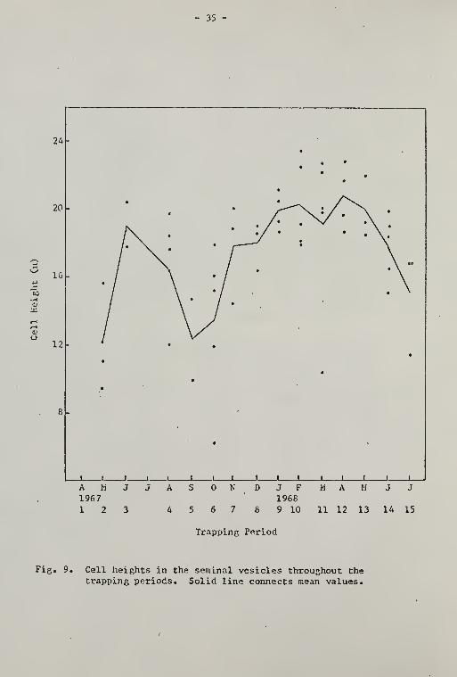
- 35 -
24

36 -
A H1967
1 2
M HJ F19689 10 11 12 13 14 15
Trapping Period
Fig. 10. DiaiTiGters of the seminal vesicles throughout thetrapping periods. Solid line connects mean values.

- 37 -
Comparison of Chnnnos in Rcproductlvo Orp.ans
with linvironmental Fcaturos
Table 7 demonstrates considerable overlapping of the peaks among
the parameters. The interpretation of this synchrony requires a close
look at the environmental features.
Fig. 11 is a summary of the monthly averages of soil temperature
maxima and minima and of rainfall. Fig. 12 shov?s the average monthly
values of soil moisture, a function of both rainfall and temperature.
Periods of high soil moisture occurred at times of high rainfall and
high temperatures, and at times of moderate rainfall cind low tempera-
tures. Figures 13 and lU were abstracted from the previous two figures
and present the mean of the environmental histories of the pocket
gophers within each period. Only the two-week-prior soil temperature
history and the four-week-prior soil moisture history are shown. The
two main reproductive peaks matched those of soil moisture fairly well,
and corresponded to intermediate periods of increasing soil temperature
•
In spite of this visual correspondence, soil moisture showed no
correlation with any one of the reproductive parameters, and soil
temperature was correlated at the .05 level of significance with only
five of the lA parameters. Since the initial analyses tested only re-
lationships between single factors — a highly artificial situation —
a multivariate analysis was chosen that would account for interaction
among the two sets of parameters.
The results of the canonical analysis are shown in Table 8. Of
the four programs that were run, only the results from the one showing
the highest correlation ~ the combination of soil moisture two weeks
prior to capture and soil temperature four weeks prior to capture —

- 38 -
Qi

- 39 -
Soil Temrif?i:ature (°F)
73

- AO -

/ll -
Trapping Period
Fig. 13. Average soil temperature histories of pocket gophers withineach trapping period for tvjo weeks prior to their capture

- /i2 -
Trappins Period
Fig, lA. Average soil moisture histories of pocket gophers within eachtrapping period for four weeks prior to their capture

- A3 -
Table 8, Results of ccmonical analysis
Parameter
Canonical Correlations1 2
.8512 . A306
Loadings
Soil h'oisture — 2 weeks
Soil Temperature — U weeks
0.0954
1.0330
1.0802
0.3299
Testis Volume -0.A592* 0.1088
Testis Tubule Diameter -0.5698* -0.4652*
Leydig Cell Diameter -0.3206* 0.5099*
Nucleus-Cell Ratio of Leydig Cell 0.0386 -0.3792*
Leydig Cell Tissue Volume -0.1977 -0.1687
Epididyrral Tubule Diameter 0.0806 0.1770
Epididymal Cell-Tubule Ratio -0.5717* 0.8601*
Seminal Vesicle Diameter 0.3826* 0.6500*
Seminal Vesicle Cell Height -0.3665* -0.9869*
Dorsolateral Prostate Cell Height 0.2438 0.2326
•indicates a highly important loading

- A4 -
are listed. The next highest correlation was frora the combination
of soil moisture four weeks prior and soil temperature four weeks pri-
or. The canonical correlation coefficients in the second set were
quite close to those in the first, suggesting that there is little
difference in the effects of soil moisture between the tvjo- and four-
week values. The other tv;o progra.iis had coefficients signific?-ntly
lower thaji these.
The canonical correlation showed that soil teriperature over a
four week period was much more important than soil moisture in
relating the two sets of variables. The second correlation implied
that some of the variables were definitely affected by soil moisture,
but the great difference between the two coefficients indicated that
these second values were not so reliable. Ko formal significance
test v;as performed; however, all coefficients greater than 0.3000
were considered highly important.
Not all the reproductive organs seemed to respond with the same
magnitude of change, or even in the same direction. Tnree of the
parameters did not show significance in either correlation: dorso-
lateral prostate cell height, epididymal tubule diameter, and Leydig
cell tissue volume. For the most part, hov/ever, the relationship was
an inverse one? the measurements of reproductive organs decreased with
an increase in soil temperature. Most of the second set of coeffi-
cients v7ere reversed, indicating that the measurements increased with
an increase in soil moisture. Both the testis tubule and seminal
vesicle cell height decreased v/ith the increase in soil moisture,
"however. Because a decrease in the ratios of Leydig cell and epididy-
mis measurements represented an increase in activity, their signs
should be interpreted in the reverse direction.

- h3 -
Immature Animal s and Pregnant Females
Little is known about the gestation and weaning periods of
G. pinetis . Howard and Childs (1959) estimated the gestation period
of T. bottae to be 30 days, but Schramm (I96I) observed two pocket
gophers (T. bottae) giving birth only I9 days after copulation; their
young weighed 2 - 3 g- Barrington (19^0) found that the weight of
three newborn G. pinetis averaged 5.1 g> and he estimated that the
gestation period in this species is 30 days. Miller (19^+6) reported
that weaning takes place in T. bottae after 35 to 37 (iays, and sexual
maturity is reached after three months. Wing (I960), however, indicated
that fromi the evidence provided by seasonal percentages, G. pinetis
of both sexes take nearly six months to reach sexual maturity.
Wing (i960) described the largest embryo in her collection as m.ea-
suring 39 mm from crown to rump. After estimating the gestation
period to be one mionth and assuming that the maximum crown-rump
length of an embryo is Uo mm, an approximation of each embryo's age
in days may be calculated. This information can then be used to
estimate the date of conception.
Criteria for separating inmature and mature individuals in this
study were, for females, the resorption of the pubic symphysis (Hisaw,
1923), and, for the m.ales, the size of the testis. In-jTiature m.ales on
this basis ranged in weight from 65 - 163 g, and immature females,
from 60 - IU6 g. If two months is arbitrarily chosen as the youngest
age at which a pocket gopher can be trapped, a hypothetical age
structure may be set up as follows:

VJeight

- A7 -
suo"C3d30uoo JO joqujn'j paacm^sg
o3xtouCuVuM-i
o
.2 -f^

- A8 -
Table 9. Hcan values of environmental parametorG over the
specified periods of time before approximateconception dates. Overall means are calculated
from capture dates of males throuchout the year.

- 'VJ -
Soil Temperature ("f)
(soqouT) II^JUXBH

- 50 -
Table 10, Sex, age, and reproductive status of animals captured in
each quarterly sample
Quarterly Sample
Group 1 2 3 h_
Percent Immature Animals 7 13 20 60
Percent of Adult Femaleswhich are Pregnant 28 50
Percent of Adult Kales and Femalesin Active Reproductive Condition 36 85 73 33
^1: 11/9/68 - 11/12/68
2j 2/20/69 - 2/23/69
3« 5/26/69 - 6/5/69
At 8/11/69 - 3/13/69

- 51 -
were enlarci^fl and dark, and if the epididymal tubules were easily
visible to the naked eye.
The projected conception dates for both immature animals and
pregnant females were combined and are shown in Fig. 17.

- 52 -
o

DISCUSSION AND CONCLUSIONS
The reproductive organs of the male pocket gopher show seasonal
changes that are related to changes in soil temperature and soil mois-
ture. The possibilities that males maintain active spermatogenesis
all year or that cessation or fluctuations in spermatogenesis occur
synchronously throughout the population were, therefore, eliminated.
Instead, it appears most likely that while some periods seem optimum
for all male pocket gophers, unfavorable periods do not cause abridge-
ment of reproductive activity to the same degree in all the males in
the population. In reaching this conclusion, much insight was gained
into age-dependent changes. Particularly valuable v/as information
provided on changes within the testis.
Even after sexual maturity is reached, the testis volume, for
example, was found to increase v/ith age. This would be due to tv7o
factors! increases in testis tubule size and in interstitial tissue.
Testis tubule diameter also increased with age, but the increase in
volume of the total Leydig cell tissue proved not to be significant.
The percentage of Leydig cell tissue volume to total volume seemed to
decrease, and the diameter of the Leydig cell itself decreased with
age. The diameter of the Leydig cell nucleus also decreased signifi-
cantly with age relative to the diameter of the entire cell.
This situation suggests that the rate of Leydig cell division
accelerates at puberty, accompanied by a disproportionate increase in
the growth rate of the cytoplasm relative to the nucleus. Although
- 53 -

- 5A -
the post-pubertal Leydig cell is larger than the pre-pubertal cell,
its diameter decreases vith age. Conav/ay (1959) recorded a similar
phenomenon in the eastern mole (Scalopus aquaticus ) . The infantile
testis was characterized by abundant but small Leydig cells with very
little cytoplasm. Early increases in testis volume were due primarily
to increases in Leydig cell volume until testis tubule enlargement
and the concurrent beginning of spermatogenesis became significant
at puberty. After this stage, the diameter of the Leydig cell decreased
by one-third, increasing again slightly at the end of the active
breeding period. The cell diameter was found to be largest during the
period of sexual inactivity.
In studies on the rat, Clegg (1966) found that the number of
Leydig cells actually declined following puberty. He suggested that
the accessory reproductive organs, which showed their maximum growth
acceleration at that stage, were more sensitive to the androgens being
produced by the Leydig cells.
Albert (1961) indicated that Leydig cells in bulls also decreased
in size from 5 to 15 years, after an initial increase in both number
and size from tv/o years to five years. According to the same source,
the namber of Leydig cells in man declines with age.
The percentage of Leydig cell tissue volume in the hippopotamus
testis decreased from 6<^i% in the very young animal to 32% at puberty,
remaining steady through old age (Laws and Clough, 1966).
The decrease in size of Leydig cells in pocket gophers as v/ell
as the decrease in percentage of total testis volume is, therefore,
not an unusual situation. Factor 3 related the increase in Leydig
cell diameter with the rise in activity of the accessory glands, as

- 55 -
well as with the increase in the diameter of the epididj'nial tubules
relative to the epididymal cell heights. Unlike the situation des-
cribed earlier in n.oles (Conaway, 1959), hormone secretion is presum-
ably associated with both a temporary increase in diameter of the
Leydig cells as X'jell as an increase in the ratio of cytoplasm to
nucleus. This evidence, together with the associations in Factor 2
which were mentioned earlier, leaves little doubt that a cyclic
increase and decrease of both Leydig cell diameter and of the tissue
voluroe is imposed on the long-term pattern of diminution.
The height of the epididymal cell was found to increase in
relation to the tubule diameter v?ith age. The cell height was also
associated with Leydig cell diameter in Factor 2. The latter response
corresponds to the observations summarized by Mann (196^), which
showed that survival of sperm in an epididymis severed from the testis
was dependent on the continued presence of the testis in the body. He
concluded that the condition of the epithelium in the epididymal tubule
V7as strongly influenced by the male sex hormone. The function of the
epididymis, Mann surmised, v?as therefore not only to serve as a
repository for sperm but also to produce a secretion somehow effec-
tive in preserving or perhaps maturing the spetTH.
Well-defined cycles during the study period were found only in
the testis volame, Leydig cell diameter and tissue volume, and
epididymal tubule diameter and cell height. The stage of spermatogen-
esis was highly dependent on age, the variation in stages among the
pocket gophers in each trapping period being sufficient to reject
the possibility of there being a synchronized population cycle in the
production of spetTnatozoa. Although definite cycles were detected in

- 56 -
the activities of the seminal vesicles and the dorsolateral prostate,
they were less distinct than the cycles mentioned earlier. Moreover,
activity in the prostate glands did not necessarily cease when sperm
viere not being produced. The seminal vesicles, hov;ever, shov;ed a pro-
nounced increase in activity in animals with active sperm production.
Price and Williaras-Ashman (1961) found that testicular hormones pro-
vided most of the control over activity of the accessory glands. They
also concluded that the chief function of these glands is to secrete
the sem.inal plasma.
Internal control of the reproductive system in the male pocket
gopher seems to rest at the hormonal level. Maximum size of the
Leydig cells reached tv;o significant peaks which corresponded to
similar peaks in size of the epididymis and accessory glands. The
lack of complete correspondence is likely attributable to a low
threshold in glandular response to testosterone. Van Tienhoven (1568)
has also suggested that maintenance of secondary sex characteristics
outside the breeding season may be the result of androgen secretion
by extra-tosticular sources such as the adrenal cortex.
Strongly influencing hormonal control, however, is environmental
control. In the canonical analysis, most of the measurements taken
of the reproductive organs decreased in magnitude, or otherwise
denoted less activity, under conditions of high soil temperature and
lov; soil moisture. This analysis, together with the tentative dates
of conception of iiiTiatura pocket gophers and embryos of pregnant
females, as well as the correspondence between environmental histories
and the peaks of reproductive activity, suggests that reproductive
activity is greatest In periods of moderate temperature and high soil
moisture.

- 57 -
Soil moisture is logically the less important of the two because
it seldom dips below 50% of the field capacity, and therefore shows
comparatively little variation. Tlie distribution of _G. pinetis may
well be limited by soil moisture, since this species is found only
in the v/ell-drained sandy soils in the Southeast (NcNab, 1966). The
possibility mentioned earlier that excessive soil moisture might
prom.pt a pocket gopher to dig to the surface, and perhaps more ex-
tensively underground as well (Killer, 1943), may be valid, but
apparently this response does not serve as the sole cue for timing
the reproductive cycle.
Temperature does affect the cycle strongly, most likely in an
indirect fashion. Because the largest proportion of the burrow lies
near the surface of the ground, it may be inferred that this is where
the greatest amount of digging takes place. The pocket gopher,
however, is a poor thermoregulator and therefore cannot long with-
stand the heat stress of digging in a shallow, warm burrow. Gunther
(1956) proposed a behavioral mechanism in which the gopher makes use
of deeper, cooler parts of its burrow system in the sui^mer, plugging
some of the shallow feeding tunnels. The diminished amount of digging
resulting from this cessation of activity would presumably lead to
diminished intraspecific contact. This regulation of acti\dty v/ould
effectively restrict most reproductive activity to certain times of
the year.
The reproductive cycle gives no evidence of being strictly timed.
Spermatogenesis, for instance, was found to be non-cyclic, at least
during this study period. A more careful study of the reproductive
tract itself might reveal storage of the sperm at some place along

- 58 -
the ductus deferens. The great variation in sperm production at any
one time convincingly suggests that the reproductive cycle is not a
strict endogenous rhythm, but is influenced by environmental fluc-
tuations and could be triggered by the right circumstances — i.e.,
favorable environmental conditions and the presence of a receptive
female — at any time of the year. Since there was a low level of
reproductive activity that continued throughout much of the study
period, at least some random encounters in the "off season" might
be fruitful.
Probably little selection pressure operates to channel repro-
duction into a strictly timed cycle. Food is certainly most abun-
dant in the spring months but is still available, particularly in a
grassy area such as Stengel Field, in sufficient quantity throughout
the year so that a newborn gopher would not starve. Contact between
pocket gophers of the same sex frequently results in harmful injuries
to one or both animals (Miller, 1964; Vaughan, 1962). Such contact,
if it should occur between a male and female, might also be harmful
if neither wore in active reproductive condition. It i.'ould obviously
be advantageous to a pocket gopher to be capable of mating should an
individual of the opposite sex be encountered. However, expending
energy tov:ard maintaining the reproductive system in an active state
during periods of diminished intraspecif ic contact could also be
wasteful. Evidence from one population of pocket gophers indicates
that the greatest nuiriber of the individuals reaches a peak in repro-
ductive activity \;hen maximum contact is expected, but seldom does
the entire population regress to an inactive state at other times.

SUI'lI>Lf\RY
The reproductivG cycle in the male pocket gopher ( GoornyG pinetis )
is closely correlated v;ith soil temperature and to a lesser decree
with soil moisture, but does not seem to be strictly controlled by
either of these parameters. Very warm external temperatures and lov;
soil moisture appear to restrict activity vjithin the burrow at certain
times of tlie year, and accordingly reduce intraspecific contacts.
Maximum reproductive activity corresponds to those environmental con-
ditions that create a friable soil.
Internal seasonal changes in the reproductive organs and glands
are presumably in turn effected by hormones, particularly testoster-
one, v;hich is secreted by the Leydig cells. Leydig cells in adult
pocket gophers are larger in diameter and more numerous than those in
immature animals. A net decrease in both size and number of Leydig
cells occurs after puberty, but the cells still increase in diameter
at times of increased reproductive activity, suggesting increased
hormonal production. This cyclic change is correlated with increases
in the height of the sf:crctory epithelium in the accessory glands and
in tlui epididymis.
Spermatogenesis V7as found to be non-cyclic during the study period.
Although the mean spermatogenic values for each period followed a
cyclic pattern similar to the one outlined by the Leydig cell diameter,
the variation about the means V7as too great to allow statistical sig-
nificance. Sperm production throughout the population therefore con-
- 59 -

- 60 -
tinned at least at a threshold level throughout the study period, and
reproductive activity at the orf'.an level was restricted more by the
condition of the accessory glands and the epididjanis than by the
availability of sperm.

LITEiU'IURli CITED
Albert, A. 1961. The maiimalian testis, p. 305-365. In U. C. Younc(ed.), Sox and internal secretions. Vol. 1, 3rd ed. Williams L
Wilkins Co., Baltimore.
Barrinp.ton, B. A. 1940. The natural history of pocket gophers. M.S.Thesis, Univ. of Fla. 19A p.
Sloora, W. and D. iV. Fawcctt. 1962. A textbook of histology. W. B.
Saunders Co., Philadelphia. 720 p.
Bond, R. M. 1946. The breeding habits of Thomonys bottao in OrangeCounty, California. J. Maminal. 27: 172-174.
Clegg, E. J. 1966, Pubertal growth in the Leydig cells and accessoryreproductive organs of the rat. J. Anat. 100: 369-379.
Conaway, C. K. 1959. The reproductive cycle of the eastern mole.J. Mammal. 40: 180-194.
Dixon, J. 1929. The breeding season of the pocket gopher in California.
J. Mammal. 10: 327-328.
English, P. F. 1932. Some habits of the pocket gopher, Goomys brevicepsbreviceps. J. Mammal. 13: 126-132.
Groome, J. R. 1940. The seasonal modification of the interstitialtissue of the testis in the fruit bat (Pteropus ) . Proc. Zool. Soc.
London 110: 37-42.
Cunther, H. C. 1956. Studies on the male reproductive system of the
California pocket gopher (Thomonys bottae navus Merriam) . Amer.
Midi. Nat. 55: 1-40.
Hammond, L. C, H, W. Lundy, and G. K. Saxena. 1967. Influence of
underground asphalt barriers on water retention and movement in
Lakeland fine sand. Soil and Crop Soc. of Fla., Proc. 27: 11-19.
Hisaw, F. L. 1923. The absorption of the pubic symphysis of the pocket
gopher Geomys bursarius (Shaw). Amer. Nat. 58: 93-96.
Howard, W. E. and H. E. Childs, Jr. 1959. Ecology of pocket gophers
with emphasis on Thomomys bottae mev;a . Hilgardia 29: 277-358.
- 61 -

- r,2 -
Johnson, 0. W. anrl I, 0. Buss. 1967. The testis of the African ele-phant (Loxodonta africana). I. Histological features, J. Heprod.Fcrt. 13: 11-21.
Kennerlj', T. E. Jr. 196-4. Microenvironmental conditions of the pocketcopher burrow, Tex. J. Sci. 26 » 395-A'!4l.
Laws, R. M. and G. CloUi-jh. 1966. Observations on reproduction in thehippopotamus llipponotamus amnliibius Linn., p. 117-1^0. In I. W,
Rowlands (ed.). Comparative biology of reproduction in mammals,Zool. Soc, of London, Academic Press,
Macklin, C. C, and M. T. Macklin. 1963. Tl^e seminal vesicles, prostateand bulbourethral glands, p. 1773-1822. In E. V. Cowdry (ed,).Special cytology. Vol. Ill, 2nd ed, llafner Publ. Co., Nev; York.
Mann, T. 196A, The biochemistry of semen and of the male reproductivetract, 2nd ed. John Wiley & Sons, Inc., Nev; York, A93 p,
McXab, B, K. 1966. The metabolism of fossorial rodents: a study ofconvergence. Ecology A7: 712-733.
Killer, 11. A. 19^6. Reproductive rates and cycles in the pocket gopher.J. KaiPu-nal. 27: 335-353.
. 19A8. Seasonal trends in burrowing of pocket gophers ( Thonomys )
»
J. Mammal, 29: 38-AA.
Miller, R. S, 1964. Ecology and distribution of pocket gophers(Goomyidae) in Colorado. Ecology 45: 256-272.
Moore, C. R,, D, Price, and T. F. Gallagher, 1930. Rat-prostatecytology as a testis hormone indicator and the prevention ofcastration changes by testis-extract injections. Amer. J, Anat,A5: 71-107,
Price, D, and H, G, Williams-Ashman, 1961, The accessory reproductiveglands of mammals, p, 366-A48. In W. C. Young (ed.). Sex and inter-nal secretions. Vol. I, 3rd ed. Williams L Wilkins Co., Baltiiaore,
Roosen-Runge, E. C. and L. 0. Giesel, Jr. 1950. Quantitative studieson spermatogenesis in the albino rat. /uiier, J, Anat, 87: 1-30.
Schramm, P, 1961, Copulation and gestation in the pocket gopher,J. Mammal, A2: 167-170,
Spencer, R, P, 1968, Changes in testicular allometric growth curves,Yale J, Siol, Med, AO: 313-319,
Steel, R, G. D. and J, H, Torrie, 1960, Principles and procedures ofstatistics, McGraw-Hill Cook Co., Inc., New York, A81 p.

- 63 -
Tcsi, J. A. 1968. Calculo del balance hidrica, p. -^2-48. In Evrel, J. J.
and A, Madriz, Zonas de vida de Venezuela. Republica do Venezuela,I-'inisterio de Agricultura y Crla, Direcci6n de Investi5aci6n.Caracas.
Van Tienhoven, A. 196P. Henroductive physi^loj-,;' of vertebrates. H. B,
Saunders Co., Philadelphia, 498 p.
Vaughan, T. A. 1962. Reproduction in the plains pocket gopher in
Colorado. J. liainmal . 43: 1-13.
, and R. M. Hansen. 1961. Activity rhythm of the plains pocket
gopher. J. Kaminal. 42: 541-543.
VJilks, B. J. 1963. Soae aspects of the ecology and population d3mainics
of the pocket gopher (Geomys_ bursarius ) in south Texas. Tex. J.
Sci. 15: 241-283.
Wing, E. S. 1960. Reproduction in the pocket gopher in north-central
Florida. J, Mammal, 41: 35-43.

APPENDIX

- 65 -
Fie. 18. Level 8 testis (lOOX)
Fig. 19. Level 3 testis (430X)
Fig. 20. Level 7 testis (A30X)

- 66 -
Fig. 21. Epididymal tubules from a male in activereproductive condition. The lumina arefilled with sperm. (lOOX)
Fig. 22. Epididymal tubules from inactive male (lOOX)

- 67 -
Fig. 23. Actively secreting acinus from dorsolateralprostate gland (430X)
Fig. 2A. Inactive acinus from dorsolateral prostategland (430X)

- 68 -
H 'Hit £jik!^'
i^
Fig. 25. Lumen and part of cell boundary from an active
seminal vesicle (lOOX)
Fig. 26. Inactive seminal vesicle (lOOX)

BIOGRAPHICAL SKETCH
Katherino Carter Ewel was born September 30, 19''4'fi, at Glens
Falls, New York. Durin;; the smmner of 1961, she attended Cornell
University on a National Science Foundation Summer Fellowship, and
Graduated from Glens Falls Hieh School in June, 1962. In June, 1966,
she received the degree of Bachelor of Arts v/ith a major in zoology
from Cornell University. During tlie smnmer of 1965, she participated
in a field biology course at Tulane University on a USDHEV Environ-
mental Training Grant and in the fall of 1966 enrolled in the Graduate
School of the University of Florida. She worked as a teaching assis-
tant in the Department of Zoology until August, 1967, and was av;arded
an NDEA Title IV Fellowship from September, 1967, through August,
1969. During the summer of 1963, she was enrolled in the tropical
biology program at the Universidad de Costa Rica under an NSF Graduate
Research Fellovjship av/arded by the Organization for Tropical Studies.
She is currently employed as a Temporary Instructor in Zoology at Duke
University while completing her work tov;ard the degree of Doctor of
Philosophy at the University of Florida.
Katherine Carter Ewel is married to John Ewel. She is a member
of the i\merican Society of Mararaalogists, the Ecological Society of
America, and the Society for the Study of Evolution.
- 69 -

This dissertation vjas prepared umler tlu' direction of thf:
chairman of the candidate's supervisory committee and has been
approved by all members of that committee. It v;as submitted to
the Dean of the Collef.e of Arts and Sciences and to the Graduate
Council, and was approved as partial fulfillment of the reqviire-
Fients for the decree of Doctor of Philosophy.
June, 1970
Dilan, Graduate Schoo
Supervisory Committee:
Chairman

. V i-^ m