Anemia epidemiology, pathophysiology, and etiology in low ...
26
Anemia epidemiology, pathophysiology, and etiology in low- and middle-income countries Camila M. Chaparro, International Nutrition Parminder S Suchdev, Emory University Journal Title: Annals of the New York Academy of Sciences Volume: Volume 1450, Number 1 Publisher: Wiley: 12 months | 2019-08-01, Pages 15-31 Type of Work: Article | Post-print: After Peer Review Publisher DOI: 10.1111/nyas.14092 Permanent URL: https://pid.emory.edu/ark:/25593/v3xgs Final published version: http://dx.doi.org/10.1111/nyas.14092 Copyright information: © 2019 New York Academy of Sciences. Accessed March 17, 2022 5:07 PM EDT
Transcript of Anemia epidemiology, pathophysiology, and etiology in low ...
Anemia epidemiology, pathophysiology, andetiology in low- and middle-income countriesCamila M. Chaparro, International NutritionParminder S Suchdev, Emory University
Journal Title: Annals of the New York Academy of SciencesVolume: Volume 1450, Number 1Publisher: Wiley: 12 months | 2019-08-01, Pages 15-31Type of Work: Article | Post-print: After Peer ReviewPublisher DOI: 10.1111/nyas.14092Permanent URL: https://pid.emory.edu/ark:/25593/v3xgs
Final published version: http://dx.doi.org/10.1111/nyas.14092
Copyright information:© 2019 New York Academy of Sciences.
Accessed March 17, 2022 5:07 PM EDT

Anemia epidemiology, pathophysiology, and etiology in low- and middle-income countries
Camila M. Chaparro1, Parminder S. Suchdev2,3,4
1Independent Consultant, International Nutrition
2Department of Pediatrics, Emory University, Atlanta, Georgia
3Emory Global Health Institute, Emory University, Atlanta, Georgia
4Nutrition Branch, Centers for Disease Control and Prevention, Atlanta, Georgia
Abstract
Anemia affects a third of the world’s population and contributes to increased morbidity and
mortality, decreased work productivity, and impaired neurological development. Understanding
anemia’s varied and complex etiology is crucial for developing effective interventions that address
the context-specific causes of anemia and for monitoring anemia control programs. We outline
definitions and classifications of anemia, describe the biological mechanisms through which
anemia develops, and review the variety of conditions that contribute to anemia development. We
emphasize the risk factors most prevalent in low- and middle-income countries, including
nutritional deficiencies, infection/inflammation, and genetic hemoglobin disorders. Recent work
has furthered our understanding of anemia’s complex etiology, including the proportion of anemia
caused by iron deficiency (ID) and the role of inflammation and infection. Accumulating evidence
indicates that the proportion of anemia due to ID differs by population group, geographical setting,
infectious disease burden, and the prevalence of other anemia causes. Further research is needed to
explore the role of additional nutritional deficiencies, the contribution of infectious and chronic
disease, as well as the importance of genetic hemoglobin disorders in certain populations.
Address for correspondence: Camila M. Chaparro, Independent Consultant, International Nutrition., [email protected] contributionsC.M.C. was responsible for overall manuscript conception, interpretation, and writing; P.S.S. contributed to manuscript conception, interpretation of data, and reviewed and wrote manuscript sections.
StatementThis manuscript was presented at the World Health Organization (WHO) technical consultation “Use and Interpretation of Hemoglobin Concentrations for Assessing Anaemia Status in Individuals and Populations,” held in Geneva, Switzerland on November 29−30 and December 1, 2017. This paper is being published individually but will be consolidated with other manuscripts as a special issue of Annals of the New York Academy of Sciences, the coordinators of which were Drs. Maria Nieves Garcia-Casal and Sant-Rayn Pasricha. The special issue is the responsibility of the editorial staff of Annals of the New York Academy of Sciences, who delegated to the coordinators preliminary supervision of both technical conformity to the publishing requirements of Annals of the New York Academy of Sciences and general oversight of the scientific merit of each article. The workshop was supported by WHO, the Centers for Disease Control and Prevention (CDC), the United States Agency for International Development (USAID), and the Bill & Melinda Gates Foundation. The authors alone are responsible for the views expressed in this paper; they do not necessarily represent the views, decisions, or policies of the WHO. The opinions expressed in this publication are those of the authors and are not attributable to the sponsors, publisher, or editorial staff of Annals of the New York Academy of Sciences.
Competing interestsThe authors declare no competing interests.
HHS Public AccessAuthor manuscriptAnn N Y Acad Sci. Author manuscript; available in PMC 2019 August 16.
Published in final edited form as:Ann N Y Acad Sci. 2019 August ; 1450(1): 15–31. doi:10.1111/nyas.14092.
Author M
anuscriptA
uthor Manuscript
Author M
anuscriptA
uthor Manuscript
Keywords
anemia; iron deficiency anemia; nutritional anemias; anemia of inflammation
Introduction
Anemia—a condition in which hemoglobin (Hb) concentration and/or red blood cell (RBC)
numbers are lower than normal and insufficient to meet an individual’s physiological needs1
—affects roughly one-third of the world’s population.2 Anemia is associated with increased
morbidity and mortality in women and children,3,4 poor birth outcomes,5,6 decreased work
productivity in adults,7 and impaired cognitive and behavioral development in children.8
Preschool children (PSC) and women of reproductive age (WRA) are particularly affected.
Establishing appropriate Hb thresholds to define anemia is essential for ensuring that anemia
is correctly identified, and its negative effects prevented. As important, understanding the
diverse and complex etiology of anemia is crucial for developing appropriate interventions
that address the context-specific causes of anemia and for monitoring the success of anemia
control programs. To that end, the primary aims of this paper are to outline definitions and
classifications of anemia; describe the biological mechanisms through which anemia
develops; review the variety of factors and conditions that contribute to anemia development,
emphasizing those most prevalent in low- and middle-income countries (LMICs); and
identify research needs. Although our primary focus is on anemia and its etiology at a
population level, the information we present on definitions and classifications of anemia, as
well as its etiology, is relevant to individual-level assessment by clinicians.
Materials and methods
We reviewed the peer-reviewed literature on definitions and classifications of anemia, global
magnitude and epidemiology of anemia, and causes of anemia, including their biological
mechanisms and public health significance. We identified references through PubMed
searches on relevant search terms (see below) and the “snowball” method in which
references of references are identified. We also consulted gray literature, particularly
documents relevant to defining anemia and nutritional status published by international
organizations. To provide estimates of the global magnitude of anemia and to elucidate the
etiology of anemia, we used three principal sources: (1) recent analyses by WHO on global
anemia prevalence between 1995 and 2016 using population-representative data on PSC and
WRA from 257 sources representing 107 countries;9,10 (2) a recent global analysis of
anemia burden between 1990 and 2010 in 20 age groups and both sexes from 187 countries
that also performed cause-specific attribution to 17 conditions from the Global Burden of
Diseases, Injuries and Risk Factors 2010 Study;2 and (3) recent analyses from the
Biomarkers Reflecting Inflammation and Nutritional Determinants of Anemia (BRINDA)
project, a large-scale collaborative study to examine the factors associated with anemia in
29,000 PSC (6−59 months of age) from 16 population-based surveys and 27,000
nonpregnant WRA (15−49 years of age) from 10 population-based surveys representing
countries in all six WHO geographic regions.11
Chaparro and Suchdev Page 2
Ann N Y Acad Sci. Author manuscript; available in PMC 2019 August 16.
Author M
anuscriptA
uthor Manuscript
Author M
anuscriptA
uthor Manuscript

Search terms
Anemia AND: causes, classification, etiology, risk factors, assessment, iron deficiency,
vitamin A deficiency, folate deficiency, B12 deficiency, riboflavin deficiency, malaria,
infection, infectious disease, schistosomiasis, hookworm, intestinal helminths, HIV,
tuberculosis, obesity, overweight, undernutrition, underweight, stunting, wasting, child
development, motor development, cognitive development, birth outcomes, birth weight,
premature birth, growth, mortality, work productivity, poverty, education; nutritional
anemias; anemia of inflammation; anemia of chronic disease; thalassemia, α-thalassemia, β-
thalassemia; sickle cell disease; sickle cell disorders; hemoglobinopathies; hemoglobin
disorders; hepcidin; iron deficiency anemia.
Defining anemia
Anemia is alternately defined as a reduced absolute number of circulating RBCs12 or a
condition in which the number of RBCs (and subsequently their oxygen-carrying capacity)
is insufficient to meet physiologic needs.1 Though most commonly diagnosed by a low Hb
concentration or a low hematocrit,12 anemia can also be diagnosed using RBC count, mean
corpuscular volume, blood reticulocyte count, blood film analysis, or Hb electrophoresis.13
At the population level and in clinical practice, Hb concentration is the most common
hematological assessment method used14 and the most common indicator used to define
anemia. The critical role of Hb to carry oxygen to the tissues explains the most common
clinical symptoms of anemia, which include fatigue, shortness of breath, bounding pulses or
palpitations, and conjunctival and palmar pallor.15 Clinical signs16 and medical history are
used to diagnose anemia when hematological data are unavailable, but they are limited in
their ability to detect anemia.17 Severe anemia (defined by WHO as Hb <70 g/L in children
under 5 years of age and Hb <80 g/L in all other age groups,1 though other definitions,
including Hb <50 g/L, are used) is of particular importance clinically, as it can result in
high-output heart failure and death.12
Defining an abnormally low Hb concentration requires understanding how Hb naturally
varies by age, sex, pregnancy status, genetic and environmental factors, and, potentially,
race. Hb varies with age, most dramatically in the first months of life (Fig. 1).18 In the
newborn, normal Hb concentrations are between 17 and 21 g/L, their highest point during
life.18,19 Hb concentration then decreases through the first 2−3 months of life before
increasing again in childhood,18,20 and then levels off throughout adulthood before declining
again in older age.21 Sex differences in Hb concentrations begin in puberty (because of the
effect of menstruation on iron stores and, subsequently, anemia) and continue throughout the
reproductive years.22,23 During pregnancy, because of the expansion of blood volume and
consequent dilution effect, Hb concentration naturally declines during the first and second
trimesters, rising gradually again in the third trimester.24 Apart from physiological factors,
behavior and environmental conditions, such as altitude and smoking, can also affect Hb
concentrations.1
The WHO Hb cutoffs for anemia (Table 1) are widely applied globally and are sex, age, and
pregnancy specific.1 These cutoffs were first established in 1968 by a nutritional anemia
study group at WHO using statistical cutoffs rather than thresholds linked to meaningful
Chaparro and Suchdev Page 3
Ann N Y Acad Sci. Author manuscript; available in PMC 2019 August 16.
Author M
anuscriptA
uthor Manuscript
Author M
anuscriptA
uthor Manuscript
health outcomes.25 Hb cutoffs were modified slightly since then to allow for additional age
divisions among children, adjustment for children in the 5−11 age group based on data of
noniron-deficient children from the United States, and creation of the categories of “mild,”
“moderate,” and “severe” anemia.26,27 Cutoffs were also supported by findings among
participants of the Second National Health and Nutrition Examination Survey (NHANES II)
who were not iron deficient.14 The need for separate cutoffs based on ethnicity/race has been
proposed (e.g., individuals of African descent have lower Hb concentrations than do
Caucasian populations, at least partially due to the greater prevalence of genetic Hb
disorders in persons of African descent), as have revisions to the cutoffs for particular age
groups (e.g., very young infants).28–32
Global magnitude of anemia
Approximately one-third of the world’s population (32.9%) was estimated to suffer from
anemia in 2010.2 The population groups most vulnerable to anemia include (1) children
under 5 years of age (42% with anemia in 2016), particularly infants and children under 2
years of age; (2) WRA (39% with anemia in 2016); and (3) pregnant women (46% with
anemia in 2016).33,34 Females were consistently at greater risk of anemia than men across
almost all geographic regions and in most age groups.2 Other at-risk groups include the
elderly, as the prevalence of anemia among adults over 50 years of age rises with advancing
age,35 though data are limited.
The prevalence of anemia also varies by geographic region. Sub-Saharan Africa, South Asia,
the Caribbean, and Oceania had the highest anemia prevalence across all age groups and
both sexes in 2010.2 At the country level, anemia among WRA and children under 5 years of
age is a moderate-to-severe public health problem (20% or greater as defined by WHO) in
the majority of WHO member states.9,10
Progress on decreasing anemia has been overall slow and uneven. For all age groups and
both sexes, anemia is estimated to have decreased roughly seven percentage points between
1990 and 2016, from 40% to 33%.2 The WHO Global Nutrition Target 2025 on anemia aims
to reduce anemia in WRA by 50% by 2025.36 Based on a global prevalence of 29–38%
anemia among WRA (nonpregnant and pregnant, respectively) as of 2011, a reduction of
1.8–2.4 percentage points per year would be required to meet this target.
Pathophysiology of anemia: consequences for development, growth, birth
outcomes, and work productivity
Anemia has significant consequences for human health, as well as for social and economic
development. In 2010, anemia accounted for 68.4 million years of life lived with disability,
or 9% of the total global disability burden from all conditions.2 Anemia has been associated
with negative health and development outcomes, including neonatal and perinatal mortality,
low birth weight,37 premature birth,5,38 and delayed child development.39
The negative effects on health and development outcomes from anemia arise from the
impacts of decreased oxygen delivery to tissues (in which multiple organ systems may be
Chaparro and Suchdev Page 4
Ann N Y Acad Sci. Author manuscript; available in PMC 2019 August 16.
Author M
anuscriptA
uthor Manuscript
Author M
anuscriptA
uthor Manuscript

affected), as well as effects related to the underlying causes of anemia, which are difficult to
disentangle. For example, in iron deficiency anemia (IDA), decreased iron availability has
well-established negative effects on brain development and functioning even prior to anemia
development.40
Etiology of anemia: conceptual models, biological mechanisms, and
classifications
At a biological level, anemia develops because of an imbalance in erythrocyte loss relative to
production; this can be due to ineffective or deficient erythropoiesis (e.g., from nutritional
deficiencies, inflammation, or genetic Hb disorders) and/or excessive loss of erythrocytes
(due to hemolysis, blood loss, or both). Anemia is frequently classified based on the
biological mechanism of causation (e.g., IDA, hemolytic anemia, and anemia of
inflammation (AI)) and/or the RBC morphology. Table 2 displays a partial list of several
common anemias and the biological mechanisms through which they develop and RBC
parameters that characterize their presentation and distinguish them from each other.41 Most
anemias have a characteristic RBC appearance, which can provide insights to the diagnosis
of anemia. However, as Table 2 displays, multiple factors can cause a similar type of RBC
morphology.41 Furthermore, as anemia may have multiple causes, even in the same
individual, hematological manifestations of a particular cause can be masked by another. For
example, the hallmark of anemia caused by vitamin B12 or folate deficiencies is macrocytic
anemia. Concomitant ID, which causes microcytosis, may mask entirely the effects of the
B12 or folate deficiency. Although indices exist in clinical practice for distinguishing anemia
etiology based on RBC parameters (e.g., IDA versus β-thalassemia—both cause
hypochromia and microcytosis), their reliability for discriminating between causes varies.42,43
Figure 2 is a conceptual model of the etiology of anemia identifying how distal factors
contribute to more proximate determinants of anemia, such as food insecurity, clean water,
and sanitation, and, ultimately, the most immediate causes of anemia (e.g., nutritional
deficiencies, disease, inflammation, and Hb disorders).13,44,45 Many of these determinants
are interrelated. Poverty, for example, is a major determinant of health and nutrition, and
poor socioeconomic position is linked to a greater risk of anemia among women and
children.13,46 Similarly, low education level is also associated with a greater risk of anemia.13 A recent analysis of 53 demographic and health surveys with Hb data found that anemia
among PSC (affecting 70% of the PSC population studied) was strongly associated with
maternal anemia, household wealth, maternal education, and low birth weight.47
It is important to note that the primary causes of mild and moderate anemia tend to differ
from the principal causes of severe anemia. Though there are limited studies on the etiology
of severe anemia,48 malaria is frequently identified as a principal cause of severe anemia,
particularly in African children. In the BRINDA project, the most consistent predictors of
severe anemia in population-based surveys of PSC were malaria, poor sanitation,
underweight, and inflammation (in African countries only); stunting, vitamin A deficiency
(VAD), and rural location were also significant determinants in high/very high infection
Chaparro and Suchdev Page 5
Ann N Y Acad Sci. Author manuscript; available in PMC 2019 August 16.
Author M
anuscriptA
uthor Manuscript
Author M
anuscriptA
uthor Manuscript

countries.49 In a study of Malawian PSC, factors associated with severe anemia included
malaria, bacteremia, hookworm infection, HIV infection, glucose-6-phosphate
dehydrogenase (G6PD) deficiency, and vitamin A (VA) and B12 deficiencies.48 In this
population, iron deficiency (ID) was protective of severe anemia, likely due to the
relationship between iron and infection. In Kenyan PSC, severe anemia was associated with
malaria, inflammation, and stunting, while predictors of more moderate forms of anemia
were ID, malaria, and α-thalassemia.50 Severe anemia is also a comorbidity of severe acute
malnutrition (SAM); for example, in India, of hospital-based children with SAM, 67% also
had severe anemia.51
As identified in Figure 2, and reflected in global analyses of anemia burden between 1990
and 2010, the most proximal risk factors for anemia include nutritional deficiencies, disease/
infection, and genetic Hb disorders.52 These conditions and several others, which are also
prevalent causes of anemia in LMIC, will be discussed in more detail below.
Nutritional anemias: iron, vitamins A and B12, folate, and riboflavin
Nutritional anemias result when concentrations of hematopoietic nutrients—those involved
in RBC production or maintenance—are insufficient to meet those demands.13 Causes of
nutrient deficiency include inadequate dietary intake, increased nutrient losses (e.g., blood
loss from parasites, hemorrhage associated with childbirth, or heavy menstrual losses),
impaired absorption (e.g., lack of intrinsic factor to aid vitamin B12 absorption, high intake
of phytate, or Helicobacter pylori infection that impair iron absorption), or altered nutrient
metabolism (e.g., VA or riboflavin deficiency affecting mobilization of iron stores). While
nutrient supplementation is a common preventive and treatment strategy for nutritional
anemias—for example, iron supplementation for the prevention of IDA—the bioavailability
and thus absorption from different nutrient supplement preparations can vary, potentially
limiting their impact.53
ID is considered the most common nutritional deficiency leading to anemia, though other
nutritional deficiencies can also cause anemia, including deficiencies of vitamins A, B12,
B6, C, D, and E, folate, riboflavin, copper, and zinc.54 Several of these nutrients—vitamins
A, B6, and B12, folic acid, and riboflavin—are needed for the normal production of RBCs;
other nutrients, such as vitamins C and E, may protect RBCs through their antioxidant
function.55 Trace elements, such as copper and zinc, are found in the structures of enzymes
that act on iron metabolism (e.g., copper and ceruloplasmin).56 Copper may also contribute
to anemia development through reductions in erythropoietin (EPO) and antioxidant enzymes
that require copper, thus increasing oxidative stress and reducing RBC life span;57 the
mechanisms through which zinc deficiency is associated with anemia are not as well
characterized.57 The extent to which each of these deficiencies contributes to the global
anemia burden is still a subject of investigation. While some of these nutrient deficiencies
are rare and may contribute little to the burden of anemia globally, deficiencies in multiple
micronutrients likely have a synergistic effect on anemia development.58
Chaparro and Suchdev Page 6
Ann N Y Acad Sci. Author manuscript; available in PMC 2019 August 16.
Author M
anuscriptA
uthor Manuscript
Author M
anuscriptA
uthor Manuscript

Iron deficiency
ID develops when dietary iron intake cannot meet iron needs over a period of time,
especially during periods of life when iron requirements are particularly high (e.g., during
periods of rapid growth and development, such as infancy and pregnancy) or when iron
losses exceed iron intake. ID typically evolves in three stages: storage iron depletion, iron-
deficient erythropoiesis, and IDA (defined as concomitant ID plus anemia).59 The WHO
recommends assessing iron status using serum ferritin or soluble transferrin receptor (sTfR).60,61 Serum ferritin, a measure of body storage iron and a sensitive measure of ID, is
elevated by the acute phase response; sTfR levels when high indicate tissue ID, but sTfR
may also be affected by inflammation and other causes of erythropoiesis.60,61 Because of the
effect of inflammation on many biomarkers of iron status, acute phase proteins (e.g., C-
reactive protein (CRP) and alpha-1-acid glycoprotein (AGP)) should be assessed when
possible.60 A fuller review of iron status indicators, outside the scope of the paper here, is
available elsewhere.61
Estimates from the late 1990s placed the number of individuals affected by ID at 2 billion,
and ID has long been assumed to contribute to approximately 50% of anemia cases globally.62,63 A recent systematic analysis of global anemia data that calculated cause-specific
attribution for 17 conditions related to anemia ranked ID as the most common cause in
almost all global regions examined.2 The WHO used the change in Hb concentration from
iron supplementation studies to estimate the “proportion of all anemia amenable to iron” as
50% of anemia among nonpregnant and pregnant women, and 42% of anemia in children.9
Recent analyses from the BRINDA project indicated that along with age and malaria, ID
was one of the factors most consistently associated with anemia, though the proportion of
anemic children and women with ID varied by infectious disease burden.46,49 Another study
that assessed the role of ID in anemia burden among PSC and nonpregnant WRA, across a
range of countries with varying rankings on the Human Development Index, showed that
between approximately a quarter to a third of anemia among PSC and WRA was associated
with ID.64 In countries where the prevalence of anemia was greater than 40% and in
countries where inflammation levels were high, ID played a much smaller role.64 Thus,
while ID remains a primary cause in many settings, the proportion of anemic individuals
with ID varies by contextual factors, and poor iron nutrition cannot be assumed to be the
primary cause in all cases. Yet, iron interventions (e.g., supplementation, fortification, and
dietary interventions) are central to most anemia control programs, and WHO currently has
17 guidelines on iron supplementation.65 Given the complex etiology of anemia, the extent
to which ID accounts for the anemia burden continues to be investigated.
Vitamin A deficiency
VAD is prevalent in many LMICs, particularly among PSC, pregnant women, and WRA. In
2005, the WHO estimated that 190 million PSC and 9.1 million pregnant women from
regions at risk of VAD were VA deficient (based on serum retinol concentrations), which
represents a third of PSC and 15% of pregnant women from these countries.66
VAD and anemia have been observed to occur in the same populations for decades, and
significant correlations between VA status biomarkers and Hb have been described in
Chaparro and Suchdev Page 7
Ann N Y Acad Sci. Author manuscript; available in PMC 2019 August 16.
Author M
anuscriptA
uthor Manuscript
Author M
anuscriptA
uthor Manuscript
multiple countries and populations including preschool and school-age children, adolescents,
and adults.67 VA supplementation has been shown to increase Hb concentrations,
hematocrit, and some iron status indices, even when administered in the absence of iron
supplements.55,67 VAD is thought to cause anemia through multiple mechanisms, including
the role of retinoids in erythropoiesis, VA’s importance for immune function, as well as
VA’s well-established role in iron metabolism.67 In contrast to IDA, which is marked by a
depletion of iron stores, anemia due to VAD is marked by an increase in iron stores in the
liver and spleen and increased serum ferritin concentrations.68 The anemia of VAD has
alternatively been described as hypochromic or microcytic and hypochromic, but other
factors—including other nutritional deficiencies and infections—occurring simultaneously
may cause inconsistencies in RBC parameters.67 BRINDA analyses showed that among
PSC, VAD was associated with anemia in close to half of the surveys (5/12) and across
levels of infectious disease burden.49 Among WRA in the BRINDA project where ID and
VAD were both assessed, VAD and ID were associated with anemia in all surveys (5/5
surveys) in both high- and low-infection burden groups.46 Estimates as to how much anemia
would be reduced by addressing VAD warrants additional research. In addition, like iron
status indices, VA biomarkers are affected by inflammation, thus complicating the
assessment of anemia due to VAD in settings where infectious disease is prevalent.69
Deficiencies of B vitamins (riboflavin, B12, and folate)
Several B vitamins are involved in Hb synthesis or iron metabolism, including riboflavin
(B2), pyridoxine (B6), cobalamin (B12), and folate. Deficiencies of these nutrients have
been associated with anemia; however, the extent to which they contribute to the global
burden of anemia varies and in some cases is unclear. Vitamin B6 deficiency is rare55 and
will not be addressed here.
Both vitamin B12 (cobalamin) and folate deficiency can lead to macrocytic anemia.
Deficiencies of these nutrients affect DNA synthesis and cell division in the bone marrow
(megaloblastic changes), such as hypersegmented neutrophils on the peripheral blood smear.70 Folate deficiency can also lead to decreased erythrocyte life span. Vitamin B12 deficiency
in LMIC most commonly results from low dietary intake of the nutrient. Its bioavailable
forms are only found in animal-source foods; but vitamin B12 deficiency can also result
from malabsorption, particularly in the elderly among whom gastric atrophy is common,71
in cases of pernicious anemia, an autoimmune disease in which autoantibodies are formed
against intrinsic factor essential for B12 absorption, and in bacterial and parasitic
coinfections.13,72 Folate deficiency tends to be more common in populations relying on
unfortif ied wheat or rice as a staple food and that consume low amounts of legumes and
green leafy vegetables.71 Pregnant women, preterm infants, and individuals living in
malaria-endemic regions (as folate is needed for malarial parasite growth)73 are at high risk
of folate deficiency.55 For women during pregnancy, folate demands increase; and starting
pregnancy with poor folate status can lead to megaloblastic anemia, which is further
exacerbated by the additional folate needs for lactation.13
Data on the prevalence of vitamin B12 and folate deficiencies at the national level are
limited.74 Out of seven countries with national data on B12 status (measured using different
Chaparro and Suchdev Page 8
Ann N Y Acad Sci. Author manuscript; available in PMC 2019 August 16.
Author M
anuscriptA
uthor Manuscript
Author M
anuscriptA
uthor Manuscript
indicators, including serum vitamin B12, homocysteine, or methylmalonic acid) primarily
from the Americas and Europe, five had levels of deficiency greater than 5% (the level above
which the authors of the study considered B12 deficiency a problem of public health
significance).74 In the BRINDA project, among the 10 surveys of WRA, four measured
vitamin B12 status;46 among these, vitamin B12 deficiency was very low (<3%) in Mexico
and the United States, but higher (approximately 15%) in Côte d’Ivoire and Colombia.46 In
the global review of vitamin B12 and folate status by McLean et al., folate deficiency was
estimated to be of public health significance (>5% deficient) in six out of eight countries
with national data, and particularly affected groups included PSC in Venezuela (33.8%),
pregnant women in Costa Rica (48.8%) and Venezuela (25.5%), and the elderly in the
United Kingdom (15.0%).74 In the BRINDA project surveys of WRA, the prevalence of
folate deficiency was >80% in both Côte d’Ivoire and Georgia, but <3% in Mexico and the
United States.46
The contribution of B12 and folate deficiencies to the global prevalence of anemia is
unknown though data suggest that it may be minimal. One review indicated that a high
prevalence of B12 or folate deficiency did not necessarily correlate with a high prevalence of
anemia except possibly for women (and their infants and children) consuming vegetarian
diets who were B12 deficient.43 Supporting this, the BRINDA project showed that vitamin
B12 and folate deficiencies were not significantly associated with anemia, though sample
sizes for studies that measured these deficiencies were limited.46
Riboflavin’s role as a cofactor in redox reactions is an important part of iron metabolism,
and riboflavin deficiency in animals can decrease iron mobilization from stores, decrease
iron absorption, increase iron losses,75 and impair globin production.55 Riboflavin
deficiency is thought to be common in many populations75 and has been documented in
pregnant and lactating women, infants, school-age children, adolescent girls, and the elderly
in both high-income and LMICs, especially where consumption of milk/dairy products and
meat (primary riboflavin sources) is low.75 Whether riboflavin deficiency is a primary
contributing factor to anemia in humans remains unclear. Riboflavin supplements provided
along with iron supplements have been shown to have a greater effect on Hb concentration
than iron supplements alone among children and pregnant women in some studies, though
not all.76 In a longitudinal study of Chinese adults, inadequate riboflavin intake was
associated with anemia at baseline and increased risk of anemia during a 5-year follow-up
period.77 However, in schoolchildren in Côte d’Ivoire, riboflavin deficiency was not
associated with Hb concentration or anemia despite a prevalence of riboflavin deficiency of
65%, though it was associated with ID.76
Other conditions associated with anemia: undernutrition and overweight/obesity
Stunting, wasting, and underweight have been associated with anemia in some studies,49,78,79 but not all.80 In analyses from the BRINDA project, stunting, underweight, and
wasting were associated with anemia in PSC in more than half of the surveys (9/15, 10/15,
and 5/15, respectively) for which these variables were available.49 These manifestations of
poor nutritional status are associated with anemia due to similar factors (though not
constituting a causal relationship), including poor maternal nutrition, inadequate home and
Chaparro and Suchdev Page 9
Ann N Y Acad Sci. Author manuscript; available in PMC 2019 August 16.
Author M
anuscriptA
uthor Manuscript
Author M
anuscriptA
uthor Manuscript
community environments, inadequate complementary feeding practices leading to poor
micronutrient and animal-source food intake, contaminated water and poor sanitation,
suboptimal breastfeeding practices, and clinical and subclinical infections.81
While more related to ID than anemia per se, overweight and obese individuals have an
increased risk for ID, as data from multiple countries show.82 The primary link between
these conditions is thought to be through hepcidin, a peptide hormone produced
predominantly by the liver and responsible for iron homeostasis, and which is elevated in the
presence of inflammation.83 The chronic subclinical inflammation present in overweight and
obese individuals increases hepcidin levels, resulting in reduced iron absorption.82 However,
Hb concentrations tend to be within the normal range.82,83
Anemia of inflammation and infection, and primary diseases associated
with anemia globally
Many diseases are associated with anemia through multiple mechanisms, including disease-
specific effects on blood loss, hemolysis or erythropoiesis, and through the effects of
inflammation on iron metabolism. The presence of an inappropriately low reticulocyte count
for the degree of anemia is used clinically to indicate conditions due to nutritional
deficiencies, decreased erythropoietin levels, aplastic anemia, or inherited bone marrow
failure syndromes.70 In global analyses of anemia burden between 1990 and 2010,
hookworm, schistosomiasis, and malaria constituted three primary causes of anemia.2
Below, we describe AI as well as the specific mechanisms for several predominant diseases
associated with anemia and prevalent in LMICs.
Anemia of inflammation
Anemia of chronic disease or AI is generally normocytic with a low reticulocyte count and
characterized to be mild-to-moderate (Hb concentrations 8−10 g/L).84 In AI,
proinflammatory cytokines released in the host defense response to infection (IL-6 in
particular, but other cytokines are also involved) alter iron metabolism so that iron is
sequestered within cells of the reticuloendothelial system (liver and spleen) and intestinal
enterocytes, and RBC production and life span are reduced.84–86 The effects on iron
metabolism are mediated by hepcidin such that inflammatory cytokines increase its
production, which downregulates the expression of ferroportin in intestinal enterocytes,
macrophages, and hepatocytes, thus blocking iron absorption and mobilization of iron from
stores into circulation.82,85,86 Inflammatory cytokines also contribute to shortened RBC life
span (potentially by activating macrophages), as well as impairing the production and
function of EPO and inhibiting normal erythroid progenitor cell proliferation and
differentiation.84,86,87
AI has been called the second most common cause of anemia after IDA,84,86 and while
disease/infections are the top causes of anemia,2 the proportion of global anemia due to
inflammation is not known, and likely varies by setting and disease burden. Among PSC in
the BRINDA project, inflammation (as assessed through the measurement of CRP and/or
AGP) was generally associated with anemia across countries (9/10 surveys). However, in a
Chaparro and Suchdev Page 10
Ann N Y Acad Sci. Author manuscript; available in PMC 2019 August 16.
Author M
anuscriptA
uthor Manuscript
Author M
anuscriptA
uthor Manuscript
pooled analysis of countries by infection burden (reflecting sanitation, drinking water
quality, and prevalence of malaria, diarrhea, or schistosomiasis), inflammation was
associated with anemia in the high- and very high–infection burden groups, but not in the
low- and moderate-infection groups. Among anemic PSC in low-, moderate-, high-, and
very high-infection burden countries, 9.1%, 13.7%, 37.4%, and 70.3%, respectively, also had
inflammation (any level).49 Thus, in countries with higher infectious disease burden, the role
of inflammation is likely larger than in countries with lower infectious disease burden.
Among WRA, inflammation was significantly associated with anemia in countries with high
and low (but not moderate) infectious disease burden; the odds of anemia among WRA with
inflammation were 90% and 50% more than the odds among WRA without inflammation in
high- and low-infection countries, respectively. In the elderly, roughly 10−32% of anemia is
thought to be due to inflammation, as circulating IL-6 levels rise with increasing age, though
there are multiple other causes of anemia—including ID and other pathologies—that
become more common with advancing age.88
Soil-transmitted helminth infections
Hookworm (Necator americanus and Ancylostoma duodenale) is the primary soil-
transmitted helminth associated with anemia. Hookworm attaches to and feeds from the
intestinal mucosa causing blood (and iron) loss and, depending on underlying iron status as
well as the presence of other risk factors, can lead to IDA. Hookworms are common in sub-
Saharan Africa and Southeast Asia, particularly in areas with poverty, poor water, sanitation,
hygiene, and infrastructure, causing an estimated 576−740 million infections.89 The severity
of blood loss and subsequent anemia risk from hookworm infection is determined by several
factors: (1) the intensity of infection (e.g., the number of hookworms an individual harbors),
(2) the species of hookworm, and (3) whether there is coinfection with multiple parasites.
Moderate- and heavy-intensity hookworm infections are associated with lower Hb in
schoolchildren, while in adults, any level of infection is associated with lower Hb.90
Children with poor iron status to begin with, however, may still be negatively affected by
even light hookworm infections.90 A. duodenale infection is associated with a greater risk of
ID and anemia because of a fivefold greater blood loss, as compared with N. americanus.91
However, the two species overlap geographically and both are endemic to many areas.91
Coinfection with multiple parasites, such as Schistosoma sp., Ascaris lumbricoides (roundworm), Trichuris trichiura (whipworm), or Plasmodium, has been shown to have an
additive effect on anemia risk, with a greater effect than would be expected if these species
had independent effects on anemia.79,92,93 Antihelminthic treatment—particularly
albendazole, and albendazole administered with praziquantel—has beneficial effects on Hb.90
In a systematic analysis that ranked the causes of global anemia burden in 2010 by
prevalence, hookworm infection was ranked as the third and fourth most prevalent causes
among males and females, respectively, though anemia due to hookworm decreased between
1990 and 2010, particularly for males.2 Anemia due to hookworm infection was a
predominant cause of anemia in East Asia and Oceania.2 Hookworm infections tend to be
less common among PSC (e.g., <2.5 years old) and may not contribute significantly to
anemia among this most vulnerable group.94 However, in a study of causes of severe anemia
Chaparro and Suchdev Page 11
Ann N Y Acad Sci. Author manuscript; available in PMC 2019 August 16.
Author M
anuscriptA
uthor Manuscript
Author M
anuscriptA
uthor Manuscript

in Malawian children, hookworm infections were more common and more intense in
children with severe anemia, and three-fourths of infected children were under 2 years of age
(and thus would not be the focus of current treatment regimens).48 The authors speculated
that younger children might be more vulnerable to severe hematologic complications from
heavy hookworm infections.
Schistosomiasis
Schistosomiasis is a parasitic disease carried by freshwater snails infected with one of five
varieties of the Schistosoma parasite and primarily occurs in sub-Saharan Africa. It affects
an estimated 240 million people in up to 78 countries and reaches peak intensity and
prevalence in 10−15-year-olds.95,96 The exact mechanisms of schistosomiasis-induced
anemia are not well understood, and the relationship may also depend on the species of
Schistosoma parasite causing the infection. Data are most consistent for a causal relationship
between S. japonicum and anemia, though anemia has been associated with the other two
primary species—S. haematobium and S. mansoni—as well.96 Schistosomiasis, similarly to
hookworm, has been shown to lead to blood loss, particularly if the intensity of infection is
high, which can contribute to IDA.96 In fact, Schistosomiasis is linked to cognitive
impairment, which may be at least in part due to the resulting ID.96 Schistosome infection
may also contribute to anemia through splenic sequestration of erythrocytes, deceased RBC
life span, autoimmune hemolysis, or AI.96 Schistosomiasis is a primary cause of anemia in
sub-Saharan Africa, particularly among females.2 In addition, between 1990 and 2010,
schistosomiasis as a cause of anemia increased for both sexes but slightly more among
females.
Malaria
Malaria caused by Plasmodium parasites can cause severe anemia, in addition to other
complications, including death. P. falciparum is the most prevalent in Africa and responsible
for the most malaria-related deaths, and P. vivax is predominant outside of sub-Saharan
Africa. Nearly half of the world’s population is at risk of malaria, though the WHO African
region bears a disproportionately high burden of malaria, accounting for 90% of malaria
cases and 92% of malaria deaths (as of 2015).97 Individuals at increased risk of contracting
malaria and developing severe disease include infants, PSC, and pregnant women; more than
two-thirds of malaria deaths occur among children under 5 years of age.97
Malaria commonly exists in areas where ID is also present; ID may protect against severe
malaria in humans,98 and the relationship between iron and malaria is complex. The parasite
requires iron for growth, and malaria significantly disturbs iron metabolism and distribution
in multiple ways, including through hemolysis, the release of heme, defective erythropoiesis,
increased iron in macrophages, and decreased iron absorption.98 The mechanism for
malaria-related anemia is multifactorial, including increased hemolysis of parasitized RBCs,
but more importantly, increased destruction of nonparasitized RBCs, which is the primary
contributor to anemia development in malaria.99 Decreased RBC production (suppressed
erythropoiesis) during and for days or weeks after acute malaria also contributes to anemia,13 as do increased red cell clearance and shortened erythrocyte survival.99 Blood loss is not a
cause of anemia due to malaria. Hepcidin is upregulated in malaria infection, which also
Chaparro and Suchdev Page 12
Ann N Y Acad Sci. Author manuscript; available in PMC 2019 August 16.
Author M
anuscriptA
uthor Manuscript
Author M
anuscriptA
uthor Manuscript
likely contributes to anemia.98 Malaria control in endemic areas can reduce anemia and
severe anemia among children under 5 by 27% and 60%, respectively.100
Malaria is one of the primary causes of anemia globally2 and is a primary cause of severe
anemia. Malaria is an even more common cause of anemia in sub-Saharan Africa,
particularly West sub-Saharan Africa, where malaria accounted for 25% of anemia
prevalence.2 Among PSC analyzed in the BRINDA project, malaria was consistently
associated with anemia in all the surveys conducted in endemic areas (5/5).49
HIV
Anemia is one of the most common hematological abnormalities among individuals infected
with HIV; it is typically characterized as a normochromic and normocytic anemia with a low
reticulocyte count, normal iron stores, and an impaired EPO response.101 Anemia prevalence
in HIV-positive individuals increases with advancing progression of the disease and is
thought to result from several factors, both indirectly and directly related to the virus. HIV
infection causes a chronic acute phase response, elevated hepcidin and AI, and altered iron
metabolism.101,102 Opportunistic infections common among HIV-positive patients can also
lead to anemia (e.g., malaria and hookworm) as do nutritional deficiencies resulting from the
virus.101 The HIV virus also appears to have direct effects on anemia by affecting
hematopoietic progenitor cells and decreasing responsiveness to EPO.101 Antiretrovirals
have been shown to reduce the incidence of anemia and increase Hb levels.101,102 Finally,
anemia among HIV patients is a predictor of the progression to AIDS, as the degree of
anemia is correlated with disease progression101 and is independently associated with
mortality.103
Tuberculosis
Anemia is common among tuberculosis (TB) patients and may be more common among
those who are coinfected with TB and HIV.104 In one study from Malawi, more than three-
quarters (77%) of TB patients without HIV were anemic, while 88% of TB/HIV coinfected
patients were anemic.105 In Indonesia, 60% of malnourished TB patients were anemic,106
while in Uganda, 71% of TB/HIV coinfected patients were anemic.107 Anemia among
pulmonary TB patients is thought to result from AI, as well as increased blood loss from
hemoptysis (blood in sputum), decreased RBC production, and poor appetite and food
intake, leading to poor nutrient status (of iron but also of other nutrients, including selenium
in one study105).104 In Tanzania, the Gambia, and South Africa, AI was the primary cause of
anemia in TB patients.108–110
Genetic HB disorders
Globally, 330,000 children are estimated to be born each year with a serious inherited Hb
disorder (83% with sickle cell anemia or one of its variants; 17% with a form of
thalassemia)31 and approximately 80% of these births occur in LMICs.111 Roughly, 5% of
the global population is estimated to carry a significant Hb variant (e.g., a gene variant that
can cause a serious disorder including sickle cell disease or a form of thalassemia); the
percentage is much higher in Africa (18%) and Asia (7%).31 Sickle cell disorders (SCDs),
Chaparro and Suchdev Page 13
Ann N Y Acad Sci. Author manuscript; available in PMC 2019 August 16.
Author M
anuscriptA
uthor Manuscript
Author M
anuscriptA
uthor Manuscript
which are associated with chronic hemolytic anemia, are the most common genetic Hb
disorder found predominantly in sub-Saharan Africa (where it is the most common genetic
disorder), followed by β- and α-thalassemia, concentrated in Southeast Asia primarily.111
Though not discussed in detail here, G6PD deficiency is one of the most common inherited
enzyme abnormalities in humans, and its distribution tends to overlap with areas where
malaria is endemic.112 In response to certain triggers—for example, ingestion of fava beans
and exposure to the antimalarial primaquine—acute hemolytic anemia can result;112 G6PD
deficiency is estimated to be within the top 35 causes of anemia globally.2 The proportion of
anemia due to genetic Hb disorders—which are currently immutable causes of anemia—in
LMICs is only expected to rise as other causes (e.g., nutritional deficiencies and infectious
diseases) are better controlled. This requires increased understanding of the contribution of
inherited blood disorders to anemia burden; a recent study from Malawi found that 60% of
analyzed samples for inherited blood disorders (sickle cell, G6PD deficiency, and α-
thalassemia) among PSC in the Malawian Demographic and Health Survey had at least one
abnormal result.113
Sickle cell disorders
In sickle cell disease, sickle-shaped RBCs—produced as a result of a defective β-globin
chain—block small blood vessels, damage large blood vessels, cause severe pain and
residual organ damage, and have a much-shortened life span, leading to chronic hemolytic
anemia.31,114 Children with SCD have an increased risk of infections and malnutrition,
which can have negative health repercussions, including painful episodes and increased
hemolysis, which can lead to severe acute anemia.114
SCDs were the fifth and seventh top causes of anemia among females and males,
respectively, in 2010.2 Though the sickle cell trait is most prevalent in Africa (where 11 per
1000 conceptions are affected),31 sickle cell disease accounts for a higher proportion of
cases in Western Europe, North America, and other high-income regions due to longer life
expectancy in these countries, as well as few other causes.2
Thalassemias
Thalassemias are a group of inherited conditions in which there are defects in the synthesis
of one or more of the globin chains that constitute Hb; α-thalassemia is caused by absent/
reduced synthesis of the α-globin chain, and β-thalassemia is caused by absent/reduced
synthesis of the β-globin chain.115 This group of autosomal recessive disorders is
characterized by hemolytic anemia and impaired erythropoiesis, among other complications
depending on the severity of the gene defect, from carriers of the trait who are
asymptomatic, to those who experience severe anemia, poor growth and skeletal
abnormalities, and death (in the cases of α- or β-thalassemia major).115
Globally, approximately 1.7% of the world’s population is estimated to carry α- or β-
thalassemia trait (e.g., asymptomatic carriers), though within particular ethnic groups—α-
thalassemia is most common in individuals from Africa and Southeast Asia, while β-
thalassemia is most often found in individuals from the Mediterranean, Africa, and
Southeast Asia—the rate can be between 5% and 30%.115 Thalassemias were estimated as
Chaparro and Suchdev Page 14
Ann N Y Acad Sci. Author manuscript; available in PMC 2019 August 16.
Author M
anuscriptA
uthor Manuscript
Author M
anuscriptA
uthor Manuscript
the sixth and ninth most prevalent causes of anemia globally among females and males,
respectively, but were ranked more highly in Australasia, Central and Eastern Europe,
Central and Southeast Asia, North Africa, and the Middle East.2
Potential directions for research
Despite advances in the understanding of anemia etiology, epidemiology, and
pathophysiology, important research gaps remain. For example, studies to optimize the
assessment of anemia using Hb (e.g., accuracy, cost-effectiveness, and defining thresholds
that have physiologic and health significance) would improve the ability to assess anemia
burden. Questions also remain on understanding the contribution of nutrition in the etiology
of anemia, as well as nonnutritional causes, such as infections and environmental factors,
and non-modifiable causes, such as inherited Hb disorders. A challenge for programs is
determining how to implement anemia control programs that simultaneously address the
context-specific causes of anemia. Without addressing these gaps in knowledge and
implementation science, the global goals to reduce anemia burden are likely to fail.
Summary and conclusions
Anemia continues to be a widespread and significant global health problem that remains to
be adequately addressed, particularly in LMICs where progress has been slow and uneven.
Though ID remains a primary cause of anemia in most regions, recent work suggests that
anemia etiology is complex and context specific. Efforts are needed to further understand
how the principal causes of anemia, including ID and other nutritional deficiencies, disease,
and Hb disorders, contribute to anemia so that appropriate interventions in specific settings
can be implemented. This work will require including biochemical measures of
micronutrient status (iron and VA primarily) and markers of inflammation, in addition to
hematological indices when assessing anemia clinically and in populations.
Acknowledgments
This work was commissioned and financially supported by the Evidence and Programme Guidance Unit, Department of Nutrition for Health and Development of the World Health Organization (WHO), Geneva, Switzerland.
Disclosure
The findings and conclusions in this report are those of the authors and do not necessarily represent the official position of the Centers for Disease Control and Prevention.
References
1. World Health Organization. 2011 Haemoglobin concentrations for the diagnosis of anaemia and assessment of severity Accessed August 4, 2017 http://www.who.int/vmnis/indicators/haemoglobin.pdf.
2. Kassebaum NJ, Jasrasaria R, Naghavi M, et al. 2014 A systematic analysis of global anemia burden from 1990 to 2010. Blood 123: 615–624. [PubMed: 24297872]
3. Black RE, Victora CG, Walker SP, et al. 2013 Maternal and child undernutrition and overweight in low-income and middle-income countries. Lancet 382: 427–451. [PubMed: 23746772]
Chaparro and Suchdev Page 15
Ann N Y Acad Sci. Author manuscript; available in PMC 2019 August 16.
Author M
anuscriptA
uthor Manuscript
Author M
anuscriptA
uthor Manuscript
4. Scott SP, Chen-Edinboro LP, Caulfield LE, et al. 2014 The impact of anemia on child mortality: an updated review. Nutrients 6: 5915–5932. [PubMed: 25533005]
5. Haider BA, Olofin I, Wang M, et al. 2013 Anaemia, prenatal iron use, and risk of adverse pregnancy outcomes: systematic review and meta-analysis. BMJ 346: f3443. [PubMed: 23794316]
6. Rasmussen K 2001 Is there a causal relationship between iron deficiency or iron-deficiency anemia and weight at birth, length of gestation and perinatal mortality? J. Nutr 131: 590S–603S. [PubMed: 11160592]
7. Haas JD & Brownlie T. 2001 Iron deficiency and reduced work capacity: a critical review of the research to determine a causal relationship. J. Nutr 131: 676S–688S; discussion 688S–690S. [PubMed: 11160598]
8. Walker SP, Wachs TD, Meeks Gardner J, et al. 2007 Child development: risk factors for adverse outcomes in developing countries. Lancet 369: 145–157. [PubMed: 17223478]
9. Stevens GA, Finucane MM, De-Regil LM, et al. 2013 Global, regional, and national trends in haemoglobin concentration and prevalence of total and severe anaemia in children and pregnant and non-pregnant women for 1995–2011: a systematic analysis of population-representative data. Lancet Glob. Health 1: e16–e25. [PubMed: 25103581]
10. World Health Organization. 2015 The global prevalence of anaemia in 2011 Geneva: World Health Organization.
11. Suchdev PS, Namaste SM, Aaron GJ, et al. 2016 Overview of the Biomarkers Reflecting Inflammation and Nutritional Determinants of Anemia (BRINDA) Project. Adv. Nutr 7: 349–356. [PubMed: 26980818]
12. Schreir SL 2018 Approach to the Adult Patient with Anemia Mentzer WC, Ed. Waltham, MA: UpToDate Inc.
13. Balarajan Y, Ramakrishnan U, Özaltin E, et al. 2011 Anaemia in low-income and middle-income countries. Lancet 378: 2123–2135. [PubMed: 21813172]
14. Centers for Disease Control. 1989 CDC criteria for anemia in children and childbearing-aged women. MMWR Morb. Mortal. Wkly. Rep 38: 400–404. [PubMed: 2542755]
15. World Health Organization. 2003 Pregnancy, childbirth, postpartum and newborn care: a guide for essential practice Geneva: World Health Organization.
16. World Health Organization. 2005 Handbook: IMCI integrated management of childhood illness Geneva: World Health Organization.
17. Chalco JP, Huicho L, Alamo C, et al. 2005 Accuracy of clinical pallor in the diagnosis of anaemia in children: a meta-analysis. BMC Pediatr 5: 46–46. [PubMed: 16336667]
18. Dewey KG & Chaparro CM. 2007 Session 4: mineral metabolism and body composition iron status of breast-fed infants. Proc. Nutr. Soc 66: 412–422. [PubMed: 17637094]
19. Jopling J, Henry E, Wiedmeier SE, et al. 2009 Reference ranges for hematocrit and blood hemoglobin concentration during the neonatal period: data from a multihospital health care system. Pediatrics 123: e333–e337. [PubMed: 19171584]
20. Lynch S 2007 Indicators of iron status of populations: red blood cell parameters. In Assessing the Iron Status of Populations: Including Literature Reviews: Report of a Joint World Health Organization/Centers for Disease Control and Prevention Technical Consultation on the Assessment of Iron Status at the Population Level, Geneva Switzerland, 6–8 April 2004 2nd ed. Geneva: World Health Organization.
21. Nilsson-Ehle H, Jagenburg R, Landahl S, et al. 2000 Blood haemoglobin declines in the elderly: implications for reference intervals from age 70 to 88. Eur. J. Haematol 65: 297–305. [PubMed: 11092459]
22. Miller EM 2014 Iron status and reproduction in US women: National Health and Nutrition Examination Survey, 1999–2006. PLoS One 9: e112216. [PubMed: 25375360]
23. Milman N, Clausen J & Byg KE. 1998 Iron status in 268 Danish women aged 18–30 years: influence of menstruation, contraceptive method, and iron supplementation. Ann. Hematol 77: 13–19. [PubMed: 9760147]
24. International Nutritional Anemia Consultative Group (INACG). 2002 Adjusting hemoglobin values in program surveys. INACG/USAID
Chaparro and Suchdev Page 16
Ann N Y Acad Sci. Author manuscript; available in PMC 2019 August 16.
Author M
anuscriptA
uthor Manuscript
Author M
anuscriptA
uthor Manuscript
25. World Health Organization. 1968 Nutritional anaemias. Report of a WHO Scientific Group Geneva: World Health Organization.
26. United Nations Children’s Fund, United Nations University & World Health Organization. 2001 Iron deficiency anaemia assessment, prevention, and control: a guide for programme managers Geneva: World Health Organization.
27. World Health Organization. 2000 The management of nutrition in major emergencies Geneva: WHO.
28. Beutler E & Waalen J. 2006 The definition of anemia: what is the lower limit of normal of the blood hemoglobin concentration? Blood 107: 1747–1750. [PubMed: 16189263]
29. Johnson-Spear MA & Yip R. 1994 Hemoglobin difference between black and white women with comparable iron status: justification for race-specific anemia criteria. Am. J. Clin. Nutr 60: 117–121. [PubMed: 8017324]
30. Domellof M, Dewey KG, Lonnerdal B, et al. 2002 The diagnostic criteria for iron deficiency in infants should be reevaluated. J. Nutr 132: 3680–3686. [PubMed: 12468607]
31. Modell B & Darlison M. 2008 Global epidemiology of haemoglobin disorders and derived service indicators. Bull. World Health Org 86: 480–487. [PubMed: 18568278]
32. World Health Organization & Centers for Disease Control and Prevention. 2007 Assessing the iron status of populations: including literature reviews: report of a Joint World Health Organization/Centers for Disease Control and Prevention Technical Consultation on the Assessment of Iron Status at the Population Level, Geneva, Switzerland, 6–8 April 2004 Geneva: World Health Organization and Centers for Disease Control and Prevention.
33. World Health Organization. 2016 Global Health Observatory data repository: prevalence of anaemia in women Accessed May 2, 2018 http://apps.who.int/gho/data/view.main.GSWCAH28REG.
34. World Health Organization. 2016 Global Health Observatory data repository: anaemia in children <5 years by region Accessed May 2, 2018 http://apps.who.int/gho/data/view.main.ANEMIACHILDRENv?lang=en.
35. Patel KV 2008 Epidemiology of anemia in older adults. Semin. Hematol 45: 210–217. [PubMed: 18809090]
36. World Health Organization. Global targets 2025 Accessed September 24, 2014 http://www.who.int/nutrition/topics/nutrition_globaltargets2025/en/.
37. Figueiredo ACMG, Gomes-Filho IS, Silva RB, et al. 2018 Maternal anemia and low birth weight: a systematic review and meta-analysis. Nutrients 10: 601.
38. Rahman MM, Abe SK, Rahman MS, et al. 2016 Maternal anemia and risk of adverse birth and health outcomes in low- and middle-income countries: systematic review and meta-analysis. Am. J. Clin. Nutr 103: 495–504. [PubMed: 26739036]
39. McCann JC & Ames BN. 2007 An overview of evidence for a causal relation between iron deficiency during development and deficits in cognitive or behavioral function. Am. J. Clin. Nutr 85: 931–945. [PubMed: 17413089]
40. Beard J 2003 Iron deficiency alters brain development and functioning. J. Nutr 133: 1468S–1472S. [PubMed: 12730445]
41. Braunstein EM 2017 Etiology of anemia 2 2017. Accessed December 5, 2018 https://www.merckmanuals.com/professional/hematology-and-oncology/approach-to-the-patient-with-anemia/etiology-of-anemia.
42. Hoffmann JJ, Urrechaga E & Aguirre U. 2015 Discriminant indices for distinguishing thalassemia and iron deficiency in patients with microcytic anemia: a meta-analysis. Clin. Chem. Lab. Med 53: 1883–1894. [PubMed: 26536581]
43. Metz J 2008 A high prevalence of biochemical evidence of vitamin B12 or folate deficiency does not translate into a comparable prevalence of anemia. Food Nutr. Bull 29: S74–S85. [PubMed: 18709883]
44. Namaste SM, Aaron GJ, Varadhan R, et al. 2017 Methodological approach for the Biomarkers Reflecting Inflammation and Nutritional Determinants of Anaemia (BRINDA) project. Am. J. Clin. Nutr 106: 333S–347S. [PubMed: 28615254]
Chaparro and Suchdev Page 17
Ann N Y Acad Sci. Author manuscript; available in PMC 2019 August 16.
Author M
anuscriptA
uthor Manuscript
Author M
anuscriptA
uthor Manuscript
45. Pasricha SR, Drakesmith H, Black J, et al. 2013 Control of iron deficiency anemia in low- and middle-income countries. Blood 121: 2607–2617. [PubMed: 23355536]
46. Wirth JP, Woodruff BA, Engle-Stone R, et al. 2017 Predictors of anemia among women of reproductive age: Biomarkers Reflecting Inflammation and Nutrition Determinants of Anemia (BRINDA) project. Am. J. Clin. Nutr 106: 416S–427S. [PubMed: 28615262]
47. Prieto-Patron A, Van der Horst K, Hutton ZV, et al. 2018 Association between anaemia in children 6 to 23 months old and child, mother, household and feeding indicators. Nutrients 10: 1269.
48. Calis JC, Phiri KS, Faragher EB, et al. 2008 Severe anemia in Malawian children. N. Engl. J. Med 358: 888–899. [PubMed: 18305266]
49. Engle-Stone R, Aaron GJ, Huang J, et al. 2017 Predictors of anemia among preschool children: Biomarkers Reflecting Inflammation and Nutritional Determinants of Anemia (BRINDA) project. Am. J. Clin. Nutr 106: 402S–415S. [PubMed: 28615260]
50. Foote EM, Sullivan KM, Ruth LJ, et al. 2013 Determinants of anemia among preschool children in rural, western Kenya. Am. J. Trop. Med. Hyg 88: 757–764. [PubMed: 23382166]
51. Thakur N, Chandra J, Pemde H, et al. 2014 Anemia in severe acute malnutrition. Nutrition 30: 440–442. [PubMed: 24332525]
52. Kassebaum N & GBD 2013 Anemia Collaborators. 2016 The global burden of anemia. Hematol. Oncol. Clin. North Am 30: 247–308. [PubMed: 27040955]
53. Zariwala MG, Somavarapu S, Farnaud S, et al. 2013 Comparison study of oral iron preparations using a human intestinal model. Sci. Pharm 81: 1123–1139. [PubMed: 24482777]
54. Wieringa FT, Dahl M, Chamnan C, et al. 2016 The high prevalence of anemia in Cambodian children and women cannot be satisfactorily explained by nutritional deficiencies or hemoglobin disorders. Nutrients 8: 348.
55. Fishman SM, Christian P & West KP. 2000 The role of vitamins in the prevention and control of anaemia. Public Health Nutr 3: 125–150. [PubMed: 10948381]
56. Hacibekiroglu T, Basturk A, Akinci S, et al. 2015 Evaluation of serum levels of zinc, copper, and Helicobacter pylori IgG and IgA in iron deficiency anemia cases. Eur. Rev. Med. Pharmacol. Sci 19: 4835–4840. [PubMed: 26744875]
57. Olivares M, Hertramph E & Uauy R. 2007 Copper and zinc interactions in anemia: a public health perspective. In Nutritional Anemia Kraemer K & Zimmermann MB, Eds.: 99–110. Basel, Switzerland: Sight and Life Press.
58. Jafari SM, Heidari G, Nabipour I, et al. 2013 Serum retinol levels are positively correlated with hemoglobin concentrations, independent of iron homeostasis: a population-based study. Nutr. Res 33: 279–285. [PubMed: 23602245]
59. Bothwell TH, Charlton RW, Cook J, et al. 1980 Iron Metabolism in Man Oxford: Blackwell Scientific Publications.
60. Suchdev PS, Williams AM, Mei Z, et al. 2017 Assessment of iron status in settings of inflammation: challenges and potential approaches. Am. J. Clin. Nutr 106: 1626s–1633s. [PubMed: 29070567]
61. Lynch S, Pfeiffer CM, Georgieff MK, et al. 2018 Biomarkers of Nutrition for Development (BOND)—iron review. J. Nutr 148: 1001s–1067s. [PubMed: 29878148]
62. Stoltzfus RJ, Mullany LC & Black RE. 2004 Chapter 3: iron deficiency anaemia. In Comparative Quantification of Health Risks: Global and Regional Burden of Disease Attributable to Selected Major Risk Factors Ezzati M, Lopez AD, Rodgers AA, et al., Eds.: 163–208. Geneva: World Health Organization.
63. Iglesias Vázquez L, Valera E, Villalobos M, et al. 2019 Prevalence of anemia in children from Latin America and the Caribbean and effectiveness of nutritional interventions: systematic review and meta-analysis. Nutrients 11: 183.
64. Petry N, Olofin I, Hurrell RF, et al. 2016 The proportion of anemia associated with iron deficiency in low, medium, and high human development index countries: a systematic analysis of national surveys. Nutrients 8: pii: E693. [PubMed: 27827838]
65. World Health Organization. eLENA: e-library of evidence for nutrition actions Accessed October 14, 2018 http://www.who.int/elena/en/.
Chaparro and Suchdev Page 18
Ann N Y Acad Sci. Author manuscript; available in PMC 2019 August 16.
Author M
anuscriptA
uthor Manuscript
Author M
anuscriptA
uthor Manuscript

66. World Health Organization. 2009 Global prevalence of vitamin A deficiency in populations at risk 1995–2005 Geneva: WHO Global Database on Vitamin A Deficiency.
67. Semba RD & Bloem MW. 2002 The anemia of vitamin A deficiency: epidemiology and pathogenesis. Eur. J. Clin. Nutr 56: 271–281. [PubMed: 11965502]
68. Michelazzo FB, Oliveira JM, Stefanello J, et al. 2013 The influence of vitamin A supplementation on iron status. Nutrients 5: 4399–4413. [PubMed: 24212089]
69. Larson LM, Namaste SM, Williams AM, et al. 2017 Adjusting retinol-binding protein concentrations for inflammation: Biomarkers Reflecting Inflammation and Nutritional Determinants of Anemia (BRINDA) project. Am. J. Clin. Nutr 106: 390s–401s. [PubMed: 28615251]
70. Kline M 2018 Rudolph’s Pediatrics 23rd ed. McGraw-Hill Education.
71. Allen LH 2008 Causes of vitamin B12 and folate deficiency. Food Nutr. Bull 29: S20–S34. [PubMed: 18709879]
72. Green R, Allen LH, Bjorke-Monsen AL, et al. 2017 Vitamin B12 deficiency. Nat. Rev. Dis. Primers 3: 17040. [PubMed: 28660890]
73. Chango A & Abdennebi-Najar L. 2011 Folate metabolism pathway and Plasmodium falciparum malaria infection in pregnancy. Nutr. Rev 69: 34–40. [PubMed: 21198633]
74. McLean E, De Benoist B & Allen LH. 2008 Review of the magnitude of folate and vitamin B12 deficiencies world-wide. Food Nutr. Bull 29: S38–S51. [PubMed: 18709880]
75. Powers HJ 2003 Riboflavin (vitamin B-2) and health. Am. J. Clin. Nutr 77: 1352–1360. [PubMed: 12791609]
76. Rohner F, Zimmermann MB, Wegmueller R, et al. 2007 Mild riboflavin deficiency is highly prevalent in school-age children but does not increase risk for anaemia in Cote d’Ivoire. Br. J. Nutr 97: 970–976. [PubMed: 17381972]
77. Shi Z, Zhen S, Wittert GA, et al. 2014 Inadequate riboflavin intake and anemia risk in a Chinese population: five-year follow up of the Jiangsu Nutrition Study. PLoS One 9: e88862. [PubMed: 24533156]
78. Ehrhardt S, Burchard GD, Mantel C, et al. 2006 Malaria, anemia, and malnutrition in African children—defining intervention priorities. J. Infect. Dis 194: 108–114. [PubMed: 16741889]
79. Magalhaes RJ & Clements AC. 2011 Mapping the risk of anaemia in preschool-age children: the contribution of malnutrition, malaria, and helminth infections in West Africa. PLoS Med 8: e1000438. [PubMed: 21687688]
80. McCuskee S, Brickley EB, Wood A, et al. 2014 Malaria and macronutrient deficiency as correlates of anemia in young children: a systematic review of observational studies. Ann. Glob. Health 80: 458–465. [PubMed: 25960095]
81. World Health Organization. 2013 WHO conceptual framework on childhood stunting Accessed June 15, 2015 http://www.who.int/nutrition/events/2013_ChildhoodStunting_colloquium_14Oct_ConceptualFramework_colour.pdf.
82. Tussing-Humphreys L, Pusatcioglu C, Nemeth E, et al. 2012 Rethinking iron regulation and assessment in iron deficiency, anemia of chronic disease, and obesity: introducing hepcidin. J. Acad. Nutr. Diet 112: 391–400. [PubMed: 22717199]
83. Cepeda-Lopez AC, Aeberli I & Zimmermann MB. 2010 Does obesity increase risk for iron deficiency? A review of the literature and the potential mechanisms. Int. J. Vitam. Nutr. Res 80: 263–270. [PubMed: 21462109]
84. Weiss G & Goodnough LT. 2005 Anemia of chronic disease. N. Engl. J. Med 352: 1011–1023. [PubMed: 15758012]
85. Nairz M, Theurl I, Wolf D, et al. 2016 Iron deficiency or anemia of inflammation?: differential diagnosis and mechanisms of anemia of inflammation. Wien. Med. Wochenschr 166: 411–423. [PubMed: 27557596]
86. Nayak L, Gardner LB & Little JA. 2018 Chapter 37—anemia of chronic diseases. In Hematology 7th ed. Hoffman R, Benz EJ, Silberstein LE, et al., Eds.: 491–496. Elsevier.
87. Wang CY & Babitt JL. 2016 Hepcidin regulation in the anemia of inflammation. Curr. Opin. Hematol 23: 189–197. [PubMed: 26886082]
Chaparro and Suchdev Page 19
Ann N Y Acad Sci. Author manuscript; available in PMC 2019 August 16.
Author M
anuscriptA
uthor Manuscript
Author M
anuscriptA
uthor Manuscript
88. McCranor BJ, Langdon JM, Prince OD, et al. 2013 Investigation of the role of interleukin-6 and hepcidin antimicrobial peptide in the development of anemia with age. Haematologica 98: 1633–1640. [PubMed: 23996485]
89. Centers for Disease Control and Prevention. 2013 Parasites 1 10, 2013. Accessed June 12, 2017 https://www.cdc.gov/parasites/sth/.
90. Smith JL & Brooker S. 2010 Impact of hookworm infection and deworming on anaemia in non-pregnant populations: a systematic review. Trop. Med. Int. Health 15: 776–795. [PubMed: 20500563]
91. Albonico M, Stoltzfus RJ, Savioli L, et al. 1998 Epidemiological evidence for a differential effect of hookworm species, Ancylostoma duodenale or Necator americanus, on iron status of children. Int. J. Epidemiol 27: 530–537. [PubMed: 9698148]
92. Ezeamama AE, McGarvey ST, Acosta LP, et al. 2008 The synergistic effect of concomitant schistosomiasis, hookworm, and trichuris infections on children’s anemia burden. PLoS Negl. Trop. Dis 2: e245. [PubMed: 18523547]
93. Brooker S, Akhwale W, Pullan R, et al. 2007 Epidemiology of plasmodium-helminth co-infection in Africa: populations at risk, potential impact on anemia, and prospects for combining control. Am. J. Trop. Med. Hyg 77: 88–98. [PubMed: 18165479]
94. Stoltzfus RJ, Chwaya HM, Montresor A, et al. 2000 Malaria, hookworms and recent fever are related to anemia and iron status indicators in 0- to 5-y old Zanzibari children and these relationships change with age. J. Nutr 130: 1724–1733. [PubMed: 10867043]
95. The Global Network For Neglected Tropical Diseases. 2014 Schistosomiasis Accessed June 12, 2017 http://www.globalnetwork.org/schistosomiasis.
96. Friedman JF, Kanzaria HK & McGarvey ST. 2005 Human schistosomiasis and anemia: the relationship and potential mechanisms. Trends Parasitol 21: 386–392. [PubMed: 15967725]
97. World Health Organization. 2017 Malaria fact sheet 4 2017. Accessed July 6, 2017 http://www.who.int/mediacentre/factsheets/fs094/en/.
98. Spottiswoode N, Duffy PE & Drakesmith H. 2014 Iron, anemia and hepcidin in malaria. Front. Pharmacol 5: 125. [PubMed: 24910614]
99. White NJ 2018 Anaemia and malaria. Malar. J 17: 371. [PubMed: 30340592]
100. Korenromp EL, Armstrong-Schellenberg JR, Williams BG, et al. 2004 Impact of malaria control on childhood anaemia in Africa—a quantitative review. Trop. Med. Int. Health 9: 1050–1065. [PubMed: 15482397]
101. Redig AJ & Berliner N. 2013 Pathogenesis and clinical implications of HIV-related anemia in 2013. Hematol. Am. Soc. Hematol. Educ. Program 2013: 377–381.
102. Minchella PA, Armitage AE, Darboe B, et al. 2015 Elevated hepcidin is part of a complex relation that links mortality with iron homeostasis and anemia in men and women with HIV infection. J. Nutr 145: 1194–1201. [PubMed: 25904736]
103. Belperio PS & Rhew DC. 2004 Prevalence and outcomes of anemia in individuals with human immunodeficiency virus: a systematic review of the literature. Am. J. Med 116(Suppl. 7A): 27s–43s. [PubMed: 15050884]
104. Papathakis P & Piwoz E. 2008 Nutrition and Tuberculosis: A Review of the Literature and Considerations for TB Control Programs Washington, DC: Africa’s Health in 2010, Academy for Educational Development.
105. van Lettow M, West CE, van der Meer JW, et al. 2005 Low plasma selenium concentrations, high plasma human immunodeficiency virus load and high interleukin-6 concentrations are risk factors associated with anemia in adults presenting with pulmonary tuberculosis in Zomba district, Malawi. Eur. J. Clin. Nutr 59: 526–532. [PubMed: 15741985]
106. Karyadi E, Schultink W, Nelwan RH, et al. 2000 Poor micronutrient status of active pulmonary tuberculosis patients in Indonesia. J. Nutr 130: 2953–2958. [PubMed: 11110853]
107. Shah S, Whalen C, Kotler DP, et al. 2001 Severity of human immunodeficiency virus infection is associated with decreased phase angle, fat mass and body cell mass in adults with pulmonary tuberculosis infection in Uganda. J. Nutr 131: 2843–2847. [PubMed: 11694606]
Chaparro and Suchdev Page 20
Ann N Y Acad Sci. Author manuscript; available in PMC 2019 August 16.
Author M
anuscriptA
uthor Manuscript
Author M
anuscriptA
uthor Manuscript
108. Hella J, Cercamondi CI, Mhimbira F, et al. 2018 Anemia in tuberculosis cases and household controls from Tanzania: contribution of disease, coinfections, and the role of hepcidin. PLoS One 13: e0195985. [PubMed: 29677205]
109. Minchella PA, Donkor S, Owolabi O, et al. 2015 Complex anemia in tuberculosis: the need to consider causes and timing when designing interventions. Clin. Infect. Dis 60: 764–772. [PubMed: 25428413]
110. Kerkhoff AD, Meintjes G, Opie J, et al. 2016 Anaemia in patients with HIV-associated TB: relative contributions of anaemia of chronic disease and iron deficiency. Int. J. Tuberc. Lung Dis 20: 193–201. [PubMed: 26792471]
111. Weatherall DJ 2010 The inherited diseases of hemoglobin are an emerging global health burden. Blood 115: 4331–4336. [PubMed: 20233970]
112. Howes RE, Battle KE, Satyagraha AW, et al. 2013 G6PD deficiency: global distribution, genetic variants and primaquine therapy. Adv. Parasitol 81: 133–201. [PubMed: 23384623]
113. McGann PT, Williams AM, Ellis G, et al. 2018 Prevalence of inherited blood disorders and associations with malaria and anemia in Malawian children. Blood Adv 2: 3035–3044. [PubMed: 30425067]
114. Ansong D, Akoto AO, Ocloo D, et al. 2013 Sickle cell disease: management options and challenges in developing countries. Mediterr. J. Hematol. Infect. Dis 5: e2013062. [PubMed: 24363877]
115. Muncie HL & Campbell JS. 2009 Alpha and beta thalassemia. Am. Fam. Physician 80: 339–344, 371. [PubMed: 19678601]
Chaparro and Suchdev Page 21
Ann N Y Acad Sci. Author manuscript; available in PMC 2019 August 16.
Author M
anuscriptA
uthor Manuscript
Author M
anuscriptA
uthor Manuscript

Figure 1. Mean hemoglobin concentrations (and –2 SD values) by age and sex. Compiled from data
from the United States, Europe, and Caucasian populations.18–21
Chaparro and Suchdev Page 22
Ann N Y Acad Sci. Author manuscript; available in PMC 2019 August 16.
Author M
anuscriptA
uthor Manuscript
Author M
anuscriptA
uthor Manuscript

Figure 2. A conceptual model of anemia etiology. Determinants outlined with heavier borders are
considered primary contributors to anemia globally. Adapted from Refs. 13,44, and 45.
Chaparro and Suchdev Page 23
Ann N Y Acad Sci. Author manuscript; available in PMC 2019 August 16.
Author M
anuscriptA
uthor Manuscript
Author M
anuscriptA
uthor Manuscript
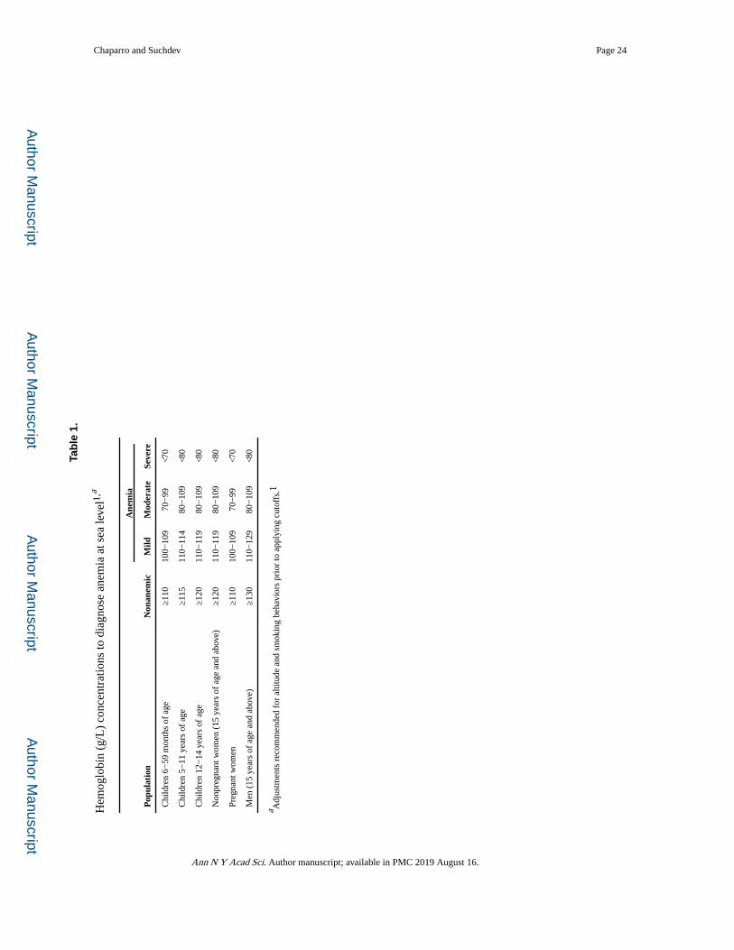
Author M
anuscriptA
uthor Manuscript
Author M
anuscriptA
uthor Manuscript
Chaparro and Suchdev Page 24
Tab
le 1
.
Hem
oglo
bin
(g/L
) co
ncen
trat
ions
to d
iagn
ose
anem
ia a
t sea
leve
l1,a
Ane
mia
Pop
ulat
ion
Non
anem
icM
ildM
oder
ate
Seve
re
Chi
ldre
n 6−
59 m
onth
s of
age
≥110
100−
109
70−
99<7
0
Chi
ldre
n 5−
11 y
ears
of
age
≥115
110−
114
80−
109
<80
Chi
ldre
n 12
−14
yea
rs o
f ag
e≥1
2011
0−11
980
−10
9<8
0
Non
preg
nant
wom
en (
15 y
ears
of
age
and
abov
e)≥1
2011
0−11
980
−10
9<8
0
Preg
nant
wom
en≥1
1010
0−10
970
−99
<70
Men
(15
yea
rs o
f ag
e an
d ab
ove)
≥130
110−
129
80−
109
<80
a Adj
ustm
ents
rec
omm
ende
d fo
r al
titud
e an
d sm
okin
g be
havi
ors
prio
r to
app
lyin
g cu
toff
s.1
Ann N Y Acad Sci. Author manuscript; available in PMC 2019 August 16.
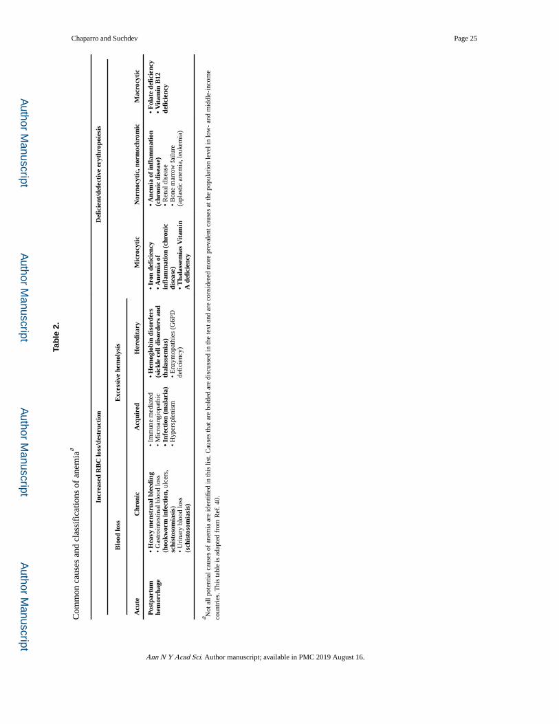
Author M
anuscriptA
uthor Manuscript
Author M
anuscriptA
uthor Manuscript
Chaparro and Suchdev Page 25
Tab
le 2
.
Com
mon
cau
ses
and
clas
sifi
catio
ns o
f an
emia
a
Incr
ease
d R
BC
loss
/des
truc
tion
Def
icie
nt/d
efec
tive
ery
thro
poie
sis
Blo
od lo
ssE
xces
sive
hem
olys
is
Acu
teC
hron
icA
cqui
red
Her
edit
ary
Mic
rocy
tic
Nor
moc
ytic
, nor
moc
hrom
icM
acro
cyti
c
Pos
tpar
tum
he
mor
rhag
e•
Hea
vy m
enst
rual
ble
edin
g•
Gas
troi
ntes
tinal
blo
od lo
ss
(hoo
kwor
m in
fect
ion,
ulc
ers,
sc
hist
osom
iasi
s)•
Uri
nary
blo
od lo
ss
(sch
isto
som
iasi
s)
• Im
mun
e m
edia
ted
• M
icro
angi
opat
hic
• In
fect
ion
(mal
aria
)•
Hyp
ersp
leni
sm
• H
emog
lobi
n di
sord
ers
(sic
kle
cell
diso
rder
s an
d th
alas
sem
ias)
• E
nzym
opat
hies
(G
6PD
de
fici
ency
)
• Ir
on d
efic
ienc
y•
Ane
mia
of
infl
amm
atio
n (c
hron
ic
dise
ase)
• T
hala
ssem
ias
Vit
amin
A
def
icie
ncy
• A
nem
ia o
f in
flam
mat
ion
(chr
onic
dis
ease
)•
Ren
al d
isea
se•
Bon
e m
arro
w f
ailu
re
(apl
astic
ane
mia
, leu
kem
ia)
• F
olat
e de
fici
ency
• V
itam
in B
12
defi
cien
cy
a Not
all
pote
ntia
l cau
ses
of a
nem
ia a
re id
entif
ied
in th
is li
st. C
ause
s th
at a
re b
olde
d ar
e di
scus
sed
in th
e te
xt a
nd a
re c
onsi
dere
d m
ore
prev
alen
t cau
ses
at th
e po
pula
tion
leve
l in
low
- an
d m
iddl
e-in
com
e co
untr
ies.
Thi
s ta
ble
is a
dapt
ed f
rom
Ref
. 40.
Ann N Y Acad Sci. Author manuscript; available in PMC 2019 August 16.