and heterodimeric troponin-C domains
10
Protein Science (1994), 3:lOlO-1019. Cambridge University Press. Printed in the USA. Copyright 0 1994 The Protein Society Relative stabilities of synthetic peptide homo- and heterodimeric troponin-C domains GARY S. SHAW,’ ROBERT S. HODGES, CYRIL M. KAY, AND BRIAN D. SYKES Department of Biochemistry and MRC Group in Protein Structure and Function, University of Alberta, Edmonton, Alberta T6G 2H7, Canada (RECEIVED March 4, 1994; ACCEPTED April 29, 1994) Abstract It has previously been shown that synthetic peptides corresponding to calcium-binding sites I11 (SCIII) and IV (SCIV) from troponin-C can undergo a calcium-induced dimerization to form the respective homodimers (Shaw CS, Hodges RS, Sykes BD, 1990, Science 249:280-283; Shaw CS et al., 1992a, JAm Chem SOC 114:6258-6259). In addition, an equimolar mixture of SCIII and SCIV has been shown to form preferentially the SCIII/SCIV heterodimer (Shaw CS et al., 1992a, J A m Chem SOC 114:6258-6259). The stabilities of these dimers havebeen investigated by using ‘H-NMR and circular dichroism spectroscopies to follow temperature- and guanidine hy- drochloride (GuHC1)-induced denaturations. It has been found that the most stable species, the SCIII/SCIV heterodimer (AG?’ = -64.8 kJ/mol), is about 13 kJ/mol more stable than the least stable species, the SCIV homodimer, while the SCIII homodimer is of intermediate stability. This trend of free energies agrees well with the trend of AGO values derived from the products of the dissociation constants for calcium binding and peptide association determined from earlier calcium-titration studies. These observations provide evidence that calcium affinity and the associationof 2-calcium binding sites are tightly linked. However, it was noted that in all cases AGO was considerably more negative than AGFZo determined from GuHCl experiments. This difference increased as the stability of the peptide complex increased, providing evidence that linear extrapolation of GuHCl data for very stable proteins may significantly underestimate the value for AGO. Keywords: calcium-binding sites; denaturation; synthetic peptides; troponin-C A common structural motif among many calcium-binding pro- teins, including troponin-C (TnC), parvalbumin, recoverin, and calmodulin, is the “EF-hand” or helix-loop-helix motif where a contiguous stretch of about 30 amino acids forms each calcium-binding site. As the name implies, the helix-loop-helix calcium-binding site is comprised of a central 12-residue loop, where calcium is coordinated, and2 flanking helices (Kretsinger & Nockolds, 1973). One observation from sequence analyses of helix-loop-helix calcium-binding proteins (Marsden et al., 1989) is that these sites usually occur in pairs, and up to8 copies of this motif may exist in a particular protein. This has been con- firmed from X-ray crystallographic studies of several of the helix-loop-helix calcium-binding proteins, which have shown that the 2 sites in parvalbumin (Kretsinger & Nockolds, 1973; Reprint requeststo: Brian D. Sykes, Department of Biochemistry and MRC Group in Protein Structure and Function, University of Alberta, Edmonton, Alberta T6G 2H7, Canada; e-mail: [email protected]. ualberta.ca. ’ Present address: Department of Biochemistry and R.S. McLaugh- lin Macromolecular Structure Facility, The University of Western On- tario, London, Ontario N6A 5C1, Canada; e-mail: shaw@merlin. biochem.uwo.ca. Declercq et al., 1988; Swain et al., 1989), oncomodulin (Ahmed et al., 1990), and calbindin Dgk (Szebenyi et al., 1986) each form a 2-site domain, whereas the 4 calcium-bindingsites in TnC (Herzberg & James, 1988; Satyshur et al., 1988) and calmodu- lin (Babu et al., 1988) form 2 independent 2-site domains. An analysis of these X-ray structuresreveals several common struc- tural components (Strynadka & James, 1989). Firstly, the 2 calcium-binding sites are pseudosymmetric, having an approx- imate 2-fold rotational relationship between helix-loop-helix mo- tifs. Secondly, a short 3-residue antiparallel b-sheet is formed between the amino acids at positions 7-9 of each calcium- binding loop. Finally, 2 distinct hydrophobic “pockets” are formed at the interface of the2-site domain. For example, in the C-terminal domain of TnC, the first hydrophobic center is formed at the interface of the incoming helix (helix E) of site 111 and the outgoing helix (helix H) of site IV. The second is formed at the interface of the outgoing helix (helix F) ofsite 111 and the incoming helix (helix G) of site IV. These common structural features undoubtedly result in the considerable stabilities of these 2-site domains. For example, parvalbumin (Williams et al., 1986; Cox et al., 1990) and the C-terminal domain of TnC (Tsalkova & Privalov, 1980, 1985) are all stable at temperatures in excess of 90 “C in the calcium- 1010
Transcript of and heterodimeric troponin-C domains

Protein Science (1994), 3:lOlO-1019. Cambridge University Press. Printed in the USA. Copyright 0 1994 The Protein Society
Relative stabilities of synthetic peptide homo- and heterodimeric troponin-C domains
GARY S. SHAW,’ ROBERT S. HODGES, CYRIL M. KAY, AND BRIAN D. SYKES Department of Biochemistry and MRC Group in Protein Structure and Function, University of Alberta, Edmonton, Alberta T6G 2H7, Canada (RECEIVED March 4, 1994; ACCEPTED April 29, 1994)
Abstract
It has previously been shown that synthetic peptides corresponding to calcium-binding sites I11 (SCIII) and IV (SCIV) from troponin-C can undergo a calcium-induced dimerization to form the respective homodimers (Shaw CS, Hodges RS, Sykes BD, 1990, Science 249:280-283; Shaw CS et al., 1992a, J A m Chem SOC 114:6258-6259). In addition, an equimolar mixture of SCIII and SCIV has been shown to form preferentially the SCIII/SCIV heterodimer (Shaw CS et al., 1992a, J A m Chem SOC 114:6258-6259). The stabilities of these dimers have been investigated by using ‘H-NMR and circular dichroism spectroscopies to follow temperature- and guanidine hy- drochloride (GuHC1)-induced denaturations. It has been found that the most stable species, the SCIII/SCIV heterodimer (AG?’ = -64.8 kJ/mol), is about 13 kJ/mol more stable than the least stable species, the SCIV homodimer, while the SCIII homodimer is of intermediate stability. This trend of free energies agrees well with the trend of AGO values derived from the products of the dissociation constants for calcium binding and peptide association determined from earlier calcium-titration studies. These observations provide evidence that calcium affinity and the association of 2-calcium binding sites are tightly linked. However, it was noted that in all cases AGO was considerably more negative than AGFZo determined from GuHCl experiments. This difference increased as the stability of the peptide complex increased, providing evidence that linear extrapolation of GuHCl data for very stable proteins may significantly underestimate the value for AGO.
Keywords: calcium-binding sites; denaturation; synthetic peptides; troponin-C
A common structural motif among many calcium-binding pro- teins, including troponin-C (TnC), parvalbumin, recoverin, and calmodulin, is the “EF-hand” or helix-loop-helix motif where a contiguous stretch of about 30 amino acids forms each calcium-binding site. As the name implies, the helix-loop-helix calcium-binding site is comprised of a central 12-residue loop, where calcium is coordinated, and 2 flanking helices (Kretsinger & Nockolds, 1973). One observation from sequence analyses of helix-loop-helix calcium-binding proteins (Marsden et al., 1989) is that these sites usually occur in pairs, and up to 8 copies of this motif may exist in a particular protein. This has been con- firmed from X-ray crystallographic studies of several of the helix-loop-helix calcium-binding proteins, which have shown that the 2 sites in parvalbumin (Kretsinger & Nockolds, 1973;
Reprint requests to: Brian D. Sykes, Department of Biochemistry and MRC Group in Protein Structure and Function, University of Alberta, Edmonton, Alberta T6G 2H7, Canada; e-mail: [email protected]. ualberta.ca. ’ Present address: Department of Biochemistry and R.S. McLaugh- lin Macromolecular Structure Facility, The University of Western On- tario, London, Ontario N6A 5C1, Canada; e-mail: shaw@merlin. biochem.uwo.ca.
Declercq et al., 1988; Swain et al., 1989), oncomodulin (Ahmed et al., 1990), and calbindin Dgk (Szebenyi et al., 1986) each form a 2-site domain, whereas the 4 calcium-binding sites in TnC (Herzberg & James, 1988; Satyshur et al., 1988) and calmodu- lin (Babu et al., 1988) form 2 independent 2-site domains. An analysis of these X-ray structures reveals several common struc- tural components (Strynadka & James, 1989). Firstly, the 2 calcium-binding sites are pseudosymmetric, having an approx- imate 2-fold rotational relationship between helix-loop-helix mo- tifs. Secondly, a short 3-residue antiparallel b-sheet is formed between the amino acids at positions 7-9 of each calcium- binding loop. Finally, 2 distinct hydrophobic “pockets” are formed at the interface of the 2-site domain. For example, in the C-terminal domain of TnC, the first hydrophobic center is formed at the interface of the incoming helix (helix E) of site 111 and the outgoing helix (helix H) of site IV. The second is formed at the interface of the outgoing helix (helix F) of site 111 and the incoming helix (helix G) of site IV.
These common structural features undoubtedly result in the considerable stabilities of these 2-site domains. For example, parvalbumin (Williams et al., 1986; Cox et al., 1990) and the C-terminal domain of TnC (Tsalkova & Privalov, 1980, 1985) are all stable at temperatures in excess of 90 “C in the calcium-
1010

Stabilities of troponin-C peptide dimers 101 1
bound form. Conversely, in the apo states these proteins are sig- nificantly less stable and are unfolded completely at much lower temperatures. A corollary of this observation is that while the folded forms of these proteins are all similar in the presence of calcium, the structures of the apo forms can be markedly dif- ferent. For example, the C-terminal domain of TnC has little or no regular structure in the absence of calcium (Drabikowski et al., 1982), whereas both apo-parvalbumin (Williams et al., 1986) and apo-calbindin Dgk (Kordel et al., 1989; Skelton et al., 1990) appear to have a similar fold compared to their respec- tive calcium-bound forms.
A novel technique to assess the calcium-binding properties of the helix-loop-helix calcium-binding proteins is to use synthetic peptides that represent a single metal-ion site (Reid et al., 1981; Garitpy et al., 1983, 1985). Recently, we have determined that this approach is particularly applicable for 2-site calcium-binding domains, which can be assembled from a pair of synthetic pep- tides each comprising a single helix-loop-helix sequence (Shaw et al., 1990). In this manner, it has been possible to form the site I11 (Shaw et al., 1990, 1991a) and site IV (Kay et al., 1991) ho- modimers of TnC in the presence of calcium. The 3-dimensional structures for these 2 TnC homodimers have now been deter- mined using NMR spectroscopy (Kay et al., 1991; Shaw et al., 1992b). In each case the 2 peptides comprising the dimer are symmetrically arranged in a head-to-tail fashion, similar in struc- ture to that observed for sites 111 and IV in the X-ray structures of TnC (Herzberg& James, 1988; Satyshur et al., 1988). It has also been shown by Monera et al. (1992) that hydrophobic in- teractions between the a-helices are not only responsible for di- merization but also have a strong influence on the calcium affinity of the 2-site domain. More recently it has been observed that a site III/site IV heterodimer from TnC is preferentially formed compared to the individual homodimers (Shaw et al., 1992a, 1992~). These observations have prompted the present work where we have used the site I11 and site 1V TnC homodi- mers and the site III/site IV heterodimer to investigate the ba- sis for the stability of the C-terminal domain of TnC in the presence of calcium. This has been accomplished by monitor- ing the chemical unfolding equilibria of each dimer by NMR and circular dichroism spectroscopies.
Results
Peptides SCIII and SCIV each represent a single helix-loop-helix calcium-binding site from chicken skeletal TnC (Fig. 1). In the absence of calcium these peptides or ones very similar in length possess little regular secondary structure as assessed by either CD or ' H NMR spectroscopy (Reid et al., 1981; Caritpy et al., 1983, 1985; Shaw et al., 1990, 1991a; Monera et al., 1992). In addition, an equimolar mixture of the SCIII and SCIV peptides shows no evidence of any interaction between the 2 peptides. However, in the presence of calcium, the SCIII, SCIV, and SCIII/SCIV peptides bind the metal ion and form a well-folded species (Shaw et al., 1992a). From early studies using CD, it was determined that similar folded peptides contained a high degree of a-helix structure and that this could be used to monitor the degree that the peptide was folded based on the ellipticity min- imum at 222 nm (Reid et al., 1981; Drabikowski et al., 1982; Monera et al., 1992).
Although C D spectroscopy provides a useful method to mon- itor the helix-coil transition in calcium-binding peptides, it yields
I11
126
little information about the 3-dimensional structure of the folded polypeptide. High-resolution NMR spectroscopy has been used to determine that single-site peptides from the C-terminal of TnC form dimers in solution only in the presence of calcium (Shaw et al., 1990, 1991a, 1992a; Kay et al., 1991; Monera et al., 1992). In the case of SCIII, an analysis of calcium titration data and NOE spectra showed that this peptide existed as a symmet- ric homodimer in solution (Shaw et al., 1990, 1991a). By anal- ogy SCIV was also shown to form a symmetric homodimer although at much higher peptide and calcium concentrations (Shaw et al., 1992a). A site IV peptide fragment from rabbit TnC was also shown to be a homodimer using sedimentation equi- librium and NMR spectroscopy (Kay et al., 1991). In addition, the preferential formation of an SCIII/SCIV heterodimer was determined using NMR spectroscopy, calcium titration experi- ments, and a comparison with the 'H-NMR spectrum of a C-terminal domain tryptic fragment (Shaw et al., 1991b, 1992a, 1992b, 1992~).
Mechanism of folding and unfolding
A mechanism for the formation of folded dimers, which in- volves calcium binding and peptide association, has been pro- posed based on calcium titration data (Shaw et al., 1991a, 1992c) and is shown below. In the unfolding pathway the calcium- saturated dimer F2Ca2 loses a calcium ( K 2 ) to form a 1-calcium dimer, F,Ca. Dissociation of this dimer (Kdimer) can form an
1012 G.S. Shaw et al.
unfolded apo-peptide, U, and the folded I-calcium monomer, FCa, which is in equilibrium ( K , ) with the unfolded apo- peptide and calcium.
F2Ca2 + F2Ca + Ca K 2 (1)
F2Ca + FCa + U Kdimer (2)
FCa + U + Ca K' (3)
Ideally, this folding pathway is most easily studied if it is sim- plified to a 2-state model, existing between folded dimer (F2Ca2) and unfolded monomer (U), as has been done in many other studies (Ho & DeGrado, 1987; Bowie & Sauer, 1989; Hodges et al., 1990; De Francesco et al., 1991; Zhu et al., 1992) using CD spectroscopy to follow the helix-to-coil transition. The complicating factor in the present pathway compared to other simple 2-state pathways is the presence of bound calcium ions. In the above mechanism, originally proposed for SCIII, at least 2 intermediates may exist, FCa and F2Ca (Shaw et al., 1991a, 1992~). It has previously been found that the unfolding reaction is dependent on peptide concentration (Monera et al., 1992), which rules out the unimolecular reaction in Equation 3 as the one monitored by CD spectroscopy. Also, we have calculated that the calcium dissociation constants for the SCIII, SCIV, and SCIII/SCIV dimers range between 0.002 and 2 mM (Shaw et al., 1991a, 1991b, 1992~). Based on these observations, we used an excess of calcium (20 mM) for CD experiments to minimize the effects that calcium would have on the denaturation experiments and to ensure that the FzCa2 species was populated through the unfolding transition.2 This allows the proposed pathway to be simplified to a 2-state mechanism (Equation 4) where only the 2-calcium folded dimer, F2Ca2, and the unfolded monomer, U, are present in significant amounts in the transition zone so that
F2Ca2 + 2U + 2Ca K, , (4)
where
and f, is the molar fraction of folded peptide, P, is the total peptide concentration, and K , is the dissociation constant for the equilibrium. From this generalized form a more specific case can be obtained when Car >> P, (Equation 6) as in the CD ex- periments where Car is at least 100-fold greater than the peptide concentration (P,). This allows a pseudo-dissociation constant Kobs to be calculated (Equation 7) from the molar fraction of folded peptide based on OZz2.
* This series of equilibria can also be expressed in a manner that may be easier to envision as a 2-state model between unfolded monomers and folded monomers and dimers by rewriting the above expressions as
P2Ca2 = 2PCa K;l i ,er
2PCa + 2U + 2Ca K ,
For these equilibria, K I is identical to that in Equation 3 and K&,,e, = ( K 2 / K , )Kdimer. However, this analysis cannot account for the appar- ent stoichiometry 0.5 Ca: 1.0 peptide, which is observed in calcium ti- trations (Shaw et al., 1991a).
where
In 'H-NMR experiments of the SCIII/SCIV heterodimer where Car 2 P, >> K I , K2, Kdrmer, Equation 5 was used to cal- culate K, . Equations 5 , 6, and 7 are identical to those used pre- viously for simple 2-state unfolding transitions (Ho & DeGrado, 1987; Bowie & Sauer, 1989; Hodges et al., 1990; De Francesco et al., 1991; Zhu et al., 1992) with the exception of terms to ac- count for calcium in the present experiments.
Thermal stability
Unfolding of the calcium-saturated SCIII/SCIV heterodimer was monitored using 'H-NMR spectroscopy as a function of temperature. In Figure 2, a series of spectra are shown for the thermal unfolding of the SCIII/SCIV heterodimer (169 PM) in the presence of 448 pM Ca2+ between 5 and 70 "C. This figure shows that at the lower temperatures (5-40 "C) the spectra are all very similar in terms of the positions and intensities of the resonances. However, as the temperature of the sample is in- creased above 40 "C, resonances characteristic of the folded form of the protein broaden, shift, and lose intensity until even- tually most of the resonances from the heterodimer are nearly gone. Because the 'H resonances marked on Figure 2 corre- spond to residues throughout the heterodimer, it is clear that this transition is a result of global unfolding of the protein rather than a localized effect. In the final spectrum at 70 "C, most of the resonances, although very broad, are found near the fre- quencies observed for the apo-peptide mixture at 30 "C (Shaw et al., 1992a).
The unfolding of the SCIII/SCIV heterodimer in the presence of calcium is shown graphically in Figure 3A, where the inte- grated intensity for the resonances noted in Figure 2 are plot- ted versus the temperature. Only resonances for the SCIII/SCIV heterodimer, which exhibit a unique frequency from those in the apo-peptide mixture, were used. The molar fraction of folded dimer, f r , was calculated from
where If is the integral of a particular 'H resonance when the peptide is fully dimerized and robs is the observed integral at a given temperature. Figure 3A shows that little thermal unfold- ing of the SCIII/SCIV heterodimer occurs between 5 and 40 "C where the fraction of folded dimer is between 90 and 100%. Above 40°C however, the proportion of dimer steadily de- creases until, at 70 "C, only about 28% of the folded dimer is present. These data were fit using a least-squares sigmoidal func- tion that had a midpoint, T, = 60°C, corresponding to the melting temperature of the SCIII/SCIV heterodimer at 169 PM. Using the data in the transition range between 30 and 70 "C, a van't Hoff plot of thermal denaturation for the SCIII/SCIV het- erodimer was constructed (Fig. 3B) based on the 2-state unfold- ing model. Over this temperature range the data yielded a linear correlation between In K , and 1/T. From this graph the slope was used to determine the enthalpy of unfolding. A value of

Stabilities of troponin-C peptide dimers 1013
7 . 6 7 . 4 7 . 2 7 . 0 6 . 8 6 . 6 6 . 4 6 . 2 6 . 0 5 . 8 5 . 6 ppm
AH,, = 183 kJ/mol was found, which is in the same range as that obtained for the unfolding of the calcium-saturated TR2C fragment of TnC (218 kJ/mol; Tsalkova & Privalov, 1985) and calcium-saturated rat a-parvalbumin (183 kJ/mol; Williams et al., 1986).
CD spectra
Representative CD spectra for the SCIII, SCIV, and SCIII/SCIV dimers and the C-terminal domain (residues 89-162) from chicken skeletal troponin-C (CTnC) are shown in Figure 4, each in the presence of excess calcium. The spectra are typical of pep- tides containing a high proportion of a-helix having minima near 208 nm and 222 nm (Greenfield & Fasman, 1969). A compari-
V%5
0.9 0 . 7 0 . 5 pp"
Fig. 2. Temperature dependence of 2 re- gions of the 500-MHz 'H-NMR spectrum of 169 pM SCIII/SCIV heterodimer in D20,50 mM KCI, 30mM imidazole d.,, and 448 pM CaCI2 at pH 7.2. A: The aromatic and some of the crCH protons. B: The up- field methyl protons in the heterodimer, each plotted as a function of temperature. Assigned resonances for the SCIII/SCIV heterodimer are indicated as: F102 6CH (O), F151 6CH (A), F154eCH (V), Ile 149aCH and Y112 crCH (W), 1104 ?CH3 and 1121 ?CH3 (A), and I113 6CH3 (V).
son of the ellipticities at 222 nm for the peptide dimers shows that the SCIII homodimer has the greatest minimum (-14,870 degrees), the SCIV homodimer has the least (- 11,630 degrees), and the SCIII/SCIV heterodimer is intermediate (-12,600 de- grees). This suggests that the SCIII dimer may have more a- helical content than does SCIV. These observations are in agreement with NMR studies, which have determined that 18- 20 residues are in an a-helix conformation in each monomer of the SCIII dimer (Shaw et al., 1992b), whereas about 17 residues are in an a-helix conformation in each monomer of the site IV homodimer (Kay et al., 1991). The ellipticity for CTnC was -15,220 degrees, slighty larger than that obtained for any of the dimeric peptides, but in good agreement with that measured previously on a tryptic fragment from rabbit skeletal TnC com-
I ' I ' I ' 1 ' I ' I . I Fig. 3. A: Thermal denaturation of the SCIII/SCIV heterodimer derived from the integrals of the 'H-NMR reso- nances indicated in Figure 2. The frac- tion of folded dimer (f,) is plotted as a function of temperature and was cal- culated as described in the Materials and methods. The first 3 data points are not
- shown. The data were iteratively fit to
plot of In K,, vs. 1/T generated from the data between 35 and 70°C in A.
data over this range ( r2 = 0.997) to yield A H = 183 kJ mol-'. From this value of A H and the value of AG = -68 kJ
0.6 -
0.4 - -28 - determine T, = 60°C. B: van't Hoff
0.2 - Linear regression was used to fit the -30 -
-32 -
0 20 40 60 80 100 120 140 Temperature ('C)
2.7 2.8 2.9 3.0 3.1 3.2 3.3 3.4 3.5 mol-' at T, = 60 QC, a value of AS = l/T ('K) x 1000 346 J K" mol-' was calculated.
1014 G.S. Shaw et al.
h
0
0
X m
7
0
-5
-10
-15
-20 200 210 220 230 240 250
Wavelength (nm)
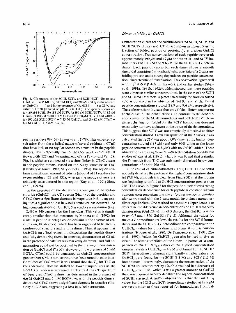
Fig. 4. CD spectra of the SCIII, SCIV, and SCIII/SCIV dimers and CTnC in 10 mM MOPS, 50 mM KCl, and 20 mM CaC12 in the absence of GuHCl (-) and in the presence of GuHCl (---) at 25 "C and either pH 7.20 (dimers) or pH 7.15 (CTnC). The spectra shown are (a) 189 p M SCIII, (b) 188 pM SCIV, (c) 190 pM SCIII/SCIV, (d) 82 pM CTnC, (e) 189 pM SCIII + 5 M GuHCl, ( f ) 188 pM SCIV + 5 M GuHCl, (g) 190 pM SCIII/SCIV + 7.33 M GuHCI, and (h) 82 pM CTnC + 6.6 M GuHCl + 5 mM EGTA.
prising residues 89-159 (Leavis et al., 1978). This expected re- sult arises from the a-helical nature of several residues in CTnC that have little or no regular secondary structure in the peptide dimers. This is especially true for the C-terminal end of site I11 (toward Gly 126) and N-terminal end of site IV (toward Val 129, Fig. l), which are connected via a short linker in CTnC absent in the peptide dimers. Based on the X-ray structure of TnC (Herzberg & James, 1988; Satyshur et al., 1988), this region con- tains a significant amount of a-helix (about 4 of 11 residues be- tween residues 122 and 132), whereas the peptide dimers are relatively unstructured in this region (Kay et al., 1991; Shaw et al., 1992b).
In the presence of the denaturing agent guanidine hydro- chloride (GuHCI), the CD spectra (Fig. 4) of the peptides and CTnC show a significant decrease in magnitude in suggest- ing that a significant loss in a-helix structure has occurred. At high concentrations of GuHCI, reaches a maximum (avg. -2,430 +. 440 degrees) for the 3 peptides. This value is signifi- cantly smaller than that measured by Monera et al. (1992) for a site I11 peptide in benign conditions and in the absence of cal- cium (-6,300 degrees), which has been suggested to be mostly random-coil structure and is not a dimer. Thus, it appears that GuHCl is an effective agent in dissociating the peptide dimers and fully denaturing them. In contrast, denaturation of CTnC in the presence of calcium was markedly different, and full de- naturation could not be obtained in the maximum concentra- tion of GuHCl used (7.8 M). However, in the presence of 5 mM EGTA, CTnC could be denatured at GuHCl concentrations greater than 6 M. A similar result has been noted in calorimet- ric studies of TnC where it was found that the T, for TnC or the C-terminal domain shifted to lower temperatures as the EGTA:Ca ratio was increased. In Figure 4 the CD spectrum of denatured CTnC is shown as determined in the presence of 6.6 M GuHCl and 5 mM EGTA. As with the peptide dimers, denatured CTnC shows a significant decrease in negative ellip- ticity at 222 nm, suggesting a loss in a-helix structure.
Dimer unfolding by GuHCI
Denaturation curves for the calcium-saturated SCIII, SCIV, and SCIII/SCIV dimers and CTnC are shown in Figure 5 as the fraction of folded peptide or protein, ff, at a given GuHCl concentration. Two concentrations of each peptide were used: approximately 190 pM and 19 pM for the SCIII and SCIV ho- modimers and 190 pM and 9.6 p M for the SCIII/SCIV hetero- dimer. Each pair of curves for each dimer shows a smooth sigmoidal transition (monophasic) characteristic of a 2-state un- folding process and a strong dependence on peptide concentra- tion, characteristic of dimerization. This observation agrees well with the 'H-NMR data in this work and earlier studies (Shaw et al., 1991a, 1991b, 1992~). which showed that these peptides were dimers at similar concentrations. In the cases of the SCIII and SCIII/SCIV dimers, a plateau near unity for fraction folded ( ff) is obtained in the absence of GuHCl and at the lowest peptide concentrations studied (18.9 and 9.6 pM, respectively). These observations indicate that only folded dimers are present at the outset of the denaturations. In contrast to the denatur- ation curves for the SCIII homodimer and SCIII/SCIV hetero- dimer, the fraction folded for the SCIV homodimer does not reach unity nor have a plateau at the outset of the denaturations. This suggests that SCIV was not completely dimerized at either concentration studied. From extrapolation of the 2 curves it was calculated that SCIV was about 93% dimer at the highest con- centration studied (1 88 pM) and only 80% dimer at the lower peptide concentration (18.8 FM) with no GuHCl added. These observations are in agreement with sedimentation equilibrium studies of Kay et al. (1991), where it was found that a similar site IV peptide from TnC was only partly dimerized below con- centrations of about 700 PM.
In the case of calcium-saturated CTnC, GuHCl alone could not fully denature the protein at the highest concentration stud- ied (7.8 M), although it is clear from Figure 5D that the protein was beginning to unfold at GuHCl concentrations greater than 7 M. The curves in Figure 5 for the peptide dimers show a strong concentration dependence for each peptide at constant calcium concentration suggesting that the unfolding reaction is bimolec- ular as proposed with the 2-state model, involving a monomer- dimer equilibrium. One method to assess this dependence is to determine the difference in concentrations of GuHCl for 50% denaturation (GuHCl,,,). In all 3 dimers, the G u H C ~ , , ~ is be- tween 0.7 and 4.8 M GuHCl (Fig. 5) . Although the values for the SCIV homodimer are low, the results for the SCIII homo- dimer and the SCIII/SCIV heterodimer are comparable to the GuHCI,,~ values for other dimeric proteins at similar concen- trations (Hodges et al., 1990; De Francesco et al., 1991; Zhu et al., 1992). Values for GuHCI,,~ can also be used to give an idea of the relative stabilities of the dimers. In particular, a com- parison of the G u H C ~ , , ~ values of the highest concentration samples reveals a GuHCI,,~ = 4.8 M is obtained for the SCIIl/ SCIV heterodimer, whereas significantly smaller values for GuHCI,,, are found for the SCIII (3.1 M) and SCIV (1.3 M) homodimers. Interestingly, decreasing the concentration of the SCIII/SCIV heterodimer by 120-fold resulted in a decrease of GuHC11,2 to 3.3 M, which is still a greater amount of GuHCl than was required to 50% denature the highest concentration of SCIII assessed. A further observation is that the G U H C I ~ , ~ values for the SCIII and SCIV homodimers studied at 18.8 pM are very similar to those reported for homodimers from cal-

Stabilities of troponin-C peptide dimers 1015
1 .o
0.8
0.6
\- 0.4
0.2
0.0 0 1 2 3 4 5 6 7 0
[GuHCI] M
0 1 2 3 4 5 6 7 8
[GuHCI] M
0 1 2
bindin D,, (F1 and F2, 3 M and 1.5 M, respectively) when stud- ied at approximately 15 pM (Linse et al., 1993). Qualitatively, this suggests that in the presence of calcium the SCIII homodi- mer from TnC is similar in stability to the F1 homodimer .from calbindin Dgk and the SCIV and F2 homodimers are of similar stabilities. However, the dimers all have significantly lower val- ues for GuHCI,,, (at the concentrations measured) than CTnC ( G u H C ~ , , ~ = 8.4 M, estimated from curve-fitting of the avail- able data, Fig. 5D).
The observations made in Figure 5 indicate that each of the dimers have different stabilities toward GuHCl denaturation with the heterodimer SCIII/SCIV being the most stable, the ho- modimer SCIV the least stable, and the homodimer SCIII of in- termediate stability. This is shown in a more quantitative manner in Figure 6 , where the free energy of unfolding AG, (where AG, = -RTln K , = -RTln Kobs - RTln [Cat]*) is plotted against the GuHCl concentration for the data points, which wer.e in the transition zone (0.20 sfr s 0.80) in Figure 5 . These data were least-squares fit according to the equation:
AG, = A G p o + m[GuHCl], (9)
where A G F o is the unfolding free energy in the absence of de- naturant (Pace, 1975), which is extrapolated from Figure 6 (Ta- ble l ) . The graphs show that for each peptide dimer the denaturation data for the 2 concentrations of each peptide lie on the same curve (within experimental error). Again, this ob- servation is consistent with the initial 2-state proposal. The
Fig. 5. Guanidine denaturation curves for the SCIII, SCIV, and SCIII/SCIV dimers and CTnC in 10 mM MOPS, 50 mM KCI, and 20 mM CaCI2. Each curve is plotted as the fraction of folded (ff) vs. [GuHCl] for (A) 188 pM SCIV (0) and 18.8 pM SCIV (O), (B) 189pM SCIII (0) and 18.9 pM SCIII (O), (C) 190 pM (SCIII + SCIV) (0) and 9.6 pM (SCIII + SCIV) (O), and (D) 82 pM CTnC. The amount of folded dimer was determined from the magnitude of e222 using f/ = (oobs - &nin)/(emax - Om,"). Each curve was iteratively fit using a sigmoidal function to yield the best fit lines shown. This process also calculated the [GuHCI],,~ values for the dimers at high and low peptide con- centrations, respectively, which are (A) 1.3 and 0.7 M for SCIV, (B) 3.1 and 2.4 M for SCIII, (C) 4.8 and 3.3 M for SCIII/SCIV, and (D) 8.4 M for CTnC.
3 4 5 6 7 8 [GuHCI] M
slopes of the lines (Table l), which can in some cases be used to assess the degree of cooperativity of unfolding, are also sim- ilar having a range of -5 to -7.9 kJ/mol M. These values are very similar to the slopes obtained for the GuHCl denaturation of several coiled-coil dimers (De Francesco et al., 1991; Zhu et al., 1992) and tetramers (Ho & DeGrado, 1987). Interestingly,
Table 1. Free energies of unfolding for dimers and C-terminal domain of TnC
Dissociation constant (x106 M)
AGOa -AGF20b Dimer KI K2 Kdimer (kJ/mol) (kJ/mol) m
SCIV2 260 260 2,000 -56.3 -51.8 -7.9 SCIII2 3 2,000 SCIII/SCIV 3
10 -75.4 -61.5 -6.5 2 10 -92.5 -64.8 -5.0
CTnCC 0.02 0.02 - -87.8 - -
a AGO was calculated from AGO = -RTln K,,,, where K,,, is the product of the individual dissociation constants. For each of the dimers,
ACTZo is the free energy based on the equilibrium constant K , according to Equation 6, where [Ca,] = 20 mM. Values are quoted as -AGf2' to clarify comparison to AGO from calcium-binding studies.
c Calcium dissociation constants are from Leavis et al. (1978) for the C-terminal domain of rabbit TnC.
K,,, = K I K2Kdimer; for CTnC, Kto, = K1 K2.

1016 G.S. Shaw et al.
55
0 1 2 3 4 5 6 7 8
2
70 65 60 55
50 45 40 35 30 25
20 15 10 5
0 0 1 2 3 4 5 6 7 8
2
0 1 2 3 4 5 6 7 8 0 1 2 3 4 5 6 7 8
[GuHCI] M [GuHCI] M [GuHCI] M
Fig. 6 . Free energies of unfolding (AG,) for the (A) SCIV, (B) SCIII, and (C) SCIII/SCIV dimers. Symbols refer to concen- trations described in Figure 5 . Values for AGu were calculated from the fraction folded (h) shown in Figure 5 using Equation 6. The slopes for each curve are (A) -7.9 kJ/mol M for SCIV, (B) -6.5 kJ/mol M for SCIII, and (C) -5.0 kJ/mol M for SCIII/SCIV.
the slopes of the curves from Figure 6 are greater for the homo- dimers SCIII and SCIV than for the SCIII/SCIV heterodimer. This observation seems to be consistent with the slopes found for the denaturation of several synthetic peptide dimers where the heterodimer usually has a smaller value for the slope than the average of the 2 possible homodimers (Zhu et al., 1992).
Discussion
Synthetic peptides have been used previously to study calcium- induced folding of the helix-loop-helix motif and isolated loop motifs in calcium-binding proteins (Reid et al., 198 1 ; Gariepy et al., 1983, 1985; Marsden et al., 1988; Shaw et al., 1990, 1991a; Monera et al., 1992). These studies have been successful in mon- itoring conformational changes that occur during calcium bind- ing and in determining the effects that specific residues have on metal ion affinity. However, 1 important area where informa- tion is lacking is in the dependency that l helix-loop-helix has on its partner in a 2-site domain and how this contributes to the stability of the domain. The determination that helix-loop-helix peptides form a calcium-induced 2-site domain has now opened up this avenue of investigation (Shaw et al., 1991b; Monera et al., 1992). Recently, we have studied the effects that specific hydrophobic residues have on both the formation and the cal- cium affinity of the 2-site domain formed from 2 site I11 (SCIII) peptides (Monera et al., 1992). In the present study, we have ex- tended this work to study the stabilities of the SCIII and SCIV homodimers and SCIII/SCIV heterodimer and compared these to the stability for the C-terminal domain of TnC.
Thermodynamics of heterodimer formation
Previously, we have shown that calcium titration studies of a mixture of SCIII and SCIV results in the formation of only the SCIII/SCIV heterodimer (Shaw et al., 1992a). The GuHCl de- naturation studies in this work provide a rationale for this. A summary of the free energies of unfolding from these studies is shown in Table 1. From these data the SCIII/SCIV hetero- dimer exhibits a free energy of dimerization of about -65 kJ/mol, making it about 3 kJ/mol more stable than the SCIII homodimer and 13 kJ/mol more stable than the SCIV homo-
dimer. These differences in energy between dimers could result from differences between the energies of the folded states, the unfolded states, or both. Because the unfolded states of all 3 di- mers are random coil, whereas the folded dimers appear to have varying degrees of a-helix (from CD spectroscopy), it is prob- able that the differences in energy arise mostly from differences in the folded states. This is similar to that suggested for synthetic Zn-binding 4-helix bundle proteins (Handel et al., 1993). Fur- ther, all 3 dimers can be formed at 25 "C (the temperature of present experiments) and their formation is reversible. This anal- ysis suggests that, on the basis of the AGYZo values calculated here, one might expect nearly identical amounts of the SCIII/ SCIV heterodimer and SCIII homodimer to be formed and a lesser amount of the SCIV homodimer be formed from an equi- molar mixture of SCIII and SCIV peptides under thermody- namic control. However, the formation of the SCIII dimer also necessitates the formation of the SCIV dimer according to the thermodynamic equilibrium
SCIIIz + SCIVz ?= 2SCIII/SCIV
so that the SCIII/SCIV heterodimer will be preferentially formed when AGFZ0 (SCIII/SCIV) < 1/2[AG,HZo (SCII12) + AG,HZo (SCIV,)] + RTln 2. From the data in Table 1, it is clear that the free energy of folding for the SCIII/SCIV heterodimer (-65 kJ/mol) is significantly larger than l/2[AGfZ0 (SCII12) + AC,H2O (SCIV,)] + R T l n 2 (-58 kJ/mol). This difference of- fers a thermodynamic rationale for the presence of heterogenic 2-site domains in calcium-binding proteins in favor of the ho- mogeneous sites from which they no doubt evolved (Kretsinger, 1987). It is also noteworthy that this observation is similar to those found for preferential formation of cup tropomyosin (Lehrer et al., 1989; Lehrer & Qian, 1990), the Fos-PIN-Jun- p l N heterodimers (O'Shea et al., 1988, 1989), and in synthetic coiled-coil proteins (Zhu et al., 1992).
One interesting comparison that can be made from this work is that between the stability of the SCIII/SCIV heterodimer and CTnC. From Table l , the AGO values for these 2 complexes are comparable, suggesting that on this basis the 2 proteins are also of comparable stability. Because of the inaccuracies of the AGfZ0 value for CTnC (and SCIII/SCIV, see below), we have

Stabilities of troponin-C peptide dimers 1017
chosen to compare the GuHCI,,, values for these 2 complexes. This approach is not straightforward because GuHCI,,~ in- creases as the concentration of SCIII/SCIV increases, whereas GuHClIl2 for CTnC is concentration independent. However, a comparison can be made if a GuHCl denaturation curve for the SCIII/SCIV heterodimer is calculated using the AGu values from the curves shown in Figure 6C for the SCIII/SCIV hetero- dimer at a total peptide concentration of 1 M (standard-state conditions). When this was done for the SCIII/SCIV heterodi- mer, it was calculated that the peptide dimer would still be 93% folded even at 8 M GuHCI, making it comparable in stability to CTnC.
Comparison of A G F o with AGO from calcium dissociation constants
The free energies of folding for the 3 dimers ( A G F O ) were compared to the free energies calculated from the product of the 3 dissociation constants for the proposed formation of the pep- tide dimers (AGO, Equations 1-3) and also to published disso- ciation constants for the C-terminal domain of TnC. In the case of the C-terminal domain of TnC, AGO has also been measured calorimetrically for the binding of 2 calcium ions (Potter & Gergely, 1975; Yamada, 1978; Yamada & Kometani, 1982; Im- aizumi et al., 1987). Although studies for rabbit TnC are not so clear, a AGO = -85 kJ/mol was obtained for bullfrog TnC (Imaizumi et al., 1987), which is in excellent agreement with the AGO derived from dissociation constants (Table 1). Similar ob- servations are also available for several other calcium-binding proteins (for example see Cox et al., 1990).
Denaturation studies that monitor the stability of calcium- binding proteins in the calcium form are rare. Urea denatura- tions have been reported for both TnC (McCubbin et al., 1982) and calmodulin (Martin & Bayley, 1986), although complete un- folding of the proteins was not accomplished. Several studies have also been reported for calbindin Dgkr but in the calcium- free state. This lack of data likely stems from the tremendous stability of these proteins in the calcium form. As shown in Table 1, it is clear from the large negative AGO values, from calcium-binding experiments, that all 3 dimers and CTnC fit this trend. However, in all cases AGF' is significantly smaller than AGO. It is also apparent from these data that the differ- ence between AG,H2' and AGO is proportional to the stability of the complex studied. Thus, for the least stable dimeric species, the SCIV homodimer, this difference is -4.5 kJ/mol versus -27.7 kJ/mol for the SCIII/SCIV heterodimer. It is also likely that a similar situation exists for CTnC, although a good esti- mate of AGFZ0 could not be obtained from GuHCl experi- ments. One possible source for these observations is the curve-fitting method used to estimate A G F O , of which linear extrapolation is the most common and was the choice in this work. However, other methods, such as a quadratic dependence on GuHCl (Wendt et al., 1988) and the GuHC1-binding model (Pace, 1979, are available and generally yield a higher value for AGYz0. Using either of these models resulted in A G F o = -52.5 kJ/mol for the SCIV homodimer, which is essentially the same value obtained from linear extrapolation. However, for the SCIII/SCIV heterodimer, this is not the case and a value of A G F o = -73.0 kJ/mol is obtained from hyperbolic fitting and -81.6 kJ/mol is obtained from the GuHC1-binding model. These values are considerably more negative than from linear
extrapolation (-64.8 kJ/mol) and are now approaching the value obtained of AGO = -92.5 kJ/mol from calcium-binding experiments. It has been suggested that the linear extrapola- tion method probably yields a A G F O , which is too low and a method such as the GuHC1-binding model yields values of AGF', which are too high (Pace, 1975). However, the results in this work may suggest that, for very stable proteins and pep- tide complexes, a nonlinear curve-fitting routine may be more appropriate to obtain a better estimate of AGO.
Cooperativity of folding and calcium binding From earlier calcium-binding studies it was found that all 3 peptides bound calcium with a range of dissociation constants and formed dimers. It has also been suggested that these 2 pro- cesses are tightly linked (Shaw et al., 1991b, 1992c; Monera et al., 1992). From the guanidine denaturation studies here, sedimentation-equilibrium studies (Kay et al., 1991), and calcium-binding studies, it is clear that SCIV not only has the highest calcium dissociation constant ( K , ) but also forms a di- mer at significantly higher concentrations than either the SCIlI homodimer or SClII/SCIV heterodimer. However, in the cases of the SCIII and SCIII/SCIV dimers, both of these species formed dimers in the presence of calcium at the lowest concen- trations tested here (19 pM and 9.6 pM, respectively) suggest- ing that the dimer dissociation constant, K d , must at least be in this range. This finding is in agreement with the calculated val- ues from calcium-titration data (Table 1). When linking these dissociation constants to the calcium-dissociation constants for the SCIII homodimer and the SCIII/SCIV heterodimer, one finds that K , (binding of calcium to SCIII) is 3.0 X M and reflects relatively tight calcium binding. This value is about 1,000-fold tighter than binding to SCIV and shows that calcium binding to the SCIII/SCIV heterodimer and to the C-terminal of TnC must be sequential and that the initial step must be cal- cium binding to site I11 only. However, it is the second calcium binding event ( K 2 ) upon which dimer formation has the great- est impact. From Table 1, it is noted that in the SCIII homodi- mer K2 >> K , , whereas in the SCIII/SCIV heterodimer KZ = K, . This suggests that in SCIII, dimer formation perturbs binding of a second molecule of calcium, whereas in the SCIII/SCIV heterodimer, calcium, binding is enhanced so that calcium bind- ing to the SCIV portion of the heterodimer is increased about 100-fold compared to that in the SCIV homodimer.
Materials and methods
Peptide synthesis and purification
Peptides comprising site I11 (residues 93-126; SCIII) and site IV (residues 129-162; SCIV) from chicken skeletal TnC were syn- thesized according to the procedures of Shaw et al. (1991b) and Monera et al. (1992) using standard t-boc chemistry on an Ap- plied Biosystems model 430A peptide synthesizer. Crude pep- tides were purified using reversed-phase HPLC, and the amino acid composition of the purified peptides was confirmed using amino acid analysis and mass spectrometry.
NMR spectroscopy
'H-NMR spectra were collected using a Varian VXR-500 spec- trometer. Peptide samples were prepared in 500 pL D20 buffer

1018 G.S. Shaw et al.
containing 30 mM imidazole-d4 and 50 mM KC1 at pH 7.30 (uncorrected meter reading). The SCIII/SCIV peptide solution was prepared from individual SCIII and SCIV peptide solutions to give a sample that contained 179 f 8 pM SCIII and 159 * 13 pM SCIV as measured by quantitative amino acid analysis (see below). Because these concentrations are the same within experimental error, an average peptide concentration of 169 pM was used.
Thermal denaturations were done over a temperature range of 5-70 "C at 5 "C intervals. At each temperature the sample was allowed to equilibrate for at least 30 min before a spectrum was acquired.
CD spectroscopy
Individual stock solutions of 1.13 mM SCIII and 1.18 mM SCIV were prepared in 1 .O mL of 10 mM 3-(N-morpholino)propane- sulfonic acid (MOPS) containing 50 mM KC1 and 20 mM CaCI2 at pH 7.20. A stock solution of 0.49 mM recombinant C-terminal domain (residues 89-162) from chicken skeletal troponin-C was prepared in 0.5 mL of the same buffer includ- ing 5 mM dithiothreitol (DTT) at pH 7.15. The concentrations of these peptide and protein solutions were then determined from triplicate amino acid analyses using a Beckman Model 6300 amino acid analyzer equipped with a 25-cm ion-exchange col- umn and post-column ninhydrin detection. For SCIII and CTnC, quantitative measurement was made by comparing the peak volumes for alanine and leucine to those of a 10-nmol stan- dard sample containing these amino acids or an internal 3-nmol norleucine standard. For SCIV, a similar procedure was used for leucine residues only because SCIV contains no alanine residues. For the SCIII/SCIV solution, the appropriate volumes from 1.30 mM SCIII and 0.93 mM SCIV solutions were combined to yield an equimolar SCIII/SCIV stock solution containing 1.15 mM total peptide.
The CD spectra of SCIII, SCIV, and SCIII/SCIV were each determined at 2 different concentrations by diluting each of the stock solutions by 6-fold and 60-fold for SCIII and SCIV and 6-fold and 120-fold for SCIII/SCIV. CD spectra for the CTnC sample were acquired using a 6-fold dilution of the stock sam- ple. Dilution was accomplished using the same 10 mM MOPS, 50 mM KCI, and 20 mM CaCI, buffer as the original stock samples were prepared in. CD spectra were measured using a JASCO 5-720 spectropolarimeter interfaced with an Epson Eq- uity computer and controlled by JASCO software. Spectra were recorded using calibrated 0.2- and 0.02-cm path length cells at 25 "C using a thermostatted cell holder and circulating water bath.
Denaturation of the peptide dimers was done by incubating aliquots of the peptide solutions at several concentrations of GuHCI. Stock 6 M and 8 M GuHCl solutions were prepared in 10 mM MOPS, 50 mM KCI, and 20 mM CaCI, buffer and used for all additions. All incubations were left to stand overnight to ensure the samples had equilibrated prior to the acquisition of the individual CD spectra.
Acknowledgments
We thank Kim Oikawa for acquiring the C D spectra for us and Paul Semchuk for peptide synthesis and purification of SCIII and SCIV. We are grateful to Dr. L. Smillie for the sample of CTnC.
References
Ahmed FR, Przybylska M, Rose DR, Birnbaum GI, Pippy ME, MacMa- nus JP. 1990. Structure of oncomodulin refined at 1.85 A resolution: An
216:127-140. example of extensive molecular aggregation via Ca2+. J Mol Bio/
Babu YS, Bugg CE, Cook WJ. 1988. Structure of calmodulin refined at 2.2 A resolution. J Mol Biol203:191-204.
Bowie JU, Sauer RT. 1989. Equilibrium dissociation and unfolding of the
Cox J, Milos M, MacManus JP. 1990. Calcium- and magnesium-binding arc repressor dimer. Biochemistry 28:7139-7143.
Declercq JP, Tinant B, Parello J, Etienne G, Huber R. 1988. Crystal struc- properties of oncomodulin. J Bioi Chem 265:6633-6637.
ture determination and refinement of pike 4.10 parvalbumin (minor com- ponent from Esox lucius). JMol Biol202:349-353.
De Francesco R, Pastore A, Vecchio G, Cortese R. 1991. Circular dichro- ism study on the conformational stability of the dimerization domain
Drabikowski W, Brzeska H, Venyaminov SY. 1982. Tryptic fragments of cal- of transcription factor LFB1. Biochemistry 30:143-147.
modulin: Ca2+ and Mg2+-induced conformational changes. J Biol Chem 257:11584-11590.
Gariepy J, Kay LE, Kuntz ID, Sykes BD, Hodges RS. 1985. Nuclear mag- netic resonance determination of metal-proton distances in a calcium binding site of rabbit skeletal troponin C. Biochemistry 24:544- 550.
Gariepy J, Sykes BD, Hodges RS. 1983. Lanthanide-induced peptide fold-
Biochemistry 22:1765-1772. ing: Variations in lanthanide affinity and induced peptide conformation.
Greenfield N, Fasman CD. 1969. Computed circular dichroism spectra for
Handel TM, Williams SA, DeGrado WF. 1993. Metal ion-dependent modu- evaluation of protein conformation. Biochemistry 8:4108-4116.
Herzberg 0, James MNG. 1988. Refined crystal structure of troponin C from lation of the dynamics of a designed protein. Science 261 :879-885.
turkey skeletal muscle at 2.0 A resolution. J Mol Biol 203:761- 779.
Ho SP, DeGrado WF. 1987. Design of a 4-helix bundle protein: Synthesis of peptides which self-associate into a helical protein. J A m Chem Soc 109:6751-6758.
Hodges RS, Zhou NE, Kay CM, Semchuk PD. 1990. Synthetic model pro- teins: Contribution of hydrophobic residues and disulfide bonds to pro- tein stability. Peptide Res 3:123-137.
Imaizumi M, Tanokura M, Yamada K. 1987. A calorimetric study on cal- cium binding by troponin C from bullfrog skeletal muscle. J Biol Chem 262:1963-7966.
Kay LE, Forman-Kay JD, McCubbin WD, Kay CM. 1991. Solution struc- ture of a polypeptide dimer comprising the fourth Ca2+ binding site of troponin C by nuclear magnetic resonance spectroscopy. Biochemistry
Kordel J, Forsen S , Chazin WJ. 1989. 'H NMR sequential resonance assign- ments secondary structure and global fold in solution of the major (trans- Pro 43) form of bovine calbindin D9k. Biochemistry 28:7065-7074.
Kraulis PJ. 1991. MOLSCRIPT: A program to produce both detailed and
950. schematic plots of protein structures. J Appl Crystaflogr 24:946-
Kretsinger RH. 1987. Calcium coordination and the calmodulin fold: Di- vergent versus convergent evolution. Cold Spring Harbor Symp Quant Biol52:499-510.
Kretsinger RH, Nockolds CE. 1973. Carp muscle calcium-binding protein. 11. Structure determination and general descriution. J Biol Chem
30:4323-4333.
248:3313-3326. Leavis PC. Rosenfeld S S , Gergely J, Grabarek Z, Drabikowski W. 1978. Pro-
teolytic fragments of troponin C. Localization of high and low affinity CaZ+ binding sites and interactions with troponin I and troponin T. J Biol Chem 253:5452-5459.
Lehrer S S , Qian Y. 1990. Unfolding/refolding studies of smooth muscle tro- pomyosin. Evidence for a chain exchange mechanism in the preferen- tial assembly of the native heterodimer. J Biol Chem 265:1134- 1138.
Lehrer SS, Qian Y, Hvidt S. 1989. Assembly of the native heterodimer of Rana esculenta tropomyosin by chain exchange. Science 246:926- 928.
Linse S, Thulin E, Sellers P. 1993. Disulfide bonds in homo- and heterodi- mers of EF-hand subdomains of calbindin Dgk: Stability calcium bind-
Marsden BJ, Hodges RS, Sykes BD. 1988. 'H NMR studies of synthetic ing and NMR studies. Protein Sci 2:985-1000.
peptide analogues of calcium-binding site I11 of rabbit skeletal tropo- nin C: Effect on the lanthanum affinity of the interchange of aspartic acid and asparagine residues at the metal-ion co-ordinating positions. Biochemistry 27:4198-4206.
"

Stabilities of troponin-C peptide dimers 1019
Marsden BJ, Shaw CS, Sykes BD. 1989. Calcium binding proteins. Eluci- dating the contributions to calcium affinity from analysis of species vari-
Martin SR, Bayley PM. 1986. The effects of Ca2+ and Cd2+ on the second- ants and peptide fragments. Biochem Cell Biol 68:587-601.
485-490. ary and tertiary structure of bovine testis calmodulin. Biochem J238:
McCubbin WD, Oikawa K, Sykes BD, Kay CM. 1982. Purification and char- acterization of troponin C from pike muscle: A comparative spectroscopic
Monera OD, Shaw CS, Zhu BY, Sykes BD, Kay CM, Hodges RS. 1992. study with rabbit skeletal muscle troponin C. Biochemistry 21:5948-5956.
Role of interchain a-helical hydrophobic interactions in Ca2+ affinity, formation and stability of a two-site domain in troponin C. Protein Sri
O'Shea EK, Rutkowski R, Kim PS. 1988. Evidence that the leucine zipper is a coiled coil. Science 243338-542.
O'Shea EK, Rutkowski R, Stafford WF, Kim PS. 1989. Preferential hetero-
245:646-648. dimer formation by isolated leucine zippers from fos and jun. Science
Pace CN. 1975. The stability of globular proteins. CRC Crit Rev Biochem 3:l-43.
Potter JD, Gergely J. 1975. The calcium and magnesium binding sites on troponin and their role in the regulation of myofibrillar adenosine tri- phosphatase. J Biol Chem 250:4628-4633.
Reid RE, Gariepy J, Saund AK, Hodges RS. 1981. Calcium-induced pro- tein folding: Structure-affinity relationships in synthetic analogs of the helix-loop-helix calcium binding unit. J Biol Chem 256:2742-2751.
Satyshur K A , Rao ST, Pyzalska D, Drendel W, Creaser M, Sundaralingam M. 1988. Refined structure of chicken skeletal muscle troponin C in the two-
Shaw CS, Findlay WA, Semchuk PD, Hodges RS, Sykes BD. 1992a. Spe- calcium state at 2-A resolution. J Biol Chem 263:1628-1647.
cific formation of a heterodimeric two-site calcium-binding domain from synthetic peptides. J Am Chem Soc 114:6258-6259.
Shaw CS, Golden LF, Hodges RS, Sykes BD. 1991a. Interactions between paired calcium-binding sites in proteins: NMR determination of the stoi- chiometry of calcium binding to a synthetic troponin C peptide. J Am Chem Soc 113:5557-5563.
Shaw CS, Hodges RS, Sykes BD. 1990. Calcium-induced peptide associa- tion to form an intact protein domain: 'H NMR structural evidence. Science 249:280-283.
Shaw CS, Hodges RS, Sykes BD. 1991b. Probing the relationship between
11945-955.
a-helix formation and calcium affinity in troponin C: 'H NMR stud- ies of calcium binding to synthetic and variant site III helix-loop-helix peptides. Biochemistry 30:8339-8347.
Shaw CS, Hodges RS, Sykes BD. 1992b. Determination of the solution struc- ture of a synthetic two-site calcium-binding homodimeric protein domain by NMR spectroscopy. Biochemistry 31:9572-9580.
Shaw CS, Hodges RS, Sykes BD. 1992c. Stoichiometry of calcium binding to a synthetic heterodimeric troponin-C domain. Biopolymers 32:391- 397.
Skelton NJ, Forsen S, Chazin WJ. 1990. 'H NMR resonance assignments secondary structure and global fold of apo bovine calbindin D9k. Bio- chemistry 295752-5761.
Strynadka NCJ, James MNG. 1989. Crystal structures of the helix-loop-helix calcium-binding proteins. Annu Rev Biochem 58:951-998.
Swain AL, Kretsinger RH, Amma EL. 1989. Restrained least squares refine- ment of native (calcium) and cadmium-substituted carp parvalbumin using X-ray crystallographic data at 1.6-A resolution. J Biol Chem 264:16620-16628.
Szebenyi DME, Moffat KJ. 1986. The refined structure of vitamin D-dependent calcium-binding protein from bovine intestine. JBiol Chem 261:8761-8777.
Tsalkova TN, Privalov PL. 1980. Stability of troponin C. Biochim Biophys Acta 624:196-204.
Tsalkova TN, Privalov PL. 1985. Thermodynamic study of domain orga- nization in troponin C and calmodulin. J Mol Biol 181:533-544.
Wendt B, Hofmann T, Martin SR, Bayley P, Brodin P, Grundstrom T, Thulin E, Linse S, Forsen S. 1988. Effect of amino acid substitutions and de- letions on the thermal stability the pH stability and unolding by urea of
Williams TC, Corson DC, Oikawa K, McCubbin WD, Kay CM, Sykes BD. bovine calbindin D9k. Eur J Biochem 175:439-445.
1986. 'H NMR studies of calcium-binding proteins. 3. Solution confor- mations of rat apo-a-parvalbumin and metal-bound rat a-parvalbumin. Biochemistry 25:1835-1846.
Yamada K. 1978. The enthalpy titration of troponin C with calcium. Bio- chim Biophys Acta 535:342-347.
Yamada K, Kometani K. 1982. The changes in heat capacity and entropy of troponin C induced by calcium binding. J Biochem 92:1505-1517.
Zhu BY, Zhou NE, Semchuk PD. Kay CM, Hodges RS. 1992. Design syn- thesis and structural characterization of model heterodimeric coiled-coil proteins. Int J Pep1 Protein Res 40:171-179.