Anatomy bones of upper limbs
46
-
Upload
shifa-medicine-collage -
Category
Health & Medicine
-
view
490 -
download
1
Transcript of Anatomy bones of upper limbs


Clavicle (collar bone)
Scapula (shoulder bone)
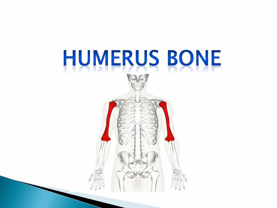
Humerus bone
Ulna Bone
Radius Bone
Bones of Hand


The clavicle is an S-shaped long bone
Lies horizontally across the root of the neck just beneath the skin
Medially it articulates with the sternum and 1st
costal cartilage
Laterally with the acromion process of the scapula
Provides the only bony link b/w the upper limb & the axial skeleton

One of the main functions of the collarbone is to hold the
arms freely and supported, away from the trunk.
Protects the underlying neurovascular structures
supplying the upper limb.
Transmits force from the upper limb to the axial
skeleton.

Sternoclavicular joint Acromio-clavicular joint
The medial(sternal) end The lateral(acromial) end
articulates with the sternum articulates with the acromial
forming the sternoclavicular process of the scapula forming
joint . Acromio-clavicular joint .
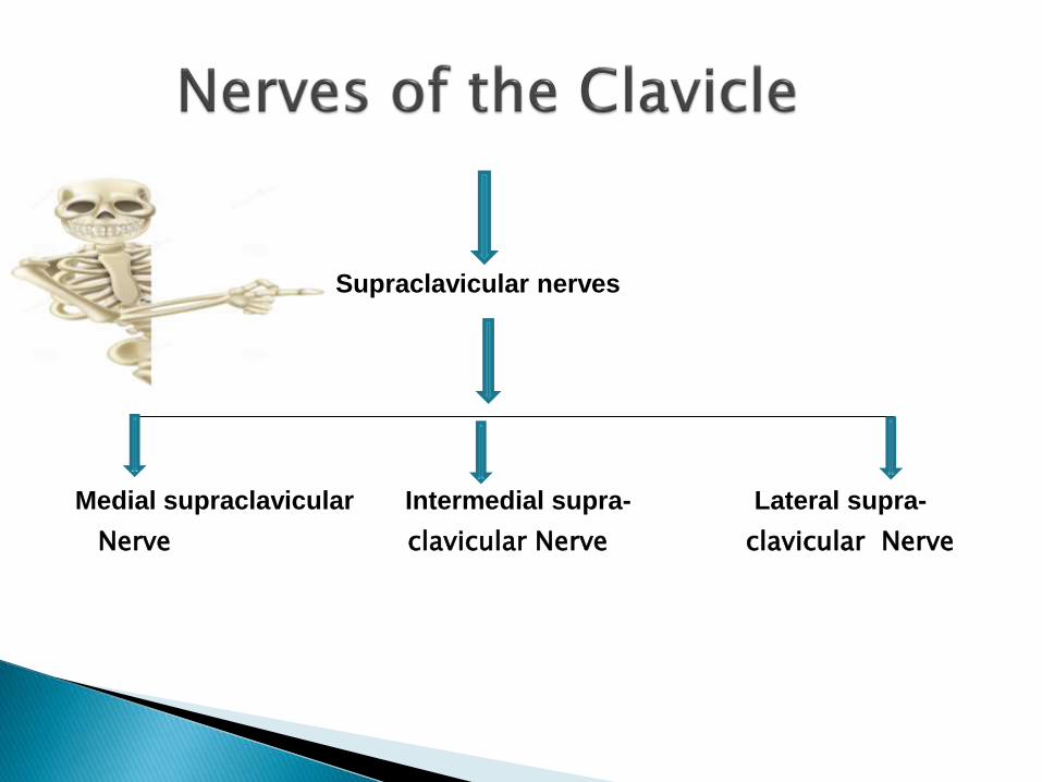
Supraclavicular nerves
Medial supraclavicular Intermedial supra- Lateral supra-
Nerve clavicular Nerve clavicular Nerve

i)Medial supraclavicular Nerve :Cross obliquely over the external
jugular vein and the clavicular & supply the skin as far as middle line.
ii)Intermedial supraclavicular Nerve :Cross the clavicle & supply
over the skin over the pectoralis major .
iii)Lateral supraclavicular Nerve :Pass obliquely across the outer surface
of the trapezius and the acromion & supply the skin of the upper and posterior part of the shoulder.


The scapula is a flat triangular bone
Lies on the posterior chest wall b/w the 2nd & 7th
ribs
In its posterior surface the spine of the scapula projects back word
The coracoid process projects upward and forward above the glenoid cavity & provides attachment for muscles & ligaments
Medial to the base of the coracoid process is the suprascapular notch
The anterior surface of the scapula is concave & form the shallow subscapular fossa

The primary function of the scapula is to attach the upper arm to the thorax, or trunk of the body. This connection stabilizes the arm and provides for arm movement at the shoulder


Dorsal scapula Artery Suprascapula Artery Branches of the subscapula
Artery

Shoulder Joint Acromioclaviclar Joint
B/w the glenoid cavity B/w the acromial process
& the head of humerus . & the lateral end of clavicle
N/S : The axillary and supra- N/S : The suprascapular Nerve
Scapular Nerves

Articulate with the scapula at
the shoulder joint & with the
radius & ulna at the
elbow joint.
It is a long bone with 2 ends
(Upper & Lower) & a shaft .

The upper end consist of : Head, neck
lesser & greater tuberosity(tubercle)
separated by the bicipital groove .
Lower end contain 2 epicondyles (lateral, medial), 2 process (trochlea & capitulum), and 3 fossae.


Shoulder Joint Elbow Joint
B/w its head & the B/w its lower end
Glenoid cavity of the scapula. Head of radius & upper end
of ulna.

i) Axillary Nerve :
The axillary nerve or the circumflex nerve is
a nerve of the human body, that originates from the brachial plexus
(upper trunk, posterior division, posterior cord) at the level of the
axilla (armpit) and carries nerve fibers from C5 and C6.
ii) Musculocutaneous nerve :
The musculocutaneous
nerve arises from the lateral cord of the brachial plexus, opposite the
lower border of the pectoralis major, its fibers being derived from C5,
C6 and C7


Radius is the lateral bone of the forearm
The proximal end articulates with the humerus at the elbow joint
At the proximal end of the radius is the small circular head
The upper surface of the head is concave
Below the head, the bone is constricted to from the neck
Below the neck is the bicipital tuberosity for the insertion of the biceps muscle

Its distal end articulates with the scaphoid and lunate bones of the hand at the wrist joint
At the distal end of the radius is the styloidprocess, this projects distally from its lateral margin

Four Joints i) Elbow Joint :
Elbow joint is the synovial hinge joint between the humerus in the upper arm and the radius and ulna in the forearm which allows the hand to be
moved towards and away from the body.ii) Superior radioulnar Joint :
The proximal radioulnar articulation (superior radioulnar joint) is a synovial trochoid or pivot joint between the circumference of the head of the radius and the ring formed by the radial notch of the ulna and the annular ligament.

iii) Inferior radioulnar joint :
The distal radioulnar articulation
(inferior radioulnar joint) is a joint between the two bones in the
forearm; the radius and ulna. It is one of two joints between the radius
and ulna, the other being the proximal radioulnar articulation.
iv) Wirst Joint :
It is actually a collection of multiple bones and
joints. The bones comprising the wrist include the distal ends of the radius and ulna, 8 carpal bones, and the proximal portions of the 5 metacarpal bones.

Radial Artery :The radial artery arises from the bifurcation of
the brachial artery in the cubital fossa. It runs distally on the anterior part of the forearm. There, it serves as a landmark for the division between the anterior and posterior compartments of the forearm, with the posterior compartment beginning just lateral to the artery. The artery winds laterally around the wrist, passing through the anatomical snuff box and between the heads of the first dorsal interosseous muscle. It passes anteriorly between the heads of the adductor pollicis, and becomes the deep palmar arch, which joins with the deep branch of the ulnar artery.

Radial Vein :
the radial veins are venae comitantes that
accompany the radial artery through the back of the hand and the lateral aspect of the forearm. They join the ulnar veins to form the brachial veins.
Radial Nerve :
The radial nerve is a nerve in the human body that supplies the upper limb. It supplies the medial, lateral, and long heads of the triceps brachii muscle of the arm, as well as all 12 muscles in the posterior osteofascial compartment of the forearm and the associated joints and overlying skin.
It originates from the brachial plexus, carrying fibers from the ventral roots of spinal nerves C5, C6, C8 & T1.
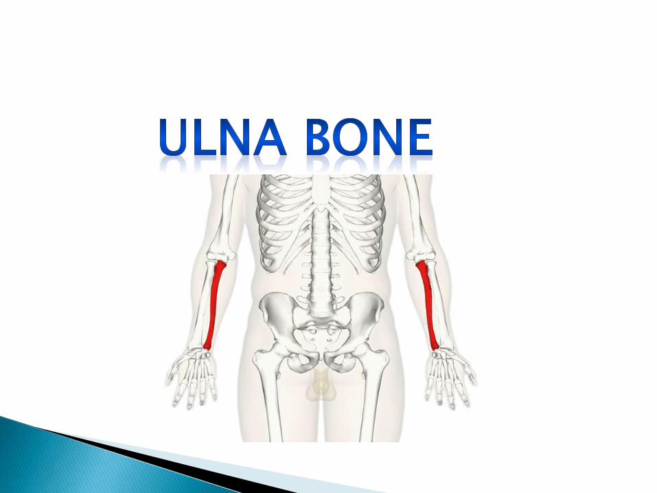
The ulna is one of two bones that make up the forearm
It is long bone with 2 ends & a shaft
The upper end consists of 2 processes( olecranon & coronoid processes ) & trochlear notch
The shaft has 3 surface (Separated by 3 borders)
Anterior , posterior , & lateral
Movement of the ulna is essential to such everyday functions as throwing a ball and driving a car.

Functionally, the ulna provides muscle attachment sites for over a dozen muscles in the upper arm
and forearm.
Ulnar Artery :The ulnar artery is the main blood vessel, with
oxygenated blood, of the medial aspect of the forearm. It arises from the brachial artery and terminates in the superficial palmar arch, which joins with the superficial branch of the radial artery. It is palpable on the anterior and medial aspect of the wrist.
Ulnar Vein : The ulnar veins are venae comitantes for the
ulnar artery. They mostly drain the medial aspect of the forearm. They arise in the hand and terminate when they join the radial veins to form the brachial veins.

Ulnar Nerve :the ulnar nerve is a nerve which runs near the
ulna bone. The ulnar collateral ligament of elbow joint is in relation with the ulnar nerve. The nerve is the largest unprotected nerve in the human body (meaning unprotected by muscle or bone), so injury is common. This nerve is directly connected to the little finger, and the adjacent half of the ring finger, supplying the palmar side of these fingers, including both front and back of the tips, perhaps as far back as the fingernail beds.


Three Joints
Elbow Joint Superior radioulnar Inferior Radioulnar
joint joint


There are eight carpal bones, made up of two rows (Proximal & distal row)
Proximal row (scaphoid, lunate, triquetrum, pisiform)
Distal row (Trapezium, trapezoid, capitate, hamate)
The 8 carpel bones form the flexor-retincaulum “carpal tunnel” which transmits the median nerve & the flexor tendons of the fingers.
5 metacarpal bones
14 phalanges :
The thumb contains only 2 phalanges(Proximal & distal)
All other fingers contain 3 phalanges(Proximal, middle, distal)


There are five metacarpal bones, each of which has a base, a shaft, and a head .
The first metacarpal bone of the thumb is the shortest and most mobile. It does not lie in the same plane as the others but occupies a more anterior position.
There are three phalanges for each of the fingers but only two for the thumb

5 Joints
i) Wirst Joint :It is actually a collection of multiple bones and joints. The
bones comprising the wrist include the distal ends of the radius and ulna, 8 carpal bones, and the proximal portions of the 5 metacarpal bones.
ii) Midcarpal Joint :The midcarpal joint is formed
by the scaphoid, lunate, and triquetral bones
in the proximal row, and the trapezium,
trapezoid, capitate, and hamate bones in
the distal row. The distal pole of the
scaphoid articulates with two trapezial
bones as a gliding type of joint.

iii) Carpo-metacarpal joint :
The carpometacarpal (CMC) joints are
five joints in the wrist that articulate the distal row of carpal bones and the proximal bases of the five metacarpal bones.
The CMC of the thumb or the first CMC, also known as the trapeziometacarpal joint (TMC).

iv) Metacarpo-phalangeal joints :The metacarpophalangeal joints
(MCP) refer to the joints between the metacarpal bones and the phalanges of the fingers. These are of the condyloid(Condyloid joint) kind, formed by the reception of the rounded heads of the metacarpal bones into shallow cavities on the proximal ends of the first phalanges, with the exception of that of the thumb, which is a ginglymus(Hinge joint).

v)Inter-phalangeal Joints :
The interphalangeal articulations of the
hand are the hinge joints between the phalanges of the hand (i.e. the finger bones)
“Proximal interphalangeal joints" those between the first (also called
proximal) and second (intermediate) phalanges
“Distal interphalangeal joints" those
between the second and third (distal)
phalanges

No Questions Plx