Anatomical, light and scanning electron micro- …vetanat.com/v13-pdf/3.pdf · Anatomy and...
22
J. Vet. Anat. Vol 7 No 2, (2014) 33 - 54 33 Anatomy and histology of the ostrich integument Zaghloul and El-Gendy Anatomical, light and scanning electron micro- scopical study of ostrich (Struthio camelus) integ- ument Doaa, M. Zaghloul 1 and Samir, El-Gendy 2 Histology &Cytology 1 and Anatomy 2 departments Faculty of Veterinary Medicine, Alex- andria University, Egypt. With 16 figures Received May, accepted for publication June, 2014 Abstract The current study dealt with the gross and microscopic anatomy of the in- tegument of male ostrich in addition to the histological features of different areas of skin. The ostrich skin is characterized by prominent feather follicles and bristles. The number of feather follicles was determined per cm 2 in different regions. The integu- ment of ostrich had many modifica- tions, which appeared as callosities and scales, nail and toe pads. They were sternal, pubic and Achilles ten- don callosities. The vacuolated epi- dermal cells were seen mainly in the skin of legs and to a lesser extent in the skin of back and Achilles areas. Higher lipogenic potential was ex- pressed by epidermis from glabrous areas of ostrich skin. The dermal pa- pillae were found in the skin of feath- ered area of neck and back and this was not a common finding in bird's skin, which may give resistance against shearing forces in these re- gions of ostrich skin. The thickness of the keratin layer of ostrich varied, be- ing thick and characteristically loose in the skin at legs, very thin and wavy at neck, while at Achilles skin area, scales and toe pad were thick and more compact, with the thickest very dense and wavy keratin layer at the nail. The dermis consisted of superfi- cial layer of dense irregular connec- tive tissue characterized by presence of many vacuoles of different sizes just under the basal lamina of the epi- thelium of epidermis and deep layer of dense regular connective tissue. This result suggested the presence of fat droplets in this layer which may be
Transcript of Anatomical, light and scanning electron micro- …vetanat.com/v13-pdf/3.pdf · Anatomy and...

J. Vet. Anat. Vol 7 No 2, (2014) 33 - 5433
Anatomy and histology of the ostrich integument Zaghloul and El-Gendy
Anatomical, light and scanning electron micro-scopical study of ostrich (Struthio camelus) integ-ument Doaa, M. Zaghloul1 and Samir, El-Gendy2 Histology &Cytology1 and Anatomy2 departments Faculty of Veterinary Medicine, Alex-
andria University, Egypt.
With 16 figures Received May, accepted for publication June, 2014
Abstract The current study dealt with the gross and microscopic anatomy of the in-tegument of male ostrich in addition to the histological features of different areas of skin. The ostrich skin is characterized by prominent feather follicles and bristles. The number of feather follicles was determined per cm2 in different regions. The integu-ment of ostrich had many modifica-tions, which appeared as callosities and scales, nail and toe pads. They were sternal, pubic and Achilles ten-don callosities. The vacuolated epi-dermal cells were seen mainly in the skin of legs and to a lesser extent in the skin of back and Achilles areas. Higher lipogenic potential was ex-pressed by epidermis from glabrous areas of ostrich skin. The dermal pa-pillae were found in the skin of feath-
ered area of neck and back and this was not a common finding in bird's skin, which may give resistance against shearing forces in these re-gions of ostrich skin. The thickness of the keratin layer of ostrich varied, be-ing thick and characteristically loose in the skin at legs, very thin and wavy at neck, while at Achilles skin area, scales and toe pad were thick and more compact, with the thickest very dense and wavy keratin layer at the nail. The dermis consisted of superfi-cial layer of dense irregular connec-tive tissue characterized by presence of many vacuoles of different sizes just under the basal lamina of the epi-thelium of epidermis and deep layer of dense regular connective tissue. This result suggested the presence of fat droplets in this layer which may be
D2
D3
D2 D4
D3
D4
Metatarsal phalangeal
pad
Digital pad
Digital pad
Digital pad
Digital pad
Digital pad
A B
R Tarso-
metatarsal
I
I
III
II III
II
VI
I
II
D4
D3
D2
Metatarsal phalangeal
pad
Claw
Claw
VI
III
V
metatarsal III
C
Fig (8): Right foot of cassowary
A) Dorsal view showing the scales covering the digits (II, III, IV)
B) Plantar view of the right foot showing the metatarsal phalangeal pad and the digital pads on each digit.
C) Latero/medial, X-ray image of the right foot of the Southern cass-owary showing: the three digits (D2, D3, D4) and the phalnges of each. Notice the position of the tarso-metatarsal phalangeal pad under-neath the joint.

J. Vet. Anat. Vol 7 No 2, (2014) 33 - 5434
Anatomy and histology of the ostrich integument Zaghloul and El-Gendy
to overcome the lack of good barrier of cutaneous water loss in epidermis. Keywords: Ostrich, Anatomy, Light microscopy, Integument, Skin modifi-cations, Scanning electron microsco-py.
Introduction The recent interest in ostrich farming has led to an increase in demand for information about this bird and how to manage it in a commercial environ-ment. Ostrich farming is important for production of feather, meat, skin and eggs (Cooper et al., 2008). Leather is one of the main products gained from ostrich farming (Meyer et al., 2002). The flightless birds have developed an adapted anatomical structure ow-ing to release from the constraints of flight. Flightlessness has led to a modification of the functional anatomy of skin (Weir and Lunam, 2004). Var-iations in the histology of the skin of volant birds occur according to spe-cies, age and different regions of the integument and are likely to be adap-tations to different environmental pressures. The avian integument as a whole is even further diversified as
claws, diverse types of scales, and various other integumentary struc-tures (Stettenheim, 2000). The functions of bird skin keep out pathogens and other potentially harmful substances, retain vital fluids and gases, and serve as a sensory organ. With feathers, the skin also plays an important role in ther-moregulation. Avian skin consists of two layers, the epidermis and dermis, and skin neither has sweat glands nor sebaceous glands (Lucas and Stet-tenheim, 1972).
As in all terrestrial vertebrates, the primary function of avian epidermis is to provide a permeability barrier to curtail excessive evaporative water loss and prevent death by dehydra-tion (Menon et al., 1996).The func-tional significance of avian epidermal lipids include anti-microbial (Purton, 1986), visual signaling and ultraviolet rays protection, and sterol precursors for vitamin D, (Menon, 1984). The avian epidermis is composed of unique sebokeratinocytes that elabo-rate and secrete sebum-like lipids as they cornify. In addition to the lipid droplets, the avian epidermis elabo-
rates, but rarely secretes, lipid-enriched organelles, the multigranular bodies, (Menon and Menon, 2000). These bodies are analogous to the lamellar bodies of mammals (Menon et al., 1991). Recent studies have in-dicated the importance of integumen-tary characteristics for phylogenetic reconstructions of bird species (Chu, 1998 and Bertelli et al., 2002).
The high vascularity near the surface of the skin aids in making the skin susceptible to bruising, which influ-ences skin quality, while the strength and flexibility of ostrich leather was attributed to the three-dimensional cross-weave arrangement of collagen fibers (Engelbrecht et al., 2009).
Skin of the ostrich is an industrial ma-terial which has high economic im-portance and any damage on the skin by cuts or scratches during the grow-out phase decreases the quality of the skin and this situation may caus-es economical loses in the ostrich industry. Regarding, ostrich leathers as exotic leather type in leather in-dustry and integument as a good model for studying wound healing and skin grafting (Mansoori et al.,
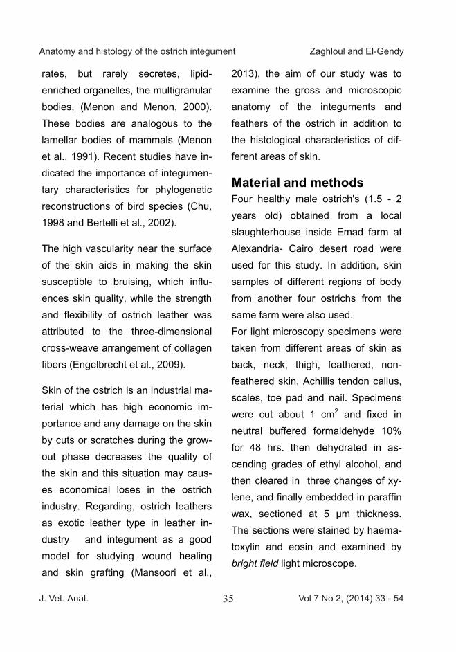
2013), the aim of our study was to examine the gross and microscopic anatomy of the integuments and feathers of the ostrich in addition to the histological characteristics of dif-ferent areas of skin.
Material and methods Four healthy male ostrich's (1.5 - 2 years old) obtained from a local slaughterhouse inside Emad farm at Alexandria- Cairo desert road were used for this study. In addition, skin samples of different regions of body from another four ostrichs from the same farm were also used. For light microscopy specimens were taken from different areas of skin as back, neck, thigh, feathered, non-feathered skin, Achillis tendon callus, scales, toe pad and nail. Specimens were cut about 1 cm2 and fixed in neutral buffered formaldehyde 10% for 48 hrs. then dehydrated in as-cending grades of ethyl alcohol, and then cleared in three changes of xy-lene, and finally embedded in paraffin wax, sectioned at 5 µm thickness. The sections were stained by haema-toxylin and eosin and examined by bright field light microscope.
J. Vet. Anat. Vol 7 No 2, (2014) 33 - 5435
Anatomy and histology of the ostrich integument Zaghloul and El-Gendy
to overcome the lack of good barrier of cutaneous water loss in epidermis. Keywords: Ostrich, Anatomy, Light microscopy, Integument, Skin modifi-cations, Scanning electron microsco-py.
Introduction The recent interest in ostrich farming has led to an increase in demand for information about this bird and how to manage it in a commercial environ-ment. Ostrich farming is important for production of feather, meat, skin and eggs (Cooper et al., 2008). Leather is one of the main products gained from ostrich farming (Meyer et al., 2002). The flightless birds have developed an adapted anatomical structure ow-ing to release from the constraints of flight. Flightlessness has led to a modification of the functional anatomy of skin (Weir and Lunam, 2004). Var-iations in the histology of the skin of volant birds occur according to spe-cies, age and different regions of the integument and are likely to be adap-tations to different environmental pressures. The avian integument as a whole is even further diversified as
claws, diverse types of scales, and various other integumentary struc-tures (Stettenheim, 2000). The functions of bird skin keep out pathogens and other potentially harmful substances, retain vital fluids and gases, and serve as a sensory organ. With feathers, the skin also plays an important role in ther-moregulation. Avian skin consists of two layers, the epidermis and dermis, and skin neither has sweat glands nor sebaceous glands (Lucas and Stet-tenheim, 1972).
As in all terrestrial vertebrates, the primary function of avian epidermis is to provide a permeability barrier to curtail excessive evaporative water loss and prevent death by dehydra-tion (Menon et al., 1996).The func-tional significance of avian epidermal lipids include anti-microbial (Purton, 1986), visual signaling and ultraviolet rays protection, and sterol precursors for vitamin D, (Menon, 1984). The avian epidermis is composed of unique sebokeratinocytes that elabo-rate and secrete sebum-like lipids as they cornify. In addition to the lipid droplets, the avian epidermis elabo-
rates, but rarely secretes, lipid-enriched organelles, the multigranular bodies, (Menon and Menon, 2000). These bodies are analogous to the lamellar bodies of mammals (Menon et al., 1991). Recent studies have in-dicated the importance of integumen-tary characteristics for phylogenetic reconstructions of bird species (Chu, 1998 and Bertelli et al., 2002).
The high vascularity near the surface of the skin aids in making the skin susceptible to bruising, which influ-ences skin quality, while the strength and flexibility of ostrich leather was attributed to the three-dimensional cross-weave arrangement of collagen fibers (Engelbrecht et al., 2009).
Skin of the ostrich is an industrial ma-terial which has high economic im-portance and any damage on the skin by cuts or scratches during the grow-out phase decreases the quality of the skin and this situation may caus-es economical loses in the ostrich industry. Regarding, ostrich leathers as exotic leather type in leather in-dustry and integument as a good model for studying wound healing and skin grafting (Mansoori et al.,
2013), the aim of our study was to examine the gross and microscopic anatomy of the integuments and feathers of the ostrich in addition to the histological characteristics of dif-ferent areas of skin.
Material and methods Four healthy male ostrich's (1.5 - 2 years old) obtained from a local slaughterhouse inside Emad farm at Alexandria- Cairo desert road were used for this study. In addition, skin samples of different regions of body from another four ostrichs from the same farm were also used. For light microscopy specimens were taken from different areas of skin as back, neck, thigh, feathered, non-feathered skin, Achillis tendon callus, scales, toe pad and nail. Specimens were cut about 1 cm2 and fixed in neutral buffered formaldehyde 10% for 48 hrs. then dehydrated in as-cending grades of ethyl alcohol, and then cleared in three changes of xy-lene, and finally embedded in paraffin wax, sectioned at 5 µm thickness. The sections were stained by haema-toxylin and eosin and examined by bright field light microscope.
J. Vet. Anat. Vol 7 No 2, (2014) 33 - 5436
Anatomy and histology of the ostrich integument Zaghloul and El-Gendy
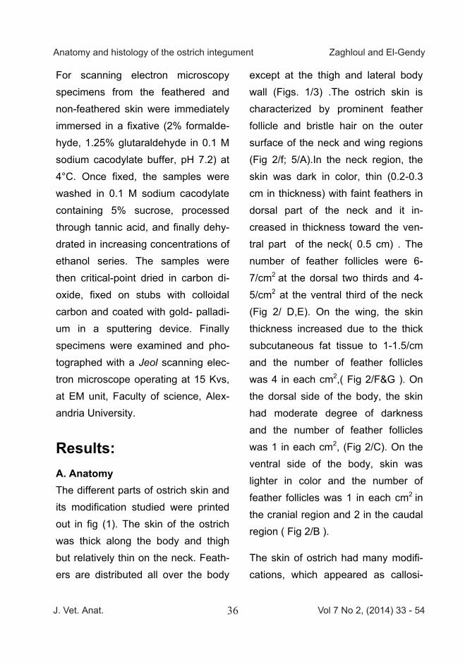
For scanning electron microscopy specimens from the feathered and non-feathered skin were immediately immersed in a fixative (2% formalde-hyde, 1.25% glutaraldehyde in 0.1 M sodium cacodylate buffer, pH 7.2) at 4°C. Once fixed, the samples were washed in 0.1 M sodium cacodylate containing 5% sucrose, processed through tannic acid, and finally dehy-drated in increasing concentrations of ethanol series. The samples were then critical-point dried in carbon di-oxide, fixed on stubs with colloidal carbon and coated with gold- palladi-um in a sputtering device. Finally specimens were examined and pho-tographed with a Jeol scanning elec-tron microscope operating at 15 Kvs, at EM unit, Faculty of science, Alex-andria University.
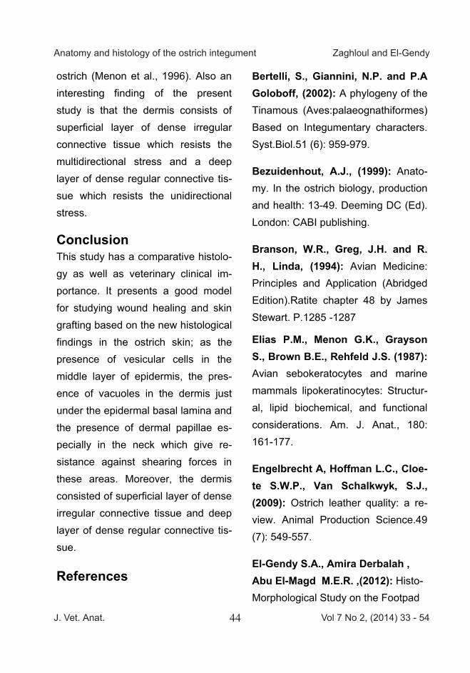
Results: A. Anatomy The different parts of ostrich skin and its modification studied were printed out in fig (1). The skin of the ostrich was thick along the body and thigh but relatively thin on the neck. Feath-ers are distributed all over the body
except at the thigh and lateral body wall (Figs. 1/3) .The ostrich skin is characterized by prominent feather follicle and bristle hair on the outer surface of the neck and wing regions (Fig 2/f; 5/A).In the neck region, the skin was dark in color, thin (0.2-0.3 cm in thickness) with faint feathers in dorsal part of the neck and it in-creased in thickness toward the ven-tral part of the neck( 0.5 cm) . The number of feather follicles were 6-7/cm2 at the dorsal two thirds and 4-5/cm2 at the ventral third of the neck (Fig 2/ D,E). On the wing, the skin thickness increased due to the thick subcutaneous fat tissue to 1-1.5/cm and the number of feather follicles was 4 in each cm2,( Fig 2/F&G ). On the dorsal side of the body, the skin had moderate degree of darkness and the number of feather follicles was 1 in each cm2, (Fig 2/C). On the ventral side of the body, skin was lighter in color and the number of feather follicles was 1 in each cm2 in the cranial region and 2 in the caudal region ( Fig 2/B ).
The skin of ostrich had many modifi-cations, which appeared as callosi-
ties, scales and toe pads. The sternal callus, located on the cranioventral side of the sternum, was 1x 8.5 x 11 cm in thickness, width and length re-spectively (Fig 1/1). The pubic callus occurred as ventral and cranial pro-jections of the pubic bone and meas-ured 1x4.2x9 cm in thickness, width and length respectively (Fig 1/2). The Achilles tendon callus was located on the proximal end of the metatarsal bone and was protected by plantar metatarsal pad (about 1 x 5x12 cm in thickness, width and length respec-tively). These callosities were charac-terized by hexagonal-shaped scales (Fig 3/B&C).
The skin on the dorsal surface of the tarsometatarsus (shank) and digits consisted of large cornified scales, while that on the rest of the shank and dorsal surface of the digits were covered by small dome-shaped scales (Fig 4/1&2). The large scales (about 15-17) varied in size. On the shank, they were nearly of the same size (0.2 x 1.5 x3 cm in thickness, width and length respectively) (Fig 4/6). They begin10 cm distal to the hock joint, then decreased in size at
the level of the metatarsal phalangeal joint (5-6) scales. The large scales occupied the dorsal surface of 4th and 3rd digits. They increased in size gradually till the end by the toe nail. They were14-15 scales on 3rd digit and 7-8 scales on the dorsal surface of digit 4th (Fig 4/1). The small dome-shaped scales were covering the re-minder of the dorsal surface of the digits (Fig 4/5), and other scales on the planter surface of the shank ap-pear hexagonal in shape like snake’s skin (Fig 3/D).
The skin modifications were found on the plantar surface of toe. The ostrich foot had four toe pads, two on the 3rd digit, one on the 4th digit and one on the metatarso-phalangeal joint. The ventral surface of toe pads was cov-ered by closely situated, papillae, which varied in direction, length and thickness (Fig 4/3). A horny nail en-closed the terminal phalanx of the third digit of the pelvic limb (Fig. 4/6). It was slightly curved and rounded at the apex and measured about 4-5 cm in length. A small nail was present on the distal part of the fourth digit.

J. Vet. Anat. Vol 7 No 2, (2014) 33 - 5437
Anatomy and histology of the ostrich integument Zaghloul and El-Gendy
For scanning electron microscopy specimens from the feathered and non-feathered skin were immediately immersed in a fixative (2% formalde-hyde, 1.25% glutaraldehyde in 0.1 M sodium cacodylate buffer, pH 7.2) at 4°C. Once fixed, the samples were washed in 0.1 M sodium cacodylate containing 5% sucrose, processed through tannic acid, and finally dehy-drated in increasing concentrations of ethanol series. The samples were then critical-point dried in carbon di-oxide, fixed on stubs with colloidal carbon and coated with gold- palladi-um in a sputtering device. Finally specimens were examined and pho-tographed with a Jeol scanning elec-tron microscope operating at 15 Kvs, at EM unit, Faculty of science, Alex-andria University.
Results: A. Anatomy The different parts of ostrich skin and its modification studied were printed out in fig (1). The skin of the ostrich was thick along the body and thigh but relatively thin on the neck. Feath-ers are distributed all over the body
except at the thigh and lateral body wall (Figs. 1/3) .The ostrich skin is characterized by prominent feather follicle and bristle hair on the outer surface of the neck and wing regions (Fig 2/f; 5/A).In the neck region, the skin was dark in color, thin (0.2-0.3 cm in thickness) with faint feathers in dorsal part of the neck and it in-creased in thickness toward the ven-tral part of the neck( 0.5 cm) . The number of feather follicles were 6-7/cm2 at the dorsal two thirds and 4-5/cm2 at the ventral third of the neck (Fig 2/ D,E). On the wing, the skin thickness increased due to the thick subcutaneous fat tissue to 1-1.5/cm and the number of feather follicles was 4 in each cm2,( Fig 2/F&G ). On the dorsal side of the body, the skin had moderate degree of darkness and the number of feather follicles was 1 in each cm2, (Fig 2/C). On the ventral side of the body, skin was lighter in color and the number of feather follicles was 1 in each cm2 in the cranial region and 2 in the caudal region ( Fig 2/B ).
The skin of ostrich had many modifi-cations, which appeared as callosi-
ties, scales and toe pads. The sternal callus, located on the cranioventral side of the sternum, was 1x 8.5 x 11 cm in thickness, width and length re-spectively (Fig 1/1). The pubic callus occurred as ventral and cranial pro-jections of the pubic bone and meas-ured 1x4.2x9 cm in thickness, width and length respectively (Fig 1/2). The Achilles tendon callus was located on the proximal end of the metatarsal bone and was protected by plantar metatarsal pad (about 1 x 5x12 cm in thickness, width and length respec-tively). These callosities were charac-terized by hexagonal-shaped scales (Fig 3/B&C).
The skin on the dorsal surface of the tarsometatarsus (shank) and digits consisted of large cornified scales, while that on the rest of the shank and dorsal surface of the digits were covered by small dome-shaped scales (Fig 4/1&2). The large scales (about 15-17) varied in size. On the shank, they were nearly of the same size (0.2 x 1.5 x3 cm in thickness, width and length respectively) (Fig 4/6). They begin10 cm distal to the hock joint, then decreased in size at
the level of the metatarsal phalangeal joint (5-6) scales. The large scales occupied the dorsal surface of 4th and 3rd digits. They increased in size gradually till the end by the toe nail. They were14-15 scales on 3rd digit and 7-8 scales on the dorsal surface of digit 4th (Fig 4/1). The small dome-shaped scales were covering the re-minder of the dorsal surface of the digits (Fig 4/5), and other scales on the planter surface of the shank ap-pear hexagonal in shape like snake’s skin (Fig 3/D).
The skin modifications were found on the plantar surface of toe. The ostrich foot had four toe pads, two on the 3rd digit, one on the 4th digit and one on the metatarso-phalangeal joint. The ventral surface of toe pads was cov-ered by closely situated, papillae, which varied in direction, length and thickness (Fig 4/3). A horny nail en-closed the terminal phalanx of the third digit of the pelvic limb (Fig. 4/6). It was slightly curved and rounded at the apex and measured about 4-5 cm in length. A small nail was present on the distal part of the fourth digit.
J. Vet. Anat. Vol 7 No 2, (2014) 33 - 5438
Anatomy and histology of the ostrich integument Zaghloul and El-Gendy
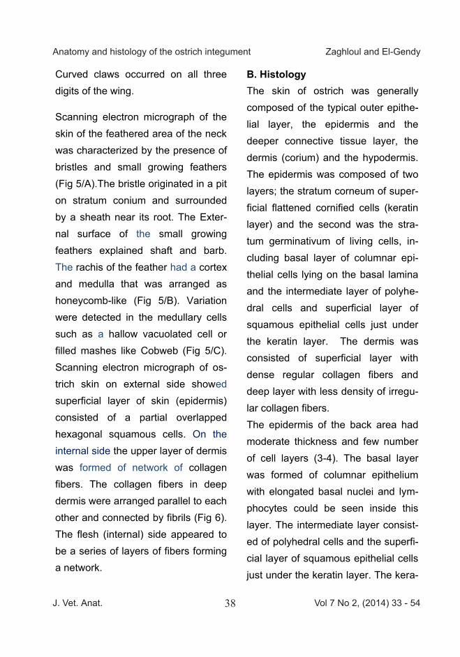
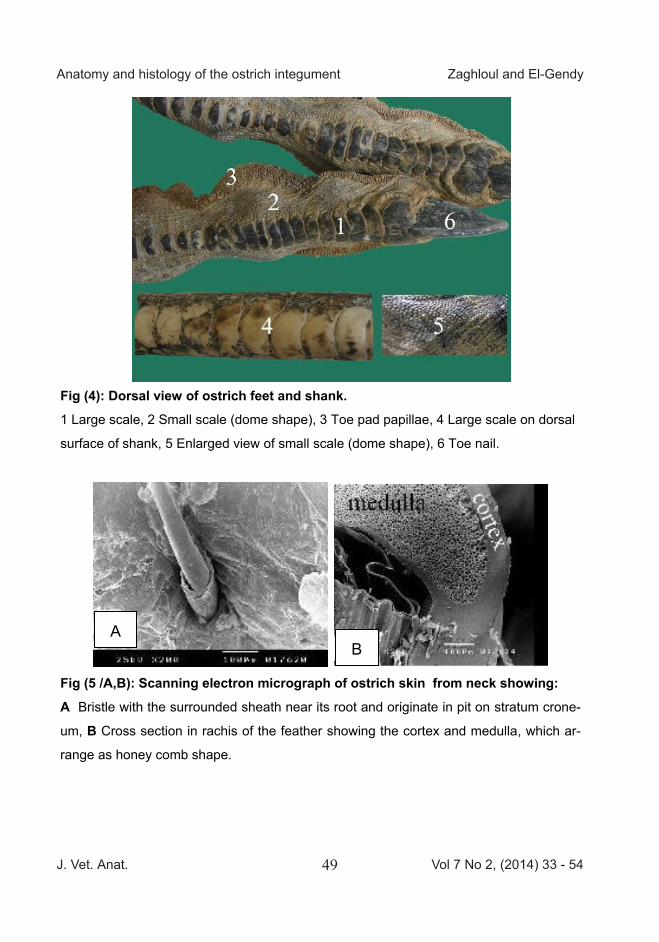
Curved claws occurred on all three digits of the wing.
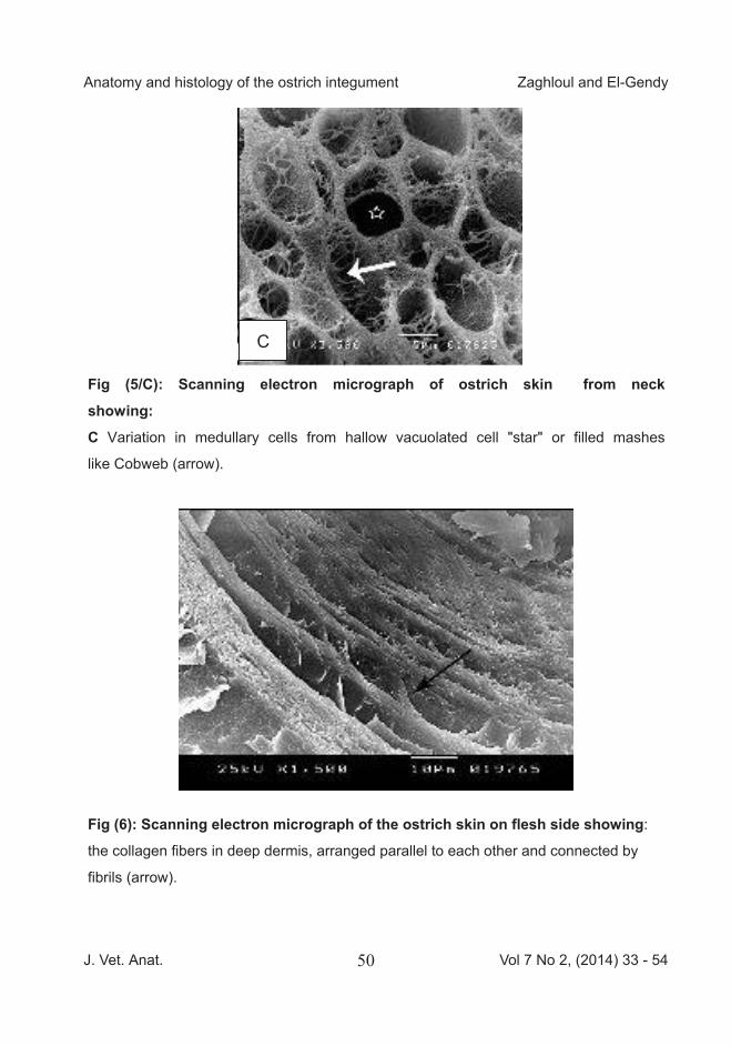
Scanning electron micrograph of the skin of the feathered area of the neck was characterized by the presence of bristles and small growing feathers (Fig 5/A).The bristle originated in a pit on stratum conium and surrounded by a sheath near its root. The Exter-nal surface of the small growing feathers explained shaft and barb. The rachis of the feather had a cortex and medulla that was arranged as honeycomb-like (Fig 5/B). Variation were detected in the medullary cells such as a hallow vacuolated cell or filled mashes like Cobweb (Fig 5/C). Scanning electron micrograph of os-trich skin on external side showed superficial layer of skin (epidermis) consisted of a partial overlapped hexagonal squamous cells. On the internal side the upper layer of dermis was formed of network of collagen fibers. The collagen fibers in deep dermis were arranged parallel to each other and connected by fibrils (Fig 6). The flesh (internal) side appeared to be a series of layers of fibers forming a network.
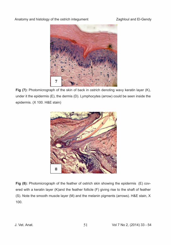
B. Histology The skin of ostrich was generally composed of the typical outer epithe-lial layer, the epidermis and the deeper connective tissue layer, the dermis (corium) and the hypodermis. The epidermis was composed of two layers; the stratum corneum of super-ficial flattened cornified cells (keratin layer) and the second was the stra-tum germinativum of living cells, in-cluding basal layer of columnar epi-thelial cells lying on the basal lamina and the intermediate layer of polyhe-dral cells and superficial layer of squamous epithelial cells just under the keratin layer. The dermis was consisted of superficial layer with dense regular collagen fibers and deep layer with less density of irregu-lar collagen fibers. The epidermis of the back area had moderate thickness and few number of cell layers (3-4). The basal layer was formed of columnar epithelium with elongated basal nuclei and lym-phocytes could be seen inside this layer. The intermediate layer consist-ed of polyhedral cells and the superfi-cial layer of squamous epithelial cells just under the keratin layer. The kera-
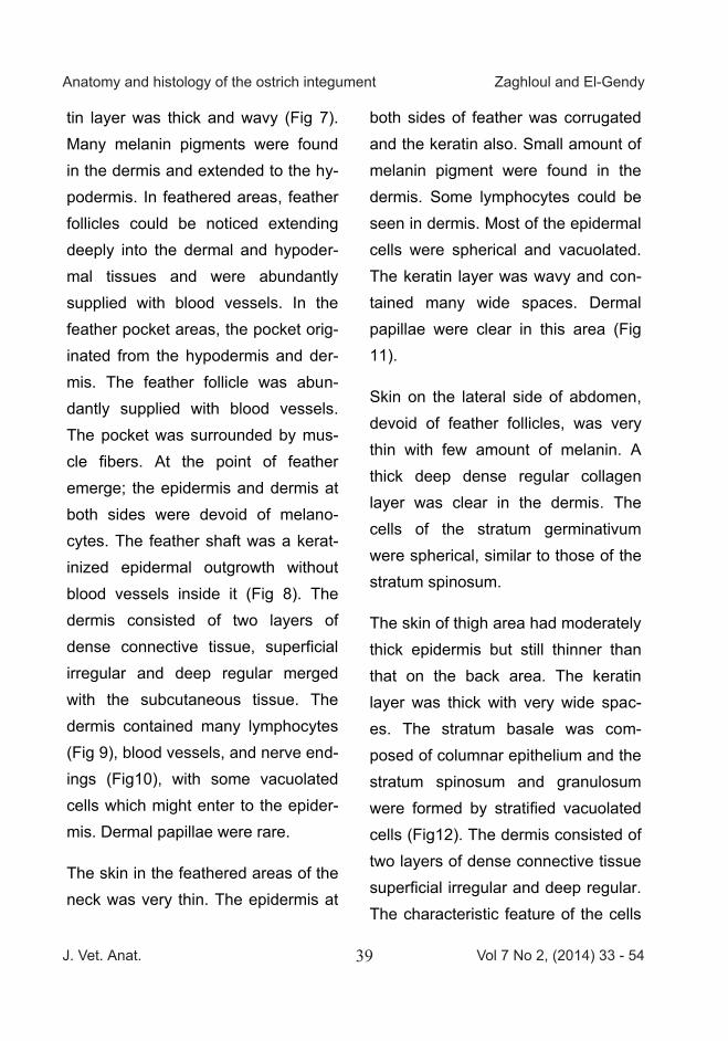
tin layer was thick and wavy (Fig 7). Many melanin pigments were found in the dermis and extended to the hy-podermis. In feathered areas, feather follicles could be noticed extending deeply into the dermal and hypoder-mal tissues and were abundantly supplied with blood vessels. In the feather pocket areas, the pocket orig-inated from the hypodermis and der-mis. The feather follicle was abun-dantly supplied with blood vessels. The pocket was surrounded by mus-cle fibers. At the point of feather emerge; the epidermis and dermis at both sides were devoid of melano-cytes. The feather shaft was a kerat-inized epidermal outgrowth without blood vessels inside it (Fig 8). The dermis consisted of two layers of dense connective tissue, superficial irregular and deep regular merged with the subcutaneous tissue. The dermis contained many lymphocytes (Fig 9), blood vessels, and nerve end-ings (Fig10), with some vacuolated cells which might enter to the epider-mis. Dermal papillae were rare.
The skin in the feathered areas of the neck was very thin. The epidermis at
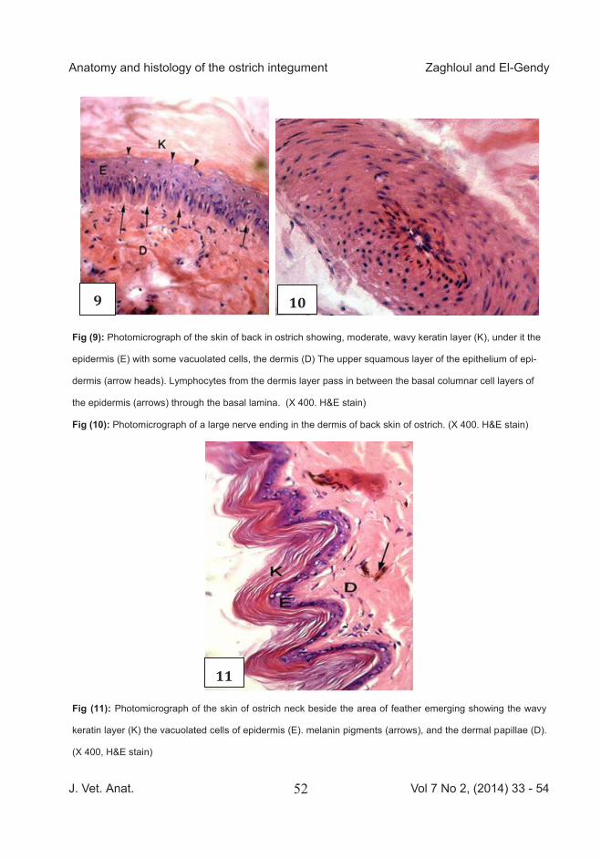
both sides of feather was corrugated and the keratin also. Small amount of melanin pigment were found in the dermis. Some lymphocytes could be seen in dermis. Most of the epidermal cells were spherical and vacuolated. The keratin layer was wavy and con-tained many wide spaces. Dermal papillae were clear in this area (Fig 11).
Skin on the lateral side of abdomen, devoid of feather follicles, was very thin with few amount of melanin. A thick deep dense regular collagen layer was clear in the dermis. The cells of the stratum germinativum were spherical, similar to those of the stratum spinosum.
The skin of thigh area had moderately thick epidermis but still thinner than that on the back area. The keratin layer was thick with very wide spac-es. The stratum basale was com-posed of columnar epithelium and the stratum spinosum and granulosum were formed by stratified vacuolated cells (Fig12). The dermis consisted of two layers of dense connective tissue superficial irregular and deep regular. The characteristic feature of the cells
J. Vet. Anat. Vol 7 No 2, (2014) 33 - 5439
Anatomy and histology of the ostrich integument Zaghloul and El-Gendy
Curved claws occurred on all three digits of the wing.
Scanning electron micrograph of the skin of the feathered area of the neck was characterized by the presence of bristles and small growing feathers (Fig 5/A).The bristle originated in a pit on stratum conium and surrounded by a sheath near its root. The Exter-nal surface of the small growing feathers explained shaft and barb. The rachis of the feather had a cortex and medulla that was arranged as honeycomb-like (Fig 5/B). Variation were detected in the medullary cells such as a hallow vacuolated cell or filled mashes like Cobweb (Fig 5/C). Scanning electron micrograph of os-trich skin on external side showed superficial layer of skin (epidermis) consisted of a partial overlapped hexagonal squamous cells. On the internal side the upper layer of dermis was formed of network of collagen fibers. The collagen fibers in deep dermis were arranged parallel to each other and connected by fibrils (Fig 6). The flesh (internal) side appeared to be a series of layers of fibers forming a network.
B. Histology The skin of ostrich was generally composed of the typical outer epithe-lial layer, the epidermis and the deeper connective tissue layer, the dermis (corium) and the hypodermis. The epidermis was composed of two layers; the stratum corneum of super-ficial flattened cornified cells (keratin layer) and the second was the stra-tum germinativum of living cells, in-cluding basal layer of columnar epi-thelial cells lying on the basal lamina and the intermediate layer of polyhe-dral cells and superficial layer of squamous epithelial cells just under the keratin layer. The dermis was consisted of superficial layer with dense regular collagen fibers and deep layer with less density of irregu-lar collagen fibers. The epidermis of the back area had moderate thickness and few number of cell layers (3-4). The basal layer was formed of columnar epithelium with elongated basal nuclei and lym-phocytes could be seen inside this layer. The intermediate layer consist-ed of polyhedral cells and the superfi-cial layer of squamous epithelial cells just under the keratin layer. The kera-
tin layer was thick and wavy (Fig 7). Many melanin pigments were found in the dermis and extended to the hy-podermis. In feathered areas, feather follicles could be noticed extending deeply into the dermal and hypoder-mal tissues and were abundantly supplied with blood vessels. In the feather pocket areas, the pocket orig-inated from the hypodermis and der-mis. The feather follicle was abun-dantly supplied with blood vessels. The pocket was surrounded by mus-cle fibers. At the point of feather emerge; the epidermis and dermis at both sides were devoid of melano-cytes. The feather shaft was a kerat-inized epidermal outgrowth without blood vessels inside it (Fig 8). The dermis consisted of two layers of dense connective tissue, superficial irregular and deep regular merged with the subcutaneous tissue. The dermis contained many lymphocytes (Fig 9), blood vessels, and nerve end-ings (Fig10), with some vacuolated cells which might enter to the epider-mis. Dermal papillae were rare.
The skin in the feathered areas of the neck was very thin. The epidermis at
both sides of feather was corrugated and the keratin also. Small amount of melanin pigment were found in the dermis. Some lymphocytes could be seen in dermis. Most of the epidermal cells were spherical and vacuolated. The keratin layer was wavy and con-tained many wide spaces. Dermal papillae were clear in this area (Fig 11).
Skin on the lateral side of abdomen, devoid of feather follicles, was very thin with few amount of melanin. A thick deep dense regular collagen layer was clear in the dermis. The cells of the stratum germinativum were spherical, similar to those of the stratum spinosum.
The skin of thigh area had moderately thick epidermis but still thinner than that on the back area. The keratin layer was thick with very wide spac-es. The stratum basale was com-posed of columnar epithelium and the stratum spinosum and granulosum were formed by stratified vacuolated cells (Fig12). The dermis consisted of two layers of dense connective tissue superficial irregular and deep regular. The characteristic feature of the cells
J. Vet. Anat. Vol 7 No 2, (2014) 33 - 5440
Anatomy and histology of the ostrich integument Zaghloul and El-Gendy
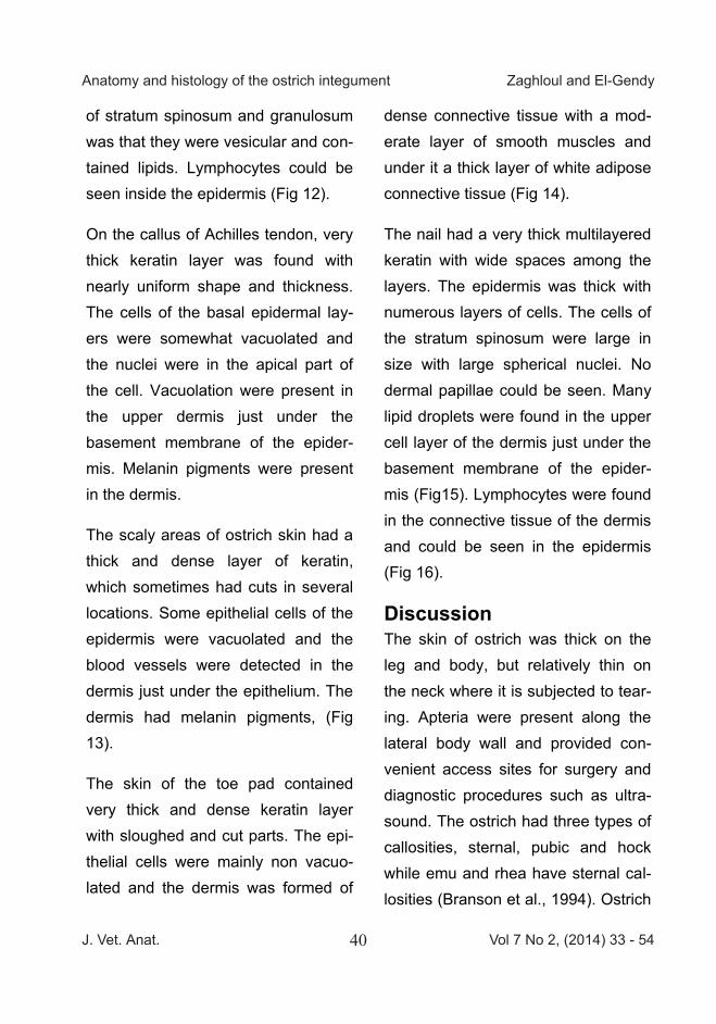
of stratum spinosum and granulosum was that they were vesicular and con-tained lipids. Lymphocytes could be seen inside the epidermis (Fig 12).
On the callus of Achilles tendon, very thick keratin layer was found with nearly uniform shape and thickness. The cells of the basal epidermal lay-ers were somewhat vacuolated and the nuclei were in the apical part of the cell. Vacuolation were present in the upper dermis just under the basement membrane of the epider-mis. Melanin pigments were present in the dermis.
The scaly areas of ostrich skin had a thick and dense layer of keratin, which sometimes had cuts in several locations. Some epithelial cells of the epidermis were vacuolated and the blood vessels were detected in the dermis just under the epithelium. The dermis had melanin pigments, (Fig 13).
The skin of the toe pad contained very thick and dense keratin layer with sloughed and cut parts. The epi-thelial cells were mainly non vacuo-lated and the dermis was formed of
dense connective tissue with a mod-erate layer of smooth muscles and under it a thick layer of white adipose connective tissue (Fig 14).
The nail had a very thick multilayered keratin with wide spaces among the layers. The epidermis was thick with numerous layers of cells. The cells of the stratum spinosum were large in size with large spherical nuclei. No dermal papillae could be seen. Many lipid droplets were found in the upper cell layer of the dermis just under the basement membrane of the epider-mis (Fig15). Lymphocytes were found in the connective tissue of the dermis and could be seen in the epidermis (Fig 16).
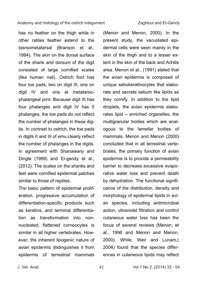
Discussion The skin of ostrich was thick on the leg and body, but relatively thin on the neck where it is subjected to tear-ing. Apteria were present along the lateral body wall and provided con-venient access sites for surgery and diagnostic procedures such as ultra-sound. The ostrich had three types of callosities, sternal, pubic and hock while emu and rhea have sternal cal-losities (Branson et al., 1994). Ostrich
has no feather on the thigh while in other ratites feather extend to the tasrsometatarsal (Branson et al., 1994). The skin on the dorsal surface of the shank and dorsum of the digit consisted of large cornified scales (like human nail). Ostrich foot has four toe pads, two on digit III, one on digit IV and one at metatarso-phalangeal joint. Because digit III has four phalanges and digit IV has 5 phalanges, the toe pads do not reflect the number of phalanges in these dig-its. In contrast to ostrich, the toe pads in digits II and III of emu clearly reflect the number of phalanges in the digits. In agreement with Shanawany and Dingle (1999) and El-gendy et al., (2012). The scales on the shanks and feet were cornified epidermal patches similar to those of reptiles. The basic pattern of epidermal prolif-eration, progressive accumulation of differentiation-specific products such as keratins, and terminal differentia-tion as transformation into non-nucleated, flattened corneocytes is similar in all higher vertebrates. How-ever, the inherent lipogenic nature of avian epidermis distinguishes it from epidermis of terrestrial mammals
(Menon and Menon, 2000). In the present study, the vacuolated epi-dermal cells were seen mainly in the skin of the thigh and to a lesser ex-tent in the skin of the back and Achilis area. Menon et al., (1991) stated that the avian epidermis is composed of unique sebokeratinocytes that elabo-rate and secrete sebum like lipids as they cornify. In addition to the lipid droplets, the avian epidermis elabo-rates lipid – enriched organelles, the multigranular bodies which are anal-ogous to the lamellar bodies of mammals. Menon and Menon (2000) concluded that in all terrestrial verte-brates, the primary function of avian epidermis is to provide a permeability barrier to decrease excessive evapo-rative water loss and prevent death by dehydration. The functional signifi-cance of the distribution, density and morphology of epidermal lipids in avi-an species, including antimicrobial action, ultraviolet filtration and control cutaneous water loss has been the focus of several reviews (Menon, et al., 1996 and Menon and Menon, 2000). While, Weir and Lunam,( 2004) found that the species differ-ences in cutaneous lipids may reflect
J. Vet. Anat. Vol 7 No 2, (2014) 33 - 5441
Anatomy and histology of the ostrich integument Zaghloul and El-Gendy
of stratum spinosum and granulosum was that they were vesicular and con-tained lipids. Lymphocytes could be seen inside the epidermis (Fig 12).
On the callus of Achilles tendon, very thick keratin layer was found with nearly uniform shape and thickness. The cells of the basal epidermal lay-ers were somewhat vacuolated and the nuclei were in the apical part of the cell. Vacuolation were present in the upper dermis just under the basement membrane of the epider-mis. Melanin pigments were present in the dermis.
The scaly areas of ostrich skin had a thick and dense layer of keratin, which sometimes had cuts in several locations. Some epithelial cells of the epidermis were vacuolated and the blood vessels were detected in the dermis just under the epithelium. The dermis had melanin pigments, (Fig 13).
The skin of the toe pad contained very thick and dense keratin layer with sloughed and cut parts. The epi-thelial cells were mainly non vacuo-lated and the dermis was formed of
dense connective tissue with a mod-erate layer of smooth muscles and under it a thick layer of white adipose connective tissue (Fig 14).
The nail had a very thick multilayered keratin with wide spaces among the layers. The epidermis was thick with numerous layers of cells. The cells of the stratum spinosum were large in size with large spherical nuclei. No dermal papillae could be seen. Many lipid droplets were found in the upper cell layer of the dermis just under the basement membrane of the epider-mis (Fig15). Lymphocytes were found in the connective tissue of the dermis and could be seen in the epidermis (Fig 16).
Discussion The skin of ostrich was thick on the leg and body, but relatively thin on the neck where it is subjected to tear-ing. Apteria were present along the lateral body wall and provided con-venient access sites for surgery and diagnostic procedures such as ultra-sound. The ostrich had three types of callosities, sternal, pubic and hock while emu and rhea have sternal cal-losities (Branson et al., 1994). Ostrich
has no feather on the thigh while in other ratites feather extend to the tasrsometatarsal (Branson et al., 1994). The skin on the dorsal surface of the shank and dorsum of the digit consisted of large cornified scales (like human nail). Ostrich foot has four toe pads, two on digit III, one on digit IV and one at metatarso-phalangeal joint. Because digit III has four phalanges and digit IV has 5 phalanges, the toe pads do not reflect the number of phalanges in these dig-its. In contrast to ostrich, the toe pads in digits II and III of emu clearly reflect the number of phalanges in the digits. In agreement with Shanawany and Dingle (1999) and El-gendy et al., (2012). The scales on the shanks and feet were cornified epidermal patches similar to those of reptiles. The basic pattern of epidermal prolif-eration, progressive accumulation of differentiation-specific products such as keratins, and terminal differentia-tion as transformation into non-nucleated, flattened corneocytes is similar in all higher vertebrates. How-ever, the inherent lipogenic nature of avian epidermis distinguishes it from epidermis of terrestrial mammals
(Menon and Menon, 2000). In the present study, the vacuolated epi-dermal cells were seen mainly in the skin of the thigh and to a lesser ex-tent in the skin of the back and Achilis area. Menon et al., (1991) stated that the avian epidermis is composed of unique sebokeratinocytes that elabo-rate and secrete sebum like lipids as they cornify. In addition to the lipid droplets, the avian epidermis elabo-rates lipid – enriched organelles, the multigranular bodies which are anal-ogous to the lamellar bodies of mammals. Menon and Menon (2000) concluded that in all terrestrial verte-brates, the primary function of avian epidermis is to provide a permeability barrier to decrease excessive evapo-rative water loss and prevent death by dehydration. The functional signifi-cance of the distribution, density and morphology of epidermal lipids in avi-an species, including antimicrobial action, ultraviolet filtration and control cutaneous water loss has been the focus of several reviews (Menon, et al., 1996 and Menon and Menon, 2000). While, Weir and Lunam,( 2004) found that the species differ-ences in cutaneous lipids may reflect
J. Vet. Anat. Vol 7 No 2, (2014) 33 - 5442
Anatomy and histology of the ostrich integument Zaghloul and El-Gendy
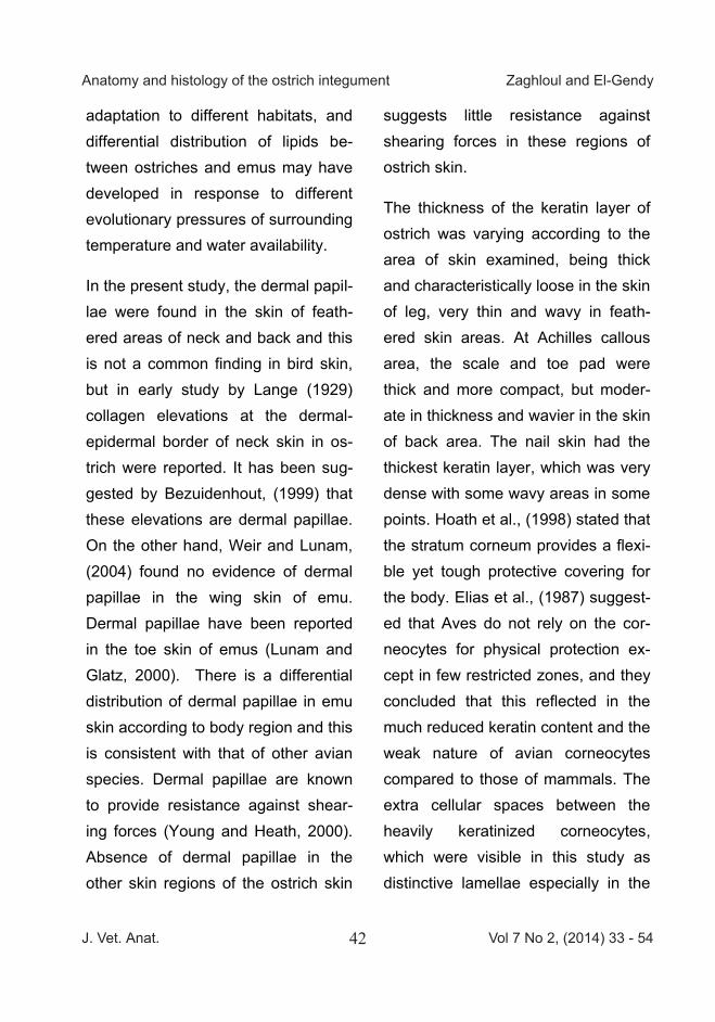
adaptation to different habitats, and differential distribution of lipids be-tween ostriches and emus may have developed in response to different evolutionary pressures of surrounding temperature and water availability.
In the present study, the dermal papil-lae were found in the skin of feath-ered areas of neck and back and this is not a common finding in bird skin, but in early study by Lange (1929) collagen elevations at the dermal-epidermal border of neck skin in os-trich were reported. It has been sug-gested by Bezuidenhout, (1999) that these elevations are dermal papillae. On the other hand, Weir and Lunam, (2004) found no evidence of dermal papillae in the wing skin of emu. Dermal papillae have been reported in the toe skin of emus (Lunam and Glatz, 2000). There is a differential distribution of dermal papillae in emu skin according to body region and this is consistent with that of other avian species. Dermal papillae are known to provide resistance against shear-ing forces (Young and Heath, 2000). Absence of dermal papillae in the other skin regions of the ostrich skin
suggests little resistance against shearing forces in these regions of ostrich skin.
The thickness of the keratin layer of ostrich was varying according to the area of skin examined, being thick and characteristically loose in the skin of leg, very thin and wavy in feath-ered skin areas. At Achilles callous area, the scale and toe pad were thick and more compact, but moder-ate in thickness and wavier in the skin of back area. The nail skin had the thickest keratin layer, which was very dense with some wavy areas in some points. Hoath et al., (1998) stated that the stratum corneum provides a flexi-ble yet tough protective covering for the body. Elias et al., (1987) suggest-ed that Aves do not rely on the cor-neocytes for physical protection ex-cept in few restricted zones, and they concluded that this reflected in the much reduced keratin content and the weak nature of avian corneocytes compared to those of mammals. The extra cellular spaces between the heavily keratinized corneocytes, which were visible in this study as distinctive lamellae especially in the
skin of leg area, may indicate the lipi-daceous contents of the lamellar granules which exocytosed forming lipids among the keratin lamellae. This agreed with the opinion of Men-on et al., (1996). Also Lucas and Stet-tenheim (1972) documented that the lipogenic activity of the epidermis is greater in certain glabrous regions of skin like the comb, wattle, interdigital web including dorsal and ventral scales, and the edge of the maxillary rictus in the domestic fowl. In agree-ment with the previous opinion, Wrench et al., (1980) found that lipo-genesis and keratinization are closely associated with each other in avian epidermis as an inverse relation ex-ists between sebogenesis and kerat-inization even within the secretory epidermis from different locations in the body. Menon and Aggarwal, (1982) suggested that some of the lipids may be utilized for the energetic of keratinization. As a final conclusion the higher lipogenic potential is ex-pressed by epidermis from glabrous areas of skin, (Menon & Menon, 2000) and this result was confirmed in the present study in the area of leg skin.
In the present study, the structure of the dermis was similar to other birds especially the high vascularization of the superficial layer in the feathered skin and this is consistent with previ-ous studies in emu (Weir & Lunam, 2004). The high vascularity and thickening of the stratum superficial to accommodate the blood vessels in featherless skin has been attributed to control of thermoregulation by heat loss via alterations in peripheral blood flow (Lucas& Stettenheim, 1972 and Philips & Sanborn, 1994). A special finding in the most superficial layer of the dermis of ostrich was the pres-ence of many vacuoles of different sizes just under the basal lamina of the epithelium of epidermis. This re-sult suggests the presence of fat droplets in this layer to compensate the absence of stratum laxum de-scribed by Weir & Lunam, (2004) in emu which was composed predomi-nantly of adipose tissue which is im-portant for insulation of the body from extreme cold (Dawson & Whittow, 2000). The presence of lipid just un-der the epidermis in ostrich may be to overcome the lack of good barrier to cutaneous water loss in epidermis of

J. Vet. Anat. Vol 7 No 2, (2014) 33 - 5443
Anatomy and histology of the ostrich integument Zaghloul and El-Gendy
adaptation to different habitats, and differential distribution of lipids be-tween ostriches and emus may have developed in response to different evolutionary pressures of surrounding temperature and water availability.
In the present study, the dermal papil-lae were found in the skin of feath-ered areas of neck and back and this is not a common finding in bird skin, but in early study by Lange (1929) collagen elevations at the dermal-epidermal border of neck skin in os-trich were reported. It has been sug-gested by Bezuidenhout, (1999) that these elevations are dermal papillae. On the other hand, Weir and Lunam, (2004) found no evidence of dermal papillae in the wing skin of emu. Dermal papillae have been reported in the toe skin of emus (Lunam and Glatz, 2000). There is a differential distribution of dermal papillae in emu skin according to body region and this is consistent with that of other avian species. Dermal papillae are known to provide resistance against shear-ing forces (Young and Heath, 2000). Absence of dermal papillae in the other skin regions of the ostrich skin
suggests little resistance against shearing forces in these regions of ostrich skin.
The thickness of the keratin layer of ostrich was varying according to the area of skin examined, being thick and characteristically loose in the skin of leg, very thin and wavy in feath-ered skin areas. At Achilles callous area, the scale and toe pad were thick and more compact, but moder-ate in thickness and wavier in the skin of back area. The nail skin had the thickest keratin layer, which was very dense with some wavy areas in some points. Hoath et al., (1998) stated that the stratum corneum provides a flexi-ble yet tough protective covering for the body. Elias et al., (1987) suggest-ed that Aves do not rely on the cor-neocytes for physical protection ex-cept in few restricted zones, and they concluded that this reflected in the much reduced keratin content and the weak nature of avian corneocytes compared to those of mammals. The extra cellular spaces between the heavily keratinized corneocytes, which were visible in this study as distinctive lamellae especially in the
skin of leg area, may indicate the lipi-daceous contents of the lamellar granules which exocytosed forming lipids among the keratin lamellae. This agreed with the opinion of Men-on et al., (1996). Also Lucas and Stet-tenheim (1972) documented that the lipogenic activity of the epidermis is greater in certain glabrous regions of skin like the comb, wattle, interdigital web including dorsal and ventral scales, and the edge of the maxillary rictus in the domestic fowl. In agree-ment with the previous opinion, Wrench et al., (1980) found that lipo-genesis and keratinization are closely associated with each other in avian epidermis as an inverse relation ex-ists between sebogenesis and kerat-inization even within the secretory epidermis from different locations in the body. Menon and Aggarwal, (1982) suggested that some of the lipids may be utilized for the energetic of keratinization. As a final conclusion the higher lipogenic potential is ex-pressed by epidermis from glabrous areas of skin, (Menon & Menon, 2000) and this result was confirmed in the present study in the area of leg skin.
In the present study, the structure of the dermis was similar to other birds especially the high vascularization of the superficial layer in the feathered skin and this is consistent with previ-ous studies in emu (Weir & Lunam, 2004). The high vascularity and thickening of the stratum superficial to accommodate the blood vessels in featherless skin has been attributed to control of thermoregulation by heat loss via alterations in peripheral blood flow (Lucas& Stettenheim, 1972 and Philips & Sanborn, 1994). A special finding in the most superficial layer of the dermis of ostrich was the pres-ence of many vacuoles of different sizes just under the basal lamina of the epithelium of epidermis. This re-sult suggests the presence of fat droplets in this layer to compensate the absence of stratum laxum de-scribed by Weir & Lunam, (2004) in emu which was composed predomi-nantly of adipose tissue which is im-portant for insulation of the body from extreme cold (Dawson & Whittow, 2000). The presence of lipid just un-der the epidermis in ostrich may be to overcome the lack of good barrier to cutaneous water loss in epidermis of
J. Vet. Anat. Vol 7 No 2, (2014) 33 - 5444
Anatomy and histology of the ostrich integument Zaghloul and El-Gendy
ostrich (Menon et al., 1996). Also an interesting finding of the present study is that the dermis consists of superficial layer of dense irregular connective tissue which resists the multidirectional stress and a deep layer of dense regular connective tis-sue which resists the unidirectional stress.
Conclusion This study has a comparative histolo-gy as well as veterinary clinical im-portance. It presents a good model for studying wound healing and skin grafting based on the new histological findings in the ostrich skin; as the presence of vesicular cells in the middle layer of epidermis, the pres-ence of vacuoles in the dermis just under the epidermal basal lamina and the presence of dermal papillae es-pecially in the neck which give re-sistance against shearing forces in these areas. Moreover, the dermis consisted of superficial layer of dense irregular connective tissue and deep layer of dense regular connective tis-sue.
References
Bertelli, S., Giannini, N.P. and P.A Goloboff, (2002): A phylogeny of the Tinamous (Aves:palaeognathiformes) Based on Integumentary characters. Syst.Biol.51 (6): 959-979.
Bezuidenhout, A.J., (1999): Anato-my. In the ostrich biology, production and health: 13-49. Deeming DC (Ed). London: CABI publishing.
Branson, W.R., Greg, J.H. and R. H., Linda, (1994): Avian Medicine: Principles and Application (Abridged Edition).Ratite chapter 48 by James Stewart. P.1285 -1287
Elias P.M., Menon G.K., Grayson S., Brown B.E., Rehfeld J.S. (1987): Avian sebokeratocytes and marine mammals lipokeratinocytes: Structur-al, lipid biochemical, and functional considerations. Am. J. Anat., 180: 161-177.
Engelbrecht A, Hoffman L.C., Cloe-te S.W.P., Van Schalkwyk, S.J., (2009): Ostrich leather quality: a re-view. Animal Production Science.49 (7): 549-557.
El-Gendy S.A., Amira Derbalah , Abu El-Magd M.E.R. ,(2012): Histo-Morphological Study on the Footpad
of Os-trich (Struthio camelus) In Rela-tion to Locomotion. J. Vet. Anat Vol 4 No 2, (2011) 77 – 97. Chu P.C., (1998): Aphylogeny of the gulls (Aves: Larinae) inferred from osteological and integumentary char-acters. Cladistics 14:1-43.
Cooper R., Mahrose M. , Elshafei M. , Mara I. ,(2008): Ostrich farming. Journal of tropical animal health and production. Vol. (40) No. (5) pp. 349 – 355.
Dawson W.R., Whittow G.C. ,(2000) : Regulation of body temperature. In Sturkie`s avian physiology: 344-390. Whittow, G.C. (Ed). San Diego: Aca-demic Press.
Hoath S.B., Visscher M., Heaton C., Neale H. ,(1998) : Skin science and the future of Dermatology. J. Cut. Med. Surg., 3:2-8.
Lange, B. (1929): special forms of fiber orientation in connective tissue of the bird skin. Anat. Anat. Anz. 67: 452-459.
Lucas, A.M. and P.R. Stettenheim, (1972): Avian Anatomy: Integument. I
&II. Agriculture Handbook 362, USDA, Washington, D.C.
Lunam, C.A. and P.C. Glatz, (2000): Declawing of farmed emus: harmful or helpful? Rural industries Research and Development Coperation Public-tion. No. 99/177.
Mansoori, F.S., S. Dehyadegari and M. Mohtadi, (2013): Histological study of ostrich after biopsy. IJVS, 8(1):53-58.
Menon, G.K. (1984): Glandular func-tions of Avian Integument: An over-view. J. Yamashina Inst. Ornith. 16:1-12.
Menon, G.K. and S.K. Aggarwal, (1982): Epidermal keratinization and lipogenesis in rictus and toe web of the domestic fowl: Comparative fine structural observations. J. Anim. Mor-phol.Physiol., 29: 19-29.
Menon, G.K., Hou, S.Y.E . and P.M. Elias, (1991): Avian permeability bar-rier function reflects mode of seques-tration and organization of stratum corneum lipids: Reevaluation utilizing Ruthenium tetroxide staining and li-
J. Vet. Anat. Vol 7 No 2, (2014) 33 - 5445
Anatomy and histology of the ostrich integument Zaghloul and El-Gendy
ostrich (Menon et al., 1996). Also an interesting finding of the present study is that the dermis consists of superficial layer of dense irregular connective tissue which resists the multidirectional stress and a deep layer of dense regular connective tis-sue which resists the unidirectional stress.
Conclusion This study has a comparative histolo-gy as well as veterinary clinical im-portance. It presents a good model for studying wound healing and skin grafting based on the new histological findings in the ostrich skin; as the presence of vesicular cells in the middle layer of epidermis, the pres-ence of vacuoles in the dermis just under the epidermal basal lamina and the presence of dermal papillae es-pecially in the neck which give re-sistance against shearing forces in these areas. Moreover, the dermis consisted of superficial layer of dense irregular connective tissue and deep layer of dense regular connective tis-sue.
References
Bertelli, S., Giannini, N.P. and P.A Goloboff, (2002): A phylogeny of the Tinamous (Aves:palaeognathiformes) Based on Integumentary characters. Syst.Biol.51 (6): 959-979.
Bezuidenhout, A.J., (1999): Anato-my. In the ostrich biology, production and health: 13-49. Deeming DC (Ed). London: CABI publishing.
Branson, W.R., Greg, J.H. and R. H., Linda, (1994): Avian Medicine: Principles and Application (Abridged Edition).Ratite chapter 48 by James Stewart. P.1285 -1287
Elias P.M., Menon G.K., Grayson S., Brown B.E., Rehfeld J.S. (1987): Avian sebokeratocytes and marine mammals lipokeratinocytes: Structur-al, lipid biochemical, and functional considerations. Am. J. Anat., 180: 161-177.
Engelbrecht A, Hoffman L.C., Cloe-te S.W.P., Van Schalkwyk, S.J., (2009): Ostrich leather quality: a re-view. Animal Production Science.49 (7): 549-557.
El-Gendy S.A., Amira Derbalah , Abu El-Magd M.E.R. ,(2012): Histo-Morphological Study on the Footpad
of Os-trich (Struthio camelus) In Rela-tion to Locomotion. J. Vet. Anat Vol 4 No 2, (2011) 77 – 97. Chu P.C., (1998): Aphylogeny of the gulls (Aves: Larinae) inferred from osteological and integumentary char-acters. Cladistics 14:1-43.
Cooper R., Mahrose M. , Elshafei M. , Mara I. ,(2008): Ostrich farming. Journal of tropical animal health and production. Vol. (40) No. (5) pp. 349 – 355.
Dawson W.R., Whittow G.C. ,(2000) : Regulation of body temperature. In Sturkie`s avian physiology: 344-390. Whittow, G.C. (Ed). San Diego: Aca-demic Press.
Hoath S.B., Visscher M., Heaton C., Neale H. ,(1998) : Skin science and the future of Dermatology. J. Cut. Med. Surg., 3:2-8.
Lange, B. (1929): special forms of fiber orientation in connective tissue of the bird skin. Anat. Anat. Anz. 67: 452-459.
Lucas, A.M. and P.R. Stettenheim, (1972): Avian Anatomy: Integument. I
&II. Agriculture Handbook 362, USDA, Washington, D.C.
Lunam, C.A. and P.C. Glatz, (2000): Declawing of farmed emus: harmful or helpful? Rural industries Research and Development Coperation Public-tion. No. 99/177.
Mansoori, F.S., S. Dehyadegari and M. Mohtadi, (2013): Histological study of ostrich after biopsy. IJVS, 8(1):53-58.
Menon, G.K. (1984): Glandular func-tions of Avian Integument: An over-view. J. Yamashina Inst. Ornith. 16:1-12.
Menon, G.K. and S.K. Aggarwal, (1982): Epidermal keratinization and lipogenesis in rictus and toe web of the domestic fowl: Comparative fine structural observations. J. Anim. Mor-phol.Physiol., 29: 19-29.
Menon, G.K., Hou, S.Y.E . and P.M. Elias, (1991): Avian permeability bar-rier function reflects mode of seques-tration and organization of stratum corneum lipids: Reevaluation utilizing Ruthenium tetroxide staining and li-

J. Vet. Anat. Vol 7 No 2, (2014) 33 - 5446
Anatomy and histology of the ostrich integument Zaghloul and El-Gendy
pase cytochemistry. Tiss. Cell 23: 445-456.
Menon, G.K. and J. Menon, (2000): Avian Epidermal lipids: Functional considerations and relationship to feathering. Amer.Zool.,40:450-552.
Menon, G.K., Maderson, P.F..A, Drewes, R.C., Baptista, .LF., Price, L.F. and P.M. Elias, (1996): Ultra-structural organization of avian stra-tum corneum lipids as the basis for facultative cutaneous water-proofing. J. Morphol., 227:1-13.
Meyer, A., Cloete, P., Brown, R. and S.J. Van Schalkwyk (2002): Declawing ostrich "Struthio camelus domesticus" chicks to minimize skin damage during rearing, south African J. of animal science V. (32) 3. pp. 192-200.
Meyer, A., Cloete, S.W.P, Brown, C.R. and S.J. Van Schalkwyk, (2003): The persistence to slaughter age of scars resulting from damage inflicted to ostrich skins during the grow-out phase. South African Jour-nal of Animal Science, 33 (1) 32-37.
Phillips, P.K. and A.F. Sanborn, (1994): An infrared, thermographic study of the surface temperature in three ratites: ostrich, emu and dou-ble-wattled cassowary. J. Therm. Bi-ol. 19:423-430.
Purton, M.D., (1986): Skin surface topography in the domestic fowl and Japanese quail. Brit. Vet. J. 142:446-452.
Shanawany,M. and J. Dingle(1999): Ostrich production system fooad and Agriculture org.
Stettenheim, P.R. (1972): The integ-ument of birds. In D.S. Farner and J.R. King (eds.), Avian Biology, Vol. II. Academic Press, New York.
Stettenheim, P.R. (2000): The integ-umentary morphology of modern birds – an over view. Am. Zool. 40:461-477.
Weir, K.A. and C.A. Lunam, (2004): A histological study of emu (Dromaius novaehollandiae) skin. J. Zool. Lond. 264:259-266.
Wrench, R., Hardy, J. A. and R.I.C. Spearman, (1980): Sebokeratocytes of avian epidermis with mammalian
comparisons. In Sperman RIC, Riley A (eds.), the skin of vertebrates. Aca-demic Press, London.
Young, B. and J.W. Heath, (2000): Wheater`s functional histology: a text and colour atlas. Edinburgh: Churchill Livingstone.
Fig (1): Ventrolateral view of ostrich body showing:
1 Sternal callus, 2 Pubic callus, 3 Lateral body wall, 4 Thigh, 5 Ventral body wall, 6
Wing.
J. Vet. Anat. Vol 7 No 2, (2014) 33 - 5447
Anatomy and histology of the ostrich integument Zaghloul and El-Gendy
pase cytochemistry. Tiss. Cell 23: 445-456.
Menon, G.K. and J. Menon, (2000): Avian Epidermal lipids: Functional considerations and relationship to feathering. Amer.Zool.,40:450-552.
Menon, G.K., Maderson, P.F..A, Drewes, R.C., Baptista, .LF., Price, L.F. and P.M. Elias, (1996): Ultra-structural organization of avian stra-tum corneum lipids as the basis for facultative cutaneous water-proofing. J. Morphol., 227:1-13.
Meyer, A., Cloete, P., Brown, R. and S.J. Van Schalkwyk (2002): Declawing ostrich "Struthio camelus domesticus" chicks to minimize skin damage during rearing, south African J. of animal science V. (32) 3. pp. 192-200.
Meyer, A., Cloete, S.W.P, Brown, C.R. and S.J. Van Schalkwyk, (2003): The persistence to slaughter age of scars resulting from damage inflicted to ostrich skins during the grow-out phase. South African Jour-nal of Animal Science, 33 (1) 32-37.
Phillips, P.K. and A.F. Sanborn, (1994): An infrared, thermographic study of the surface temperature in three ratites: ostrich, emu and dou-ble-wattled cassowary. J. Therm. Bi-ol. 19:423-430.
Purton, M.D., (1986): Skin surface topography in the domestic fowl and Japanese quail. Brit. Vet. J. 142:446-452.
Shanawany,M. and J. Dingle(1999): Ostrich production system fooad and Agriculture org.
Stettenheim, P.R. (1972): The integ-ument of birds. In D.S. Farner and J.R. King (eds.), Avian Biology, Vol. II. Academic Press, New York.
Stettenheim, P.R. (2000): The integ-umentary morphology of modern birds – an over view. Am. Zool. 40:461-477.
Weir, K.A. and C.A. Lunam, (2004): A histological study of emu (Dromaius novaehollandiae) skin. J. Zool. Lond. 264:259-266.
Wrench, R., Hardy, J. A. and R.I.C. Spearman, (1980): Sebokeratocytes of avian epidermis with mammalian
comparisons. In Sperman RIC, Riley A (eds.), the skin of vertebrates. Aca-demic Press, London.
Young, B. and J.W. Heath, (2000): Wheater`s functional histology: a text and colour atlas. Edinburgh: Churchill Livingstone.
Fig (1): Ventrolateral view of ostrich body showing:
1 Sternal callus, 2 Pubic callus, 3 Lateral body wall, 4 Thigh, 5 Ventral body wall, 6
Wing.
J. Vet. Anat. Vol 7 No 2, (2014) 33 - 5448
Anatomy and histology of the ostrich integument Zaghloul and El-Gendy
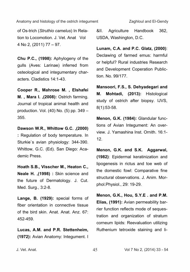
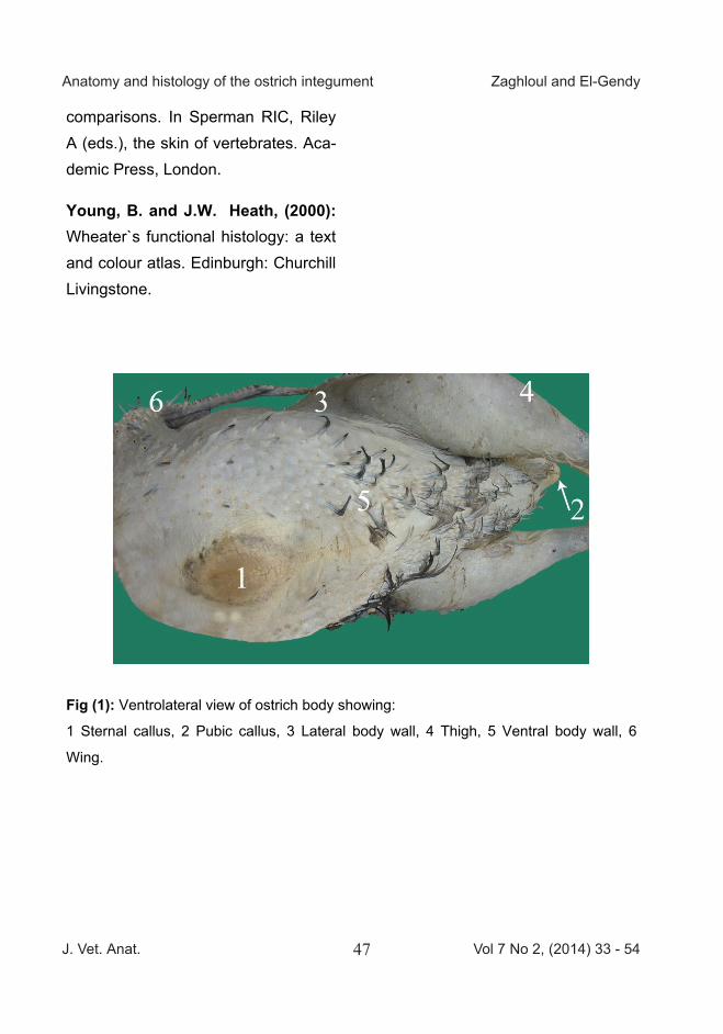
Fig (2): Different types of feathered skin of ostrich. A Skin On the cloaca region. B Skin on the ventral body region, C Skin on the dorsal
body region; D Skin on the ventral part of neck, E Skin on the dorsal part of neck. F
Part of skin on wing contain bristle feather. G Longitudinal section at skin of wing.
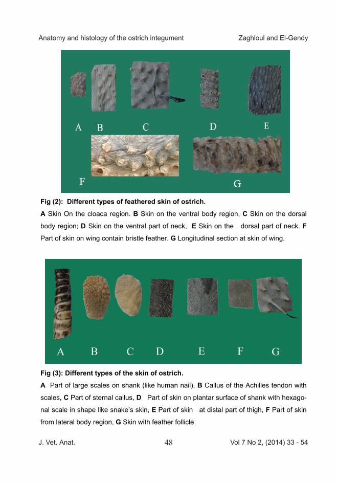
Fig (3): Different types of the skin of ostrich. A Part of large scales on shank (like human nail), B Callus of the Achilles tendon with
scales, C Part of sternal callus, D Part of skin on plantar surface of shank with hexago-
nal scale in shape like snake’s skin, E Part of skin at distal part of thigh, F Part of skin
from lateral body region, G Skin with feather follicle
Fig (4): Dorsal view of ostrich feet and shank. 1 Large scale, 2 Small scale (dome shape), 3 Toe pad papillae, 4 Large scale on dorsal
surface of shank, 5 Enlarged view of small scale (dome shape), 6 Toe nail.
Fig (5 /A,B): Scanning electron micrograph of ostrich skin from neck showing: A Bristle with the surrounded sheath near its root and originate in pit on stratum crone-
um, B Cross section in rachis of the feather showing the cortex and medulla, which ar-
range as honey comb shape.
B A
J. Vet. Anat. Vol 7 No 2, (2014) 33 - 5449
Anatomy and histology of the ostrich integument Zaghloul and El-Gendy
Fig (2): Different types of feathered skin of ostrich. A Skin On the cloaca region. B Skin on the ventral body region, C Skin on the dorsal
body region; D Skin on the ventral part of neck, E Skin on the dorsal part of neck. F
Part of skin on wing contain bristle feather. G Longitudinal section at skin of wing.
Fig (3): Different types of the skin of ostrich. A Part of large scales on shank (like human nail), B Callus of the Achilles tendon with
scales, C Part of sternal callus, D Part of skin on plantar surface of shank with hexago-
nal scale in shape like snake’s skin, E Part of skin at distal part of thigh, F Part of skin
from lateral body region, G Skin with feather follicle
Fig (4): Dorsal view of ostrich feet and shank. 1 Large scale, 2 Small scale (dome shape), 3 Toe pad papillae, 4 Large scale on dorsal
surface of shank, 5 Enlarged view of small scale (dome shape), 6 Toe nail.
Fig (5 /A,B): Scanning electron micrograph of ostrich skin from neck showing: A Bristle with the surrounded sheath near its root and originate in pit on stratum crone-
um, B Cross section in rachis of the feather showing the cortex and medulla, which ar-
range as honey comb shape.
B A
J. Vet. Anat. Vol 7 No 2, (2014) 33 - 5450
Anatomy and histology of the ostrich integument Zaghloul and El-Gendy
Fig (5/C): Scanning electron micrograph of ostrich skin from neck
showing:
C Variation in medullary cells from hallow vacuolated cell "star" or filled mashes
like Cobweb (arrow).
Fig (6): Scanning electron micrograph of the ostrich skin on flesh side showing:
the collagen fibers in deep dermis, arranged parallel to each other and connected by
fibrils (arrow).
C
Fig (7): Photomicrograph of the skin of back in ostrich denoting wavy keratin layer (K),
under it the epidermis (E), the dermis (D). Lymphocytes (arrow) could be seen inside the
epidermis. (X 100. H&E stain)
Fig (8): Photomicrograph of the feather of ostrich skin showing the epidermis (E) cov-
ered with a keratin layer (K)and the feather follicle (F) giving rise to the shaft of feather
(S). Note the smooth muscle layer (M) and the melanin pigments (arrows). H&E stain, X
100.
8
7
J. Vet. Anat. Vol 7 No 2, (2014) 33 - 5451
Anatomy and histology of the ostrich integument Zaghloul and El-Gendy
Fig (5/C): Scanning electron micrograph of ostrich skin from neck
showing:
C Variation in medullary cells from hallow vacuolated cell "star" or filled mashes
like Cobweb (arrow).
Fig (6): Scanning electron micrograph of the ostrich skin on flesh side showing:
the collagen fibers in deep dermis, arranged parallel to each other and connected by
fibrils (arrow).
C
Fig (7): Photomicrograph of the skin of back in ostrich denoting wavy keratin layer (K),
under it the epidermis (E), the dermis (D). Lymphocytes (arrow) could be seen inside the
epidermis. (X 100. H&E stain)
Fig (8): Photomicrograph of the feather of ostrich skin showing the epidermis (E) cov-
ered with a keratin layer (K)and the feather follicle (F) giving rise to the shaft of feather
(S). Note the smooth muscle layer (M) and the melanin pigments (arrows). H&E stain, X
100.
8
7
J. Vet. Anat. Vol 7 No 2, (2014) 33 - 5452
Anatomy and histology of the ostrich integument Zaghloul and El-Gendy
Fig (9): Photomicrograph of the skin of back in ostrich showing, moderate, wavy keratin layer (K), under it the
epidermis (E) with some vacuolated cells, the dermis (D) The upper squamous layer of the epithelium of epi-
dermis (arrow heads). Lymphocytes from the dermis layer pass in between the basal columnar cell layers of
the epidermis (arrows) through the basal lamina. (X 400. H&E stain)
Fig (10): Photomicrograph of a large nerve ending in the dermis of back skin of ostrich. (X 400. H&E stain)
Fig (11): Photomicrograph of the skin of ostrich neck beside the area of feather emerging showing the wavy
keratin layer (K) the vacuolated cells of epidermis (E). melanin pigments (arrows), and the dermal papillae (D).
(X 400, H&E stain)
10 9
11
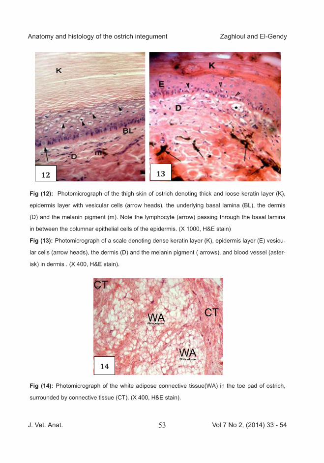
Fig (12): Photomicrograph of the thigh skin of ostrich denoting thick and loose keratin layer (K),
epidermis layer with vesicular cells (arrow heads), the underlying basal lamina (BL), the dermis
(D) and the melanin pigment (m). Note the lymphocyte (arrow) passing through the basal lamina
in between the columnar epithelial cells of the epidermis. (X 1000, H&E stain)
Fig (13): Photomicrograph of a scale denoting dense keratin layer (K), epidermis layer (E) vesicu-
lar cells (arrow heads), the dermis (D) and the melanin pigment ) arrows), and blood vessel (aster-
isk) in dermis . (X 400, H&E stain).
Fig (14): Photomicrograph of the white adipose connective tissue(WA) in the toe pad of ostrich,
surrounded by connective tissue (CT). (X 400, H&E stain).
12
14
13
J. Vet. Anat. Vol 7 No 2, (2014) 33 - 5453
Anatomy and histology of the ostrich integument Zaghloul and El-Gendy
Fig (9): Photomicrograph of the skin of back in ostrich showing, moderate, wavy keratin layer (K), under it the
epidermis (E) with some vacuolated cells, the dermis (D) The upper squamous layer of the epithelium of epi-
dermis (arrow heads). Lymphocytes from the dermis layer pass in between the basal columnar cell layers of
the epidermis (arrows) through the basal lamina. (X 400. H&E stain)
Fig (10): Photomicrograph of a large nerve ending in the dermis of back skin of ostrich. (X 400. H&E stain)
Fig (11): Photomicrograph of the skin of ostrich neck beside the area of feather emerging showing the wavy
keratin layer (K) the vacuolated cells of epidermis (E). melanin pigments (arrows), and the dermal papillae (D).
(X 400, H&E stain)
10 9
11
Fig (12): Photomicrograph of the thigh skin of ostrich denoting thick and loose keratin layer (K),
epidermis layer with vesicular cells (arrow heads), the underlying basal lamina (BL), the dermis
(D) and the melanin pigment (m). Note the lymphocyte (arrow) passing through the basal lamina
in between the columnar epithelial cells of the epidermis. (X 1000, H&E stain)
Fig (13): Photomicrograph of a scale denoting dense keratin layer (K), epidermis layer (E) vesicu-
lar cells (arrow heads), the dermis (D) and the melanin pigment ) arrows), and blood vessel (aster-
isk) in dermis . (X 400, H&E stain).
Fig (14): Photomicrograph of the white adipose connective tissue(WA) in the toe pad of ostrich,
surrounded by connective tissue (CT). (X 400, H&E stain).
12
14
13
J. Vet. Anat. Vol 7 No 2, (2014) 33 - 5454
Anatomy and histology of the ostrich integument Zaghloul and El-Gendy
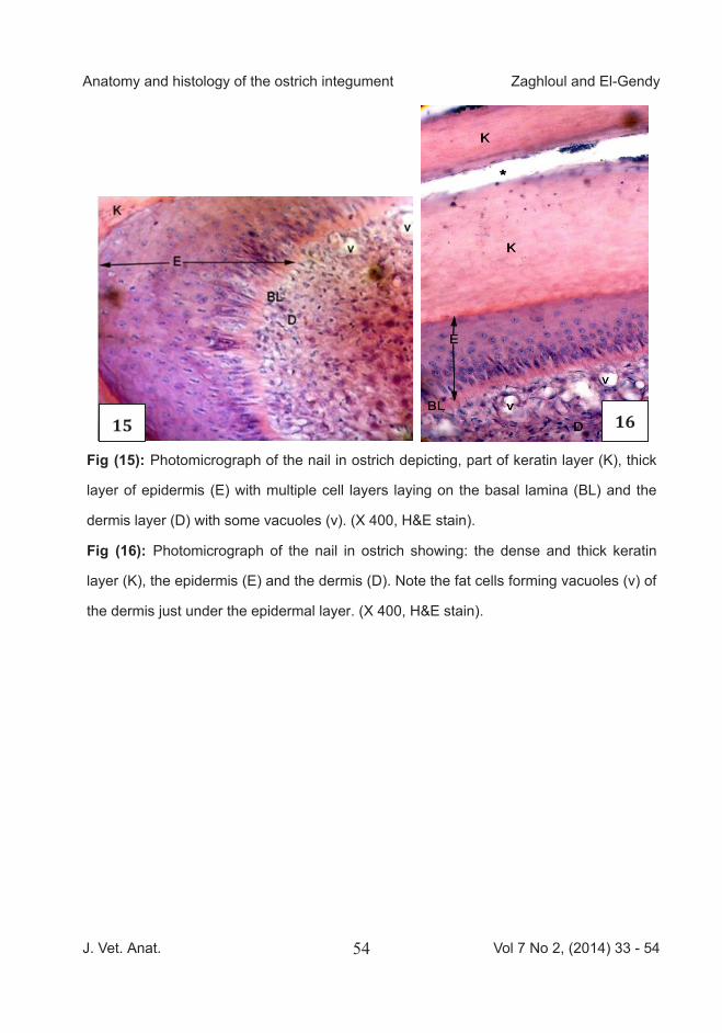
Fig (15): Photomicrograph of the nail in ostrich depicting, part of keratin layer (K), thick
layer of epidermis (E) with multiple cell layers laying on the basal lamina (BL) and the
dermis layer (D) with some vacuoles (v). (X 400, H&E stain).
Fig (16): Photomicrograph of the nail in ostrich showing: the dense and thick keratin
layer (K), the epidermis (E) and the dermis (D). Note the fat cells forming vacuoles (v) of
the dermis just under the epidermal layer. (X 400, H&E stain).
16 15
Some Morphological Studies on the Jaw Joint of the Australian Saltwater Crocodile (Croco-dylus porosus)
A. S. Saber1 and A. Hassanin2 1 Department of Anatomy and Embryology, Faculty of Veterinary Medicine, Uni-versity of Sadat City, Sadat City, Egypt & Dicipline of Veterinary Sciences, College of Public Health, Medical and Veterinary Sciences, James Cook University, Townsville, Australia 2 Department of Cytology and Histology, Faculty of Veterinary Medicine, Univer-sity of Sadat City, Sadat City, Egypt.
__________________________________________________________ With 10 figures, 4 tables Received May, accepted for publication June 2014
Abstract The saltwater crocodile is the largest of all living reptiles. It is also known and proved that it has the largest biting forces. The magnitude of the biting force ex-erted by the jaw leads to thoughts about the anatomical structure and construction of the jaw joint in this reptile. Thirteen skulls of the saltwater crocodile (C. porosus) were used for this study. Some skulls were used for X-raying and morphology, and the others for histological slides which were prepared from the articular cartilages, capsule and collateral ligaments. The quadrate/articular joint (jaw joint) was diarthroidal, formed by two
articular surfaces, which were fibrocartilage in nature at the periphery of the articular sur-face, hyaline in the rest of the articular surface, and ossifies on reaching the underlying bone.
The thick lateral and thin medial collateral ligaments were formed of collagenous fibers. An articu-lar disc was missing in the croc-odile quadrate/articular joint. The joint was surrounded by a complex massive group of mus-cles responsible for the firm clo-sure and opening of the mouth. The results were supported by 10 images and 4 tables, and were discussed with other am-phibians, domestic animals and man when needed.