Analysis of the Dose Escalation in the Scalp of Patients due to Titanium Plate...
27
Monte Carlo Analysis of the Dose Enhancement in the Scalp of Patients due to Titanium Plate Backscatter during Post-Operative Radiotherapy Michael T. Hardin
Transcript of Analysis of the Dose Escalation in the Scalp of Patients due to Titanium Plate...

Monte Carlo Analysis of the Dose Enhancement in the Scalp of
Patients due to Titanium Plate Backscatter during Post-Operative
Radiotherapy
Michael T. Hardin

Motivation
• It has been reported that patients receiving post-operative radiotherapy are demonstrating necrosis of the scalp superior to the placement of titanium fixation plates.
• Healthy scalp tissue is approximately 3-5 mm thick.
• Patients’ scalp tissue have become paper-thin and fragile, requiring surgery.

• Synthes and Biomet titanium plates of thickness 0.4 mm and 0.38 mm respectively.

Motivation
• This is a clinically significant issue. • Why is this issue occurring? • What is the dose enhancement to the scalp? • How can this issue be remedied?

Theoretical Considerations
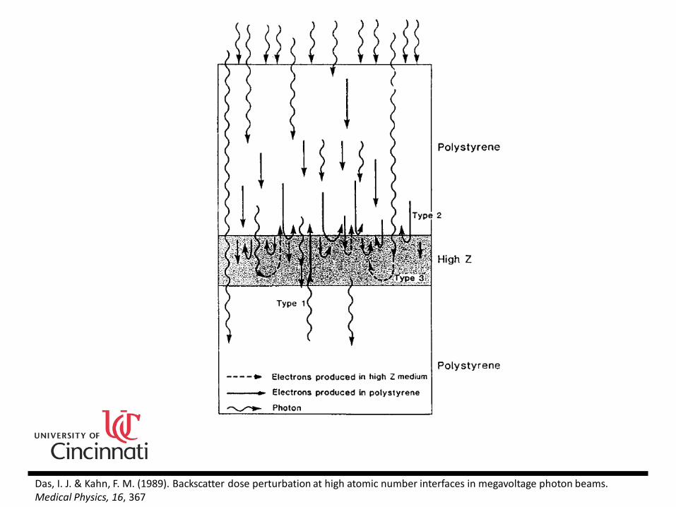
• Das & Kahn, 1989 divide the backscattered dose at an interface into the dose due to: (a) the backscattered photons (b) the backscattering of secondary electrons set in motion in the soft tissue medium above the inhomogeneity (c) the backscattering of secondary electrons set in motion within the inhomogeneity
Das, I. J. & Kahn, F. M. (1989). Backscatter dose perturbation at high atomic number interfaces in megavoltage photon beams. Medical Physics, 16, 367
Das, I. J. & Kahn, F. M. (1989). Backscatter dose perturbation at high atomic number interfaces in megavoltage photon beams. Medical Physics, 16, 367
Theoretical Considerations
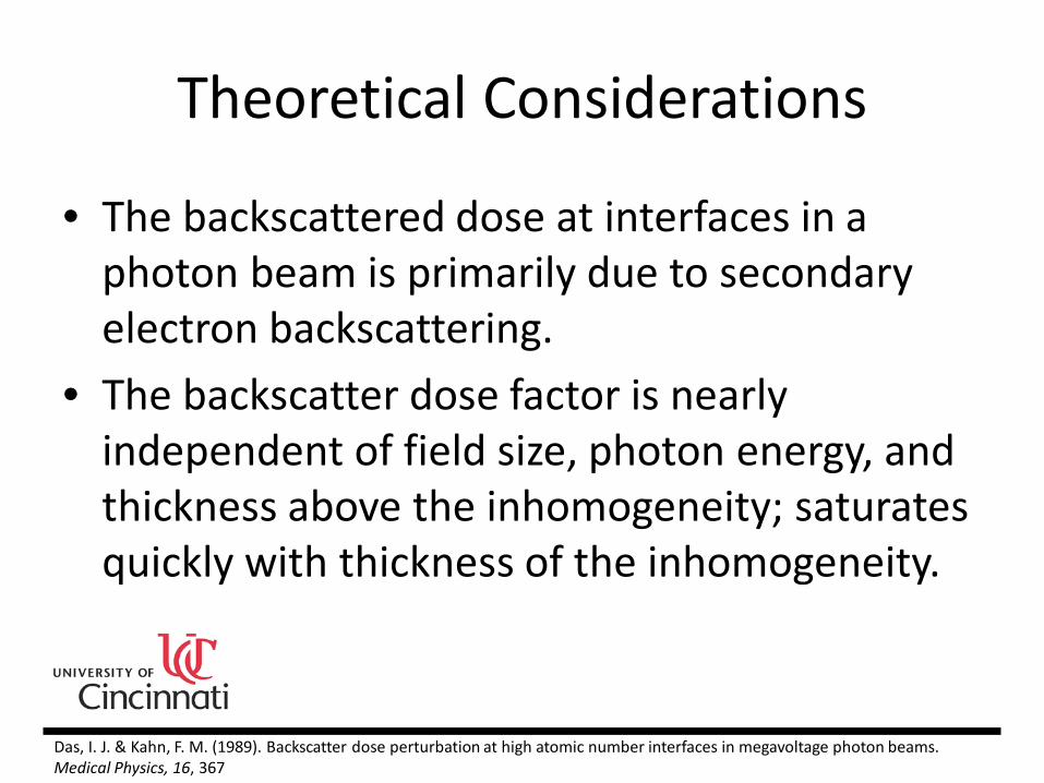
• The backscattered dose at interfaces in a photon beam is primarily due to secondary electron backscattering.
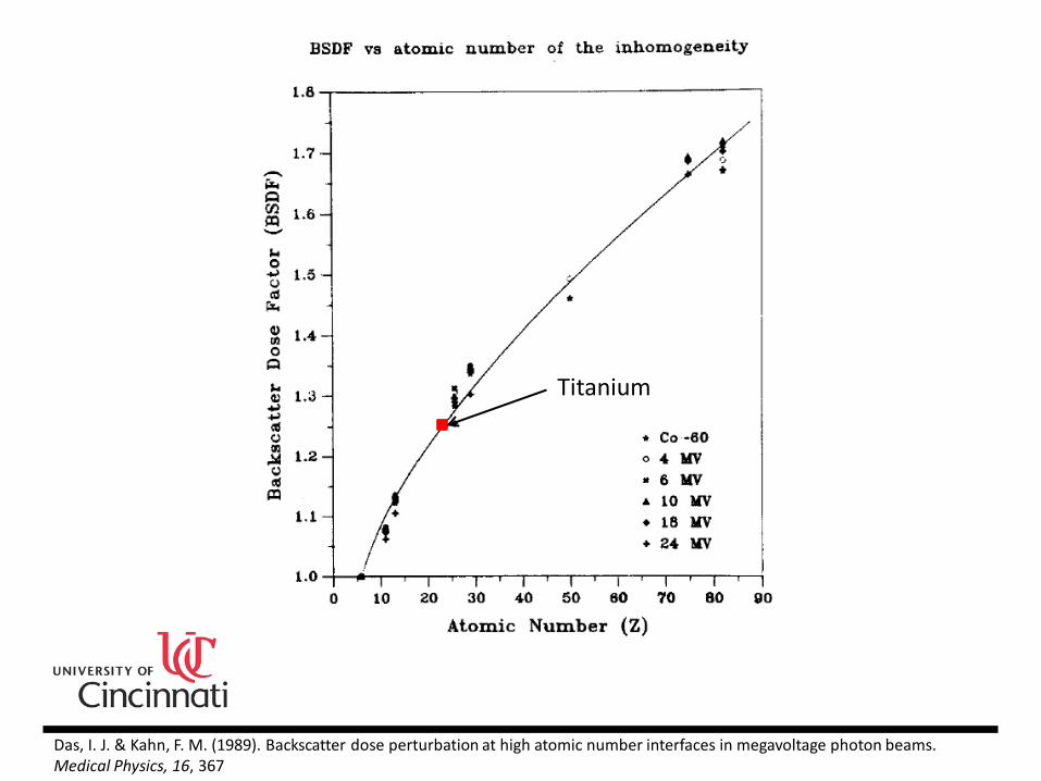
• The backscatter dose factor is nearly independent of field size, photon energy, and thickness above the inhomogeneity; saturates quickly with thickness of the inhomogeneity.
Das, I. J. & Kahn, F. M. (1989). Backscatter dose perturbation at high atomic number interfaces in megavoltage photon beams. Medical Physics, 16, 367
Das, I. J. & Kahn, F. M. (1989). Backscatter dose perturbation at high atomic number interfaces in megavoltage photon beams. Medical Physics, 16, 367
Titanium

Clinical Measurements
• Kodak EDR2 and ISP Gafchromic MD-55 film were used to quantify the dose enhancement.
• Titanium plates were placed inside the film packets to replicate the clinical scenario.
• Different amounts of buildup were examined. • Films were scanned and analyzed in RIT.

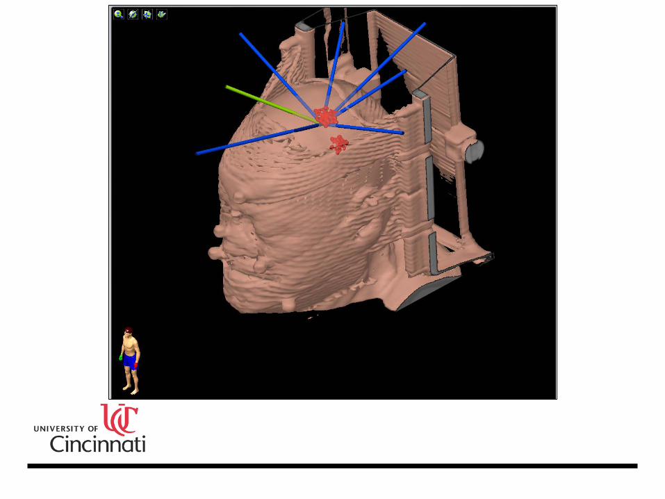
• Placement of the titanium plate.

• Phantom setup.
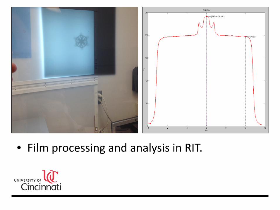
• Film processing and analysis in RIT.
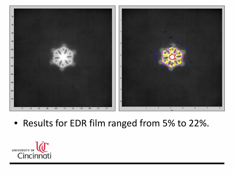
• Results for EDR film ranged from 5% to 22%.

• Film processing and analysis in RIT.

• Results for Gafchromic film and Synthes plate ranged from 8% to 13%.
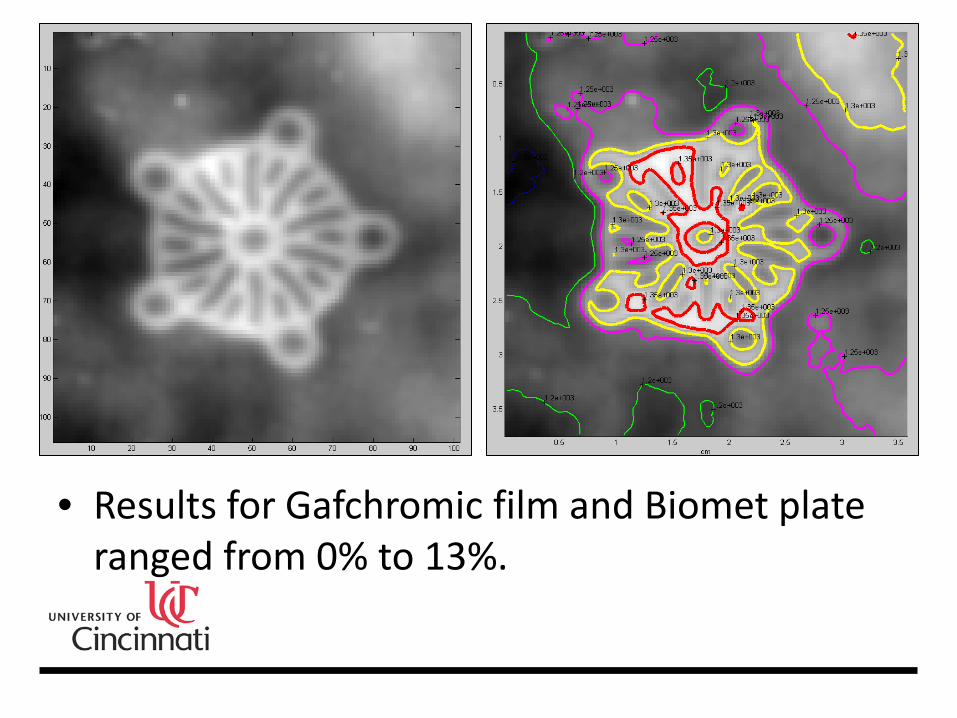
• Results for Gafchromic film and Biomet plate ranged from 0% to 13%.

-15%
-10%
-5%
0%
5%
10%
15%
0 15 30 45 60 75 90 105 120 135 150 165 180 195
Rela
tive
Dose
Incr
ease
Gantry Angle (Degrees)
Gafchromic Film Relative Dose Increase vs Gantry Angle
Synthes
Biomet

Monte Carlo Modeling
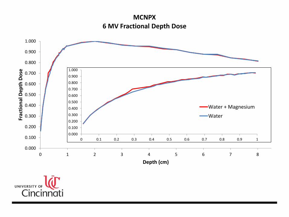
• MCNPX was used to model and simulate the transport of photons and electrons within a box of water containing a thin titanium plate.
• Shimozato et al., 2010 found the maximum relative differences between simulation and TPS results on the entrance and exit sides of the plate to be 23.1% and -12.7% respectively.
Shimozato, T., Yasui, K., Kawanami, R., Habara, K., Aoyama, Y., Tabushi, K., & Obata, Y. (2010). Dose distribution near thin titanium plate for skull fixation irradiated by a 4-MV photon beam. Journal of Medical Physics, 35(2), 81-87.

• Dynamic 3D plotting in MCNP Visual Editor.
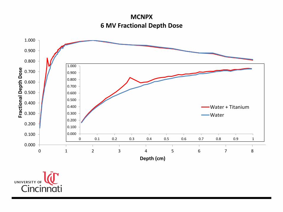
0.000
0.100
0.200
0.300
0.400
0.500
0.600
0.700
0.800
0.900
1.000
0 1 2 3 4 5 6 7 8
Frac
tiona
l Dep
th D
ose
Depth (cm)
MCNPX 6 MV Fractional Depth Dose
0.000
0.100
0.200
0.300
0.400
0.500
0.600
0.700
0.800
0.900
1.000
0 0.1 0.2 0.3 0.4 0.5 0.6 0.7 0.8 0.9 1
Water + TitaniumWater

-5%
0%
5%
10%
15%
20%
25%
30%
0 1 2 3 4 5 6 7 8
Perc
ent D
ose
Enha
ncem
ent
Depth (cm)
MCNPX 6 MV Dose Enhancement
Water + Titanium vs Water
0.000
0.100
0.200
0.300
0.400
0.500
0.600
0.700
0.800
0.900
1.000
0 1 2 3 4 5 6 7 8
Frac
tiona
l Dep
th D
ose
Depth (cm)
MCNPX 6 MV Fractional Depth Dose
0.000
0.100
0.200
0.300
0.400
0.500
0.600
0.700
0.800
0.900
1.000
0 0.1 0.2 0.3 0.4 0.5 0.6 0.7 0.8 0.9 1
Water + Magnesium
Water

-5%
0%
5%
10%
15%
20%
25%
30%
0 1 2 3 4 5 6 7 8
Perc
ent D
ose
Enha
ncem
ent
Depth (cm)
MCNPX 6 MV Dose Enhancement
Water + Magnesium vs Water
Water + Titanium vs Water

1
10
100
0.05 0.10 0.15 0.20 0.25 0.30 0.35 0.40 0.45
Rela
tive
Ener
gy
Energy Bin (MeV)
MCNPX • 6 MV Relative Energy of Deposited Radiation
0.27
0.29
0.31
0.33
0.35
0.37
Cell Depth (cm)
Conclusion
• The maximum relative dose enhancement on the entrance and exit sides of the plate are 26.4% and 7.1% respectively with error < 1%.
• Future work involves research and development of improved surgical fixation devices and/or techniques. Additionally, what biological mechanisms are responsible for this effect?
References • Das, I. J. & Kahn, F. M. (1989). Backscatter dose perturbation at high atomic
number interfaces in megavoltage photon beams. Medical Physics, 16, 367. • Feng, Y. (2006). A monte carlo simulation and deconvolution study of detector
response function for small field measurements. (Unpublished doctoral dissertation). University of Cincinnati, Cincinnati, Ohio.
• Kassing, W. M. (2001). A monte carlo investigation of the radiation dose distribution in intravascular brachytherapy. (Unpublished doctoral dissertation). University of Cincinnati, Cincinnati, Ohio.
• Los Alamos National Laboratory. (2003). MCNP – A general monte carlo n-particle transport code, version 5. Los Alamos, New Mexico: X-5 Monte Carlo Team.
• Shimozato, T., Yasui, K., Kawanami, R., Habara, K., Aoyama, Y., Tabushi, K., & Obata, Y. (2010). Dose distribution near thin titanium plate for skull fixation irradiated by a 4-MV photon beam. Journal of Medical Physics, 35(2), 81-87.