
An Integrated Microfluidic Real-Time PCR System for ... · control and online fluorescence...
4
An Integrated Microfluidic Real-Time PCR System for Pathogen Detection Ling Zhu *,# , Li Li **,# , Dong An ** , Maohua Wang**, Long Zhang*, YiKun Wang*, Zhigang Li*, Yong Liu*, Gong Zhang***, Francis Lin*** * Anhui Institute of Optics & Fine Mechanics, Chinese Academy of Sciences, Hefei, Anhui, 230031, China, [email protected] ** China Agricultural University, Beijing, 100083, China, [email protected] *** University of Manitoba, Winnipeg, MB, R3T 2N2, Canada, [email protected] # These authors contributed equally to this work. ABSTRACT The development of microfluidics-based PCR for rapid genetic analysis with direct application in pathogen detection is an emerging research field. System integration to effectively connect individual function modules is an important aspect for the practical use of microfluidics PCR for diagnosis applications. Here we present our preliminary development of a fully-integrated microfluidic Real-Time PCR system with thermal cycler and online fluorescence detection module. The PCR microfluidic chip was fabricated in PDMS for rapid prototyping. The thermal cycler allows rapid temperature cycling with high accuracy (i.e. the average heating and cooling rate are 4.46℃/s and 4.36℃/s respectively). Using this developed system, we successfully demonstrated real-time PCR detection of E. coli O157:H7 with 20µL of PCR reaction mixture. Further development will lead to a more compact and portable microfluidic Real- Time PCR system that offers a useful tool for various biomedical research and clinical applications. Keywords: microfluidics, lab-on-a-chip, real-time PCR, point-of-care, pathogen detection. 1 INTRODUCTION Foodborne pathogens are important causes for human diseases. The detection and identification of pathogens in food is essential for ensuring the safety of food products for both human and animals. Pathogens such as Escherichia coli, Staphylococcus aureus and avian influenza virus have been commonly associated with diseases outbreaks [1, 2]. Microbiological, biochemical and immunological techniques have been commonly used for pathogen detection and identification. However, these detection methods are time consuming mainly due to microorganisms culturing required for the tests [3]. In addition, immunological methods are limited for its accuracy because of cross-reactivity [4]. By contrast, molecular biology based methods, especially polymerase chain reaction (PCR), offer an advantageous approach in pathogen detection sensitivity and accuracy [5, 6]. Enabled by microfluidics, chip-based PCR methods have been increasingly developed with improved miniaturization, reaction control and throughput [7, 8]. While commonly used in microfluidic PCR, offline signal detection methods suffer from low detection efficiency [9, 10]. By contrast, micro-fluidics-based Real-Time PCR allows rapid test with high sensitivity, specificity and reproducibility for pathogen detection [11]. Efforts to integrate the control and detection components for microfluidic Real-Time PCR with satisfying portability will facilitate its practical use for point-of-care applications. Toward this direction, here we present our preliminary development of a fully-integrated and portable microfluidic Real-Time PCR system with thermal cycling control and online fluorescence detection (Fig. 1). The performance of this developed system for pathogen detection is validated by successful Real-Time PCR amplification and detection of E. coli O157:H7, an E. coli strain that causes foodborne illness. 2 MATERIALS AND METHODS The developed microfluidic real-time PCR system consists of a PDMS microfluidic device, integrated temperature control and online fluorescence detection module in a portable format. 2.1 Microfluidic device The microfluidic device for PCR was designed using SolidWorks, and fabricated in polydimethylsiloxane (PDMS) by simple tape transfer-based soft-lithography method [12, 13]. Specifically, Sylgard 184 (Dow Corning, MI, USA) was stirred thoroughly with curing agent in a weight ratio of 10:1. The mixture was poured onto the tape based master and cured for 3 hours at 75 ℃. The PDMS replicas were removed from the mold and cut down to match the glass substrate. Two through holes for inlet and outlet were punched out of PDMS by a sharpened needle. The surface of NSTI-Nanotech 2012, www.nsti.org, ISBN 978-1-4665-6276-9 Vol. 3, 2012 52
Transcript of An Integrated Microfluidic Real-Time PCR System for ... · control and online fluorescence...
An Integrated Microfluidic Real-Time PCR System for Pathogen Detection
Ling Zhu*,#, Li Li**,#, Dong An**, Maohua Wang**, Long Zhang*, YiKun Wang*, Zhigang Li*, Yong Liu*, Gong Zhang***, Francis Lin***
* Anhui Institute of Optics & Fine Mechanics, Chinese Academy of Sciences,
Hefei, Anhui, 230031, China, [email protected] ** China Agricultural University, Beijing, 100083, China, [email protected]
*** University of Manitoba, Winnipeg, MB, R3T 2N2, Canada, [email protected] # These authors contributed equally to this work.
ABSTRACT
The development of microfluidics-based PCR for rapid
genetic analysis with direct application in pathogen detection is an emerging research field. System integration to effectively connect individual function modules is an important aspect for the practical use of microfluidics PCR for diagnosis applications. Here we present our preliminary development of a fully-integrated microfluidic Real-Time PCR system with thermal cycler and online fluorescence detection module. The PCR microfluidic chip was fabricated in PDMS for rapid prototyping. The thermal cycler allows rapid temperature cycling with high accuracy (i.e. the average heating and cooling rate are 4.46℃/s and 4.36℃/s respectively). Using this developed system, we successfully demonstrated real-time PCR detection of E. coli O157:H7 with 20µL of PCR reaction mixture. Further development will lead to a more compact and portable microfluidic Real-Time PCR system that offers a useful tool for various biomedical research and clinical applications.
Keywords: microfluidics, lab-on-a-chip, real-time PCR, point-of-care, pathogen detection.
1 INTRODUCTION
Foodborne pathogens are important causes for human diseases. The detection and identification of pathogens in food is essential for ensuring the safety of food products for both human and animals. Pathogens such as Escherichia coli, Staphylococcus aureus and avian influenza virus have been commonly associated with diseases outbreaks [1, 2]. Microbiological, biochemical and immunological techniques have been commonly used for pathogen detection and identification. However, these detection methods are time consuming mainly due to microorganisms culturing required for the tests [3]. In addition, immunological methods are limited for its accuracy because of cross-reactivity [4]. By contrast, molecular biology based methods, especially
polymerase chain reaction (PCR), offer an advantageous approach in pathogen detection sensitivity and accuracy [5, 6]. Enabled by microfluidics, chip-based PCR methods have been increasingly developed with improved miniaturization, reaction control and throughput [7, 8]. While commonly used in microfluidic PCR, offline signal detection methods suffer from low detection efficiency [9, 10]. By contrast, micro-fluidics-based Real-Time PCR allows rapid test with high sensitivity, specificity and reproducibility for pathogen detection [11]. Efforts to integrate the control and detection components for microfluidic Real-Time PCR with satisfying portability will facilitate its practical use for point-of-care applications. Toward this direction, here we present our preliminary development of a fully-integrated and portable microfluidic Real-Time PCR system with thermal cycling control and online fluorescence detection (Fig. 1). The performance of this developed system for pathogen detection is validated by successful Real-Time PCR amplification and detection of E. coli O157:H7, an E. coli strain that causes foodborne illness.
2 MATERIALS AND METHODS
The developed microfluidic real-time PCR system consists of a PDMS microfluidic device, integrated temperature control and online fluorescence detection module in a portable format.
2.1 Microfluidic device
The microfluidic device for PCR was designed using SolidWorks, and fabricated in polydimethylsiloxane (PDMS) by simple tape transfer-based soft-lithography method [12, 13]. Specifically, Sylgard 184 (Dow Corning, MI, USA) was stirred thoroughly with curing agent in a weight ratio of 10:1. The mixture was poured onto the tape based master and cured for 3 hours at 75℃. The PDMS replicas were removed from the mold and cut down to match the glass substrate. Two through holes for inlet and outlet were punched out of PDMS by a sharpened needle. The surface of
NSTI-Nanotech 2012, www.nsti.org, ISBN 978-1-4665-6276-9 Vol. 3, 201252
the PDMS replica and a clean glass coverslide were treated with air plasma for 1min using a plasma cleaner (PDC-32G, Harrick, NY) and brought into conformal contact to form an irreversible seal. This assembly produced the micro-reaction chamber for PCR. The PCR reaction chamber has total volume of 20 µL and two air channels on either side of the PCR reaction chamber were used to reduce temperature variation in the reaction chamber. A similar design was reported previously with a different fabricatoin method [14].
2.2 Sample preparation
E. coli O157:H7 (2×10³ CFU/m and 2×10⁶ CFU/ml) and fluorescent SYBR Green I were prepared for PCR. The final PCR reaction mixture includes 10µL of SYBR Green I (TaKaRa.DRR-420A), 2 µL of E. coli O157:H7 suspension, 1µL of primer mix (ACAAAACACTTTATGACCGTT, AATCCTATAGCAG CGCAGA), 5µL of BSA (Bovine Serum Albumin) at the concentration of 0.05% and 2µL of ddH2O. The BSA was used to passivate the microchannel and improve the biocompatibility of the microfluidic device.
2.3 Real-Time PCR system and operation
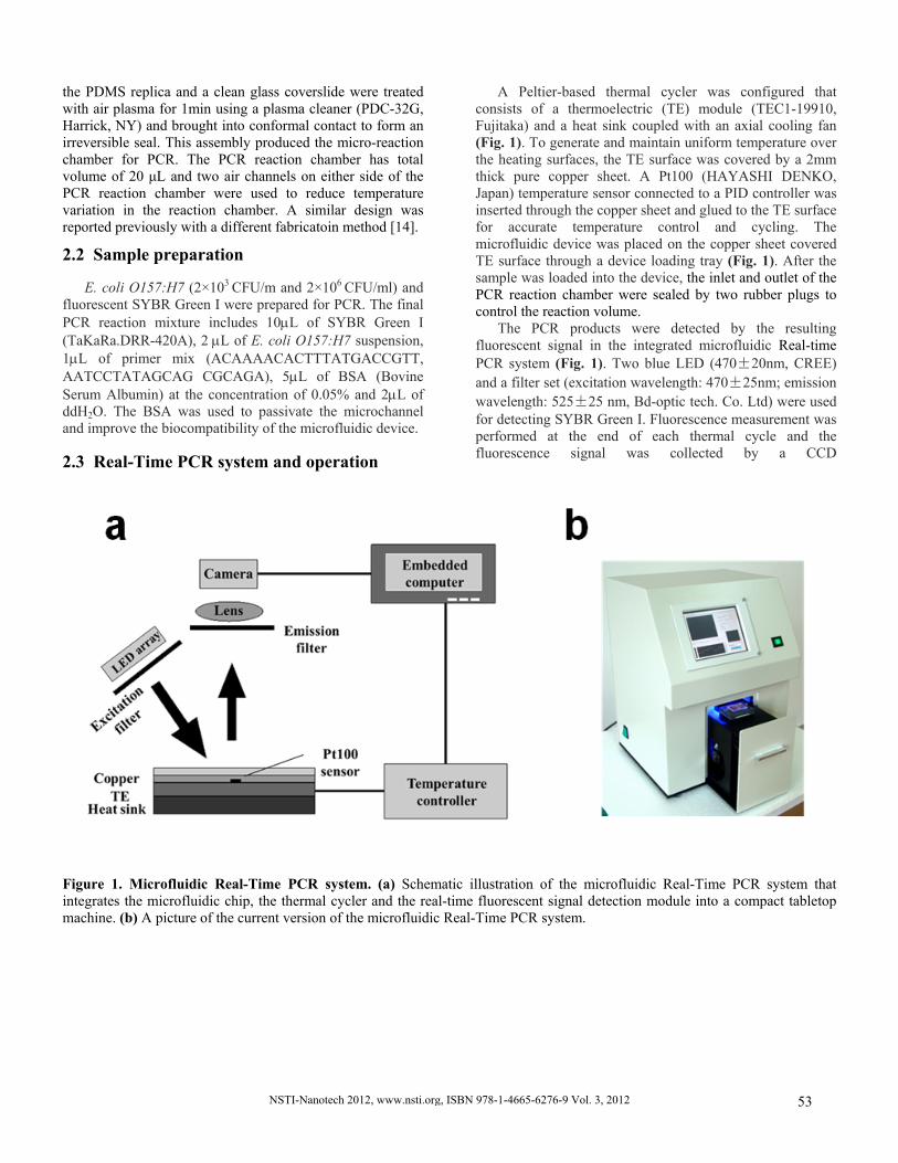
A Peltier-based thermal cycler was configured that consists of a thermoelectric (TE) module (TEC1-19910, Fujitaka) and a heat sink coupled with an axial cooling fan (Fig. 1). To generate and maintain uniform temperature over the heating surfaces, the TE surface was covered by a 2mm thick pure copper sheet. A Pt100 (HAYASHI DENKO, Japan) temperature sensor connected to a PID controller was inserted through the copper sheet and glued to the TE surface for accurate temperature control and cycling. The microfluidic device was placed on the copper sheet covered TE surface through a device loading tray (Fig. 1). After the sample was loaded into the device, the inlet and outlet of the PCR reaction chamber were sealed by two rubber plugs to control the reaction volume.
The PCR products were detected by the resulting fluorescent signal in the integrated microfluidic Real-time PCR system (Fig. 1). Two blue LED (470±20nm, CREE) and a filter set (excitation wavelength: 470±25nm; emission wavelength: 525±25 nm, Bd-optic tech. Co. Ltd) were used for detecting SYBR Green I. Fluorescence measurement was performed at the end of each thermal cycle and the fluorescence signal was collected by a CCD
Figure 1. Microfluidic Real-Time PCR system. (a) Schematic illustration of the microfluidic Real-Time PCR system that integrates the microfluidic chip, the thermal cycler and the real-time fluorescent signal detection module into a compact tabletop machine. (b) A picture of the current version of the microfluidic Real-Time PCR system.
NSTI-Nanotech 2012, www.nsti.org, ISBN 978-1-4665-6276-9 Vol. 3, 2012 53
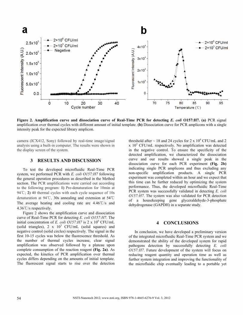
Figure 2. Amplification curve and dissociation curve of Real-Time PCR for detecting E. coli O157:H7. (a) PCR signal amplification over thermal cycles with different amount of initial template. (b) Dissociation curve for PCR amplicons with a single intensity peak for the expected library amplicon.
camera (ICX412, Sony) followed by real-time image/signal analysis using a built-in computer. The results were shown in the display screen of the system.
3 RESULTS AND DISCUSSION
To test the developed microfluidic Real-Time PCR system, we performed PCR with E. coli O157:H7 following the general operation procedures as described in the Method section. The PCR amplifications were carried out according to the following program: 1) Pre-denaturation for 10min at 94℃; 2) 40 thermal cycles with each cycle sequence of 10s denaturation at 94℃, 30s annealing and extension at 54℃ The average heating and cooling rate are 4.46℃/s and 4.36℃/s respectively.
Figure 2 shows the amplification curve and dissociation curve of Real-Time PCR for detecting E. coli O157:H7. The initial concentration of E. coli O157:H7 is 2 x 10⁶ CFU/mL (solid triangles), 2 x 10³ CFU/mL (solid squares) and negative control (solid circles) respectively. The signal in the first 10-15 cycles was below the fluorescence threshold. As the number of thermal cycles increase, clear signal amplification was observed followed by a plateau upon complete consumption of the reaction reagent (Fig. 2a). As expected, the kinetics of PCR amplification over thermal cycles differs depending on the amounts of initial template. The fluorescent signal starts to rise over the detection
threshold after ~ 18 and 24 cycles for 2 x 10⁶ CFU/mL and 2 x 10³ CFU/mL respectively. No amplification was detected in the negative control. To ensure the specificity of the detected amplification, we characterized the dissociation curve and our results showed a single peak in the dissociation curve for each PCR experiment (Fig. 2b) indicating single PCR amplicons and thus excluding any non-specific amplification products. A single PCR experiment was completed within an hour and we expect that this time can be further reduced by optimizing the system performance. Thus, the developed microfluidic Real-Time PCR system was successfully validated in detecting E. coli O157:H7. The system was also validated for PCR detection of a housekeeping gene glyceraldehyde-3-phosphate dehydrogenase (GAPDH) in a separate study.
4 CONCLUSIONS
In conclusion, we have developed a preliminary version of the integrated microfluidic Real-Time PCR system and we demonstrated the ability of the developed system for rapid pathogens detection by successfully detecting E. coli O157:H7. Future development of the system will focus on reducing reagent quantity and operation time as well as further system integration and improving the functionality of the microfluidic chip eventually leading to a portable yet
NSTI-Nanotech 2012, www.nsti.org, ISBN 978-1-4665-6276-9 Vol. 3, 201254
reliable Real-Time PCR product for rapid, inexpensive and accurate biodiagnostic applications at the point of care.
ACKNOWLEDGEMENTS This work was supported by a grant from the National High Technology Research and Development Program of China (863 Program) (No. 2011AA100704-1).
REFERENCES
[1] C.S. Zhang, J.L. Xu, W.L. Ma, W.L.
Zheng, Biotechnology Advances, 2006, 24, 243-284. [2] R. Naveen, Y. Zhang, H.B. Liu, C.C. Dai, R. Kaushik,
B. Ratnaharika, H.Q. Gong, Sensors and Actuators B, 2010, 145, 543-552.
[3] R. Naveen, R. Zhang, H.B. Liu, et al. Sensors and Actuators B: Chemical, 2010, 145(1): 543-552.
[4] A. Rompre, P. Servais, J. Baudart, et al. Journal of Microbiological Methods, 2002, 49(1): 31-54.
[5] D. Lees. Viruses and bivalve shellfish, Int. J. Food Microbiol., 2000, 59, 81-116.
[6] C.S. Zhang, D. Xing, Chem. Rev., 2010, 8, 4910-4947. [7] C. D. Chin, V. Linder and S. K. Sia. Lab Chip, 2007, 7,
41–57. [8] L. Chen, A. Manz and P. J. Day. Lab Chip, 2007, 7,
1413–1423. [9] Q. Xiang, B. Xu, R. Fu, D. Li, Biomedical
Microdevices, 2005, 4, 273-279. [10] Q. Xiang, B. Xu, D. Li, Biomedical Microdevices,
2007, 4, 443-449. [11] A. F. Sauer-Budge, P. Mirer, A. Chatterjee, et al. Lab
Chip, 2009, 9, 2803-2810. [12] N. Pulak, F. Derek, K. Yuliya A, Z. Ahmet, B. Brittany,
G. Greg, Lab on a Chip, 2010, 10, 2286-2291. [13] R, Kumar, R. L. Smith, M. G. Pappas, Lab on a chip,
Chips & Tips, 30 June 2009. [14] X. Qiu, M. G. Mauk, D. Chen, C. Liu and H. H. Bau,
Lab on a Chip, 2010, 10, 3170-3177.
NSTI-Nanotech 2012, www.nsti.org, ISBN 978-1-4665-6276-9 Vol. 3, 2012 55


