All cellulose electrospun water purification membranes ... · 20–30% of the membranes used in...
13
ORIGINAL PAPER All cellulose electrospun water purification membranes nanotextured using cellulose nanocrystals Lee A. Goetz . Narges Naseri . Santhosh S. Nair . Zoheb Karim . Aji P. Mathew Received: 10 November 2017 / Accepted: 12 March 2018 / Published online: 16 March 2018 Ó The Author(s) 2018 Abstract Cellulose acetate (CA) fibers were elec- trospun on a mesh template to create specific surface and pore structures for membrane applications. The mesh template CA fiber mats were impregnated with cellulose nanocrystals at varying weight percentages. The membranes showed nanotextured surfaces and improved mechanical properties post impregnation. More importantly, the hydrophilicity of the original CA fibers was increased from a hydrophobic contact angle of 102°–0° thereby creating an anti-fouling membrane surface structure. The membranes showed rejection of 20–56% for particles of 0.5–2.0 lm, indicating potential of these membranes in rejecting microorganisms from water. Furthermore, high rejec- tion of dyes (80–99%) by adsorption and potential application as highly functional affinity membranes was demonstrated. These membranes can therefore be utilized as all-cellulose, green, scalable and low cost high flux membranes ( [ 20,000 LMH) for water cleaning applications in food industry where microor- ganisms and charged contaminants are to be removed. Keywords Electrospinning Cellulose nanocrystals Water permeability Size exclusion Adsorption Introduction Membranes and membrane processes are now an integral part of the food processing chain; an estimated 20–30% of the membranes used in manufacturing industries worldwide are used by the food industry (Daufin et al. 2001; Inc 2004; Lipnizki 2010). In food industry biological contaminants is a major concern and fresh-cut produces include minimally processed products, which result in very high microbial loads, the initial microbiological contamination ranging from 10 5 to 10 7 CFU/g (Francis et al. 1999). The most significant are Listeria monocytogenes, which can grow in the cooling chain, and Enterobacteri- aceae, like Salmonella enterica or Escherichia coli (De Oliveira et al. 2011). The European commission has identified that fresh-food microorganisms associ- ated with outbreaks include bacteria, viruses, and parasites, and the World Health Organization reported in 2015 the first-ever estimates of disease burden caused by 31 foodborne agents (bacteria, viruses, Electronic supplementary material The online version of this article (https://doi.org/10.1007/s10570-018-1751-1) con- tains supplementary material, which is available to authorized users. L. A. Goetz Z. Karim A. P. Mathew Division of Materials Science, Lulea ˚ University of Technology, 971 87 Lulea ˚, Sweden N. Naseri S. S. Nair A. P. Mathew (&) Department of Materials and Environmental Chemistry, Stockholm University, 106 91 Stockholm, Sweden e-mail: [email protected] 123 Cellulose (2018) 25:3011–3023 https://doi.org/10.1007/s10570-018-1751-1
Transcript of All cellulose electrospun water purification membranes ... · 20–30% of the membranes used in...

ORIGINAL PAPER
All cellulose electrospun water purification membranesnanotextured using cellulose nanocrystals
Lee A. Goetz . Narges Naseri . Santhosh S. Nair . Zoheb Karim .
Aji P. Mathew
Received: 10 November 2017 / Accepted: 12 March 2018 / Published online: 16 March 2018
� The Author(s) 2018
Abstract Cellulose acetate (CA) fibers were elec-
trospun on a mesh template to create specific surface
and pore structures for membrane applications. The
mesh template CA fiber mats were impregnated with
cellulose nanocrystals at varying weight percentages.
The membranes showed nanotextured surfaces and
improved mechanical properties post impregnation.
More importantly, the hydrophilicity of the original
CA fibers was increased from a hydrophobic contact
angle of 102�–0� thereby creating an anti-fouling
membrane surface structure. The membranes showed
rejection of 20–56% for particles of 0.5–2.0 lm,
indicating potential of these membranes in rejecting
microorganisms from water. Furthermore, high rejec-
tion of dyes (80–99%) by adsorption and potential
application as highly functional affinity membranes
was demonstrated. These membranes can therefore be
utilized as all-cellulose, green, scalable and low cost
high flux membranes ([ 20,000 LMH) for water
cleaning applications in food industry where microor-
ganisms and charged contaminants are to be removed.
Keywords Electrospinning � Cellulosenanocrystals � Water permeability � Size exclusion �Adsorption
Introduction
Membranes and membrane processes are now an
integral part of the food processing chain; an estimated
20–30% of the membranes used in manufacturing
industries worldwide are used by the food industry
(Daufin et al. 2001; Inc 2004; Lipnizki 2010). In food
industry biological contaminants is a major concern
and fresh-cut produces include minimally processed
products, which result in very high microbial loads,
the initial microbiological contamination ranging
from 105 to 107 CFU/g (Francis et al. 1999). The
most significant are Listeria monocytogenes, which
can grow in the cooling chain, and Enterobacteri-
aceae, like Salmonella enterica or Escherichia coli
(De Oliveira et al. 2011). The European commission
has identified that fresh-food microorganisms associ-
ated with outbreaks include bacteria, viruses, and
parasites, and the World Health Organization reported
in 2015 the first-ever estimates of disease burden
caused by 31 foodborne agents (bacteria, viruses,
Electronic supplementary material The online version ofthis article (https://doi.org/10.1007/s10570-018-1751-1) con-tains supplementary material, which is available to authorizedusers.
L. A. Goetz � Z. Karim � A. P. Mathew
Division of Materials Science, Lulea University of
Technology, 971 87 Lulea, Sweden
N. Naseri � S. S. Nair � A. P. Mathew (&)
Department of Materials and Environmental Chemistry,
Stockholm University, 106 91 Stockholm, Sweden
e-mail: [email protected]
123
Cellulose (2018) 25:3011–3023
https://doi.org/10.1007/s10570-018-1751-1

parasites, toxins and chemicals) at global and regional
levels (Directorate 2002; Shannon et al. 2008).
Water purification technology in food industry also
faces some major technical barriers, especially while
dealing with removal of small molecules and viruses
and nanofiltration or reverse osmosis process is
accompanied with high energy consumption and low
water flux (Manth et al. 2003; Shahmansouri and
Bellona 2015). Ultrafiltration membranes systems can
be a solution as they are typically more compact,
require lower pressure than reverse osmosis or
nanofiltration membrane systems, and also have the
ability to remove harmful biologics while maintaining
a relatively high flux, though the removal of smaller
molecules and ions can still be challenging. In
addition, membrane fouling compromises the benefits
of membranes due to reduced performance, reduced
flux and thereby increase of cost (Flemming et al.
1997; Mohammad et al. 2012).
Food industry is a major water consuming sector
and about 70% of the water consumption in these
sectors is for cleaning and decontamination processes
which calls for intelligent design of membrane oper-
ations aimed at reducing the dose of disinfectant
agents and saving water by improving recyclability. A
new generation of ultrafiltration membranes including
biocidal functionalization with the aim of limiting
biofouling and controlling the level of pathogens in
process water is therefore needed. Reduction of water
and energy inputs in the food chain provided by the
development of nanotechnology advances and the use
of novel approaches to control of water borne
microorganisms would also result in a better micro-
biological quality of processed food and cleaning of
water.
Membranes with charge have been successful in the
recent years also due to better fouling resistance. The
utilization of biological materials, such as cellulose
and chitin, in membrane applications is a current
research goal and its nanoscaled derivatives have
shown promising applications in the membrane and
water filtration as functional entities with high flux and
high rejection driven by surface adsorption. Our
studies have shown that nanocellulose in native form
or with additional surface functionalization (TEMPO
oxidation, enzymatic phosphorylation, cationization,
etc.) have significant adsorption efficiency towards
metal ions, nitrates, dyes, humic acid, etc. from
industry effluents (Karim et al. 2014; Liu et al. 2015;
Ma et al. 2011a, b; Mautner et al. 2016; Sehaqui et al.
2015; Gopakumar et al. 2017) One challenge however
is the processing of membrane with control of pore
structure and layering of the different layers of the
membrane without compromising on the membrane
flux, functionality and rejection capacity and the green
image of ‘nanocellulose’.
Electrospinning, being an established process, is
becoming a popular route in producing membranes
with an open yet interconnected pore structure and
high surface area (Gopal et al. 2006; Huang et al.
2003). Electrospun membranes are used for removal
of micron scaled pollutants in water and there are
several attempts to improve the functionality and
stability of these membranes by addition of nanopar-
ticles, blending with functional polymers, crosslink-
ing, etc. (Huang et al. 2013; Ma et al. 2005; Qin et al.
2015). The electrospun fibrous membranes have also
shown potential as an excellent support layer for a
secondary active surface-by coating individual fiber or
by creating a more complete secondary layer of a
bilayered composite membrane (Ma et al. 2011a, b;
Gopi et al. 2017) Ma et al. (2011b) have shown that
TEMPO oxidized cellulose nanocrystals with negative
functional groups can be used to functionalize elec-
trospun membranes, which provides a route to reject
bacteria via size exclusion and reject virus via
adsorption. We have also shown that stand-alone and
biobased electrospun membranes of cellulose acetate
with sufficient mechanical properties and flux for
membrane as well as anti-fouling performance can be
obtained via impregnation using chitin nanocrystals
(Goetz et al. 2016). While typically the electrospun
mat is randomly aligned, there has also been recent
interest in electrospinning fibers on to templates to
create aligned electrospun mats. However, templated
electrospun membranes have not been exploited for
functionalization with nanocrysals and its use in water
filtration.
This work seeks to demonstrate an effective way to
create high flux-renewable membranes for water
filtration with the adsorption functionality of biobased
cellulose nanocrystals on the mesh-template electro-
spun cellulose acetate fiber mats. The work aims to
exploit the potential of cellulose nanocrystals func-
tionalized electrospun cellulose acetate applications to
reject 0.05–2.0 lm particles via size exclusion while
providing additional functionality as adsorption,
antifouling and improved mechanical stability.
3012 Cellulose (2018) 25:3011–3023
123

Experimental section
Materials
Cellulose acetate (CA), Mn 50,000, was purchased
from Sigma-Aldrich Chemistry, USA. Acetic acid
(96%, EMSURE�) and acetone, analysis grade, were
purchased fromMerck KGaA (Germany). Polystyrene
latex microspheres were purchased from Alfa Aesar in
2.5wt% suspension with particle sizes of 0.05, 0.5, and
2.5 lm, individually. All chemicals were used as
received without further purification.
Cellulose nanocrystals (CNCs) were prepared fol-
lowing the process reported earlier by Mathew et al.
(2014). Unbarked wood was hydrolyzed using dilute
acid in a bioethanol pilot plant and refined by solvent
extraction and bleaching to obtain pure cellulose.
Cellulose, after bioethanol process in a plant was
supplied by SP Processum, Ornskoldsvik, Sweden
(17wt%). The purified cellulose from bioethanol
process was made into 1.5wt% suspensions, mixed
by shear mixture and passed through the homogenizer,
10 times to obtain a thick gel of CNCs.
Preparation
Cellulose acetate, 5.0 g (Mn 50,000), was dissolved in
a 45 g (1:1) mixture of concentrated acetic acid and
acetone and stirred overnight (12 h) to ensure com-
plete dissolution (Inc 2004). The viscosity and
conductivity of the cellulose acetate electrospinning
solution were 1144 mPa.s and 8.67 mScm-1, respec-
tively. Electrospinning of the cellulose acetate solu-
tion was undertaken using the 150 mm Laboratory
Electrospinning Platform, Electrospinz-ES1a, New
Zealand, attached to a high voltage supplier, with the
solution pumped through a 20 ml plastic syringe, (BD
Plasti-Pak syringe, USA), using a single syringe pump
(Aladdin-1000, World Precision Instrument, USA).
The cellulose acetate fibers were successfully electro-
spun on stainless steel wire mesh (150 mesh, Fors-
bergs Metallduk AB, Sweden), with a supplied voltage
of 9 kV, 150 mm tip to collector distance, and a flow
rate of 10 ml h-1. Electrospinning was performed for
2 h at room temperature.
A schematic representation of the membrane pro-
cessing is given in Fig. 1. The mesh template electro-
spun cellulose acetate mats were impregnated with the
CNC suspensions via Buchner funnel filtration
apparatus with the cellulose acetate membrane on a
90 mm diameter glass frit. Varying weight percent-
ages of crystals were filtered through the electrospun
mesh mats (Fig. 1a) and are described in Table (in
Fig. 1). The mesh side was the top surface during
impregnation.
Characterization
Atomic Force Microscopy (AFM) AFM images of
CNC shows typical cellulose nanocrystal structure and
the diameters were found to be in the range of
5–10 nm but are longer compared to typical wood
based CNCs. The resulting nanocrystal impregnated
cellulose acetate membranes were air dried for 24 h
and then heated to 100 �C for 10 min to ensure
binding between the cellulose and the cellulose acetate
fibers (Fig. 1b). Membranes were weighed on an
analytical balance before and after impregnation to
determine actual mass of the nanocrystals on the
electrospun cellulose acetate mat.
Scanning Electron Microscopy (SEM) The surface
morphology of the electrospun fibers and the mem-
brane were examined using a FEG-SEM (Zeiss,
Merlin). The fiber samples were placed on conductive
tape and sputter coated with tungsten. Images were
taken operating at 2.5 kV and 8 mmworking distance.
The retention of the polystyrene latex microspheres
was imaged using SEM (TVM SEM), imaged at
15 kV and about 20 mm working distance.
Flux and Permeability Flux and permeability were
determined using a dead-end cell (HP 4750, Sterlitech,
USA) with N2 gas to maintain constant pressure. The
time for 300 ml of distilled water to pass through the
membranes was recorded and used for the flux
calculations. Flux, J, was calculated as
J ¼ Qp
Am
ð1Þ
where Qp is the filtrate volume through the membrane
per time and Am is the area of the membrane. Am
(14.6 cm2) is a constant value provided by Sterlitech.
Permeability was calculated as flux (J) per pressure
(bar) multiplied by membrane thickness (mm).
Tensile Properties Tensile tests were performed
using a universal testing machine, Shimadzu Auto-
graph AG–X (Japan), with a load cell of 500 N. Test
specimens (50 mm 9 5 mm) were conditioned at
45% relative humidity for 1 week and were mounted
Cellulose (2018) 25:3011–3023 3013
123

on paper windows for ease of handling and mounting.
A preload of 0.1 N was applied and a strain rate of
2 mm/min and gauge length of 20 mmwere used. The
stress–strain curves were plotted from the measured
load and sample extension (measured by video
camera).
The stress is defined as
r ¼ F
A0
ð2Þ
and the strain as
e ¼ lnL
L0
� �ð3Þ
where F is the force at break, Ao is the area of cross-
section of the tensile sample, and L0 is the initial
sample length and L is the sample length at break. The
elastic modulus was calculated from the initial part of
the slope from the stress–strain curve. 4–6 test samples
were tested for each material and the average values
are reported.
Contact Angle Equilibrium contact angles of the
membrane surfaces against deionized water were
measured in triplicate using a Kruss EasyDrop
DSA1 (Germany) system. The surface energy was
calculated using the Neumann equation of states
approach (Moy and Neumann 1987; Li and Neumann
1992).
Microfiltration Performance by Particle Retention
The retention performance of the membranes using
polystyrene latex microspheres was evaluated by
passing 20 ml of the 0.05, 0.5, and 2.5 lm polybead
suspensions (Alfa Aesar, USA) through the mem-
branes at an applied N2 gas pressure of 0.2 bar.
Particle suspension concentrations were analyzed
using UV–Vis spectrometer (PerkinElmer, Lambda
2S, USA).
Membrane Performance by Dye Adsorption The
dye adsorption was carried out in static mode by
treating the membranes (0.05 g) with 50 ml of Vic-
toria Blue 2B with three different concentrations such
as 10, 25, and 50 mg/L, respectively. The dye
adsorption on membranes of CA, CA-37CNC, and
CA-45CNC were continuously monitored at different
time intervals and quantitatively determined by UV–
Vis spectrometer (PerkinElmer, Lambda 2S, Sweden).
The absorbance of Victoria Blue 2B was calculated by
Beer Lambert’s law.
The amount of dye adsorbed was calculated using
the formula as given below;
q ¼ C0 � Ctð ÞV=WÞ ð4Þ
Fig. 1 Scheme showing the processing of CA membranes via
electrospinning. a Mesh collector used for collecting fibers; the
SEM images of corresponding membranes showing the mesh
pattern and the photograph of the membrane. b AFM image of
the cellulose nanocrystals (CNCs) and the SEM image of the
membranes showing the CNC coated electrospun fibers.
Table showing the composition of the prepared materials and
the codes which are used for each sample
3014 Cellulose (2018) 25:3011–3023
123
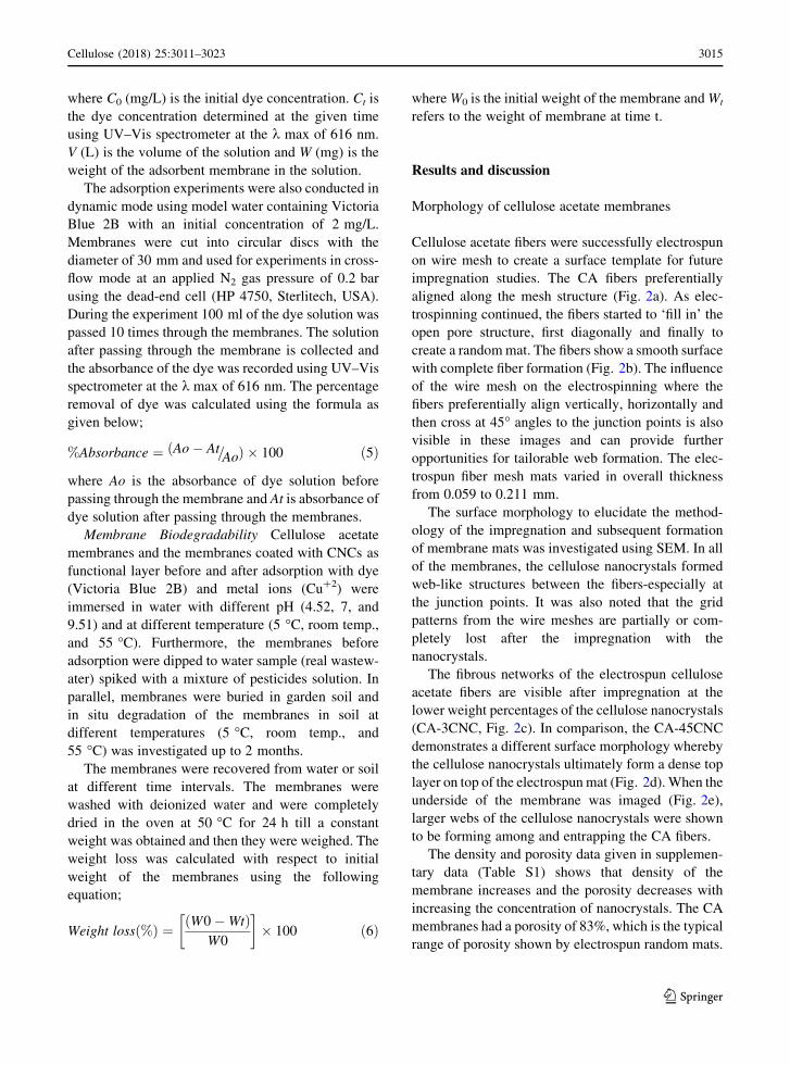
where C0 (mg/L) is the initial dye concentration. Ct is
the dye concentration determined at the given time
using UV–Vis spectrometer at the k max of 616 nm.
V (L) is the volume of the solution and W (mg) is the
weight of the adsorbent membrane in the solution.
The adsorption experiments were also conducted in
dynamic mode using model water containing Victoria
Blue 2B with an initial concentration of 2 mg/L.
Membranes were cut into circular discs with the
diameter of 30 mm and used for experiments in cross-
flow mode at an applied N2 gas pressure of 0.2 bar
using the dead-end cell (HP 4750, Sterlitech, USA).
During the experiment 100 ml of the dye solution was
passed 10 times through the membranes. The solution
after passing through the membrane is collected and
the absorbance of the dye was recorded using UV–Vis
spectrometer at the k max of 616 nm. The percentage
removal of dye was calculated using the formula as
given below;
%Absorbance ¼ ðAo� At=AoÞ � 100 ð5Þ
where Ao is the absorbance of dye solution before
passing through the membrane and At is absorbance of
dye solution after passing through the membranes.
Membrane Biodegradability Cellulose acetate
membranes and the membranes coated with CNCs as
functional layer before and after adsorption with dye
(Victoria Blue 2B) and metal ions (Cu?2) were
immersed in water with different pH (4.52, 7, and
9.51) and at different temperature (5 �C, room temp.,
and 55 �C). Furthermore, the membranes before
adsorption were dipped to water sample (real wastew-
ater) spiked with a mixture of pesticides solution. In
parallel, membranes were buried in garden soil and
in situ degradation of the membranes in soil at
different temperatures (5 �C, room temp., and
55 �C) was investigated up to 2 months.
The membranes were recovered from water or soil
at different time intervals. The membranes were
washed with deionized water and were completely
dried in the oven at 50 �C for 24 h till a constant
weight was obtained and then they were weighed. The
weight loss was calculated with respect to initial
weight of the membranes using the following
equation;
Weight loss %ð Þ ¼ W0�Wtð ÞW0
� �� 100 ð6Þ
whereW0 is the initial weight of the membrane andWt
refers to the weight of membrane at time t.
Results and discussion
Morphology of cellulose acetate membranes
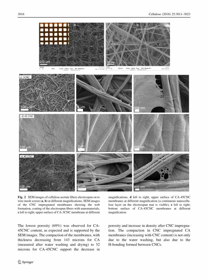
Cellulose acetate fibers were successfully electrospun
on wire mesh to create a surface template for future
impregnation studies. The CA fibers preferentially
aligned along the mesh structure (Fig. 2a). As elec-
trospinning continued, the fibers started to ‘fill in’ the
open pore structure, first diagonally and finally to
create a randommat. The fibers show a smooth surface
with complete fiber formation (Fig. 2b). The influence
of the wire mesh on the electrospinning where the
fibers preferentially align vertically, horizontally and
then cross at 45� angles to the junction points is also
visible in these images and can provide further
opportunities for tailorable web formation. The elec-
trospun fiber mesh mats varied in overall thickness
from 0.059 to 0.211 mm.
The surface morphology to elucidate the method-
ology of the impregnation and subsequent formation
of membrane mats was investigated using SEM. In all
of the membranes, the cellulose nanocrystals formed
web-like structures between the fibers-especially at
the junction points. It was also noted that the grid
patterns from the wire meshes are partially or com-
pletely lost after the impregnation with the
nanocrystals.
The fibrous networks of the electrospun cellulose
acetate fibers are visible after impregnation at the
lower weight percentages of the cellulose nanocrystals
(CA-3CNC, Fig. 2c). In comparison, the CA-45CNC
demonstrates a different surface morphology whereby
the cellulose nanocrystals ultimately form a dense top
layer on top of the electrospunmat (Fig. 2d).When the
underside of the membrane was imaged (Fig. 2e),
larger webs of the cellulose nanocrystals were shown
to be forming among and entrapping the CA fibers.
The density and porosity data given in supplemen-
tary data (Table S1) shows that density of the
membrane increases and the porosity decreases with
increasing the concentration of nanocrystals. The CA
membranes had a porosity of 83%, which is the typical
range of porosity shown by electrospun random mats.
Cellulose (2018) 25:3011–3023 3015
123
The lowest porosity (69%) was observed for CA-
45CNC content, as expected and is supported by the
SEM images. The compaction of the membranes, with
thickness decreasing from 143 microns for CA
(measured after water washing and drying) to 52
microns for CA-45CNC support the decrease in
porosity and increase in density after CNC impregna-
tion. The compaction in CNC impregnated CA
membranes (increasing with CNC content) is not only
due to the water washing, but also due to the
H-bonding formed between CNCs.
Fig. 2 SEM images of cellulose acetate fibers electrospun on to
wire mesh screen (a, b) at different magnifications. SEM images
of the CNC impregnated membranes showing the web
formation, coating of the electrospun fibers with nanomaterials,
c left to right; upper surface of CA-3CNCmembrane at different
magnifications, d left to right; upper surface of CA-45CNC
membranes at different magnification (a continuous nanocellu-
lose layer on the electrospun mat is visible), e left to right;
bottom surface of CA-45CNC membranes at different
magnification
3016 Cellulose (2018) 25:3011–3023
123

Flux, permeability and wettability of membranes
The water flux through the membranes was studied to
understand the effect of coating on the permeability
and the data is given in Fig. 3. Figure 3 shows that the
water flux increases with pressure in all cases, as
expected. However, the trend line shows that slope
increases for the functionalized membranes, indicat-
ing that water transport through the membranes is
favored after coating with cellulose nanocrystals, in
spite of the decreased pore diameters.
The water flux values are in the range of
5000–22,000 Lm-2 h-1 bar-1 at 0.5–2 bars for CA
and CA functionalized with 3 wt% of cellulose
nanocrystals. However, for high CNC content (45%)
the flux was 500–5000 Lm-2 h-1 bar-1 and was
attained only at a high pressure of 6–8 bars. The
surface morphology of these membranes explains the
flux behavior, especially for CA-45CNC, which had
drastically lower flux and permeability. Our earlier
report on a similar system of CA membranes impreg-
nated with 5 wt% chitin nanocrystals (ChNCs) also
showed a high flux of 18,000 Lm-2 h-1 bar-1. In
comparison, Ma et al. have reported a flux of
5900 Lm-2 h-1 bar-1 for PAN nanofiber mats with
a support layer impregnated with cellulose
nanocrystals and is lower than our current membrane
(Ma et al. 2011b).
The flux and permeability through the membranes
at 1 bar are given in supplementary data (Table S1).
The flux increases slightly after impregnation with
nanocrystals. However, permeability systematically
decreased as a function of nanocrystal loading which
is a direct function of pore structure and porosity; SEM
images show a closing of pore structure as well as
decrease in porosity with increased nanocrystal
content.
The pore structure of the membranes with 3 wt%
nanocrystal loading and absence of a support layer is
considered favorable for high flux. Another possible
explanation for the high flux is the increased
hydrophilicity of the cellulose acetate membranes
coated with nanocrystals confirmed by the contact
angle measurements (Table 1).
The cellulose acetate membrane had a hydrophobic
contact angle of 101.9� while the CA-3CNC mem-
branes demonstrated extreme hydrophilicity with a
measured contact angle of 0�. In the case of CA-
45CNC relatively higher contact angle of 50.3� was
observed and is probably due to the tighter CNC
network that reduced the porosity. It is well docu-
mented that surface hydrophilicity can change the
membrane flux as well as membrane anti-fouling
performance (Ding et al. 2016; Kaur et al. 2007; Luo
et al. 2005) and we have confirmed this in our earlier
study where ChNC were coated on CA membranes
(Goetz et al. 2016).
The impact of CNC on the surface roughness and
antifouling of the membranes is not evaluated in this
study though the SEM images clearly show that the
impregnation with CNC increased the surface rough-
ness; e.g. surface of the fibers (CA-3CNC) and are not
as smooth as CA fibers. However, increasing the CNC
loading (CA-45CNC) decreased the surface roughness
due to formation of a compact film of CNC on the
surface.
The work of adhesion and surface energy calcula-
tions also shows that there is an increase in surface
activity after coating with the nanocrystals compared
to pure CA membranes. Thus, surface functionaliza-
tion with biobased nanocrystals can be a potential
route to convert hydrophobic membrane surfaces into
superhydrophilic and more interactive surfaces.
Fig. 3 The water flux through the CA and the nanocrystal
functionalized membranes as a function of pressure
Cellulose (2018) 25:3011–3023 3017
123
Mechanical properties
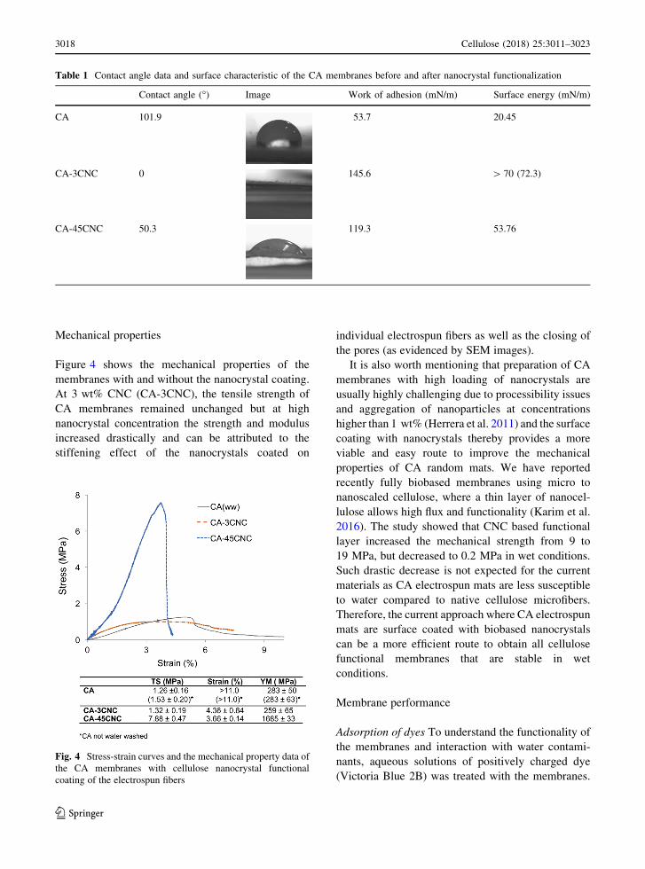
Figure 4 shows the mechanical properties of the
membranes with and without the nanocrystal coating.
At 3 wt% CNC (CA-3CNC), the tensile strength of
CA membranes remained unchanged but at high
nanocrystal concentration the strength and modulus
increased drastically and can be attributed to the
stiffening effect of the nanocrystals coated on
individual electrospun fibers as well as the closing of
the pores (as evidenced by SEM images).
It is also worth mentioning that preparation of CA
membranes with high loading of nanocrystals are
usually highly challenging due to processibility issues
and aggregation of nanoparticles at concentrations
higher than 1 wt% (Herrera et al. 2011) and the surface
coating with nanocrystals thereby provides a more
viable and easy route to improve the mechanical
properties of CA random mats. We have reported
recently fully biobased membranes using micro to
nanoscaled cellulose, where a thin layer of nanocel-
lulose allows high flux and functionality (Karim et al.
2016). The study showed that CNC based functional
layer increased the mechanical strength from 9 to
19 MPa, but decreased to 0.2 MPa in wet conditions.
Such drastic decrease is not expected for the current
materials as CA electrospun mats are less susceptible
to water compared to native cellulose microfibers.
Therefore, the current approach where CA electrospun
mats are surface coated with biobased nanocrystals
can be a more efficient route to obtain all cellulose
functional membranes that are stable in wet
conditions.
Membrane performance
Adsorption of dyes To understand the functionality of
the membranes and interaction with water contami-
nants, aqueous solutions of positively charged dye
(Victoria Blue 2B) was treated with the membranes.
Table 1 Contact angle data and surface characteristic of the CA membranes before and after nanocrystal functionalization
Contact angle (�) Image Work of adhesion (mN/m) Surface energy (mN/m)
CA 101.9 53.7 20.45
CA-3CNC 0 145.6 [ 70 (72.3)
CA-45CNC 50.3 119.3 53.76
Fig. 4 Stress-strain curves and the mechanical property data of
the CA membranes with cellulose nanocrystal functional
coating of the electrospun fibers
3018 Cellulose (2018) 25:3011–3023
123

The solution was evaluated at defined time intervals
using a UV–Vis spectrometer. For 10 mg/L of dye
concentration, it was found that CA, CA-37CNC, and
CA-45CNC adsorbed 1.9, 87.5, and 99.9%, respec-
tively within 24 h. It should be noted that 82.6% dye
was adsorbed in 1 h for CA-45CNC; however in the
case of CA-37CNC the adsoprtion was 36.4%
(Fig. 5a). Adsorption on pristine CA adsorption, in
comparison, was very random, (13.5% in 1 h) and it
was observed that the dyes were getting adsorbed and
desorbed at various time intervals due to the lack of
electrostatic interactions. The dye adsorption for all
the three membranes at concentration of 10 mg/L
were tabulated and results are shown in Fig. 5a. The
color degradation from the initial to final (24 h) was
shown as photograph inset in Fig. 5a.
For higher dye concentrations such as 25 and
50 mg/L, CA-45CNC was active and removed 84.36
and 52.62%, respectively. In the case of CA-45CNC, a
combination of adsorption and size exclusion can be
expected due to the compact nature of the CNC
coating. In the case of 25 mg/L, adsorption improved
by increasing the contact time by 24 h, however, for
50 mg/L, the adsorption reached saturation. The
adsorption results are given in supplementary data
(Table S2). The increase of CNC content to 45 wt%
not only increased the rejection rate but also improved
the mechanical robustness of the membrane. Figure 5a
inset shows the CA-45CNC after the 48 h treatment
with the dye solution.
To understand the effect of cross-flow filtration on
dye removal, water containing positively charged dye
was filtered through the membranes at a pressure of
0.2 bar. The removal of dyes after passing through the
membranes was visually observable from the
decreased intensity of blue color in the dye solutions
(Fig. 5b). CAmembranes without CNCs also removed
only 41% of the dye whereas the removal capacity
increased to 86% when 3 wt% CNC was coated. The
increase of CNC content to 45 wt% further increased
the rejection to 96%.
The mechanism of adsorption is the electrostatic
interaction between the negatively charged CNCs and
the positively charged dye irrespective of static or
dynamic modes (Karim et al. 2014). In the case of CA-
45CNC a combination of adsorption and size exclu-
sion was expected due to the dense nature of the CNC
coating.
Particle rejection The membrane performance via
size exclusion was evaluated using particle analysis
and the results are given in Table 2. It was concluded
from the above results that a competition between the
mechanical properties and the water flux exists and
therefore CA membranes with CNC loadings between
3, 8, 37 and 45 wt% CNC were used to study size
exclusion phenomenon. CA membranes water washed
and dried was used as control to account for the
compaction effect. The CA membranes did not show
significant rejection of 0.05 and 0.5 lm particles and
23% rejection of 2 lm particles. CNC impregnated
Fig. 5 The contaminant removal capability of the membranes
via a static adsorption and b cross-flow filtration. a Performance
of dye adsorption for concentration of 10 mg/L; a initial dye
color, the color after 24 h adsorption with membranes b CA,
c CA-37CNC, and d CA-45CNC. The inset shows the image of
CA-45CNC after 48 h adsorption. b Photographs showing the
removal of dyes by cross-flow filtration for 10 min using CA
electrospun membranes and the CNC coated membranes. The
membranes after the filtration and the percentage removal of
dyes are given
Cellulose (2018) 25:3011–3023 3019
123
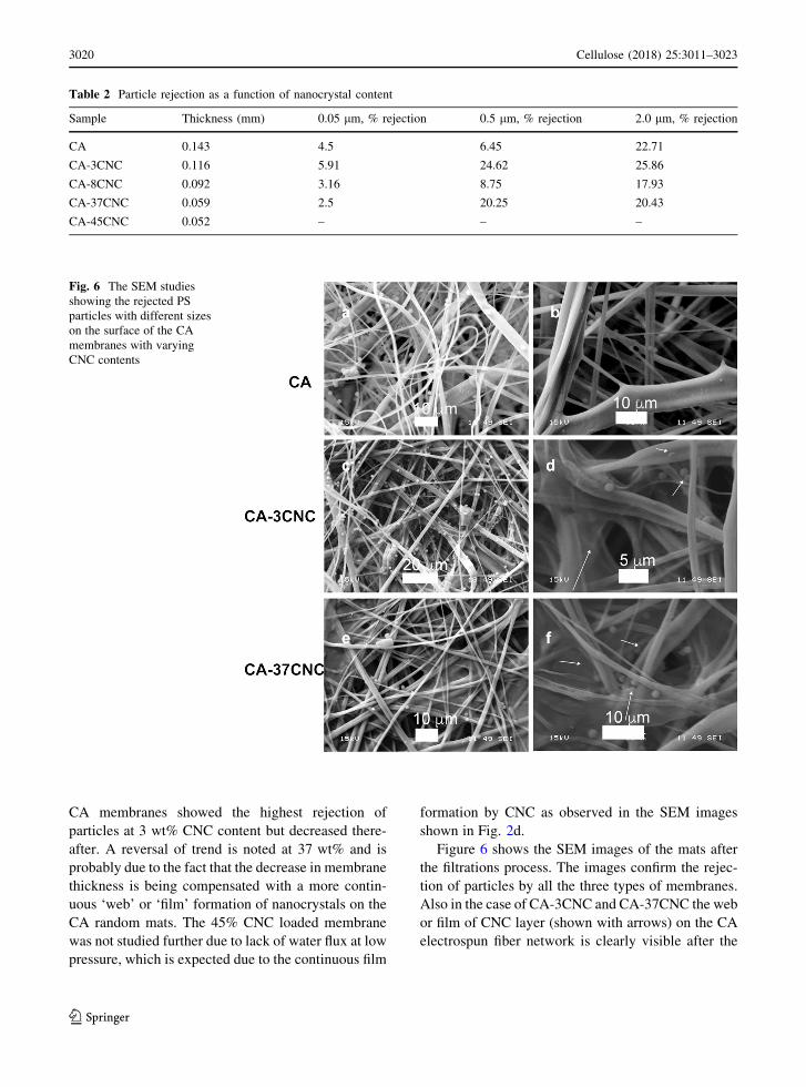
CA membranes showed the highest rejection of
particles at 3 wt% CNC content but decreased there-
after. A reversal of trend is noted at 37 wt% and is
probably due to the fact that the decrease in membrane
thickness is being compensated with a more contin-
uous ‘web’ or ‘film’ formation of nanocrystals on the
CA random mats. The 45% CNC loaded membrane
was not studied further due to lack of water flux at low
pressure, which is expected due to the continuous film
formation by CNC as observed in the SEM images
shown in Fig. 2d.
Figure 6 shows the SEM images of the mats after
the filtrations process. The images confirm the rejec-
tion of particles by all the three types of membranes.
Also in the case of CA-3CNC and CA-37CNC the web
or film of CNC layer (shown with arrows) on the CA
electrospun fiber network is clearly visible after the
Table 2 Particle rejection as a function of nanocrystal content
Sample Thickness (mm) 0.05 lm, % rejection 0.5 lm, % rejection 2.0 lm, % rejection
CA 0.143 4.5 6.45 22.71
CA-3CNC 0.116 5.91 24.62 25.86
CA-8CNC 0.092 3.16 8.75 17.93
CA-37CNC 0.059 2.5 20.25 20.43
CA-45CNC 0.052 – – –
Fig. 6 The SEM studies
showing the rejected PS
particles with different sizes
on the surface of the CA
membranes with varying
CNC contents
3020 Cellulose (2018) 25:3011–3023
123

filtration process, confirming the stability of the CNC
layer in cross flow filtration.
The rejection of 0.5 and 2.0 lm beads can be
correlated to potential rejection of microorganisms as
bacteria (1–2 lm), yeast (5–10 lm), protozoa
(50 lm) etc. via size exclusion during filtration.
Owing to the scalability of CA based electrospun
membranes and the advancements in CNC scaled up,
the cost efficient commercial processing of all-cellu-
lose electrospun membranes for water purification is
viable.
Membrane biodegradability
Degradation of the membranes in water and soil was
investigated up to two months and the visual appear-
ances of membranes at different conditions are shown
in Fig. 7.
In the case of degradation in water medium at
different pH and temperatures as well as water sample
(real wastewater) spiked with a mixture of pesticides
solution, the membranes were mainly stable after
2 months and no degradation was observed regarding
the visual appearance and weight loss. The water
sample mixed with pesticides was not good enough for
the growth of microorganism and after 2 months, the
membranes showed good stability, especially after
coating with CNCs. Thus, they are considered as
potential candidates for water purification.
Soil burial tests for biodegradation of the mem-
branes showed a different trend, at different temper-
atures and with different contaminant adsorption.
Biodegradability of membranes in soil depends on the
composition of soil (such as organic/inorganic matters
and oxygen/carbon dioxide and water content), tem-
perature and pH. So, degradation rate and range can be
changed in different region and season. In soil, the
membranes were degraded more compared to the
water medium (the values are shown in the inset) and it
was also observed that higher temperature resulted in
disintegration of the membranes and changes in the
visual appearance after 2 months of incubation.
Fig. 7 Biodegradation of the membranes after 2 months in real wastewater containing pesticides and in soil
Cellulose (2018) 25:3011–3023 3021
123

However, no significant difference was noted in
degradation of membranes before and after adsorp-
tion. Furthermore, the membranes did not show a
systematic difference in degradation in soil due to
CNC coating.
Conclusions
Cellulose nanocrystals were successfully infused on to
the electrospun cellulose acetate fiber networks result-
ing in highly efficient surface treatment approach to
prepare biobased membranes that combine size exclu-
sion, adsorption and super hydrophilicity. The CA
acetate mats spun on wire meshes showed grid
patterns, which was considered beneficial in control-
ling the pore structure of the microfiber support layer.
An hierarchical morphology is shown by the mem-
branes where micron scaled electrospun fiber network
is surface coated with nanocrystals, especially at lower
nanocrystal loading, whereas a continuous web or
coating and a concomitant increase in mechanical
strength and modulus of the membranes as observed at
higher CNC concentrations. The water flux through
the membranes was very high (22,000 LMH) and
decreased when the nanocrystal network became more
continuous at higher concentration. Moreover, the
contact angle of the membranes dropped to 0� after
coating with nanocrystals indicating increased
hydrophilicity and potential anti-fouling of these
membranes towards hydrophobic entities. On the
other hand, the high rejection of charged entities as
dyes, attributable to adsorption confirms the applica-
tion as affinity membranes. The pore structure was in
microfiltration range and the networks showed mod-
erate rejection of particles with 0.5–2 microns.
Biodegradation of the membranes was favorable for
its application in water purification; showing limited
degradation in polluted water and showing a tendency
to degrade in soil for membranes with and without
adsorbed contaminants as dyes and metal ions.
The study demonstrates an easy and successful
route to functionalize and nanotexture electrospun
fibers with CNCs. The resulting biobased membranes
showed enhanced mechanical properties, high water
flux, hydrophilicity/anti-fouling and pollutant removal
capability via adsorption and size exclusion.
Acknowledgments The authors gratefully acknowledge
financial support from FORMAS, under EU FP7, ERANET
SUSFOOD funding, for CEREAL project; Dnr No. 222-2014-
18.
Open Access This article is distributed under the terms of the
Creative Commons Attribution 4.0 International License (http://
creativecommons.org/licenses/by/4.0/), which permits unrest-
ricted use, distribution, and reproduction in any medium, pro-
vided you give appropriate credit to the original author(s) and
the source, provide a link to the Creative Commons license, and
indicate if changes were made.
References
Daufin G, Escudier J, Carrere H, Berot S, Fillaudeau L, Decloux
M (2001) Recent and emerging applications of membrane
processes in the food and dairy industry. Food Bioprod
Process 79:89–102
De Oliveira MA, De Souza VM, Bergamini AMM,Martinis De,
Pereira Elaine Cristina (2011) Microbiological quality of
ready-to-eat minimally processed vegetables consumed in
Brazil. Food Control 22:1400–1403
Ding Z, Zhong L, Wang X, Zhang L (2016) Effect of lignin-
cellulose nanofibrils on the hydrophilicity and mechanical
properties of polyethersulfone ultrafiltration membranes.
High Perform Polym 28:1192–1200
Directorate C (2002) Risk profile on the microbiological con-
tamination of fruits and vegetables eaten raw
Flemming H, Schaule G, Griebe T, Schmitt J, Tamachkiarowa A
(1997) Biofouling-the Achilles heel of membrane pro-
cesses. Desalination 113:215–225
Francis GA, Thomas C, O’beirne D (1999) The microbiological
safety of minimally processed vegetables. Int J Food Sci
Technol 34:1–22
Goetz LA, Jalvo B, Rosal R, Mathew AP (2016) Superhy-
drophilic anti-fouling electrospun cellulose acetate mem-
branes coated with chitin nanocrystals for water filtration.
J Membr Sci 510:238–248
Gopakumar DA, Pasquini D, HenriqueMA, deMorais LC, Yves
Grohens Y, Thomas S (2017) Meldrum’s acid modified
cellulose nanofiber-based polyvinylidene fluoride micro-
filtration membrane for dye water treatment and nanopar-
ticle removal. ACS Sustain Chem Eng 5(2):2026–2033
Gopal R, Kaur S, Ma Z, Chan C, Ramakrishna S, Matsuura T
(2006) Electrospun nanofibrous filtration membrane.
J Membr Sci 281:581–586
Gopi S, Balakrishnan P, Pius A, Thomas S (2017) Chitin
nanowhisker (ChNW)-functionalized electrospun PVDF
membrane for enhanced removal of Indigo carmine. Car-
bohyd Polym 165:115–122
Herrera N, Mathew A, Wang L, Oksman K (2011) Randomly
oriented and aligned cellulose fibres reinforced with cel-
lulose nanowhiskers, prepared by electrospinning. Plast
Rubber Compos 40:57–64
Huang Z, Zhang Y, Kotaki M, Ramakrishna S (2003) A review
on polymer nanofibers by electrospinning and their
3022 Cellulose (2018) 25:3011–3023
123

applications in nanocomposites. Compos Sci Technol
63:2223–2253
Huang L, Manickam SS, McCutcheon JR (2013) Increasing
strength of electrospun nanofiber membranes for water
filtration using solvent vapor. J Membr Sci 436:213–220
Inc I (2004) Membrane technology benefits the food processing
industry. Filtr Sep 41:32–33
Karim Z, Mathew AP, Grahn M, Mouzon J, Oksman K (2014)
Nanoporous membranes with cellulose nanocrystals as
functional entity in chitosan: removal of dyes from water.
Carbohyd Polym 112:668–676
Karim Z, Mathew AP, Kokol V, Wei J, Grahn M (2016) High-
flux affinity membranes based on cellulose nanocompos-
ites for removal of heavy metal ions from industrial efflu-
ents. RSC Adv 6:20644–20653
Kaur S, Ma Z, Gopal R, Singh G, Ramakrishna S, Matsuura T
(2007) Plasma-induced graft copolymerization of poly
(methacrylic acid) on electrospun poly (vinylidene fluo-
ride) nanofiber membrane. Langmuir 23:13085–13092
Li D, Neumann A (1992) Equation of state for interfacial ten-
sions of solid-liquid systems. Adv Coll Interface Sci
39:299–345
Lipnizki F (2010) Cross-flowmembrane applications in the food
industry. In: Peinemann KV, Pereira S, Giorno L (eds)
Membranes for food applications. Willey, Weinheim,
pp 1–23
Liu P, Borrell PF, Bozic M, Kokol V, Oksman K, Mathew AP
(2015) Nanocelluloses and their phosphorylated deriva-
tives for selective adsorption of Ag?, Cu2? and Fe3?
from industrial effluents. J Hazard Mater 294:177–185
Luo M, Zhao J, Tang W, Pu C (2005) Hydrophilic modification
of poly (ether sulfone) ultrafiltration membrane surface by
self-assembly of TiO2 nanoparticles. Appl Surf Sci
249:76–84
Ma Z, Kotaki M, Ramakrishna S (2005) Electrospun cellulose
nanofiber as affinity membrane. J Membr Sci 265:115–123
Ma H, Burger C, Hsiao BS, Chu B (2011a) Nanofibrous
microfiltration membrane based on cellulose nano-
whiskers. Biomacromol 13:180–186
Ma H, Hsiao BS, Chu B (2011b) Ultrafine cellulose nanofibers
as efficient adsorbents for removal of UO22 in water. ACS
Macro Letters 1:213–216
Manth T, Gabor M, Oklejas E (2003) Minimizing RO energy
consumption under variable conditions of operation.
Desalination 157:9–21
Mathew AP, Oksman K, Karim Z, Liu P, Khan SA, Naseri N
(2014) Process scale up and characterization of wood cel-
lulose nanocrystals hydrolysed using bioethanol pilot
plant. Ind Crops Prod 58:212–219
Mautner A, Maples HA, Sehaqui H, Zimmermann T, de Larraya
UP, Mathew AP, Lai CY, Li K, Bismarck A (2016) Nitrate
removal from water using a nanopaper ion-exchanger.
Environ Sci Water Res Technol 2:117–124
Mohammad AW, Ng CY, Lim YP, Ng GH (2012) Ultrafiltration
in food processing industry: review on application, mem-
brane fouling, and fouling control. Food Bioprocess
Technol 5:1143–1156
Moy E, Neumann A (1987) Solid/liquid interfacial tensions
from contact angle data and direct force measurements.
J Colloid Interface Sci 119:296–297
Qin A, Li X, Zhao X, Liu D, He C (2015) Preparation and
characterization of nano-chitin whisker reinforced PVDF
membrane with excellent antifouling property. J Membr
Sci 480:1–10
Sehaqui H, de Larraya UP, Tingaut P, Zimmermann T (2015)
Humic acid adsorption onto cationic cellulose nanofibers
for bioinspired removal of copper (ii) and a positively
charged dye. Soft Matter 11:5294–5300
Shahmansouri A, Bellona C (2015) Nanofiltration technology in
water treatment and reuse: applications and costs. Water
Sci Technol 71:309–319
Shannon MA, Bohn PW, Elimelech M, Georgiadis JG, Marinas
BJ, Mayes AM (2008) Science and technology for water
purification in the coming decades. Nature 452:301–310
Cellulose (2018) 25:3011–3023 3023
123