Curcumin-PVP Loaded Electrospun Membranes with …
11
55 ISSN 1229-9197 (print version) ISSN 1875-0052 (electronic version) Fibers and Polymers 2020, Vol.21, No.1, 55-65 Curcumin-PVP Loaded Electrospun Membranes with Conferred Antibacterial and Antitumoral Activities Gyuldzhan Yakub 1 , Antoniya Toncheva 1 , Veselin Kussovski 2 , Reneta Toshkova 3 , Ani Georgieva 3 , Elena Nikolova 3 , Nevena Manolova 1 * , and Iliya Rashkov 1 1 Laboratory of Bioactive Polymers, Institute of Polymers, Bulgarian Academy of Sciences, Sofia 1113, Bulgaria 2 Institute of Microbiology, Bulgarian Academy of Sciences, Sofia 1113, Bulgaria 3 Institute of Experimental Morphology, Pathology and Anthropology with Museum, Bulgarian Academy of Sciences, Sofia 1113, Bulgaria (Received May 8, 2019; Revised July 18, 2019; Accepted July 19, 2019) Abstract: Electrospun membranes containing curcumin were prepared from poly(L-co-D,L-lactic) acid and polyvinylpyrrolidone. The effect of curcumin concentration on the solution viscosity and the morphology of fiber was studied. Curcumin solubility in aqueous solutions was enhanced by the formation of curcumin/polyvinylpyrrolidone water- soluble complex. Curcumin physico-chemical and therapeutic properties within the membranes were preserved upon UV-Vis light irradiation, as a part of the membranes sterilization. The biomaterials showed antibacterial activity against pathogenic microorganisms such as Staphylococcus aureus and Candida albicans. In-vitro experiments against HeLa and Graffi tumor cells and white blood cells (peritoneal macrophages and spleen lymphocytes) revealed potential biomedical application of the membranes. Keywords: Nanomaterials, Electrospinning, Polylactic acid, Curcumin, Antibacterial and antitumoral activity Introduction During the last years the biocompatible polymers gained significant scientific interest with a focus on the production of new generation polymer devices with direct application in the medical and pharmaceutical field. A promising approach in their effective nanostructuration has turned out to be the use of electrospinning. Some of the major advantages of the fibrous mats are the relative ease in their fabrication, their large specific surface area and lightness, as well as the possibility to confer them desired properties through the incorporation of bioactive substances, nanoparticles or oligomers. Therefore, the developed active materials can be used in the manufacturing process of drug delivery systems, antibacterial and anesthetic wound dressing, scaffolds for tissue engineering or for open wounds treatment [1-3]. Curcumin (Curc), also known as yellow gold, is a naturally occurring substance with polyphenol structure usually extracted from Curcuma Longa plant having a rich palette of therapeutic properties (antioxidant, anti-inflammatory, antibacterial, antifungal, etc.) [4]. However, its low water- solubility and relative chemical instability (alkaline pH) significantly reduce its bioavailability and prevents living organisms from easily absorbing it [5,6]. In the scientific literature multiple solutions were proposed on tackling this issue by incorporating Curc in liposomes, micelles, emulsions and nanoparticles [7]. Another promising approach to improve its water-solubility and long-term local delivery is Curc loading in micro- and nanofibrous materials. They are characterized with a significant level of loading capacity and encapsulation efficiency. It was demonstrated that the antioxidant water-solubility can be improved by choosing appropriate fibers polymer matrix. Following this idea electrospun mats were successfully produced from: poly(L- co-D,L-lactic)acid/poly(ethylene glycol) blends or polylactic acid/polyvinylpyrrolidone (PLA/PVP) blends [8,9], polylactic acid/cellulose acetate [10] or cellulose acetate/PVP [11,12], poly(vinyl alcohol) containing β-cyclodextrin [13], poly(ε- caprolactone)/tragacanth [14] or mats made out of chitosan/ polylactic acid blends [15]. An interesting covalent binding between the polymer chains [(meth)acrylate family polymers] and Curc was also described [16]. As part of the latest trends in modern medicine, the fabrication of effective and less toxic biomaterials with conferred antitumoral properties is largely discussed. Recently it was demonstrated that the electrospun mats can undergo significant progress in this field [17]. Well known for its biocompatibility and relatively low toxicity, Curc represents a good candidate for replacement of common antitumoral agents and has already been loaded in polylactic acid fibers with a proven cytotoxic activity against C6 glioma cells in mice [18], in polylactic acid/cellulose acetate and polylactic acid/silk fibroin with an impact on breast cancer cells [10], in PVP for melanoma cancerous cells studies [19] in poly(lactic-co-glycolic acid) in treatment tests with human carcinoma [20] and in amphiphilic block copolymer of poly(ε-caprolactone)-poly(ethylene glycol)- poly(ε-caprolactone) fibers for post-operative interventions in rats with brain tumors [21]. However, the design of these polymer materials is not always offering the possibility to enhance the water-solubility of Curc, and at the same time preserve its chemical structure and therapeutic properties for *Corresponding author: [email protected] DOI 10.1007/s12221-020-9473-z
Transcript of Curcumin-PVP Loaded Electrospun Membranes with …
55
ISSN 1229-9197 (print version)
ISSN 1875-0052 (electronic version)
Fibers and Polymers 2020, Vol.21, No.1, 55-65
Curcumin-PVP Loaded Electrospun Membranes with Conferred Antibacterial
and Antitumoral Activities
Gyuldzhan Yakub1, Antoniya Toncheva
1, Veselin Kussovski
2, Reneta Toshkova
3, Ani Georgieva
3,
Elena Nikolova3, Nevena Manolova
1*, and Iliya Rashkov
1
1Laboratory of Bioactive Polymers, Institute of Polymers, Bulgarian Academy of Sciences, Sofia 1113, Bulgaria2Institute of Microbiology, Bulgarian Academy of Sciences, Sofia 1113, Bulgaria
3Institute of Experimental Morphology, Pathology and Anthropology with Museum, Bulgarian Academy of Sciences,
Sofia 1113, Bulgaria
(Received May 8, 2019; Revised July 18, 2019; Accepted July 19, 2019)
Abstract: Electrospun membranes containing curcumin were prepared from poly(L-co-D,L-lactic) acid andpolyvinylpyrrolidone. The effect of curcumin concentration on the solution viscosity and the morphology of fiber wasstudied. Curcumin solubility in aqueous solutions was enhanced by the formation of curcumin/polyvinylpyrrolidone water-soluble complex. Curcumin physico-chemical and therapeutic properties within the membranes were preserved upon UV-Vislight irradiation, as a part of the membranes sterilization. The biomaterials showed antibacterial activity against pathogenicmicroorganisms such as Staphylococcus aureus and Candida albicans. In-vitro experiments against HeLa and Graffi tumorcells and white blood cells (peritoneal macrophages and spleen lymphocytes) revealed potential biomedical application of themembranes.
Keywords: Nanomaterials, Electrospinning, Polylactic acid, Curcumin, Antibacterial and antitumoral activity
Introduction
During the last years the biocompatible polymers gained
significant scientific interest with a focus on the production
of new generation polymer devices with direct application in
the medical and pharmaceutical field. A promising approach
in their effective nanostructuration has turned out to be the
use of electrospinning. Some of the major advantages of the
fibrous mats are the relative ease in their fabrication, their
large specific surface area and lightness, as well as the
possibility to confer them desired properties through the
incorporation of bioactive substances, nanoparticles or
oligomers. Therefore, the developed active materials can be
used in the manufacturing process of drug delivery systems,
antibacterial and anesthetic wound dressing, scaffolds for
tissue engineering or for open wounds treatment [1-3].
Curcumin (Curc), also known as yellow gold, is a
naturally occurring substance with polyphenol structure
usually extracted from Curcuma Longa plant having a rich
palette of therapeutic properties (antioxidant, anti-inflammatory,
antibacterial, antifungal, etc.) [4]. However, its low water-
solubility and relative chemical instability (alkaline pH)
significantly reduce its bioavailability and prevents living
organisms from easily absorbing it [5,6]. In the scientific
literature multiple solutions were proposed on tackling this
issue by incorporating Curc in liposomes, micelles, emulsions
and nanoparticles [7]. Another promising approach to
improve its water-solubility and long-term local delivery is
Curc loading in micro- and nanofibrous materials. They are
characterized with a significant level of loading capacity and
encapsulation efficiency. It was demonstrated that the
antioxidant water-solubility can be improved by choosing
appropriate fibers polymer matrix. Following this idea
electrospun mats were successfully produced from: poly(L-
co-D,L-lactic)acid/poly(ethylene glycol) blends or polylactic
acid/polyvinylpyrrolidone (PLA/PVP) blends [8,9], polylactic
acid/cellulose acetate [10] or cellulose acetate/PVP [11,12],
poly(vinyl alcohol) containing β-cyclodextrin [13], poly(ε-
caprolactone)/tragacanth [14] or mats made out of chitosan/
polylactic acid blends [15]. An interesting covalent binding
between the polymer chains [(meth)acrylate family
polymers] and Curc was also described [16].
As part of the latest trends in modern medicine, the
fabrication of effective and less toxic biomaterials with
conferred antitumoral properties is largely discussed.
Recently it was demonstrated that the electrospun mats can
undergo significant progress in this field [17]. Well known
for its biocompatibility and relatively low toxicity, Curc
represents a good candidate for replacement of common
antitumoral agents and has already been loaded in polylactic
acid fibers with a proven cytotoxic activity against C6
glioma cells in mice [18], in polylactic acid/cellulose acetate
and polylactic acid/silk fibroin with an impact on breast
cancer cells [10], in PVP for melanoma cancerous cells
studies [19] in poly(lactic-co-glycolic acid) in treatment tests
with human carcinoma [20] and in amphiphilic block
copolymer of poly(ε-caprolactone)-poly(ethylene glycol)-
poly(ε-caprolactone) fibers for post-operative interventions
in rats with brain tumors [21]. However, the design of these
polymer materials is not always offering the possibility to
enhance the water-solubility of Curc, and at the same time
preserve its chemical structure and therapeutic properties for*Corresponding author: [email protected]
DOI 10.1007/s12221-020-9473-z
56 Fibers and Polymers 2020, Vol.21, No.1 Gyuldzhan Yakub et al.
long-term release profile.
The aim of this study is to produce active and stable upon
UV-Vis irradiation electrospun materials with conferred
antibacterial and antitumor properties through the incorporation
of Curc into biocompatible polymer matrix made of PLA
and PVP. The morphology of the fibers is evaluated by
scanning electronic and fluorescent microscopy and the
fibers mean diameter is discussed in a direct correlation with
the spinning solution dynamic viscosity. The complex
formation, based on hydrogen bonds between Curc and the
polymer chains, is studied in detail and its impact on the
preservation of the properties of the active substance is
evaluated upon UV-Vis irradiation. Special attention is paid
to Curc release profile and this in relation with the polymer
matrix composition. The antibacterial properties of the PLA/
PVP/Curc membranes are studied against pathogen
microorganisms such as Staphylococcus aureus (S. aureus)
and Candida albicans (C. albicans). In addition, the
cytotoxic effect of PVP/Curc and PLA(/PVP)/Curc fibers is
investigated performing experiments with tumor HeLa and
Graffi cell lines. The immune response of the membranes is
monitored after contact with white blood cells (peritoneal
macrophages and spleen lymphocytes).
Experimental
Materials
The membranes were produced from poly(L-co-D,L-lactic
acid) [PLA, Boehringer Ingelheim Chemicals (Germany)]
with L/D,L=70/30, Mn=78 500 g/mol, Mw=165 850 g/mol
and Mw/Mn=2.1], polyvinylpyrrolidone (PVP, Fluka) with
Mr=360 000 g/mol and curcumin (Curc) with M=368,38 g/
mol (Merck). Dichloromethane (DCM), dimethyl sulfoxide
(DMSO) and ethanol (EtOH) were provided by Merck and
Fluka, respectively. For the microbiology tests Staphylococcus
aureus 509 and Candida albicans 74 from National bank for
industrial microorganisms and cell cultures were used.
Cervical cancer HeLa cells line were cultivated in a DMEM
media (Gibco, Austria) with 10 % fetal bovine serum
(Gibco, Austria), 100 U/ml penicillin and 0.1 mg/ml
streptomycin while using 75 cm3 tissue plastic flasks.
Trypsin- ethylenediaminetetraacetic acid, penicillin and
streptomycin were acquired by FlowLab, Australia. For the
culture cell tests, DMSO and 3-(4,5-dimethylthiazol-2-yl)-
2,5-diphenyltetrazolium bromide (МТТ) were purchased
from Sigma Aldrich, Germany.
Preparation of the Fibrous Materials
PVP/Curc fibers were produced from spinning solutions
obtained after mixing a solution containing PVP (0.49 g)
dissolved in 4 ml EtOH and a solution containing different
amount of Curc (10, 15 and 25 wt% with respect to the final
polymer weight) dissolved in 4.2 ml of EtOH. PLA/PVP/
Curc fibers were obtained after mixing a solution of PLA
(0.35 g) dissolved in 5.2 ml DCM and a solution of PVP
(0.35 g) dissolved in 2.1 ml DMSO. Then the respective
amount of Curc was added to the final solution.
The electrospinning setup consisted of a high voltage
power supply (up to 30 kV), a pump for controlled spinning
solution delivering (NE-300 Just Infusion Syringe Pump
(New Era Pump Systems Inc., USA)), a syringe equipped
with a metal needle (gauge of 20 GX1½’’) connected to the
positively charged electrode and a grounded rotating
aluminum collector (diameter of 5.6 cm). In the case of the
PVP/Curc fibers the working conditions were: spinning
solution feeding rate of 3 ml/h, applied voltage of 25 kV, tip
to the collector distance of 10 cm and collector rotation
speed of 2000 rpm. For PLA/PVP and PLA/PVP/Curc
membranes these parameters were: 3 ml/h, 29 kV, 20 cm and
2000 rpm, respectively.
Characteristics of the Spinning Solutions and the
Fibrous Materials
Detailed morphological analyses of the membranes were
done using scanning electron microscope (SEM Jeol JSM-
5510 and Philips SEM 515) after vacuum gold-coating the
samples. The mean fiber diameter was determined using
ImageJ software, while measuring the diameter of at least 50
individual fibers. Fluorescent micrographs were obtained
operating with fluorescent microscope (NU-2; Carl Zeiss
Jena, Germany) at maximum wavelength of excitation (λex)
of 420 nm and maximum wavelength of emission (λem) of
470 nm. The dynamic spinning solution viscosity was
measured at temperature of 25 oС with Brookfield viscometer
and Brookfield TC-102 thermostat.
Infrared spectra (Fourier transformation, FT-IR) of the
membranes were done with IRAffinity-1 spectrophotometer
(Shimadzu Co., Japan) supplied with a MIRacle ATR add-on
(crystal diamond; depth penetration of IR beam of 2 μm in
the range from 4000 to 600 cm-1 and resolution of 4 cm-1;
PIKE Technologies, USA). All data were corrected for H2O
and CO2 (IRsolution software). In order to study the
materials degree of crystallinity, X-ray diffraction analyses
(XRD; D8 Bruker Advance dust diffractometer) were done
at room temperature with filtered Cu Kα source of radiation
and a luminescent detector in the 2θ range from 0 o to 80 o,
step of 0.02o and a countdown timer of 1 sec/step. The
fibrous materials thermal properties were studied performing
thermogravimetric analysis (TGA; TGA Q5000) by heating
the samples from 0 to 1000oC and differentially scanning
calorimetry (DSC; DSC Perkin Elmer DSC 8500) in the
range from 0 to 250 oC while applying heating/cooling speed
of 10oC/min under nitrogen atmosphere. Curc content in the
fibrous materials was determined spectrophotometrically
[DU 800 UV, Beckman Coulter spectrophotometer] based
on calibration curves at wavelength of 426 nm [EtOH was
used as a solvent for PVP/Curc membranes and DCM/
DMSO (75/25 wt/wt) for (PLA/)PVP/Curc fibers].
Curcumin-PVP Loaded Electrospun Membranes Fibers and Polymers 2020, Vol.21, No.1 57
The impact of UV-Vis light irradiation (light range from
260 to 600 nm and source to sample distance of 43 cm) on
Curc physico-chemical properties loaded in the fibers was
studied after 30, 60 and 120 min or irradiation time with
respective radiation dosage of 261, 522 and 1044 J/cm2.
After this step, the membranes were dissolved in EtOH and
the solution absorption was registered at wavelength of
426 nm. The Curc residual amount within the membranes
presented an average value from three separate experimental
setups, applying equation (1):
Curc residual amount (%) = (Cirr/C0) × 100 (1)
where Cirr is the content of Curc in the membranes after 30,
60 or 120 min of UV-Vis irradiation and C0 is the Curc
content in the relative non-irradiated membrane.
The surface chemical composition of the membranes was
studied by X-ray photoelectron spectroscopy (XPS). The
measurements were taken in a vacuum UHV camera with an
ESCALAB-Mkll (VG Scientific) electronic spectrometer
applying monochrome Mg Kα source irradiation with final
resolution of 1 eV. An energy calibration was performed,
using as reference the binding energy for C-H hydrogen
bonds at 285 eV (C1s).
Hydrophobic/hydrophilic surface behavior of the membranes
was determined after measuring the water contact angle
(Easy Drop DSA20E KRUSS GmbH apparatus). To find out
the correct values, 10 µl drops of deionized water were
deposited on the membranes surface at room temperature
after measuring at least 10 independent drop profiles for
each sample.
The morphological changes in the PLA/PVP and PLA/
PVP/Curc membranes after 24 h immersion in distilled
water and subsequent freeze-drying were evaluated with
SEM. The weight loss was obtained by measuring the
weight difference of the sample before their immersion and
after their lyophilization.
Curc Release Profile
The release profile of the bioactive substance from the
fibers was studied at a temperature of 37 oС, in an acetate
buffer/PVP=90/10 v/v (PVP with Mr of 360 000 g/mol;
buffer ionic strength of 0.1) as release medium with pH of
5.5. PLA/Curc30 and PLA50/PVP50/Curc15 membranes were
immerged in 100 ml of the prepared buffer, while applying a
stirring of 200 rpm. At specific intervals of time, aliquots
(2 ml) were withdrawn and the solution absorption at a
wavelength of 439 nm was registered. Afterwards, the
withdrawn volumes were replaced with fresh buffer
solution. The amount of the diffused from the membranes
Curc was calculated using calibration curve and the results
were presented as average values of three separate trials per
membrane. The initial Curc amount in the PLA/Curc30 and
PLA50/PVP50/Curc15 membranes was determined after
dissolving the fibers in appropriate volume of DCM/
DMSO=75/25 (wt/wt) and measuring the solution absorption at
wavelength of 426 nm.
Microbiological Assays
The minimum inhibitory concentration (MIC) of Curc was
studied against two pathogen microorganisms: Gram-
positive bacteria S. aureus and the fungus C. albicans at
bacterial suspensions concentration of 106 cells/ml. The
microbiological tests with the PVP/Curc and PLA/PVP/Curc
membranes were performed upon irradiation (420 nm) or
in a dark environment following previously described
methodology [8].
Tumor Cell Viability
HeLa cancer cells were first cultured in DMEM by adding
10 % FBS, 100 U/ml penicillin, and 0.1 mg/ml streptomycin
and placed in CO2 incubator (37 oC, 90 % humidity and 5 %
CO2). At 80 to 90 % of cell confluence, the cell culture was
treated with 0.25 % trypsin-EDTA solution and then counted
using hemocytometer. As a next step, 96 well culture plates
were used with cells concentration of 2×10⁴ cell/well. After
24 h of incubation the culture medium was removed and the
HeLa cells were put in presence of PVP, PLA50/PVP50,
PLA50/PVP50/Curc15 and PVP/Curc15 membranes for 24, 48
and 72 h of contact. In the case of the Graffi tumor cells,
RPMI-1640 culture media was used (enriched with 10 %
fetal bovine serum, 100 U/ml penicillin and 0.1 mg/ml
streptomycin) and cell СО2 incubator at 37 oC, 90 %
humidity and 5 % CO2. The Graffi cell culture was treated
with Trypsin-EDTA, counted with Trypan blue and split in
96 well culture plates (2×10⁴ cells/well). After 24 h of
incubation, the culture medium was removed and the cells
were put in contact with PVP, PLA50/PVP50, PLA50/PVP50/
Curc15 and PVP/Curc15 membranes for 24, 48 and 72 h of
contact. In both HeLa and Graffi cells studies, Curc was
used as a positive control. After culturing, the cells were
washed out twice with PBS (рН 7.4) and additionally
incubated with 100 µl МТТ solution (Sigma Chemical Co.)
for 3 h at 37 oC. Then the supernatants were took out and
100 µl lysing solution (DMSO/ethanol=1:1) was added in
order to dissolve any presence of formazan crystals. The
cells viability was evaluated applying MTT test [22] using
ELISA plate reader (TECAN, SunriseTM, Grödig/Salzburg,
Austria). The cell vitality was expressed as percentage based
on equation (2):
Cell viability (%) = (OD570exp/OD570control) × 100 (2)
where OD570exp is the absorption value (λ=570 nm) for the
cell culture at given time of contact with the membranes or
after the addition of Curc, and OD570 control is the absorption
value (λ=570 nm) for the control cell culture.
58 Fibers and Polymers 2020, Vol.21, No.1 Gyuldzhan Yakub et al.
Proliferation Cell Tests of Murine Spleen Lymphocytes
and Peritoneal Macrophages
For this study male and female healthy mice (6 to 8 weeks
old, weigh of 18-20 g) were purchased from a certified
laboratory (SBALHZ-Oncology, Sofia, Bulgaria). All animals
were grown at standard temperature (20±5 oC), were fed
dietary standard pellets and had free access to water. They
were carried in accordance with the requirements of the
recommended institutionally recommended guidelines. The
animal experiments were conducted in accordance with the
requirements of the Animal Ethics Committee. After
scarifying the animals, peritoneal cells were isolated by a
carful procedure of peritoneal cavity washing with 10 ml of
sterile cold PBS (pH 7.2). As next step the cell suspensions
were washed twice with RPMI 1640 medium containing
10 % fetal bovine serum. In order to determine the cell
viability (in contact with the membranes and presence of
Curc) was evaluated with a hematocytometer, while using
tryptone blue dye as cell viability test using 96-well plate
(1×10⁶ cells/ml) with RPMI-1640 containing 10 % fetal
bovine serum.
In the case of the spleen cells study, the mice were
sacrificed by cervical dislocation. The spleens organs were
aseptically removed, placed and cut in small pieces in 60 ml
tissue culture vessels containing 5 ml culture medium. After
this step, the tissue fragments were filtered through nylon
tissue and the cell suspensions were placed on a Ficoll pack
density gradient and centrifuged at 2200 rpm for 20 min at
4oC. The collected lymphocyte-enriched fraction was rinsed
twice with RPMI-1640 medium containing 10 % fetal
bovine serum, centrifuged and resuspended in the same
medium at a concentration of 1×10⁶ cells/ml. The number of
cells and their viability (in contact with the membranes and
in the presence of Curc) were determined by tests with
tryptone blue dye.
Results and Discussion
In the present contribution, biocompatible PVP and PLA-
based membranes loaded with the natural product Curc were
produced by electrospinning while conferring desired
morphological characteristics to the fibers. The electrospinning
of the PVP and PVP/Curc (Curc content of 10 or 15 wt%)
spinning solutions led to the deposition of defect free
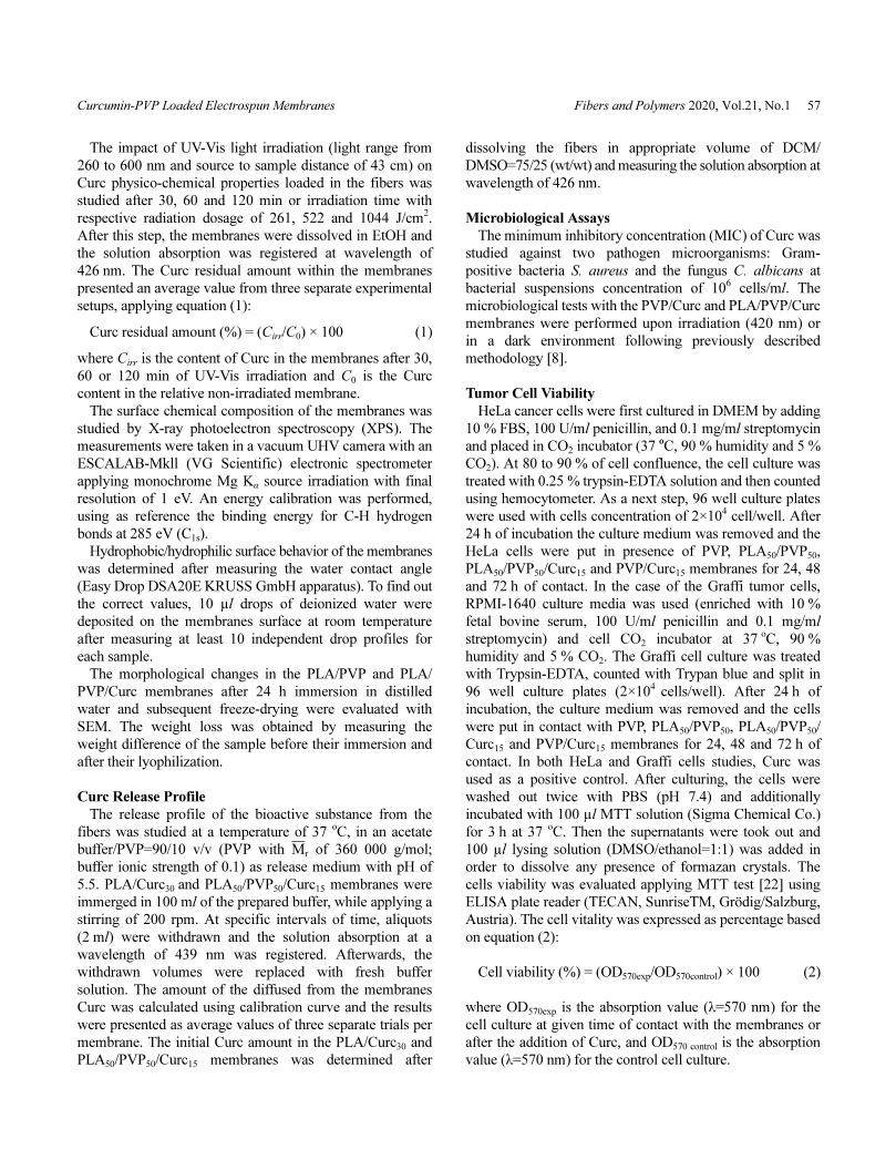
cyclical fibers (Figure 1(a) and (b)). It was found that the
fibers’ morphology and their specific surface area can be
subject of additional design by increasing the content of the
natural substance and this is achievable without changing
the electrospinning process parameters. For example, in the
case of PVP/Curc20 and PVP/Curc25 membranes, the fibers
were ribbon-like shaped (Figure 1(c)). In the literature, such
structures are described as micro- and nanoribbons and can
often be the result of electrospinning of concentrated
solutions at conditions of high degree of humidity at short
capillary to collector distance [23,24]. Their unique anisotropic
structure (high aspect ratio values) could be of particular
interest for the preparation of membranes with specific
properties (optical, physico-mechanical) or systems with
desired drug release rate. In this study, we observed a change
in the fibers morphology from cylindrical to ribbon-like
(Curc content higher than 15 wt%). Since hydrogen bond
complex formation proceeds between PVP and Curc [25],
the spinning solution dynamic viscosity changes (Table 1)
and has an effect on the fiber morphology.
From the already published data it is known that the
electrospinning of concentrated polymer solutions leads to
the formation of fibers with higher diameter values, largely
because of the higher degree of the polymer chains
entanglements [26]. Here, we demonstrated that another
factor could be the degree of complex formation between the
spinning solution components: increase in diameter with the
Figure 1. SEM micrographs of membranes of PVP (a), PVP/
Curc15 (b), PVP/Curc25 (c) and PLA50/PVP50/Curc15 (d). The
collector rotation direction is indicated with an arrow. SEM
micrographs magnification: ×1500; fluorescent microscopy images.
Table 1. Average values of dynamic viscosity of the spinning
solutions and average fiber diameter
Membrane
composition
Dynamic viscosity
(cP)
Mean diameter
(nm)
PVP 50.4±0.2 502±130
PVP/Curc10 57.3±0.1 560±126
PVP/Curc15 57.5±0.1 692±125
PVP/Curc20 56.0±0.1 764±164
PVP/Curc25 58.0±0.1 1073±177
PLA50/PVP50 108.8±1.6 939±184
PLA50/PVP50/Curc15 119.6±2.3 1064±191
Curcumin-PVP Loaded Electrospun Membranes Fibers and Polymers 2020, Vol.21, No.1 59
increase of the antioxidant amount (Table 1). It was found
that PVP fibers had a diameter of 502±13 nm, while the
cross section of PVP/Curc25 was almost twice as large
(1073±177 nm) at a viscosity values of 58.0±0.1 cP. As far
as the PLA50/PVP50 membranes, the fibers were cylindrical
with an average diameter of 939±184 nm with presence of
local inter-fiber adhesion zones (Table 1). These instabilities
of the electrospinning process were stabilized with the
addition of the hydrophobic Curc to the PLA50/PVP50
solution due to hydrogen bonds complex formation between
Curc and the macromolecules. Such improvement of the
fibers morphology (from defects interconnected fibers to
well defined individual fibers), after adding Curc to
electrospinning spinning solutions was recently discussed
[27]. In addition to this, PLA50/PVP50/Curc15 fibers were
obtained with diameter of 1064±191 nm at viscosity values
of 119.6±2.3 cP. Taking advantage of the polyphenol
fluorescent properties, it was possible to observe its
distribution along the fibers length using fluorescence
optical microscopy (Figure 1).
A confirmation for the complex formation between the
bioactive substance and the polymer chains in the
electrospun materials was obtained by IR-spectroscopy
(Supporting information, Figure S1). The main changes in
the spectra were related to: (i) displacement of the
characteristic for C=O band in the structure of PVP from
1651 cm-1 to 1657 cm-1 (PVP/Curc membranes), (ii)
displacement of the characteristic for PLA C=O band from
1749 to 1755 cm-1 (PVP/PLA/Curc membranes) and (iii)
disappearance of the characteristic band for Curc at 3508
cm-1 (ОН group from the Curc phenolic group) in the case of
PVP/Curc and PLA/PVP/Curc membranes. This data were
in agreement with the published results relative to the
intermolecular interaction between PVP and Curc or PVP
and D,L-units of the poly(D,L-lactic acid) [28,29].
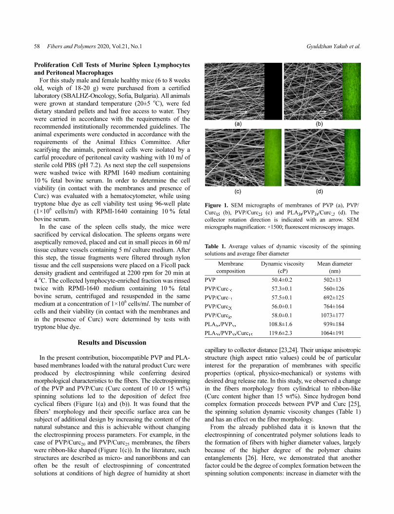
The release profile of a given bioactive substance is
directly depending on its physical state (crystal or amorphous)
in the membrane, its specific location in the fibers (on their
surface or in their volume), its interaction with the polymer
chains (complex formation) and its physico-chemical
stability in the final polymer material. It only follows, that it
was of a particular interest to find out if Curc was in an
amorphous or crystal state in the electrospun (PLA)PVP/
Curc fibers. It is known that Curc diffraction peaks can be
found at 2θ values of 14.5, 17.2, 17.7, 24.6, 25.2o [30]
(Figure 2(a)). It is interesting to note that although polylactic
acid of L/D,L=70/30 composition is amorphous [31,32],
presence of crystalline phase for PLA was detected at 2θ
value of 22.0 o. This may be attributed to strain-induced
changes in the crystal morphology, similarly to reports in
Figure 2. X-ray diffraction spectra of Curc (crystalline state) (a) and PLA50/PVP50/Curc15 (1) and PVP/Curc25 (2) membranes (b). DSC
thermograms of Curc (1) and membranes of PVP (2), PVP/Curc5 (3), PVP/Curc10 (4), PVP/Curc15 (5), PVP/Curc20 (6) and PVP/Curc25 (7)
presented in (c) and membranes of PLA (1), PLA50/PVP50 (2) and PLA50/PVP50/Curc15 (3) presented in (d).
60 Fibers and Polymers 2020, Vol.21, No.1 Gyuldzhan Yakub et al.
literature [33]. For PVP/Curc25 and PLA50/PVP50/Curc15
membranes the peaks for Curc were not observed, revealing
its amorphous state. We assumed that this was a result of the
complex formation between the polyphenol and the
macromolecules preventing the formation of Curc crystal
structures (absence of Curc melting point; DSC thermograms
presented in Figure 2(c) and (d)). These overall results were
a prerequisite for a gradual release of Curc from the fibrous
materials by avoiding burst release for continuous therapeutic
effect (see Release profile section). Additionally, after 9-
month storage period of the nanofibers (temperature of
25 oС, dark environment), the polyphenol compound remained
in amorphous state.
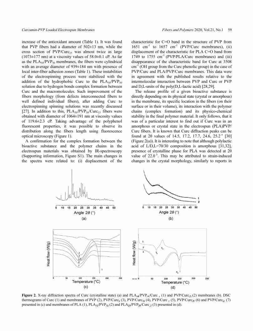
Up to this date, the question of Curc chemical stability and
storage once incorporated in the micro- or nanofibers is still
barely studied in the literature. It is established that Curc
could be subject to photo-destruction when light-irradiated
(UV-Vis spectrum range) in solution or in its crystal dry
state. As a result, several degradation products can be
obtained (vanillin, vanillic acid, ferulic acid and other) thus
losing the polyphenol therapeutic properties [34]. This limits
in higher degree the applications of final Curc-loaded
devices as drug delivery systems, packaging materials with
antibacterial properties, wound healings and others. In our
study, the fresh obtained membranes were irradiated with
UV-Vis light source (260-600 nm) for 30, 60 and 120 min.
After 120 min of light exposure, the greatest residual amount
of Curc was observed for the PVP/Curc membrane (85 %)
and in less in the fibers containing the polyester (around
70 % for PLA/Curc and 75 % for PLA70/PVP30/Curc Figure
3). This was an indication that the presence of the
hydrophilic polymer favors the preservation of Curc chemical
structure upon intensive irradiation. In addition, the amount
of Curc after 9 months of storage (dark environment at
25oC) did not change significantly remaining 95 % of its
initial content in the fibers. On the basis of the obtained
results, we assumed that the electrospun materials could be
suitable for biomedical application and be a subject of
surface sterilization upon UV-irradiation without losing the
physico-chemical and therapeutic properties of the incorporated
bioactive substance.
The deconvoluted C1s spectra of the fibrous materials
were also studied in detail (membranes of PVP(/Curc) and
PLA/PVP(/Curc) (Supporting information, Figure S2). In all
cases three peaks at 288 eV, 286 eV and 285 eV were
observed, characteristic for carbon atom engaged in
chemical -C=O, C-O, C-N and C-H/C-C bonds. For all the
materials, the largest surface area carries the peak at 285 eV,
which is an evident proof of the fibers surface enrichment
with C-H/C-C chemical bonds. For the PVP membrane, the
experimentally derived relationship for the peaks surface
was [C-H/C-C]/[С-О, C-N]/[C=О]=48/34/18. In this case,
we concluded that the PVP macromolecules were oriented in
such a way upon the fibers production that the carbonyl
group and the nitrogen atom were located with priority in
volume of the fibers, while the -(CH2)3- skeleton was placed
on their surface. This result is in line with the data in the
literature concerning a heightened affinity to air environments
of hydrophobic fragments in different type of surfaces [35].
Additional surface hydrophobization was noticed in the PVP
fibers after the Curc addition. The peaks surface relation for
the PVP/Curc10 membrane was [C-H/C-C]/[С-О, C-N]/
[C=О]=65/24/11, and 62/26/12 for PVP/Curc25. This
illustrated a tendency towards achieving new macromolecular
stable state in presence of the antioxidant hydrophobic
molecule: further migration of the polymer molecule
fragments to the volume of the fibers. Similar decreases of
the surface area for C-H/C-C and C=О were also observed in
presence of the polyester. The peaks for the PLA50/PVP50
membranes were [C-H/C-C]/[С-О, C-N]/[C=О]=51/28/21
and 53/27/20 for PLA50/PVP50/Curc15. For those two types,
we concluded that the surface was enriched by fragments of
the PLA macromolecules and to a lesser degree by the Curc
(Curc 15 wt%). As a final note, in the fibers containing Curc
we also observed a peak at 291 eV, consistent with π→π*
satellite peak, characteristic for the benzene rings of Curc.
It is well known that PVP is a water soluble and
biocompatible polymer easily used for the production of
Figure 3. Curc residual amount in the PVP/Curc (a) and PLA/(PVP/)Curc (b) membranes, after 30, 60 and 120 min of irradiation with UV-
Vis light source (wavelength range: from 200 to 600 nm, samples source distance of 43 cm).
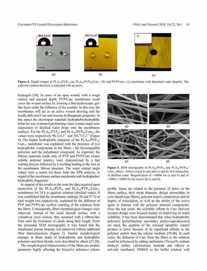
Curcumin-PVP Loaded Electrospun Membranes Fibers and Polymers 2020, Vol.21, No.1 61
hydrogels [36]. In cases of an open wound, with a rough
surface and unequal depth, PVP/Curc membranes could
cover the wound surface by forming a thin hydroscopic gel-
like layer under the influence of the exudate. In this way, the
membranes will act as an active wound dressing and the
locally delivered Curc and exercise its therapeutic properties. In
this aspect the electrospun materials hydrophobic/hydrophilic
behavior was evaluated performing water contact angle tests
(deposition of distilled water drops onto the membranes
surface). For the PLA50/PVP50 and PLA50/PVP50/Curc15 the
values were respectively 96.2±4.3o and 103.7±3.2 o (Figure
4). The higher hydrophobic character of the PLA50/PVP50/
Curc15 membrane was explained with the presence of two
hydrophobic components in the fibers - the biocompatible
polyester and the polyphenol compound. As expected, the
fibrous materials made only of PVP and PVP/Curc (water
soluble polymer matrix), were characterized by a fast
wetting process followed by a swelling leading to the loss of
the membranes fibrous structure. The water contact angle
values were a match for those from the XPS analysis, in
regard of the membranes surface enrichment with hydrophobic/
hydrophilic fragments.
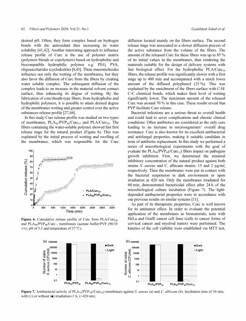
In support of the results so far were the data acquired upon
immersion of the PLA50/PVP50 and PLA50/PVP50/Curc15
membranes for 24 h in aqueous solution (distilled water). It
was established that the membranes undergo 50 and 57 % of
total weight loss respectively, explained by the diffusion of
PVP and PVP/Curc (yellow coloring of the solution) from
the fibers. Consequently, fibers morphological changes were
observed: instead of the usual smooth surface with a
cylindrical cross section, they assumed with a ribbon-like
form until the formation of canals along their length. From
the presented SEM micrographs, it was found that the
membranes porous structure was preserved without additional
fiber interconnection (Figure 5). Similar morphological
changes in fibers made of hydrophobic and hydrophilic
polymers and their blends, were described by others [37,38].
The morphological characteristics of the fibers are another
parameter highly affecting the bioactive substance release
profile. Some are related to the presence of pores on the
fibers surface, their mean diameter, design (monolithic or
core-sheath type fibers), polymer matrix composition and its
degree of reticulation, as well as the ability of the active
agent to interact with the polymer material components.
Over the last years, the scientific efforts in Curc delivery
systems design were focused mainly on improving its water
solubility. It has been demonstrated that when hydrophobic
polymers [poly(butylene succinate), poly(ε-caprolactone)]
are used, the quantity of the released lipophilic natural
product is lower because of its significant affinity to the
polymer matrix than the release medium [39,40]. In such
cases, the diffusion of Curc from the fibers to the solution
could be influenced by adding surfactants (Tween20, sodium
dodecyl sulfate, cetrimonium bromide and others) or
solvents (methanol, DMSO) to the buffer solution with
Figure 4. Digital images of PLA50/PVP50 (a), PLA50/PVP50/Curc15 (b) and PVP/Curc25 (c) membrane with deposited water droplets. The
collector rotation direction is indicated with an arrow.
Figure 5. SEM micrographs of PLA50/PVP50 and PLA50/PVP50/
Curc15 fibers - before (a and b) and after (c and d) 24 h immersion
in distilled water. Magnification of ×10000 for (a and b) and of
×5000 (×10000 for the insets) for (c and d).
62 Fibers and Polymers 2020, Vol.21, No.1 Gyuldzhan Yakub et al.
desired pH. Often, they form complex based on hydrogen
bonds with the antioxidant thus increasing its water
solubility [41,42]. Another interesting approach to influence
release profile of Curc is the use of polymer matrix
(polymers blends or copolymers) based on hydrophobic and
biocompatible hydrophilic polymer e.g. PEG, PVA,
oligosaccharides (cyclodextrin) [8,43]. These macromolecules
influence not only the wetting of the membranes, but they
also favor the diffusion of Curc from the fibers by creating
water soluble complex. The subsequent diffusion of the
complex leads to an increase in the material solvent contact
surface, thus enhancing its degree of wetting. By the
fabrication of core/sheath-type fibers, from hydrophobic and
hydrophilic polymers, it is possible to attain desired degree
of the membranes wetting and greater control over the active
substances release profile [27,44].
In this study Curc release profile was studied on two types
of membranes, PLA50/PVP50/Curc15 and PLA/Curc30. The
fibers containing the water-soluble polymer showed fast first
release stage for the natural product (Figure 6). This was
explained by the initial process of wetting and swelling of
the membranes, which was responsible for the Curc
diffusion located mainly on the fibers surface. The second
release stage was associated to a slower diffusion process of
the active substance from the volume of the fibers. The
amount of the released Curc for these fibers was up to 85 %
of its initial values in the membranes, thus rendering the
materials suitable for the design of delivery systems with
fast biological effect. For the hydrophobic PLA/Curc30
fibers, the release profile was significantly slower with a first
stage up to 400 min and accompanied with a much lower
amount of the diffused polyphenol (25 %). This was
explained by the enrichment of the fibers surface with C-H/
C-C chemical bonds, which makes their level of wetting
significantly lower. The maximum amount of the released
Curc was around 70 % in this case. These results reveal that
PVP facilitate Curc release.
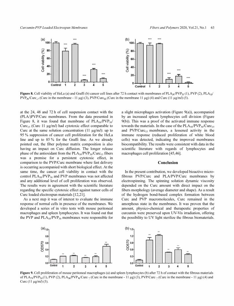
Bacterial infections are a serious threat to overall health
and could lead to sever complications and chronic clinical
conditions. Often antibiotics are considered as the only cure
leading to an increase in microorganisms’ overall drug
resistance. Curc is also known for its excellent antibacterial
and antifungal properties, making it suitable candidate in
term of antibiotic replacement. In this study we performed a
series of microbiological experiments with the goal of
evaluate the PLA50/PVP50(/Curc15) fibers impact on pathogens
growth inhibition. First, we determined the minimal
inhibitory concentration of the natural product against both
strains S. aureus and C. аlbicans strains: 15 and 2 µg/ml,
respectively. Then the membranes were put in contact with
the bacterial suspension in dark environment or upon
irradiation at 420 nm. Only the membranes irradiated for
60 min, demonstrated bactericidal effect after 24 h of the
microbiological culture incubation (Figure 7). The light-
depended antibacterial properties were in accordance with
our previous results on similar systems [11].
As part of its therapeutic properties, Curc is well known
for its antitumor effect. In order to evaluate the potential
application of the membranes as biomaterials, tests with
HeLa and Graffi cancer cell lines (cells in cancer forms of
cervical cancer and myeloid tumor) were performed. The
kinetics of the cell viability were established via MTT test,
Figure 6. Cumulative release profile of Curc from PLA/Curc30
and PLA50/PVP50/Curc15 membranes (acetate buffer/PVP (90/10
v/v), pH of 5.5 and temperature of 37 oC).
Figure 7. Antibacterial activity of PLA50/PVP50(/Curc15) membranes against S. aureus (a) and C. albicans (b). Incubation time of 30 min,
with (□) or without (■) irradiation (1 h, λ=420 nm).
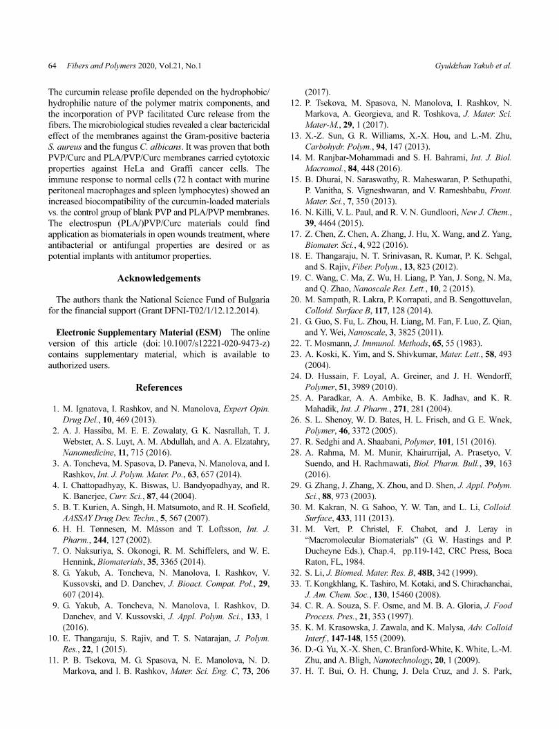
Curcumin-PVP Loaded Electrospun Membranes Fibers and Polymers 2020, Vol.21, No.1 63
at the 24, 48 and 72 h of cell suspension contact with the
(PLA/)PVP/Curc membranes. From the data presented in
Figure 8, it was found that membrane of PLA50/PVP50/
Curc15 (Curc 11 µg/ml) had cytotoxic effect comparable to
Curc at the same solution concentration (11 µg/ml): up to
95 % suppression of cancer cell proliferation for the HeLa
line and up to 85 % for the Graffi line. As we already
pointed out, the fiber polymer matrix composition is also
having an impact on Curc diffusion. The longer release
phase of the antioxidant from the PLA50/PVP50/Curc15 fibers
was a premise for a persistent cytotoxic effect, in
comparison to the PVP/Curc membrane where fast delivery
is occurring accompanied with short biological effect. At the
same time, the cancer cell viability in contact with the
control PLA50/PVP50 and PVP membranes was not affected
and any additional level of cell proliferation was observed.
The results were in agreement with the scientific literature
regarding the specific cytotoxic effect against tumor cells of
Curc loaded electrospun materials [12,21].
As a next step it was of interest to evaluate the immune
response of normal cells in presence of the membranes. We
developed a series of in vitro tests with mouse peritoneal
macrophages and spleen lymphocytes. It was found out that
the PVP and PLA50/PVP50 membranes were responsible for
a slight microphages activation (Figure 9(a)), accompanied
by an increased spleen lymphocytes cell division (Figure
9(b)). This was a proof of the activated immune response
towards the materials. In the case of the PLA50/PVP50/Curc15
and PVP/Curc15 membranes, a lessened activity in the
immune response (reduced proliferation of white blood
cells) was detected, indicating the improved membranes
biocompatibility. The results were consistent with data in the
scientific literature with regards of lymphocytes and
macrophages cell proliferation [45,46].
Conclusion
In the present contribution, we developed bioactive micro-
fibrous PVP/Curc and PLA/PVP/Curc membranes by
electrospinning. The spinning solution dynamic viscosity
depended on the Curc amount with direct impact on the
fibers morphology (average diameter and shape). As a result
of the hydrogen bond-based complex formation between
Curc and PVP macromolecules, Curc remained in the
amorphous state in the membranes. It was proven that the
amount, physico-chemical and therapeutic properties of
curcumin were preserved upon UV-Vis irradiation, offering
the possibility to UV light sterilize the fibrous biomaterials.
Figure 8. Cell viability of HeLa (a) and Graffi (b) cancer cell lines after 72 h contact with membranes of PLA50/PVP50 (1), PVP (2), PLA50/
PVP50/Curc15 (Curc in the membrane - 11 µg) (3), PVP/Curc15 (Curc in the membrane 11 µg) (4) and Curc (11 µg/ml) (5).
Figure 9. Cell proliferation of mouse peritoneal macrophages (a) and spleen lymphocytes (b) after 72 h of contact with the fibrous materials
of PLA50/PVP50 (1), PVP (2), PLA50/PVP50/Curc15 (Curc in the membrane - 11 µg) (3), PVP/Curc15 (Curc in the membrane - 11 µg) (4) and
Curc (11 µg/ml) (5).
64 Fibers and Polymers 2020, Vol.21, No.1 Gyuldzhan Yakub et al.
The curcumin release profile depended on the hydrophobic/
hydrophilic nature of the polymer matrix components, and
the incorporation of PVP facilitated Curc release from the
fibers. The microbiological studies revealed a clear bactericidal
effect of the membranes against the Gram-positive bacteria
S. aureus and the fungus C. albicans. It was proven that both
PVP/Curc and PLA/PVP/Curc membranes carried cytotoxic
properties against HeLa and Graffi cancer cells. The
immune response to normal cells (72 h contact with murine
peritoneal macrophages and spleen lymphocytes) showed an
increased biocompatibility of the curcumin-loaded materials
vs. the control group of blank PVP and PLA/PVP membranes.
The electrospun (PLA/)PVP/Curc materials could find
application as biomaterials in open wounds treatment, where
antibacterial or antifungal properties are desired or as
potential implants with antitumor properties.
Acknowledgements
The authors thank the National Science Fund of Bulgaria
for the financial support (Grant DFNI-T02/1/12.12.2014).
Electronic Supplementary Material (ESM) The online
version of this article (doi: 10.1007/s12221-020-9473-z)
contains supplementary material, which is available to
authorized users.
References
1. М. Ignatova, I. Rashkov, and N. Manolova, Expert Opin.
Drug Del., 10, 469 (2013).
2. A. J. Hassiba, M. E. E. Zowalaty, G. K. Nasrallah, T. J.
Webster, A. S. Luyt, A. M. Abdullah, and A. A. Elzatahry,
Nanomedicine, 11, 715 (2016).
3. A. Toncheva, M. Spasova, D. Paneva, N. Manolova, and I.
Rashkov, Int. J. Polym. Mater. Po., 63, 657 (2014).
4. I. Chattopadhyay, K. Biswas, U. Bandyopadhyay, and R.
K. Banerjee, Curr. Sci., 87, 44 (2004).
5. B. T. Kurien, A. Singh, H. Matsumoto, and R. H. Scofield,
AASSAY Drug Dev. Techn., 5, 567 (2007).
6. H. H. Tønnesen, M. Másson and T. Loftsson, Int. J.
Pharm., 244, 127 (2002).
7. O. Naksuriya, S. Okonogi, R. M. Schiffelers, and W. E.
Hennink, Biomaterials, 35, 3365 (2014).
8. G. Yakub, A. Toncheva, N. Manolova, I. Rashkov, V.
Kussovski, and D. Danchev, J. Bioact. Compat. Pol., 29,
607 (2014).
9. G. Yakub, A. Toncheva, N. Manolova, I. Rashkov, D.
Danchev, and V. Kussovski, J. Appl. Polym. Sci., 133, 1
(2016).
10. E. Thangaraju, S. Rajiv, and T. S. Natarajan, J. Polym.
Res., 22, 1 (2015).
11. P. B. Tsekova, M. G. Spasova, N. E. Manolova, N. D.
Markova, and I. B. Rashkov, Mater. Sci. Eng. C, 73, 206
(2017).
12. P. Tsekova, M. Spasova, N. Manolova, I. Rashkov, N.
Markova, A. Georgieva, and R. Toshkova, J. Mater. Sci.
Mater-M., 29, 1 (2017).
13. X.-Z. Sun, G. R. Williams, X.-X. Hou, and L.-M. Zhu,
Carbohydr. Polym., 94, 147 (2013).
14. M. Ranjbar-Mohammadi and S. H. Bahrami, Int. J. Biol.
Macromol., 84, 448 (2016).
15. B. Dhurai, N. Saraswathy, R. Maheswaran, P. Sethupathi,
P. Vanitha, S. Vigneshwaran, and V. Rameshbabu, Front.
Mater. Sci., 7, 350 (2013).
16. N. Killi, V. L. Paul, and R. V. N. Gundloori, New J. Chem.,
39, 4464 (2015).
17. Z. Chen, Z. Chen, A. Zhang, J. Hu, X. Wang, and Z. Yang,
Biomater. Sci., 4, 922 (2016).
18. E. Thangaraju, N. T. Srinivasan, R. Kumar, P. K. Sehgal,
and S. Rajiv, Fiber. Polym., 13, 823 (2012).
19. C. Wang, C. Ma, Z. Wu, H. Liang, P. Yan, J. Song, N. Ma,
and Q. Zhao, Nanoscale Res. Lett., 10, 2 (2015).
20. M. Sampath, R. Lakra, P. Korrapati, and B. Sengottuvelan,
Colloid. Surface B, 117, 128 (2014).
21. G. Guo, S. Fu, L. Zhou, H. Liang, M. Fan, F. Luo, Z. Qian,
and Y. Wei, Nanoscale, 3, 3825 (2011).
22. T. Mosmann, J. Immunol. Methods, 65, 55 (1983).
23. A. Koski, K. Yim, and S. Shivkumar, Mater. Lett., 58, 493
(2004).
24. D. Hussain, F. Loyal, A. Greiner, and J. H. Wendorff,
Polymer, 51, 3989 (2010).
25. A. Paradkar, A. A. Ambike, B. K. Jadhav, and K. R.
Mahadik, Int. J. Pharm., 271, 281 (2004).
26. S. L. Shenoy, W. D. Bates, H. L. Frisch, and G. E. Wnek,
Polymer, 46, 3372 (2005).
27. R. Sedghi and A. Shaabani, Polymer, 101, 151 (2016).
28. A. Rahma, M. M. Munir, Khairurrijal, A. Prasetyo, V.
Suendo, and H. Rachmawati, Biol. Pharm. Bull., 39, 163
(2016).
29. G. Zhang, J. Zhang, X. Zhou, and D. Shen, J. Appl. Polym.
Sci., 88, 973 (2003).
30. M. Kakran, N. G. Sahoo, Y. W. Tan, and L. Li, Colloid.
Surface, 433, 111 (2013).
31. M. Vert, P. Christel, F. Chabot, and J. Leray in
“Macromolecular Biomaterials” (G. W. Hastings and P.
Ducheyne Eds.), Chap.4, pp.119-142, CRC Press, Boca
Raton, FL, 1984.
32. S. Li, J. Biomed. Mater. Res. B, 48B, 342 (1999).
33. T. Kongkhlang, K. Tashiro, M. Kotaki, and S. Chirachanchai,
J. Am. Chem. Soc., 130, 15460 (2008).
34. C. R. A. Souza, S. F. Osme, and M. B. A. Gloria, J. Food
Process. Pres., 21, 353 (1997).
35. K. M. Krasowska, J. Zawala, and K. Malysa, Adv. Colloid
Interf., 147-148, 155 (2009).
36. D.-G. Yu, X.-X. Shen, C. Branford-White, K. White, L.-M.
Zhu, and A. Bligh, Nanotechnology, 20, 1 (2009).
37. H. T. Bui, O. H. Chung, J. Dela Cruz, and J. S. Park,
Curcumin-PVP Loaded Electrospun Membranes Fibers and Polymers 2020, Vol.21, No.1 65
Macromol. Res., 22, 1288 (2014).
38. M. Bognitzki, T. Frese, M. Steinhart, A. Greiner, J. H.
Wendorff, A. Schaper, and M. Hellwig, Polym. Eng. Sci.,
41, 982 (2001).
39. E. Llorens, H. Ibañez, L. J. del Valle, and J. Puiggalí,
Mater. Sci. Eng. C, 49, 472 (2015).
40. J. Shubham, M. Sai Rama Krishna, and C. Kaushik,
Biomed. Mater., 11, 1 (2016).
41. L. Deng, X. Kang, Y. Liu, F. Feng, and H. Zhang, Food
Chem., 231, 70 (2017).
42. R. Sedghi, A. Shaabani, Z. Mohammadi, F. Y. Samadia,
and E. Isaei, Carbohydr. Polym., 159, 1 (2017).
43. W. Chen, H. EI-Hamshary, S. S. Al-Deyab, and X. Mo,
Adv. Polym. Tech., 37, 647 (2018).
44. Z. Aytac and T. Uyar, Int. J. Pharm., 518, 177 (2017).
45. H. Yang, W. Xu, Z. Zhou, J. Liu, X. Li, L. Chen, J. Weng,
and Z. Yu, Exp. Clin. Endocrinol. Diabetes, 123, 360
(2015).
46. S. Antony, R. Kuttan, and G. Kuttan, Immunol. Invest., 28,
291 (1999).