
Algicidal Activity of Streptomyces eurocidicus JXJ …aem.asm.org/content/82/17/5132.full.pdf ·...
12
Algicidal Activity of Streptomyces eurocidicus JXJ-0089 Metabolites and Their Effects on Microcystis Physiology Bing-Huo Zhang, a,b Zhang-Gui Ding, c Han-Quan Li, a Xiao-Zhen Mou, d Yu-Qin Zhang, e Jian-Yuan Yang, a En-Min Zhou, b,c Wen-Jun Li b,c College of Life Science, Jiujiang University, Jiujiang, People’s Republic of China a ; State Key Laboratory of Biocontrol and Guangdong Provincial Key Laboratory of Plant Resources, School of Life Sciences, Sun Yat-Sen University, Guangzhou, People’s Republic of China b ; Yunnan Institute of Microbiology, Yunnan University, Kunming, People’s Republic of China c ; Department of Biological Sciences, Kent State University, Kent, Ohio, USA d ; Institute of Medicinal Biotechnology, Chinese Academy of Medical Sciences and Peking Union Medical College, Beijing, People’s Republic of China e ABSTRACT Copper sulfate (CuSO 4 ) has been widely used as an algicide to control harmful cyanobacterial blooms (CyanoHABs) in freshwa- ter lakes. However, there are increasing concerns about this application, due mainly to the general toxicity of CuSO 4 to other aquatic species and its long-term persistence in the environment. This study reported the isolation and characterization of two natural algicidal compounds, i.e., tryptamine and tryptoline, from Streptomyces eurocidicus JXJ-0089. At a concentration of 5 g/ml, both compounds showed higher algicidal efficiencies than CuSO 4 on Microcystis sp. FACHB-905 and some other harmful cyanobacterial strains. Tryptamine and tryptoline treatments induced a degradation of chlorophyll and cell walls of cyanobacte- ria. These two compounds also significantly increased the intracellular oxidant content, i.e., superoxide anion radical (O 2 ) and malondialdehyde (MDA), but reduced the activity of intracellular reductants, i.e., superoxide dismutase (SOD), of cyanobacte- ria. Moreover, tryptamine and tryptoline treatments significantly altered the internal and external contents of microcystin-LR (MC-LR), a common cyanotoxin. Like CuSO 4 , tryptamine and tryptoline led to releases of intracellular MC-LR from Microcystis, but with lower rates than CuSO 4 . Tryptamine and tryptoline (5 g/ml) in cyanobacterial cultures were completely degraded within 8 days, while CuSO 4 persisted for months. Overall, our results suggest that tryptamine and tryptoline could potentially serve as more efficient and environmentally friendly alternative algicides than CuSO 4 in controlling harmful cyanobacterial blooms. IMPORTANCE Cyanobacterial harmful algal blooms (CyanoHABs) in aquatic environments have become a worldwide problem. Numerous ef- forts have been made to seek means to prevent, control, and mitigate CyanoHABs. Copper sulfate (CuSO 4 ), was once a common algicide to treat and control CyanoHABs. However, its application has become limited due to concerns about its general toxicity to other aquatic species and its long-term persistence in the environment. There is a great need for algicides with higher specific- ity and low environmental impacts. This study reports the isolation and characterization of two natural algicidal compounds from a streptomycete strain, Streptomyces eurocidicus JXJ-0089. Our results suggest that the identified algicides could poten- tially serve as more efficient and environmentally friendly alternative algicides than CuSO 4 in controlling harmful cyanobacte- rial blooms. H armful cyanobacterial blooms (CyanoHABs) have become a global phenomenon and are occurring with increasing inten- sity, area of infection, and frequency (1). One danger that Cyano- HABs have posed to the environment and human health is the production and release of cyanotoxins. The most frequently de- tected cyanotoxins in freshwater systems are microcystins (MCs), a group of potent liver toxins (2, 3). An MC contamination inci- dent in 1996 caused the death of 53 patients in Caruaru, Brazil (4). A more recent MC contamination event in the public water sys- tem of Toledo, OH (USA), caused a drinking water crisis that impacted about a half million residents (reported by NBC News). Over 90 variants of MCs have been identified so far (3, 5), among which microcystin-LR (MC-LR) is the most common and toxic. MC-LR accounts for 45.5 to 99.8% of the MCs in bloom-impacted natural waters (6) and 57% of MCs produced by Microcystis aeruginosa cultures (7). Therefore, MC-LR often serves as the model for studies related to microcystin production, degradation, and toxicity (2, 4–8). Copper sulfate (CuSO 4 ) has a long history of being used as an algicide to treat nuisance algal blooms, including CyanoHABs (9). However, the application of CuSO 4 has disadvantages, such as secondary pollution and low selectivity toward toxic cyanobacte- ria (10). Therefore, development of more effective and environ- mentally friendly treatments is required. Received 19 April 2016 Accepted 7 June 2016 Accepted manuscript posted online 17 June 2016 Citation Zhang B-H, Ding Z-G, Li H-Q, Mou X-Z, Zhang Y-Q, Yang J-Y, Zhou E-M, Li W-J. 2016. Algicidal activity of Streptomyces eurocidicus JXJ-0089 metabolites and their effects on Microcystis physiology. Appl Environ Microbiol 82:5132–5143. doi:10.1128/AEM.01198-16. Editor: M. J. Pettinari, University of Buenos Aires Address correspondence to Xiao-Zhen Mou, [email protected], or Wen-Jun Li, [email protected]. Supplemental material for this article may be found at http://dx.doi.org/10.1128 /AEM.01198-16. Copyright © 2016, American Society for Microbiology. All Rights Reserved. crossmark 5132 aem.asm.org September 2016 Volume 82 Number 17 Applied and Environmental Microbiology on July 18, 2018 by guest http://aem.asm.org/ Downloaded from
Transcript of Algicidal Activity of Streptomyces eurocidicus JXJ …aem.asm.org/content/82/17/5132.full.pdf ·...

Algicidal Activity of Streptomyces eurocidicus JXJ-0089 Metabolitesand Their Effects on Microcystis Physiology
Bing-Huo Zhang,a,b Zhang-Gui Ding,c Han-Quan Li,a Xiao-Zhen Mou,d Yu-Qin Zhang,e Jian-Yuan Yang,a En-Min Zhou,b,c
Wen-Jun Lib,c
College of Life Science, Jiujiang University, Jiujiang, People’s Republic of Chinaa; State Key Laboratory of Biocontrol and Guangdong Provincial Key Laboratory of PlantResources, School of Life Sciences, Sun Yat-Sen University, Guangzhou, People’s Republic of Chinab; Yunnan Institute of Microbiology, Yunnan University, Kunming,People’s Republic of Chinac; Department of Biological Sciences, Kent State University, Kent, Ohio, USAd; Institute of Medicinal Biotechnology, Chinese Academy of MedicalSciences and Peking Union Medical College, Beijing, People’s Republic of Chinae
ABSTRACT
Copper sulfate (CuSO4) has been widely used as an algicide to control harmful cyanobacterial blooms (CyanoHABs) in freshwa-ter lakes. However, there are increasing concerns about this application, due mainly to the general toxicity of CuSO4 to otheraquatic species and its long-term persistence in the environment. This study reported the isolation and characterization of twonatural algicidal compounds, i.e., tryptamine and tryptoline, from Streptomyces eurocidicus JXJ-0089. At a concentration of 5�g/ml, both compounds showed higher algicidal efficiencies than CuSO4 on Microcystis sp. FACHB-905 and some other harmfulcyanobacterial strains. Tryptamine and tryptoline treatments induced a degradation of chlorophyll and cell walls of cyanobacte-ria. These two compounds also significantly increased the intracellular oxidant content, i.e., superoxide anion radical (O2
�) andmalondialdehyde (MDA), but reduced the activity of intracellular reductants, i.e., superoxide dismutase (SOD), of cyanobacte-ria. Moreover, tryptamine and tryptoline treatments significantly altered the internal and external contents of microcystin-LR(MC-LR), a common cyanotoxin. Like CuSO4, tryptamine and tryptoline led to releases of intracellular MC-LR from Microcystis,but with lower rates than CuSO4. Tryptamine and tryptoline (5 �g/ml) in cyanobacterial cultures were completely degradedwithin 8 days, while CuSO4 persisted for months. Overall, our results suggest that tryptamine and tryptoline could potentiallyserve as more efficient and environmentally friendly alternative algicides than CuSO4 in controlling harmful cyanobacterialblooms.
IMPORTANCE
Cyanobacterial harmful algal blooms (CyanoHABs) in aquatic environments have become a worldwide problem. Numerous ef-forts have been made to seek means to prevent, control, and mitigate CyanoHABs. Copper sulfate (CuSO4), was once a commonalgicide to treat and control CyanoHABs. However, its application has become limited due to concerns about its general toxicityto other aquatic species and its long-term persistence in the environment. There is a great need for algicides with higher specific-ity and low environmental impacts. This study reports the isolation and characterization of two natural algicidal compoundsfrom a streptomycete strain, Streptomyces eurocidicus JXJ-0089. Our results suggest that the identified algicides could poten-tially serve as more efficient and environmentally friendly alternative algicides than CuSO4 in controlling harmful cyanobacte-rial blooms.
Harmful cyanobacterial blooms (CyanoHABs) have become aglobal phenomenon and are occurring with increasing inten-
sity, area of infection, and frequency (1). One danger that Cyano-HABs have posed to the environment and human health is theproduction and release of cyanotoxins. The most frequently de-tected cyanotoxins in freshwater systems are microcystins (MCs),a group of potent liver toxins (2, 3). An MC contamination inci-dent in 1996 caused the death of 53 patients in Caruaru, Brazil (4).A more recent MC contamination event in the public water sys-tem of Toledo, OH (USA), caused a drinking water crisis thatimpacted about a half million residents (reported by NBC News).Over 90 variants of MCs have been identified so far (3, 5), amongwhich microcystin-LR (MC-LR) is the most common and toxic.MC-LR accounts for 45.5 to 99.8% of the MCs in bloom-impactednatural waters (6) and 57% of MCs produced by Microcystisaeruginosa cultures (7). Therefore, MC-LR often serves as themodel for studies related to microcystin production, degradation,and toxicity (2, 4–8).
Copper sulfate (CuSO4) has a long history of being used as an
algicide to treat nuisance algal blooms, including CyanoHABs (9).However, the application of CuSO4 has disadvantages, such assecondary pollution and low selectivity toward toxic cyanobacte-ria (10). Therefore, development of more effective and environ-mentally friendly treatments is required.
Received 19 April 2016 Accepted 7 June 2016
Accepted manuscript posted online 17 June 2016
Citation Zhang B-H, Ding Z-G, Li H-Q, Mou X-Z, Zhang Y-Q, Yang J-Y, Zhou E-M, LiW-J. 2016. Algicidal activity of Streptomyces eurocidicus JXJ-0089 metabolites andtheir effects on Microcystis physiology. Appl Environ Microbiol 82:5132–5143.doi:10.1128/AEM.01198-16.
Editor: M. J. Pettinari, University of Buenos Aires
Address correspondence to Xiao-Zhen Mou, [email protected], or Wen-Jun Li,[email protected].
Supplemental material for this article may be found at http://dx.doi.org/10.1128/AEM.01198-16.
Copyright © 2016, American Society for Microbiology. All Rights Reserved.
crossmark
5132 aem.asm.org September 2016 Volume 82 Number 17Applied and Environmental Microbiology
on July 18, 2018 by guesthttp://aem
.asm.org/
Dow
nloaded from

Natural products, particularly indole derivatives, have shownalgicidal properties on cyanobacteria. For example, gramine,which has been extracted from a number of higher plants (11–13),can inhibit the growth of M. aeruginosa (14). Bacillamides that areproduced by Thermoactinomyces (15, 16), Bacillus (17–19), andMicrobispora aerata (20) have shown selective lytic effects on Mi-crocystis aeruginosa, Aphanizomenon gracile, Anabaena circinalis,and Anabaenopsis circularis (21). Alkaloid 12-epi-hapalindole F,which has been isolated from the filamentous cyanobacteriumFischerella sp. CENA 19, can inhibit the growth of Microcystis andSynechococcus (22). Three �-carbolines (harmane, norharmane,and norharmalane) that have been isolated from Pseudomonas(23) and the cyanobacterium Nodularia harveyana (24, 25) haveshown algicidal activity against Microcystis, Anabaena, and Oscil-latoria (25). Tryptamine, a product of tryptophan decarboxyl-ation in plants (26), has shown selective algicidal activity againstAphanizomenon, Anabaena, and Microcystis (27).
Algicidal compounds inhibit cyanobacterial growth mainly byinterfering with the morphology and physiology of cyanobacteria.For example, lysine can cause severe damage to the cell wall of M.aeruginosa (28); tryptamine and gramine can reduce chlorophyll a(Chl-a) content, degrade superoxide dismutase (SOD), and in-duce the production of reactive oxygen species (ROS) and malon-dialdehyde (MDA) in cyanobacterial cells (14, 27). Some algicidalcompounds, such as amoxicillin and spiramycin, can also affectintracellular cyanotoxin synthesis (7).
In the present study, we report the isolation and identificationof Streptomyces eurocidicus JXJ-0089 and its two algicidal com-pounds. Streptomyces species produce the largest number of bio-active microbial metabolites (29), many of which are algicides.These include lysine (28), niromycin A (30), anthracidin (31),nanaomycin A methyl ester (32), spiramycin (33), triterpenoidsaponin (34), and some unknown proteins (35). Our study ex-tended the above list to include two more indole alkaloids, i.e.,tryptamine and its metabolite tryptoline (36).
MATERIALS AND METHODSCyanobacterial liquid cultures. Eight CyanoHAB strains (FACHB-245,FACHB-252, FACHB-905, FACHB-1092, FACHB-1112, FACHB-1171,FACHB-1284, and FACHB-1285) that are commonly found in Cyano-HABs were obtained from the Freshwater Algae Culture Collection at theInstitute of Hydrobiology (FACHB collection), Chinese Academy of Sci-ences (http://algae.ihb.ac.cn/english/Cultrues.aspx). The organisms werecultured in HGZ medium at 25°C with an illumination of 30 to 50 �molphoton/m2/s in a 12-h light/dark cycle as described previously (37). Theculturing media and conditions were the same for all cyanobacterial cul-tures used in this study unless otherwise noted.
Morphology and 16S rRNA gene sequence analyses of cyanobacte-ria. The morphology of the tested cyanobacteria was observed by usinglight microscope (Olympus BX43, Tokyo, Japan) and scanning electronmicroscopy (Vega Iitescna, Brno, Czech Republic). Genomic DNAs ofthese cyanobacterial strains were isolated, and the 16S rRNA genes wereamplified using primers F1 (5=-TTGATCCTGGCTCAGGATGA-3=) (38)and 1480 (5=-AGTCCTACCTTAGGCATCCCCCTCC-3=) (39). PCR am-plifications were performed in a volume of 50 �l containing 1 �l genomicDNA, 1 U Taq DNA polymerase (TaKaRa, Japan), 1� PCR buffer with 2.5mM MgCl2, 1 �l of each primer, and 200 mM concentrations of eachdeoxyribonucleoside triphosphate (dNTP). PCR was performed in a T100thermal cycler (Bio-Rad, USA). The amplification program was set at95°C for 5 min, followed by 40 cycles of 95°C for 50 s, 57°C for 50 s, and72°C for 1.5 min and a final extension at 72°C for 5 min. PCR ampliconswere sequenced by the Shenggong Company in Shanghai, China. The
resultant 16S rRNA gene sequences were analyzed as described previously(40), and the neighbor-joining (41) phylogenetic tree was constructedusing the MEGA version 5 (42), with bootstrap values based on 1,000replications (43).
Cyanobacterial lawn preparation. Microcystis, the most common ge-nus of bloom-causing cyanobacteria (44), was used as the model in lawnassays to screen for algicidal actinomycetes and compounds. One hun-dred milliliters of Microcystis sp. FACHB-905 culture (�5 � 107 CFU/ml)was mixed with 300 ml of HGZ medium (containing 1.5% agar) at 45°C,poured into petri dishes, and incubated for 2 weeks before being tested foralgicidal activities.
Isolation and identification of algicidal strain JXJ-0089. Actinomy-cetal strains were isolated from soil samples collected from Lushan Moun-tain (29°42=N, 116°26=E), China, using a serial dilution technique. Thealgicidal activities of obtained isolates against Microcystis sp. FACHB-905were first screened by the agar block method. Briefly, bacterial isolateswere grown on ISP 2 medium agar blocks (yeast extract-malt extract agar[45]), which were then placed onto the Microcystis sp. FACHB-905 lawnand incubated for 3 days. Agar blocks without the isolates were used as thenegative control. Isolates with a transparent lysis zone developed on theMicrocystis sp. FACHB-905 lawn were putatively assigned as algicidal bac-teria. Both the thickness of the Microcystis sp. FACHB-905 lawn and thediameter of the algicidal bacterial lysis zone were measured using a verniercaliper. Putative algicidal bacteria were further cultured with a solid me-dium (rice, 100 g; soybean meal, 15 g; glucose, 1 g; peptone, 1 g; yeastextract, 1 g; K2HPO4, 0.3 g; MgSO4 · 7H2O, 0.5 g; H2O, 120 ml; natural pHvalue; sterilized at 121°C for 1 h) at 28°C for 20 days. Afterward, the solidcultures were soaked with methanol-acetone-ethyl acetate (1:1:1, vol/vol/vol) to extract bacterial metabolites. Cell extracts were tested for algicidalactivities using the disk method. Briefly, a paper disk (0.6 cm in diameter)was soaked with 50 �l of extract solution, dried at 50°C to remove thesolvents, and then placed on the Microcystis sp. FACHB-905 lawn. Thelawn was incubated for three more days under the same conditions asthose described previously. The extracts of the solid medium without theisolates were used as the negative control. Extracts of one isolate, i.e.,JXJ-0089, formed transparent zones around the disks, and this isolate wasselected for further analysis.
In order to determine the taxonomic identity of strain JXJ-0089, wecarried out the following tests. JXJ-0089 was grown on ISP 2 culturingplates at 28°C for 14 days, after which the mycelia of strain JXJ-0089 werecollected and examined for their morphological characteristics by lightmicroscopy and scanning electron microscopy. The growth of JXJ-0089 atvarious temperatures (4 to 50°C), pHs (4.0 to 11.0), and NaCl contents (0to 10%, wt/vol) was examined according to the method of Xu et al. (46)using ISP 2 medium as the basal medium. Catalase activity was deter-mined based on the production of bubbles after the addition of a drop of3% (vol/vol) H2O2. Oxidase activity was tested according to the method ofKovacs (47). Other phenotypic characteristics were determined accordingto Goodfellow (48) and Williams et al. (49). Enzyme activities were testedby using the commercial API ZYM system (bioMérieux, Marcy l’Etoile,France). The analyses of isomer of diaminopimelic acid and whole-cellsugars were performed according to the procedures developed by Hase-gawa et al. (50) and Tang et al. (51). Polar lipids were extracted accordingto Minnikin et al. (52) and analyzed as described by Collins and Jones(53). Analysis of fatty acid was performed using standard protocols (54).The genomic DNA was isolated, and the 16S rRNA gene was amplifiedusing the universal bacterial primers 27F (5=-AGAGTTTGATCMTGGCTCAG-3=) and 1492R (5=-TACGYTACCTTGTTACGACTT-3=). The re-sultant 16S rRNA gene sequence was analyzed as described above. Thegenomic relatedness between strain JXJ-0089 and Streptomyces eurocidicusNRRL B-1676T was determined by the thermal renaturation method (55),using a UV-1700 spectrophotometer (Shimadzu) equipped with a DCW-2008 thermo bath.
Streptomyces sp. strain JXJ-0089 was deposited at the China Pharma-
Algicidal Indole Alkaloids from Streptomyces sp.
September 2016 Volume 82 Number 17 aem.asm.org 5133Applied and Environmental Microbiology
on July 18, 2018 by guesthttp://aem
.asm.org/
Dow
nloaded from

ceutical Culture Collection (CPCC), and the deposition number is CPCC204140.
Isolation and identification of algicidal compounds. Cell extract ofstrain JXJ-0089 was purified with silica gel columns (Qingdao HaiyangChemical Co., Ltd.) using chloroform-methanol (1:0¡0:1, vol/vol) as theeluent of gradient elution. The resultant fractions were examined for al-gicidal activities using the disk method on the Microcystis sp. FACHB-905lawn as described above. Putative algicidal fractions in the cell extract werepurified again using silica gel columns with chloroform-methanol (5:1,saturated with water) as the eluent. The separated fractions were retestedfor algicidal activities using the disk method. The confirmed active frac-tions were finally purified with Sephadex LH-20 (GE Healthcare) usingchloroform-methanol (1:1, vol/vol) as the eluent.
The chemical structures of the two identified algicidal compoundswere determined by two spectral analyses. First, nuclear magnetic reso-nance (NMR) spectra (one-dimensional [1D] and 2D) were obtained ona Bruker DRX-500 MHz instrument with tetramethylsilane (TMS) as theinternal standard. Then, mass spectrum analysis was performed using anAutoSpec Premier P776 spectrometer (Waters, USA).
Algicidal activities of tryptamine and tryptoline on CyanoHABstrains. The algicidal activities of two compounds purified from strainJXJ-0089 extracts, i.e., tryptamine, tryptoline, and three other com-pounds, i.e., tryptophan (precursor of tryptamine) (26), gramine (an al-gicidal compound with similar molecular weight as tryptoline) (14), andCuSO4 (all purchased from Sinopharm Chemical Reagent Co., Ltd.,Shanghai, China; the purities of all these compounds were �97.5%), wereexamined on Microcystis sp. FACHB-905 lawns as described above. Foreach compound, algicidal activities were tested at four doses, i.e., 5, 10, 15,and 20 �g/disk.
Both tryptamine and tryptoline were also added to liquid cultures ofeight cyanobacterial strains at different concentrations (final concentra-tions, 1.0, 2.0, 3.0, 4.0, and 5.0 �g/ml). The effects of CuSO4 on cyano-bacterial strains were tested at a final concentration of 5.0 �g/ml. Cyano-bacterial cultures without addition of algicidal compounds were used ascontrols. All the treatments and controls were set up in triplicates. After 3days of incubation, cyanobacterial cultures were centrifuged at 4,860 � gfor 20 min at 4°C, and the sediments were first frozen at �20°C for 24 hand then mixed with 90% ethanol (85°C) and extracted in the dark at 20°Cfor 4 h before the contents of Chl-a were measured (56). Algicidal effi-ciency was calculated based on the following equation: algicidal efficiency(%) � (1 � Ct/Cc) � 100%, where Cc and Ct represent the contents ofChl-a of the control and the algicidal compound-treated samples, respec-tively. The 50% effective concentration (EC50) value was calculated usinglinear interpolation analysis.
Effects of tryptamine and tryptoline on the morphology of cyano-bacterial strains. Microcystis sp. FACHB-1112 was found to be the mostsensitive to algicidal compounds among the 8 tested cyanobacterialstrains; it was used to subsequently study the influences of the algicidalcompounds on the morphology of cyanobacteria by scanning electronmicroscopy. For sample preparation, Microcystis sp. FACHB-1112 cellswere harvested from HGZ liquid culture at the exponential phase andthen exposed to algicidal compounds (5 �g/ml) for 3 days. Treated Mi-crocystis sp. FACHB-1112 cells were collected by centrifugation for 10 minat 25°C, 2,000 � g. The resultant sediment was soaked in a solution con-taining 2.5% glutaraldehyde and phosphate-buffered saline (PBS) solu-tion (100 mM, pH 7.0) at 4°C for 1 h. After removing the solution, cellswere successively dehydrated for 4 min by 50, 70, 80, 90, and 100% etha-nol. Finally, cells were dried on clean coverslips in vacuum drying appa-ratus and coated with gold for analysis. Microcystis sp. FACHB-1112 cellswithout treatments with algicidal compounds were used as controls. Themorphology of filamentous cyanobacteria was observed by using an op-tical microscope after a 3-day exposure to algicidal compounds (5 �g/ml).
Effects of algicidal compounds on Microcystis sp. FACHB-905.FACHB-905 is a Microcystis sp. strain with the ability to produce a highcontent of microcystins (57); it was used in this test. Tryptamine, trypto-
line, or CuSO4 (final concentration, 5 �g/ml) was added to triplicate freshHGZ cultures of Microcystis sp. FACHB-905 (1 � 107 CFU/ml), and thecultures were incubated for a total of 8 days under the same conditions asthose described previously. Samples were taken from the culture every 24h and then centrifuged at 4,860 � g for 20 min at 4°C. The resultantsupernatants were used for measuring the concentrations of tryptamineand tryptoline and the contents of extracellular MC-LR. The resultantsediments were used for measuring the contents of Chl-a and intracellularMC-LR.
The concentrations of tryptamine and tryptoline were measured usinghigh-pressure liquid chromatography (HPLC) after the supernatantswere filtered through a 0.45-�m filter. The HPLC system consisted of anAgilent 1200 LC equipped with a quaternary pump, vacuum degasser,autosampler, diode-array detector, and LC 3D ChemStation and an Agi-lent LC C18 column (Zorbax Eclipse Plus C18, 5 �m, 4.6 by 150 mm;Agilent, USA). The mobile phase in channel A was water, and the mo-bile phase in channel B was methanol. Elution was performed with45% B for 15 min at a flow rate of 1 ml/min. The absorbance wasmonitored at 280 nm.
Extracellular MC-LR content was measured according to a methoddescribed previously (37). Before measurement, the supernatant was con-centrated to 1/10 of its original volume by freeze-drying and filteredthrough a 0.45-�m filter. The intracellular MC-LR was extracted as fol-lows. After three cycles of freezing at �80°C and thawing at 40°C, cellsediments were extracted with 10 ml of acetone-deionized water (1:1,vol/vol) and freeze-dried. The dried material was then resuspended in 0.5ml of methanol-deionized water (1:1, vol/vol) and filtered through a0.45-�m filter, and then the filtrate was analyzed for intracellular MC-LRby HPLC as described previously (37).
The Chl-a content and the algicidal efficiencies of tryptamine, trypto-line, and CuSO4 were examined using the method described previously.
O2�, MDA, and SOD activity assays of Microcystis sp. FACHB-905.
O2�, MDA, and SOD activities were measured at 0, 12, 24, 36, 48, and 60
h during treatment of Microcystis sp. FACHB-905 (1 � 107 CFU/ml) withalgicidal compounds. Both control (without the algicidal compounds)and treatment samples (treated with 5 �g/ml of tryptamine or tryptoline)were collected and centrifuged at 4,860 � g for 20 min at 4°C. The cellextract was prepared by using the cell pellets according to the methoddescribed by Hong et al. (14). Briefly, cell pellets were homogenized byusing an ultrasonic cell pulverizer (JY92-2DN; Xinzhi Co., Ningbo,China) at 200 W for 5 min (ultrasonic time, 2 s; rest time, 8 s) under icebath cooling. Then, the homogenate was centrifuged at 12,000 � g for 10min at 4°C. The content of protein in the cell extract was measured byusing the Coomassie brilliant blue G-250 method (58).
O2� content was determined using a spectrophotometric method
(59). The reaction for measuring the content of O2� included two steps:
(i) 2 ml cell extract, 1.5 ml PBS solution (65 mM, pH 7.8), and 0.5 mlhydroxylamine hydrochloride (10 mM) were mixed and incubated at25°C for 20 min; (ii) 2 ml reaction mixture from step i, 2 ml sulfanilic acid(17 mM, containing 270 mM hydrochloric acid), and 2 ml �-naphthyla-mine (7 mM, containing 400 mM acetic acid) were mixed and incubatedat 30°C for 30 min. Then, the absorbance of the reaction mixture of step iiwas measured at 530 nm, and the O2
� content was calculated from thestandard curve as described previously (59).
SOD activity was determined using the nitroblue tetrazolium (NBT)method (58). The reaction mixture contained 1.9 ml PBS solution (50mM, pH 7.8, containing 100 �M EDTA), 0.3 ml methionine solution (220mM), 0.3 ml NBT solution (1.25 mM), 0.3 ml riboflavin solution (33�M), and 0.2 ml cell extract. The negative control was wrapped in alumi-num foil to prevent any photochemical reaction of NBT, while no cellextract was added to the positive control to ensure the full photochemicalreaction of NBT. The absorbances of the reaction mixtures were measuredat 560 nm after 20 min of irradiation with 50 �mol photon/m2/s at 25°C.One unit of SOD activity is defined as the amount of enzyme required toinhibit 50% photochemical reaction of NBT.
Zhang et al.
5134 aem.asm.org September 2016 Volume 82 Number 17Applied and Environmental Microbiology
on July 18, 2018 by guesthttp://aem
.asm.org/
Dow
nloaded from

MDA contents were determined using the thiobarbituric acid (TBA)method (58). Two milliliter cell extract and 2 ml thiobarbituric acid re-agent (0.5% in 10% trichloroacetic acid) were mixed together andheated for 20 min at 100°C. The mixture was cooled and centrifuged at12,000 � g for 5 min at 4°C. The absorbance of the supernatant wasmeasured at 450 nm, 532 nm, and 600 nm. MDA contents were calcu-lated based on the following equation: CMDA (�M) � 6.45 � (OD532 �OD600) � 0.56 � OD450, where OD450, OD532, and OD600 representthe absorbance values of the supernatants at 450 nm, 532 nm, and 600nm, respectively.
Statistical analyses. Statistical analyses were performed using theSPSS15.0 software. Data were expressed as the means standard de-viations (SD). Comparisons of algicidal efficiency, SOD activity, andthe contents of O2
�, MDA, and MC-LR between treatments and con-trols were carried out using analysis of variance (ANOVA) followed byTukey’s pairwise comparisons. Significance was set at P values of0.05.
Accession number(s). Near-full-length sequences of 16S rRNA genes(1,038 to 1,572 bp) of JXJ-0089, FACHB-245, FACHB-252, FACHB-905,FACHB-1092, FACHB-1112, FACHB-1171, FACHB-1284, and FACHB-1285 were submitted to GenBank database under the accession numbersKP193140 and KU645905 to KU645912, respectively.
RESULTSTaxonomic affiliation of cyanobacterial strains. Cyanobacterialstrains FACHB-905, FACHB-1112, FACHB-1284, FACHB-1285,and FACHB-1092 were spherical and unicellular with a size of 2to 5 �m in diameter. Strains FACHB-245, FACHB-252, andFACHB-1171 were filamentous cyanobacteria, and heterocystswere clearly observed in the chains of FACHB-245.
Analysis of the 16S rRNA gene sequences revealed that
FACHB-905, FACHB-1112, FACHB-1284, and FACHB-1285 be-longed to the genus Microcystis, and they formed distinct cladeswith the strains of genus Microcystis (Fig. 1); another four strains,FACHB-245, FACHB-252, FACHB-1092, and FACHB-1171, be-longed to the genera Anabaena, Nostoc, Synechococcus, and Apha-nizomenon, and they formed distinct clades with the strains ofthese genera, respectively (Table 1; see also Fig. S1 in the supple-
FIG 1 Neighbor-joining tree based on 16S rRNA gene sequences, showing the phylogenetic relationships between strains FACHB-905, FACHB-1284, FACHB-1112, and FACHB-1285 and the closest genera of cyanobacteria. Bootstrap values (expressed as percentages of 1,000 replications) of �50% are given at nodes.Bar, 1% sequence divergences.
TABLE 1 Cyanobacterial strains and their closest relatives determinedby 16S rRNA gene sequence comparison
Straindesignationa
16S rRNAgene GenBankaccession no. Most similar strain
Similarity(%)
FACHB-245 KU645905 Anabaena variabilisATCC 29413
99.52
FACHB-252 KU645906 Nostoc entophytumIAM M-267
99.42
FACHB-905 KU645907 Microcystis aeruginosaNIES-843T
99.13
FACHB-1092 KU645908 Synechococcus elongatusPCC 7942
99.57
FACHB-1112 KU645909 Microcystis ichthyoblabeTC24
99.29
FACHB-1171 KU645910 Aphanizomenonflos-aquae PMC9401
99.93
FACHB-1284 KU645911 Microcystis flos-aquaeUWOCC N
99.05
FACHB-1285 KU645912 Microcystis aeruginosaNIES-843T
99.13
a FACHB, Freshwater Algae Culture Collection at the Institute of Hydrobiology,Chinese Academy of Sciences.
Algicidal Indole Alkaloids from Streptomyces sp.
September 2016 Volume 82 Number 17 aem.asm.org 5135Applied and Environmental Microbiology
on July 18, 2018 by guesthttp://aem
.asm.org/
Dow
nloaded from

mental material). All of their 16S rRNA gene sequences were de-posited in GenBank (Table 1).
Isolation and characterization of algicidal strain JXJ-0089.Strain JXJ-0089 developed transparent zones around its agar plugson the Microcystis sp. FACHB-905 lawn after 3 days of incubation.Meanwhile, no transparent zone was observed around any agarblocks of no-cell controls. Furthermore, metabolites extractedfrom the solid cultures of strain JXJ-0089 showed algicidal activi-ties on the Microcystis sp. FACHB-905 lawn. Therefore, strain JXJ-0089 and its metabolites were selected for further analysis.
Multiple lines of evidence showed that JXJ-0089 was a strain ofS. eurocidicus. JXJ-0089 grew well on ISP 2, ISP 3, ISP 5, peptone-dextrose agar (PDA), Gauze’s medium no. 1, and nutrient agarbut moderately on ISP 4 and poorly on Czapek’s agar. The tem-perature range of its growth was 4 to 45°C (with optimum growthat about 30°C). JXJ-0089 tolerated NaCl up to 7% (wt/vol). ThepH range of JXJ-0089 growth was 5 to 10 (with optimum growthat pH 7 to 8). At maturity, the aerial mycelia formed whorledstraight spore chains similar to what is seen with S. eurocidicus(60). The spores were off-white and ellipsoid. The strain couldutilize glucose, arabinose, fructose, galactose, lactose, maltose,mannose, sorbitol, succinic acid, or xylose as the sole carbonsource but not cellobiose, dulcite, inositol, mannitol, raffinose,rhamnose, sodium malate, trehalose, or xylitol. The bacteriumcould also use alanine, arginine, asparagine, glycine, histidine,hydroxyproline, hypoxanthine, lysine, methionine, ornithine,phenylalanine, proline, serine, threonine, tyrosine, valine, or xan-thineas as the sole nitrogen source but not glutamic acid. StrainJXJ-0089 showed positive for catalase, degradation of Tween 20,40, 60, and 80, milk peptonization and coagulation, gelatin lique-faction, starch hydrolysis, carboxymethylcellulose (CMC) degra-dation, melanin formation, and H2S production but negative forurease test. The cell wall contains LL-diaminopimelic acid (LL-DAP) and glycine, and the whole-cell hydrolysates contain rham-nose (40.16%), galactose (27.35%), glucose (14.64%), mannose(9.04%), and ribose (8.81%). Phospholipids are diphosphatidyl-glycerol, phosphatidylethanolamine, phosphatidylinositol man-nosides, and an unknown phospholipid. The major fatty acids areiso-C16:0 (27.0%), anteiso-C15:0 (25.2%), anteiso-C17:0 (9.4%),iso-C15:0 (8.1%), iso-C14:0 (7.5%), anteiso-C17:1w9c (6.0%), iso-HC16:1 (3.4%), C16:1 (2.5%), iso-C17:0 (2.3%), and cyclo-C17:0
(2.1%). Analysis of the near-full-length 16S rRNA gene sequence(1,529 bp) revealed that strain JXJ-0089 belongs to the genusStreptomyces and forms a distinct clade with Streptomyces euroci-dicus NRRL B-1676T (99.73% sequence similarity; see Fig. S2 inthe supplemental material). The DNA-DNA hybridization valuebetween strain JXJ-0089 and S. eurocidicus NBRC 13491T was81% 3%, which was higher than 70%, the threshold value sug-gested for the delineation of genomic species (61). Based on thedata above, strain JXJ-0089 was annotated as S. eurocidicus JXJ-0089.
Isolation and identification of algicidal compounds. Two ac-tive compounds in the metabolites of JXJ-0089 were isolated ascolorless crystal (in methanol). Data from NMR (Table 2) andelectrospray ionization mass spectrometry (ESI-MS) ([M � H]�
at m/z 161 and [2M � H]� at m/z 321; [M � H]� at m/z 173 and[2M � H]� at m/z 345) analyses determined that the two activecompounds were tryptamine (C10H12N2) and tryptoline(C11H12N2). These results were confirmed by 2D NMR analysis(see Fig. S3 in the supplemental material).
Algicidal activities of tryptamine and tryptoline. Lysis zoneson the Microcystis sp. FACHB-905 lawn (�4.77 mm thick) clearlyappeared around disks soaked with tryptoline and CuSO4 (5 to 20�g/disk) after 12 h of incubation and around disks soaked withtryptamine (5 to 20 �g/disk) after 24 h of incubation. At any testeddose of the algicidal compounds, the lysis zones were the clearestaround disks with CuSO4, followed by tryptoline and thentryptamine in the first 2 days. After 4 days, the lysis zones causedby the three compounds showed similar extents of transparency(see Fig. S4 in the supplemental material). In addition, the diam-eters of lysis zones around CuSO4 disks were larger than thosecaused by tryptamine (P 0.05) and similar to those caused bytryptoline at the dose of 5 �g/disk. However, the diameters of lysiszones around CuSO4 disks were smaller than those around disksof tryptamine and tryptoline at each of the higher (10, 15, and 20�g/disk) doses (P 0.05 and P 0.01, respectively) (Table 3).The other two tested compounds, i.e., tryptophan and gramine,showed no or significantly lower algicidal activity, respectively, atany of the tested doses (P 0.01).
In liquid media, the algicidal activities of both tryptamine andtryptoline generally increased with the concentration of the com-pounds (P 0.05) (Fig. 2). At 1 �g/ml, tryptamine showed noinfluence on the growth of Microcystis sp. FACHB-1284 and evenpromoted the growth of Microcystis sp. FACHB-1285 by 16% (P 0.05). At most tested doses, tryptoline exhibited stronger algicidalactivities on the 8 tested cyanobacterial strains than did tryptam-ine (P 0.05) (Fig. 2 and Table 4). At 5 �g/ml, tryptoline andtryptamine exhibited stronger algicidal activities on 5 and 3 of 8tested cyanobacteria than CuSO4, respectively (P 0.05) (Fig. 3).
TABLE 2 Spectral data of 13C NMR and 1H NMR (in dimethylsulfoxide-d6) for structure determination of algicidal compounds
Position
C (ppm) H (ppm)
Tryptamine Tryptoline Tryptamine Tryptoline
1 23.12, C-1 18.10, C-1 3.03, overlapped 2.92, t, J � 5.70, 5.752 39.38, C-2 40.18, C-2 3.03, overlapped 4.31, s3 109.60, C-3 41.39, C-3 3.41, s4 111.63, C-4 105.52, C-4 7.36, d, J � 8.15 118.21, C-5 111.39, C-5 7.55, d, J � 7.85 7.35, d, J � 8.056 118.52, C-6 117.88, C-6 6.98, t, J � 7.45, 7.45 7.43, d, J � 7.87 121.21, C-7 118.96, C-7 7.07, t, J � 7.25, 7.80 7.00, t, J � 7.25, 7.508 123.40, C-8 121.60, C-8 7.23, d, J � 2.3 7.09, t, J � 7.50, 7.559 126.88, C-9 126.00, C-910 136.35, C-10 126.69, C-1011 136.06, C-11 8.24, br s12 11.07, br s 9.72, br s13 11.14, br s
TABLE 3 Disk inhibition assay of algicidal compounds on the lawn ofMicrocystis sp. strain FACHB-905a
Algicidalcompound
Diam of lysis zones (mm)
5 �g/disk 10 �g/disk 15 �g/disk 20 �g/disk
Tryptoline 9.35 0.45 13.05 0.48* 15.35 0.53** 17.57 0.49**Tryptamine 8.57 0.25* 13.05 0.36* 16.57 0.37** 20.33 0.38**Tryptophan 0 0 0 0 0 0 0 0Gramine 0 0 8.29 0.30** 9.59 0.49** 11.33 0.18**Copper sulfate 9.62 0.47 11.97 0.38 12.93 0.17 14.79 0.67a Data are expressed as means standard deviations (n � 3); statistical comparisonswith copper sulfate treatments were made using ANOVA (*, P 0.05; **, P 0.01).
Zhang et al.
5136 aem.asm.org September 2016 Volume 82 Number 17Applied and Environmental Microbiology
on July 18, 2018 by guesthttp://aem
.asm.org/
Dow
nloaded from

Influences of algicidal compounds on Chl-a and MC-LR con-tents of Microcystis sp. FACHB-905. The algicidal efficiencies oftryptamine, tryptoline, and CuSO4 on Microcystis sp. FACHB-905increased from 6%, 8%, and 17% on day 1 to 80%, 100%, and 98%on day 8, respectively. Meanwhile, tryptamine and tryptoline weredegraded (Table 5). The degradation rates were low in the first 3days (8% and 12% degradation, respectively) and quickly in-creased afterwards. By day 8, neither tryptamine nor tryptolinecould be detected in Microcystis sp. FACHB-905 cultures.
Intracellular MC-LR contents of the nontreated Microcystis sp.FACHB-905 cells (controls) gradually increased from 213.4 �g/liter on day 1 to 300.1 �g/liter on day 8 of incubation. In tryptam-ine- and tryptoline-treated cells, the intracellular MC-LR contentsreached 303.9 �g/liter and 287.7 �g/liter, respectively, within 1day and significantly decreased to 76.9 �g/liter and 17.5 �g/literon day 8, respectively (Fig. 4a). In contrast, the intracellularMC-LR content in CuSO4-treated cells significantly decreased onday 1 relative to the controls (�70% reduction), and no MC-LRwas detected on day 4 (Fig. 4a).
Extracellular MC-LR contents of the nontreated Microcystis sp.FACHB-905 cells (controls) remained largely unchanged duringthe first 3 days of incubation (�200 �g/liter) and doubled by days7 and 8 (Fig. 4b). In contrast, significant increases in extracellularMC-LR contents were observed much earlier in algicidal com-
pound treatments, i.e., on days 1, 2, and 3 for CuSO4, tryptamine,and tryptoline, respectively (P 0.01) (Fig. 4b).
The sum of the intracellular and extracellular contents, i.e., thetotal MC-LR contents, were significantly higher in algicide-treated cyanobacterial cells than no-treatment controls (P 0.05)after 24 h of incubation (Fig. 4c). A similar pattern (higher totalMC-LR content in treatments than controls) was maintained untilday 5 for both tryptoline and CuSO4 treatments and until day 6 fortryptamine treatment. Afterwards, the total MC-LR contents inthe treatments became significantly lower than in the controls. Onday 8, total MC-LR contents in tryptamine, tryptoline, and CuSO4
treatments were 17%, 47%, and 49% lower than those of controls(P 0.01), respectively.
Morphological damage of cyanobacteria by tryptamine andtryptoline. Tested cyanobacterial cells were seriously damaged af-ter exposure to tryptamine and tryptoline. The surfaces of Micro-cystis sp. FACHB-1112 cells were crumpled and collapsed after 3days of exposure to tryptamine or tryptoline (5 �g/ml). Trypto-line appeared to cause more serious damage than tryptamine atthe same concentrations (Fig. 5). In addition, after tryptamine ortryptoline (5 �g/ml) treatment, filamentous cyanobacteria, in-cluding Anabaena sp. FACHB-245, Aphanizomenon sp. FACHB-1171, and Nostoc sp. FACHB-252, gradually changed from blue-green to yellow. Moreover, the cell strands of most of the
FIG 2 Algicidal efficiencies of tryptamine and tryptoline after 3 days of treatment at various concentrations. Error bars indicate standard deviations for the threereplicates. Comparisons between tryptamine and tryptoline treatments were performed using ANOVA. Significant differences are shown by asterisks: *, P 0.05;**, P 0.01.
Algicidal Indole Alkaloids from Streptomyces sp.
September 2016 Volume 82 Number 17 aem.asm.org 5137Applied and Environmental Microbiology
on July 18, 2018 by guesthttp://aem
.asm.org/
Dow
nloaded from

filamentous cyanobacteria broke up, and vast elliptic cells swelled,became intumescent spheres, and then ruptured (see Fig. S5 in thesupplemental material).
O2�, MDA, and SOD. The nontreated Microcystis sp. FACHB-
905 cells maintained their intracellular contents of O2� (between
2.34 and 2.84 �g/mg protein), MDA (between 0.0035 and 0.0040�mol/mg protein), and SOD (between 44.65 and 46.14 U/mg pro-tein) at low levels throughout the incubation (Fig. 6). In tryptam-ine- and tryptoline-treated (5 �g/ml) cells, the contents of O2
�
and MDA were significantly higher (Fig. 6a and b) (P 0.01).
FIG 2 continued
TABLE 4 EC50s of tryptamine and tryptoline on eight strains ofcyanobacteria after 3 days of incubationa
Cyanobacterium tested
EC50 (�g/ml)
Tryptamine Tryptoline
Microcystis sp. strain FACHB-905 3.00 0.09 2.54 0.05**Microcystis sp. strain FACHB-1112 2.31 0.11 0.96 0.12**Microcystis sp. strain FACHB-1284 3.70 0.17 2.35 0.30**Microcystis sp. strain FACHB-1285 �5 1.17 0.15**Synechococcus sp. strain FACHB-1092 1.82 0.09 0.58 0.02**Anabaena sp. strain FACHB-245 3.26 0.06 1.68 0.03**Nostoc sp. strain FACHB-252 4.48 0.28 2.77 0.35**Aphanizomenon sp. strain FACHB-1171 4.19 0.01 2.05 0.08**a Data are expressed as means standard deviations (n � 3); statistical comparisonswere made between tryptoline and tryptamine treatments using ANOVA (**, P 0.01).
FIG 3 Algicidal efficiencies of tryptamine, tryptoline, and CuSO4 at 5 �g/mlon 8 cyanobacterial strains: 1, Microcystis sp. FACHB-905; 2, Microcystis sp.FACHB-1112; 3, Microcystis sp. FACHB-1284; 4, Microcystis sp. FACHB-1285;5, Anabaena sp. FACHB-245; 6, Nostoc sp. FACHB-252; 7, Synechococcus sp.FACHB-1092; 8, Aphanizomenon sp. FACHB-1171. Error bars indicate stan-dard deviations for the three replicates. Comparisons between tryptamine ortryptoline and CuSO4 treatments were performed using ANOVA. Significantdifferences are shown by asterisks: *, P 0.05; **, P 0.01.
Zhang et al.
5138 aem.asm.org September 2016 Volume 82 Number 17Applied and Environmental Microbiology
on July 18, 2018 by guesthttp://aem
.asm.org/
Dow
nloaded from

After 60 h, O2� and MDA contents were 368% (11.29 �g/mg
protein) and 385% (0.0119 �mol/mg protein) higher than thoseof controls, respectively, in tryptamine-treated cells and 516%(14.84 �g/mg protein) and 535% (0.0172 �mol/mg protein)higher than those of controls, respectively, in tryptoline-treatedcells (P 0.01). SOD activities were also affected remarkably byboth tryptamine and tryptoline (5 �g/ml) (Fig. 6c). SOD activityquickly increased to 61.39 U/mg protein in tryptamine treatmentsand 70.00 U/mg protein in tryptoline treatments after 24 h, whichwere 37% and 56% higher than those of the control, respectively
(P 0.01). The SOD activities decreased quickly afterwards forboth tryptamine- and tryptoline-treated cells.
DISCUSSION
S. eurocidicus can secrete many active compounds, such as tertio-mycin, azomycin, and eurocidin (62). In this study, we report thatStreptomyces eurocidicus JXJ-0089 can secrete tryptamine andtryptoline. These two compounds have previously been foundonly in plants (26) and animals (36). Tryptamine and tryptolineshowed algicidal activities on most of the tested bloom-formingcyanobacteria strains that were similar to or higher than (up to2.5-fold) that of a commonly used algicide, CuSO4 (P 0.05 orP 0.01), suggesting their potential as alternative reagents to treatCyanoHABs.
One concern in using algicides to treat CyanoHABs is that theymay increase the production and release of cyanotoxins (7, 33,63), which may increase toxin levels and aggravate rather thansolve water quality problems (63, 64). As has been observed,CuSO4, tryptamine, and tryptoline caused similar increases in theintracellular, extracellular, and total MC-LR contents in Microcys-tis sp. FACHB-905 cultures. However, CuSO4 caused 78.8% re-lease of the intracellular MC-LR on day 1, which resulted in a steepincrease (121.1%) of extracellular MC-LR content. In contrast,tryptamine and tryptoline caused a 2 to 4% release of the intracel-lular MC-LR and a 1% increase of the extracellular MC-LR con-tent on day one. The low release of toxins also was observed on day2 for both tryptamine (17%) and tryptoline (3%). Therefore,
TABLE 5 Algicidal efficiencies and degradation of the activecompounds during incubation with Microcystis sp. FACHB-905a
Culturetime(days)
Algicidal efficiency (%) Degradation rate (%)
Tryptamine Tryptoline CuSO4 Tryptamine Tryptoline
1 6 3 8 0 17 4 4 2 1 22 15 2 20 4 25 2 5 1 5 33 25 2 37 4 41 7 8 2 12 14 42 1 59 3 63 0 15 2 33 35 56 3 83 2 75 2 32 2 57 26 70 2 94 1 89 1 48 4 72 27 77 1 97 1 95 0 73 7 83 18 80 1 100 2 98 1 100 0 100 0a Data are expressed as means standard deviations (n � 3); 5 �g/ml of eachcompound was used.
FIG 4 Dynamics of MC-LR contents in Microcystis sp. FACHB-905 cells in response to algicidal compound treatments. Error bars indicate standard deviationsfor the three replicates. Comparisons between treatments and controls were performed using ANOVA. Significant differences are shown by asterisks: *, P 0.05;**, P 0.01.
Algicidal Indole Alkaloids from Streptomyces sp.
September 2016 Volume 82 Number 17 aem.asm.org 5139Applied and Environmental Microbiology
on July 18, 2018 by guesthttp://aem
.asm.org/
Dow
nloaded from

CuSO4 was more powerful to release the intracellular MC-LR andincrease the extracellular MC-LR content than both tryptamineand tryptoline. When comparing tryptamine and tryptoline, thelatter showed higher algicidal efficiency (P 0.05 or P 0.01),quicker reaction, and shorter residue time than the former.
Another concern in applying algicides to treat CyanoHABs isleaving harmful chemical residuals that cause secondary contam-ination (65). CuSO4 is not only an algicide but also a fungicide andinsecticide (66, 67). Similarly, the function of tryptamine andtryptoline is much broader than lysing cyanobacteria. Tryptamineis a natural insecticide (68) and the precursor of phytohormoneindole-3-acetic acid (IAA) (69). Tryptamine and tryptoline alsocarry out important physiological functions in animal cells, suchas neuromodulation (70, 71), modulating the effect of serotonin,
antidopaminergic effect (72), and selective inhibition of mono-amine oxidase (73). Due to their broad biological functions andpotential to cause negative impacts on nonalgal or noncyanobac-terial organisms, rapid degradation of algicides after treatment isdesirable. On the other hand, the short decomposition time ofalgicides causes low algal cell lysing efficiency and typically re-quires frequent reapplications to maintain algicides at effectiveconcentrations. For example, the algicide transferulic acid can becompletely broken down within 48 h after application; even withmultiple applications (6 times in 2 months, at a final concentra-tion of 5 �M every time), it still failed to control a bloom ofOscillatoria perornata (74). CuSO4 is resistant to decomposition; itcan be persistent at high concentrations for months in aquaticenvironments, though it probably loses its toxicity to cyanobacte-
FIG 5 Scanning electron microscopy images of Microcystis sp. FACHB-1112 after algicidal compound treatments. (a) Crumpled cells (arrow) in tryptaminetreatments; (b) collapsed cells (arrows) and crumpled cells in tryptoline treatments; (c) cells in control samples.
FIG 6 Influences of tryptamine and tryptoline on intracellular contents of O2� (a), MDA (b), and SOD (c) in Microcystis sp. FACHB-905 cells. Error bars
indicate standard deviations for the three replicates. Comparisons between treatments and controls were performed using ANOVA. Significant differences areshown by asterisks: *, P 0.05; **, P 0.01.
Zhang et al.
5140 aem.asm.org September 2016 Volume 82 Number 17Applied and Environmental Microbiology
on July 18, 2018 by guesthttp://aem
.asm.org/
Dow
nloaded from
ria within 48 h because of complexation after application (75).This is a main reason for the recent reduction in use of CuSO4 totreat CyanoHABs. Our results suggest that tryptamine and tryp-toline (5 �g/ml) may remain effective for about 1 week beforebeing completely decomposed. Therefore, tryptamine and trypto-line have longer effective times than transferulic acid and will notbe persistent in the environment like CuSO4.
Our study shows that tryptamine and tryptoline inhibit thegrowth of cyanobacterial cells in multiple ways, including pro-moting ROS production (e.g., O2
�), inhibiting the synthesis ofantioxidants, degrading and/or inhibiting chlorophyll, damagingthe cellular membrane system, and degrading cell walls. Churro etal. (27) found that Aphanizomenon gracile (cyanobacterium) wasless efficient in scavenging ROS induced by tryptamine than Ank-istrodesmus falcatus (Chlorophyceae), and serious lipid peroxida-tion happened only in A. gracile after tryptamine exposure, whichled to irreversible membrane damages and no growth recoveryonly in A. gracile. This suggested that promoting ROS productionand inhibiting the synthesis of antioxidants are probably the mainmechanisms involved in tryptamine and tryptoline killing cyano-bacteria, which may lead to a number of secondary effects, such asdegradation and/or inhibition of chlorophyll and damage to cel-lular membrane systems and cell walls of cyanobacteria. Similarly,CuSO4 toxicity may also result from inhibiting the antioxidants(such as reduced glutathione and catalase) and inducing the accu-mulations of ROS in the algal cells (76). In contrast, lysine has nosignificant influence on SOD activity and cellular membrane sys-tem (77), it might replace meso-diaminopimelic acid of pepti-doglycan (an important component of cyanobacterial cell wall),which resulted in the collapse of the cell wall of M. aeruginosa (78).
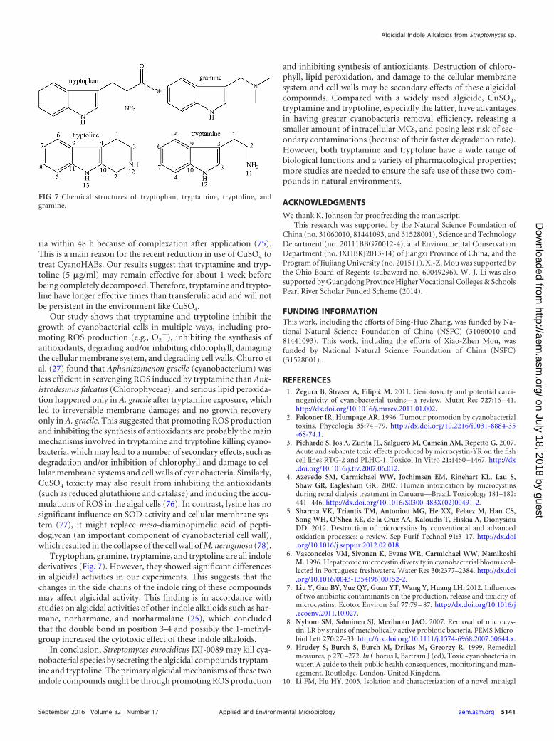
Tryptophan, gramine, tryptamine, and tryptoline are all indolederivatives (Fig. 7). However, they showed significant differencesin algicidal activities in our experiments. This suggests that thechanges in the side chains of the indole ring of these compoundsmay affect algicidal activity. This finding is in accordance withstudies on algicidal activities of other indole alkaloids such as har-mane, norharmane, and norharmalane (25), which concludedthat the double bond in position 3-4 and possibly the 1-methyl-group increased the cytotoxic effect of these indole alkaloids.
In conclusion, Streptomyces eurocidicus JXJ-0089 may kill cya-nobacterial species by secreting the algicidal compounds tryptam-ine and tryptoline. The primary algicidal mechanisms of these twoindole compounds might be through promoting ROS production
and inhibiting synthesis of antioxidants. Destruction of chloro-phyll, lipid peroxidation, and damage to the cellular membranesystem and cell walls may be secondary effects of these algicidalcompounds. Compared with a widely used algicide, CuSO4,tryptamine and tryptoline, especially the latter, have advantagesin having greater cyanobacteria removal efficiency, releasing asmaller amount of intracellular MCs, and posing less risk of sec-ondary contaminations (because of their faster degradation rate).However, both tryptamine and tryptoline have a wide range ofbiological functions and a variety of pharmacological properties;more studies are needed to ensure the safe use of these two com-pounds in natural environments.
ACKNOWLEDGMENTS
We thank K. Johnson for proofreading the manuscript.This research was supported by the Natural Science Foundation of
China (no. 31060010, 81441093, and 31528001), Science and TechnologyDepartment (no. 20111BBG70012-4), and Environmental ConservationDepartment (no. JXHBKJ2013-14) of Jiangxi Province of China, and theProgram of Jiujiang University (no. 201511). X.-Z. Mou was supported bythe Ohio Board of Regents (subaward no. 60049296). W.-J. Li was alsosupported by Guangdong Province Higher Vocational Colleges & SchoolsPearl River Scholar Funded Scheme (2014).
FUNDING INFORMATIONThis work, including the efforts of Bing-Huo Zhang, was funded by Na-tional Natural Science Foundation of China (NSFC) (31060010 and81441093). This work, including the efforts of Xiao-Zhen Mou, wasfunded by National Natural Science Foundation of China (NSFC)(31528001).
REFERENCES1. Žegura B, Štraser A, Filipic M. 2011. Genotoxicity and potential carci-
nogenicity of cyanobacterial toxins—a review. Mutat Res 727:16 – 41.http://dx.doi.org/10.1016/j.mrrev.2011.01.002.
2. Falconer IR, Humpage AR. 1996. Tumour promotion by cyanobacterialtoxins. Phycologia 35:74 –79. http://dx.doi.org/10.2216/i0031-8884-35-6S-74.1.
3. Pichardo S, Jos A, Zurita JL, Salguero M, Cameán AM, Repetto G. 2007.Acute and subacute toxic effects produced by microcystin-YR on the fishcell lines RTG-2 and PLHC-1. Toxicol In Vitro 21:1460 –1467. http://dx.doi.org/10.1016/j.tiv.2007.06.012.
4. Azevedo SM, Carmichael WW, Jochimsen EM, Rinehart KL, Lau S,Shaw GR, Eaglesham GK. 2002. Human intoxication by microcystinsduring renal dialysis treatment in Caruaru—Brazil. Toxicology 181–182:441– 446. http://dx.doi.org/10.1016/S0300-483X(02)00491-2.
5. Sharma VK, Triantis TM, Antoniou MG, He XX, Pelaez M, Han CS,Song WH, O’Shea KE, de la Cruz AA, Kaloudis T, Hiskia A, DionysiouDD. 2012. Destruction of microcystins by conventional and advancedoxidation processes: a review. Sep Purif Technol 91:3–17. http://dx.doi.org/10.1016/j.seppur.2012.02.018.
6. Vasconcelos VM, Sivonen K, Evans WR, Carmichael WW, NamikoshiM. 1996. Hepatotoxic microcystin diversity in cyanobacterial blooms col-lected in Portuguese freshwaters. Water Res 30:2377–2384. http://dx.doi.org/10.1016/0043-1354(96)00152-2.
7. Liu Y, Gao BY, Yue QY, Guan YT, Wang Y, Huang LH. 2012. Influencesof two antibiotic contaminants on the production, release and toxicity ofmicrocystins. Ecotox Environ Saf 77:79 – 87. http://dx.doi.org/10.1016/j.ecoenv.2011.10.027.
8. Nybom SM, Salminen SJ, Meriluoto JAO. 2007. Removal of microcys-tin-LR by strains of metabolically active probiotic bacteria. FEMS Micro-biol Lett 270:27–33. http://dx.doi.org/10.1111/j.1574-6968.2007.00644.x.
9. Hrudey S, Burch S, Burch M, Drikas M, Greorgy R. 1999. Remedialmeasures, p 270 –272. In Chorus I, Bartram J (ed), Toxic cyanobacteria inwater. A guide to their public health consequences, monitoring and man-agement. Routledge, London, United Kingdom.
10. Li FM, Hu HY. 2005. Isolation and characterization of a novel antialgal
FIG 7 Chemical structures of tryptophan, tryptamine, tryptoline, andgramine.
Algicidal Indole Alkaloids from Streptomyces sp.
September 2016 Volume 82 Number 17 aem.asm.org 5141Applied and Environmental Microbiology
on July 18, 2018 by guesthttp://aem
.asm.org/
Dow
nloaded from

allelochemical from Phragmites communis. Appl Environ Microbiol 71:6545– 6553. http://dx.doi.org/10.1128/AEM.71.11.6545-6553.2005.
11. Semenov BB, Granik VG. 2004. Chemistry of N-(1H-indol-3-ylmethyl)-N,N-dimethylamine (gramine): a review. Pharm Chem J 38:287–310.http://dx.doi.org/10.1023/B:PHAC.0000048140.06266.63.
12. Coulman BE. 1995. Bellevue reed canarygrass (Phalaris arundinacea L.).Can J Plant Sci 75:473– 474. http://dx.doi.org/10.4141/cjps95-082.
13. Hautala EL, Holopainen JK. 1995. Gramine and free amino-acids asindicators of fluoride-induced stress in barley and its consequences toinsect herbivory. Ecotoxicol Environ Saf 31:238 –245. http://dx.doi.org/10.1006/eesa.1995.1069.
14. Hong Y, Hu HY, Xie X, Sakoda A, Sagehashi M, Li FM. 2009. Gramine-induced growth inhibition, oxidative damage and antioxidant responsesin freshwater cyanobacterium Microcystis aeruginosa. Aquat Toxicol 91:262–269. http://dx.doi.org/10.1016/j.aquatox.2008.11.014.
15. Konda Y, Suzuki Y, Omura S, Onda M. 1976. Alkaloid from Thermo-actinomyces species. Chem Pharm Bull (Tokyo) 24:92–96. http://dx.doi.org/10.1248/cpb.24.92.
16. Korkmaz C, Hames-Kocabas E, Uzel A, Bedir E. 2008. Tryptaminederived amides with thiazole ring system from Thermoactinomyces strainTA66-2. Magn Reson Chem 46:80 – 83. http://dx.doi.org/10.1002/mrc.2101.
17. Jeong S, Ishida K, Ito Y, Okada S, Murakami M. 2003. Bacillamide, anovel algicide from the marine bacterium, Bacillus sp. SY-1, against theharmful dinoflagellate, Cochlodinium polykrikoides. Tetrahedron Lett 44:8005– 8007.
18. Socha A, Long R, Rowley D. 2007. Bacillamides from a hypersalinemicrobial mat bacterium. J Nat Prod 70:1793–1795. http://dx.doi.org/10.1021/np070126a.
19. Jin LJ, Ma W, Peng CS, Yin Y, Xu BL, Zhang FL, Guo YW, Li ZY. 2011.Bacillamide C production by the optimized cultivation of the Bacillusatrophaeus strain C89 associated with the South China Sea sponge Dysideaavara. Process Biochem 46:1153–1159. http://dx.doi.org/10.1016/j.procbio.2011.02.003.
20. Ivanova V, Kolarova M, Aleksieva K. 2007. Microbiaeratin, a new nat-ural indole alkaloid from a Microbispora aerata strain, isolated from Liv-ingston Island, Antarctica. Prep Biochem Biotechnol 37:161–168. http://dx.doi.org/10.1080/10826060701199122.
21. Churro C, Alverca E, Sam-Bento F, Paulino S, Figueira VC, Bento AJ,Prabhakar S, Lobo AM, Calado AJ, Pereira P. 2009. Effects of bacil-lamide and newly synthesized derivatives on the growth of cyanobacteriaand microalgae cultures. J Appl Phycol 21:429 – 442. http://dx.doi.org/10.1007/s10811-008-9388-3.
22. Etchegaray A, Rabello E, Dieckmann R, Moon DH, Fiore MF, DöhrenHV, Tsai SM, Neilan BA. 2004. Algicide production by the filamentouscyanobacterium Fischerella sp. CENA 19. J Appl Phycol 16:237–243. http://dx.doi.org/10.1023/B:JAPH.0000048509.77816.5e.
23. Kodani S, Imoto A, Mitsutani A, Murakami M. 2002. Isolation andidentification of the antialgal compound, harmane (1-methyl-�-carboline), produced by the algicidal bacterium, Pseudomonas sp. K44-1. JAppl Phycol 14:109 –114. http://dx.doi.org/10.1023/A:1019533414018.
24. Volk RB. 2005. Screening of microalgal culture media for the presence ofalgicidal compounds and isolation and identification of two bioactive me-tabolites, excreted by the cyanobacteria Nostoc insulare and Nodulariaharveyana, respectively. J Appl Phycol 17:339 –347. http://dx.doi.org/10.1007/s10811-005-7292-7.
25. Volk RB. 2006. Antialgal activity of several cyanobacterial exometabolites.J Appl Phycol 18:145–151. http://dx.doi.org/10.1007/s10811-006-9085-z.
26. Stevens LH, Blom TJM, Verpoorte R. 1993. Subcellular localization oftryptophan decarboxylase, strictosidine synthase and strictosidine gluco-sidase in suspension cultured cells of Catharanthus roseus and Tabernae-montana divaricata. Plant Cell Rep 12:563–576.
27. Churro C, Fernandes AS, Alverca E, Sam-Bento F, Paulino S, FigueiraVC, Bento AJ, Prabhakar S, Lobo AM, Martins LL, Mourato MP,Pereira P. 2010. Effects of tryptamine on growth, ultrastructure, andoxidative stress of cyanobacteria and microalgae cultures. Hydrobiologia649:195–206. http://dx.doi.org/10.1007/s10750-010-0245-4.
28. Yamamoto Y, Kouchiwa T, Hodoki Y. 1998. Distribution and identifi-cation of actinomycetes lysing cyanobacteria in a eutrophic lake. J ApplPhycol 10:391–397. http://dx.doi.org/10.1023/A:1008077414808.
29. Bérdy J. 2005. Bioactive microbial metabolites. J Antibiot 58:1–26. http://dx.doi.org/10.1038/ja.2005.1.
30. El-Sherbiny SA, Ghaly MF, El-Ayoty YM, Fleafil NS. 2007. Niromycin A:
an antialgal substance produced by Streptomyces endus N40. Res J Micro-biol 2:606 – 618. http://dx.doi.org/10.3923/jm.2007.606.618.
31. El-Sherbiny SA, El-Ayoty YM, Ghaly MF, Fleafil NS. 2009. Evaluationfor the production of antialgal substance from Streptomyces neyagawaen-sis. Biotechnology (Faisalabad) 8:405– 415. http://dx.doi.org/10.3923/biotech.2009.405.415.
32. Feng Y, Chang XX, Zhao LX, Li XP, Li WJ, Jiang Y. 2013. NanaomycinA methyl ester, an actinomycete metabolite: algicidal activity and thephysiological response of Microcystis aeruginosa. Ecol Eng 53:306 –312.http://dx.doi.org/10.1016/j.ecoleng.2012.12.066.
33. Liu Y, Zhang J, Gao BY, Feng SP. 2014. Combined effects of twoantibiotic contaminants on Microcystis aeruginosa. J Hazard Mater 279:148 –155. http://dx.doi.org/10.1016/j.jhazmat.2014.07.002.
34. Luo JF, Wang Y, Tang SS, Liang JW, Lin WT, Luo LX. 2013. Isolationand identification of algicidal compound from Streptomyces and algicidalmechanism to Microcystis aeruginosa. PLoS One 8:1–14. http://dx.doi.org/10.1371/journal.pone.0076444.
35. Choi HJ, Kim BH, Kim JD, Han MS. 2005. Streptomyces neyagawaensisas a control for the hazardous biomass of Microcystis aeruginosa (Cyano-bacteria) in eutrophic fresh waters. Biol Control 33:335–343. http://dx.doi.org/10.1016/j.biocontrol.2005.03.007.
36. Rommelspacher H, Bade P, Coper H, Kossmehl G. 1976. Inhibition ofthe reuptake of serotonin by tryptoline. Naunyn Schmiedebergs ArchPharmacol 292:93–95. http://dx.doi.org/10.1007/BF00506495.
37. Zhang BH, Chen W, Li HQ, Zhou EM, Hu WY, Duan YQ, MohamadOA, Gao R, Li WJ. 2015. An antialgal compound produced by Strepto-myces jiujiangensis JXJ 0074T. Appl Microbiol Biotechnol 99:7673–7683.http://dx.doi.org/10.1007/s00253-015-6584-3.
38. Otsuka S, Suda S, Li R, Watanabe M, Oyaizu H, Matsumoto S, Wa-tanabe MM. 1998. 16S rDNA sequences and phylogenetic analyses ofMicrocystis strains with and without phycoerythrin. FEMS Microbiol Lett164:119 –124. http://dx.doi.org/10.1111/j.1574-6968.1998.tb13076.x.
39. Li XC, Yang YM, Li RH. 2015. Phenotypic and genotypic validation ofthe rare species Sphaerospermopsis eucompacta comb. nov. (Nostocales,Cyanobacteria) isolated from China. Phycologia 54:299 –306. http://dx.doi.org/10.2216/14-102.1.
40. Li WJ, Xu P, Schumann P, Zhang YQ, Pukall R, Xu LH, Stackebrant E,Jiang CL. 2007. Georgenia ruanii sp. nov., a novel actinobacterium iso-lated from forest soil in Yunnan (China), and emended description of thegenus Georgenia. Int J Syst Evol Microbiol 57:1424 –1428. http://dx.doi.org/10.1099/ijs.0.64749-0.
41. Saitou N, Nei M. 1987. The neighbor-joining method: a new method forreconstructing phylogenetic tree. Mol Biol Evol 4:406 – 425.
42. Tamura K, Peterson D, Peterson N, Stecher G, Nei M, Kumar S. 2011.MEGA5: molecular evolutionary genetics analysis using maximum likeli-hood, evolutionary distance, and maximum parsimony methods. MolBiol Evol 28:2731–2739. http://dx.doi.org/10.1093/molbev/msr121.
43. Felsenstein J. 1985. Confidence limits on phylogenies: an approach usingthe bootstrap. Evolution 39:783–791. http://dx.doi.org/10.2307/2408678.
44. Hua XH, Li JH, Li JJ, Zhang LH, Cui Y. 2009. Selective inhibition of thecyanobacterium, Microcystis, by a Streptomyces sp. Biotechnol Lett 31:1531–1535. http://dx.doi.org/10.1007/s10529-009-0051-0.
45. Shirling EB, Gottlieb D. 1966. Methods for characterization of Strepto-myces species. Int J Syst Bacteriol 16:313–340. http://dx.doi.org/10.1099/00207713-16-3-313.
46. Xu P, Li WJ, Tang SK, Zhang YQ, Chen GZ, Chen HH, Xu H, Jiang CL.2005. Naxibacter alkalitolerans gen. nov., sp. nov., a novel member of thefamily Oxalobacteraceae isolated from China. Int J Syst Evol Microbiol55:1149 –1153. http://dx.doi.org/10.1099/ijs.0.63407-0.
47. Kovacs N. 1956. Identification of Pseudomonas pyocyanea by the oxidasereaction. Nature 178:703–704.
48. Goodfellow M. 1971. Numerical taxonomy of some nocardioform bacteria. JGen Microbiol 69:33–80. http://dx.doi.org/10.1099/00221287-69-1-33.
49. Williams ST, Goodfellow M, Alderson G, Wellington EMH, SneathPHA, Sackin MJ. 1983. Numerical classification of Streptomyces and re-lated genera. J Gen Microbiol 129:1743–1813.
50. Hasegawa T, Takizaea M, Tanida S. 1983. A rapid analysis for chemicalgrouping aerobic actinomycetes. J Gen Appl Microbiol 29:319 –322. http://dx.doi.org/10.2323/jgam.29.319.
51. Tang SK, Wang Y, Chen Y, Lou K, Cao LL, Xu LH, Li WJ. 2009.Zhihengliuella alba sp. nov., and emended description of the genus Zhi-hengliuella. Int J Syst Evol Microbiol 59:2025–2032. http://dx.doi.org/10.1099/ijs.0.007344-0.
Zhang et al.
5142 aem.asm.org September 2016 Volume 82 Number 17Applied and Environmental Microbiology
on July 18, 2018 by guesthttp://aem
.asm.org/
Dow
nloaded from

52. Minnikin DE, Collins MD, Goodfellow M. 1979. Fatty acid and polarlipid composition in the classification of Cellulomonas, Oerskovia and re-lated taxa. J Appl Bacteriol 47:87–95. http://dx.doi.org/10.1111/j.1365-2672.1979.tb01172.x.
53. Collins MD, Jones D. 1980. Lipids in the classification and identificationof coryneform bacteria containing peptidoglycan based on 2,4-diaminobutyric acid. Appl Bacteriol 48:459 – 470. http://dx.doi.org/10.1111/j.1365-2672.1980.tb01036.x.
54. Sasser M. 2001. Identification of bacteria by gas chromatography of cel-lular fatty acids. Technical note 101. Microbial ID, Inc, Newark, DE.
55. De Ley P, Cattoir H, Reeynaerts A. 1970. The quantitative measurementof DNA hybridization from renaturation rates. Eur J Biochem 12:133–142. http://dx.doi.org/10.1111/j.1432-1033.1970.tb00830.x.
56. Chen YW, Chen KN, Hu YH. 2006. Discussion on possible error forphytoplankton chlorophyll-a concentration analysis using hot-ethanolextraction method. J Lake Sci 18:550 –552. http://dx.doi.org/10.18307/2006.0519.
57. Yang LY, Xiao L. 2011. Outburst, jeopardize and control of cyanobacte-rial bloom in lakes, p 212. Science Press, Beijing, China.
58. Chen JX, Wang XF. 2006. Plant physiology experiment guidance, 2nd ed,p 68 –77. South China University of Technology Press, Guangzhou,China.
59. Zhang BH, Chen W, Li HQ, Yang JY, Zha DM, Duan YQ, Hozzein WN,Xiao M, Gao R, Li WJ. 2016. L-valine, an antialgal amino acid fromStreptomyces jiujiangensis JXJ 0074T. Appl Microbiol Biotechnol 100:4627– 4636. http://dx.doi.org/10.1007/s00253-015-7150-8.
60. Hatano K, Nishii T, Kasai H. 2003. Taxonomic re-evaluation of whorl-forming Streptomyces (formerly Streptoverticillium) species by using phe-notypes, DNA-DNA hybridization and sequences of gyrB, and proposal ofStreptomyces luteireticuli (ex Katoh and Arai 1957) corrig., sp. nov., nom.rev. Int J Syst Evol Microbiol 53:1519 –1529. http://dx.doi.org/10.1099/ijs.0.02238-0.
61. Wayne LG, Brenner DJ, Colwell RR, Grimont PAD, Kandler O,Krichevsky MI, Moore LH, Moore WEC, Murray RGE, Stackebrandt E,Starr MP, Truper HG. 1987. Report of the ad hoc committee on recon-ciliation of approaches to bacterial systematics. Int J Syst Bacteriol 37:463–464. http://dx.doi.org/10.1099/00207713-37-4-463.
62. Osato T, Ueda M, Fukuyama S, Yagishita K, Okami Y, Umezawa H.1955. Production of tertiomycin (a new antibiotic substance), azomycinand eurocidin by S. eurocidicus. J Antibiot 8:105–109.
63. Ross C, Santiago-Vázquez L, Paul V. 2006. Toxin release in response tooxidative stress and programmed cell death in the cyanobacterium Micro-cystis aeruginosa. Aquat Toxicol 78:66 –73. http://dx.doi.org/10.1016/j.aquatox.2006.02.007.
64. Griffiths DJ, Saker ML. 2003. The Palm Island mystery disease 20 yearson: a review of research on the cyanotoxin cylindrospermopsin. EnvironToxicol 18:78 –93. http://dx.doi.org/10.1002/tox.10103.
65. Meepagala KM, Schrader KK, Wedge DE, Duke SO. 2005. Algicidal andantifungal compounds from the roots of Rutagraveolens and synthesis oftheir analogs. Phytochemistry 66:2689 –2695. http://dx.doi.org/10.1016/j.phytochem.2005.09.019.
66. Lee KS, Kim EH, Lee YS, Lee SH, Seo YB, Hwang SA, Cho JY. 2009.Control efficacy of Bordeaux Mixture against powdery mildew on Omija(Schizandra chinensis). J Korean Soc Appl Biol Chem 52:58 – 62.
67. Martins F, Soares ME, Oliveira I, Pereira JA, de Lourdes Bastos M,Baptista P. 2012. Tolerance and bioaccumulation of copper by the ento-mopathogen Beauveria bassiana (Bals.-Criv.) Vuill. exposed to variouscopper-based fungicides. Bull Environ Contam Toxicol 89:53– 60. http://dx.doi.org/10.1007/s00128-012-0628-5.
68. Thomas J, Akroush A, Adamus G. 1999. The indole alkaloid tryptamineproduced in transgenic Petunia hybrida. Plant Physiol Biochem 37:665–670. http://dx.doi.org/10.1016/S0981-9428(00)80096-0.
69. Knaggs AR. 2003. The biosynthesis of shikimate metabolites. Nat ProdRep 20:119 –136. http://dx.doi.org/10.1039/b100399m.
70. Berry M. 2004. Mammalian central nervous system traceamines, phar-macologic amphetamines, physiologic neuromodulators. J Neurochem90:257–271. http://dx.doi.org/10.1111/j.1471-4159.2004.02501.x.
71. Fantegrossi W, Murnane K, Reissig C. 2008. The behavioral pharmacol-ogy of hallucinogens. Biochem Pharmacol 75:17–33. http://dx.doi.org/10.1016/j.bcp.2007.07.018.
72. Rommelspacher H, Kauffmann H, Cohnitz CH, Coper H. 1977. Phar-macological properties of tetrahydronorharmane (tryptoline). NaunynSchmiedebergs Arch Pharmacol 298:83–91. http://dx.doi.org/10.1007/BF00508615.
73. Glover V, Liebowitz J, Armando I, Sandler M. 1982. �-Carbolines asselective monoamine oxidase inhibitors: in vivo implications. J NeuralTransm 54:209 –218. http://dx.doi.org/10.1007/BF01254930.
74. Schrader KK, Duke SO, Kingsbury SK, Tucker CS, Duke MV, DionigiCP, Millie DF, Zimba PV. 2000. Evaluation of ferulic acid for controllingthe musty-odor cyanobacterium, Oscillatoria perornata, in aquacultureponds. J Appl Aquacult 10:1–16.
75. Burch M. 2009. Review of available techniques for control of algal blooms.Presented at the Integrated Management of Algal Bloom Workshop, Su-zhou, China. http://www.docin.com/p-84752397.html.
76. Stauber JL, Florence TM. 1987. Mechanism of toxicity of ionic copperand copper complexes to algae. Mar Biol 94:511–519. http://dx.doi.org/10.1007/BF00431397.
77. Lin BG, Yang LY, Xiao L, Ji J, Yuan Y, Yao Y. 2008. Mechanism of lysineinhibiting Microcystis aeruginosa cells. J Ecol Rural Environ 24:68 –72.
78. Takamura Y, Yamada T, Kimoto A, Kanehama N, Tanaka T, NakadairaS, Yagi O. 2004. Growth inhibition of Microcystis cyanobacteria by L-lysine and disappearance of natural Microcystis blooms with spraying. Mi-crobes Environ 19:31–39. http://dx.doi.org/10.1264/jsme2.19.31.
Algicidal Indole Alkaloids from Streptomyces sp.
September 2016 Volume 82 Number 17 aem.asm.org 5143Applied and Environmental Microbiology
on July 18, 2018 by guesthttp://aem
.asm.org/
Dow
nloaded from


![Probability Review - University of Rochestergmateosb/ECE440/Slides/block_2...Markov’s inequality I RV X with E[jXj] < 1, constant a > 0 I Markov’s inequality states )P(jXj](https://static.fdocuments.net/doc/165x107/5f70cef5b1aea64dd340cfbe/probability-review-university-of-gmateosbece440slidesblock2-markovas.jpg)
