
Alagebrium inhibits neointimal hyperplasia and restores ... · wall shear stress by reducing...
11
Alagebrium inhibits neointimal hyperplasia and restores distributions of wall shear stress by reducing downstream vascular resistance in obese and diabetic rats Hongfeng Wang, 1 Dorothee Weihrauch, 2 Judy R. Kersten, 2 Jeffrey M. Toth, 1,3 Anthony G. Passerini, 4 Anita Rajamani, 4 Sonja Schrepfer, 5,6 and John F. LaDisa, Jr. 1,7,8 1 Department of Biomedical Engineering, Marquette University, Milwaukee, Wisconsin; 2 Department of Anesthesiology, Medical College of Wisconsin, Milwaukee, Wisconsin; 3 Department of Orthopaedic Surgery, Medical College of Wisconsin, Milwaukee, Wisconsin; 4 Department of Biomedical Engineering, University of California Davis, Davis, California; 5 Transplant and Stem Cell Immunobiology Laboratory, University Heart Center and Cardiovascular Research Center, University of Hamburg, Hamburg, Germany; 6 Department of Cardiothoracic Surgery, Stanford University School of Medicine, Stanford, California; 7 Department of Medicine, Division of Cardiovascular Medicine, Medical College of Wisconsin, Milwaukee, Wisconsin; 8 Biotechnology and Bioengineering Center, Medical College of Wisconsin, Milwaukee, Wisconsin Submitted 20 February 2014; accepted in final form 3 August 2015 Wang H, Weihrauch D, Kersten JR, Toth JM, Passerini AG, Rajamani A, Schrepfer S, LaDisa JF Jr. Alagebrium inhibits neointimal hyperplasia and restores distributions of wall shear stress by reducing downstream vascular resistance in obese and diabetic rats. Am J Physiol Heart Circ Physiol 309: H1130 –H1140, 2015. First published August 7, 2015; doi:10.1152/ajpheart.00123.2014.—Mech- anisms of restenosis in type 2 diabetes mellitus (T2DM) are incom- pletely elucidated, but advanced glycation end-product (AGE)-in- duced vascular remodeling likely contributes. We tested the hypoth- esis that AGE-related collagen cross-linking (ARCC) leads to increased downstream vascular resistance and altered in-stent hemo- dynamics, thereby promoting neointimal hyperplasia (NH) in T2DM. We proposed that decreasing ARCC with ALT-711 (Alagebrium) would mitigate this response. Abdominal aortic stents were implanted in Zucker lean (ZL), obese (ZO), and diabetic (ZD) rats. Blood flow, vessel diameter, and wall shear stress (WSS) were calculated after 21 days, and NH was quantified. Arterial segments (aorta, carotid, iliac, femoral, and arterioles) were harvested to detect ARCC and protein expression, including transforming growth factor- (TGF-) and receptor for AGEs (RAGE). Downstream resistance was elevated (60%), whereas flow and WSS were significantly decreased (44% and 56%) in ZD vs. ZL rats. NH was increased in ZO but not ZD rats. ALT-711 reduced ARCC and resistance (46%) in ZD rats while decreasing NH and producing similar in-stent WSS across groups. No consistent differences in RAGE or TGF- expression were observed in arterial segments. ALT-711 modified lectin-type oxidized LDL receptor 1 but not RAGE expression by cells on decellularized matrices. In conclusion, ALT-711 decreased ARCC, increased in- stent flow rate, and reduced NH in ZO and ZD rats through RAGE- independent pathways. The study supports an important role for AGE-induced remodeling within and downstream of stent implanta- tion to promote enhanced NH in T2DM. coronary artery disease; hemodynamics; hyperglycemia; interven- tional cardiology; restenosis NEW & NOTEWORTHY Alagebrium (ALT-711) decreased advanced glycation end- product–related collagen cross-linking and arteriolar stiffness in stented obese and diabetic rats, resulting in decreased downstream resistance and increased in-stent blood flow and wall shear stress. ALT-711 was effective at reducing neointimal hyperplasia and promoting endothelial proliferation regardless of glycemic status. IT IS ESTIMATED THAT 25.8 MILLION Americans are diabetic (8.3% of the population), and an additional 79 million have the metabolic syndrome, a constellation of clinical findings that substantially increases the risk for developing type 2 diabetes mellitus (T2DM) (7). Cardiovascular disease (CVD) accounts for 68% of diabetes-related deaths among people aged 65 yr or older, as these patients have a two- to fourfold increased risk of developing coronary and peripheral artery disease (7, 19). Bare-metal stents (BMS) can restore blood flow (BF) be- yond a vascular occlusion, but restenosis occurring primarily as a result of excessive neointimal hyperplasia (NH) limits their success. These cases require revascularization of the restenotic lesion and have cost the U.S. healthcare system more than $2.5 billion since 1999 (40). Drug-eluting stents (DES) have also been used to combat CVD but are less effective for patients with T2DM (18, 32). Sirolimus and paclitaxel (anti- proliferative agents) used with early-generation DES impede healing of the intima after implantation, thereby inhibiting coverage of the stent linkages by endothelial cells (ECs) and making the vessel more prone to late thrombosis (20, 22). Newer generation DES with zotarolimus and everolimus more favorably inhibit the proliferation of smooth muscle cells (SMC) and inflammatory cells (26), but improvements in mortality, myocardial infarction, stent thrombosis, and target lesion revascularization afforded to patients with normoglyce- mia with these newer DES have not translated to patients with diabetes (11, 44). Mechanisms for the elevated restenosis rates after stenting in patients with T2DM compared with normoglycemia have not yet been fully elucidated. It has been suggested that the current paradigm concerning the use of stents in patients with T2DM applies methods from retrospective revascularization studies conducted in patients with normoglycemia to T2DM in hopes that poor outcomes do not occur (3). In contrast, some inves- tigators have examined pharmacological agents such as anti- sense oligonucleotides and L-arginine supplementation to im- Address for reprint requests and other correspondence: J. LaDisa, Jr., 1637 West Wisconsin Ave., Milwaukee, WI 53233 (e-mail: [email protected]). Am J Physiol Heart Circ Physiol 309: H1130 –H1140, 2015. First published August 7, 2015; doi:10.1152/ajpheart.00123.2014. 0363-6135/15 Copyright © 2015 the American Physiological Society http://www.ajpheart.org H1130
-
Upload
vuongquynh -
Category
Documents
-
view
215 -
download
1
Transcript of Alagebrium inhibits neointimal hyperplasia and restores ... · wall shear stress by reducing...
Alagebrium inhibits neointimal hyperplasia and restores distributions ofwall shear stress by reducing downstream vascular resistance in obese anddiabetic rats
Hongfeng Wang,1 Dorothee Weihrauch,2 Judy R. Kersten,2 Jeffrey M. Toth,1,3 Anthony G. Passerini,4
Anita Rajamani,4 Sonja Schrepfer,5,6 and John F. LaDisa, Jr.1,7,8
1Department of Biomedical Engineering, Marquette University, Milwaukee, Wisconsin; 2Department of Anesthesiology,Medical College of Wisconsin, Milwaukee, Wisconsin; 3Department of Orthopaedic Surgery, Medical College of Wisconsin,Milwaukee, Wisconsin; 4Department of Biomedical Engineering, University of California Davis, Davis, California;5Transplant and Stem Cell Immunobiology Laboratory, University Heart Center and Cardiovascular Research Center,University of Hamburg, Hamburg, Germany; 6Department of Cardiothoracic Surgery, Stanford University School ofMedicine, Stanford, California; 7Department of Medicine, Division of Cardiovascular Medicine, Medical College ofWisconsin, Milwaukee, Wisconsin; 8Biotechnology and Bioengineering Center, Medical College of Wisconsin, Milwaukee,Wisconsin
Submitted 20 February 2014; accepted in final form 3 August 2015
Wang H, Weihrauch D, Kersten JR, Toth JM, Passerini AG,Rajamani A, Schrepfer S, LaDisa JF Jr. Alagebrium inhibitsneointimal hyperplasia and restores distributions of wall shear stressby reducing downstream vascular resistance in obese and diabetic rats.Am J Physiol Heart Circ Physiol 309: H1130–H1140, 2015. Firstpublished August 7, 2015; doi:10.1152/ajpheart.00123.2014.—Mech-anisms of restenosis in type 2 diabetes mellitus (T2DM) are incom-pletely elucidated, but advanced glycation end-product (AGE)-in-duced vascular remodeling likely contributes. We tested the hypoth-esis that AGE-related collagen cross-linking (ARCC) leads toincreased downstream vascular resistance and altered in-stent hemo-dynamics, thereby promoting neointimal hyperplasia (NH) in T2DM.We proposed that decreasing ARCC with ALT-711 (Alagebrium)would mitigate this response. Abdominal aortic stents were implantedin Zucker lean (ZL), obese (ZO), and diabetic (ZD) rats. Blood flow,vessel diameter, and wall shear stress (WSS) were calculated after 21days, and NH was quantified. Arterial segments (aorta, carotid, iliac,femoral, and arterioles) were harvested to detect ARCC and proteinexpression, including transforming growth factor-� (TGF-�) andreceptor for AGEs (RAGE). Downstream resistance was elevated(60%), whereas flow and WSS were significantly decreased (44% and56%) in ZD vs. ZL rats. NH was increased in ZO but not ZD rats.ALT-711 reduced ARCC and resistance (46%) in ZD rats whiledecreasing NH and producing similar in-stent WSS across groups. Noconsistent differences in RAGE or TGF-� expression were observedin arterial segments. ALT-711 modified lectin-type oxidized LDLreceptor 1 but not RAGE expression by cells on decellularizedmatrices. In conclusion, ALT-711 decreased ARCC, increased in-stent flow rate, and reduced NH in ZO and ZD rats through RAGE-independent pathways. The study supports an important role forAGE-induced remodeling within and downstream of stent implanta-tion to promote enhanced NH in T2DM.
coronary artery disease; hemodynamics; hyperglycemia; interven-tional cardiology; restenosis
NEW & NOTEWORTHY
Alagebrium (ALT-711) decreased advanced glycation end-product–related collagen cross-linking and arteriolar stiffness
in stented obese and diabetic rats, resulting in decreaseddownstream resistance and increased in-stent blood flow andwall shear stress. ALT-711 was effective at reducing neointimalhyperplasia and promoting endothelial proliferation regardlessof glycemic status.
IT IS ESTIMATED THAT 25.8 MILLION Americans are diabetic (8.3%of the population), and an additional 79 million have themetabolic syndrome, a constellation of clinical findings thatsubstantially increases the risk for developing type 2 diabetesmellitus (T2DM) (7). Cardiovascular disease (CVD) accountsfor 68% of diabetes-related deaths among people aged 65 yr orolder, as these patients have a two- to fourfold increased risk ofdeveloping coronary and peripheral artery disease (7, 19).
Bare-metal stents (BMS) can restore blood flow (BF) be-yond a vascular occlusion, but restenosis occurring primarilyas a result of excessive neointimal hyperplasia (NH) limitstheir success. These cases require revascularization of therestenotic lesion and have cost the U.S. healthcare system morethan $2.5 billion since 1999 (40). Drug-eluting stents (DES)have also been used to combat CVD but are less effective forpatients with T2DM (18, 32). Sirolimus and paclitaxel (anti-proliferative agents) used with early-generation DES impedehealing of the intima after implantation, thereby inhibitingcoverage of the stent linkages by endothelial cells (ECs) andmaking the vessel more prone to late thrombosis (20, 22).Newer generation DES with zotarolimus and everolimus morefavorably inhibit the proliferation of smooth muscle cells(SMC) and inflammatory cells (26), but improvements inmortality, myocardial infarction, stent thrombosis, and targetlesion revascularization afforded to patients with normoglyce-mia with these newer DES have not translated to patients withdiabetes (11, 44).
Mechanisms for the elevated restenosis rates after stenting inpatients with T2DM compared with normoglycemia have notyet been fully elucidated. It has been suggested that the currentparadigm concerning the use of stents in patients with T2DMapplies methods from retrospective revascularization studiesconducted in patients with normoglycemia to T2DM in hopesthat poor outcomes do not occur (3). In contrast, some inves-tigators have examined pharmacological agents such as anti-sense oligonucleotides and L-arginine supplementation to im-
Address for reprint requests and other correspondence: J. LaDisa, Jr., 1637 WestWisconsin Ave., Milwaukee, WI 53233 (e-mail: [email protected]).
Am J Physiol Heart Circ Physiol 309: H1130–H1140, 2015.First published August 7, 2015; doi:10.1152/ajpheart.00123.2014.
0363-6135/15 Copyright © 2015 the American Physiological Society http://www.ajpheart.orgH1130
prove endothelial function, but these pharmacological agentsfailed to alleviate restenosis in T2DM (15, 27).
Although local changes in cytokine release and cell signal-ing undoubtedly play a role in this process, the inciting changesin local and distal fluid dynamics and vascular biomechanicsassociated with T2DM, which may also contribute to resteno-sis, have been relatively ignored. For example, adverse struc-tural modifications are known to occur throughout the arterialsystem in response to T2DM, including increased vascularstiffness attributable to advanced glycation end-products(AGEs), which are formed through nonenzymatic reactionbetween glucose and proteins. It has been reported that AGEscan cause tissue damage in the cardiovascular system bycross-linking with collagen, thus disrupting the vessel wall andaltering compliance (i.e., increasing stiffness) (39). Thesechanges may be manifested by an increase in vascular resis-tance (DVR) at the arterioles that may have an importantimpact within the upstream stented region in terms of localflow patterns, blood pressure (BP), and altered wall shear stress(WSS) that have previously been correlated with NH (29).
AGEs, which form more rapidly during diabetes as a con-sequence of chronic hyperglycemia (38, 49), also play animportant role in cell signaling to influence NH. For example,they may react with a receptor (RAGE) to increase oxidativestress, expression of transforming growth factor-� (TGF-�),and extracellular matrix accumulation (35).
Alagebrium (ALT-711; 3-phenacyl-4, 5-dimethylthiazoliumchloride) has been shown to cleave AGE-related collagencross-linking (ARCC), thereby decreasing vessel resistanceand atherosclerosis (2, 10). ALT-711 may consequently furtherdecrease NH in T2DM by modulating in-stent hemodynamicssecondary to its effects on downstream resistance vessels.Therefore, we tested the hypothesis that ARCC leads to in-creased DVR and altered hemodynamics in the stented region,promoting enhanced NH after BMS implantation in T2DM.We further proposed that that decreasing ARCC with ALT-711would mitigate this response.
MATERIALS AND METHODS
In vivo protocol. Stents were deployed into the abdominal aorta(AAo) of male Zucker lean (ZL), obese (ZO), and diabetic (ZD) rats(n � 9/group) in the absence or presence of ALT-711 (i.e., treatment).After 21 days, three rats from each group were randomly selected forAAo casting and measurement of local BF and vessel diameter tocalculate in-stent WSS. Remaining rats underwent quantification ofNH, ARCC, and protein expression by Western blotting. Additionaldetails are provided below.
In vivo experimental preparation. All procedures were approved bythe Animal Care and Use Committees of the Medical College ofWisconsin and Marquette University and conformed to the Guide forthe Care and Use of Laboratory Animals published by the U.S.National Institutes of Health. ZL, ZO, and ZD rats (Charles RiverLaboratories, Wilmington, MA) were selected randomly for stentingat 12 wk of age after �48 h of acclimation. Rats were anesthetized inan induction chamber using 1–2% isoflurane then placed on a warmsurface and fitted with a nose cone attached to an anesthetic vaporizerto maintain the proper plane of anesthesia (�0.5–3.0%).
Stent implantation in vivo. AAo stenting was performed underanesthesia and sterile conditions (12, 36). Local BF patterns and thesubsequent severity of NH are influenced by stent geometry (29).Stainless steel balloon expandable stents (316L) specially designedfor the rat AAo (2.5 � 8 mm) and with a known geometric pattern
were therefore purchased from Burpee Materials Technology (Eaton-town, NJ) and crimped on 2.5 � 12 mm rapid-exchange deliverycatheters (Polymerex, San Diego, CA). In preparation for stenting, thesite above the AAo and iliac arteries was shaved and cleaned. The skinwas then incised by a midsagittal incision. Under microscopic view,the AAo and iliac arteries were carefully separated free from sur-rounding vessels and tissue. Small aortic side branches were tempo-rarily clamped to limit the backflow of blood. Vascular clips wereplaced to isolate the region undergoing stenting near the aorta-iliacbifurcation. A small incision was made in the isolated segment, andblood was removed by rinsing with heparinized saline (100 U/kg) toprevent acute thrombosis. The tip of a delivery catheter and guide wirewere then inserted. Clamps were then briefly removed as necessary tothread the stent to an infrarenal segment of the AAo. The stent wasthen inflated to securely anchor it against the AAo wall using 10–20%overexpansion (17). Successful deployment was confirmed visuallythrough the AAo wall. After removal of the stent delivery catheter, theincision was closed using an 8-0 suture. The vascular clips were thenremoved, and the abdomen was closed with 4-0 silk suture while theskin was closed with 4-0 dissolvable Vicryl suture using stitches justbelow the skin.
Surgical and postoperative care. Quantitative criteria includingoxygen saturation, respiration, temperature, pulse, mucous membranecolor, and capillary refill time were continuously monitored during thesurgical procedure and 3–4 h after its completion. Buprenorphine(0.05 mg/kg ip) was used as analgesia for 2 days. Animals alsoreceived antibiotic prophylaxis (20 mg/kg cefazolin ip) for 4 days andaspirin in their food or drinking water (20 mg/day) for the duration ofthe experiment to prevent thrombosis.
ALT-711 treatment in vivo. An inhibitor of ARCC, ALT-711(Iron-Dragon, Newport Beach, CA) was delivered at a dose of 1.0mg/kg per day for 21 days using an osmotic minipump (ALZET,Cupertino, CA) to avoid daily injections. Before skin was closed afterstenting, a minipump was filled, placed just beneath the muscle layer,and positioned with its delivery portal facing cranially. All otherprocedures were equivalent, and four additional ZL rats underwentstenting and minipump insertion with vehicle alone (i.e., saline).
Hemodynamic data acquisition. Rats were anesthetized 21 daysafter stenting as described above, and the right carotid artery wasisolated. A fluid-filled catheter connected to a BP transducer (HarvardApparatus, Holliston, MA) was calibrated and inserted from thecarotid artery into the aorta. The AAo proximal to the stent was alsodissected free from connective tissue and the vena cava, so BF couldbe recorded with a transit-time flow probe connected to a flow meter(Transonic Systems, Ithaca, NY). BP and BF data were sampled usingan A/D converter interfacing with a laptop running WINDAQ soft-ware (DATAQ Instruments, Akron, OH). Rats were then euthanizedby overdose pentobarbital sodium (100 mg/kg iv).
Calculation of in-stent WSS. The stented region was carefullydissected from connective tissue after euthanasia. A small incisionwas made in the suprarenal AAo, and a catheter containing Batson’sNo. 17 Corrosion Compound (Polysciences, Warrington, PA) wassecured within the vessel (29). A four-way stopcock was connected tothe catheter and BP transducer. A syringe was then connected to theopen end of the stopcock to flush the vessel with saline before theplastic compound was injected and maintained at the mean BPmeasured for each rat. Care was taken to ensure no bubbles wereinjected, and mean BP was maintained, thereby capturing the geom-etry of the flow domain. After curing (2–3 h), surrounding tissue wascaustically removed with Batson’s No. 17 Maceration Solution, leav-ing a cast of the flow domain. In-stent WSS was calculated as 4�Q/�r3, where Q is the mean measured flow rate, viscosity was assumedto be 6.2 cP (53), and r is the radius determined from an average of threedigital micrometer measurements within the stented region.
Quantification of NH. The stented AAo of rats not undergoingcasting was rinsed with saline to remove any blood and fixed in 4%paraformaldehyde for �24 h. Vessels were then dehydrated in 70%
H1131ALT-711 INHIBITS NH AND RESTORES WSS VIA DISTAL RESISTANCE
AJP-Heart Circ Physiol • doi:10.1152/ajpheart.00123.2014 • www.ajpheart.org
ethanol for 2 h, 95% ethanol for 2 h, and 100% ethanol twice for 2 h.Following dehydration, samples underwent preinfiltration for 2 h andinfiltration for 24 h. Samples were then embedded in glycol methac-rylate (EB Sciences, Agawam, MA), sectioned at room temperature in5-�m intervals using a microtome equipped with a tungsten carbideblade (Ted Pella, Redding, CA), and stained with hematoxylin andeosin (H and E). Briefly, plastic stent sections were stained by Gill’shematoxylin for 15 min followed by three distilled water washes. Scott’stap water was used for 2 min followed by an additional three washes withdistilled water. Sections were then counterstained with eosin for 5 min,dehydrated carefully using 96% and 100% ethanol, cleared in xylene, andmounted with a xylene-based mounting medium. Images were obtainedusing a Zeiss Universal microscope with �2.5 and �4 objective lensescoupled to a 16-bit Leica DFC 280 digital camera using Image Pro Plus5.1 image analysis software running on a Windows XP workstation. NHwas quantified from the middle of the stented region to avoid contribu-tions resulting from flow disturbances or pronounced vessel injury. Thepercentage of vascular lumen in which NH occurred was quantified bysubtracting the luminal area from that bounded by the stent with NIHImageJ software (29).
Analysis of ARCC and protein expression. Vessels from elasticarteries (aorta and carotid arteries), peripheral muscular arteries (fem-oral and iliac arteries), and smallest distal arteries (cremaster arteri-oles) were harvested. Portions of these vessels from six rats before andafter ALT-711 treatment were separately allocated for ARCC orprotein analysis. All vessels were snap frozen in liquid nitrogen andstored at 80°C to avoid protein degradation after rinsing with 4°Csaline to remove any blood.
Vessel segments analyzed for ARCC underwent pepsin digestion(43). Briefly, vessel segments were lyophilized for �8 h to obtaintheir dry weight. Dry samples (10 mg) were then treated with 4 mol/lguanidine-HCL in 0.05 mol/l sodium acetate (pH 5.8) at 4°C for 24 hto remove proteoglycans. After centrifugation for 30 min at 30,000 g,the residue was collected and washed three times using 0.5 mol/lacetic acid. The collagen residue was added to a solution of 1 mg/mlpepsin in 0.5 mol/l acetic acid at 4°C for 3 days, and undigestedmaterial was discarded by centrifugation for 20 min. AGE-relatedfluorescence of the supernatant was measured by 365-nm excitationand 418-nm emission before being reacted with Sirius red in 0.5 mol/lacetic acid and incubated at room temperature for 20 min. Sampleswere then centrifuged at 2,500 g for 10 min, and the absorbance of thesupernatant was read at 540 nm against a 0.5 mol/l acetic acid blank.A series dilution of collagen I (Life Technologies, Grand Island, NY)was then used to generate a standard concentration and absorbancecurve. Previous studies found a linear relationship between Sirius red andoptical density and that a 0.5 �mol/l concentration was suitable forcollagen quantification without saturation (45). The results are presentedas AGE-related fluorescence divided by collagen concentration.
Protein for Western blotting analysis was isolated as previouslydescribed (48) and quantified using a spectrophotometer (BeckmanCoulter, Brea, CA) with the Bradford method and bovine serumalbumin as a standard (4). After stabilization on ice for 30 min, 30 �gof the protein was added to the same amount of Laemmli buffer andincubated in a thermomixer (Eppendorf, Hauppauge, NY) for 5 min at97°C. The treated protein mix was then loaded into a 4–20% poly-acrylamide gel (Criterion; Bio-Rad, Hercules, CA). The gel was run
for 10 min at 100 V followed by 50 min at 150 V, and transferoccurred at 100 V for 1 h. The polyvinylidene fluoride membrane wasthen blocked for 1 h at room temperature. The membrane was reactedwith primary antibodies (Table 1) overnight and then secondaryantibodies (Table 1) for 1 h at room temperature. The membrane waswashed, incubated in enhanced chemiluminescence solution for 5min, and developed using a molecular imager (Bio-Rad).
Statistical procedures for analysis of in vivo data. Statisticalanalysis was conducted using multiple ANOVA followed by Tukey-Kramer and post hoc analysis. Changes within and between groupswere considered statistically significant when P 0.05, and all datawere expressed as means � SE.
In vitro cell culture and treatment protocols. To elucidate thepotential contribution of ARCC to NH, the AAo of additional groupsof ZL, ZO, and ZD rats in the absence or presence of ALT-711treatment (n � 3/group; 6 groups) was harvested as described above.The extracellular matrix was then isolated by incubating and agitatingin 1% SDS for 12 h at room temperature to dissolve cells. The nextday, a Triton X-100 wash (30 min) was performed followed by athorough PBS rinse (15 min). The isolated matrix was then snapfrozen and pulverized with mortar and pestle. The powdered matrixwas suspended in Dulbecco’s PBS and sonicated on ice for 30 s.Human aortic ECs (HAECs) and umbilical vein ECs (HUVECs) wereprepared and maintained in endothelial growth medium-2 with 2%FBS and 1� antibiotic-antimycotic solution before being seeded onthe decellularized matrices for experiments at passages 5–7. The choiceof cell type was selected based on their successful use in prior shearing(HAECs) and static (HUVECs) investigations as discussed below.
Cell-shearing experiments. Hydrodynamic experiments were per-formed on HAEC monolayers using a microfluidic flow chamberdevice based on Hele-Shaw stagnation flow theory (14, 46). Thedevice induces a linearly decreasing WSS profile along the center lineof the longitudinal axis for a given flow rate when vacuum adhered toa cell monolayer, facilitating the study of a wide range in WSSmagnitudes within a single cell monolayer, while preserving informa-tion related to the spatial heterogeneity of response. The flow rate waschosen to deliver WSS magnitudes ranging from �0–16 dynes/cm2,which captures a range of values experienced in the intrastrut regionof an implanted stent within a single experiment.
HAECs were seeded on decellularized matrices derived from theAAo of ZD, ZL, and ZO rats with or without ALT-711 (20 �g/ml inDulbecco’s PBS with 1� antibiotic-antimycotic). Collagen I-coated(100 �g/ml; Invitrogen, Carlsbad, CA) tissue culture substrates wereused as a control. Experiments were performed when cells reached80–90% confluence. Flow chambers were sealed to the HAEC mono-layers via a vacuum network. Flow was driven by a Masterflex L/Speristaltic pump (Cole-Parmer, Vernon Hills, IL) in a humidifiedchamber heated to 37°C for 4 h. Leibovitz-15 medium (GIBCO,Carlsbad, CA), supplemented with 10% FBS, endothelial BulletKit(Lonza, Walkersville, MD), and 1� antibiotic-antimycotic solution,was used as the flow medium to maintain pH in the absence of CO2.
Immunofluorescence staining of sheared cells. Sheared cells wererinsed with PBS and fixed in 4% paraformaldehyde (Electron Micros-copy Sciences, Hatfield, PA) for RAGE analysis. Samples wereblocked in 20% donkey serum (Invitrogen) and 1% human serumalbumin (CSL Behring, King of Prussia, PA) and incubated with
Table 1. Antibodies used for WB or IF
Antibody Company Catalog No. Host Primary Antibody Secondary Antibody
AGEs Abcam ab23722 Rabbit 1:500 1:2,000RAGE Santa Cruz Biotechnology sc-8230 Goat 1:300 (WB) 1:100 (IF) 1:2,000 1:400TGF-� Santa Cruz Biotechnology sc-146 Rabbit 1:400 1:2,000�-Tubulin Abcam ab6046 Rabbit 1:5,000 1:10,000
�-Tubulin was used as loading control for Western blot (WB). IF, immunofluorescence; AGES, advanced glycation end-products; RAGE, receptor for AGEs;TGF-�, transforming growth factor-�.
H1132 ALT-711 INHIBITS NH AND RESTORES WSS VIA DISTAL RESISTANCE
AJP-Heart Circ Physiol • doi:10.1152/ajpheart.00123.2014 • www.ajpheart.org
polyclonal goat anti-RAGE (Santa Cruz Biotechnology, Santa Cruz,CA) at a concentration of 2 �g/ml, for 2 h. Samples were thenincubated with secondary Alexa Fluor-546 (Life Technology) rabbitanti-goat (1:400) for 1 h. Coverslips were mounted usingVECTASHIELD mounting medium with DAPI (Vector Laboratories,Burlingame, CA). Representative images were taken at �40 magni-fication at each WSS magnitude, corresponding to fixed axial positionwithin the chamber. Images were quantified using ImageJ software.The local background-subtracted mean fluorescence intensity wascalculated for each cell (�20–40 over 5 representative images) andaveraged for a given WSS for each shear experiment (n � 3–4).
EC and SMC proliferation assay. HUVECs and aortic SMCs weresimilarly cultured on decellularized matrices from the AAo of ZD,ZL, and ZO rats with or without ALT-711 for 72 h using 24-wellplates (Fisher Scientific, Waltham, MA) in endothelial growth me-dium complete media (Lonza) and SMC media (Lonza). After 24 h,cells were trypsinized, and the number of cells was determined witha hemacytometer.
Statistical procedures for analysis of in vitro data. Data wereanalyzed using Minitab 17 Statistical software. Data were tested fornormality using the Kolmogorov-Smirnov test. Differences in RAGEexpression over multiple shear conditions for a given matrix wereassessed using repeated-measures ANOVA, followed by a Dunnett’s posthoc test, comparing each group to the static (0 dynes/cm2) condition.Differences between two groups were assessed using Student’s t-test.Two-tailed P values of 0.05 were considered statistically significant.
RESULTS
Body weight, heart rate (HR), and mean BP were similar inZL, ZO, and ZD groups in the absence or presence of ALT-711. Implantation of osmotic pumps with saline alone did notaffect blood glucose concentration, HR, NH, ARCC, or proteinexpression results compared with respective values in ZL ratswithout osmotic pumps.
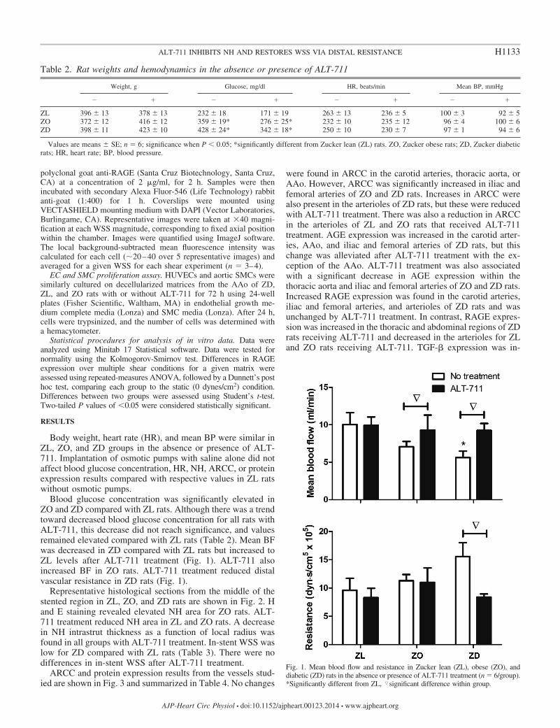
Blood glucose concentration was significantly elevated inZO and ZD compared with ZL rats. Although there was a trendtoward decreased blood glucose concentration for all rats withALT-711, this decrease did not reach significance, and valuesremained elevated compared with ZL rats (Table 2). Mean BFwas decreased in ZD compared with ZL rats but increased toZL levels after ALT-711 treatment (Fig. 1). ALT-711 alsoincreased BF in ZO rats. ALT-711 treatment reduced distalvascular resistance in ZD rats (Fig. 1).
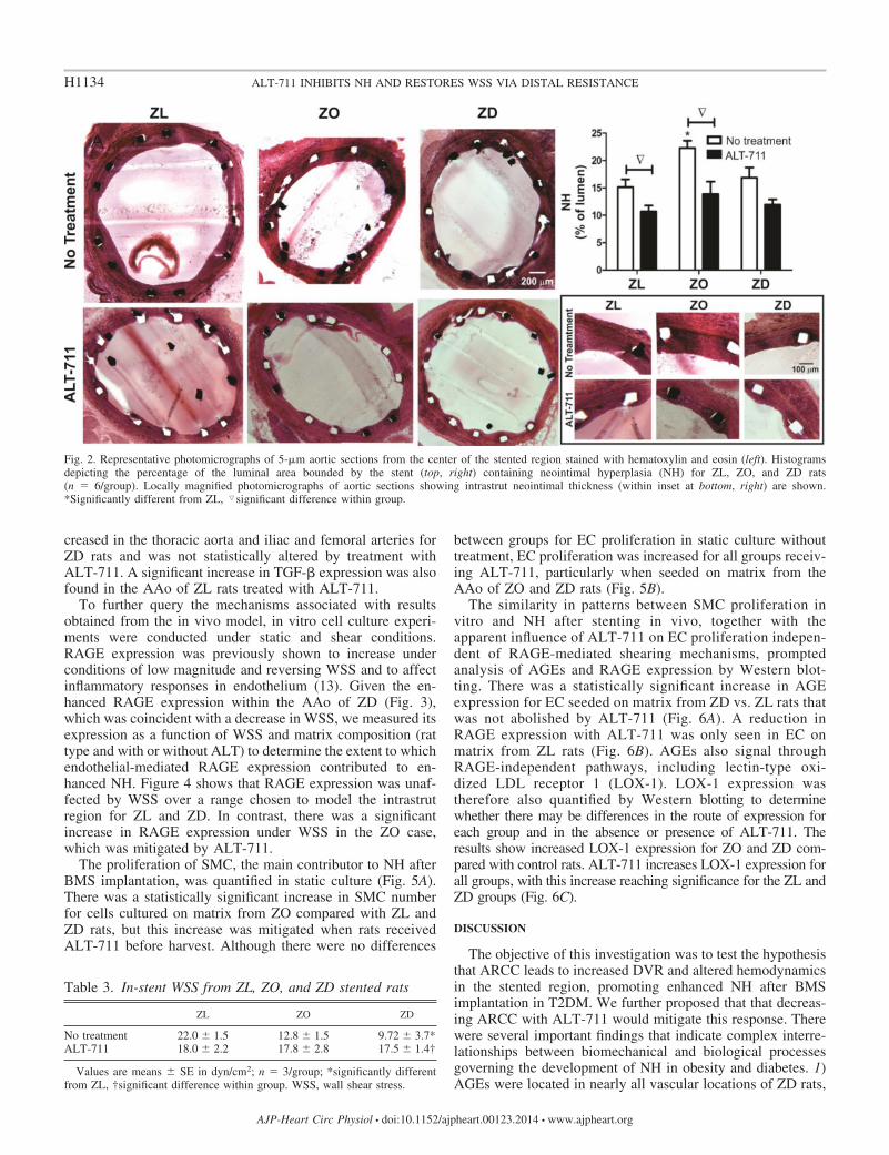
Representative histological sections from the middle of thestented region in ZL, ZO, and ZD rats are shown in Fig. 2. Hand E staining revealed elevated NH area for ZO rats. ALT-711 treatment reduced NH area in ZL and ZO rats. A decreasein NH intrastrut thickness as a function of local radius wasfound in all groups with ALT-711 treatment. In-stent WSS waslow for ZD compared with ZL rats (Table 3). There were nodifferences in in-stent WSS after ALT-711 treatment.
ARCC and protein expression results from the vessels stud-ied are shown in Fig. 3 and summarized in Table 4. No changes
were found in ARCC in the carotid arteries, thoracic aorta, orAAo. However, ARCC was significantly increased in iliac andfemoral arteries of ZO and ZD rats. Increases in ARCC werealso present in the arterioles of ZD rats, but these were reducedwith ALT-711 treatment. There was also a reduction in ARCCin the arterioles of ZL and ZO rats that received ALT-711treatment. AGE expression was increased in the carotid arter-ies, AAo, and iliac and femoral arteries of ZD rats, but thischange was alleviated after ALT-711 treatment with the ex-ception of the AAo. ALT-711 treatment was also associatedwith a significant decrease in AGE expression within thethoracic aorta and iliac and femoral arteries of ZO and ZD rats.Increased RAGE expression was found in the carotid arteries,iliac and femoral arteries, and arterioles of ZD rats and wasunchanged by ALT-711 treatment. In contrast, RAGE expres-sion was increased in the thoracic and abdominal regions of ZDrats receiving ALT-711 and decreased in the arterioles for ZLand ZO rats receiving ALT-711. TGF-� expression was in-
Table 2. Rat weights and hemodynamics in the absence or presence of ALT-711
Weight, g Glucose, mg/dl HR, beats/min Mean BP, mmHg
� � � �
ZL 396 � 13 378 � 13 232 � 18 171 � 19 263 � 13 236 � 5 100 � 3 92 � 5ZO 372 � 12 416 � 12 359 � 19* 276 � 25* 232 � 10 235 � 12 96 � 4 100 � 6ZD 398 � 11 423 � 10 428 � 24* 342 � 18* 250 � 10 230 � 7 97 � 1 94 � 6
Values are means � SE; n � 6; significance when P 0.05; *significantly different from Zucker lean (ZL) rats. ZO, Zucker obese rats; ZD, Zucker diabeticrats; HR, heart rate; BP, blood pressure.
Fig. 1. Mean blood flow and resistance in Zucker lean (ZL), obese (ZO), anddiabetic (ZD) rats in the absence or presence of ALT-711 treatment (n � 6/group).*Significantly different from ZL, �significant difference within group.
H1133ALT-711 INHIBITS NH AND RESTORES WSS VIA DISTAL RESISTANCE
AJP-Heart Circ Physiol • doi:10.1152/ajpheart.00123.2014 • www.ajpheart.org
creased in the thoracic aorta and iliac and femoral arteries forZD rats and was not statistically altered by treatment withALT-711. A significant increase in TGF-� expression was alsofound in the AAo of ZL rats treated with ALT-711.
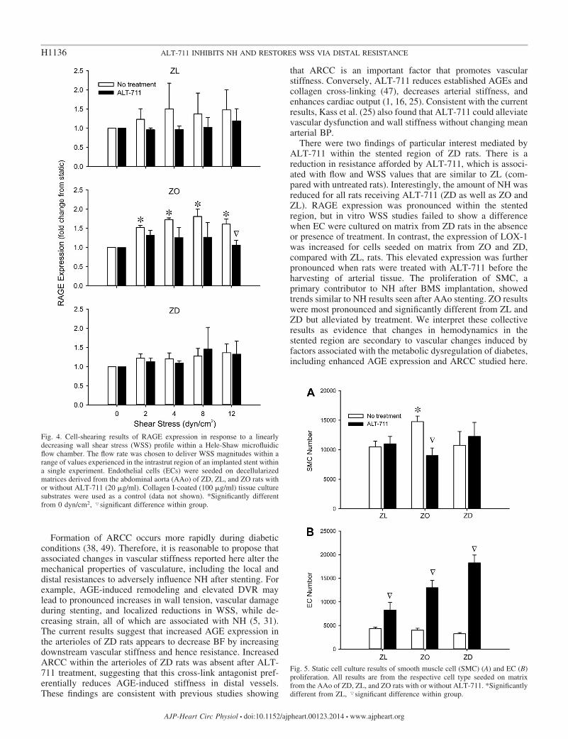
To further query the mechanisms associated with resultsobtained from the in vivo model, in vitro cell culture experi-ments were conducted under static and shear conditions.RAGE expression was previously shown to increase underconditions of low magnitude and reversing WSS and to affectinflammatory responses in endothelium (13). Given the en-hanced RAGE expression within the AAo of ZD (Fig. 3),which was coincident with a decrease in WSS, we measured itsexpression as a function of WSS and matrix composition (rattype and with or without ALT) to determine the extent to whichendothelial-mediated RAGE expression contributed to en-hanced NH. Figure 4 shows that RAGE expression was unaf-fected by WSS over a range chosen to model the intrastrutregion for ZL and ZD. In contrast, there was a significantincrease in RAGE expression under WSS in the ZO case,which was mitigated by ALT-711.
The proliferation of SMC, the main contributor to NH afterBMS implantation, was quantified in static culture (Fig. 5A).There was a statistically significant increase in SMC numberfor cells cultured on matrix from ZO compared with ZL andZD rats, but this increase was mitigated when rats receivedALT-711 before harvest. Although there were no differences
between groups for EC proliferation in static culture withouttreatment, EC proliferation was increased for all groups receiv-ing ALT-711, particularly when seeded on matrix from theAAo of ZO and ZD rats (Fig. 5B).
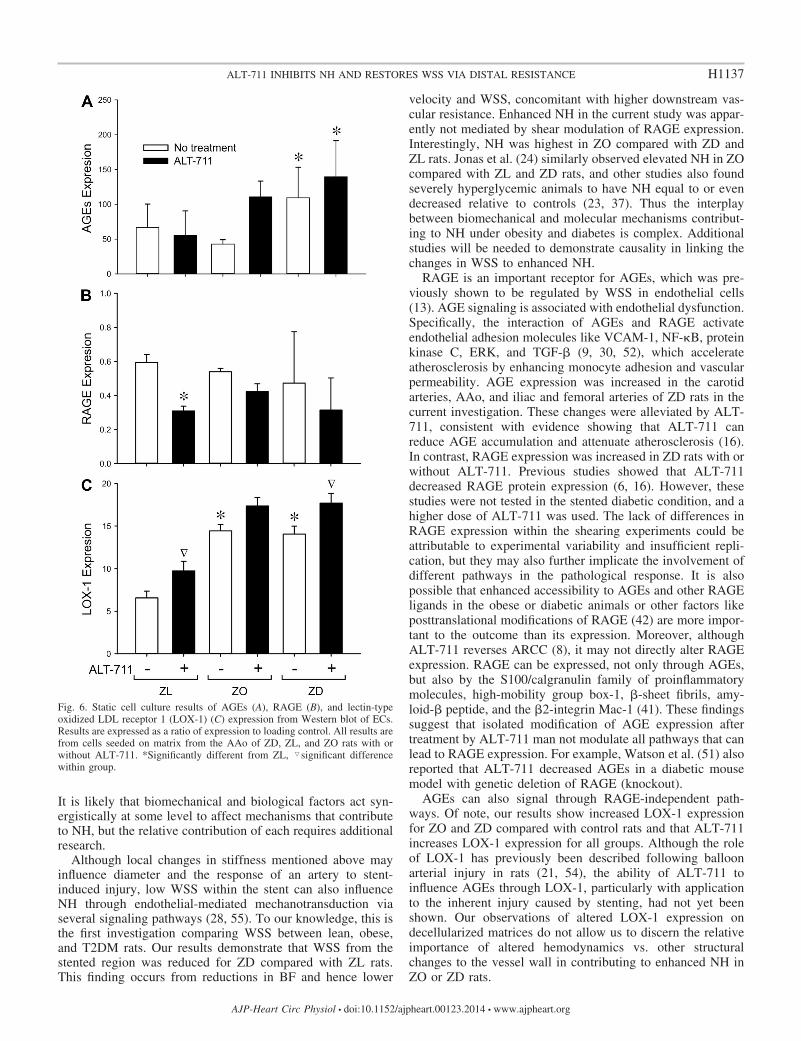
The similarity in patterns between SMC proliferation invitro and NH after stenting in vivo, together with theapparent influence of ALT-711 on EC proliferation indepen-dent of RAGE-mediated shearing mechanisms, promptedanalysis of AGEs and RAGE expression by Western blot-ting. There was a statistically significant increase in AGEexpression for EC seeded on matrix from ZD vs. ZL rats thatwas not abolished by ALT-711 (Fig. 6A). A reduction inRAGE expression with ALT-711 was only seen in EC onmatrix from ZL rats (Fig. 6B). AGEs also signal throughRAGE-independent pathways, including lectin-type oxi-dized LDL receptor 1 (LOX-1). LOX-1 expression wastherefore also quantified by Western blotting to determinewhether there may be differences in the route of expression foreach group and in the absence or presence of ALT-711. Theresults show increased LOX-1 expression for ZO and ZD com-pared with control rats. ALT-711 increases LOX-1 expression forall groups, with this increase reaching significance for the ZL andZD groups (Fig. 6C).
DISCUSSION
The objective of this investigation was to test the hypothesisthat ARCC leads to increased DVR and altered hemodynamicsin the stented region, promoting enhanced NH after BMSimplantation in T2DM. We further proposed that that decreas-ing ARCC with ALT-711 would mitigate this response. Therewere several important findings that indicate complex interre-lationships between biomechanical and biological processesgoverning the development of NH in obesity and diabetes. 1)AGEs were located in nearly all vascular locations of ZD rats,
Fig. 2. Representative photomicrographs of 5-�m aortic sections from the center of the stented region stained with hematoxylin and eosin (left). Histogramsdepicting the percentage of the luminal area bounded by the stent (top, right) containing neointimal hyperplasia (NH) for ZL, ZO, and ZD rats(n � 6/group). Locally magnified photomicrographs of aortic sections showing intrastrut neointimal thickness (within inset at bottom, right) are shown.*Significantly different from ZL, �significant difference within group.
Table 3. In-stent WSS from ZL, ZO, and ZD stented rats
ZL ZO ZD
No treatment 22.0 � 1.5 12.8 � 1.5 9.72 � 3.7*ALT-711 18.0 � 2.2 17.8 � 2.8 17.5 � 1.4†
Values are means � SE in dyn/cm2; n � 3/group; *significantly differentfrom ZL, †significant difference within group. WSS, wall shear stress.
H1134 ALT-711 INHIBITS NH AND RESTORES WSS VIA DISTAL RESISTANCE
AJP-Heart Circ Physiol • doi:10.1152/ajpheart.00123.2014 • www.ajpheart.org

confirming their elevated presence in T2DM; 2) increasingstiffness, as indicated by ARCC, was localized to the arteriolesof ZD rats but alleviated by treatment with ALT-711; 3) meanBF decreased in ZD rats, concomitantly with increases inarteriolar resistance; 4) WSS within the stented region was lowin untreated ZD rats but comparable to ZL with ALT-711
treatment; 5) NH within the stented region was increased in ZObut not ZD rats, but treatment reduced NH in all groups; 6)LOX-1, but not TGF-� or RAGE, expression was elevated byALT-711, illustrating that different AGE-mediated pathwaysmay mediate the local NH response. These findings are dis-cussed in more detail below.
Fig. 3. Advanced glycation end-product (AGE)-related collagen cross-linking as well as transforming growth factor-� (TGF-�) and receptor for AGE (RAGE)protein expression results from the vessels studied (n � 6/group). *Significantly different from ZL, #significantly different from ZO rats, �significant differencewithin group.
Table 4. Summary of AGE-related collagen cross-linking and protein expression for vessels in group of rats
Changes vs. ZL
Carotid Arteries Thoracic Aorta Abdominal AortaIliac and Femoral
Arteries Arterioles
ZO ZD ZO ZD ZO ZD ZO ZD ZO ZD
Collagen cross-linking NC NC NC NC NC NC 1 1 NC \1AGEs NC \1 NC NC NC 1 NC \1 NC NCRAGE NC 1 NC NC NC NC NC 1 NC 1TGF-� NC NC NC 1 NC NC NC 1 NC NC
N � 6; NC, no change; 1, significant increase; \, increase alleviated by ALT-711.
H1135ALT-711 INHIBITS NH AND RESTORES WSS VIA DISTAL RESISTANCE
AJP-Heart Circ Physiol • doi:10.1152/ajpheart.00123.2014 • www.ajpheart.org
Formation of ARCC occurs more rapidly during diabeticconditions (38, 49). Therefore, it is reasonable to propose thatassociated changes in vascular stiffness reported here alter themechanical properties of vasculature, including the local anddistal resistances to adversely influence NH after stenting. Forexample, AGE-induced remodeling and elevated DVR maylead to pronounced increases in wall tension, vascular damageduring stenting, and localized reductions in WSS, while de-creasing strain, all of which are associated with NH (5, 31).The current results suggest that increased AGE expression inthe arterioles of ZD rats appears to decrease BF by increasingdownstream vascular stiffness and hence resistance. IncreasedARCC within the arterioles of ZD rats was absent after ALT-711 treatment, suggesting that this cross-link antagonist pref-erentially reduces AGE-induced stiffness in distal vessels.These findings are consistent with previous studies showing
that ARCC is an important factor that promotes vascularstiffness. Conversely, ALT-711 reduces established AGEs andcollagen cross-linking (47), decreases arterial stiffness, andenhances cardiac output (1, 16, 25). Consistent with the currentresults, Kass et al. (25) also found that ALT-711 could alleviatevascular dysfunction and wall stiffness without changing meanarterial BP.
There were two findings of particular interest mediated byALT-711 within the stented region of ZD rats. There is areduction in resistance afforded by ALT-711, which is associ-ated with flow and WSS values that are similar to ZL (com-pared with untreated rats). Interestingly, the amount of NH wasreduced for all rats receiving ALT-711 (ZD as well as ZO andZL). RAGE expression was pronounced within the stentedregion, but in vitro WSS studies failed to show a differencewhen EC were cultured on matrix from ZD rats in the absenceor presence of treatment. In contrast, the expression of LOX-1was increased for cells seeded on matrix from ZO and ZD,compared with ZL, rats. This elevated expression was furtherpronounced when rats were treated with ALT-711 before theharvesting of arterial tissue. The proliferation of SMC, aprimary contributor to NH after BMS implantation, showedtrends similar to NH results seen after AAo stenting. ZO resultswere most pronounced and significantly different from ZL andZD but alleviated by treatment. We interpret these collectiveresults as evidence that changes in hemodynamics in thestented region are secondary to vascular changes induced byfactors associated with the metabolic dysregulation of diabetes,including enhanced AGE expression and ARCC studied here.
Fig. 5. Static cell culture results of smooth muscle cell (SMC) (A) and EC (B)proliferation. All results are from the respective cell type seeded on matrixfrom the AAo of ZD, ZL, and ZO rats with or without ALT-711. *Significantlydifferent from ZL, �significant difference within group.
Fig. 4. Cell-shearing results of RAGE expression in response to a linearlydecreasing wall shear stress (WSS) profile within a Hele-Shaw microfluidicflow chamber. The flow rate was chosen to deliver WSS magnitudes within arange of values experienced in the intrastrut region of an implanted stent withina single experiment. Endothelial cells (ECs) were seeded on decellularizedmatrices derived from the abdominal aorta (AAo) of ZD, ZL, and ZO rats withor without ALT-711 (20 �g/ml). Collagen I-coated (100 �g/ml) tissue culturesubstrates were used as a control (data not shown). *Significantly differentfrom 0 dyn/cm2, �significant difference within group.
H1136 ALT-711 INHIBITS NH AND RESTORES WSS VIA DISTAL RESISTANCE
AJP-Heart Circ Physiol • doi:10.1152/ajpheart.00123.2014 • www.ajpheart.org
It is likely that biomechanical and biological factors act syn-ergistically at some level to affect mechanisms that contributeto NH, but the relative contribution of each requires additionalresearch.
Although local changes in stiffness mentioned above mayinfluence diameter and the response of an artery to stent-induced injury, low WSS within the stent can also influenceNH through endothelial-mediated mechanotransduction viaseveral signaling pathways (28, 55). To our knowledge, this isthe first investigation comparing WSS between lean, obese,and T2DM rats. Our results demonstrate that WSS from thestented region was reduced for ZD compared with ZL rats.This finding occurs from reductions in BF and hence lower
velocity and WSS, concomitant with higher downstream vas-cular resistance. Enhanced NH in the current study was appar-ently not mediated by shear modulation of RAGE expression.Interestingly, NH was highest in ZO compared with ZD andZL rats. Jonas et al. (24) similarly observed elevated NH in ZOcompared with ZL and ZD rats, and other studies also foundseverely hyperglycemic animals to have NH equal to or evendecreased relative to controls (23, 37). Thus the interplaybetween biomechanical and molecular mechanisms contribut-ing to NH under obesity and diabetes is complex. Additionalstudies will be needed to demonstrate causality in linking thechanges in WSS to enhanced NH.
RAGE is an important receptor for AGEs, which was pre-viously shown to be regulated by WSS in endothelial cells(13). AGE signaling is associated with endothelial dysfunction.Specifically, the interaction of AGEs and RAGE activateendothelial adhesion molecules like VCAM-1, NF- B, proteinkinase C, ERK, and TGF-� (9, 30, 52), which accelerateatherosclerosis by enhancing monocyte adhesion and vascularpermeability. AGE expression was increased in the carotidarteries, AAo, and iliac and femoral arteries of ZD rats in thecurrent investigation. These changes were alleviated by ALT-711, consistent with evidence showing that ALT-711 canreduce AGE accumulation and attenuate atherosclerosis (16).In contrast, RAGE expression was increased in ZD rats with orwithout ALT-711. Previous studies showed that ALT-711decreased RAGE protein expression (6, 16). However, thesestudies were not tested in the stented diabetic condition, and ahigher dose of ALT-711 was used. The lack of differences inRAGE expression within the shearing experiments could beattributable to experimental variability and insufficient repli-cation, but they may also further implicate the involvement ofdifferent pathways in the pathological response. It is alsopossible that enhanced accessibility to AGEs and other RAGEligands in the obese or diabetic animals or other factors likeposttranslational modifications of RAGE (42) are more impor-tant to the outcome than its expression. Moreover, althoughALT-711 reverses ARCC (8), it may not directly alter RAGEexpression. RAGE can be expressed, not only through AGEs,but also by the S100/calgranulin family of proinflammatorymolecules, high-mobility group box-1, �-sheet fibrils, amy-loid-� peptide, and the �2-integrin Mac-1 (41). These findingssuggest that isolated modification of AGE expression aftertreatment by ALT-711 man not modulate all pathways that canlead to RAGE expression. For example, Watson et al. (51) alsoreported that ALT-711 decreased AGEs in a diabetic mousemodel with genetic deletion of RAGE (knockout).
AGEs can also signal through RAGE-independent path-ways. Of note, our results show increased LOX-1 expressionfor ZO and ZD compared with control rats and that ALT-711increases LOX-1 expression for all groups. Although the roleof LOX-1 has previously been described following balloonarterial injury in rats (21, 54), the ability of ALT-711 toinfluence AGEs through LOX-1, particularly with applicationto the inherent injury caused by stenting, had not yet beenshown. Our observations of altered LOX-1 expression ondecellularized matrices do not allow us to discern the relativeimportance of altered hemodynamics vs. other structuralchanges to the vessel wall in contributing to enhanced NH inZO or ZD rats.
Fig. 6. Static cell culture results of AGEs (A), RAGE (B), and lectin-typeoxidized LDL receptor 1 (LOX-1) (C) expression from Western blot of ECs.Results are expressed as a ratio of expression to loading control. All results arefrom cells seeded on matrix from the AAo of ZD, ZL, and ZO rats with orwithout ALT-711. *Significantly different from ZL, �significant differencewithin group.
H1137ALT-711 INHIBITS NH AND RESTORES WSS VIA DISTAL RESISTANCE
AJP-Heart Circ Physiol • doi:10.1152/ajpheart.00123.2014 • www.ajpheart.org
We found that TGF-� expression was not influenced bytreatment with ALT-711, which is inconsistent with previousstudies showing increased TGF-� protein expression in regionsadjacent to stent struts as early as 5 days after stenting (34, 50).TGF-� promotes extracellular matrix production and cellularproliferation as evidenced by enhanced NH when the TGF-�gene was transferred into normal porcine arteries (33). Ourresults pointing to the importance of extracellular matrix in theNH response may have differed from this prior investigationbecause we sampled tissue adjacent but not within the stentedregion. The stented region of rats in the current investigationwas used for WSS calculation or quantification of NH.
The current static cell culture results indicate EC prolifera-tion increases with ALT-711, particularly in the ZO and ZDcases. These results suggest that, in addition to the ability toprevent ARCC, ALT-711 may have a beneficial effect onre-endothelialization, which could have important ramifica-tions for reducing the likelihood of late stent thrombosis. Thefunctionality of these ECs and whether ALT-711 has a similarimpact in the setting of the various drug-eluting stent agents(e.g., everolimus, paclitaxel, and zotarolimus) remain to bedetermined in future experiments conducted through follow-upstudies.
The current results should be interpreted relative to themethods applied. BP was measured using a fluid-filled catheterplaced in the carotid artery, but the flow transducer waspositioned just above the stented region for measurement offlow into the stented region. There is a distance between thesetwo measurement locations, so the relationship between BP,flow, and resistance calculated may not account for the portionof flow distributed to the liver and organs in the abdomen. Thealternative approach of obtaining all measurements in the sameposition would require a fluid-filled needle positioned abovethe stented region for BP measurement, which is technicallychallenging and would add to potential complications for thecurrent experimental protocol. In addition, the stent was de-ployed below the renal vascular region so as to not disturb thekidneys, which can also influence flow and resulting BP.Differences in body weight would be expected for ZO and ZDvs. ZL rats. The age of rats used here might have precluded thisoccurrence, which is expected to result in inflammation andlikely further exacerbate the neointimal response after stenting.
In summary, the results demonstrated that the cross-linking antagonist, ALT-711, decreased ARCC and arterio-lar stiffness in obese and diabetic rats after stent implanta-tion. ALT-711 decreased DVR, increased local BF, and ledto consistent values of WSS within the stented region,suggesting that AGE-induced vascular remodeling can alterin-stent hemodynamics. This is predicted to play an impor-tant role in NH formation although the underlying mecha-nisms require further study, particularly in the context of thecomplex milieu of diabetes. The finding that ALT-711treatment reduced NH in lean, obese, and diabetic ratsthrough RAGE-independent pathways, while simultane-ously promoting endothelial proliferation, suggests that thisagent may be effective to decrease stent restenosis regard-less of patient glycemic status.
ACKNOWLEDGMENTS
The authors thank John Tessmer and David Schwabe (Department ofAnesthesiology, Medical College of Wisconsin) for technical assistance with
the experimental protocol, as well as Thomas J. Eddinger, PhD (Department ofBiological Sciences, Marquette University) and Laura M. Ellwein, PhD (De-partment of Mathematics and Applied Mathematics, Virginia CommonwealthUniversity) for scientific contributions during the early stages of this investi-gation.
GRANTS
This work was supported by a Junior Faculty Award from the AmericanDiabetes Foundation (7-08-JF-25 to J. LaDisa).
DISCLOSURES
No conflicts of interest, financial or otherwise, are declared by the authors.
AUTHOR CONTRIBUTIONS
Author contributions: H.W., D.W., J.R.K., A.G.P., and J.F.L. conceptionand design of research; H.W., D.W., A.G.P., A.R., and J.F.L. performedexperiments; H.W., D.W., J.R.K., J.M.T., A.G.P., A.R., S.S., and J.F.L.analyzed data; H.W., D.W., J.R.K., J.M.T., A.G.P., S.S., and J.F.L. interpretedresults of experiments; H.W., D.W., J.M.T., A.G.P., and J.F.L. preparedfigures; H.W., J.R.K., and J.F.L. drafted manuscript; H.W., D.W., J.R.K.,J.M.T., A.G.P., S.S., and J.F.L. edited and revised manuscript; H.W., D.W.,J.R.K., J.M.T., A.G.P., A.R., S.S., and J.F.L. approved final version ofmanuscript.
REFERENCES
1. Asif M, Egan J, Vasan S, Jyothirmayi GN, Masurekar MR, Lopez S,Williams C, Torres RL, Wagle D, Ulrich P, Cerami A, Brines M,Regan TJ. An advanced glycation endproduct cross-link breaker canreverse age-related increases in myocardial stiffness. Proc Natl Acad SciUSA 97: 2809–2813, 2000.
2. Bakris GL, Bank AJ, Kass DA, Neutel JM, Preston RA, Oparil S.Advanced glycation end-product cross-link breakers. A novel approach tocardiovascular pathologies related to the aging process. Am J Hypertens17: 23S–30S, 2004.
3. Berry C, Tardif JC, Bourassa MG. Coronary heart disease in patientswith diabetes. II: Recent advances in coronary revascularization. J AmColl Cardiol 49: 643–656, 2007.
4. Bradford MM. A rapid and sensitive method for the quantitation ofmicrogram quantities of protein utilizing the principle of protein-dyebinding. Anal Biochem 72: 248–254, 1976.
5. Brownlee M. Advanced protein glycosylation in diabetes and aging. AnnuRev Med 46: 223–234, 1995.
6. Candido R, Forbes JM, Thomas MC, Thallas V, Dean RG, Burns WC,Tikellis C, Ritchie RH, Twigg SM, Cooper ME, Burrell LM. A breakerof advanced glycation end products attenuates diabetes-induced myocar-dial structural changes. Circ Res 92: 785–792, 2003.
7. Center for Disease Control and Prevention. National Diabetes FactSheet: General Information and National Estimates on Diabetes in theUnited States. Atlanta, GA: National Center for Chronic Disease Preven-tion and Health Promotion, 2011.
8. Cooper ME. Importance of advanced glycation end products in diabetes-associated cardiovascular and renal disease. Am J Hypertens 17: 31S–38S,2004.
9. Cortizo AM, Lettieri MG, Barrio DA, Mercer N, Etcheverry SB,McCarthy AD. Advanced glycation end-products (AGEs) induce con-certed changes in the osteoblastic expression of their receptor RAGE andin the activation of extracellular signal-regulated kinases (ERK). Mol CellBiochem 250: 1–10, 2003.
10. Coughlan MT, Forbes JM, Cooper ME. Role of the AGE crosslinkbreaker, alagebrium, as a renoprotective agent in diabetes. Kidney IntSuppl 106: S54–S60, 2007.
11. De la Torre Hernandez JM, Alfonso F, Sanchez Recalde A, JimenezNavarro MF, Perez de Prado A, Hernandez F, Abdul-Jawad AltisentO, Roura G, Garcia Camarero T, Elizaga J, Rivero F, Gimeno F,Calviño R, Moreu J, Bosa F, Rumoroso JR, Bullones JA, Gallardo A,Fernandez Diaz JA, Ruiz Arroyo JR, Aragon V, Masotti M; ES-TROFA-LM Study Group. Comparison of paclitaxel-eluting stents(Taxus) and everolimus-eluting stents (Xience) in left main coronaryartery disease with 3 years follow-up (from the ESTROFA-LM registry).Am J Cardiol 111: 676–683, 2013.
H1138 ALT-711 INHIBITS NH AND RESTORES WSS VIA DISTAL RESISTANCE
AJP-Heart Circ Physiol • doi:10.1152/ajpheart.00123.2014 • www.ajpheart.org

12. Deuse T, Ikeno F, Robbins RC, Schrepfer S. Imaging in-stent resteno-sis: an inexpensive, reliable, and rapid preclinical model. J Vis Exp 14:1346, 2009.
13. DeVerse JS, Bailey KA, Jackson KN, Passerini AG. Shear stressmodulates RAGE-mediated inflammation in a model of diabetes-inducedmetabolic stress. Am J Physiol Heart Circ Physiol 302: H2498–H2508,2012.
14. DeVerse JS, Sandhu AS, Mendoza N, Edwards CM, Sun C, SimonSI, Passerini AG. Shear stress modulates VCAM-1 expression inresponse to TNF-� and dietary lipids via interferon regulatory factor-1in cultured endothelium. Am J Physiol Heart Circ Physiol 305:H1149 –H1157, 2013.
15. Dudek D, Legutko J, Heba G, Bartus S, Partyka L, Huk I, Dembin-ska-Kiec A, Kaluza GL, Dubiel JS. L-Arginine supplementation does notinhibit neointimal formation after coronary stenting in human beings: anintravascular ultrasound study. Am Heart J 147: E12, 2004.
16. Forbes JM, Yee LT, Thallas V, Lassila M, Candido R, Jandeleit-Dahm KA, Thomas MC, Burns WC, Deemer EK, Thorpe SR, CooperME, Allen TJ. Advanced glycation end product interventions reducediabetes-accelerated atherosclerosis. Diabetes 53: 1813–1823, 2004.
17. Garasic JM, Edelman ER, Squire JC, Seifert P, Williams MS, RogersC. Stent and artery geometry determine intimal thickening independent ofarterial injury. Circulation 101: 812–818, 2000.
18. Gilbert J, Raboud J, Zinman B. Meta-analysis of the effect of diabeteson restenosis rates among patients receiving coronary angioplasty stenting.Diabetes Care 27: 990–994, 2004.
19. Goldfine AB, Beckman JA. Life and death in Denmark: lessons aboutdiabetes and coronary heart disease. Circulation 117: 1914–1917, 2008.
20. Hakim DA, Mintz GS, Sanidas E, Rusinova R, Weisz G, Leon MB,Moses JW, Stone GW, Maehara A. Serial gray scale intravascularultrasound findings in late drug-eluting stent restenosis. Am J Cardiol 111:695–699, 2013.
21. Hinagata J, Kakutani M, Fujii T, Naruko T, Inoue N, Fujita Y, MehtaJL, Ueda M, Sawamura T. Oxidized LDL receptor LOX-1 is involved inneointimal hyperplasia after balloon arterial injury in a rat model. Car-diovasc Res 69: 263–271, 2006.
22. Hioki H, Kumazaki S, Izawa A. Critical in-stent restenosis followingfracture of biolimus-eluting stent: a report of 2 cases. J Invasive Cardiol25: E11–E13, 2013.
23. Indolfi C, Torella D, Cavuto L, Davalli AM, Coppola C, Esposito G,Carriero MV, Rapacciuolo A, Di Lorenzo E, Stabile E, Perrino C,Chieffo A, Pardo F, Chiariello M. Effects of balloon injury on neointi-mal hyperplasia in streptozotocin-induced diabetes and in hyperinsuline-mic nondiabetic pancreatic islet-transplanted rats. Circulation 103: 2980–2986, 2001.
24. Jonas M, Edelman ER, Groothuis A, Baker AB, Seifert P, Rogers C.Vascular neointimal formation and signaling pathway activation in re-sponse to stent injury in insulin-resistant and diabetic animals. Circ Res97: 725–733, 2005.
25. Kass DA, Shapiro EP, Kawaguchi M, Capriotti AR, Scuteri A, de-Groof RC, Lakatta EG. Improved arterial compliance by a novel ad-vanced glycation end-product crosslink breaker. Circulation 104: 1464–1470, 2001.
26. Khan W, Farah S, Domb AJ. Drug eluting stents: developments andcurrent status. J Control Release 161: 703–712, 2012.
27. Kutryk MJ, Foley DP, van den Brand M, Hamburger JN, van derGiessen WJ, deFeyter PJ, Bruining N, Sabate M, Serruys PW. Localintracoronary administration of antisense oligonucleotide against c-mycfor the prevention of in-stent restenosis: results of the randomized inves-tigation by the Thoraxcenter of antisense DNA using local delivery andIVUS after coronary stenting (ITALICS) trial. J Am Coll Cardiol 39:281–287, 2002.
28. LaDisa JF Jr, Olson LE, Hettrick DA, Warltier DC, Kersten JR,Pagel PS. Axial stent strut angle influences wall shear stress after stentimplantation: analysis using 3D computational fluid dynamics models ofstent foreshortening. Biomed Eng Online 4: 59, 2005.
29. LaDisa JF Jr, Olson LE, Molthen RC, Hettrick DA, Pratt PF,Hardel MD, Kersten JR, Warltier DC, Pagel PS. Alterations in wallshear stress predict sites of neointimal hyperplasia after stent implan-tation in rabbit iliac arteries. Am J Physiol Heart Circ Physiol 288:H2465–H2475, 2005.
30. Meerwaldt R, Links T, Zeebregts C, Tio R, Hillebrands JL, Smit A.The clinical relevance of assessing advanced glycation endproducts accu-mulation in diabetes. Cardiovasc Diabetol 7: 29, 2008.
31. Morrison TM, Choi G, Zarins CK, Taylor CA. Circumferential andlongitudinal cyclic strain of the human thoracic aorta: age-related changes.J Vasc Surg 49: 1029–1036, 2009.
32. Moses JW, Leon MB, Popma JJ, Fitzgerald PJ, Holmes DR,O’Shaughnessy C, Caputo RP, Kereiakes DJ, Williams DO, TeirsteinPS, Jaeger JL, Kuntz RE. Sirolimus-eluting stents versus standard stentsin patients with stenosis in a native coronary artery. N Engl J Med 349:1315–1323, 2003.
33. Nabel EG, Shum L, Pompili VJ, Yang ZY, San H, Shu HB, Liptay S,Gold L, Gordon D, Derynck R, Nabel GJ. Direct transfer of transform-ing growth factor beta 1 gene into arteries stimulates fibrocellular hyper-plasia. Proc Natl Acad Sci USA 90: 10759–10763, 1993.
34. Nikol S, Isner JM, Pickering JG, Kearney M, Leclerc G, Weir L.Expression of transforming growth factor-beta 1 is increased in humanvascular restenosis lesions. J Clin Invest 90: 1582–1592, 1992.
35. Noh H, King GL. The role of protein kinase C activation in diabeticnephropathy. Kidney Int Suppl S49–S53, 2007.
36. Oyamada S, Ma X, Wu T, Robich MP, Wu H, Wang X, Buchholz B,McCarthy S, Bianchi CF, Sellke FW, Laham R. Trans-iliac rat aortastenting: a novel high throughput preclinical stent model for restenosis andthrombosis. J Surg Res 166: e91–e95, 2011.
37. Park SH, Marso SP, Zhou Z, Foroudi F, Topol EJ, Lincoff AM.Neointimal hyperplasia after arterial injury is increased in a rat modelof non-insulin-dependent diabetes mellitus. Circulation 104: 815–819,2001.
38. Peppa M, Uribarri J, Vlassara H. The role of advanced glycation endproducts in the development of atherosclerosis. Curr Diab Rep 4: 31–36,2004.
39. Peppa M, Vlassara H. Advanced glycation end products and diabeticcomplications: a general overview. Hormones (Athens) 4: 28–37, 2005.
40. Ryan J, Cohen DJ. Are drug-eluting stents cost-effective? It depends onwhom you ask. Circulation 114: 1736–1743, 2006.
41. Schmidt AM, Yan SD, Yan SF, Stern DM. The biology of the receptorfor advanced glycation end products and its ligands. Biochim Biophys Acta1498: 99–111, 2000.
42. Srikrishna G, Huttunen HJ, Johansson L, Weigle B, Yamaguchi Y,Rauvala H, Freeze HH. N-glycans on the receptor for advanced glycationend products influence amphoterin binding and neurite outgrowth. JNeurochem 80: 998–1008, 2002.
43. Stefek M, Gajdosik A, Gajdosikova A, Krizanova L. p-Dimethylami-nobenzaldehyde-reactive substances in tail tendon collagen of streptozo-tocin-diabetic rats: temporal relation to biomechanical properties andadvanced glycation endproduct (AGE)-related fluorescence. Biochim Bio-phys Acta 1502: 398–404, 2000.
44. Stone GW, Kedhi E, Kereiakes DJ, Parise H, Fahy M, Serruys PW,Smits PC. Differential clinical responses to everolimus-eluting and Pacli-taxel-eluting coronary stents in patients with and without diabetes melli-tus. Circulation 124: 893–900, 2011.
45. Tas¸kiran DT, Tas¸kiran E, Yercan H, Kutay FZ. Quantificationtotal collagen in rabbit tendon by the Sirius red. Tr J Med Sci 29: 7–9,1999.
46. Tsou JK, Gower RM, Ting HJ, Schaff UY, Insana MF, Passerini AG,Simon SI. Spatial regulation of inflammation by human aortic endothelialcells in a linear gradient of shear stress. Microcirculation 15: 311–323, 2008.
47. Vasan S, Zhang X, Kapurniotu A, Bernhagen J, Teichberg S, BasgenJ, Wagle D, Shih D, Terlecky I, Bucala R, Cerami A, Egan J, UlrichP. An agent cleaving glucose-derived protein crosslinks in vitro and invivo. Nature 382: 275–278, 1996.
48. Vladic N, Ge ZD, Leucker T, Brzezinska AK, Du JH, Shi Y, WarltierDC, Pratt PF Jr, Kersten JR. Decreased tetrahydrobiopterin and dis-rupted association of Hsp90 with eNOS by hyperglycemia impair myo-cardial ischemic preconditioning. Am J Physiol Heart Circ Physiol 301:H2130–H2139, 2011.
49. Vlassara H, Palace MR. Diabetes and advanced glycation endproducts. JIntern Med 251: 87–101, 2002.
50. Ward MR, Agrotis A, Kanellakis P, Hall J, Jennings G, Bobik A.Tranilast prevents activation of transforming growth factor-beta system,leukocyte accumulation, and neointimal growth in porcine coronary arter-ies after stenting. Arterioscler Thromb Vasc Biol 22: 940–948, 2002.
51. Watson AM, Gray SP, Jiaze L, Soro-Paavonen A, Wong B, CooperME, Bierhaus A, Pickering R, Tikellis C, Tsorotes D, Thomas MC,Jandeleit-Dahm KA. Alagebrium reduces glomerular fibrogenesis andinflammation beyond preventing RAGE activation in diabetic apolipopro-tein E knockout mice. Diabetes 61: 2105–2113, 2012.
H1139ALT-711 INHIBITS NH AND RESTORES WSS VIA DISTAL RESISTANCE
AJP-Heart Circ Physiol • doi:10.1152/ajpheart.00123.2014 • www.ajpheart.org

52. Wendt T, Tanji N, Guo J, Hudson BI, Bierhaus A, Ramasamy R,Arnold B, Nawroth PP, Yan SF, D’Agati V, Schmidt AM. Glucose,glycation, and RAGE: implications for amplification of cellular dysfunc-tion in diabetic nephropathy. J Am Soc Nephrol 14: 1383–1395, 2003.
53. Windberger U, Bartholovitsch A, Plasenzotti R, Korak KJ, Heinze G.Whole blood viscosity, plasma viscosity and erythrocyte aggregation innine mammalian species: reference values and comparison of data. ExpPhysiol 88: 431–440, 2003.
54. Yao EH, Fukuda N, Ueno T, Matsuda H, Matsumoto K, Nagase H,Matsumoto Y, Takasaka A, Serie K, Sugiyama H, Sawamura T. Novelgene silencer pyrrole-imidazole polyamide targeting lectin-like oxidizedlow-density lipoprotein receptor-1 attenuates restenosis of the artery afterinjury. Hypertension 52: 86–92, 2008.
55. Yazdani SK, Nakano M, Otsuka F, Kolodgie FD, Virmani R. Ather-oma and coronary bifurcations: before and after stenting. EuroIntervention6, Suppl J: J24–J30, 2010.
H1140 ALT-711 INHIBITS NH AND RESTORES WSS VIA DISTAL RESISTANCE
AJP-Heart Circ Physiol • doi:10.1152/ajpheart.00123.2014 • www.ajpheart.org


