Afternoon Session Cases - BC Cancer€¢ ERCP demonstrated distal CBD obstruction and stent placed...
32
Afternoon Session Cases
Transcript of Afternoon Session Cases - BC Cancer€¢ ERCP demonstrated distal CBD obstruction and stent placed...

Afternoon Session Cases
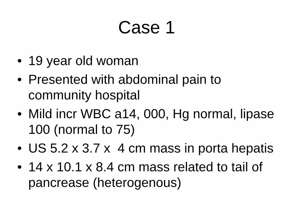
Case 1
• 19 year old woman• Presented with abdominal pain to
community hospital• Mild incr WBC a14, 000, Hg normal, lipase
100 (normal to 75)• US 5.2 x 3.7 x 4 cm mass in porta hepatis• 14 x 10.1 x 8.4 cm mass related to tail of
pancrease (heterogenous)

Case 1
• CT scan• 12 x 9 x 16 cm retrogastric mass related to
tail of pancreas• Heterogenous, with possible punctate
calcifications• Lesion at porta 3.6 x 4.1 may be lymph
node• Free fluid in pelvis and pericholecystic
fluid, possible mass in cul –de -sac
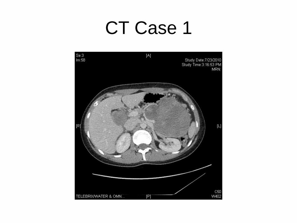
CT Case 1
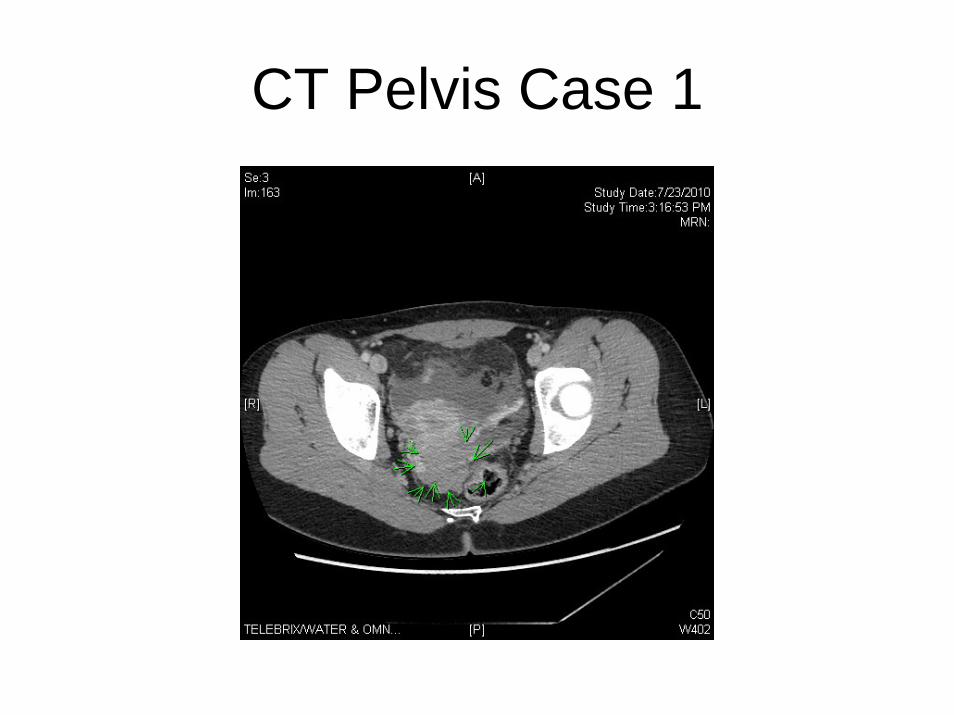
CT Pelvis Case 1
• Pelvic US• 3.8 x 1.5 x 3.4 cm mass consistent with
drop metastases
• CEA and CA 19-9 are normal
• What further investigations?

Case 1
• Percutaneous core biopsy attempted• Non diagnostic• Repeat biopsy- solid pseudopapillary
tumour of the pancreas• Recommendations?
Case 1
• Taken to OR• Distal pancreatectomy, cholecystectomy,
splenectomy, resection/biopsy of peritoneal implants
• Any role for chemotherapy?

• Solid pseudopapillary tumor, otherwise known as solid and cystic tumor or Frantz tumor, is an unusual form of pancreatic carcinoma. Its natural history differs from the more common pancreatic adenocarcinoma in that it has a female predilection, is more indolent, and carries a better prognosis. Metastatic disease can occur, usually involving the liver, and its management is not well defined.
HPB (Oxford). 2003; 5(4): 264–267.
Case 2
• Incidental finding on CT scan of pancreatic cystic lesion
• No preceding history of pancreatitis or abdominal pain
• Normal LFTs, amylase and lipase
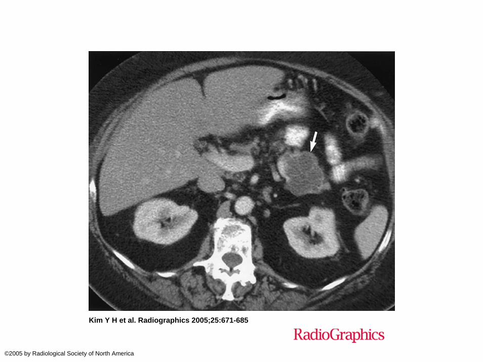
Kim Y H et al. Radiographics 2005;25:671-685
©2005 by Radiological Society of North America
Case 2
• What is the differential diagnosis?• How would you investigate?• Is there a role for percutaneous biopsy?• Is there a role for EUS and biopsy?• How would you manage?
Case 2
• Another pancreatic cystic lesion:
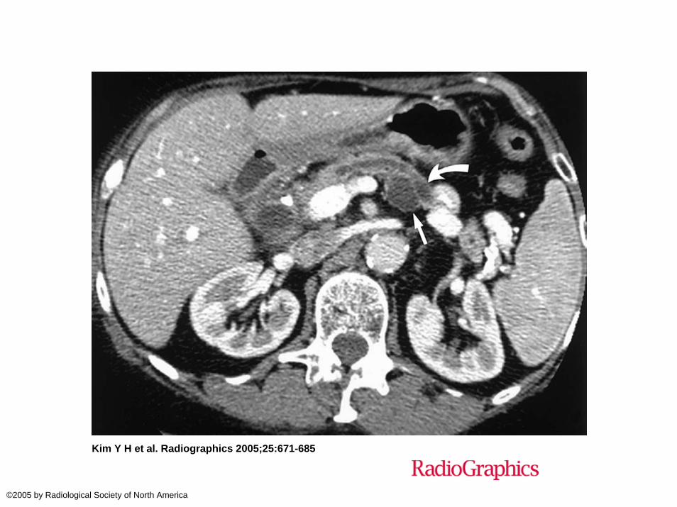
Kim Y H et al. Radiographics 2005;25:671-685
©2005 by Radiological Society of North America
Case 2
• How does this imaging differ?• Would you recommend any different
investigations?
Case 3
• 65 year old otherwise healthy man• US done for investigation of vague
epigastric pain associated with abnormal LFTs
• Mass identified in head of pancreas• ERCP demonstrated distal CBD
obstruction and stent placed• CXR normal• CT scan arranged
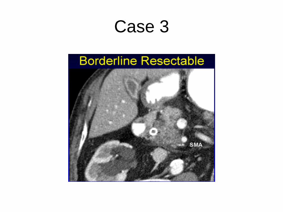
Case 3
Case 3
• What is the appropriate next step?• Is there a role for neoadjuvant therapy or
should attempted surgical resection be undertaken?
• Are there any official guidelines for this type of problem?

Case 3
• NCCN guidelines in Oncology (NCCN.org)• Borderline Resectable Pancreatic
Adenocarcinoma recommendations are for neoadjuvant chemotherapy with repeat imaging to reassess for resectability– Selects those with favorable biology– Enhance the chance of a complete (R0,
R1)resection – Treats occults M1 disease
•••
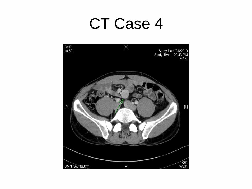
Case 4
• 53 year old man with anorexia and fatigue• Abnormal liver enzymes
• Bilirubin 63• Alk phos 378• GGT 481• ALT 359 • AST 98
Case 4
• US 2.5cm mass in right lobe liver, 2 lesionas 8mm and 11mm in left lobe of liver
• ERCP shows stenotic ampulla but otherwise normal and LFTs normalize
Case 4
• CT scan (triphasic)• Liver lesions: 2.5 cm subcapsular in
segment V and 10 mm lesion in segment II (adj to middle hepatic vein)
• Hypervascular on arterial phase and wash out on portal phase
• 5.1 cm mesenteric mass in distal SB with multiple enhancing nodes
• Hypervascularity of distal ileum (subtle)
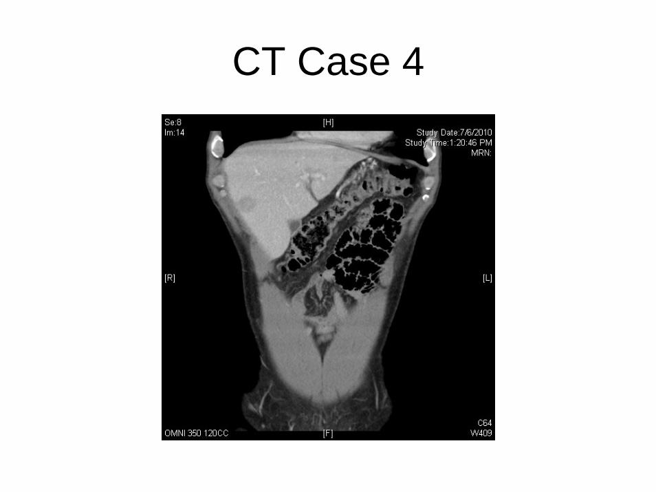
CT Case 4
CT Case 4
Case 4
• Radiologist recommendations• Percutaneous biopsy• Octreotide scan• Further investigations?
Case 4
• Octreotide scan positive• 24 hour urine 5HIAA- normal• Serum chromogranin A - normal• Further history- no episodes of flushing,
diarrhea or bronchospasm, no evidence of right sided heart failure
Case 4
• Recommendations?• What if the liver disease was non-
resectable?• What if the liver disease was resectable
but the primary was not?• Does the fact that the patient is
asymptomatic influence your recommendations?
Case 5
• 41 year old premenopausal woman• T1N0 ER neg PR 1+ Her 2 neg left breast
ca• BCS and SLNBx, Radiation and adj
chemo• c/o epigastric pain 4 months after
completion of adjuvant treatment• US shows 8mm mass LL liver
Case 5
• CT one month later 2cm mass seg 4B• Biopsy confirms metastatic breast cancer• Possible 1 cm pulmonary nodule• Begins palliative chemo
Case 5
• Liver mass continues to grow • Lung nodule stable• Chemo changed (sequentially)• Further enlargement of liver lesion and
additional small nodules seen in left lobe• HPB consult requested• Recommendations?

Case 5
• PET scan ordered to rule out other disease
• Confirms 3 lesions in Left lobe of Liver• Largest now almost 10 cm• No other disease identified• Is there value in hepatic resection for
chemo-resistant metastatic breast cancer?• Are there any factors that influence
outcome?

• Patients with intrahepatic disease progression (5-year OS, 0%) and those with stable disease (5-year OS, 12%) during preoperative chemotherapy administration were 3.5 times more likely to die compared with patients responding to preoperative systemic therapy (5-year OS, 42%; P = 0.008).
Ann Surg. 2006 December; 244(6): 897–908.