ADVANCED ENDOUROLOGY - download.e-bookshelf.de...Essential Urology: A Guide to Clinical Practice,...
30
ADVANCED ENDOUROLOGY
Transcript of ADVANCED ENDOUROLOGY - download.e-bookshelf.de...Essential Urology: A Guide to Clinical Practice,...

ADVANCED ENDOUROLOGY
CURRENT CLINICAL UROLOGYEric A. Klein, MD, SERIES EDITOR
Advanced Endourology: The Complete Clinical Guide, edited by Stephen Y. Nakadaand Margaret S. Pearle, 2006
Oral Pharmacotherapy for Male Sexual Dysfunction: A Guide to ClinicalManagement, edited by Gregory A. Broderick, 2005
Urological Emergencies, edited by Hunter Wessells and Jack W. McAninch, 2005Management of Prostate Cancer, Second Edition, edited by Eric A. Klein, 2004Essential Urology: A Guide to Clinical Practice, edited by Jeannette M. Potts, 2004Management of Benign Prostatic Hypertrophy, edited by Kevin T. McVary, 2004Laparoscopic Urologic Oncology, edited by Jeffrey A. Cadeddu, 2004Pediatric Urology, edited by John P. Gearhart, 2003Essential Urologic Laparoscopy: The Complete Clinical Guide, edited
by Stephen Y. Nakada, 2003Urologic Prostheses: The Complete Practical Guide to Devices, Their
Implantation, and Patient Follow-Up, edited by Culley C. Carson, III, 2002Male Sexual Function: A Guide to Clinical Management, edited by John J.
Mulcahy, 2001Prostate Cancer Screening, edited by Ian M. Thompson, Martin I. Resnick,
and Eric A. Klein, 2001Bladder Cancer: Current Diagnosis and Treatment, edited by Michael J. Droller, 2001Office Urology: The Clinician’s Guide, edited by Elroy D. Kursh and
James C. Ulchaker, 2001Voiding Dysfunction: Diagnosis and Treatment, edited by Rodney A. Appell, 2000Management of Prostate Cancer, edited by Eric A. Klein, 2000

ADVANCEDENDOUROLOGYTHE COMPLETE CLINICAL GUIDE
Edited by
STEPHEN Y. NAKADA, MDThe University of Wisconsin Medical SchoolMadison, WI
and
MARGARET S. PEARLE, MD, PhDThe University of Texas Southwestern Medical CenterDallas, TX

© 2006 Humana Press Inc.999 Riverview Drive, Suite 208Totowa, New Jersey 07512
humanapress.com
For additional copies, pricing for bulk purchases, and/or information about other Humana titles,contact Humana at the above address or at any of the following numbers: Tel.: 973-256-1699;Fax: 973-256-8341, E-mail: [email protected]; or visit our Website: http://humanapress.com
All rights reserved. No part of this book may be reproduced, stored in a retrieval system, or transmitted in any form orby any means, electronic, mechanical, photocopying, microfilming, recording, or otherwise without written permissionfrom the Publisher.
All articles, comments, opinions, conclusions, or recommendations are those of the author(s), and do not necessarily reflectthe views of the publisher.
Due diligence has been taken by the publishers, editors, and authors of this book to assure the accuracy of the informationpublished and to describe generally accepted practices. The contributors herein have carefully checked to ensure that thedrug selections and dosages set forth in this text are accurate and in accord with the standards accepted at the time ofpublication. Notwithstanding, as new research, changes in government regulations, and knowledge from clinical experi-ence relating to drug therapy and drug reactions constantly occurs, the reader is advised to check the product informationprovided by the manufacturer of each drug for any change in dosages or for additional warnings and contraindications.This is of utmost importance when the recommended drug herein is a new or infrequently used drug. It is the responsibilityof the treating physician to determine dosages and treatment strategies for individual patients. Further it is the responsi-bility of the health care provider to ascertain the Food and Drug Administration status of each drug or device used in theirclinical practice. The publisher, editors, and authors are not responsible for errors or omissions or for any consequencesfrom the application of the information presented in this book and make no warranty, express or implied, with respect tothe contents in this publication.
Production Editor: Amy Thau
Cover Illustration: Cover art provided by Dr. Stephen Y. Nakada, from his intraoperative photo collection.
Cover design by Patricia F. Cleary.
This publication is printed on acid-free paper. ∞ANSI Z39.48-1984 (American National Standards Institute) Permanence of Paper for Printed Library Materials.
Photocopy Authorization Policy:Authorization to photocopy items for internal or personal use, or the internal or personal use of specific clients, is grantedby Humana Press Inc., provided that the base fee of US $30.00 per copy is paid directly to the Copyright Clearance Centerat 222 Rosewood Drive, Danvers, MA 01923. For those organizations that have been granted a photocopy license fromthe CCC, a separate system of payment has been arranged and is acceptable to Humana Press Inc. The fee code for usersof the Transactional Reporting Service is: [1-58829-446-3/06 $30.00].
Printed in the United States of America. 10 9 8 7 6 5 4 3 2 1eISBN:1-59259-954-0Library of Congress Cataloging-in-Publication DataAdvanced endourology : the complete clinical guide / edited by Stephen Y. Nakada and Margaret S. Pearle. p. ; cm. -- (Current clinical urology) Includes bibliographical references and index. ISBN 1-58829-446-3 (alk. paper) 1. Endourology. I. Nakada, Stephen Y. II. Pearle, Margaret Sue. III. Series. [DNLM: 1. Urologic Diseases--surgery. 2. Endoscopy--methods. 3. Urinary Calculi--surgery. WJ 168 A2437 2005] RD572.A36 2005 617.4'60597--dc22
2005010940

Dedication
To our spouses, Deanna and Jack, who remind us that there is more to life than endourology.
v

Preface
vii
Endourology is one of the most important subspecialties in the field of urology becauseof the widespread use of endoscopy for the diagnosis and treatment of a variety of uppergenitourinary tract pathologies. Although most clinical urologists incorporate some basicendourology into their practices, complex upper tract pathology and anatomy requiremore advanced endoscopic skills and instrumentation.
Advanced Endourology: The Complete Clinical Guide is intended as a resource guidefor all aspects of clinical endourology, particularly the more advanced procedures. Thisvolume encompasses endourological applications for upper urinary tract calculi, stric-tures, and urothelial cancer. It will also serve as a comprehensive overview of availableendoscopes and instrumentation.
Advanced Endourology: The Complete Clinical Guide is unique in that most of itsindividual chapters include videos that clearly illustrate critical portions of the techniquesand provide tips and tricks from the experts. Every practicing urologist should have thisbook in his or her library, with the accompanying DVD kept near a DVD player, for quickaccess to detailed procedural instruction and immediate review of the videos.
Stephen Y. Nakada, MD
Margaret S. Pearle, MD, PhD

Contents
ix
Dedication..................................................................................................................... vPreface ........................................................................................................................ viiContributors ................................................................................................................xiCompanion DVD ......................................................................................................xiii
Part I. Diagnosis and Instrumentation
1 Endoscopic Imaging and Instrumentation ........................................................................ 3David S. Chou and Elspeth M. McDougall
2 Access, Stents, and Urinary Drainage ............................................................................19Ben H. Chew and John D. Denstedt
3 Percutaneous Access to the Urinary Tract .....................................................................43Samuel C. Kim and James E. Lingeman
4 Lateralizing Essential Hematuria ...................................................................................61Murali K. Ankem and Stephen Y. Nakada
5 Diagnosis and Surveillance of Upper Tract Tumors .....................................................71Douglas W. Soderdahl and Michael D. Fabrizio
Part II. Calculus Therapy
6 Ureteroscopy ...................................................................................................................87David E. Rapp and Glenn S. Gerber
7 Ureteropyeloscopy for Calculi ......................................................................................105Michael J. Conlin
8 Percutaneous Stone Removal .......................................................................................121Louis Eichel and Ralph V. Clayman
9 Calculus Therapy: Combined Approaches ...................................................................145Véronique Tran, Ramakrishna Venkatesh, and Jaime Landman
Part III. Incisional Therapy10 Antegrade Endopyelotomy ...........................................................................................159
Beng Jit Tan, Assaad El-Hakim, Benjamin R. Lee, and Arthur D. Smith
11 Retrograde Endopyelotomy ..........................................................................................183W. Patrick Springhart and Glenn M. Preminger
12 Percutaneous Endopyeloplasty .....................................................................................197David S. Sharp, Mihir M. Desai, Mahesh R. Desai, and Inderbir S. Gill
13 Endoureterotomy ...........................................................................................................211Khaled S. Hafez and J. Stuart Wolf, Jr.
14 Treatment of Caliceal Diverticula ................................................................................229Edward D. Matsumoto and Margaret S. Pearle

x Contents
Part IV. Ablative Therapy15 Percutaneous Approach to Upper Urinary Tract Tumors ............................................253
Ioannis M. Varkarakis and Thomas W. Jarrett
16 Ureteroscopic Treatment of Upper Tract Neoplasms ..................................................267Demetrius H. Bagley
Part V. Complications of Endourology17 Complications of Percutaneous Approaches, Including Incisions ..............................283
Brian R. Matlaga, Ojas D. Shah, and Dean G. Assimos
18 Complications of Ureteroscopic Approaches, Including Incisions .............................299Farjaad M. Siddiq and Raymond J. Leveillee
Part VI. Pediatric Minimally Invasive Surgery19 Pediatric Endourology ..................................................................................................323
Christina Kim and Steven G. Docimo
Index .........................................................................................................................351

Contributors
xi
MURALI K. ANKEM, MD • Division of Urology, Department of Surgery, Universityof Wisconsin-Madison Medical School, Madison, WI
DEAN G. ASSIMOS, MD • Department of Urology, Wake Forest University Schoolof Medicine, Winston-Salem, NC
DEMETRIUS H. BAGLEY, MD • Departments of Urology and Radiology, JeffersonMedical College, Thomas Jefferson University, Philadelphia, PA
BEN H. CHEW, MSc, MD, FRCSC • St. Joseph’s Health Centre, University of WesternOntario, London, ON, Canada
DAVID S. CHOU, MD • Island Urology, Honolulu, HIRALPH V. CLAYMAN, MD • Department of Urology, University of California Irvine
Medical Center, Orange, CAMICHAEL J. CONLIN, MD • Division of Urology and Renal Transplantation, Oregon
Health and Sciences University, Portland, ORJOHN D. DENSTEDT, MD, FRCSC • St. Joseph’s Health Centre, University of Western
Ontario, London, ON, CanadaMAHESH R. DESAI, MD • Department of Urology, Muljibhai Patel Urological Hospital,
Nadiad, IndiaMIHIR M. DESAI, MD • Section of Laparoscopic and Minimally Invasive Surgery,
Glickman Urological Institute, Cleveland Clinic Foundation, Cleveland, OHSTEVEN G. DOCIMO, MD • Department of Urology, University of Pittsburgh School
of Medicine, Pittsburgh, PALOUIS EICHEL, MD • Department of Urology, University of California Irvine Medical
Center, Orange, CAMICHAEL D. FABRIZIO, MD • Department of Urology, Eastern Virginia Medical School,
Norfolk, VAGLENN S. GERBER, MD • Section of Urology, Department of Surgery, University
of Chicago Pritzker School of Medicine, Chicago, ILINDERBIR S. GILL, MD • Section of Laparoscopic and Minimally Invasive Surgery,
Glickman Urological Institute, Cleveland Clinic Foundation, Cleveland, OHKHALID S. HAFEZ, MD • Department of Urology, University of Michigan Medical School,
Ann Arbor, MIASSAAD EL-HAKIM, MD • Department of Urology, Long Island Jewish Medical Center,
New Hyde Park, NYTHOMAS W. JARRETT, MD • The James Buchanan Brady Urological Institute, The Johns
Hopkins Medical Institutions, Baltimore, MDCHRISTINA KIM, MD • Pediatric Urology, Department of Urology, University
of Pittsburgh School of Medicine, Pittsburgh, PASAMUEL C. KIM, MD • Indiana Kidney Stone Institute, Methodist Hospital Institute for
Kidney Stone Disease, and Indiana University School of Medicine, Indianapolis, INJAIME LANDMAN, MD • Division of Urology, Washington University School of Medicine,
St. Louis, MO

xii Contributors
BENJAMIN R. LEE, MD • Department of Urology, Long Island Jewish Medical Center,New Hyde Park, NY
RAYMOND J. LEVEILLEE, MD • Department of Urology, University of Miami Schoolof Medicine, Miami, FL
JAMES E. LINGEMAN, MD • Institute for Kidney Stone Disease, Methodist Hospital,and Indiana University School of Medicine, Indianapolis, IN
BRIAN R. MATLAGA, MD, MPH • Department of Urology, Wake Forest University Schoolof Medicine, Winston Salem, NC
EDWARD D. MATSUMOTO, MD, FRCSC • Division of Urology, Department of Surgery,St. Joseph’s Hospital, McMaster University, Hamilton, ON, Canada
ELSPETH M. MCDOUGALL, MD, FRCSC • Department of Urology, University of CaliforniaIrvine Medical Center, Orange, CA
STEPHEN Y. NAKADA, MD • Division of Urology, Department of Surgery, Universityof Wisconsin-Madison Medical School, Madison, WI
MARGARET S. PEARLE, MD, PhD • Department of Urology, The University of TexasSouthwestern Medical Center, Dallas, TX
GLENN M. PREMINGER, MD • Division of Urology, Duke University Medical Center,Durham, NC
DAVID E. RAPP, MD • Section of Urology, Department of Surgery, University of ChicagoPritzker School of Medicine, Chicago, IL
OJAS D. SHAH, MD • Department of Urology, New York University School of Medicine,New York, NY
DAVID S. SHARP, MD • Section of Laparoscopic and Minimally Invasive Surgery,Glickman Urological Institute, Cleveland Clinic Foundation, Cleveland, OH
FARJAARD, M. SIDDIQ, MD • Department of Urology, University of Miami Schoolof Medicine, Miami, FL
ARTHUR D. SMITH, MD • Department of Urology, Long Island Jewish Medical Center,New Hyde Park, NY
DOUGLAS W. SODERDAHL, MD • Department of Urology, Eastern Virginia MedicalSchool, Norfolk, VA
W. PATRICK SPRINGHART, MD • Division of Urology, Duke University Medical Center,Durham, NC
BENG JIT TAN, MD • Department of Urology, Long Island Jewish Medical Center, NewHyde Park, NY
VÉRONIQUE TRAN, MD • Division of Urology, Washington University School of Medicine,St. Louis, MO
IOANNIS M. VARKARAKIS, MD, PhD • The James Buchanan Brady Urological Institute,The Johns Hopkins Medical Institutions, Baltimore, MD
RAMAKRISHNA VENKATESH, MD, FRCSC • Division of Urology, Washington UniversitySchool of Medicine, St. Louis, MO
J. STUART WOLF, JR., MD • Department of Urology, University of Michigan MedicalSchool, Ann Arbor, MI

Companion DVD
xiii
The companion DVD to this volume contains video segments in support of the book,organized in sections corresponding to the book. The DVD can be played in any DVDplayer attached to a NTSC television set. The DVD may also be viewed using anycomputer with a DVD drive and DVD-compatible playback software, such as AppleDVD Player, Windows Media Player 8 or higher (Win XP), PowerDVD, or WinDVD.

I DIAGNOSIS AND INSTRUMENTATION

Endoscopic Imaging and Instrumentation
David S. Chou, MD and Elspeth M. McDougall, MD
CONTENTS
INTRODUCTION
INSTRUMENTATION
CONCLUSION
REFERENCES
1
SUMMARY
With the advancement of materials science and optics, endoscopes have undergonemajor refinements since Bozzini’s lichtleither, leading to the development of the mod-ern endoscopes. This chapter presents the basic physics and characteristics of both rigidand flexible endoscopy. Included is a discussion on video systems and the integratedoperating rooms. The future of cystoscopes, ureteroscopes, and nephroscopes for bothrigid and flexible devices is presented. In addition to presenting the present-day endo-scopes and delineating their features, this chapter includes discussions of the limitingfactors of some of these fragile instruments and future trends to look forward to. It isimportant for the urologist to have a clear understanding of the characteristics of thesehighly technical instruments in order to make appropriate choices when purchasingthese devices, and in understanding the nuances of handling them in their clinical prac-tice. In addition, discussion of care and sterilization has been presented with recentresearch data reported to help in the decision-making process of acquiring these endo-scopes and using them clinically. With the availability of a wide range of rigid, semi-rigid, flexible endoscopes, and specifically designed working instruments, most of theupper urinary tract lesions encountered in urology can be effectively diagnosed andtreated in a minimally invasive approach. Continued refinements may potentiallyimprove the optics, durability, and efficacy of these instruments as technologicaladvances are incorporated into the design of endoscopes and accessory instruments.
Key Words: Endoscope; optics; light source; ureteroscopes; video imaging system;integrated operating room; cystoscopes; nephroscopes; rigid; flexible; semi-rigid;working channel; irrigation channel; deflection; sterilization.
From: Advanced Endourology: The Complete Clinical GuideEdited by: S. Y. Nakada and M. S. Pearle © Humana Press Inc., Totowa, NJ
3

INTRODUCTION
The goal of endoscopy is to access and treat organs, through natural or artificial ori-fices in the body, with a telescope. The gradual evolution toward the modern endo-scopes began with Philipp Bozzini’s construction of the “lichtleiter” in 1806 for directinspection and treatment of the uterus and bladder (1). These early endoscopes werecumbersome and impractical, made of hollow examining tubes with illumination bycandle light directed by a mirror. With the advancement of material science and optics,endoscopes have undergone major refinements since Bozzini’s lichtleither, leading tothe development of the modern endoscopes.
Optics
The first major improvement in optics was made by Nitze in 1877 by using a series ofprecisely aligned thin lenses within a tube (1). The optical image is relayed from the distalend of the scope to the ocular lens where it can be viewed. The next breakthrough in opticsdid not occur until 1960 when Harold Hopkins developed the rod–lens system (Fig. 1) (2).A more durable and smaller diameter scope was made possible by replacing the conven-tional thin lenses with long, contoured glass rods. The rods now served as the transmissionmedium and the thin pockets interspersed between the glass rods acted as lenses. The lightreflecting off an object is detected by the objective lens at the distal tip and the image istransmitted via the rod–lens system back to the ocular lens where it is viewed by the sur-geon’s eye or captured by a camera. The rod–lens system offers better light transmission,reduced image distortion, wider viewing angle, and improved image brightness by ninefold. The size, or the degree of magnification, of the image is dependent on the diameter ofthe lenses, therefore a smaller caliber telescope, such as a ureteroscope, would have a smallerimage than a larger caliber cystoscope. Although the Hopkins lens system provides excel-lent visualization and clarity when the shaft is straight, in straight cystoscopy and nephroscopy,significant deterioration can occur when torque is placed on the scope, as during passagethrough the ureter. The lenses and air spacers may come out of alignment, and up to halfof the image may disappear, leading to a crescent field defect, or a “half-moon” appear-ance. Further stress on the shaft may lead to permanent lens damage or misalignment.Therefore, as demands for ureteroscopes increased, semirigid ureteroscopes or miniscopes
4 Chou and McDougall
Fig. 1. Traditional and Hopkins rod–lens designs.

that incorporate flexible fiberoptics within rigid shafts were designed to circumvent opticalproblems encountered during passage through a tortuous ureter.
Light Source
Throughout this period, the light source also underwent considerable modification.Trouve in 1873 moved the light source from the outside to the inner tip of the endoscopeusing a glowing hot platinum wire (1). This was later replaced by a small incandescentlight bulb. A major step toward modern endoscopy was made in the 1960s with theintroduction of fiberoptic cable that enabled the transmission of light from an outsidesource. Fiberoptic cables provided more illumination with a cool light which made cys-toscopy safer; it also made smaller profile scopes with larger irrigation and workingchannels possible. The fiberoptic cable may be built into the design of the scope, or itmay be attached via a light post to the scope.
INSTRUMENTATION
Early endoscopic procedures were limited by the lack of accessory instruments totreat disease. As the optics of rigid endoscopes underwent continuous refinement, moresophisticated accessory instruments evolved to broaden their therapeutic potentials. Thefirst true endoscopic procedure was performed by Desormeaux in 1853, extracting apapilloma from the urethra through an urethroscope. The usefulness of electrocauterywas demonstrated in 1874 when Bottini performed blind electrosurgery of the prostate.A lever was introduced by Albarran in 1897 allowing the ability to control the electrode.This was improved by Freudenber in 1900 with the addition of an endocope for visual-ization. High-frequency current was introduced by Beer in 1910 which revolutionizedthe field of therapeutic endoscopic procedures. Subsequently, the first resectoscope wasconstructed in 1926 by Stern. It was modified by McCarthy in 1931, with the additionof a lever to move the cutting loop. This basic design is still used today for modernresectosopes. Subsequently, surgeons developed different loops, catheters, and wirebaskets that could be passed through the endoscopes for the treatment of stone disease.Today, these instruments have become increasingly more powerful, with the develop-ment of ultrasonic, pneumatic, electrohydraulic, and laser lithotriptors.
Ureteroscopes
In 1912, Hugh Hampton Young performed the first ureteroscopic procedure using apediatric cystoscope in a 2-month-old child with posterior urethral valve (3). Our modernday concept of enodoscopy of the ureter and renal pelvis was made possible first byMarshall in 1960 with the advent of a 3-mm flexible fiberoscope (4). Similarly, in 1968,Takayasu and Aso developed the first flexible pelviureteroscope with an operatingchannel (5). The first rod–lens ureteroscopy was performed by Lyon to explore the dis-tal ureter with a 11-Fr pediatric cystoscope in 1977 (6). Ureteral orifice dilation was per-formed cystoscopically with Jewett sounds prior to insertion of the scope. The originalureteroscope was made by Richard Wolf Medical Instruments (Vernon Hills, IL) in 1979,modeled after a pediatric cystoscope, and was available with 13-, 14.5-, and 16-Frsheaths (7). The first practical ureteroscope was developed in 1980 and 1981 by EnriquePerez-Castro and the Karl Storz Company (Culver City, CA) (8). However, theseureteroscopes utilized the rod–lens optical system and were limited by their size and thelack of adequate instrumentation for stone fragmentation and removal. They werepurely instruments for diagnosis and not for therapeutic efficacy.
Chapter 1 / Endoscopic Imaging and Instrumentation 5

The application of fiberoptic technology was the next major step in the developmentof ureteroscopes. This was based on the principle of total internal reflection; light trav-eling inside of an ultrathin glass fiber surrounded by a cladding with a lower refractoryindex can be transmitted over a long distance with minimal degradation. A coherentfiberoptic bundle contains thousands of individual fibers with identical orientation atthe ends of each bundle so the exact image is transmitted to the eyepiece. Therefore theimage obtained by fiberoptic bundles is not a single image but a composite matrix ofeach fiber within the bundle, giving it a “honeycomb” appearance (Fig. 2A). The earlyflexible ureteroscopes were limited by the lack of irrigation, active deflection, or instru-mentation. Continuous refinements have led to the 7.5-Fr flexible ureteroscopes withhigh pixel densities today. These ureteroscopes contain two coherent bundles for lighttransmission and one noncoherent bundle for image transmission, a working/irrigationchannel to allow both irrigation and insertion of instruments, and active dual deflection,as well as secondary passive deflection. As demands for reliable rigid ureteroscopesgrew, the fiberoptic technology was applied to a new generation of “miniscopes” orsemirigid fiberoptic uretroscopes. The flexibility of the fiberoptic bundles allowed forthe metal shaft to be flexed up to 2-in. off the vertical axis without significant imagedistortion. It also allowed a significant reduction of the outside diameter of the endo-scopes, while maintaining larger working channels and greater irrigation flow ratecompared to the rod–lens system. Semirigid ureteroscopes with small distal diametersof 4.5 to 8.5 Fr became available, making the inspection of the distal to midureter pos-sible without routine dilation of the intramural ureter. At the same time, a host of newgraspers, baskets, biopsy forceps, and laser fibers were also developed specifically forureteroscopic procedures.
Video System
The images transmitted by the endoscopes may be viewed directly from the eyepieceor indirectly on a television monitor using a video system. A video system offers a largeviewable area for binocular vision that can be viewed by multiple persons simultane-ously, and with greater surgeon comfort and ergonomics. Specially designed camerasmay contain “beamsplitters” (Fig. 3) to accommodate urologists who are more comfort-able using direct visiualization through the endoscope eyepiece while projecting thesame image on a television monitor for viewing by the operating assistant. A video sys-tem may include a camera and control device, television monitor, printer, and a videocapture device. At the heart of modern digital imaging is the charge-coupled device(CCD), an integrated circuit designed to respond to light. A digital image is composedof millions of tiny dots of information or pixels. Each pixel corresponds to a charge gen-erated by the CCD proportional to the intensity of the light striking it. Although singleCCD chip cameras are still common, newer cameras for endoscopic procedures containa prism-based 3 chip (multisensor) system to create a high-resolution image. Light fromthe image is split by a prism into the three primary colors: red, blue, and green to gen-erate three CCD arrays. The information from each of the CCD is then merged by acomputer into a single color pixel. The information is converted into a signal that isprocessed and refreshed up to 60 times per second and transmitted to a television mon-itor to form a complete image. New digital filters can be built into the camera systemto eliminate the “honeycomb” appearance of the endoscope image at the expense of res-olution (Fig. 2B). Continuous refinements in the video system are ongoing with theadvancements in digital technology.
6 Chou and McDougall

Integrated Operating Rooms
As the equipment for endourology has become more sophisticated, the trend is nowtoward integration of all operating room functions and equipment controls into one cen-tral control unit which may even have touch screen or voice control capabilities, suchas the OR 1™ system by Karl Storz or the Endoalpha™ Centralized OR system byOlympus (Melville, NY). Thus, the management of multiple complex systems can besimplified. Recent studies on surgeon fatigue and discomfort during minimally invasivesurgeries has brought attention to the ergonomics of endoscopic procedures (9). Thesurgeon’s comfort, hand–eye coordination, and visualization can be greatly improvedby using flat-screen, liquid crystal display monitors mounted on booms placed in closerange to the surgeon’s direct line of vision, the surgeon’s hands, and endoscope. Theintegrated operating room provides an efficient and ergonomic work environment forthe entire surgical team. This also provides a multidisciplinary, minimally invasive sur-gical suite. Single flat-screen monitors accommodate laparoscopic surgery, whereas thetriple flat-screen monitors, on a single boom, provide simultaneous endoscopic and flu-oroscopic visualization during endoscopy (Fig. 4).
Chapter 1 / Endoscopic Imaging and Instrumentation 7
Fig. 3. Beamsplitter camera (Karl Storz Inc, Culver City, CA).
Fig. 2. (A) Honeycomb appearance of the modern fiberopotic endoscope image. (B) The use of a dig-ital filter eliminates the honeycomb effect, but also reduces the resolution of the image.

Future Trends
New development in video technology has allowed the cameras to become increasinglyminiaturized with high resolution image output. Despite the advancements in traditionaloptical systems, they may eventually be replaced by digital and electronic imaging with-out a viewing lens. A small CCD chip can be mounted at the distal tip of the scope to trans-mit digitized information via a single fiber to a processor that can reconstruct the imageon a television monitor. This will allow for a smaller scope profile with larger workingchannels while producing a superior image. Three-dimensional imaging may become pos-sible if two CCD chips are used to create a stereoscopic vision. Finally, although still inits infancy, noninvasive virtual endoscopy from emerging computed tomography tech-niques may be used for surveillance of the entire urinary tract in the future (10–12).
Rigid Cystoscopes
A rigid or flexible cystourethroscope may be used for direct visual inspection of thebladder. The rigid scopes offer a better image quality, larger working channels, and greatercontrol, whereas the flexible scopes offer better access to visualizing all areas of the blad-der and greater patient comfort. Basic components of the modern rigid cystoscope includethe sheath, bridge, obturator, and telescope. The size of the sheath is expressed in French(Fr), which is a measure of the outer circumference of the scope in millimeters (1 mm =3 Fr). Available sizes range from 8 to 12 Fr for pediatric endoscopes and up to 16 to 25 Frfor adult endoscopes. The bridge attaches to the sheath and allows for the attachment ofirrigation tubing and the passage of the telescope and instruments. A deflecting Alberans
8 Chou and McDougall
Fig. 4. Storz OR1™ at University of California Irvine Medical Center.

bridge may be used to control deflection of flexible instruments as they pass through thedistal portion of the instrument. The obturator may be inserted into the sheath to create asmooth tip for insertion. Viewing obturators allow the zero degree telescope to be insertedto enable direct visualization for passage of the instrument into and through the urethra.The standard telescopes available are 0 (direct), 12 (operative), 25 or 30 (forward-oblique), 70 (right angle), and 110 to 120° (retrospective). The telescopes contain therod–lens system for image transmission and provide illumination via fiberoptic fibers.
Flexible Cystoscopes
The flexible cystourethroscope can also be used as a percutaneous nephroscope. Thebasic components include fiberoptic bundles, within a flexible shaft, to provide illumina-tion and image transmission to the eyepiece, and a large, 6.4 to 7.5 Fr, channel to accom-modate irrigation and ancillary instruments. The tip of the scope can be deflected in eitherdirection from 180 to 220° with a thumb control. There are a wide variety of long, flexibleinstruments that can be passed through the working channel including grasping forceps,biopsy forceps, lithotripsy and electrocautery probes, and basket entrapping devices. A newdigital cystonephroscope (Fig. 5) made by American Cytoscope Makers, Inc. (ACMI;Southborough, MA) has recently become available and is the first scope to address someof the unique demands of flexible nephroscopy. Besides the improvements in image qual-ity, the digital cystonephroscope is capable of additional flexion perpendicular to the tradi-tional up and down axis of deflection of the flexible cystoscopes. This may facilitate easieraccess of the calyceal system from the percutaneous nephrostomy tract sheath.
Semirigid Ureteroscopes
The newer generation of semirigid ureteroscopes contain fiberoptic bundles larger thanthose in a flexible ureteroscope. Therefore the image is comparable to those derived froma rod–lens system, and the “honeycomb” effect is further reduced by new fiber-packingtechniques and an advanced camera system. A straight working channel for passage of arigid instrument is possible in scopes that take advantage of the flexibility of fiberopticsand have an offset eyepiece. Most of the available semirigid ureteroscopes have round oroval tip designs, but scopes with smooth, triangular tips have recently become available,designed to ease insertion into the ureteral orifice. The shafts of these scopes are taperedsuch that they gradually enlarge from 5 to 8.5 Fr at the distal tip to 7.8 to 14.5 Fr at theproximal shaft. This design increases the proximal strength of the scope while providinga gradual dilation of the ureter as the instrument is advanced. The distal and lower middleureter in men and the renal pelvis in women may be accessed using a 31-cm ureteroscope,whereas a 40-cm ureteroscope may be needed to reach the renal pelvis in male patients.The scopes may be designed with one large channel for both instrumentation and irriga-tion, or with two channels to separate instrumentation and irrigation. A single, straight,large working channel is possible in ureteroscopes with an offset eyepiece. In contrast,two channel scopes allow passage of a working instrument without diminution in the flowof the irrigant fluid. They usually have a 3.4-Fr working channel that can accommodate astandard 3-Fr instrument and a 2.1- to 2.4-Fr irrigation channel. Some of the currentlyavailable semirigid ureteroscopes and their features are listed in Table 1.
Flexible Ureteroscopes
Several state-of-the-art flexible ureteroscopes are available with a small distal diame-ter ranging from 4.9 to 11 Fr, and a relatively large working channel up to 3.6 Fr. These
Chapter 1 / Endoscopic Imaging and Instrumentation 9

scopes all contain imaging and light transmission fiberoptic bundles, a working channel,and a deflecting mechanism. However, each may have variations in dimensions, imagetransmission, working channel size, degrees of active deflection, the deflection mechanism,and tip design depending on the manufacturer. The newer scopes have working lengthsbetween 54 and 70 cm. As in the semirigid scopes, they have tapered shaft designs withthe proximal shaft size between 5.8 and 11 Fr. The smaller tip design has greatly reducedthe need for ureteral dilation and decreased the ureteral complication rate. Some of thecurrently available flexible ureteroscopes and their specifications are listed in Table 2.
Optics
Each scope contains a coherent fiberoptic bundle for image transmission and one ortwo larger noncoherent light transmitting fiberoptic bundles. In general, two sets oflight transmission bundles provide a more even illumination and decreased shadowing.The light cord which carries light from the lightsource to the ureteroscope may beincorporated into the design of the scope, or it may be attached onto a connecting poston the scope. The former uses a continuous bundle from the lightsource to the tip ofthe scope to provide a better illumination and relatively better visibility, whereas thelatter offers the ability to replace the light cord separately if it should become dam-aged. In vitro evaluation of select, available ureteroscopes was undertaken atUniversity of California-Irvine to compare the resolution and distortion of the uretero-scopes using test targets lined with dots of varying diameters at preset distances. Theimages of the test target, viewed through the ureterscopes, were analyzed. Resolutionwas defined as the imaging system’s ability to distinguish object detail, measured inline pairs per millimeter. Distortion was defined as an optical error (aberration) in thelens that causes a difference in magnification of the object at different points in theimage. It is calculated as [(Actual distance–Predicted distance)/Predicted distance] ×100. This was expressed in terms of a percentage. These studies have demonstrated the
10 Chou and McDougall
Fig. 5. Digital Cystonephroscope (ACMI, Southborough, MA).

Tab
le 1
Spec
ific
atio
ns o
f R
igid
and
Sem
irig
id U
rete
rosc
ope
Ang
leE
yepi
ece
Wor
king
Ti
p si
ze
Mid
seg
men
t P
roxi
mal
C
hann
elof
vie
w
Mod
elde
sign
leng
th (
cm)
Tip
shap
e(F
r)si
ze (
Fr)
size
(F
r)N
o. c
hann
els
size
(F
r)(d
egre
es)
AC
MI
MR
-6/ M
R-6
LSt
raig
ht33
/41
Bev
eled
/tria
ngle
6.9
8.3
10.2
23.
4, 2
.35
MR
O-6
33/ M
RO
-642
Off
set
33/4
2B
evel
ed/tr
iang
le6.
98.
310
.22
3.4,
2.3
5M
RO
-733
Off
set
33B
evel
ed/tr
iang
le7.
79.
210
.81
5.4
5M
RO
- 74
2O
ffse
t42
Bev
eled
/tria
ngle
7.7
––10
.81
5.4
5
Oly
mpu
sA
2940
A/ 2
941A
Off
set
43/3
3O
val
6.4
––7.
81
4.2
7A
2942
AO
ffse
t43
Ova
l8.
6––
9.8
16.
67
A29
48A
/ A29
49A
Stra
ight
43/3
3O
val
6.4
––7.
81
4.2
7A
2944
AO
ffse
t43
Tri
angu
lar
7.5
––9.
02
3.4,
2.4
7A
2946
A/ A
2943
ASt
raig
ht33
/43
Tri
angu
lar
7.5
––9.
02
3.6,
2.5
7
Stor
z27
410S
K/ 2
7410
SLSt
raig
ht o
r of
fset
34/4
3T
rian
gula
r7.
59
10.5
23.
6, 2
.50
2743
0K/ 2
7430
LO
ffse
t34
/43
Ova
l8
910
.51
+2
irri
gatio
n5
027
023S
A/ 2
7023
SBSt
raig
ht34
/43
Ova
l10
1213
1 +
2 ir
riga
tion
5.5,
30
2783
0ASt
raig
ht25
Tri
angu
lar
7.5
9.0
10.5
23.
6,2.
50
Wol
f87
02.4
02/ 8
712.
402
Stra
ight
31/4
2.5
Ova
l6
7.5
––1
4.2
087
02.5
33/ 8
702.
534
Off
set
31/ 4
3O
val
67.
511
14.
20
8703
.402
/ 870
7.40
2St
raig
ht o
r of
fset
42.5
/ 31
Ova
l8
9.8
111
5.2
1087
08.5
1St
raig
ht o
r of
fset
31.5
/ 33/
43
Ova
l6.
58.
511
24.
2, 2
.55
8704
.401
/ 871
4.40
1O
ffse
t31
/ 42.
5O
val
8.5
11.5
14.5
16.
210
W87
03.5
34/ 8
703.
533
Off
set
31.5
/ 43
Ova
l8
9.8
141
5.2
1087
21.4
02/ 8
721.
401
Stra
ight
31/4
2.5
Ova
l4.
5––
––1
2.5
0
11

Tab
le 2
Man
ufac
ture
rs’ S
peci
fica
tion
s of
Fle
xibl
e U
rete
rosc
opes
Act
ive
Act
ive
prim
ary
seco
ndar
y W
orki
ngW
orki
ng
Tip
Mid
shaf
t P
roxi
mal
Tip
defl
ecti
on
defl
ecti
onD
efle
ctin
g ch
anne
lA
ngle
M
odel
leng
thdi
amet
erdi
amet
erdi
amet
erde
sign
(deg
rees
)(d
egre
es)
mec
hani
smsi
ze (
Fr)
of v
iew
Com
men
ts
AC
MI
DU
R-8
656.
759.
410
.1B
evel
ed17
0/18
0––
Bot
h3.
69
DU
R-8
E64
6.75
9.4
10.1
Bev
eled
170/
180
0/13
0B
oth
3.6
9A
UR
-765
7.2
7.4
11B
evel
ed17
0/18
0––
Bot
h3.
69
Oly
mpu
sU
RF-
P370
6.9
––8.
4Ta
pere
d18
0/18
0––
Cou
nter
-int
uitiv
e3.
60
Inte
grat
ed
Lig
htco
rd
Stor
z11
274A
AU
707.
48.
58.
9R
ound
120/
170
––In
tuiti
ve3.
60
1127
4AA
707.
48.
58.
9R
ound
170/
120
––C
ount
er-i
ntui
tive
3.6
011
274S
P70
7.4
8.5
8.9
Rou
nd12
0/17
0––
Intu
itive
3.6
0In
tegr
ated
V
ideo
head
1127
4SPU
707.
48.
58.
9R
ound
170/
120
––C
ount
er-i
ntui
tive
3.6
0In
tegr
ated
V
ideo
head
1127
8AU
165
6.7
7.5
8.4
Rou
nd27
0/27
0––
Cou
nter
-int
uitiv
e3.
60
1127
8AU
656.
77.
58.
4R
ound
270/
270
––In
tuiti
ve3.
60
Wol
f73
25.1
72/
70/4
5/20
7.5
7.5
7.5
Tape
red
130/
160
––In
tuiti
ve3.
60
7325
.152
/73
25.1
2273
30.0
72/
70/4
57.
49
9Ta
pere
d13
0/16
0––
Intu
itive
4.5
073
30.0
5273
31.0
0160
7.4
99
Tape
red
130/
160
––In
tuiti
ve3.
60
12

Wolf flexible ureteroscopes to have the best resolution with the least amount of distor-tion compared to the other commercially available flexible ureteroscopes (Table 3).
Tip Design
In general, most of these ureteroscopes have a 0° angle of visualization. However,some have a 9° angle for the visualization of instruments as they are advanced out ofthe working/irrigation channel. The majority of the flexible ureteroscopes have flushtips, however, some of the flexible scopes have a beveled, triangular tip which in the-ory may facilitate insertion into the ureteral orifice and decrease ureteral trauma. Thesebeveled tip endoscopes also allow the manufactures to claim a smaller tip diameter,which rapidly enlarges to the distal shaft size, whereas the scopes with flush tips main-tain the small distal diameter for several millimeters (Fig. 6).
Working/Irrigation Channels
Most of the modern flexible ureteroscopes have a single 3.6-Fr working channel withthe exception of the Wolf 9-Fr ureteroscope that has a 4.5-Fr working channel. The largercaliber allows for a higher flow rate and insertion of larger instruments. Because the sin-gle channel is used for both passage of instruments and irrigation, an instrument in thechannel will reduce the irrigant flow rate. The loss of flow may be compensated by pres-surizing the irrigant fluid and the use of smaller, less than 1.9-Fr caliber instruments.It appears that the 200-μ laser fiber has the least deleterious effects on the flow rate,whereas the 3.0-Fr basket causes the greatest reduction in the flow rate (Table 4).
Scope Deflection
The active deflection of the tip of the flexible ureteroscope is manually controlled viaa lever mechanism on the handle. Depending on the model, the tip may deflect from 130to 270 degrees in either direction in the same plane. The scope may be designed with intu-itive or counterintuitive deflection directions. In the more common intuitive deflection
Chapter 1 / Endoscopic Imaging and Instrumentation 13
Table 3Resolution and Distortion of Flexible Ureteroscopes
ResolutionWolf 7325.172: 25.39 lines/mm (BEST)Wolf 7330.072: 22.62 lines/mm Olympus URF-P3: 12.70 lines/mmACMI Dur-8: 14.30 lines/mmACMI DUR-8E: 11.30 lines/mmStorz Flex X: 9.54 lines/mm (WORST)
DistortionWolf 7330.072; 11.9% (Lowest distortion)Olympus URF-P3: 13.6%Wolf 7325.172: 18.4%ACMI Dur-8: 28.8%ACMI DUR-8E: 34.2%Storz Flex X: 38.1% (Highest distortion)
Distortion (%) = [(Actual Distance – Predicted Distance)/Predicted Distance] × 100.

scopes, the tip deflects in the same direction as the movement of the thumb lever, asopposed to counterintuitive deflection where the tip deflects in the opposite direction tothe movement of the thumb lever. Whereas most of the scopes can be deflected 120 to180° in either direction, the recently introduced “Flex-X” flexible ureteroscope (KarlStorz America Inc, Culver City, CA) can be deflected 270° in either direction (Fig. 7A).Another new ureteroscope, the “DUR-8 Elite” (ACMI Corp, Southborough, MA) incor-porates a more proximal secondary 130° one way deflection in addition to the primary170/180° up and down deflection (Fig. 7B). Besides the active deflection, flexible uretero-scopes also contain a passive deflecting segment; it is a more flexible segment of thescope that is placed several centimeters proximal to the active deflectable segment. This
14 Chou and McDougall
Table 4Irrigation Flow Rate (cc/min) at 100 mmHg and Percent Reduction With Various Instruments
ACMI Dur Stortz Olympus Wolf Wolf 8 Elite Flex-X URF-P3 7325.172 7330.072
Empty 60 56 65.5 70.5 153200-μ laser 33.3 (44.5%) 28.5 (49%) 36 (45%) 37 (47.5%) 110 (28%)400-μ laser 12 (80%) 8.5 (84%) 11 (83%) 11 (84%) 63 (58%)1.9-Fr EHL 17.5 (71%) 13.7 (75%) 18.5 (71.8%) 19 (73%) 81 (47%)
(ACMI)2.2-Fr basket 15.1 (75%) 11.5 (79.5%) 11 (83%) 14 (80.1%) 79 (48.4%)3.0-Fr basket 3.7 (94%) 2.7 (95.1%) 5 (92.3%) 4 (94.3%) 45 (70.5%)2.6-Fr grasping 4.5 (92.5%) 3.1 (94.5%) 5.5 (85.7%) 5 (93%) 53 (65.4%)
forceps(microvasive)
ACMI, Advanced Cytoscope Makers, Inc.; EHL, electrohydraulic lithotripsy.
Fig. 6. Comparison of flexible ureteroscope tip design. (A) Flush tip of Flex-X ureteroscope (KarlStorz Inc, Culver City, CA). (B) Beveled tip of Dur-8 Elite (ACMI, Southborough, MA).
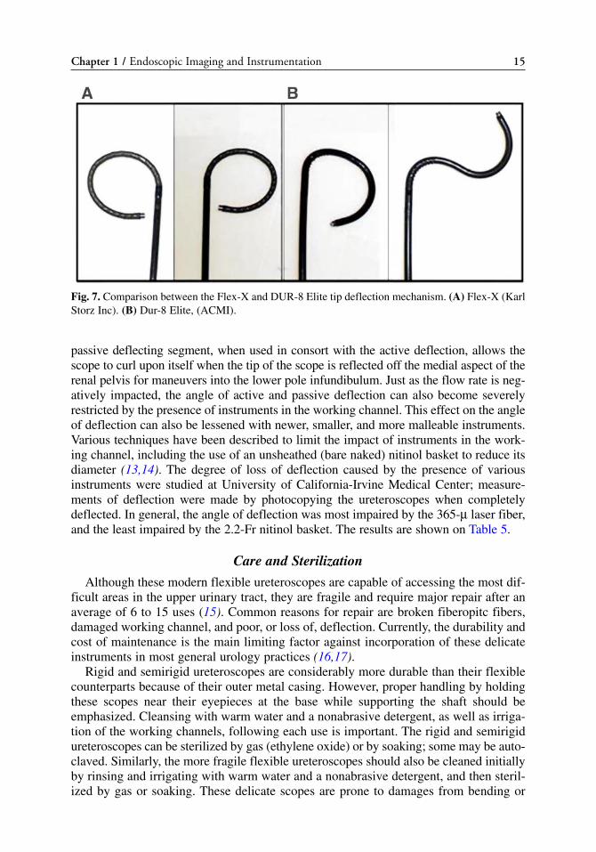
passive deflecting segment, when used in consort with the active deflection, allows thescope to curl upon itself when the tip of the scope is reflected off the medial aspect of therenal pelvis for maneuvers into the lower pole infundibulum. Just as the flow rate is neg-atively impacted, the angle of active and passive deflection can also become severelyrestricted by the presence of instruments in the working channel. This effect on the angleof deflection can also be lessened with newer, smaller, and more malleable instruments.Various techniques have been described to limit the impact of instruments in the work-ing channel, including the use of an unsheathed (bare naked) nitinol basket to reduce itsdiameter (13,14). The degree of loss of deflection caused by the presence of variousinstruments were studied at University of California-Irvine Medical Center; measure-ments of deflection were made by photocopying the ureteroscopes when completelydeflected. In general, the angle of deflection was most impaired by the 365-μ laser fiber,and the least impaired by the 2.2-Fr nitinol basket. The results are shown on Table 5.
Care and Sterilization
Although these modern flexible ureteroscopes are capable of accessing the most dif-ficult areas in the upper urinary tract, they are fragile and require major repair after anaverage of 6 to 15 uses (15). Common reasons for repair are broken fiberopitc fibers,damaged working channel, and poor, or loss of, deflection. Currently, the durability andcost of maintenance is the main limiting factor against incorporation of these delicateinstruments in most general urology practices (16,17).
Rigid and semirigid ureteroscopes are considerably more durable than their flexiblecounterparts because of their outer metal casing. However, proper handling by holdingthese scopes near their eyepieces at the base while supporting the shaft should beemphasized. Cleansing with warm water and a nonabrasive detergent, as well as irriga-tion of the working channels, following each use is important. The rigid and semirigidureteroscopes can be sterilized by gas (ethylene oxide) or by soaking; some may be auto-claved. Similarly, the more fragile flexible ureteroscopes should also be cleaned initiallyby rinsing and irrigating with warm water and a nonabrasive detergent, and then steril-ized by gas or soaking. These delicate scopes are prone to damages from bending or
Chapter 1 / Endoscopic Imaging and Instrumentation 15
Fig. 7. Comparison between the Flex-X and DUR-8 Elite tip deflection mechanism. (A) Flex-X (KarlStorz Inc). (B) Dur-8 Elite, (ACMI).

Tab
le 5
Perc
ent
Loss
of
Def
lect
ion
Wit
h Va
riou
s In
stru
men
ts Oly
mpu
sA
CM
I D
UR
8 E
lite
Stor
z F
LE
X-X
UR
F-P
3W
olf
7325
.172
Wol
f 73
30.0
27
Inst
rum
ent
Dow
nU
pA
ctiv
e se
cond
ary
Act
ive
1+2
Dow
nU
pD
own
Up
Dow
nU
pD
own
Up
200-
μla
ser
fibe
r18
.3%
21.1
%22
.7%
7.1%
9.3%
12.4
%9%
11%
5.4%
11.5
%5.
1%3.
1%36
5-μ
lase
r fi
ber
46.3
%39
.5%
28%
25.9
%26
.8%
31.8
%34
.1%
43.9
%38
.8%
35.9
%33
.3%
28.1
%1.
9-Fr
EH
L9.
1%11
.2%
18.9
%6.
8%8.
5%12
.4%
14.1
%14
.2%
12%
10.7
%19
.2%
10.9
%2.
2-Fr
bas
ket
4.9%
14.5
%16
.7%
6%2.
4%9.
9%12
%9.
7%0.
7%5.
3%13
.5%
4.3%
3-Fr
bas
ket
12.2
%17
.1%
18.2
%7.
9%10
.6%
12.9
%13
.2%
14.8
%17
.4%
6.9%
14.7
%3.
1%2.
6-Fr
gra
sper
22%
20.4
%25
.8%
14.3
%15
.9%
21.9
%17
.4%
14.8
%19
.9%
12.2
%16
.7%
14.1
%
EH
L, e
lect
rohy
drau
lic li
thot
rips
y.
16

trauma to the distal tip or the eyepiece. Therefore, every effort should be made to main-tain them in a straight orientation during cleansing and use. In addition, the flexibleureteroscopes require venting during gas sterilization, either by manually opening avent near the irrigation port near the light post, or some may have an automatic,patented, Autoseal system. Liquid steriliztion may be accomplished by soaking in 2.4%glutaraldehyde (i.e., Cidex, Advanced Sterilization Products, Irvine, CA) or 35% perox-yacetic acid (i.e., Steris, Mentor, OH). Peroxyacetic acid is harsh on flexible endoscopesand has been demonstrated to be associated with higher flexible cystoscope repair costs(18). However, the durability of the flexible ureteroscopes may also be effected by thetechnique and number of personnel involved in the cleaning and maintenance ratherthan the technical demands of the procedure and the endoscopists’ technique (19). Theroutine use of newer ureteroscopic accessories such as ureteral access sheaths, nitinoldevices, and 200-μ holmium laser fibers can decrease the strain on the flexible uretero-scopes and significantly increase the longevity (17).
Rigid and Flexible Nephroscopes
The rigid nephroscopes have undergone little change since the advent of percuta-neous nephrostolithotomy. In general, they provide excellent visualization with arod–lens system and an offset eyepiece to allow passage of large, straight instrumentsfor stone fragmentation, such as the ultrasonic lithotriptor or the lithoclast. Variouslengths are available, ranging from 17.5 to 30 cm, to accommodate a variety of patientbody habitus. Sheaths range from 15 to 27 Fr in size; “mini-nephroscopes” with asmaller, 11-Fr diameter which can be used as a compact cystoscope are also available.A flexible cystoscope may be used as a nephroscope when needed. A new digital cys-tonephroscope by ACMI has been developed and offers additional flexion to the tradi-tional up/down plane to meet the demands of percutaneous nephrostolithotomy (Fig. 5).
CONCLUSION
Since the initial concept of inspecting a body cavity using a light and image trans-mitting system, significant development and advancements have been made in the fieldof urologic endoscopy. With the availability of a wide range of rigid, semirigid, flexi-ble endoscopes, and specifically designed working instruments, most upper urinarytract lesions can be effectively diagnosed and treated in a minimally invasive approach.Continued refinements to these instruments may potentially improve the optics, dura-bility and efficacy of the treatment as technological advances are incorporated into thedesign of the endoscopes and accessory instruments.
REFERENCES1. Reuter MA, Reuter HJ. The development of the cystoscope. J Urol, 1997; 159: 638–640.2. Hopkins HH. Optical principles of the endoscope. In: Endoscopy (Berci, G, ed.), Appleton-Century-
Crofts, New York, 1976, pp. 3–26.3. Young HH, Mckay RW. Congenital valvular obstruction of the prostatic urethra. Surg Gynecol
Obstet, 1929; 48: 509–512.4. Marshal VV. Fiberoptics in urology. J Urol, 1964; 91: 110–113.5. Takagi T, Go T, Takayasu N, Aso Y. A small caliber fiberscope for the visualization of the urinary
tract, biliary tract, and spinal canal. Surgery, 1968; 64: 1033–1036.6. Lyon ES, Kyker JS, Schoenberg, HW. Transurethral ureteroscopy in women: A ready addition to the
urological armamentarium. J Urol, 1978; 119: 35–38.
Chapter 1 / Endoscopic Imaging and Instrumentation 17

7. Lyon ES, Banno JJ, Schoenberg HW. Transurethral ureteroscopy in men using juvenile cystoscopyequipment. J Urol, 1979; 122: 152–155.
8. Perez-Castro EE, Martinez-Piniero JA. Transurethral ureteroscopy- a current urological procedure.Arch Esp Urol, 1980; 33: 445–448.
9. Wolf SJ, Jr., Marcovich R, Gill IS, et al. Survey of Neuromuscular injuries to the patient and surgeonduring urologic lapaoscopic surgery. Urology, 2000; 55: 831–836.
10. Takebayashi S, Hosaka M, Kubota Y, et al. Computerized tomographic ureteroscopy for diagnosingureteral tumors. J Urol, 2000; 163: 42.
11. Fielding JR, Hoyte L, Okon SA, et al. Tumor detection by virtual cystoscopy with color mapping ofbladder wall thickness. J Urol, 2002; 167: 559–562.
12. Takebayashi S, Hosaka M, Takase K, Kubota N, Kishida T, Matsubara S. Computerized tomographynephroscopic images of renal pelvic carcinoma. J Urol, 1999; 162: 315–318.
13. Monga M, Dretler SP, Landman J, Slaton JW, Conradie MC, Clayman RV. Maximizing ureteroscopedeflection: “Play it straight”. Urology, 2002; 60: 902–905.
14. Landman J, Monga M, El-Gabry EA, et al. Bare naked baskets: Ureteroscope deflection and flow char-acteristics with intact and disassembled ureteroscopic nitinol stone baskets. J Urol, 2002; 167:2377–2379.
15. Afane JS, Olweny EO, Bercowsky E, et al. Flexible ureteroscopes: A single center evaluation of thedurability and function of the new endoscopes smaller than 9 Fr. J Urol, 2000; 164: 1164–1168.
16. Landman J, Lee DI, Lee C, Monga M. Evaluation of overall costs of currently available small flexi-ble ureteroscopes. Urology, 2003; 62: 218–222.
17. Pietrow PK, Auge BK, Delvecchio FC, et al. Techniques to maximize flexible ureteroscope longetiv-ity. Urology, 2002; 60: 784–788.
18. Fuselier Jr., HA, Mason C. Liquied sterilization versus high level disinfection in the urologic office.Urology, 1997; 50: 337–340.
19. McDougall EM, Alberts G, Deal KJ, Nagy JM 3rd. Does the cleaning technique influence the dura-bility of the <9 flexible ureteroscope? J Endourol, 2001; 15: 615–618.
18 Chou and McDougall

Access, Stents, and Urinary Drainage
Ben H. Chew, MSc, MD, FRCSC
and John D. Denstedt, MD, FRCSC
CONTENTS
INTRODUCTION
ACCESS TECHNIQUES
STENTING TECHNIQUE
STENT COMFORT, INFECTION, AND ENCRUSTATION:THE ROLE OF NEW BIOMATERIALS AND COATINGS
TIPS AND TRICKS
CONCLUSION
REFERENCES
2
SUMMARY
Ureteral access is necessary in many endourological procedures includingureteroscopy and ureteral stenting. Technologies such as ureteral access sheaths, bal-loon dilators, and coaxial dilators may be helpful in facilitating ureteral access in diffi-cult cases. This chapter describes a stenting technique that relies on fluoroscopicguidance once the initial guidewire is placed and the cystoscope is removed.
Key Words: Ureter; stent; calculi; ureteroscopy; nephrostomy tube; shockwavelithotripsy.
INTRODUCTION
Ureteral stents are a mainstay in the urological armamentarium and are utilized in thetreatment of urolithiasis including postureteroscopy, preshockwave lithotripsy, and to relievesymptomatic renal colic. Routine stenting postureteroscopy and intracorporeal lithotripsy,once the standard of care, have been shown to be unnecessary following uncomplicatedureteroscopy and stone manipulation. Advances such as laser lithotripsy and smaller uretero-scopes have minimized the potential morbidity of ureteroscopy to the point that theindwelling stent has become the most morbid part of the procedure. Ureteral stents maycause considerable side effects ranging from dysuria, urgency and frequency to hematuria
From: Advanced Endourology: The Complete Clinical GuideEdited by: S. Y. Nakada and M. S. Pearle © Humana Press Inc., Totowa, NJ
19

and suprapubic pain. There is an emerging body of literature that routine stenting pos-tureteroscopy is not necessary and that the need for stenting should be determined on a caseby case basis.
Stents are also used to provide urinary drainage in nongenitourinary causes ofureteral obstruction, such as pregnancy and malignant ureteral obstruction. An alterna-tive and effective method of urinary drainage is the percutaneous nephrostomy tubewhich is easily placed in patients with significant hydronephrosis and may be evenmore successful than retrograde ureteral stenting when urinary drainage is required as aresult of obstruction of the distal ureter. Incompressible stents incorporating metal intothe stent material have been used to provide urinary drainage to patients with malignantureteral obstruction. Conversely, biodegradable stents have been developed to provideureteral drainage temporarily following an endourological procedure before degradingand being excreted in the urine, thus obviating the need for cystoscopic stent removal.Other stent advancements will see coatings, new materials, and drugs loaded directlyinto the stent material or coated on the stent surface to improve comfort and reducebiofilm formation, infection, and encrustation.
Access to the ureter is required any time closed endoscopic ureteral procedures are tobe carried out including during ureteral stenting and in association with diagnostic andtherapeutic ureteroscopy for urolithiasis. More detail will be provided in other chaptersregarding procedure specific aspects of ureteroscopy and percutaneous procedures; thischapter will focus on initially gaining retrograde access to the ureter, aspects relatedto ureteral stenting and a comparative analysis of alternative methods of urinary drainage.A brief summary of new stent technologies and biomaterials will also be presented.
Indications to Access the Ureter
Achievement of ureteral access is necessary for performing retrograde endoscopicprocedures such as ureteroscopy, or for placing a ureteral stent. Table 1 lists commonindications for ureteral stent placement.
Stones
Urolithiasis represents one of the more common reasons to insert a ureteral stent.Clinical indications for stenting include patients with intractable pain, those withinfected pyonephrosis, or patients with impaired renal function from obstruction. Inaddition, ureteral stenting is often employed as an adjunct to shockwave lithotripsy orendoscopic procedures in patients requiring surgical stone management.
Ureteral Stones: Retrograde Ureteral Stenting vs Nephrostomy Tube Drainage
Pyonephrosis with an obstructing stone requires urgent decompression using either ret-rograde ureteral stent placement or antegrade percutaneous nephrostomy tube drainage(1). Whether urinary drainage to bypass the obstruction is best accomplished via a ureteralstent or a nephrostomy tube is a subject of debate. The first randomized clinical trial tocompare these two methods in obstructed, infected patients was performed by Pearle et al.(2) in 42 patients with obstructing urolithiasis and pyonephrosis. The time to deferves-cence, length of stay in hospital, pain symptoms, and normalization of leukocytosis didnot differ between these two groups suggesting that urinary decompression by either ret-rograde ureteral stenting or antegrade percutaneous nephrostomy tube insertion are bothequally effective in treating obstructed pyonephrosis. However, patients had significantlyless fluoroscopy exposure (2.6 minutes less) when they were stented in a retrograde fashion.
20 Chew and Denstedt